Embed Size (px)
Citation preview
Tracheotomy is a common procedure for multiple medical indications.1 To providesafe and competent care, nursing staff must understand the immediate postopera-tive and long-term management of tracheostomy patients. Each institution shouldhave its own standard policies and procedures for caring for these patients. Basic min-imal care usually consists of cleaning or changing the inner cannula, caring for the
stoma, and suctioning at least 3 times a day. Depending on the thickness and quantity of secretions,more frequent inspection of the inner cannula may be necessary.
Tracheostomy tubes are made from various materials. Mitchell et al2 recommend that a plastic tracheostomy tube be used for initial placement. Metal tracheostomy tubes are rigid, lack a cuff, andcannot be attached to a ventilator or a bag-valve mask. For these reasons and the cost of materials andproduction, metal tubes are not commonly used in hospitals today.3 Some plastic tracheostomy tubes
Tracheostomy Care and Complications in the Intensive Care UnitLINDA L. MORRIS, PhD, APN, CCNS
ANDREA WHITMER, RN, MSN, ACNP-BC
ERIK McINTOSH, RN, MSN, ACNP-BC
This article has been designated for CNE credit. A closed-book, multiple-choice examination follows this article, which tests your knowledge of the following objectives:
1. Identify evidence-based recommendations for care, indications for placement, and general types of tracheostomies2. Describe postoperative care of patients with a new tracheostomy3. Describe the assessment and emergency interventions for patients with tracheostomy
©2013 American Association of Critical-Care Nurses doi: http://dx.doi.org/10.4037/ccn2013518
CNE Continuing Nursing Education
Cover
Tracheotomy is a common procedure in intensive care units, and nurses must provide proper care to tra-cheostomy patients to prevent complications. One of the most important considerations is effective mobi-lization of secretions, and a suction catheter is the most important tool for that purpose. Each bedsideshould be equipped with a functional suctioning system, an oxygen source, a manual resuscitation bag,and a complete tracheostomy kit, which should accompany patients wherever they go in the hospital. Com-plications include infection, tracheomalacia, skin breakdown, and tracheoesophageal fistula. Tracheostomyemergencies include hemorrhage, tube dislodgement and loss of airway, and tube obstruction; such emer-gencies are managed more effectively when all necessary supplies are readily available at the bedside. Thisarticle describes how to provide proper care in the intensive care unit, strategies for preventing complica-tions, and management of tracheostomy emergencies. (Critical Care Nurse. 2013;33[5]:18-22,24-31)
18 CriticalCareNurse Vol 33, No. 5, OCTOBER 2013 www.ccnonline.org
conform to a patient’s anatomy as the plastic softens atbody temperature, and silicone tracheostomy tubes canconform to the size and shape of a patient’s trachea.3
In this article, we discuss management in the inten-sive care unit (ICU) of patients with a new tracheostomy.We include indications for tracheostomy, tube place-ment, patient care, prevention of complications, andemergency management.
Indications for TracheotomyIndications for placing tracheostomy tubes can be
grouped into 4 general categories: ventilation, airwayobstruction, airway protection, and secretions. The firstcategory applies to patients who require long-termmechanical ventilation because of chronic respiratoryfailure, who cannot maintain respiratory function unas-sisted, or who cannot be weaned from ventilatory sup-port. Numerous studies4-9 have been done to determinethe optimal interval from orotracheal intubation toplacement of a tracheostomy tube, but no definitive rec-ommendations have been made because of varied resultsin different populations of patients and in patients withdifferent comorbid conditions. The American College ofChest Physicians10 recommends consideration of a tra-cheostomy for patients who require an endotrachealtube for more than 21 days. Benefits of establishing atracheostomy rather than using an endotracheal tubeinclude decreasing direct laryngeal injury, improvingcomfort, and improving activities of daily living such asmobility, speech, and eating.11
Patients who have tumors within the airway, paralyzedvocal cords, swelling, stricture, or unusual airway anatomyare another category for tracheostomy because of airwayobstruction that compromises normal respiration. A thirdcategory includes patients who cannot protect their air-way and patients with an inefficient swallow and/or coughmechanism, common situations in patients who have a
high spinal cord injury, cerebrovascular accident, or trau-matic brain injury. Last, patients who cannot mobilize ormanage their secretions may also require a tracheostomy.
Tracheostomy PlacementA tracheostomy tube may be placed surgically or
percutaneously. Surgical placement is done in the oper-ating room or at the bedside, generally with use of gen-eral anesthesia. A stoma is created by using an opensurgical technique. Landmarks are identified, and a skinincision is made below the cricoid cartilage. The isthmusof the thyroid gland is exposed, cross-clamped, and lig-ated. Thetrachea canthen bevisualized.A commontechnique isto create a “trap door” (Björk flap) in which a small partof the tracheal cartilage is pulled down and sutured tothe skin. This flap is thought to facilitate reinsertion ofthe tracheostomy tube if accidental decannulation occurs,especially in patients with difficult anatomy or obesity.12,13
Percutaneous tracheotomy is generally performedsolely on intubated patients and, unlike surgical tra-cheotomy, can be performed without direct visualiza-tion of the trachea. Bronchoscopy is used to guide andconfirm placement of the tracheostomy tube within thetrachea and is considered standard of care.13 In contrastto an open surgical incision, a small opening is createdwith a needle and then dilated.13 Contraindications topercutaneous tracheotomy include uncorrected coagu-lopathy, infection at the incision site, high ratio of posi-tive end-expiratory pressure to fraction of inspired oxygen,elevated intracranial pressure, tracheal obstruction,unusual neck anatomy, and the need for emergencyairway management.11,14
Linda L. Morris is a clinical nurse specialist in respiratory care at Northwestern Memorial Hospital and an associate professor of clinicalanesthesiology, Feinberg School of Medicine, Northwestern University, Chicago, Illinois. She is also on the board of directors of the GlobalTracheostomy Collaborative, an international group of specialists dedicated to research and quality outcomes for patients with tracheostomies.
Andrea Whitmer is the acute care nurse practitioner for the intensivist program at Elkhart General Hospital’s critical care unit in Elkhart,Indiana.
Erik McIntosh is a nurse practitioner on an inpatient internal medicine unit at Rush University Medical Center, Chicago, Illinois.Corresponding author: Linda L. Morris, PhD, APN, CCNS, FCCM, Northwestern Memorial Hospital, 251 E Huron St, Feinberg Pavilion, Ste 8-330, Chicago, IL 60611 (e-mail: [email protected]).
To purchase electronic or print reprints, contact the American Association of Critical-Care Nurses, 101 Columbia, Aliso Viejo, CA 92656. Phone, (800) 899-1712 or (949) 362-2050 (ext 532); fax, (949) 362-2049; e-mail, [email protected].
Authors
www.ccnonline.org CriticalCareNurse Vol 33, No. 5, OCTOBER 2013 19
Indications for placing tracheostomy tubes
can be grouped into 4 general categories:
ventilation, airway obstruction, airway
protection, and secretions.
Postoperative CareImmediate postoperative priorities of care for a patient
with a new tracheostomy include ensuring that the tra-cheostomy tube is securely in place and is patent. Routinecare, as well as prompt management of postoperativecomplications, can be facilitated by ensuring that properequipment and supplies are quickly available. Table 1lists the contents of a tracheostomy kit that should bepresent at the patient’s bedside and, per recommenda-tions, should accompany the patient whenever he or sheis away from the ICU.2,15,16
The American Academy of Otolaryngology Head andNeck Surgery recently published consensus statementsfor tracheostomy care.2 These statements were developedby a multidisciplinary panel of experts on care of patientswith a tracheostomy. Using a modified Delphi method,the panel members completed surveys on various aspectsof care. Consensus was reached on 77 statements thataddress the initial change of the tracheostomy tube, man-agement of emergencies and complications, prerequisitesfor decannulation, management of tube cuffs and com-munication devices, and specific needs of patients and thepatients’ caregivers. These statements are an importantdocument because few randomized studies have addressedthese issues, possibly because of the difficulty in studydesign and recruitment and because of ethical concerns.Table 2 provides some of these important statements.
Scheduled Changes of Tracheostomy Tubes
Currently, no empirical evidence indicates a stan-dardized time for changing a tracheostomy tube, and
changes are typically done according to the preferenceof the health care provider. White et al17 suggest thatindications for changing a tracheostomy tube includethe need for a different size tube, tube malfunction,need for a different type of tube, and routine changesfor ongoing airway management and prevention ofinfection. They suggest that a tracheostomy tube shouldbe changed every 7 to 14 days after initial insertion, butthey acknowledge that no evidence supports that rec-ommendation. Mitchell et al2 recommend replacing theinitial tracheostomy tube within 10 to 14 days afterplacement if a percutaneous procedure was used toestablish the tracheostomy and within 3 to 7 days if a
Table 1 Bedside tracheostomy kit
Tracheostomy tube of the same size and type currently in placeTracheostomy tube 1 size smaller than the one currently in
placeObturator Suction catheters (usually 12F or 14F)Yankauer suction catheterFunctional suctioning system, canisterManual resuscitation bag and oxygenEndotracheal tube of appropriate sizeTracheostomy cleaning kitDisposable inner cannulas (not required for single-cannula
tubes)10-mL syringe (not required for cuffless tubes)Tracheostomy holder or tiesDrain spongesHydrogen peroxidePhysiological salineIntubation equipmentOxygen source
1. All supplies to replace a tracheostomy tube should be at the bedside or within reach.
2. If no aspiration, tracheostomy tube cuffs should be deflated when a patient no longer requires mechanical ventilation.
3. The first change of a tracheostomy tube should normally be performed by an experienced physician with assistance from another clinician.
4. Use of a defined tracheostomy care protocol for patient and caregiver education before discharge will improve patients’ outcomes and decrease complications.
5. Patients and their caregivers should be informed of what to do in an emergency before discharge.
6. In an emergency, a dislodged tube from a mature tracheostomy should be replaced with the same size tube or a tube 1 size smaller or an endotracheal tube through the tracheostomy wound.
7. In an emergency, patients with a dislodged tracheostomy tube that cannot be reinserted should be intubated.
8. A patient should not be discharged from the hospital with the tracheostomy tube sutured in place.
9. Acute occlusion of a tracheostomy tube is most likely caused by a mucous plug, obstructing granuloma, or insertion of the tube into a false track.
10. A patient can be turned in bed once the security of the tube has been assessed to avoid accidental decannulation.
Table 2 Consensus statements for tracheostomy care
20 CriticalCareNurse Vol 33, No. 5, OCTOBER 2013 www.ccnonline.org
surgical procedure was used. A tracheostomy tube insertedpercutaneously fits more tightly within the stoma thandoes a tube that was inserted through a surgical incision.If a tracheostomy tube is changed prematurely, the tis-sue of the dilated stoma tract is more likely to recoil thanit would if the change were done later.18 In addition,Mitchell et al2 recommend that patients should not bedischarged from the hospital with the tracheostomy tubesutured in place because the first tracheostomy tubechange should be done before discharge.
Changing tracheostomy tubes can correct problemsthat cause ventilator asynchrony, improve comfort byreducing tube size, and correct a cuff leak due to tracheo-malacia or malposition or fracture of the tracheostomytube or flange.17 Most manufacturers recommend chang-ing the tubes every 1 to 2 months; however, Yaremchuk19
found that routine tube changes every 2 weeks decreasedthe formation of granulation tissue. In a study by Björlinget al,20 electron micrographs of plastic tubes revealed vis-ible surface changes after 30 days in all types of tubesstudied: polyvinyl chloride, silicone, and polyurethane.
Cleaning and Replacing the Inner Cannula
The primary purpose of the inner cannula is to pre-vent tube obstruction by allowing regular cleaning orreplacement. Many episodes of tube obstruction can beprevented with simple inspection and cleaning or chang-ing of the inner cannula. Many plastic tubes are cleanedwith a solution of full- or half-strength hydrogen perox-ide and sterile water,21-23 but some sources24 recommendusing physiological saline alone. Silicone tubes and metaltubes can be damaged by using hydrogen peroxide. Theconnection joints in metal tubes can be damaged byhydrogen peroxide and by enzyme cleaners, so these tubesshould be cleaned by soaking in warm water with milddishwashing liquid to soften secretions and then using atracheal brush. Silicone tends to absorb cleaning products,so physiological saline alone should be used to clean sili-cone tubes. For these reasons, it is important to check man-ufacturers’ instructions for cleaning tracheostomy tubes.25
No studies have been done to determine the optimalfrequency for cleaning the inner cannula; however, thecannula should be inspected regularly, perhaps at least 3times per day, depending on the volume and thicknessof the patient’s secretions.21,25 Burns et al26 maintain thatinner cannulas do not need to be changed at all; how-ever, their study had some limitations and was restricted
to surgical patients who may not have had commoncomorbid conditions such as pneumonia.
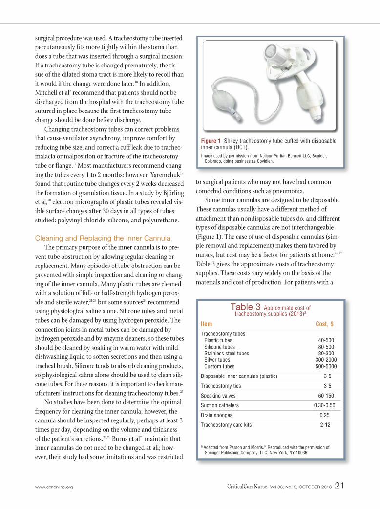
Some inner cannulas are designed to be disposable.These cannulas usually have a different method ofattachment than nondisposable tubes do, and differenttypes of disposable cannulas are not interchangeable(Figure 1). The ease of use of disposable cannulas (sim-ple removal and replacement) makes them favored bynurses, but cost may be a factor for patients at home.25,27
Table 3 gives the approximate costs of tracheostomysupplies. These costs vary widely on the basis of thematerials and cost of production. For patients with a
Table 3 Approximate cost of tracheostomy supplies (2013)a
Item
Tracheostomy tubes:Plastic tubesSilicone tubesStainless steel tubesSilver tubesCustom tubes
Disposable inner cannulas (plastic)
Tracheostomy ties
Speaking valves
Suction catheters
Drain sponges
Tracheostomy care kits
Cost, $
40-50080-50080-300
300-2000500-5000
3-5
3-5
60-150
0.30-0.50
0.25
2-12
a Adapted from Parson and Morris.28 Reproduced with the permission ofSpringer Publishing Company, LLC, New York, NY 10036.
Figure 1 Shiley tracheostomy tube cuffed with disposableinner cannula (DCT).Image used by permission from Nellcor Puritan Bennett LLC, Boulder,
Colorado, doing business as Covidien.
www.ccnonline.org CriticalCareNurse Vol 33, No. 5, OCTOBER 2013 21
long-term tracheostomy, inquiring about the care thepatients receive at home is a good idea. The informationshould include the last time the tube was completelychanged, use of inner cannulas, and suctioning at home.
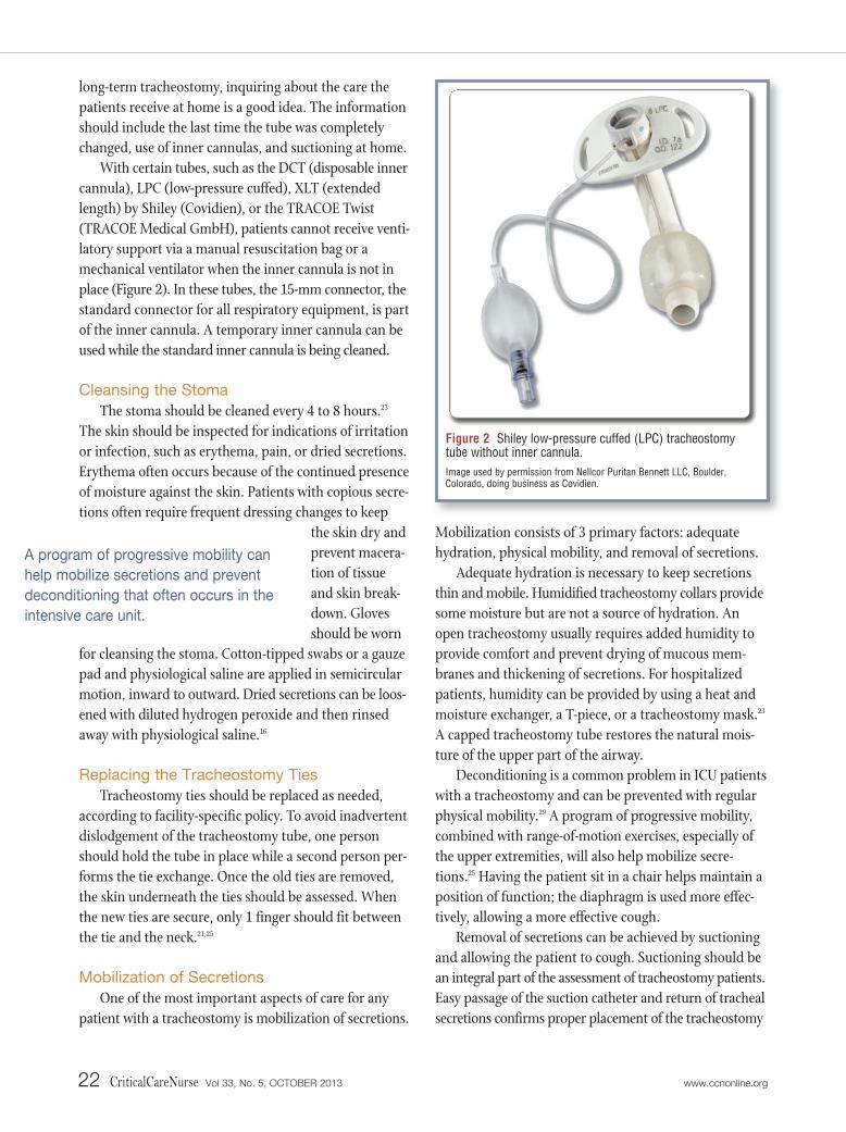
With certain tubes, such as the DCT (disposable innercannula), LPC (low-pressure cuffed), XLT (extendedlength) by Shiley (Covidien), or the TRACOE Twist(TRACOE Medical GmbH), patients cannot receive venti-latory support via a manual resuscitation bag or amechanical ventilator when the inner cannula is not inplace (Figure 2). In these tubes, the 15-mm connector, thestandard connector for all respiratory equipment, is partof the inner cannula. A temporary inner cannula can beused while the standard inner cannula is being cleaned.
Cleansing the Stoma
The stoma should be cleaned every 4 to 8 hours.23
The skin should be inspected for indications of irritationor infection, such as erythema, pain, or dried secretions.Erythema often occurs because of the continued presenceof moisture against the skin. Patients with copious secre-tions often require frequent dressing changes to keep
the skin dry andprevent macera-tion of tissueand skin break-down. Glovesshould be worn
for cleansing the stoma. Cotton-tipped swabs or a gauzepad and physiological saline are applied in semicircularmotion, inward to outward. Dried secretions can be loos-ened with diluted hydrogen peroxide and then rinsedaway with physiological saline.16
Replacing the Tracheostomy Ties
Tracheostomy ties should be replaced as needed,according to facility-specific policy. To avoid inadvertentdislodgement of the tracheostomy tube, one personshould hold the tube in place while a second person per-forms the tie exchange. Once the old ties are removed,the skin underneath the ties should be assessed. Whenthe new ties are secure, only 1 finger should fit betweenthe tie and the neck.21,25
Mobilization of Secretions
One of the most important aspects of care for anypatient with a tracheostomy is mobilization of secretions.
Mobilization consists of 3 primary factors: adequatehydration, physical mobility, and removal of secretions.
Adequate hydration is necessary to keep secretionsthin and mobile. Humidified tracheostomy collars providesome moisture but are not a source of hydration. Anopen tracheostomy usually requires added humidity toprovide comfort and prevent drying of mucous mem-branes and thickening of secretions. For hospitalizedpatients, humidity can be provided by using a heat andmoisture exchanger, a T-piece, or a tracheostomy mask.23
A capped tracheostomy tube restores the natural mois-ture of the upper part of the airway.
Deconditioning is a common problem in ICU patientswith a tracheostomy and can be prevented with regularphysical mobility.29 A program of progressive mobility,combined with range-of-motion exercises, especially ofthe upper extremities, will also help mobilize secre-tions.25 Having the patient sit in a chair helps maintain aposition of function; the diaphragm is used more effec-tively, allowing a more effective cough.
Removal of secretions can be achieved by suctioningand allowing the patient to cough. Suctioning should bean integral part of the assessment of tracheostomy patients.Easy passage of the suction catheter and return of trachealsecretions confirms proper placement of the tracheostomy
22 CriticalCareNurse Vol 33, No. 5, OCTOBER 2013 www.ccnonline.org
Figure 2 Shiley low-pressure cuffed (LPC) tracheostomytube without inner cannula.Image used by permission from Nellcor Puritan Bennett LLC, Boulder, Colorado, doing business as Covidien.
A program of progressive mobility can
help mobilize secretions and prevent
deconditioning that often occurs in the
intensive care unit.
tube. Shapiro et al30 established that vital capacity shouldbe at least 15 mL/kg to clear secretions. When coughstrength is less than 15 mL/kg, or the cough reflex isdiminished, more frequent suctioning may be required.
Cuff Pressure
The purpose of the cuff is to provide a closed systemto allow effective ventilation and/or airway protection.Cuff pressure should be 20 to 25 cm H2O with most tra-cheostomy tubes.21,25 Monitoring cuff pressure is impor-tant because underinflation of the cuff promotes leakageof secretions around the cuff, a situation that can con-tribute to ventilator-associated pneumonia.25 However,overinflation of the cuff can cause numerous long-termcomplications, including tracheomalacia, tracheoinnom-inate artery fistula, tracheal ulcerations, fibrosis, trachealstenosis, and tracheoesophageal fistula.21
If a leak around the cuff persists, the pilot balloonmaybe ineffective in sealing the airway or the trachea mayhave lost its rigid composition. A persistent leak will bemanifested by audible noises around the tracheostomytube and loss of returned volumes with ventilation. Forexample, if the tidal volume is set at 700 mL, and the
returned vol-umes are only500 mL, thepatient is notgetting the
benefit of the entire tidal volume. If the cuff continues torequire more air to seal the airway, the pilot balloon maybe ineffective,25 the tracheostomy tube may be too smallfor the airway, or tracheomalacia may have developed.31
Patient and Caregiver EducationPatient education on care of the tracheostomy tube
and stoma is of utmost importance in preventing manycomplications. The Joint Commission32 has identified thesafety implications of patient education. Patients andcaregivers should be taught how to perform basic care ofthe tracheostomy, including the importance of changingthe tracheotomy tube as scheduled, cleaning or replacingthe inner cannula, cleansing the stoma, and replacingthe tracheostomy holder or ties.
The clinical consensus statements on tracheostomycare2 recommend that when possible patient and familyeducation should begin before the tracheostomy is done.Before discharge, patients and their caregivers should
receive a checklist of supplies that should be taken withthe patient at all times. Patients and caregivers should beevaluated before discharge to assess their competency incaring for the tracheostomy. They should know what todo in an emergency. They should be informed of thetype, size, and length of the tracheostomy tube; how andwhen to use suctioning; how to clean the stoma and thetube itself; how to change the ties; indications of respira-tory distress; how to use all home equipment; and signsand symptoms of infection and skin breakdown. Finally,a home care instruction manual should be given topatients and caregivers before the patient is discharged.2
Most manufacturers of tracheostomy tubes provide ahome care manual for their products. Otherwise, North-west Memorial Hospital in Chicago has published aninstruction manual33 that can be downloaded from theInternet. Emergencies at home are an essential part of thedischarge discussion. Possible home emergencies shouldbe discussed before discharge, ideally allowing patientsand their family members to discuss and demonstratekey skills such as suctioning, use of the manual resuscita-tion bag, and reinsertion of the tracheostomy tube.
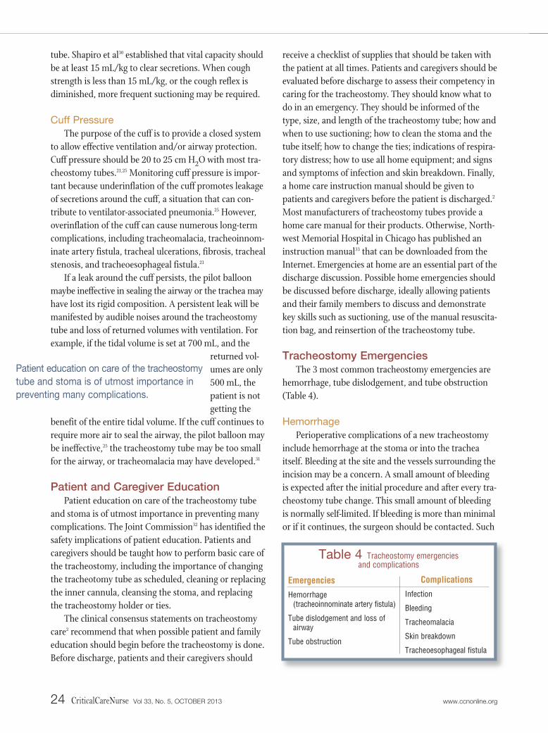
Tracheostomy EmergenciesThe 3 most common tracheostomy emergencies are
hemorrhage, tube dislodgement, and tube obstruction(Table 4).
Hemorrhage
Perioperative complications of a new tracheostomyinclude hemorrhage at the stoma or into the tracheaitself. Bleeding at the site and the vessels surrounding theincision may be a concern. A small amount of bleedingis expected after the initial procedure and after every tra-cheostomy tube change. This small amount of bleedingis normally self-limited. If bleeding is more than minimalor if it continues, the surgeon should be contacted. Such
24 CriticalCareNurse Vol 33, No. 5, OCTOBER 2013 www.ccnonline.org
Patient education on care of the tracheostomy
tube and stoma is of utmost importance in
preventing many complications.
Table 4 Tracheostomy emergencies and complications
Emergencies
Hemorrhage (tracheoinnominate artery fistula)
Tube dislodgement and loss ofairway
Tube obstruction
Complications
Infection
Bleeding
Tracheomalacia
Skin breakdown
Tracheoesophageal fistula
bleeding may indicate that the site should be explored,and a vessel may require ligation.34
Fortunately, one of the most deadly complications,tracheoinnominate fistula, is rare. In this complication,the innominate artery is eroded through the trachea,causing exsanguination within minutes. The reportedincidence35 is 0.7%, and the mortality rate36-38 is almost100%. This massive hemorrhage can be due to pressurenecrosis from cuffs with high pressures, improper place-ment of the cannula tip (from direct weight or torque onthe tracheostomy tube from the ventilator circuit), lowplacement of the tube, hyperextension of the head,radiotherapy, and steroid use. Most often, the complica-tion occurs 3 to 4 weeks after the surgery.37 Managementincludes oxygenation, cuff overinflation to tamponadethe bleeding, and translaryngeal intubation with directdigital compression,39 followed by immediate surgeryfor repair.40 In some studies37,41 the fistula was success-fully treated with endovascular embolization.
Tube Dislodgement and Loss of Airway
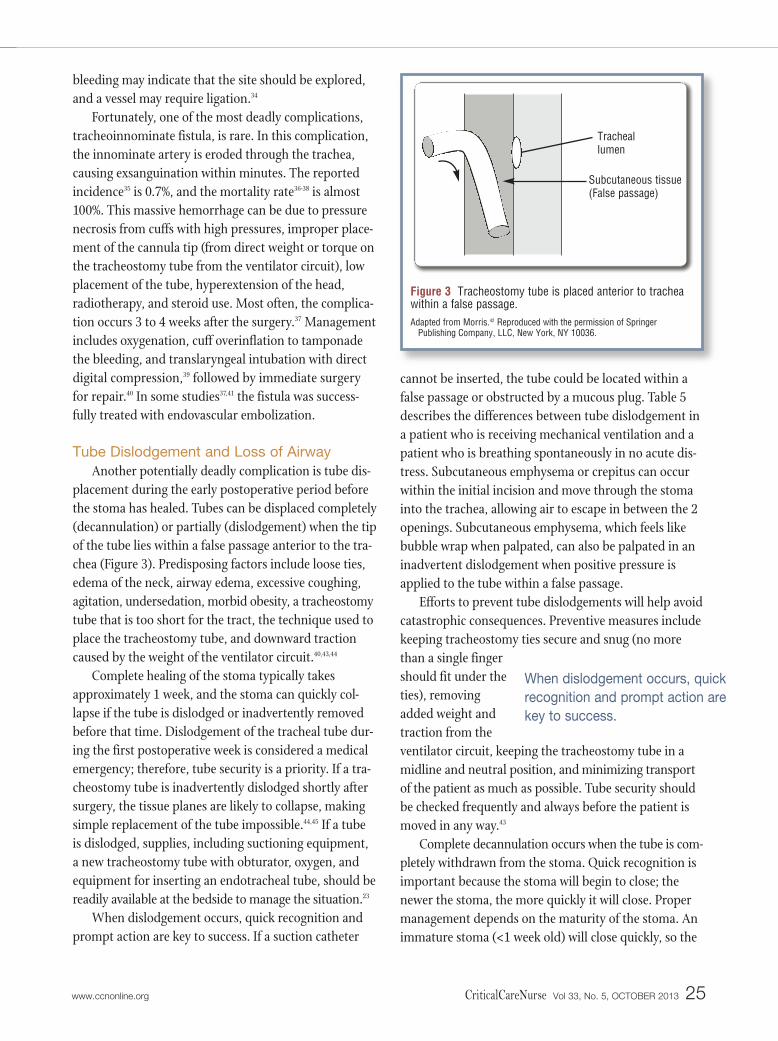
Another potentially deadly complication is tube dis-placement during the early postoperative period beforethe stoma has healed. Tubes can be displaced completely(decannulation) or partially (dislodgement) when the tipof the tube lies within a false passage anterior to the tra-chea (Figure 3). Predisposing factors include loose ties,edema of the neck, airway edema, excessive coughing,agitation, undersedation, morbid obesity, a tracheostomytube that is too short for the tract, the technique used toplace the tracheostomy tube, and downward tractioncaused by the weight of the ventilator circuit.40,43,44
Complete healing of the stoma typically takesapproximately 1 week, and the stoma can quickly col-lapse if the tube is dislodged or inadvertently removedbefore that time. Dislodgement of the tracheal tube dur-ing the first postoperative week is considered a medicalemergency; therefore, tube security is a priority. If a tra-cheostomy tube is inadvertently dislodged shortly aftersurgery, the tissue planes are likely to collapse, makingsimple replacement of the tube impossible.44,45 If a tubeis dislodged, supplies, including suctioning equipment,a new tracheostomy tube with obturator, oxygen, andequipment for inserting an endotracheal tube, should bereadily available at the bedside to manage the situation.23
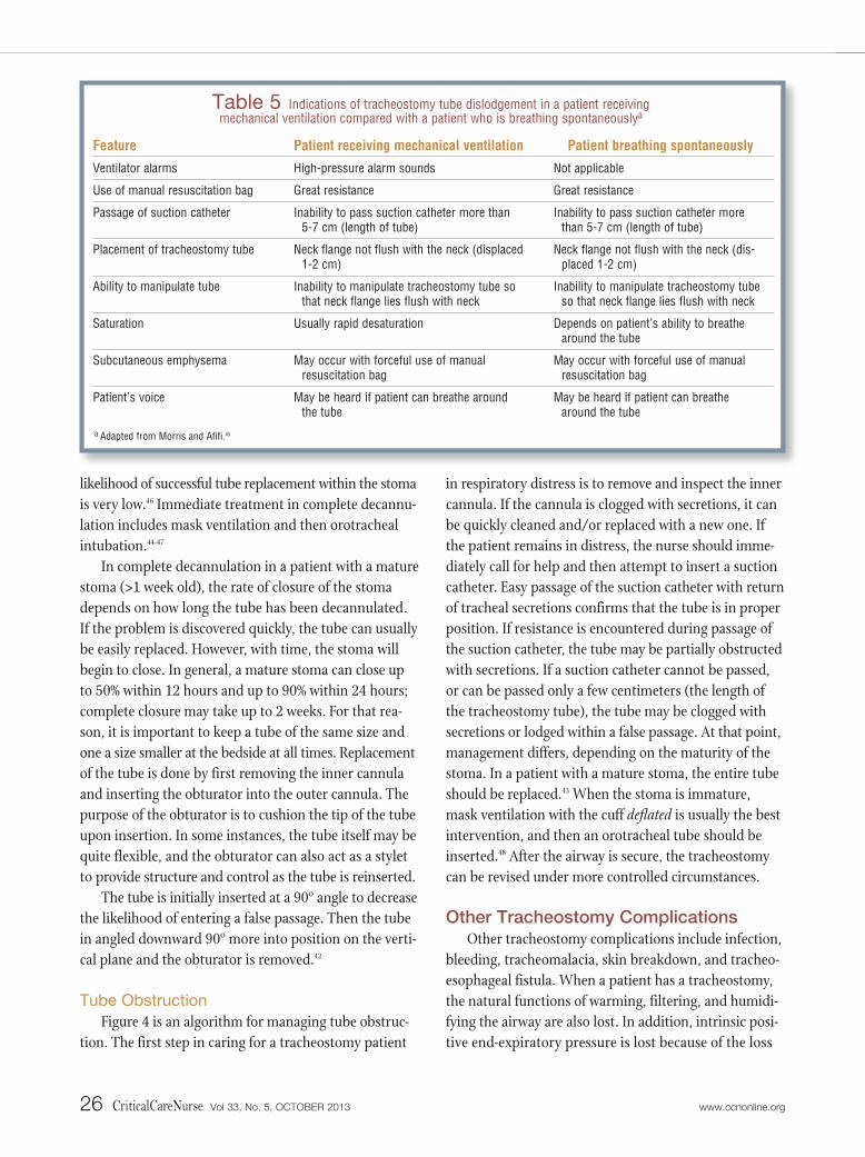
When dislodgement occurs, quick recognition andprompt action are key to success. If a suction catheter
cannot be inserted, the tube could be located within afalse passage or obstructed by a mucous plug. Table 5describes the differences between tube dislodgement ina patient who is receiving mechanical ventilation and apatient who is breathing spontaneously in no acute dis-tress. Subcutaneous emphysema or crepitus can occurwithin the initial incision and move through the stomainto the trachea, allowing air to escape in between the 2openings. Subcutaneous emphysema, which feels likebubble wrap when palpated, can also be palpated in aninadvertent dislodgement when positive pressure isapplied to the tube within a false passage.
Efforts to prevent tube dislodgements will help avoidcatastrophic consequences. Preventive measures includekeeping tracheostomy ties secure and snug (no morethan a single fingershould fit under theties), removingadded weight andtraction from theventilator circuit, keeping the tracheostomy tube in amidline and neutral position, and minimizing transportof the patient as much as possible. Tube security shouldbe checked frequently and always before the patient ismoved in any way.43
Complete decannulation occurs when the tube is com-pletely withdrawn from the stoma. Quick recognition isimportant because the stoma will begin to close; thenewer the stoma, the more quickly it will close. Propermanagement depends on the maturity of the stoma. Animmature stoma (<1 week old) will close quickly, so the
When dislodgement occurs, quick
recognition and prompt action are
key to success.
www.ccnonline.org CriticalCareNurse Vol 33, No. 5, OCTOBER 2013 25
Figure 3 Tracheostomy tube is placed anterior to tracheawithin a false passage.Adapted from Morris.42 Reproduced with the permission of Springer
Publishing Company, LLC, New York, NY 10036.
Subcutaneous tissue(False passage)
Tracheallumen
likelihood of successful tube replacement within the stomais very low.46 Immediate treatment in complete decannu-lation includes mask ventilation and then orotrachealintubation.44-47
In complete decannulation in a patient with a maturestoma (>1 week old), the rate of closure of the stomadepends on how long the tube has been decannulated.If the problem is discovered quickly, the tube can usuallybe easily replaced. However, with time, the stoma willbegin to close. In general, a mature stoma can close upto 50% within 12 hours and up to 90% within 24 hours;complete closure may take up to 2 weeks. For that rea-son, it is important to keep a tube of the same size andone a size smaller at the bedside at all times. Replacementof the tube is done by first removing the inner cannulaand inserting the obturator into the outer cannula. Thepurpose of the obturator is to cushion the tip of the tubeupon insertion. In some instances, the tube itself may bequite flexible, and the obturator can also act as a styletto provide structure and control as the tube is reinserted.
The tube is initially inserted at a 90º angle to decreasethe likelihood of entering a false passage. Then the tubein angled downward 90º more into position on the verti-cal plane and the obturator is removed.42
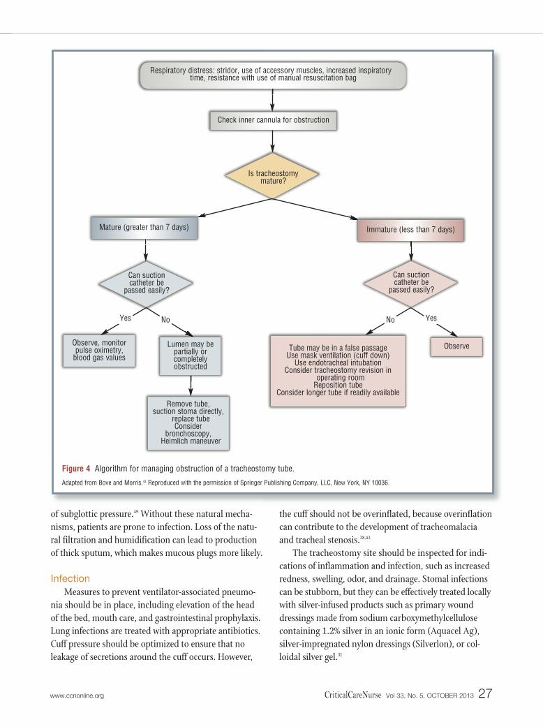
Tube Obstruction
Figure 4 is an algorithm for managing tube obstruc-tion. The first step in caring for a tracheostomy patient
in respiratory distress is to remove and inspect the innercannula. If the cannula is clogged with secretions, it canbe quickly cleaned and/or replaced with a new one. Ifthe patient remains in distress, the nurse should imme-diately call for help and then attempt to insert a suctioncatheter. Easy passage of the suction catheter with returnof tracheal secretions confirms that the tube is in properposition. If resistance is encountered during passage ofthe suction catheter, the tube may be partially obstructedwith secretions. If a suction catheter cannot be passed,or can be passed only a few centimeters (the length ofthe tracheostomy tube), the tube may be clogged withsecretions or lodged within a false passage. At that point,management differs, depending on the maturity of thestoma. In a patient with a mature stoma, the entire tubeshould be replaced.43 When the stoma is immature,mask ventilation with the cuff deflated is usually the bestintervention, and then an orotracheal tube should beinserted.48 After the airway is secure, the tracheostomycan be revised under more controlled circumstances.
Other Tracheostomy ComplicationsOther tracheostomy complications include infection,
bleeding, tracheomalacia, skin breakdown, and tracheo -esophageal fistula. When a patient has a tracheostomy,the natural functions of warming, filtering, and humidi-fying the airway are also lost. In addition, intrinsic posi-tive end-expiratory pressure is lost because of the loss
26 CriticalCareNurse Vol 33, No. 5, OCTOBER 2013 www.ccnonline.org
Feature
Ventilator alarms
Use of manual resuscitation bag
Passage of suction catheter
Placement of tracheostomy tube
Ability to manipulate tube
Saturation
Subcutaneous emphysema
Patient’s voice
Patient receiving mechanical ventilation
High-pressure alarm sounds
Great resistance
Inability to pass suction catheter more than 5-7 cm (length of tube)
Neck flange not flush with the neck (displaced1-2 cm)
Inability to manipulate tracheostomy tube sothat neck flange lies flush with neck
Usually rapid desaturation
May occur with forceful use of manual resuscitation bag
May be heard if patient can breathe aroundthe tube
Patient breathing spontaneously
Not applicable
Great resistance
Inability to pass suction catheter morethan 5-7 cm (length of tube)
Neck flange not flush with the neck (dis-placed 1-2 cm)
Inability to manipulate tracheostomy tubeso that neck flange lies flush with neck
Depends on patient’s ability to breathearound the tube
May occur with forceful use of manualresuscitation bag
May be heard if patient can breathearound the tube
Table 5 Indications of tracheostomy tube dislodgement in a patient receiving mechanical ventilation compared with a patient who is breathing spontaneouslya
a Adapted from Morris and Afifi.45
of subglottic pressure.49 Without these natural mecha-nisms, patients are prone to infection. Loss of the natu-ral filtration and humidification can lead to productionof thick sputum, which makes mucous plugs more likely.
Infection
Measures to prevent ventilator-associated pneumo-nia should be in place, including elevation of the headof the bed, mouth care, and gastrointestinal prophylaxis.Lung infections are treated with appropriate antibiotics.Cuff pressure should be optimized to ensure that noleakage of secretions around the cuff occurs. However,
the cuff should not be overinflated, because overinflationcan contribute to the development of tracheomalaciaand tracheal stenosis.38,43
The tracheostomy site should be inspected for indi-cations of inflammation and infection, such as increasedredness, swelling, odor, and drainage. Stomal infectionscan be stubborn, but they can be effectively treated locallywith silver-infused products such as primary wounddressings made from sodium carboxymethylcell ulosecontaining 1.2% silver in an ionic form (Aquacel Ag), silver-impregnated nylon dressings (Silverlon), or col-loidal silver gel.31
www.ccnonline.org CriticalCareNurse Vol 33, No. 5, OCTOBER 2013 27
Check inner cannula for obstruction
Tube may be in a false passageUse mask ventilation (cuff down)
Use endotracheal intubationConsider tracheostomy revision in
operating roomReposition tube
Consider longer tube if readily available
ObserveObserve, monitorpulse oximetry,
blood gas values
Remove tube, suction stoma directly,
replace tube Consider
bronchoscopy, Heimlich maneuver
Is tracheostomymature?
Can suctioncatheter be
passed easily?
Can suctioncatheter be
passed easily?
Immature (less than 7 days)Mature (greater than 7 days)
Lumen may be partially orcompletely obstructed
Yes No YesNo
Figure 4 Algorithm for managing obstruction of a tracheostomy tube.
Adapted from Bove and Morris.43 Reproduced with the permission of Springer Publishing Company, LLC, New York, NY 10036.
Respiratory distress: stridor, use of accessory muscles, increased inspiratorytime, resistance with use of manual resuscitation bag
Bleeding
Because of tracheal irritation, patients who requirefrequent suctioning may experience bloody or blood-tinged secretions.23 Many clinicians tend to suction lessoften in response to bloody secretions; however, less fre-quent suctioning could lead to accumulation of thickersecretions, making mucous plugs a concern. A betterchoice is to switch to red rubber suction catheters. Thesoft blunt tip of a rubber catheter does not irritate thetracheal wall, and tracheal healing is more rapid than witha regular suction catheter. Red rubber catheters cannot,however, be used in patients with a latex allergy becauselatex is a product of rubber.
Tracheomalacia
As mentioned earlier, tracheomalacia can occur withsustained overinflation of a cuffed tracheostomy tubeand can be definitively diagnosed by using broncho scopy.Tracheomalacia is the breakdown of the natural rigidstructure of the trachea that leads to a flaccid airway inthe affected area.31,38,50 Tracheomalacia is common in theICU and can occur in tracheostomy patients beginning 1week after the tracheotomy procedure. It is manifestedby the presence of a cuff leak (air escaping around thecuff ) combined with overinflation of the cuff and highcuff pressures. Another contributing factor is tractionfrom the weight of the ventilator circuit, including in-
line devicessuch as suc-tion systems,heat-moistureexchangers,
and filters. Methods to prevent tracheomalacia aredirected toward the cause: keep the tracheostomy tubein neutral position, limit traction against the tube, andavoid overinflation of the cuff. Short-term treatment fortracheomalacia is placement of a longer tracheostomytube to bypass the affected area.31,50
Skin Breakdown
Downward traction of the tracheostomy tube canoccur with too much weight pulling down on it, as dis-cussed previously. Traction against the tube can bedirected out and downward by pulling against the tubeor inward, with the neck flange digging into the neck.These traction forces must be prevented and the tubekept in a neutral position. In addition to tracheomalacia,
tube dislodgement, and inadvertent decannulation,traction can also be a factor in skin breakdown. Inwardtraction can contribute to skin erosion under the neckflange; outward traction can contribute to erosion, dis-lodgement, decannulation, and enlargement of thestoma from the inside.
Skin integrity can be compromised at any time in apatient with a tracheostomy. Regular skin inspection isimportant to prevent complications. Keeping the site asdry as possible with drain sponges or skin barrier dress-ings such as those made from carboxymethylcellulose(Aquacel), polyurethane foam (Lyofoam), or siliconefoam (Mepilex) can prevent skin breakdown and preventinfection. Skin care may be difficult while the suturesremain in place until the stoma matures. The use of cotton-tip applicators can help in reaching the tightplaces. When a cotton-tip applicator is soaked withhydrogen peroxide and sterile physiological saline, driedblood and secretions can be more easily removed, espe-cially from tight spaces.
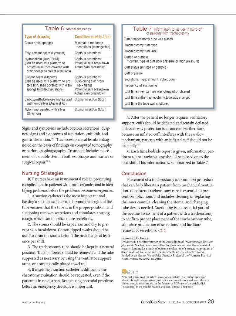
Maintaining the tracheostomy tube in a continuousneutral position can ensure skin integrity while a patientis receiving oxygen via mechanical ventilation or someother delivery device. A continuous neutral position canbe achieved by making sure the oxygen delivery devicesare not putting any weight on or pulling on or twistingthe tracheos tomy tube or by placing a bolster (rolledtowel) under the ventilator circuit. Areas of skin break-down should be staged and treated as necessary. Protec-tive dressings can be used as needed to provide a cushion,collect secretions, and encourage timely healing. Severaldressing products are available that guard against skinbreakdown from flange pressure and are extraabsorbentto soak up excessive tracheal secretions. However, pack-aged precut tracheostomy gauze is often sufficient toprotect the peristomal skin. The gauze should not coverthe opening of the tracheostomy tube.21 Table 6 liststypes of stomal dressings and indications for their use.
Tracheoesophageal Fistula
Tracheoesophogeal fistula occurs when the tracheaand the esophagus communicate through an adjacentperforation in each. A hallmark of tracheoesophagealfistula is the presence of tube feedings within the tracheo -stomy tube. This type of fistula can be due to overinfla-tion of the cuff in patients who also have a feeding tubeor to direct trauma during the tracheotomy procedure.
28 CriticalCareNurse Vol 33, No. 5, OCTOBER 2013 www.ccnonline.org
The first step in caring for a tracheostomy
patient in respiratory distress is to remove
and inspect the inner cannula.
Signs and symptoms include copious secretions, dysp-nea, signs and symptoms of aspiration, cuff leak, andgastric distention.38,43 Tracheoesophageal fistula is diag-nosed on the basis of findings on computed tomographyor barium esophagography. Treatment includes place-ment of a double stent in both esophagus and trachea orsurgical repair.50,51
Nursing StrategiesICU nurses have an instrumental role in preventing
complications in patients with tracheostomies and in iden-tifying problems before the problems become emergencies.
1. A suction catheter is the most important tool.Passing a suction catheter well beyond the length of thetube ensures that the tube is in the proper position, andsuctioning removes secretions and stimulates a strongcough, which can mobilize more secretions.
2. The stoma should be kept clean and dry to pre-vent skin breakdown. Cotton-tipped swabs should beused to clean the stoma behind the neck flange at leastonce per shift.
3. The tracheostomy tube should be kept in a neutralposition. Traction forces should be removed and the tubesupported as necessary by using the ventilator supportarms, or a strategically placed towel roll.
4. If inserting a suction catheter is difficult, a tra-cheostomy evaluation should be requested, even if thepatient is in no distress. Recognizing potential problemsbefore an emergency develops is important.
5. After the patient no longer requires ventilatorysupport, cuffs should be deflated and remain deflated,unless airway protection is a concern. Furthermore,because an inflated cuff interferes with the swallowmechanism, patients with an inflated cuff should not befed orally.52
6. Each time bedside report is given, information per-tinent to the tracheostomy should be passed on to thenext shift. This information is summarized in Table 7.
ConclusionPlacement of a tracheostomy is a common procedure
that can help liberate a patient from mechanical ventila-tion. Consistent tracheostomy care is essential to pre-vent complications and includes cleaning or replacingthe inner cannula, cleaning the stoma, and changingtube ties as needed. Suctioning is an essential part ofthe routine assessment of a patient with a tracheostomyto confirm proper placement of the tracheostomy tube,stimulate production of secretions, and facilitateremoval of secretions. CCN
Financial DisclosuresDr Morris is a coeditor/author of the 2010 edition of Tracheostomies: The Com-plete Guide. She has been a consultant for Covidien and was the recipient ofresearch funding for a study of outcome evaluation of a structured program ofdeep breathing and arm exercises for patients with new tracheostomies,funded by an Eleanor Wood-Price Grant: A Project of the Woman’s Board ofNorthwestern Memorial Hospital.
www.ccnonline.org CriticalCareNurse Vol 33, No. 5, OCTOBER 2013 29
Table 6 Stomal dressings
Type of dressing
Gauze drain sponges
Polyurethane foam (Lyofoam)
Hydrocolloid (DuoDERM)(Can be used as a platform to
protect skin, then covered withdrain sponge to collect secretions)
Silicone foam (Mepilex)(Can be used as a platform to pro-
tect skin, then covered with drainsponge to collect secretions)
Carboxymethylcellulose impregnatedwith ionic silver (Aquacel Ag)
Nylon impregnated with silver(Silverlon)
Condition used to treat
Minimal to moderatesecretions (manageable)
Copious secretions
Copious secretionsPotential skin breakdownActual skin breakdown
Copious secretionsCushioning skin from
neck flangePotential skin breakdownActual skin breakdown
Stomal infection (local)
Stomal infection (local)
Table 7 Information to include in hand-off of patients with tracheostomy
Date tracheostomy tube was placed
Tracheostomy tube type
Tracheostomy tube size
Cuffed or cufflessIf cuffed, type of cuff (low pressure or high pressure)
Cuff status (inflated or deflated)
Cuff pressure
Secretions: type, amount, color, odor
Frequency of suctioning
Last time inner cannula was changed or cleaned
Last time entire tracheostomy tube was changed
Last time the tube was suctioned
Now that you’ve read the article, create or contribute to an online discussionabout this topic using eLetters. Just visit www.ccnonline.org and select the arti-cle you want to comment on. In the full-text or PDF view of the article, click“Responses” in the middle column and then “Submit a response.”
References1. Garner JM, Shoemaker-Moyle M, Franzese CB. Adult outpatient tra-
cheostomy care: practices and perspectives. Otolaryngol Head Neck Surg.2007;136(2):301-306.
2. Mitchell RB, Hussey HM, Setzen G, et al. Clinical consensus statement:tracheostomy care. Otolaryngol Head Neck Surg. 2013;148(1):6-20.doi:10.1177/0194599812460376.
3. Hess D. Tracheostomy tubes and related appliances. Respir Care. 2005;50(4):497-510.
4. Masoudifar M, Aghadavoudi O, Nasrollahi L. Correlation between tim-ing of tracheostomy and duration of mechanical ventilation in patientswith potentially normal lungs admitted to intensive care unit. Adv Bio-med Res. 2012;1:25. doi:10.4103/2277-9175.98148.
5. Esteban A, Anzueto A, Alia I, et al. How is mechanical ventilationemployed in the intensive care unit? An international utilization review.Am J Respir Crit Care Med. 2000;161(5):1450-1458.
6. Ahmed N, Kuo YH. Early versus late tracheostomy in patients withsevere traumatic head injury. Surg Infect (Larchmt). 2007;8(3):343-347.
7. Arabi YM, Alhashemi JA, Tamim HM, et al. The impact of time to tra-cheostomy on mechanical ventilation duration, length of stay, and mor-tality in intensive care unit patients. J Crit Care. 2009;24:435-440.
8. Hsu CL, Chen KY, Chang CH, Jerng JS, Yu CJ, Yang PC. Timing of tra-cheostomy as a determinant of weaning success in critically ill patients: aretrospective study. Crit Care. 2005;9:R46-R52.
9. Brook AD, Sherman G, Malen J, Kollef MH. Early versus late tracheostomyin patients who require prolonged mechanical ventilation. Am J Crit Care.2000;9:352-359.
10. Plummer AL, Gracey DR. Consensus conference on artificial airways inpatients receiving mechanical ventilation. Chest. 1989;96(1):178-180.
11. Bittner ED, Schmidt UH. The ventilator liberation process: update ontechnique, timing, and termination of tracheostomy. Respir Care. 2012;57(10):1626-1634.
12. Malata CM, Foo IT, Simpson KH, Batchelor AG. An audit of Björk flaptracheostomies in head and neck plastic surgery. Br J Oral MaxillofacSurg. 1996;34(1):42-46.
13. Bove MJ, Afifi MS. Tracheotomy procedure. In: Morris LL, Afifi MS, eds.Tracheostomies: The Complete Guide. New York, NY: Springer PublishingCo LLC; 2010:17-40.
14. Ernst A, Silvestri CA, Johnstone D. Interventional pulmonary procedures:guidelines from the American College of Chest Physicians. Chest. 2003;123(5):1693-1717.
15. Truman J, Arsenault L, Edson T. A go kit and caddy: airway managementpreparedness for patients who have a tracheostomy or a laryngectomy.ORL Head Neck Nurs. 2007; 25(4):7-13, 26.
16. Nance-Floyd B. Tracheostomy care: an evidence-based guide to suction-ing and dressing changes. Am Nurs Today. 2011;6(7):14-16.
17. White AC, Kher S, O’Connor HH. When to change a tracheostomy tube.Respir Care. 2010;55(8):1069-1075.
18. McGrath BA, Bates L, Atkinson D, Moore JA. Multidisciplinary guide-lines for the management of tracheostomy and laryngectomy airwayemergencies. Anaesthesia. 2012;67(9):1025-1041.
19. Yaremchuk K. Regular tracheostomy tube changes to prevent formationof granulation tissue. Laryngoscope. 2003;113:1-10.
20. Björling G, Axelsson S, Johansson UB, et al. Clinical use and material wearof polymeric tracheostomy tubes. Laryngoscope. 2007;117(9):1552-1559.
21. Smith N, Pravikoff D. Tracheostomy tube: Care of. CINAHL NursingGuide, CINAHL Information Systems. 2012; Mar 02. (5p)
22. Hudak M, Hickey MM. Nursing management of the patient with a tracheostomy. In: Myers EM, Johnson JT, eds. Tracheotomy: Airway Man-agement, Communication, and Swallowing. 2nd ed. San Diego, CA: PluralPublishing Inc, 2008:147-168.
23. Neville-Regan E, Dallachiesa L. How to care for a patient with a tracheostomy.Nursing. 2009;39(8):34-39.
24. Lippincott’s Nursing Procedures and Skills. Philadelphia, PA: LippincottWilliams & Wilkins; 2013.
25. Morris LL. Care of the tracheostomy patient. In: Morris LL, Afifi MS, eds.Tracheostomies: The Complete Guide. New York, NY: Springer PublishingCo LLC; 2010:211-241.
26. Burns SM, Spilman M, Wilmoth D, et al. Are frequent inner cannulachanges necessary? A pilot study. Heart Lung. 1998;27(1):58-62.
27. Crimlisk JT, O’Donnell C, Grillone GA. Standardizing adult tracheostomytube styles: what is the clinical and cost-effective impact? Dimens Crit CareNurs. 2006;25(1):35-43.
28. Parson J, Morris LL. Rehabilitation and recovery. In: Morris LL, Afifi MS,eds. Tracheostomies: The Complete Guide. New York, NY: Springer PublishingCo LLC; 2010:323-347.
29. Morris PE, Goad A, Thompson C, et al. Early intensive care unit mobilitytherapy in the treatment of acute respiratory failure. Crit Care Med.2008;36(8):2238-2243.
30. Shapiro BA, Harrison RA, Kacmarek RM, Cane RD. Clinical Application ofRespiratory Care. 3rd ed. Chicago, IL: Year Book Medical Publishers; 1985.
31. Morris LL. Special considerations for the tracheostomy patient. In: MorrisLL, Afifi MS, eds. Tracheostomies: The Complete Guide. New York, NY:Springer Publishing Co LLC; 2010:159-180.
32. The Joint Commission on Accreditation of Healthcare Organizations. Safercare for patients with tracheostomies. The Joint Commission Perspectives onPatient Safety. 2010;10(4). http://www.passy-muir.com/sites/default/files/pdf/safer_care_for_patients_with_tracheostomies.pdf. Accessed July 1, 2013.
33. Northwestern Memorial Hospital. Tracheostomy care at home. http://www.nmh.org/ccurl/758/673/tracheostomy-care-home-09-05.pdf. Pub-lished September 2005. Accessed July 1, 2013.
34. Walkevar RR, Myers EN. Technique and complications of tracheostomy inadults. In: Myers EN, Johnson JT, eds. Tracheotomy: Airway Management:Communication, and Swallowing. 2nd ed. San Diego, CA: Plural PublishingInc; 2008:35-67.
35. Scalise P, Prunk SR, Healy D, Votto J. The incidence of tracheoarterial fis-tula in patients with chronic tracheostomy tubes: a retrospective study of544 patients in a long-term care facility. Chest. 2005;128(6):3906-3909.
36. Goldenberg D, Ari EG, Golz A, Danino J, Netzer A, Joachims HZ. Tra-cheotomy complications: a retrospective study of 1130 cases. OtolaryngolHead Neck Surg. 2000;123(4):495-500.
37. Palchik E, Bakken AM, Saad N, Saad WE, Davies MG. Endovascular treat-ment of tracheoinnominate artery fistula: a case report. Vasc Endovasc Surg.2007;41(3):258-261.
38. Sue RD, Susanto I. Long-term complications of artificial airways. Clin ChestMed. 2003;24:457-471.
39. National Tracheostomy Safety Project. http://www.tracheostomy.org.uk/Resources/Printed%20Resources/National%20Tracheostomy%20Safety%20Project%20Resource.pdf. Published 2013. Accessed July 1, 2013.
40. Grant CA, Dempsey G, Harrison J, Jones T. Tracheo-innominate artery fis-tula after percutaneous tracheostomy: three case reports and a clinicalreview. Br J Anaesth. 2005;(96)1:127-131.
41. Hamaguchi S, Nakajima Y. Two cases of tracheoinnominate artery fistulafollowing tracheostomy treated successfully by endovascular embolizationof the innominate artery. J Vasc Surg. 2012;55(2):545-547.
42. Morris LL. Fitting and changing a tracheostomy tube. In: Morris LL, Afifi MS,eds. Tracheostomies: The Complete Guide. New York, NY: Springer PublishingCo LLC; 2010:115-158.
43. Bove MJ, Morris LL. Complications and emergency procedures. In: Morris LL,Afifi MS, eds. Tracheostomies: The Complete Guide. NY: Springer PublishingCo LLC; 2010:277-302.
44. Rajendram R, McGuire N. Repositioning a displaced tracheostomy tubewith an Aintree intubation catheter mounted on a fibre-optic broncho-scope. Br J Anaesth. 2006;97(4):576-579.
45. Morris LL, Afifi MS. The dreaded false passage: management of tracheostomytube dislodgement. Emerg Med News. 2011;33(8). http://journals.lww.com/em-news/Fulltext/2011/07081/The_Dreaded_False_Passage_Management_of.3.aspx. Accessed July 2, 2013.
46. O’Connor HH, White AC. Tracheostomy decannulation. Respir Care.2010;55(8):1076-1081.
47. McGuire G, El-Beheiry H, Brown D. Loss of the airway during tracheostomy:rescue oxygenation and re-establishment of the airway. Can J Anesth.2001;48(7):697-700.
48. McGrath BA, Bates L, Atkinson D, Moore JA. Multidisciplinary guidelinesfor the management of tracheostomy and laryngectomy airway emergencies.Anaesthesia. 2012;67:1025-1041.
49. Kazandjian MS, Dikeman KJ. Communication options for tracheostomy andventilator-dependent patients. In: Myers EN, Johnson JT, eds. Tracheotomy:Airway Management, Communication, and Swallowing. San Diego. CA: PluralPublishing Inc; 2008:187-214.
50. Epstein SK, Late complications of tracheostomy. Respir Care. 2005;50(4):542-549.51. Walvekar RR, Myers EN. Technique and complications of tracheostomy in
adults. In: Myers EN, Johnson JT, eds. Tracheotomy: Airway Management, Com-munication, and Swallowing. San Diego. CA: Plural Publishing Inc; 2008:35-67.
52. Ding R, Logemann JA. Swallow physiology in patients with trach cuffinflated or deflated: a retrospective study. Head Neck. 2005;27(9):809-813.
30 CriticalCareNurse Vol 33, No. 5, OCTOBER 2013 www.ccnonline.org
To learn more about caring for patients with a tracheostomy, read“Peak Flow Rate During Induced Cough: A Predictor of SuccessfulDecannulation of a Tracheotomy Tube in Neurosurgical Patients”by Chanet al in the American Journal of Critical Care, 2010;19:278-284.Available at www.ajcconline.org.