Embed Size (px)
Citation preview

Transbilayer Movement of Fully Ionized Taurine-Conjugated Bile Salts Dependsupon Bile Salt Concentration, Hydrophobicity, and Membrane Cholesterol Content†,‡
Joanne M. Donovan*,§,| and Audrey A. Jackson§
Brockton/West Roxbury Department of Veterans Affairs Medical Center, 1400 VFW Parkway, Boston, Massachusetts 02132, andDepartment of Medicine, HarVard Medical School, Brigham and Women’s Hospital, and HarVard DigestiVe Diseases Center,
75 Francis Street, Boston, Massachusetts 02115
ReceiVed March 17, 1997; ReVised Manuscript ReceiVed June 13, 1997X
ABSTRACT: Taurine-conjugated bile salts mediate rapid transmembrane flux of divalent cations, irrespectiveof whether bile salts and divalent cations are initially on the same or opposite side of the membrane. Wetherefore hypothesized that ionized bile salts can equilibrate between membrane hemileaflets. Wequantitated bile salt binding to large unilamellar egg yolk phosphatidylcholine (EYPC)( cholesterol(Ch) vesicles under conditions in which one or both hemileaflets were initially exposed to bile salts. Atunbound taurodeoxycholate (TDC) concentrations>0.2 mM, the dependence of binding on TDCconcentration after 30 min was indistinguishable for vesicles prepared by either method and did not changefrom 30 minutes to 24 h. At unbound TDC concentrations<0.1 mM, the ratio of bound/free TDC toEYPC vesicles doubled over a single exponential time course. Equilibration times were greater for themore hydrophilic bile salts taurocholate and tauroursodeoxycholate, for EYPC/Ch vesicles, and at lowertemperatures. For glycine-conjugated bile salts, time-dependent changes in binding did not occur, consistentwith more rapid equilibration of the small fraction of the protonated form. We conclude that fully ionizedconjugated bile salts translocate between lipid bilayer hemileaflets, in contrast to previous observationsthat equilibration of fully ionized unconjugated bile salts occurs at a negligible rate in small unilamellarvesicles. The rate of “flip-flop” increases with increases in intramembrane bile salt concentration andhydrophobicity but decreases with cholesterol content and lower temperature. We speculate thatphysiologically, even in the absence of a specific membrane transporter, bile salts can gain access tointracellular compartments and mediate increases in divalent cation flux that may underlie cytotoxicity.
Bile salts interact with membranes in diverse physiologicalsystems of pathophysiological significance (7). At micellarconcentrations, bile salts solubilize membranes, releaseintracellular enzymes, and cause cell death (18, 19). More-over, bile salts also appear to be cytotoxic at the much lowersubmicellar concentrations within hepatocytes (19, 27, 39,42, 50). Nonetheless, the detergent properties of bile saltsare central to their physiological functions: secretion ofbiliary lipids at the canalicular membrane (12, 46), solubi-lization of cholesterol in bile (5, 8), facilitation of lipidabsorption (9), and as recently shown, absorption of divalentcations in the small intestine (37, 38). The toxic detergenteffects of bile salts can be tempered: hydrophilic bile saltssuch as ursodeoxycholic acid conjugates protect against thecytotoxic and disruptive effects of hydrophobic bile salts (16,18). Despite extensive knowledge of the physical chemistryof bile salt-containing biliary lipid aggregates and their ability
to solubilize cholesterol (5, 10, 29), far less is knownregarding the mechanism by which bile salts interact withlipid membranes (5, 7) and, under differing circumstances,mediate either cytotoxicity or cytoprotection.
A major avenue of bile salt cytotoxicity has been presumedto be alterations in membrane permeability. However, wehave recently demonstrated that, at the relatively lowconcentrations present within the hepatocyte (42), bile saltsdo not alter membrane permeability to non-ionic solutes suchas water (2). In contrast, we found that hydrophobic bilesalts increase membrane permeability to divalent cations byseveral orders of magnitude (1). Curiously, an increase inmagnesium permeability was observed even under conditionswhen magnesium and taurine-conjugated bile salts wereinitially present on opposite sides of the membrane (1)(Jackson, A. A., Donovan, J. M., unpublished data). Wehypothesized that taurine-conjugated bile salts could rapidlygain access to both membrane hemileaflets.
Previous studies suggested that un-ionized bile acids can“flip -flop”, i.e., the protonated, uncharged species canredistribute across membrane bilayers rapidly, witht1/2 valuesless than 1 s (6). Because pKa′ values of unconjugated andpresumably glycine-conjugated bile salts in phospholipidbilayers approach neutral pH values (4), the unchargedfraction rapidly equilibrates across lipid bilayers. In contrast,NMR studies of fully ionized unconjugated bile salts did notshow equilibration of species in the outer and inner mem-brane hemileaflets (6). Thus, taurine conjugates, which havepKa′ values below 1 and remain fully ionized under physi-
† Supported in part by research funding from the Veterans Admin-istration, and Center Grant DK 34854 from the National Institutes ofHealth (U.S. Public Health Service).
‡ Presented in part at the National Meeting of the AmericanGastroenterological Association, San Francisco, CA, May 19-22, 1996,and published in abstract form [Donovan, J. M., & Jackson, A. A.(1996)Gastroenterology 110,A896].* Correspondence should be addressed to Brockton/West Roxbury
Department of Veterans Affairs Medical Center, 1400 VFW Parkway,Boston, MA 02132. E-mail address: [email protected].
§ Brockton/West Roxbury Department of Veterans Affairs MedicalCenter.
| Brigham and Woman’s Hospital and Harvard Digestive DiseasesCenter.
X Abstract published inAdVance ACS Abstracts,September 1, 1997.
11444 Biochemistry1997,36, 11444-11451
S0006-2960(97)00592-8 CCC: $14.00 © 1997 American Chemical Society

ological conditions, have not been believed to flip-flopacross membrane bilayers.We hypothesize that rapid translocation of fully ionized
bile salts across lipid bilayers could explain our observationsthat bile salts facilitate transmembrane transport of divalentcations when located on opposite sides of the bilayer (1)(Jackson, A. A., Donovan, J. M., unpublished data). Byquantitating bile salts bound to large unilamellar vesiclesunder conditions where one or both membrane hemileafletswere initially exposed to aqueous bile salt solutions, wedemonstrate that bile salts indeed redistribute across mem-brane bilayers. We have examined effects of bile saltconcentration, hydrophobicity, and conjugation as well asmembrane composition and temperature on bile salt flip-flop. These results suggest that bile salts can undergotranslocation across cell membranes via an additionalpathway, flip-flop of fully ionized as well as uncharged bilesalts, as well as via previously characterized high-affinitybile salt transport proteins (30, 45, 48).
EXPERIMENTAL PROCEDURES
Bile salts (Sigma, St. Louis, MO), grade I egg yolkphosphatidylcholine (EYPC,1 Lipid Products, South Nutfield,U.K.), and cholesterol (Nu-Chek Prep, Elysian, MN) wereused as received, or purified as previously described (13).By high-performance liquid chromatography (HPLC) (36)(Beckman Instruments, Wakefield, MA), bile salt purity withrespect to other conjugates was>98%. Thin-layer chroma-tography demonstrated that bile salt and EYPC purity were>99% (13). All other chemicals were of highest reagentgrade purity.Vesicle Preparation.Unilamellar vesicles were prepared
by coprecipitation of EYPC( Ch from MeOH/CHCl3,drying first under a stream of N2 and then under reducedpressure, followed by resuspension in aqueous solution (150mM NaCl, 1 mM NaN3, pH 7.4) with or without bile saltsat the appropriate final concentration (0-1.5 mM). For largeunilamellar vesicles, EYPC( Ch lipid dispersions wereextruded multiple times through two 0.1-µM Nucleporefilters (Corning Costar Corp., Cambridge, MA) in a high-pressure vesicle extruder (Model HPVE-S, Sciema TechnicalServices, Ltd., Richmond, BC, Canada) (20). In the absenceof bile salts, lipid concentrations were 30-60 mg/mL toallow concentrated solutions of vesicles to be added to bilesalt solutions (see below). For systems prepared with bilesalts, the initial concentration of EYPC( Ch was 3 mg/mL. Hence, for systems prepared with bile salts in bothmembrane hemileaflets, each data point shown represents aseparate preparation of vesicles.Bile Salt/Vesicle Binding.Large unilamellar EYPC or
EYPC/Ch (molar ratio 2:1) vesicles (3 mg/mL) wereincubated for periods from 0.25 to 24 h at 37°C with varyingbile salt concentrations (0.1-2.0 mM). Concentrated vesiclesuspensions were added to larger volumes of bile salt-containing solutions to ensure that vesicles were not exposedto bile salt concentrations significantly greater than finalconcentrations after membrane adsorption of bile salts.Unbound bile salt concentrations were determined bycentrifugal ultrafiltration (30000 MWCOmicroconcentrators,
Amicon Division, W. R. Grace & Co., Beverly, MA) (13).Total and unbound bile salt concentrations were quantifiedby HPLC (36). Incubations and centrifugal ultrafiltrationwere performed at 37°C, except for selected experiments,in which both incubation and centrifugal ultrafiltration wereconducted at 5 or 22°C. Results are expressed as the means( SD for 3-5 experiments, with duplicate measurementsperformed for each individual experiment.Quasielastic Light Scattering.Vesicle size (mean hydro-
dynamic diameter) was measured by quasielastic lightscattering (2, 11). The intensity of scattered light at a 90°angle during and after incubation with bile salts was alsomeasured as an index of the relative mass of vesicles.Essentially all the intensity of scattered light is derived fromvesicles (11). For all bile salt concentrations shown, therewas no decrease in scattered light intensity that suggestedmixed micelle formation.Determination of pKa′ Values. To exclude the possibility
that the pKa′ value of EYPC-bound TDC increased suf-ficiently so that transmembrane migration occurred by meansof the protonated form, equilibrium titrations of 6.6 mM TDCalone or with 39 mM EYPC were conducted at 22°C withcontinuous magnetic stirring, as described previously (44).The latter conditions were chosen such that>95% of TDCwas bound to EYPC. Titration with 1 M HCl was conductedfrom pH 10 to pH 2.
RESULTS
The basic approach was to measure bile salt binding tounilamellar vesicles under conditions that would allow initialaccess of bile salts to one or both hemileaflets. When bilesalts are added to suspensions of large unilamellar vesicles,bile salts can partition into the outer membrane hemileaflet,but not into the inner membrane hemileaflet unless trans-membrane flip-flop occurs. When aqueous bile salt solu-tions are added to anhydrous EYPC( Ch lipid films, bilesalts partition into all portions of the lipid membrane. Theprocess of extrusion allows access of aqueous solution toboth sides of the membrane bilayer (20, 28) and produceslarge unilamellar vesicles that contain bile salts in bothhemileaflets. The calculated internal volume of thesevesicles based on vesicle diameter (2) was negligible(<0.001% of total volume) (32). Thus, entrapped aqueousbile salts did not contribute to observed binding. Moreover,the ratio of surface area in the outer hemileaflet to that inthe inner hemileaflet is calculated to be 1.01. We tested thehypothesis that, for a given free bile salt concentration,approximately twice as much bile salt would be bound whenboth membrane hemileaflets were accessible as would whenonly a single membrane hemileaflet was accessible.Bile Salt Binding to EYPC Vesicles.Figure 1 displays
binding of the hydrophilic bile salt TUDC to EYPC vesiclesunder two conditions: (1) as bile salts were added externallyto EYPC vesicles and incubated for 30 min or (2) whenTUDC was present during EYPC vesicle formation byextrusion. Under both conditions, TUDC binding increasedmonotonically with free bile salt concentration. However,for any given free bile salt concentration, approximately twiceas much TUDC was bound when both hemileaflets wereaccessible as when only the external hemileaflet was acces-sible. This is consistent with the hypothesis that transmem-brane transfer of TUDC does not occur over 30 min; hence,
1 Abbreviations: EYPC, egg yolk phosphatidylcholine; Ch, choles-terol; TDC, taurodeoxycholate; TUDC, tauroursodeoxycholate; TC,taurocholate; GDC, glycodeoxycholate; GUDC, glycoursodeoxycholate.
Bile Salt “Flip-Flop” Biochemistry, Vol. 36, No. 38, 199711445
binding is constrained to the outer membrane hemileaflet,at least for this time period.Figure 2 displays binding of the hydrophobic bile salt TDC
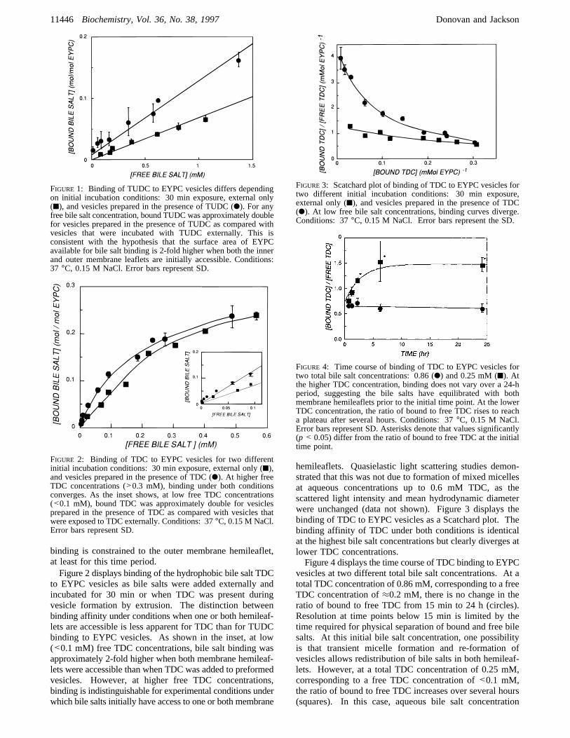
to EYPC vesicles as bile salts were added externally andincubated for 30 min or when TDC was present duringvesicle formation by extrusion. The distinction betweenbinding affinity under conditions when one or both hemileaf-lets are accessible is less apparent for TDC than for TUDCbinding to EYPC vesicles. As shown in the inset, at low(<0.1 mM) free TDC concentrations, bile salt binding wasapproximately 2-fold higher when both membrane hemileaf-lets were accessible than when TDC was added to preformedvesicles. However, at higher free TDC concentrations,binding is indistinguishable for experimental conditions underwhich bile salts initially have access to one or both membrane
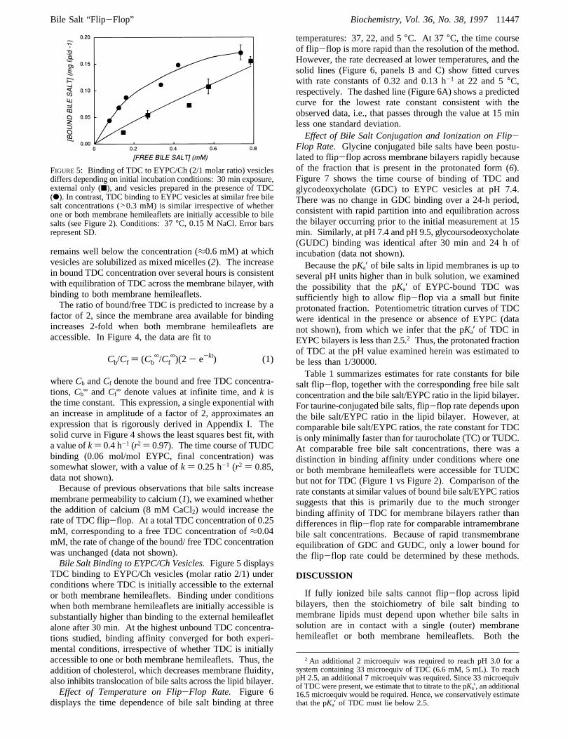
hemileaflets. Quasielastic light scattering studies demon-strated that this was not due to formation of mixed micellesat aqueous concentrations up to 0.6 mM TDC, as thescattered light intensity and mean hydrodynamic diameterwere unchanged (data not shown). Figure 3 displays thebinding of TDC to EYPC vesicles as a Scatchard plot. Thebinding affinity of TDC under both conditions is identicalat the highest bile salt concentrations but clearly diverges atlower TDC concentrations.Figure 4 displays the time course of TDC binding to EYPC
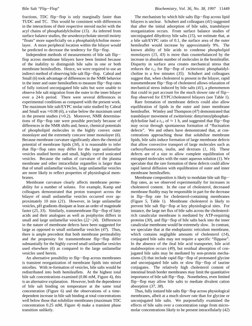
vesicles at two different total bile salt concentrations. At atotal TDC concentration of 0.86 mM, corresponding to a freeTDC concentration of≈0.2 mM, there is no change in theratio of bound to free TDC from 15 min to 24 h (circles).Resolution at time points below 15 min is limited by thetime required for physical separation of bound and free bilesalts. At this initial bile salt concentration, one possibilityis that transient micelle formation and re-formation ofvesicles allows redistribution of bile salts in both hemileaf-lets. However, at a total TDC concentration of 0.25 mM,corresponding to a free TDC concentration of<0.1 mM,the ratio of bound to free TDC increases over several hours(squares). In this case, aqueous bile salt concentration
FIGURE 1: Binding of TUDC to EYPC vesicles differs dependingon initial incubation conditions: 30 min exposure, external only(9), and vesicles prepared in the presence of TUDC (b). For anyfree bile salt concentration, bound TUDC was approximately doublefor vesicles prepared in the presence of TUDC as compared withvesicles that were incubated with TUDC externally. This isconsistent with the hypothesis that the surface area of EYPCavailable for bile salt binding is 2-fold higher when both the innerand outer membrane leaflets are initially accessible. Conditions:37 °C, 0.15 M NaCl. Error bars represent SD.
FIGURE 2: Binding of TDC to EYPC vesicles for two differentinitial incubation conditions: 30 min exposure, external only (9),and vesicles prepared in the presence of TDC (b). At higher freeTDC concentrations (>0.3 mM), binding under both conditionsconverges. As the inset shows, at low free TDC concentrations(<0.1 mM), bound TDC was approximately double for vesiclesprepared in the presence of TDC as compared with vesicles thatwere exposed to TDC externally. Conditions: 37°C, 0.15 M NaCl.Error bars represent SD.
FIGURE 3: Scatchard plot of binding of TDC to EYPC vesicles fortwo different initial incubation conditions: 30 min exposure,external only (9), and vesicles prepared in the presence of TDC(b). At low free bile salt concentrations, binding curves diverge.Conditions: 37°C, 0.15 M NaCl. Error bars represent the SD.
FIGURE 4: Time course of binding of TDC to EYPC vesicles fortwo total bile salt concentrations: 0.86 (b) and 0.25 mM (9). Atthe higher TDC concentration, binding does not vary over a 24-hperiod, suggesting the bile salts have equilibrated with bothmembrane hemileaflets prior to the initial time point. At the lowerTDC concentration, the ratio of bound to free TDC rises to reacha plateau after several hours. Conditions: 37°C, 0.15 M NaCl.Error bars represent SD. Asterisks denote that values significantly(p < 0.05) differ from the ratio of bound to free TDC at the initialtime point.
11446 Biochemistry, Vol. 36, No. 38, 1997 Donovan and Jackson
remains well below the concentration (≈0.6 mM) at whichvesicles are solubilized as mixed micelles (2). The increasein bound TDC concentration over several hours is consistentwith equilibration of TDC across the membrane bilayer, withbinding to both membrane hemileaflets.The ratio of bound/free TDC is predicted to increase by a
factor of 2, since the membrane area available for bindingincreases 2-fold when both membrane hemileaflets areaccessible. In Figure 4, the data are fit to
whereCb andCf denote the bound and free TDC concentra-tions,Cb
∞ andCf∞ denote values at infinite time, andk is
the time constant. This expression, a single exponential withan increase in amplitude of a factor of 2, approximates anexpression that is rigorously derived in Appendix I. Thesolid curve in Figure 4 shows the least squares best fit, witha value ofk) 0.4 h-1 (r2 ) 0.97). The time course of TUDCbinding (0.06 mol/mol EYPC, final concentration) wassomewhat slower, with a value ofk ) 0.25 h-1 (r2 ) 0.85,data not shown).Because of previous observations that bile salts increase
membrane permeability to calcium (1), we examined whetherthe addition of calcium (8 mM CaCl2) would increase therate of TDC flip-flop. At a total TDC concentration of 0.25mM, corresponding to a free TDC concentration of≈0.04mM, the rate of change of the bound/ free TDC concentrationwas unchanged (data not shown).Bile Salt Binding to EYPC/Ch Vesicles.Figure 5 displays
TDC binding to EYPC/Ch vesicles (molar ratio 2/1) underconditions where TDC is initially accessible to the externalor both membrane hemileaflets. Binding under conditionswhen both membrane hemileaflets are initially accessible issubstantially higher than binding to the external hemileafletalone after 30 min. At the highest unbound TDC concentra-tions studied, binding affinity converged for both experi-mental conditions, irrespective of whether TDC is initiallyaccessible to one or both membrane hemileaflets. Thus, theaddition of cholesterol, which decreases membrane fluidity,also inhibits translocation of bile salts across the lipid bilayer.Effect of Temperature on Flip-Flop Rate. Figure 6
displays the time dependence of bile salt binding at three
temperatures: 37, 22, and 5°C. At 37 °C, the time courseof flip-flop is more rapid than the resolution of the method.However, the rate decreased at lower temperatures, and thesolid lines (Figure 6, panels B and C) show fitted curveswith rate constants of 0.32 and 0.13 h-1 at 22 and 5°C,respectively. The dashed line (Figure 6A) shows a predictedcurve for the lowest rate constant consistent with theobserved data, i.e., that passes through the value at 15 minless one standard deviation.Effect of Bile Salt Conjugation and Ionization on Flip-
Flop Rate. Glycine conjugated bile salts have been postu-lated to flip-flop across membrane bilayers rapidly becauseof the fraction that is present in the protonated form (6).Figure 7 shows the time course of binding of TDC andglycodeoxycholate (GDC) to EYPC vesicles at pH 7.4.There was no change in GDC binding over a 24-h period,consistent with rapid partition into and equilibration acrossthe bilayer occurring prior to the initial measurement at 15min. Similarly, at pH 7.4 and pH 9.5, glycoursodeoxycholate(GUDC) binding was identical after 30 min and 24 h ofincubation (data not shown).Because the pKa′ of bile salts in lipid membranes is up to
several pH units higher than in bulk solution, we examinedthe possibility that the pKa′ of EYPC-bound TDC wassufficiently high to allow flip-flop via a small but finiteprotonated fraction. Potentiometric titration curves of TDCwere identical in the presence or absence of EYPC (datanot shown), from which we infer that the pKa′ of TDC inEYPC bilayers is less than 2.5.2 Thus, the protonated fractionof TDC at the pH value examined herein was estimated tobe less than 1/30000.Table 1 summarizes estimates for rate constants for bile
salt flip-flop, together with the corresponding free bile saltconcentration and the bile salt/EYPC ratio in the lipid bilayer.For taurine-conjugated bile salts, flip-flop rate depends uponthe bile salt/EYPC ratio in the lipid bilayer. However, atcomparable bile salt/EYPC ratios, the rate constant for TDCis only minimally faster than for taurocholate (TC) or TUDC.At comparable free bile salt concentrations, there was adistinction in binding affinity under conditions where oneor both membrane hemileaflets were accessible for TUDCbut not for TDC (Figure 1 vs Figure 2). Comparison of therate constants at similar values of bound bile salt/EYPC ratiossuggests that this is primarily due to the much strongerbinding affinity of TDC for membrane bilayers rather thandifferences in flip-flop rate for comparable intramembranebile salt concentrations. Because of rapid transmembraneequilibration of GDC and GUDC, only a lower bound forthe flip-flop rate could be determined by these methods.
DISCUSSION
If fully ionized bile salts cannot flip-flop across lipidbilayers, then the stoichiometry of bile salt binding tomembrane lipids must depend upon whether bile salts insolution are in contact with a single (outer) membranehemileaflet or both membrane hemileaflets. Both the
2 An additional 2 microequiv was required to reach pH 3.0 for asystem containing 33 microequiv of TDC (6.6 mM, 5 mL). To reachpH 2.5, an additional 7 microequiv was required. Since 33 microequivof TDC were present, we estimate that to titrate to the pKa′, an additional16.5 microequiv would be required. Hence, we conservatively estimatethat the pKa′ of TDC must lie below 2.5.
FIGURE 5: Binding of TDC to EYPC/Ch (2/1 molar ratio) vesiclesdiffers depending on initial incubation conditions: 30 min exposure,external only (9), and vesicles prepared in the presence of TDC(b). In contrast, TDC binding to EYPC vesicles at similar free bilesalt concentrations (>0.3 mM) is similar irrespective of whetherone or both membrane hemileaflets are initially accessible to bilesalts (see Figure 2). Conditions: 37°C, 0.15 M NaCl. Error barsrepresent SD.
Cb/Cf ) (Cb∞/Cf
∞)(2- e-kt) (1)
Bile Salt “Flip-Flop” Biochemistry, Vol. 36, No. 38, 199711447

convergence of bile salt binding to one vs both hemileafletsat high bile salt concentrations as well as the observedincrease in bile salt binding over time imply that bile saltsgain access to both membrane hemileaflets. We haveinterpreted time-dependent changes in bile salt affinity forEYPC ( Ch vesicles as reflecting an increase in theaccessible membrane area and infer from these data incombination with titration studies that fully ionized taurineconjugates undergo flip-flop. An intrinsic advantage of thisexperimental approach is that the system is not perturbed
by addition of exogenous molecules. The dependence ofthe time course of bile salt/membrane binding on bile salthydrophobicity, concentration, membrane composition, andtemperature provide further support that increases in bindingaffinity represent membrane flip-flop.The finding that bile salts flip-flop across membranes in
a concentration-dependent fashion is not unexpected. At bilesalt concentrations approaching those that solubilize mem-branes, bile salts must gain access to both membranehemileaflets (26) in order to mediate observed increases inmembrane permeability to large molecules (1, 40, 41), andultimately, to solubilize membranes as mixed micelles.Changes in the outer membrane hemileaflet alone arepredicted to be insufficient to alter membrane permeabilityto polar solutes, which depends on the composition of boththe inner and the outer membrane hemileaflet (31). Indeed,osmotic water permeability can be estimated from the inde-pendent contributions of each hemileaflet (31). If structuralchanges induced by bile salts were confined to the externalmembrane hemileaflet, permeability would be predicted toincrease by a factor of 2 at the most. However, we havepreviously demonstrated that bile salts increase membranepermeability to divalent cations by up to several orders ofmagnitude (1). We infer that bile salts must alter thestructural organization of both inner and outer membranehemileaflets.
Since glycine-conjugated bile salts rapidly translocateacross the membrane as their protonated neutral species (6),the time dependence of binding of glycine-conjugated speciesserves as an internal control for our inference that membranebinding reflects available surface area. The lack of time-dependent changes in binding affinity of GUDC or GDCimplies either that flip-flop occurs prior to the initial timepoint (15 min) or over a time scale much longer than 24 h.The latter is inconsistent with knowledge of the rapid flip-flop of other un-ionized species (21). Furthermore, the strongtemperature dependence of flip-flop of taurine conjugatesspecies is also consistent with relatively high energyintermediates.
For identical free bile salt concentrations, flip-flop ismuch faster for TDC than for TUDC (Figures 1 and 2). Thisis not unexpected given the much lower binding affinity ofTUDC for lipid bilayers than TDC (16, 17). However, Table1 shows that, for comparable intramembrane bile salt mole
FIGURE 6: Time course of bound/free bile salt concentrations for TDC at (A) 37, (B) 22, and (C) 5°C. Solid lines at 22 and 5°C representsingle exponential fits. Dashed line at 37°C represents a single exponential fit with intercept att ) 0 at one-half the final value (see textfor details). Conditions: total bile salt concentration, 0.86 mM; EYPC, 3 mg/mL. Error bars represent SD. Asterisks denote that valuessignificantly (p < 0.05) differ from the ratio of bound to free TDC at the initial time point.
FIGURE 7: Time course of free bile salt concentration for (A) TDCand (B) GDC. GDC binding is unchanged over 24 h, consistentwith rapid equilibration across the membrane bilayer. In contrast,at this low concentration, TDC equilibrates more slowly with at1/2of 1.7 h for the single exponential fit shown. Conditions: total bilesalt concentration, 0.22 mM; EYPC, 3 mg/mL; 37°C. Error barsrepresent SD. Asterisks denote that values significantly (p< 0.05)differ from the free TDC at the initial time point.
Table 1: Comparison of Rate Constants for Bile Salt Flip-Flop
bilesalt
vesiclecomposition
[free bile salt]a,b
(mM)[bound bile salt]b
(mol/mol of EYPC)kc
(h-1)
TDC EYPC 0.04( 0.005 0.05( 0.005 0.4TDC EYPC 0.25( 0.01 0.15( 0.01 >4TDC EYPC/Ch
(2/1 mol/mol)0.4( 0.05 0.13( 0.02 0.06
TC EYPC 0.22( 0.01 0.04( 0.005 0.3TUDC EYPC 0.5( 0.02 0.06( 0.005 0.25GDC EYPC 0.05( 0.005 0.04( 0.005 >4GUDC EYPC 0.19( 0.01 0.02( 0.002 >4
a Values are given as mean( SD. b Free bile salt concentration at24 h. cRate constant derived from eq 1 (see text for details).
11448 Biochemistry, Vol. 36, No. 38, 1997 Donovan and Jackson
fractions, TDC flip-flop is only marginally faster thanTUDC and TC. This would be consistent with differencesin the interactions of their respective steroid nuclei with theacyl chains of phosphatidylcholine (15). As inferred fromsurface balance studies, the ursodeoxycholate steroid moiety“floats” more superficially on a phosphatidylcholine mono-layer. A more peripheral location within the bilayer wouldbe predicted to decrease the tendency for flip-flop.Independent methods of measurements of bile salt flip-
flop across membrane bilayers have been limited becauseof the inability to distinguish bile salts in one or bothmembrane hemileaflets. Hence, we were required to use anindirect method of observing bile salt flip-flop. Cabral andSmall (6) took advantage of differences in the NMR behaviorin the inner and outer hemileaflets to measure flip-flop ratesof fully ionized unconjugated bile salts but were unable toobserve bile salt migration from the outer to the inner bilayerover a 24-h period. There were several differences inexperimental conditions as compared with the present work.The maximum bile salt/EYPC molar ratio studied by Cabraland Small was≈0.08 (6), a value below the maximum usedin the present studies (≈0.2). Moreover, NMR determina-tions of flip-flop rate were possible precisely because ofdifferences in the NMR shifts and, hence, chemical potentialsof phospholipid molecules in the highly convex outermonolayer and the extremely concave inner monolayer (6).Because membrane curvature significantly alters the chemicalpotential of membrane lipids (34), it is reasonable to inferthat flip-flop rates may differ for the large unilamellarvesicles studied herein and small, highly curved sonicatedvesicles. Because the radius of curvature of the plasmamembrane and other intracellular organelles is larger thanthat of small unilamellar vesicles, large unilamellar vesiclesare more likely to reflect properties of physiological mem-branes.Membrane curvature clearly affects membrane perme-
ability for a number of solutes. For example, Kamp andcolleagues demonstrated that proton transport across thebilayer of small unilamellar vesicles occurred over ap-proximately 10 min (21). However, in large unilamellarvesicles, pH gradients dissipate at least an order of magnitudefaster (25, 33). Similarly, transmembrane flip-flop of fattyacids and their analogues as well as porphyrins differs insmall and large unilamellar vesicles (22-24). Differencesin the nature of membrane defects have been suggested forlarge as opposed to small unilamellar vesicles (47). Thus,there is ample precedent that both membrane permeabilityand the propensity for transmembrane flip-flop differsubstantially for the highly curved small unilamellar vesiclesused elsewhere (6) as compared to the large unilamellarvesicles used herein.An alternative possibility to flip-flop across membranes
is transient reorganization of membrane lipids into mixedmicelles. With re-formation of vesicles, bile salts would beredistributed into both hemileaflets. At the highest totalbile salt concentrations examined (0.86 mM, Figure 4), thisis an alternative explanation. However, both the dependenceof bile salt binding on temperature at the same totalconcentration (Figure 6) and the observations of a time-dependent increase in bile salt binding at total concentrationswell below those that solubilize membranes (maximum TDCconcentration 0.25 mM, Figure 4) make a transient phasetransition unlikely.
The mechanism by which bile salts flip-flop across lipidbilayers is unclear. Schubert and colleagues (41) suggestedthat after the initial adsorption of bile salts, transbilayerreorganization occurs. From surface balance studies ofunconjugated dihydroxy bile salts (15), we estimate that, ata bile salt/EYPC ratio of 0.1, the surface area of the outerhemileaflet would increase by approximately 9%. Theknown ability of bile acids to condense phospholipidmonolayers (15, 43) is more than counterbalanced by theincrease in absolute number of molecules in the hemileaflet.Disparity in surface area creates mechanical stress thatreduces thet1/2 for flip-flop of zwitterionic phosphatidyl-choline to a few minutes (35). Schubert and colleaguessuggest that, when cholesterol is present in the bilayer, rapidtransmembrane flip-flop of cholesterol allows relaxation ofmechanical stress induced by bile salts (41), a phenomenonthat could in part account for the much slower rate of flip-flop observed for EYPC/cholesterol bilayers (see Table 1).Rare formation of membrane defects could also allow
equilibration of lipids in the outer and inner membranehemileaflet. Wimley and Thompson (47) demonstrated thattransbilayer movement of zwitterionic dimyristoylphosphati-dylcholine had at1/2 of ≈ 1 h, and suggested that flip-flopmay occur through spontaneous formation of “fluctuationdefects”. We and others have demonstrated that, at con-centrations approaching those that solubilize membranelipids, bile salts induce formation of large membrane defectsthat allow convective transport of large molecules such ascarboxyfluorescein, inulin, and dextrans (1, 16). Thesedefects are of a size to allow complete equilibration ofentrapped molecules with the outer aqueous solution (1). Wespeculate that the rare formation of these defects could allowrapid lateral diffusion with equilibration of outer and innermembrane hemileaflets.Membrane composition is likely to modulate bile salt flip-
flop rates, as we observed experimentally for increases incholesterol content. In the case of cholesterol, decreasedmembrane fluidity may be responsible in part for the decreasein flip-flop rate for cholesterol-containing membranes(Figure 5, Table 1). Membrane cholesterol is likely toprevent bile salt flip-flop at key physiological sites. Forinstance, the large net flux of bile salts across the cholesterol-rich canalicular membrane is mediated by ATP-requiringproteins (30), and flip-flop of bile salts back into the innercanalicular membrane would be extremely slow. In contrast,we speculate that at the endoplasmic reticulum membrane,which contains negligible amounts of cholesterol (14),conjugated bile salts may not require a specific “flippase”.In the absence of the ileal bile acid transporter, bile acidmalabsorption occurs (49), but residual absorption of con-jugated bile salts may be mediated by non-protein mecha-nisms (3) that include rapid flip-flop of protonated glycineand unconjugated bile salts or slow flip-flop of taurineconjugates. The relatively high cholesterol content ofintestinal brush border membranes may limit the quantitativeimportance of bile salt flip-flop. Nonetheless, even limitedflip-flop may allow bile salts to mediate divalent cationabsorption (37, 38).Thus, fully ionized bile salts flip-flop across physiological
membranes, albeit at a much slower rate than for glycine orunconjugated bile salts. We purposefully examined theeffects of bile salts over a concentration range from micro-molar concentrations likely to be present intracellularly (42)
Bile Salt “Flip-Flop” Biochemistry, Vol. 36, No. 38, 199711449

to micellar concentrations present in the intestine. The goalof the present study is to determine the physicochemicalprocesses that may occur under physiological conditions. Asrecently estimated by Setchell and colleagues (42), physi-ological hepatic bile salt concentrations are in the lowmicromolar range and increase during cholestasis. Since bilesalt toxicity is observed at concentrations higher thanphysiological, we hypothesize that, within the range betweenphysiological low micromolar concentrations and higherconcentrations found in cholestasis, bile salts begin to interactwith membranes and alter their fundamental barrier function.We speculate that the ability of fully ionized bile salts togain access to both membrane hemileaflets may contributeto bile salt toxicity by creating transmembrane pathways thatincrease divalent cation permeability (1).
ACKNOWLEDGMENT
The authors appreciate stimulating and helpful discussionwith Ariela Albalak, Ph.D.
APPENDIX I: MATHEMATICAL MODEL OF BILESALT FLIP -FLOP
The time dependence of binding can be modeled byassuming that the available surface area of EYPC increasesby a factor of 2 from time zero to infinite time. This assumesthat the rate of flip-flop is slow as compared with the rateof bile salt partitioning into the outer membrane hemi-leaflet. This assumption is justified by rapid equilibrationof other amphiphiles with membrane vesicles (23, 51) andby our observation that GDC and GUDC binding isunchanged over a 24-h period (Figure 7). The rate oftransmembrane transport in either direction is assumed tobe first order with respect to the bile salt concentration inthat hemileaflet. Thus, the net rate of accumulation of bilesalts in the inner leaflet is
whereCin andCout represent the concentration of bile saltsin the inner and outer hemileaflet, respectively, andkflip isthe rate constant.
We assume that initially bile salts are only bound to theouter leaflet
whereKeq is the equilibrium binding constant for bile saltbinding to EYPC membranes,Cf is the free bile salt con-centration, and [EYPC] is the total EYPC concentration. Theeffective concentration of EYPC is one-half of the total, sinceonly the outer hemileaflet is assumed to be accessible.
Using the equations for mass balance:
whereCb is the total bound bile salt concentration, andrearranging, we obtain the expression
Integration fromt ) 0, at which timeCin ) 0, to time tyields an expression forCin:
substitutingR ) K × EYPC) Cb∞/Cf
∞ whereCb∞ andCf
∞
denote the ratio at equilibrium, we obtain
At t ) 0, this simplifies to 1/2(Cb∞/Cf
∞), and att ) ∞, thisexpression simplifies toCb
∞/Cf∞.
For typical values ofCb∞/Cf
∞, the value ofkflip is within15% of the value ofk from eq 1. Hence, values derivedfrom curves fitted to the simpler expression shown in eq 1are used through this work.
REFERENCES
1. Albalak, A., Jackson, A. A., and Donovan, J. M. (1997) inBile Acids in Gastroenterology(Hofmann, A. F., Paumgartner,G., and Stiehl, A., Eds.) pp 256-266, Kluwer AcademicPublishers, Dordecht, The Netherlands.
2. Albalak, A., Zeidel, M. L., Zucker, S., Jackson, A. A., andDonovan, J. M. (1996)Biochemistry 35, 7936-7945.
3. Amelsberg, A., Schteingart, C. D., Ton-Nu, H. T., andHofmann, A. F. (1996)Gastroenterology 110, 1098-1106.
4. Cabral, D. J., Hamilton, J. A., and Small, D. M. (1986)J.Lipid Res. 27, 334-343.
5. Cabral, D. J., and Small, D. M. (1989) inHandbook ofPhysiologysThe Gastrointestinal System III, Section 6(Schultz,S. G., Forte, J. G., and Rauner, B. B., Eds.) pp 621-662,American Physiology Society, Waverly Press, Baltimore, MD.
6. Cabral, D. J., Small, D. M., Lilly, H. S., and Hamilton, J. A.(1987)Biochemistry 26, 1801-1804.
7. Carey, M. C. (1985) inSterols and Bile Acids(Danielsson,H., and Sjo¨vall, J., Eds.) pp 345-403, Elsevier, Amsterdam.
8. Carey, M. C. (1988) inBile Acids in Health and Disease(Northfield, T. C., Jazrawi, R. P., and Zentler-Munro, P. L.,Eds.) pp 61-82, Kluwer Academic, Dordrecht, The Nether-lands.
9. Carey, M. C., and Hernell, O. (1992)Sem. Gastrol. Dis. 3,189-208.
10. Carey, M. C., and Small, D. M. (1978)J. Clin. InVest. 61,998-1026.
11. Cohen, D. E., Fisch, M. R., and Carey, M. C. (1990)Hepatology 12, 113S-121S.
12. Cohen, D. E., Leighton, L. S., and Carey, M. C. (1992)Am.J. Physiol. 263, G386-G395.
13. Donovan, J. M., and Jackson, A. A. (1993)J. Lipid Res. 34,1121-1129.
14. Evans, W. H., and Hardison, W. G. M. (1985)Biochem. J.232, 33-36.
15. Fahey, D. A., Carey, M. C., and Donovan, J. M. (1995)Biochemistry 34, 10886-10897.
16. Heuman, D. M., and Bajaj, R. (1994)Gastroenterology 106,1333-1341.
17. Heuman, D. M., Bajaj, R. S., and Lin, Q. A. (1996)J. LipidRes. 37, 562-573.
dCin
dt) kflip[ (Ct - Cin)
1+ K × EYPC2
K × EYPC2
- Cin] (5)
Cin )CtK × EYPC
2(1+ K × EYPC)[1-
exp[-2(1+ K × EYPC)
(2+ K × EYPC)kflipt]] (6)
Cb
Cf)(2+ R) - exp(- 2(1+ R)
(2+ R)kflipt)
(2+ R)R
+ exp(- 2(1+ R)(2+ R)
kflipt) (7)
dCin
dt) kflip(Cout - Cin) (2)
Keq)Cout
Cf([EYPC]2 ) (3)
Cf ) Ct - Cout - Cin and Cb ) Cout + Cin (4)
11450 Biochemistry, Vol. 36, No. 38, 1997 Donovan and Jackson
18. Heuman, D. M., Hylemon, P. B., Pandak, W. M., andVlahcevic, Z. R. (1991)Hepatology 14, 920-926.
19. Heuman, D. M., Mills, A. S., McCall, J., Hylemon, P. B.,Pandak, W. M., and Vlahcevic, Z. R. (1991)Gastroenterology100, 203-211.
20. Hope, M. J., Bally, M. B., Webb, G., and Cullis, P. R. (1985)Biochim. Biophys. Acta 812, 55-65.
21. Kamp, F., Westerhoff, H. V., and Hamilton, J. A. (1993)Biochemistry 89, 11074-11086.
22. Kamp, F., Zakim, D., Zhang, F. L., Noy, N., and Hamilton, J.A. (1995)Biochemistry 34, 11928-11937.
23. Kleinfeld, A. M., and Storch, J. (1993)Biochemistry 32, 2053-2061.
24. Kuzelova, K., and Brault, D. (1995)Biochemistry 33, 9447-9459.
25. Lande, M. B., Donovan, J. M., and Zeidel, M. L. (1995)J.Gen. Physiol. 106, 67-84.
26. Lasch, J. (1995)Biochim. Biophys. Acta 1241, 269-292.27. Lowe, P. J., and Coleman, R. (1981)Biochim. Biophys. Acta
640, 55-65.28. MacDonald, R. C., MacDonald, R. I., Menco, B. P. M.,
Takeshita, K., Subbarao, N. K., and Hu, L. R. (1991)Biochim.Biophys. Acta 1061, 297-303.
29. Mazer, N. A., Benedek, G. B., and Carey, M. C. (1980)Biochemistry 19, 601-615.
30. Meier, P. J. (1995)Am. J. Physiol. 32, G801-G812.31. Negrete, H. O., Rivers, R. L., Gough, A. H., Colombini, M.,
and Zeidel, M. L. (1996)J. Biol. Chem. 271, 11627-11630.32. New, R. R. C. (1990)Liposomes, A Practical Approach,
Oxford University Press, Oxford.33. Priver, N. A., Rabon, E. C., and Zeidel, M. L. (1993)
Biochemistry 32, 2459-2468.34. Qiu, R. Z., and Macdonald, R. C. (1994)Biochim. Biophys.
Acta 1191, 343-353.35. Raphael, R. M., and Waugh, R. E. (1996)Biophys. J. 71,
1374-1388.
36. Rossi, S. S., Converse, J. L., and Hofmann, A. F. (1987)J.Lipid Res. 28, 589-595.
37. Sanyal, A. J., Hirsch, J. I., and Moore, E. W. (1994)Am. J.Physiol. 266, G318-G323.
38. Sanyal, A. J., Hirsch, J. I., and Moore, E. W. (1994)Gastroenterology 106, 866-874.
39. Schmucker, D. L., Ohta, M., Kanai, S., Sato, Y., and Kitani,K. (1990)Hepatology 12, 1216-1221.
40. Schubert, R., Beyer, K., Wolburg, H., and Schmidt, K. H.(1986)Biochemistry 25, 5263-5269.
41. Schubert, R., and Schmidt, K.-H. (1988)Biochemistry 27,8787-8794.
42. Setchell, K. D. R., Rodrigues, C. M. P., Clerici, C., Solinas,A., Morelli, A., Gartung, C., and Boyer, J. (1997)Gastroen-terology 112, 226-235.
43. Small, D. M. (1971) inThe Bile Acids, Vol. I(Nair, P. P., andKritchevsky, D., Eds.) pp 249-356, Plenum Press, New York.
44. Staggers, J., Hernell, O., Stafford, R. J., and Carey, M. C.(1990)Biochemistry 29, 2028-2040.
45. Stieger, B., Oneill, B., and Meier, P. J. (1992)Biochem. J.284, 67-74.
46. Verkade, H. J., Vonk, R. J., and Kuipers, F. (1995)Hepatology21, 1174-1189.
47. Wimley, W. C., and Thompson, T. E. (1991)Biochemistry30, 1702-1709.
48. Wong, M. H., Oelkers, P., Craddock, A. L., and Dawson, P.A. (1994)J. Biol. Chem. 269, 1340-1347.
49. Wong, M. H., Oelkers, P., and Dawson, P. A. (1995)J. Biol.Chem. 270, 27228-27234.
50. Yousef, I. M., Barnwell, S., Gratton, F., Tuchweber, B., Weber,A., and Roy, C. C. (1987)Am. J. Physiol. 252, G84-G91.
51. Zucker, S. D., Storch, J., Zeidel, M. L., and Gollan, J. L. (1992)Biochemistry 31, 3184-3192.
BI9705927
Bile Salt “Flip-Flop” Biochemistry, Vol. 36, No. 38, 199711451


![Differences in Nanostructure and Hydrophobicity of …downloads.hindawi.com/journals/abb/2018/5305847.pdfsurfaces among cicadas [18, 22, 35]. Some species show very weak hydrophobicity](https://img.pdfslide.net/doc/110x75/5f2b7ebc5322004a2502ee65/differences-in-nanostructure-and-hydrophobicity-of-surfaces-among-cicadas-18-22.jpg)