Embed Size (px)
Citation preview
684 Copyright © 2012 The Korean Society of Cardiology
Korean Circulation Journal
Introduction
Aortic stenosis (AS) is the most common valvular heart disease in adults,1) and is associated with significant morbidity and mortality in advanced cases. The incidence of AS is on the rise as a result of
Original Article
http://dx.doi.org/10.4070/kcj.2012.42.10.684Print ISSN 1738-5520 • On-line ISSN 1738-5555
Transcatheter Aortic Valve Implantation: Early Experience in KoreaWon-Jang Kim, MD1, Young-Hak Kim, MD1, Jong-Young Lee, MD1, Duk-Woo Park, MD1, Soo-Jin Kang, MD1, Seung-Whan Lee, MD1, Cheol Whan Lee, MD1, Dae-Hee Kim, MD1, Jong-Min Song, MD1, Duk-Hyun Kang, MD1, Jae-Kwan Song, MD1, Joon Bum Kim, MD2, Sung Ho Jung, MD2, Suk Jung Choo, MD2, Cheol Hyun Chung, MD2,Jae Won Lee, MD2, Ji-Yeon Sim, MD3, In-Cheol Choi, MD3, Seong-Wook Park, MD1, and Seung-Jung Park, MD1
1Departments of Cardiology, 2Thoracic and Cardiovascular Surgery and 3Anesthesiology and Pain Medicine, University of Ulsan College of Medicine, Asan Medical Center, Seoul, Korea
Background and Objectives: Transcatheter aortic valve implantation (TAVI) was recently introduced in Korea. The present report describes the experience of early TAVI cases.Subjects and Methods: Between March, 2010 and October, 2011, 48 patients with severe symptomatic aortic stenosis (AS) were screened at the Asan Medical Center to determine their suitability for surgical intervention. Of these, 23 were considered unsuitable and underwent TAVI. Procedural success rates, procedure-related complications, and clinical outcomes were evaluated in the TAVI patients.Results: Transfemoral (n=20) or transapical (n=3) TAVI was performed. The mean age of patients was 75.9±5.4 years and 57% were fe-males. The mean logistic European System for Cardiac Operative Risk Evaluation was 25.6±5.1%. Implantation was successful in 22 pa-tients (19 transfemoral, three transapical). After successful implantation, the mean aortic valve area increased from 0.68±0.14 cm2 to 1.45±0.33 cm2. There were no procedure-related complications or mortality. The patients showed no paravalvular aortic regurgitation with ≥ moderate degree and remained stable without progression during follow-up. During follow-up (interquartile range, 1.1-12.9), all pa-tients were alive without any occurrence of valve failure.Conclusion: TAVI procedure is feasible in patients with inoperative symptomatic AS leading to hemodynamic and clinical improvement. With accumulation of experience, proper patient selection and development of device technologies, TAVI should decrease adverse events and expand the indications in the near future. (Korean Circ J 2012;42:684-691)
KEY WORDS: Aortic stenosis; Valve prosthesis.
Received: February 15, 2012Revision Received: April 30, 2012Accepted: May 2, 2012Correspondence: Seung-Jung Park, MD, Department of Cardiology, Uni-versity of Ulsan College of Medicine, Cardiac Center, Asan Medical Center, 88 Olympic-ro 43-gil, Songpa-gu, Seoul 138-736, KoreaTel: 82-2-3010-4812, Fax: 82-2-475-6898E-mail: [email protected]
• The authors have no financial conflicts of interest.
This is an Open Access article distributed under the terms of the Creative Commons Attribution Non-Commercial License (http://creativecommons.org/licenses/by-nc/3.0) which permits unrestricted non-commercial use, distribution, and reproduction in any medium, provided the original work is properly cited.
the aging population. Several studies have investigated the efficacy of surgical aortic valve replacement for AS, and there is now a ge-neral consensus in the field that this procedure prolongs survival.2)3) However, one-third of patients with severe symptomatic AS in need of treatment are declined for surgery due to advanced age, multiple comorbidity, and a high risk of procedure-related mortality, factors which are associated with reduced life expectancy and poor quality of life.1)4) The proportion of untreated patients is expected to further increase because of the aging population and improving treatment mo-dalities in patients with chronic and advanced medical conditions.5)
With introduction of transcatheter aortic valve implantation (TAVI), those untreated patients are adapted to alternative treatment mo-dalities. In 2002, Cribier et al.6) performed the first TAVI, and many patients worldwide have undergone the procedure by means of two different device technologies. A recently published randomized trial for the inoperable symptomatic severe AS showed that TAVI signifi-cantly reduced the rates of death, repeat hospitalization, and car-
685Won-Jang Kim, et al.
http://dx.doi.org/10.4070/kcj.2012.42.10.684www.e-kcj.org
diac symptoms compared with the standard procedures including balloon aortic valvuloplasty.7) The Edward SAPIENTM (Edwards Life-sciences LLC, Irvine, CA, USA)8) and CoreValve ReValving® System (Me-dtronic LLC, Irvine, CA, USA) prosthetic heart valve obtained Confor-mité Européenne mark approval in August and April 2007, respec-tively. Both technologies are currently subjected to post-marketing surveillance studies in Europe. In 2009, Korean Food and Drug Ad-ministration authorized conduct of the first explorative studies at the Asan Medical Center in Korea. Here, we report procedural suc-cess rates and clinical outcomes for the Edwards SAPIENTM and XTTM in the early TAVI cases in Korea.
Subjects and Methods
Study populationBetween March 2010 and October 2011, 48 patients with severe
symptomatic AS were screened at the Asan Medical Center to de-termine if they were at high risk of surgical mortality and were not proper candidates for receiving surgical operation. Patient prefer-ence alone for a percutaneous procedure was not considered ade-quate if surgery was an option.9) Twenty-three patients were consi-dered unsuitable for surgery due to the presence of multiple co-morbid disorders and/or the high risk of procedure-related mortality. These patients were referred for a percutaneous procedure, and were assessed by a team of intervention cardiologists, echocardiograph-ic cardiologists, and cardiac surgeons.
Prior to TAVI, all patients underwent transthoracic (TTE), trans-esophageal echocardiography (TEE), iliofemoral angiography, coro-nary and aortic root angiography, and computed tomographic (CT) angiography. Clinical evaluation and TTE and/or CT were obtained within 24 hours of the procedure and at 1, 6, and 12 months of fol-low-up.
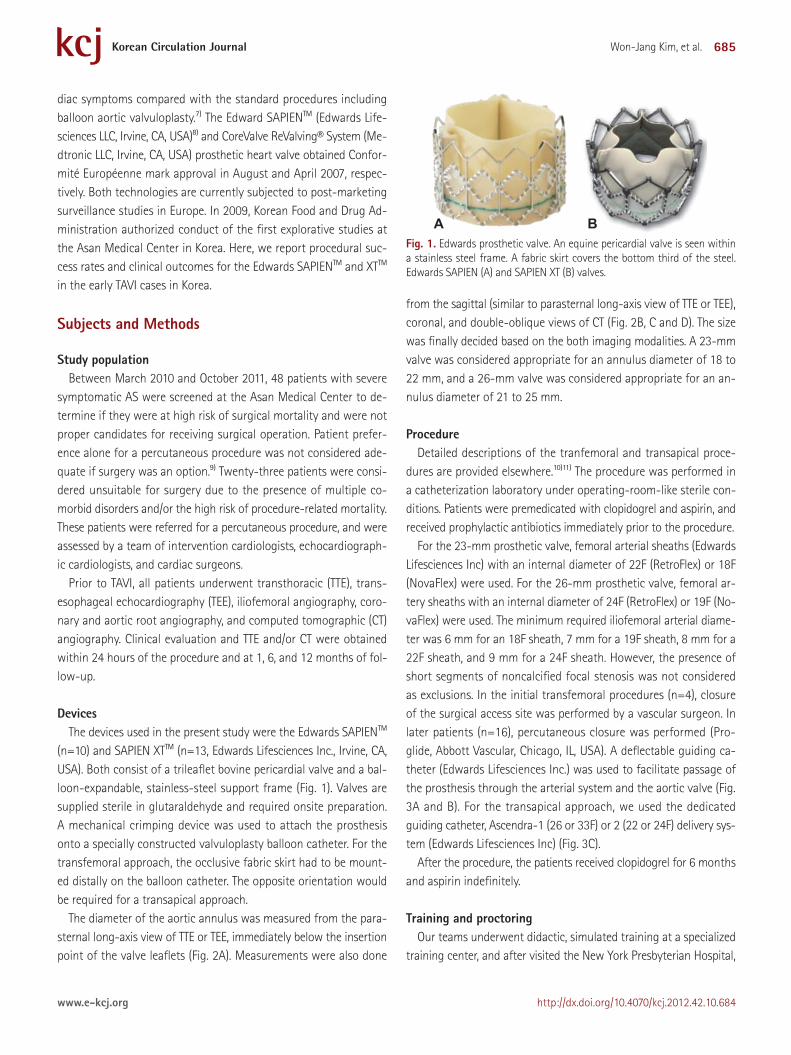
DevicesThe devices used in the present study were the Edwards SAPIENTM
(n=10) and SAPIEN XTTM (n=13, Edwards Lifesciences Inc., Irvine, CA, USA). Both consist of a trileaflet bovine pericardial valve and a bal-loon-expandable, stainless-steel support frame (Fig. 1). Valves are supplied sterile in glutaraldehyde and required onsite preparation. A mechanical crimping device was used to attach the prosthesis onto a specially constructed valvuloplasty balloon catheter. For the transfemoral approach, the occlusive fabric skirt had to be mount-ed distally on the balloon catheter. The opposite orientation would be required for a transapical approach.
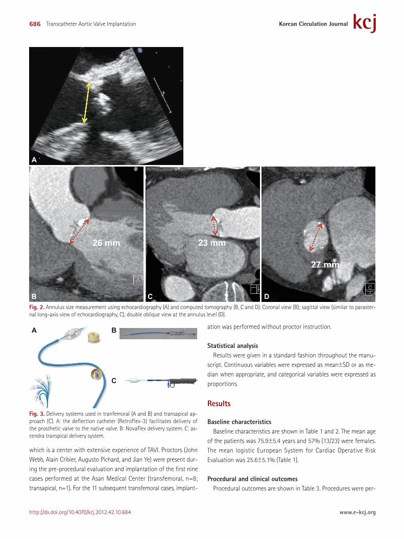
The diameter of the aortic annulus was measured from the para-sternal long-axis view of TTE or TEE, immediately below the insertion point of the valve leaflets (Fig. 2A). Measurements were also done
from the sagittal (similar to parasternal long-axis view of TTE or TEE), coronal, and double-oblique views of CT (Fig. 2B, C and D). The size was finally decided based on the both imaging modalities. A 23-mm valve was considered appropriate for an annulus diameter of 18 to 22 mm, and a 26-mm valve was considered appropriate for an an-nulus diameter of 21 to 25 mm.
ProcedureDetailed descriptions of the tranfemoral and transapical proce-
dures are provided elsewhere.10)11) The procedure was performed in a catheterization laboratory under operating-room-like sterile con-ditions. Patients were premedicated with clopidogrel and aspirin, and received prophylactic antibiotics immediately prior to the procedure.
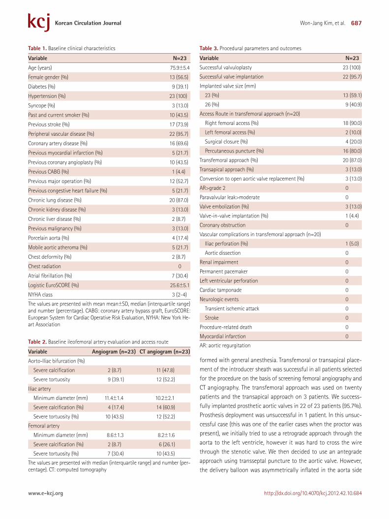
For the 23-mm prosthetic valve, femoral arterial sheaths (Edwards Lifesciences Inc) with an internal diameter of 22F (RetroFlex) or 18F (NovaFlex) were used. For the 26-mm prosthetic valve, femoral ar-tery sheaths with an internal diameter of 24F (RetroFlex) or 19F (No-vaFlex) were used. The minimum required iliofemoral arterial diame-ter was 6 mm for an 18F sheath, 7 mm for a 19F sheath, 8 mm for a 22F sheath, and 9 mm for a 24F sheath. However, the presence of short segments of noncalcified focal stenosis was not considered as exclusions. In the initial transfemoral procedures (n=4), closure of the surgical access site was performed by a vascular surgeon. In later patients (n=16), percutaneous closure was performed (Pro-glide, Abbott Vascular, Chicago, IL, USA). A deflectable guiding ca-theter (Edwards Lifesciences Inc.) was used to facilitate passage of the prosthesis through the arterial system and the aortic valve (Fig. 3A and B). For the transapical approach, we used the dedicated guiding catheter, Ascendra-1 (26 or 33F) or 2 (22 or 24F) delivery sys-tem (Edwards Lifesciences Inc) (Fig. 3C).
After the procedure, the patients received clopidogrel for 6 months and aspirin indefinitely.
Training and proctoringOur teams underwent didactic, simulated training at a specialized
training center, and after visited the New York Presbyterian Hospital,
A B Fig. 1. Edwards prosthetic valve. An equine pericardial valve is seen within a stainless steel frame. A fabric skirt covers the bottom third of the steel. Edwards SAPIEN (A) and SAPIEN XT (B) valves.
686 Transcatheter Aortic Valve Implantation
http://dx.doi.org/10.4070/kcj.2012.42.10.684 www.e-kcj.org
which is a center with extensive experience of TAVI. Proctors (John Webb, Alain Cribier, Augusto Pichard, and Jian Ye) were present dur-ing the pre-procedural evaluation and implantation of the first nine cases performed at the Asan Medical Center (transfemoral, n=8; transapical, n=1). For the 11 subsequent transfemoral cases, implant-
ation was performed without proctor instruction.
Statistical analysisResults were given in a standard fashion throughout the manu-
script. Continuous variables were expressed as mean±SD or as me-dian when appropriate, and categorical variables were expressed as proportions.
Results
Baseline characteristicsBaseline characteristics are shown in Table 1 and 2. The mean age
of the patients was 75.9±5.4 years and 57% (13/23) were females. The mean logistic European System for Cardiac Operative Risk Evaluation was 25.6±5.1% (Table 1).
Procedural and clinical outcomesProcedural outcomes are shown in Table 3. Procedures were per-
Fig. 3. Delivery systems used in tranfemoral (A and B) and transapical ap-proach (C). A: the deflection catheter (RetroFlex-3) facilitates delivery of the prosthetic valve to the native valve. B: NovaFlex delivery system. C: as-cendra transpical delivery system.
A B
C
Fig. 2. Annulus size measurement using echocardiography (A) and computed tomography (B, C and D). Coronal view (B); sagittal view (similar to paraster-nal long-axis view of echocardiography, C); double oblique view at the annulus level (D).
A
B C D
687Won-Jang Kim, et al.
http://dx.doi.org/10.4070/kcj.2012.42.10.684www.e-kcj.org
formed with general anesthesia. Transfemoral or transapical place-ment of the introducer sheath was successful in all patients selected for the procedure on the basis of screening femoral angiography and CT angiography. The transfemoral approach was used on twenty patients and the transapical approach on 3 patients. We success-fully implanted prosthetic aortic valves in 22 of 23 patients (95.7%). Prosthesis deployment was unsuccessful in 1 patient. In this unsuc-cessful case (this was one of the earlier cases when the proctor was present), we initially tried to use a retrograde approach through the aorta to the left ventricle, however it was hard to cross the wire through the stenotic valve. We then decided to use an antegrade approach using transseptal puncture to the aortic valve. However, the delivery balloon was asymmetrically inflated in the aorta side
Table 1. Baseline clinical characteristics
Variable N=23
Age (years) 75.9±5.4
Female gender (%) 13 (56.5)
Diabetes (%) 9 (39.1)
Hypertension (%) 23 (100)
Syncope (%) 3 (13.0)
Past and current smoker (%) 10 (43.5)
Previous stroke (%) 17 (73.9)
Peripheral vascular disease (%) 22 (95.7)
Coronary artery disease (%) 16 (69.6)
Previous myocardial infarction (%) 5 (21.7)
Previous coronary angioplasty (%) 10 (43.5)
Previous CABG (%) 1 (4.4)
Previous major operation (%) 12 (52.7)
Previous congestive heart failure (%) 5 (21.7)
Chronic lung disease (%) 20 (87.0)
Chronic kidney disease (%) 3 (13.0)
Chronic liver disease (%) 2 (8.7)
Previous malignancy (%) 3 (13.0)
Porcelain aorta (%) 4 (17.4)
Mobile aortic atheroma (%) 5 (21.7)
Chest deformity (%) 2 (8.7)
Chest radiation 0
Atrial fibrillation (%) 7 (30.4)
Logistic EuroSCORE (%) 25.6±5.1
NYHA class 3 (2-4)
The values are presented with mean mean±SD, median (interquartile range) and number (percentage). CABG: coronary artery bypass graft, EuroSCORE: European System for Cardiac Operative Risk Evaluation, NYHA: New York He-art Association
Table 2. Baseline ileofemoral artery evaluation and access route
Variable Angiogram (n=23) CT angiogram (n=23)
Aorto-Iliac bifurcation (%)
Severe calcification 2 (8.7) 11 (47.8)
Severe tortuosity 9 (39.1) 12 (52.2)
Iliac artery
Minimum diameter (mm) 11.4±1.4 10.2±2.1
Severe calcification (%) 4 (17.4) 14 (60.9)
Severe tortuosity (%) 10 (43.5) 12 (52.2)
Femoral artery
Minimum diameter (mm) 8.6±1.3 8.2±1.6
Severe calcification (%) 2 (8.7) 6 (26.1)
Severe tortuosity (%) 7 (30.4) 10 (43.5)
The values are presented with median (interquartile range) and number (per-centage). CT: computed tomography
Table 3. Procedural parameters and outcomes
Variable N=23
Successful valvuloplasty 23 (100)
Successful valve implantation 22 (95.7)
Implanted valve size (mm)
23 (%) 13 (59.1)
26 (%) 9 (40.9)
Access Route in transfemoral approach (n=20)
Right femoral access (%) 18 (90.0)
Left femoral access (%) 2 (10.0)
Surgical closure (%) 4 (20.0)
Percutaneous puncture (%) 16 (80.0)
Transfemoral approach (%) 20 (87.0)
Transapical approach (%) 3 (13.0)
Conversion to open aortic valve replacement (%) 3 (13.0)
AR>grade 2 0
Paravalvular leak>moderate 0
Valve embolization (%) 3 (13.0)
Valve-in-valve implantation (%) 1 (4.4)
Coronary obstruction 0
Vascular complications in transfemoral approach (n=20)
Iliac perforation (%) 1 (5.0)
Aortic dissection 0
Renal impairment 0
Permanent pacemaker 0
Left ventricular perforation 0
Cardiac tamponade 0
Neurologic events 0
Transient ischemic attack 0
Stroke 0
Procedure-related death 0
Myocardial infarction 0
AR: aortic regurgitation
688 Transcatheter Aortic Valve Implantation
http://dx.doi.org/10.4070/kcj.2012.42.10.684 www.e-kcj.org
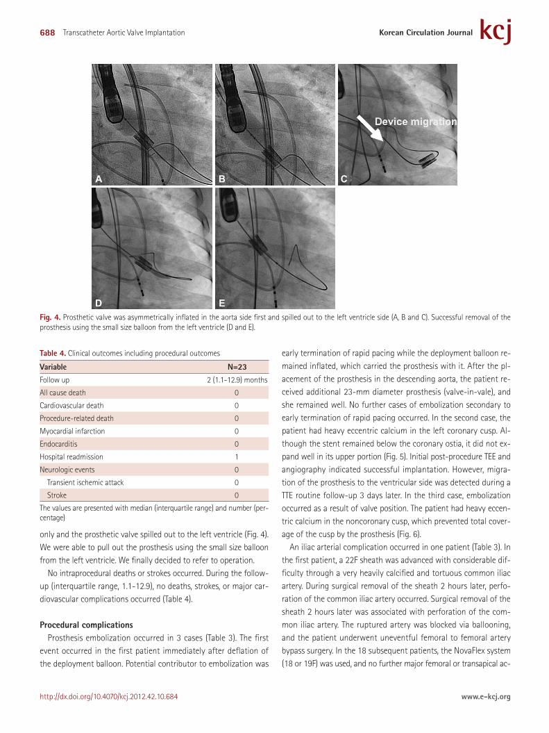
only and the prosthetic valve spilled out to the left ventricle (Fig. 4). We were able to pull out the prosthesis using the small size balloon from the left ventricle. We finally decided to refer to operation.
No intraprocedural deaths or strokes occurred. During the follow-up (interquartile range, 1.1-12.9), no deaths, strokes, or major car-diovascular complications occurred (Table 4).
Procedural complicationsProsthesis embolization occurred in 3 cases (Table 3). The first
event occurred in the first patient immediately after deflation of the deployment balloon. Potential contributor to embolization was
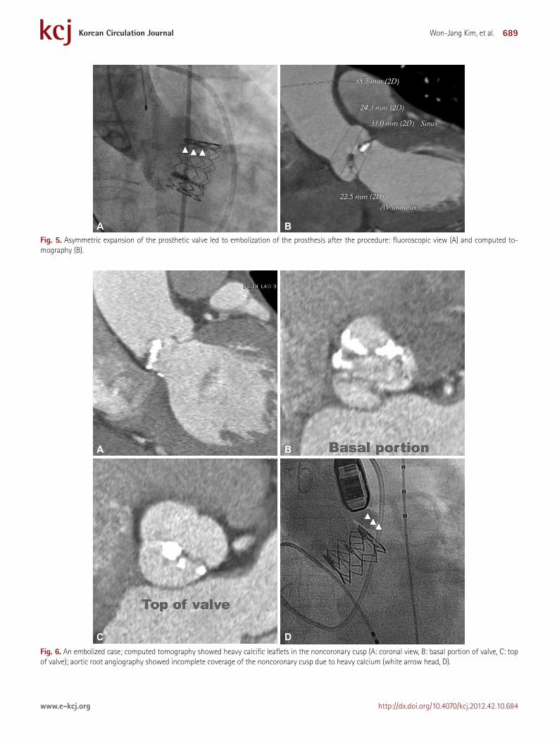
early termination of rapid pacing while the deployment balloon re-mained inflated, which carried the prosthesis with it. After the pl-acement of the prosthesis in the descending aorta, the patient re-ceived additional 23-mm diameter prosthesis (valve-in-vale), and she remained well. No further cases of embolization secondary to early termination of rapid pacing occurred. In the second case, the patient had heavy eccentric calcium in the left coronary cusp. Al-though the stent remained below the coronary ostia, it did not ex-pand well in its upper portion (Fig. 5). Initial post-procedure TEE and angiography indicated successful implantation. However, migra-tion of the prosthesis to the ventricular side was detected during a TTE routine follow-up 3 days later. In the third case, embolization occurred as a result of valve position. The patient had heavy eccen-tric calcium in the noncoronary cusp, which prevented total cover-age of the cusp by the prosthesis (Fig. 6).
An iliac arterial complication occurred in one patient (Table 3). In the first patient, a 22F sheath was advanced with considerable dif-ficulty through a very heavily calcified and tortuous common iliac artery. During surgical removal of the sheath 2 hours later, perfo-ration of the common iliac artery occurred. Surgical removal of the sheath 2 hours later was associated with perforation of the com-mon iliac artery. The ruptured artery was blocked via ballooning, and the patient underwent uneventful femoral to femoral artery bypass surgery. In the 18 subsequent patients, the NovaFlex system (18 or 19F) was used, and no further major femoral or transapical ac-
Fig. 4. Prosthetic valve was asymmetrically inflated in the aorta side first and spilled out to the left ventricle side (A, B and C). Successful removal of the prosthesis using the small size balloon from the left ventricle (D and E).
A
D
B
E
C
Table 4. Clinical outcomes including procedural outcomes
Variable N=23Follow up 2 (1.1-12.9) months
All cause death 0
Cardiovascular death 0
Procedure-related death 0
Myocardial infarction 0
Endocarditis 0
Hospital readmission 1
Neurologic events 0
Transient ischemic attack 0
Stroke 0
The values are presented with median (interquartile range) and number (per-centage)
689Won-Jang Kim, et al.
http://dx.doi.org/10.4070/kcj.2012.42.10.684www.e-kcj.org
Fig. 5. Asymmetric expansion of the prosthetic valve led to embolization of the prosthesis after the procedure: fluoroscopic view (A) and computed to-mography (B).
A B
Fig. 6. An embolized case; computed tomography showed heavy calcific leaflets in the noncoronary cusp (A: coronal view, B: basal portion of valve, C: top of valve); aortic root angiography showed incomplete coverage of the noncoronary cusp due to heavy calcium (white arrow head, D).
C
A
D
B
690 Transcatheter Aortic Valve Implantation
http://dx.doi.org/10.4070/kcj.2012.42.10.684 www.e-kcj.org
cess site complications or hematomas occurred.No stroke, renal insufficiency, coronary artery obstruction, or con-
duction abnormality occurred in the cohort (Table 3).
Prosthetic valve function and follow-up measurementsValve function was assessed by echocardiography immediately af-
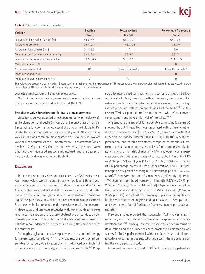
ter implantation, and again 24 hours and 9 months later. In all pa-tients, valve function remained essentially unchanged (Table 5). Pa-ravalvular aortic regurgitation was generally mild. Although para-valvular leak was common, most cases were trivial or mild. No late valve failure occurred. At the 9-month follow-up assessment (which involved 17/23 patients, 74%), the improvements in the aortic valve area and the mean gradient were maintained, and the degree of paravalvular leak was unchanged (Table 5).
Discussion
The present report describes an experience of 23 TAVI cases in Ko-rea. Twenty valves were implanted transfemorally and three trans-apically. Successful prosthesis implantation was achieved in 22 pa-tients. In the cases that failed, difficulties were encountered in the passage of the wire through the stenotic valve and in the position-ing of the prosthesis, in which open replacement was performed. Prosthesis embolization and a major vascular complication occurred in three cases and one case, respectively. However, no death, stroke, renal insufficiency, coronary artery obstruction, or conduction ab-normality occurred in the cohort, and all complications occurred in patients who underwent the procedure during the early period of the study cases.
Although surgical aortic valve replacement is a standard therapy for severe symptomatic AS,2)3)12) many patients are considered un-suitable for surgery due to excessive risk, advanced age, high risk of procedure-related mortality, and multiple comorbidity.1)4) Prog-
nosis following medical treatment is poor, and although balloon aortic valvuloplasty provides both a temporary improvement in valvular function and symptom relief, it is associated with a high rate of procedure-related complications and mortality.13) For this reason, TAVI is a good alternative for patients who refuse conven-tional surgery and have a high risk of mortality.7)8)14)
A recent randomized trial for inoperable symptomatic severe AS showed that at 1 year, TAVI was associated with a significant re-duction in mortality rate {30.7% vs. 50.7%; hazard ratio with TAVI, 0.55; 95% confidence interval (CI), 0.40-0.74; p<0.001}, repeat hos-pitalization, and cardiac symptoms compared to standard treat-ments such as balloon aortic valvuloplasty.7) In a randomized trial for patients with a high risk of mortality, TAVI and open heart surgery were associated with similar rates of survival at both 1 month (3.4% vs. 6.5%, p=0.07) and 1 year (24.2% vs. 26.8%, p=0.44; a reduction of 2.6 percentage points in TAVI; upper limit of 95% CI, 3.0 per-centage points; predefined margin, 7.5 percentage points; Pnoninferiority= 0.001).14) However, the rate of stroke was significantly higher for TAVI than for open heart surgery at 1 month (5.5% vs. 2.4%, p= 0.04) and 1 year (8.3% vs. 4.3%, p=0.04). Major vascular complica-tions were also significantly higher in TAVI at 1 month (11.0% vs. 3.2%, p<0.001). In contrast, the surgery group showed a significant-ly higher incidence of major bleeding (9.3% vs. 19.5%, p<0.001) and new-onset of atrial fibrillation (8.6% vs. 16.0%, p=0.006) at 1 month.14)
Previous studies reported that successful TAVI involves a learn-ing curve, and that outcomes improve with experience and device development.15)16) Athough our experience was limited in terms of its duration and the number of cases, prosthesis implantation was successful in 22 patients (96%) with one failed case and all com-plications occurred in patients who underwent the procedure dur-ing the early period of study.
Important factors in successful TAVI include adequate patient se-
Table 5. Echocardiographic characteristics
VariableBaseline(n=23)
Postprocedure(n=23)
Follow-up of 9 months (n=17)
Left ventricular ejection fraction (%) 59.5±8.8 64.4±7.0 63.0±3.8
Aortic valve area (cm2) 0.68±0.14 1.45±0.33 1.35±0.4
Aortic annulus diameter (mm) 21.4±2.0 NA NA
Mean transaortic valve gradient (mm Hg) 59.4±22.4 16.6±5.7 15.6±7.1
Peak transaortic valve gradient (mm Hg) 98.7±34.5 30.5±9.5 29.1±11.9
Moderate to severe AR 0 0 0
Aortic paravalvular leak NA Trivial (trivial-mild) Trivial (trivial-mild)*
Moderate to severe MR 0 0 0
Moderate to severe pulmonary HTN 0 0 0
The values are presented with median (interquartile range) and number (percentage). *Three cases of trivial paravalvular leak were disappeared. AR: aortic regurgitation, NA: not available, MR: mitral regurgitation, HTN: hypertension
691Won-Jang Kim, et al.
http://dx.doi.org/10.4070/kcj.2012.42.10.684www.e-kcj.org
lection, correct sizing,17-19) positioning, 20 coaxial alignment, timely reduction of cardiac output, operator techniques,15)16) and organized team approach.20) In the present cohort, the team approach ensured reduction, early detection, and treatment of complications. Our ini-tial experience was mainly derived by the early version of prosthe-sis and delivery systems and caused a major vascular complication. However, we were able to reduce the complications and increase the success rate after the introduction of new smaller, atraumatic, more advanced delivery system (NovaFlex) and prosthesis (Edwards SA-PIEN XTTM valve).
In conclusion, TAVI is a new technology that is of potential bene-fit to patients with severe symptomatic AS, particularly for those who are inoperable or at high surgical risk. With advancements in device technology, procedure related complications will decrease and prosthesis durability will improve. This will allow an expansion of the indications for TAVI. Although our initial experiences are promising, application of this procedure should be limited to pa-tients who are poor candidates for surgical valve replacement.
AcknowledgmentsThis study was supported by a grant of the Korea Healthcare tech-
nology R&D Project, Ministry of Health and Welfare, Republic of Ko-rea (A102065).
References1. Iung B, Baron G, Butchart EG, et al. A prospective survey of patients with
valvular heart disease in Europe: the Euro Heart Survey on Valvular He-art Disease. Eur Heart J 2003;24:1231-43.
2. Kvidal P, Bergström R, Hörte LG, Ståhle E. Observed and relative surviv-al after aortic valve replacement. J Am Coll Cardiol 2000;35:747-56.
3. Schwarz F, Baumann P, Manthey J, et al. The effect of aortic valve repl-acement on survival. Circulation 1982;66:1105-10.
4. Bach DS, Siao D, Girard SE, Duvernoy C, McCallister BD Jr, Gualano SK. Evaluation of patients with severe symptomatic aortic stenosis who do not undergo aortic valve replacement: the potential role of sub-jectively overestimated operative risk. Circ Cardiovasc Qual Outcomes 2009;2:533-9.
5. Nkomo VT, Gardin JM, Skelton TN, Gottdiener JS, Scott CG, Enriquez-Sa-rano M. Burden of valvular heart diseases: a population-based study. Lancet 2006;368:1005-11.
6. Cribier A, Eltchaninoff H, Bash A, et al. Percutaneous transcatheter implantation of an aortic valve prosthesis for calcific aortic stenosis: first human case description. Circulation 2002;106:3006-8.
7. Leon MB, Smith CR, Mack M, et al. Transcatheter aortic-valve implan-tation for aortic stenosis in patients who cannot undergo surgery. N Engl J Med 2010;363:1597-607.
8. Webb JG, Pasupati S, Humphries K, et al. Percutaneous transarterial aortic valve replacement in selected high-risk patients with aortic ste-nosis. Circulation 2007;116:755-63.
9. Vassiliades TA Jr, Block PC, Cohn LH, et al. The clinical development of percutaneous heart valve technology: a position statement of the So-ciety of Thoracic Surgeons (STS), the American Association for Thoracic Surgery (AATS), and the Society of Cardiovascular Angiography and Intervention (SCAI). Catheter Cardiovasc Interv 2005;65:73-9.
10. Lichtenstein SV, Cheung A, Ye J, et al. Transapical transcatheter aortic valve implantation in humans: initial clinical experience. Circulation 2006;114:591-6.
11. Webb JG, Chandavimol M, Thompson CR, et al. Percutaneous aortic valve implantation retrograde from the femoral artery. Circulation 2006; 113:842-50.
12. Bonow RO, Carabello BA, Kanu C, et al. ACC/AHA 2006 guidelines for the management of patients with valvular heart disease: a report of the American College of Cardiology/American Heart Association Task Force on Practice Guidelines (writing committee to revise the 1998 Guidelines for the Management of Patients with Valvular Heart Dis-ease): developed in collaboration with the Society of Cardiovascular Anesthesiologists: endorsed by the Society for Cardiovascular Angiog-raphy and Interventions and the Society of Thoracic Surgeons. Circul-ation 2006;114:e84-231.
13. Otto CM, Mickel MC, Kennedy JW, et al. Three-year outcome after balloon aortic valvuloplasty: insights into prognosis of valvular aortic stenosis. Circulation 1994;89:642-50.
14. Smith CR, Leon MB, Mack MJ, et al. Transcatheter versus surgical aortic-valve replacement in high-risk patients. N Engl J Med 2011;364: 2187-98.
15. Gurvitch R, Tay EL, Wijesinghe N, et al. Transcatheter aortic valve im-plantation: lessons from the learning curve of the first 270 high-risk patients. Catheter Cardiovasc Interv 2011;78:977-84.
16. Buellesfeld L, Wenaweser P, Gerckens U, et al. Transcatheter aortic valve implantation: predictors of procedural success: the Siegburg-Bern experience. Eur Heart J 2010;31:984-91.
17. Kurra V, Kapadia SR, Tuzcu EM, et al. Pre-procedural imaging of aortic root orientation and dimensions: comparison between X-ray angio-graphic planar imaging and 3-dimensional multidetector row com-puted tomography. JACC Cardiovasc Interv 2010;3:105-13.
18. Messika-Zeitoun D, Serfaty JM, Brochet E, et al. Multimodal assess-ment of the aortic annulus diameter: implications for transcatheter aortic valve implantation. J Am Coll Cardiol 2010;55:186-94.
19. Ng AC, Delgado V, van der Kley F, et al. Comparison of aortic root di-mensions and geometries before and after transcatheter aortic valve implantation by 2- and 3-dimensional transesophageal echocardiog-raphy and multislice computed tomography. Circ Cardiovasc Imaging 2010;3:94-102.
20. Serruys PW, Piazza N, Cribier A, Webb JG, Laborde JC, Jaegere PD. Transcatheter aortic valve implantation: tips and tricks to avoid failure. 1st ed. New York: Informa Healthcare;2009. p.645-57.