Embed Size (px)
Citation preview
THE JOURNAL OF BIOLOGICAL CHEMISTRY 0 1987 by The American Society for Biochemistry and Molecular Biology, Inc.
Vol. 262, No. 27, Issue of September 25, pp. 13309-13315,1987 Printed in U.S.A.
Transcriptional and Translational Control of the Message for Transition Protein 1, a Major Chromosomal Protein of Mammalian Spermatids*
(Received for publication, November 24, 1986)
Mohammad A. Heidaran and W. Stephen KistlerS From the Department of Chemistry, University of South Carolina, Columbia, South Carolina 29208
The spermatid transition proteins comprise a set of basic chromosomal proteins that appear during the period when spermatids are undergoing nuclear elon- gation and condensation, about midway between the end of meiosis and the release of spermatozoa from the seminiferous tubule. The transition proteins replace the histones but are themselves subsequently replaced by protamines, and they are not found in sperm nuclei. We have used a cDNA clone for the smallest transition protein (TP1,54 amino acids) to show that its message first appears postmeiotically in late round spermatids. Thus production of TPl is an example of haploid gene expression. The message remains translationally in- active for some 3-4 days before translation occurs in early elongating spermatids. While translationally re- pressed, TP1 message is nonpolysomal and has a dis- crete size of about 590 bases, including a 140 residue poly(A) tail. In contrast, polysome-associated message is of heterogeneous size due to variability of poly(A) lengths.
During mammalian spermatogenesis diploid spermatogonia divide mitotically several times to provide a population of spermatocytes that then proceed through meiosis to become haploid spermatids. Spermatid development involves a strik- ing transformation of a morphologically unremarkable cell into the highly specialized spermatozoon (1, 2). One of the distinctive remodeling steps that occurs involves the change of nucleosomal chromatin to the highly condensed chromatin found in the sperm nucleus (3, 4). In mammals, early sper- matids, which have nucleosomal chromatin, contain a mixture of somatic histones as well as testis-specific variants of H1 (5) and H2B (6) and sometimes H2A (7) and H3 (8,9). About midway through spermatid development, the nucleus begins to condense, to elongate, and to become resistant to mechan- ical disruption (10, 11). These nuclear changes coincide with the replacement of both the somatic-type and testis-specific histones with a set of spermatid-specific chromosomal pro- teins now generally referred to as “transition proteins” (12, 13). Four transition proteins have been identified in the rat (12), and the situation is similar although not always identical
* This work was supported by Grant HD 10793 from the United States Public Health Service. A preliminary account was presented at the Ninth Testis Workshop, Nashville, TN, October 14-17, 1986, and at the meeting of the American Society for Cell Biology, Atlanta, GA, Nov. 18-22, 1985 (Heidaran, M. A., and Kistler, W. S. (1985) J. Cell. Bioi. 101, 366 (Abstr. 1387)). The costs of publication of this article were defrayed in part by the payment of page charges. This article must therefore be hereby marked “advertisement” in accord- ance with 18 U.S.C. Section 1734 solely to indicate this fact.
$ TO whom correspondence should be addressed.
in other mammals (13). Later still in spermatid development, the transition proteins are replaced by the distinctive CYS- teine-rich protamines found in the sperm nuclei of eutherian mammals (14-16).
RNA synthesis is not detected in spermatids once nuclear condensation begins (17-20). Accordingly, the production of messenger RNA for any proteins that appear after nuclear condensation begins must depend on RNA made at an earlier time. Indeed, as the pioneering studies of Dixon and colleagues (21, 22) have demonstrated, protamine message in the trout appears early in spermatogenesis (prior to meiosis) and is stored as a translationally inert RNA-protein particle until activation occurs late in spermatid development. Recently Hecht and associates (23) have examined this problem in mice and found that protamine message appears following meiosis in early haploid cells where it is translationally repressed until nuclear condensation is well under way. In mammals devel- oping germ cells remain joined by cytoplasmic bridges (24), and this arrangement presumably mitigates or eliminates many, although not necessarily all (25), of the problems inherent to haploid gene expression.
TP1’ was the first transition protein isolated (26), and is a 54-amino acid molecule notable for its high content of argi- nine and lysine and lack of an obvious globular region (27). It is present in many mammals including man (14, 26, 28). The recent isolation of a cDNA clone for TP1’ permits analysis of TP1 message by molecular hybridization tech- niques. Based on the studies described below, we conclude that TP1 message first appears in late round spermatids but is not translated for some 3-4 days. Thus, like mouse prota- mine, TP1 message is transcribed in haploid cells and regu- lated at the translational level.
EXPERIMENTAL PROCEDURES
Materials-Sprague-Dawley rats were obtained from Harlan- Sprague-Dawley (Indianapolis, IN). For developmental studies, ani- mals were from Building 207 of the Madison, WI facility. Plasmid pMH2 is a cDNA clone that contains the entire coding region for rat TP1.’ Plasmid pRPA-1 is a cDNA clone of rat &actin that probably hybridizes to all classes of actin message and was kindly provided by P. Gunning (Veterans Administration Medical Center, Palo Alto, CA).
Isolation of RNA and Northern Blotting-Total high molecular weight RNA was isolated from tissues by proteinase K digestion of homogenates in buffered sodium dodecyl sulfate followed by phenol/ chloroform extraction, and precipitation from sodium acetate as
The abbreviations used are: TP1, transition protein 1. (Note that this protein has been designated simply T P previously. This change will bring the terminology in conformity with the other TP proteins (see Ref. 12).) HEPES, 4-(2-hydroxyethyl)-l-piperazineethanesul- fonic acid.
* Heidaran, M. A., and Kistler, W. S. (1987) Gene 54, 281-284. submitted.
13309

13310 Control of TPl Synthesis
described by Lee et al. (30). For standard Northern blots, RNA was denatured in a buffered mixture of formamide and formaldehyde and separated electrophoretically through agarose gels containing 2.2 M formaldehyde (31). It was transferred to nitrocellulose (BA85, Schleicher and Schuell) as described by Thomas (32). Prehybridiza- tion, hybridization, and washing were carried out as described by Maniatis et al. (33) for Southern blots with the final stringency wash in 75 mM NaCI, 15 mM Tris-HC1, 1 mM EDTA, 0.1% sodium dodecyl sulfate, pH 8 (0.5 X SET) at 65 "C for 20 min.
For Northern blots from polyacrylamide gels, RNA was denatured in 50% (v/v) formamide, 5 mM EDTA, pH 8, for 2.5 min at 100 "C. It was separated on a sequencing type gel (34) (12-cm long x 0.15-cm thick) containing 8 M urea and run at a voltage sufficient to maintain the surface temperature a t about 50 "C. RNA was then transferred electrophoretically in a transblot cell (Bio-Rad) to a Zeta-Probe nylon membrane (Bio-Rad) according to the protocol provided by the sup- plier.
Preparation and Analysis of Polysomes-These procedures were based on those of Kleene et al. (23) with modifications (35, 36). Briefly, 1 g of decapsulated testis was homogenized in 4 ml of ice- cold 100 mM KCl, 10 mM MgCl,, 20 mM HEPES, pH 7.5, 1 mg/ml heparin, 90 pg/ml cycloheximide with 10 strokes of a motor-driven type-B Teflon/glass homogenizer. The homogenate was centrifuged (5,000 X g ) for 5 min to pellet intact cells and nuclei. The supernatant was brought to 0.5% (v/v) Triton NlOl and centrifuged (12,000 X g ) for 10 min. The supernatant (2 ml) was layered onto a 5-40% (w/v) exponential gradient of sucrose prepared in 20 mM HEPES, pH 7.5, 100 mM KCl, 1 mM MgCl,, and centrifuged for 3 h at 25,000 rpm in a Beckman SW-27 rotor at 4 "C. For control gradients in which polysomes were dissociated, EDTA was added to the 12,000 X g supernatant to make 100 mM. Gradients were fractionated using an ISCO (Lincoln, NE) 640 instrument and a UA-5 monitor. RNA was precipitated by adding 0.1 volume of 2 M sodium acetate and 2 volumes of ethanol. Precipitates were collected by centrifugation and dissolved in 1 ml of 0.1% sodium dodecyl sulfate, 10 mM Tris-HC1, 1 mM EDTA, pH 8, and digested with proteinase K (10 pglml). RNA was then extracted with phenol/chloroform and precipitated with ethanol from 0.2 M sodium acetate. For gradients of EDTA-released samples, 10 pg of carrier tRNA was added to each fraction prior to the initial ethanol precipitation. A sample of the RNA (1/50) isolated from each polysomal gradient fraction was incubated in 6% (v/v) formaldehyde, 1 M NaC1,50 mM sodium phosphate, pH 6.9,5 mM EDTA for 15 min at 55 'C. The reaction was chilled on ice and applied as dots to BA85 nitrocellulose using a Scheicher and Schuell Minifold filtration de- vice. The filter was then treated as for other Northern blots.
Digestion of RNA with RNase H-Selective digestion of poly(A) tract was achieved by use of RNase H (37). The reaction mixture (50 pl) contained 20 pg of total high molecular weight RNA, 2 rg of oligo(dT) (Collaborative Research, Woburn, MA), in distilled water and was heated to 65 "C for 3 min. A %o volume of lox RNase H buffer was added ( lx buffer contains 100 mM KCl, 20 mM Tris-HC1, pH 7.5,lO mM MgCl,, 50 mg/ml sucrose), and samples were incubated on ice for 15 min. RNase H (6 units, Bethesda Research Laboratories) was added, and the samples were incubated for 45 min at 37 "C, extracted with phenol/chloroform, and ethanol precipitated.
In Vitro Protein Synthesis by Testis Tubules-Fresh adult testis was placed on ice immediately following death of the animal. The capsule was removed, and portions of tubules and interstitial tissue (0.2 g) were teased out using forceps. The tubules were placed in 25- ml Erlenmyer flasks containing 3 ml of Krebs-Ringer buffer (38) (119 mM NaCl, 4.75 mM KCl, 16.1 mM Na2HP04, 1.19 mM KHzPOI, 1.19 mM MgSO,, 2 mg/ml glucose, adjusted to pH 7.4 with HCl) and kept on ice. Flasks were transferred to a shaking water bath at 34 "C and incubated in the presence of 10 pCi [35S]methionine (Du Pont-New England Nuclear) for 2 h. Reactions were stopped by adding trichlo- roacetic acid to 4%. As a source of carrier TP1, the protein from 0.2 g of adult testis that is soluble in 4% but precipitated by 20% trichloroacetic acid was added. The contents were then homogenized with a Polytron (Brinkman Instruments) and worked up to select proteins soluble in 4% but precipitated by 20% trichloroacetic acid (26). Samples corresponding to l/3 of the flask contents were separated electrophoretically in a 15% (w/v) polyacrylamide gel slab containing 0.9 M acetic acid and 2.5 M urea (39), and a fluorogram was prepared (40). For quantitation of radioactivity incorporated into TP1, dupli- cate gels were stained with Coomassie Blue and sliced into 2-mm slices in the region of the TP1 band. The slices were dissolved in hydrogen peroxide and counted in a scintillation counter as previously described (26).
Miscelluneow-Radioactive probes were prepared from plasmid restriction fragments that were separated electrophoretically in pol- yacrylamide gels and recovered by electroelution in dialysis bags (41). Radioactive label was introduced by nick translation (42) using a [32P]deoxynucleotide from Du Pont-New England Nuclear and DNA polymerase I from Boehringer. For histological examination, testis samples were fixed in Carnoy's solution, embedded in paraffin, sec- tioned, and stained with hematoxylin and eosin. Biochemicals were generally from Sigma. Measurements of pH were made at room temperature. Kodak X-Omat AR film was used for autoradiograms with Dupont Lightning Plus intensifying screens where appropriate. Wherever testis weight is specified, it refers to the combined weight of both testes from one animal.
RESULTS
TP1 appears in spermatid development during the period when nuclear condensation and elongation occur (12, 26), approximately steps 9-15 (43). Our initial goal was to deter- mine whether TP1 message appears coincident with the pro- tein or significantly earlier, perhaps even prior to the meiotic divisions. Mammalian spermatogenesis is a lengthy process, and about 9 days separate the second meiotic division from the beginning of nuclear condensation in spermatids (43). Thus, if TP1 message were to be transcribed premeiotically, it would have to appear at least 9 days before the synthesis of TP1 protein. In order to investigate this possibility, we de- cided to compare appearance of TP1 message and TP1 syn- thesis as the first cohorts of germ cells developed in young rats. Sensitive and unambiguous detection of message was made possible by the recent isolation of a cDNA clone for TP1.'
TP1 message was determined by extracting total high mo- lecular weight RNA and using the TP1 cDNA insert of pMH2 to identify specific sequences on Northern blots. To monitor TP1 synthesis, teased seminiferous tubules were incubated in uitro in the presence of labeled methionine. TP1 was extracted in 4% trichloroacetic acid. Following electrophoresis in acid urea gel slabs, the radioactive band was identified by fluorog- raphy and quantitated by excision and scintillation counting. Because few proteins are soluble in 4% trichloroacetic acid, isotope incorporation into TP1 can be determined unambig- uously without recourse to immunological precipitation. Prot- amine is the only protein that migrates near TP1 in this electrophoretic system, but rat protamine is not extracted by 4% trichloroacetic acid. In any event, rat protamine does not contain methionine (44).
In preliminary studies we found age alone to be an unreli- able measure of the stage of sexual development of Sprague- Dawley rats but that testis weight was closely correlated with the appearance of TP1 message or protein. TP1 message is first detected in animals whose combined testes weighed slightly less than 0.5 g, corresponding in this case to an age of 22-24 days (Fig. 1). In contrast, TP1 synthesis by seminif- erous tubules was first detected in testes whose combined weight was about 0.9 g, corresponding to animals about 26- 27 days old (Fig. 2). When results from the two experiments are expressed quantitatively, and plotted to show appearance of message and protein as a function of increasing testes weight, it is clear that TP1 message appears distinctly before TP1 synthesis (Fig. 3). The lag between message appearance and protein synthesis is some 3-4 days based on correlation of testis weights and ages. Histological examination of testis samples taken over this stage of development shows that message is first detected when many tubules have substantial populations of round spermatids, and the most advanced tubules have spermatids at about step 7 (Fig. 4-4). By the time TP1 synthesis is detectable, the most advanced sper- matids are now elongating at stages 9-13 (Fig. 4B). Since the lag between appearance of message and protein is much less


13312 Control of TPl Synthesis
FIG. 4. Testis histology of young animals. Testes were fixed in Carnoy's solution, embedded in paraffin, sectioned, and stained with hematoxylin and eosin (X 160). A ) , material from testes of combined weight of 0.65 g. At this stage of development, TP1 message is readily detected, but TP1 synthesis is not. Most tubules contain round spermatids although those in the upper center and upper left contain pachytene spermatocytes as the most advanced cell type. B ) , material from testes of combined weight of 1.0 g. TP1 synthesis is detected in organs of this weight. The most advanced tubules contain elongating spermatids of steps 9-13.
cytoplasm of round spermatids in a translationally inert form, perhaps as some type of ribonucleoprotein particle. In the adult, when all stages of spermatogenesis are present and some cells are synthesizing TP1, a substantial fraction of TP1 message is polysomal.
I t was of interest to see whether the polyadenylation state of TP1 message correlated with its association with active ribosomes. Poly(A) tracts can be removed selectively by di- gesting RNA with RNase H in the presence of oligo(dT) (37). Northern blotting can then be used to determine the size of the intact and deadenylated messages. We initially examined unfractionated RNA from immature animals since we had already shown that it occurs largely if not entirely in untrans- lated form. Untreated message migrated as a sharp band on a denaturing polyacrylamide gel with a size of about 590 nu- cleotides (Fig. 7, lane A ) . RNase H treatment reduced the
T P I
9 .. 0 - .
A C T I N
(B)
. .. I . .
*.I I * *
. . . . f . . *.. . .. ....*. ...... I.. t.. ...... -1.1.. ......
I.... f.. f. . I . . . ..*.. ..... . ..... . f.... I . . . . t f f . .
2 4 6 8 FRA C T/ON
FIG. 5. Location of TPl message in gradient-fractionated testis polysomes from an adult rat. Samples of a postmitochon- drial supernatant were separated in a 5-4076 exponential gradient of sucrose under standard conditions ( A ) or after adjusting each sample to 100 mM EDTA (R). RNA was prepared from each fraction and a sample spotted onto nitrocellulose and probed with either the insert for TPl cDNA clone pMH2 or the insert from a cDNA clone for rat @-actin. At the top of each profile is shown the autoradiogram resulting from the dot hybridization of each fraction. Exposure time was 2 days for the actin filters and 9 days for the TP1 filters. The dots probed for TP1 were scanned with a densitometer, using film exposed in its linear response range. The data from these scans are plotted as a bar graph along with the absorbance of each gradient a t 254 nm. The film density is in arbitrary units. The direction of sedimentation is from left to right.
ACTIN
( 0 -
P
.*+ ..* *.. f f f + + * Q - ...* 1 *+. +*. . ..I.. t+. -. .+...
I ..... t'.... ... f..
I
2 4 6 8 2 4 6 < F R A C T / O N
FIG. 6. Location of TP1 message in gradient-fractionated testis polysomes from an immature rat. Details are the same as given in the legend to Fig. 5 except that polysomes were prepared from pooled testes in the weight range of 0.4-0.6 g/pair. At this stage of development TP1 message is present, but TP1 translation is not detectable. Densitometric scans of TP1 message shown as bar graphs are on a different scale than shown in Fig. 5.
Control of TP1 Synthesis 13313
I) 4 x
A B C
1 - 5 1 7 -630
-39 6 -344
I)
-298
-220
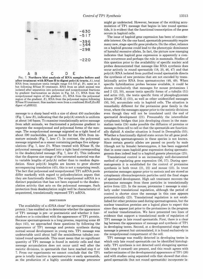
D E FIG. 7. Northern blot analysis of RNA samples before and
after treatment with RNase H to digest poly(A) tracts. A ) , total RNA from immature testis (weight range 0.4-0.6 g). B ) , same as A but following RNase H treatment. RNA from an adult animal was isolated after separation into polysomal and nonpolysomal fractions by gradient fractionation as shown in Fig. 5A. C), RNA from the nonpolysomal region of the gradient. D), RNA from the polysomal region of the gradient. E ) , RNA from the polysomal region following RNase H treatment. Size markers were from a combined HinfI ,EcoRI digest of pBR322.
message to a sharp band with a size of about 450 nucleotides (Fig. 7, lane B ) , indicating that the poly(A) stretch is uniform a t about 140 bases. To examine translationally active message from adult animals, we fractionated a polysome gradient to separate the nonpolysomal and polysomal forms of the mes- sage. The nonpolysomal message migrated as a tight band of about 590 nucleotides, just as found for the RNA from im- mature animals (Fig. 7, lane C). In contrast, the polysomal message migrated as a smear containing perhaps five subpop- ulations (Fig. 7, lane D). When treated with RNase H, the polysomal message collapsed into a tight band corresponding to the deadenylated message (Fig. 7, lane E ) . This indicates that the disperse size range of the untreated material was due to variable lengths of poly(A) rather than to random degra- dation. Since poly(A) lengths are known to grow shorter accompanying translation (45), this is not a surprising result. The fact that polysomal and nonpolysomal TP1 mRNA pools differ markedly with regard to polyadenylation argues that they are functionally distinct. The nonpolysomal mRNA is a distinct population that has not been exposed to the deaden- ylation activity that acts on the polysomal messages. Such protection from deadenylation might well be characteristic of sequestered, translationally repressed TP1 mRNA.
DISCUSSION
The availability of a cDNA clone2 for spermatid transition protein 1 has enabled us to determine whether the appearance of TP1 message is pre- or postmeiotic and whether it fore- shadows or is coincident with the appearance of TP1 protein. Because spermatogenesis is a long process, requiring some 48 days (43), we approached these questions by following the appearance of TP1 message and protein synthesis during normal sexual development in young rats. TP1 message was undetectable until about half the seminiferous tubules con- tained round spermatids. This must mean that no significant quantity of TP1 message is found in meiotic cells and that message accumulation does not occur until well after the meiotic divisions, in spermatids that have reached step 6 or 7. From our experiments we cannot conclude that the TP1 gene is totally inactive in spermatocytes or early spermatids, as the production of a highly unstable message precursor
might go undetected. However, because of the striking accu- mulation of TP1 message that begins in late round sperma- tids, it is evident that the functional transcription of the gene occurs in haploid cells.
The issue of haploid gene expression has been of consider- able interest. On the one hand, spermatids presumably require many new, stage-specific gene products. On the other, reliance on a haploid genome could lead to the phenotypic dominance of harmful recessive alleles. In fact, the picture now emerging indicates that haploid gene expression is apparently a com- mon occurrence and perhaps the rule in mammals. Studies of this question prior to the availability of specific nucleic acid probes demonstrated that message-like RNA synthesis does occur actively in round spermatids (18, 20, 46, 47) and that poly(A) RNA isolated from purified round spermatids directs the synthesis of new proteins that are not encoded by trans- lationally active RNA from spermatocytes (48, 49). When specific hybridization probes became available, it could be shown conclusively that messages for mouse protamines 1 and 2 (23, 50), mouse testis specific forms of a-tubulin (51) and actin (52), the testis specific form of phosphoglycerate kinase (PGK-2) (53), as well as other unidentified messages (50, 54), accumulate only in haploid cells. The situation is remarkably different for the protamine gene family in the trout, where the messages appear prior to the meiotic divisions even though they will not be translated until the end of spermatid development (21). Presumably the intercellular cytoplasmic bridges that join developing clones in the mam- malian testis (24) make possible the diffusion of proteins or messages from cell to cell so that the spermatids are function- ally diploid. A similar situation is found in Drosophila (55). Whether a functionally diploid state occurs for all gene prod- ucts during spermatogenesis in these species is not known. Since certain genetic alleles are passed on poorly by male (though not by female) heterozygotes, it has been suggested that in some cases haploid gene expression during spermato- genesis does indeed lead to a haploid gamete phenotype (25).
Translational control is an increasingly well-documented method of regulating gene expression (56, 57). During sper- matogenesis it is established for the control of protamine synthesis in both trout (21, 22) and mouse (23). In trout protamine messages appear prior to meiosis and are stored as cytoplasmic ribonucleoprotein particles until the final stages of spermatid development. High salt treatment recovers the protamine messages from these particles in translationally active form (22). In the mouse, protamine 1 message is simi- larly under translational regulation, although the period of control is shorter since the message in this case appears postmeiotically (23). Translational regulation is not estab- lished for other proteins used during spermatogenesis, but the nuclear transition proteins are a logical place to expect this since they appear just prior to the protamines in the program of nuclear transformation. We have presented two lines of evidence that support a translational mode of regulation of TP1 message in late round spermatids. First, there is a clear lag between the appearance of message and synthesis of TP1 in developing testes. Second, a t a developmental stage when message is present but untranslated, it is found exclusively in the nonpolysomal compartment of the cell.
TP1 message was readily detected in developing testes in which only late round spermatids can be identified histologi- cally. TP1 synthesis is not detected until elongating sperma- tids (step 9 onward) are present, and this result agrees with earlier estimates of the developmental appearance of TP l (26 ) and with studies using separated cells that showed that elon- gated spermatids (but not round spermatids) incorporate la-
13314 Control of TPl Synthesis
beled amino acids into TP1 (12, 58). Similarly, in the mouse the transition proteins are first detected in step 12 spermatids (59-61). The exact point at which TP1 is first made is difficult to determine. Spermatids pass through the early stages of nuclear elongation quickly, steps 9-11 requiring only about 1 day (43). As a result these cells are relatively scarce in the testis at any age and are difficult to obtain as highly purified populations (29,62). Whether or not synthesis begins in these early elongating cells, it continues in cell populations enriched for steps 13-15 (12). Our developmental studies agree with these results, as TP1 synthesis was first observed in testes that have elongating spermatids up to about step 13.
In testes of 0.4-0.6 g, which contain late round spermatids but no elongating cells (step 9 onward), TP1 message was found exclusively in the nonpolysomal compartment, and this message had a uniform polyadenylation of about 140 residues. In adult testis, where all stages of germ cell development are present, about '/3 of the TP1 message was found associated with small polysomes. Presumably these polysomes arise from the elongating spermatids in which the message is expressed. Since the length of time the message is expressed compared to the time it is stored is not known exactly, we cannot calculate precisely what proportion of the message ought to be polysomal and nonpolysomal in the adult testis, even if it were entirely polysomal in expressing cells. We may make an approximate calculation by assuming that message appears at the beginning of step 7 (late round spermatid) and that it is translated constantly from the beginning of step 9 until the end of step 14. As step 7 is long (3 days) while steps 9-11 are short (1 day altogether), this model has TP1 message present for 4 days in a repressed state and translated for 4 days. Therefore, we would expect only half of the message in the adult testis to be polysomal. This is rather close to the observed pattern. A shorter period of translation would ob- viously correlate with an even smaller proportion of polysomal message. It is unlikely that the nonpolysomal message from either adult or immature testes in an artifactual product of polysome destruction during handling. RNase H digestion showed that the message in the nonpolysomal region of the gradient has an intact and homogeneous poly(A) segment (-140 nucleotides), while the message from the polysomal region of the gradient has heterogeneous polyadenylation. Accordingly, the nonpolysomal message has been cycled min- imally if a t all through the polysomal population. A reasonable conclusion is that it originates from the stored message pres- ent in round spermatids. In fact, the case for TP1 message is very similar to that found for mouse protamine 1 message, which, even in adult testes, occurs largely as a nonpolysomal particle characterized by uniform polyadenylation. The poly- soma1 message is in a minority and has distinctly shorter poly(A) tails (23).
Thus far, translational level control has been described only for nuclear proteins made late in spermatogenesis. It will be interesting to see what other proteins are subject to transla- tional level control during spermatogenesis and to what extent the mechanisms of control are shared.
Acknowledgments-We are grateful to Dr. Richard Showman and Dr. Lewis Bowman of the Biology Department for assistance in establishing conditions for polysome isolation and separation. We thank Dr. Peter Gunning for providing the @-actin clone.
REFERENCES 1. Bellve, A. R. (1979) in Oxford Review of Reproductive Biology
(Finn, C. A., ed) Vol. 1, pp. 159-260, Oxford University Press, Oxford
2. Fawcett D. W. (1975) Den Biol. 4 4 , 394-436
3. Kierszenbaum, A. L., and Tres, L. L. (1978) Fed. Proc. 37,2512- 2516
4. Loir, M., Bouvier, D., Fornells, M., Lanneau, M., and Subirana, J. A. (1985) Chromosoma (Bed.) 9 2 , 304-312
5. Seyedin, S. M., and Kistler, W. S. (1980) J. Biol. Chem. 255, 5949-5954
6. Shires, A., Carpenter, M. P., and Chalkley, R. (1976) J. Biol. Chem. 251,4155-4158
7. Trostle-Weige, P. K., Meistrich, M. L., Brock, W. A., Nishioka, K., and Bremer, J. W. (1982) J. Bwl. Chem. 267 , 5560-5567
8. Trostle-Weige, P. K., Meistrich, M. L., Brock, W. A., and Ni- shioka, K. (1984) J. Biol. Chem. 259,8769-8776
9. Zweidler, A. (1984) in Histone Genes: Structure, Organization, and Regulation (Stein, G. S., Stein, J. L., and Marzluff, W. F., eds) pp. 339-371, John Wiley & Sons, New York
10. Grimes, S. R., Jr., Meistrich, M. L., Platz, R. D., and Hnilica, L. S. (1977) Exp. Cell Res. 110 , 31-39
11. Loir, M., and Lanneau, M. (1978) Exp. Cell Res. 115,231-243 12. Meistrich, M. L., Brock, W. A., Grimes, S. R., Jr., Platz, R. D.,
13. Lanneau, M., and Loir, M. (1982) J. Reprod. F e d . 6 5 , 163-170 14. Coelingh, J. P., Monfoort, C. H., Rozijn, T. H., Gevers Leuven,
J. A., Schiphof, R., Steyn-Parve, E. P., Braunitzer, G., Schrank, B., and Ruhfus, A. (1972) Biochim. Biophys. Acta 2 8 5 , 1-14
15. Kleene, K. C., Distel, R. J., and Hecht, N. B. (1985) Biochemistry
16. McKay, D. J., Renaux, B. S., and Dixon, G. H. (1985) Biosci.
17. Monesi, V. (1965) Exp. Cell Res. 39 , 197-224 18. Loir, M. (1972) Ann. Biol. Anim. Biochim. Biophys. 12,203-219 19. Kierszenbaum, A. L., and Tres, L. L. (1974) J. CeU Biol. 60,39-
20. Geremia, R., Boitani, C., Conti, M., and Monesi, V. (1977) Cell
21. Iatrou, K., Spira, A. W., and Dixon, G. H. (1978) Dev. Biol. 6 4 ,
22. Sinclair, G. D., and Dixon, G. H. (1982) Biochemistry 21,1869-
23. Kleene, K. C., Distel, R. J., and Hecht, N. B. (1984) Dev. Biol.
24. Dym, M., and Fawcett, D. W. (1971) Biol. Reprod. 4, 195-215 25. Erickson, R. P. (1978) Fed. Proc. 37,2517-2521 26. Kistler, W. S., Geroch, M. E,, and Williams-Ashman, H. G. (1973)
J. Bwl. Chem. 248,45324543 27. Kistler, W. S., Noyes, C., Hsu, R., and Heinrikson, R. L. (1975)
J. Biol. Chem. 250,1847-1853 28. Kistler, W. S., Geroch, M. E., and Williams-Ashman, H. G. (1975)
Invest. Urol. 12 , 346-350 29. Meistrich, M. L., Longtin, J., Brock, W. A., Grimes, S. R., and
Mace, M. L. (1981) Biol. Reprod. 25,1065-1077 30. Lee, D. C., McKnight, G. S., and Palmiter, R. D. (1978) J. Biol.
Chem. 253,3494-3503 31. Lehrach, H., Diamond, D., Wozney, J. N., and Boedtker, H.
(1977) Biochemistry 16 , 4743-4751 32. Thomas, P. S. (1980) Proc. Natl. Acad. Sci. U. S. A. 77, 5201-
5205 33. Maniatis, T., Hardison, R. C., Lacy, E., Lauer, J., OConnell, C.,
Quon, D., Sim, G. K., and Efstratiadis, A. (1978) Cell 15,687- 701
34. Maxam, A., and Gilbert, W. (1980) Methods Enzymol. 6 5 , 499- 560.
35. Palacios, R., Palmiter, R. D., and Schimke, R. T. (1972) J. Biol. Chem. 247,2316-2321
36. Walden, W. E., Godefroy-Colburn, T., and Thach, R. E. (1981) J. Biol. Chem. 256,11739-11746
37. Sippel, A. E., Stavrianopoulos, J. G., Schutz, G., and Feigelson, P. (1974) Proc. Natl. Acad. Sci. U. S. A. 71,46354639
38. Krebs, H. A. (1933) Z. Physiol. Chem. 217 , 191-227 39. Panyim, S., and Chalkley, R. (1969) Arch. Biochem. Biophys.
40. Ostrowski, M. C., Kistler, M. K., and Kistler, W. S. (1982)
41. McDonell, M. W., Simon, M. N., and Studier, F. W. (1977) J.
42. Maniatis, T., Jeffrey, A., and Kleid, D. G. (1975) Proc. Natl.
43. Clermont, Y. (1972) Physiol. Rev. 5 2 , 198-236
and Hnilica, L. S. (1978) Fed. Proc. 37 , 2522-2525
2 4 , 719-722
Rep. 4,383-391
53
Differ. 5 , 343-355
82-98
1877
105,71-79
130,337-346
Biochemistry 21,3525-3529
Mol. Biol. 110,119-146
A C ~ . sci. u. s. A. 72,1184-11aa

Control of TPl Synthesis 13315
44. Kistler, W. S., Keim, P. S., and Heinrikson, R. L. (1976) Bwchim. 53. Erickson, R. P., Michelson, A. M., Rosenberg, M. P., Sanchez,
45. Sheiness, D., and Darnell, J. E. (1973) Nature New Biol. 241, 54. Dudley, K., Potter, J., Lyon, M. F., and Willison, K. R. (1984)
46. Soderstrom, K.-O., and Parvinen, M. (1976) J. Cell Biol. 70,239- 55. Lindsley, D. L., and Tokuyasu, K. T. (1980) in The Genetics and 246 Biology of Drosophila (Ashburner, M., and Wright, T. R. F.,
47. Geremia, R., D’Agostino, A., and Monesi, V. (1978) Exp. Cell Res. eds) Vol. 2, pp. 225-294, Academic Press, New York
48. Fujimoto, H., and Erickson, R. P. (1982) Biochem. Biophys. Res. 57. Austin, S. A., and Kay, J. E. (1982) Essays Biochem. 18, 79-119 Commun. 108,1369-1375 58. Platz, R. D., Grimes, S. R., Meistrich, M. L., and Hnilica, L. S.
49. Gold, B., Stern, L., Bradley, F. M., and Hecht, N. B. (1983) J. (1975) J. BWl. Chem. 250, 5791-5800
50. Hecht, N. B., Bower, P. A., Waters, S. H., Yelicj, P. C., and 60. Mayer, J. F., Jr., Chang, T. S. K., and Zirkin, B. R. (1981) Bwl.
51. Distel, R. J., Kleene, K. C., and Hecht, N. B. (1984) Science 224, 61. Balhorn, R., Weston, S., Thomas, C., and Wyrobek, A. J. (1984)
52. Waters, S. H., Distel, R. J., and Hecht, N. B. (1985) Mol. Cell. 62. Romrell, L. J., Bellve, A. R., and Fawcett, D. W. (1976) Deu. Bwl.
Bwphys. Acta 427, 752-757 E., and Orkin, S. H. (1985) Bwsci. Rep. 5, 1087-1091
265-268 Nucleic Acids Res. 12,4281-4293
11 1,23-30 56. Lodish, H. F. (1976) Annu. Reu. Biochem. 45, 39-72
EXP. Z00l. 225, 123-134. 59. Mayer, J. F., and Zirkin, B. R. (1979) J. Cell Bwl. 81,403-410
Distel, R. J. (1986) Exp. Cell Res. 164, 183-190 Reprod. 25, 1041-1051
68-70 Exp. Cell Res. 150,298-308
Biol. 5,1649-1654 49,119-131