Embed Size (px)
Citation preview

University of New MexicoUNM Digital Repository
Biomedical Sciences ETDs Electronic Theses and Dissertations
7-1-2009
Transcriptional networks of lung airway epithelialciliogenesisXiaobo Sun
Follow this and additional works at: https://digitalrepository.unm.edu/biom_etds
This Thesis is brought to you for free and open access by the Electronic Theses and Dissertations at UNM Digital Repository. It has been accepted forinclusion in Biomedical Sciences ETDs by an authorized administrator of UNM Digital Repository. For more information, please [email protected].
Recommended CitationSun, Xiaobo. "Transcriptional networks of lung airway epithelial ciliogenesis." (2009). https://digitalrepository.unm.edu/biom_etds/107


DEDICATION
This thesis is dedicated to my parents and wife Qian who have supported me all
the way since the beginning of my studies.
Also, this thesis is dedicated to those who believe in the richness of learning.
iii

iv
ACKNOWLEDGMENT
First of all, I would like to express my deepest sense of gratitude to my mentor
and supervisor Dr. Kevin Harrod for his excellent and patient guidance in both of my
experiments and thesis writings, encouragement and brilliant instructions throughout
this study.
I would express my gratitude to my Scholarship sponsor from Lovelace
Respiratory Research Institute and Biomedical Sciences Graduate Program of UNM.
I am also thankful to my committee members, Dr. Brian Hjelle, Dr. Robert Rubin,
for their valuable opinions and advice in this study, and in revisions of this thesis.
I also would give special thanks to Dr. Wanli Lei, my experimental instructor and
colleague in this study, for his constant and patient guidance in my experiments,
generous assistance on editing thesis writing.
I also thank my colleague Dr. Junita Martinez, Richard Jaramillo, Kimberly
Ortega and Bilan Li for sharing their experience and knowledge in this study.

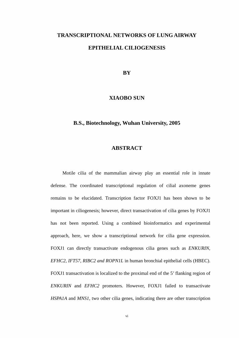
TRANSCRIPTIONAL NETWORKS OF LUNG AIRWAY
EPITHELIAL CILIOGENESIS
BY
XIAOBO SUN
B.S., Biotechnology, Wuhan University, 2005
ABSTRACT
Motile cilia of the mammalian airway play an essential role in innate
defense. The coordinated transcriptional regulation of cilial axoneme genes
remains to be elucidated. Transcription factor FOXJ1 has been shown to be
important in ciliogenesis; however, direct transactivation of cilia genes by FOXJ1
has not been reported. Using a combined bioinformatics and experimental
approach, here, we show a transcriptional network for cilia gene expression.
FOXJ1 can directly transactivate endogenous cilia genes such as ENKURIN,
EFHC2, IFT57, RIBC2 and ROPN1L in human bronchial epithelial cells (HBEC).
FOXJ1 transactivation is localized to the proximal end of the 5’ flanking region of
ENKURIN and EFHC2 promoters. However, FOXJ1 failed to transactivate
HSPA1A and MNS1, two other cilia genes, indicating there are other transcription
vi

factor(s) involved in the ciliogenesis. Motif discovery analysis indicates ETS and
RFX binding sites located in promoters of several cilia genes (IFT57, HSPA1A,
MNS1, RIBC2 and ROPN1L). QRT-PCR indicates 5 of 27 human ETS
transcription factors members (ETV1, ETV5, SPDEF, SPIC and ESE1) and 4 of 7
RFX transcription factors (RFX1, RFX2, RFX5 and RFXANK) are regulated
coincidently with the differentiation of human bronchial epithelial cells and the
appearance of cilia, suggesting these transcription factors are involved in gene
regulations during lung airway epithelial ciliogenesis. ETS family members ESE1
and ETV1 both can transactivate endogenous EFHC2 and ENKURIN while SPIC
represses EFHC2. An RFX family member RFX1 transactivates endogenous
ENKURIN, HSPA1A, ROPN1L, and RFX5 transactivates EFHC2, HSPA1A and
RIBC2. Furthermore, another transcription factor, GATA2, transactivates
endogenous ENKURIN and EFHC2, and its transactivation also locates in the
proximal end of the 5’ flanking region of ENKURIN and EFHC2 promoters. Our
work defines a transcriptional network that regulates expression of cilia genes
during airway epithelial differentiation and highlights the importance of multiple
transcription factors in ciliogenesis.
vii
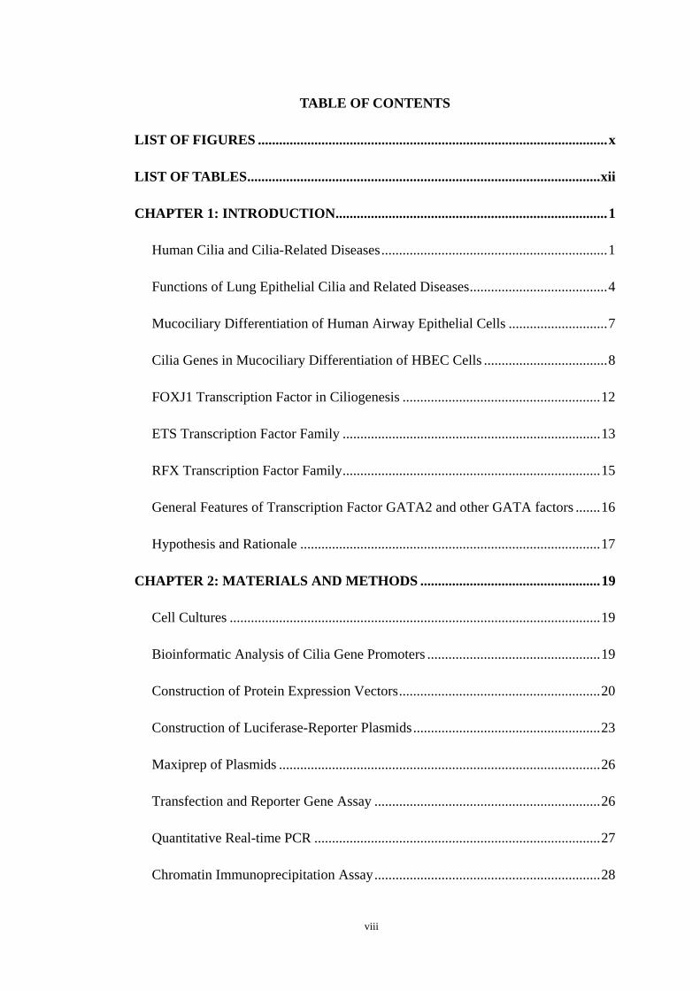
TABLE OF CONTENTS
LIST OF FIGURES ...................................................................................................x
LIST OF TABLES....................................................................................................xii
CHAPTER 1: INTRODUCTION.............................................................................1
Human Cilia and Cilia-Related Diseases................................................................1
Functions of Lung Epithelial Cilia and Related Diseases.......................................4
Mucociliary Differentiation of Human Airway Epithelial Cells ............................7
Cilia Genes in Mucociliary Differentiation of HBEC Cells ...................................8
FOXJ1 Transcription Factor in Ciliogenesis ........................................................12
ETS Transcription Factor Family .........................................................................13
RFX Transcription Factor Family.........................................................................15
General Features of Transcription Factor GATA2 and other GATA factors .......16
Hypothesis and Rationale .....................................................................................17
CHAPTER 2: MATERIALS AND METHODS ...................................................19
Cell Cultures .........................................................................................................19
Bioinformatic Analysis of Cilia Gene Promoters .................................................19
Construction of Protein Expression Vectors.........................................................20
Construction of Luciferase-Reporter Plasmids.....................................................23
Maxiprep of Plasmids ...........................................................................................26
Transfection and Reporter Gene Assay ................................................................26
Quantitative Real-time PCR .................................................................................27
Chromatin Immunoprecipitation Assay................................................................28
viii

CHAPTER 3: RESULTS ........................................................................................32
FOXJ1 in human airway epithelial differentiation and ciliogenesis.....................32
ETS transcription factor family in human airway epithelial differentiation and
ciliogenesis............................................................................................................39
RFX transcription factor family in human airway epithelial differentiation and
ciliogenesis............................................................................................................44
GATA2 in Human Lung Airway Epithelial Differentiation and Ciliogenesis .....47
CHAPTER 4: DISCUSSION ..................................................................................55
REFERENCE...........................................................................................................65
ix

LIST OF FIGURES
Figure 1. Axonemal Structure of Motile 9+2 Cilia................................................ 3
Figure 2. Scanning electron microscope image of lung trachea epithelium......... 5
Figure 3. Mucociliary differentiation of HBEC cells during ALI culture ............. 9
Figure 4. List of the 37 cilia genes...................................................................... 10
Figure 5. The members and their homologs of ETS family ................................ 14
Figure 6. Matrices of the binding sequences of ETS family and RFX family..... 21
Figure 7. Promoter structures of ENKURIN and EFHC2 in luciferase reporter
vectors .................................................................................................................. 25
Figure 8. Expression of FOXJ1, EFHC2, ENKURIN and IFT57 were up-regulated
during lung epithelial differentiation and ciliogenesis......................................... 33
Figure 9. FOXJ1 transactivates IFT57, EFHC2 and ENKURIN promoters. ........35
Figure 10. Effects of FOXJ1 on endogenous cilia gene expression .....................36
Figure 11. Cis-element(s) responsive for FOXJ1 transactivation were located in the
proximal promoter regions of ENKURIN. ........................................................... 37
Figure 12. Cis-element(s) responsive for FOXJ1 transactivation were located in the
proximal promoter regions of EFHC2. ................................................................ 38
Figure 13. Direct binding of FOXJ1 to the proximal promoter of ENKURIN. ... 40
Figure 14. Expression level changes of ETS transcription factors during lung
epithelial ciliogenesis........................................................................................... 42
Figure 15. Expression level changes of ETS factors during the initiation of NHBE
cell differentiation and ciliogenesis ..................................................................... 43
x

Figure 16. Numbers of ETS binding motifs in proximal cilia gene promoters .. 45
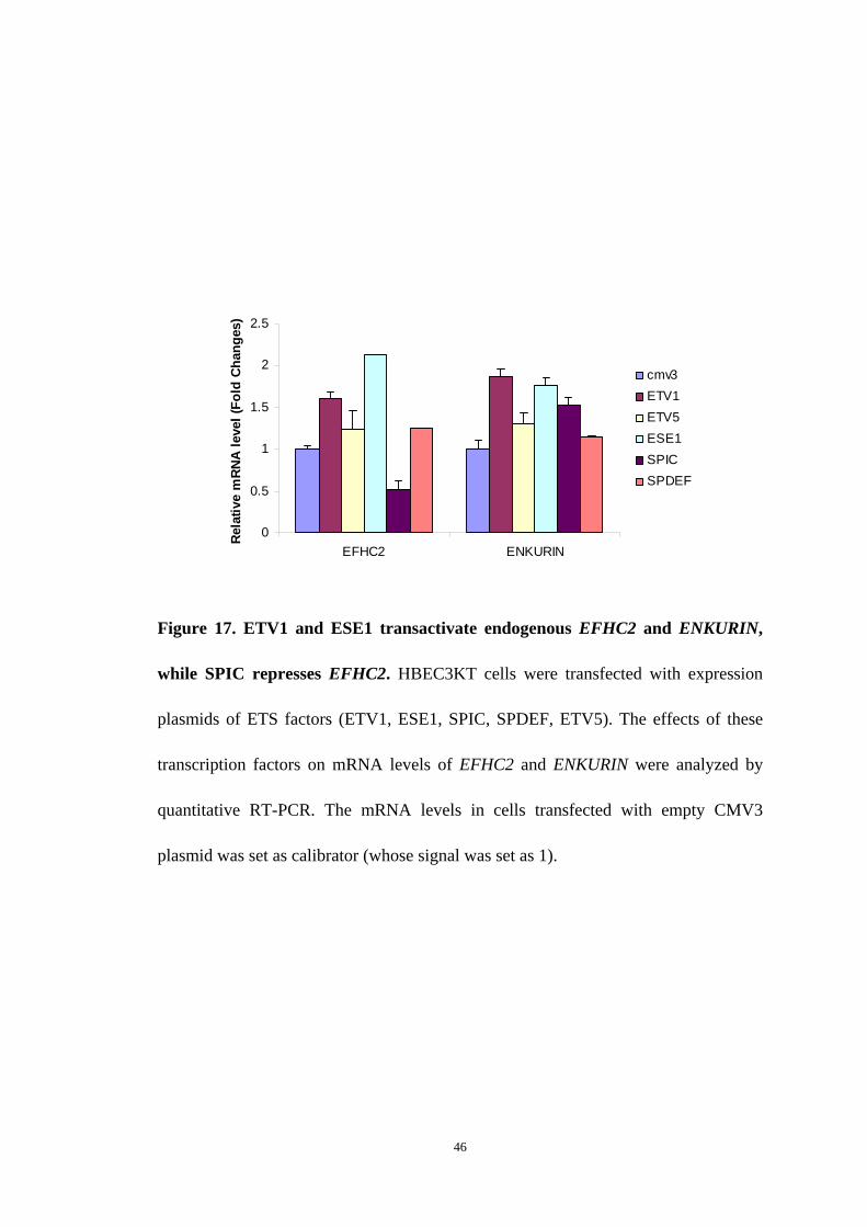
Figure 17. ETV1 and ESE1 transactivate endogenous EFHC2 and ENKURIN,
while SPIC represses EFHC2. ............................................................................. 46
Figure 18. Expression level changes of RFX factors during the initiation of NHBE
cell differentiation and ciliogenesis. .....................................................................48
Figure 19. Numbers of RFX binding motifs in proximal cilia gene promoters.. 49
Figure 20. RFX1 transactivates endogenous ENKURIN, HSPA1A and ROPN1L;
and RFX5 transactivates endogenous EFHC2, HSPA1A and RIBC2.................. 50
Figure 21. GATA2 transactivates EFHC2 and ENKURIN promoters..................52
Figure 22. GATA2 transactivates endogenous EFHC2 and ENKURIN.............. 53
Figure 23. GATA2 transactivates proximal regions of ENKURIN and EFHC2
promoters. ............................................................................................................ 54
Figure 24. Transcriptional network in ciliogenesis............................................. 64
xi

xii
LIST OF TABLES
Table 1. PCR primers for amplifying cDNAs of transcription factors ......................22
Table 2. PCR primers for amplifying proximal promoter and 5’UTR exon of cilia genes.
.................................................................................................................................... 24
Table 3. qRT-PCR primer sites for ETS, RFX family and cilia genes...................... 29

Chapter 1: Introduction
Human Cilia and Cilia-Related Diseases
Cilium is an organelle found in eukaryotic cells, which projects from the cell
surface and is membrane bounded. It contains a microtubule cytoskeleton, the ciliary
axoneme, surrounded by the ciliary membrane. The ciliary axoneme derives from and
keeps the nine-fold symmetry of centriole, which is nearly identical to, and often
becomes a ciliary basal body (1).
There are two major patterns of mammalian cilial axoneme. Those in multiciliated
epithelial cells are motile 9+2 type, possessing dynein arms. The other pattern is
single, nonmotile 9+0 primary cilia which are found on virtually every cell in the
body (2). These nonmotile 9+0 cilia do not assemble the central microtubule which
appears in the motile 9+2 cilia and lack of the molecular motors, termed axonemal
dyneins (both ODA and IDA). Primary cilia function as various specialized sensory
structures with chemosensory, mechanosensory, or positional sensory activity. They
incorporate specific receptors and channels into their ciliary membrane and thus can
respond to either mechanical stimuli or defined ligands. The presence of primary
cilium is important for cell cycle, cell differentiation and controlling tissue
homeostasis (1). Loss of primary cilia leads to abnormal cell function, cell division
and can cause diseases such as polycystic kidney disease (3, 4, 5, 6, 7). In addition,
unique motile 9+0 cilia possessing dynein arms with left-right dynein (8, 9) are found
at the mammalian embryonic node during development, determining left-right
asymmetry of the body (10, 11, 12, 13).
1

The ciliary axoneme of motile cilia grows from and continues the nine-fold
symmetry of the centriole, which is polymerized from αβ tubulin heterodimers and the
precursor of a ciliary basal body. The protein complex of axonemes is attached to the
microtubules, including outer and inner dynein arms (ODAs and IDAs), the radial
spokes, and the central pair projections. The ODAs and IDAs are molecular motors
with ATPase activity, and can change their conformation alternatively by utilizing the
energy of ATP hydrolysis to move along their neighboring outer microtubule doublets.
This activity generates the force to make the microtubule doublets slide with respect
to one another, producing a sliding motion (14, 15). The radial spokes resist this
sliding motion which is converted into a local bending. The highly extensible
interdoublets protein, nexin, keeps adjacent doublets together during this process
(Figure 1).
The wave-like or beating motion of motile cilia is used either for locomotion such
as in spermatozoa, or in the case of cilia along the respiratory tract and nodal cilia, to
generate mucosal or fluid flow (16-18). Non-motile (or primary) cilia are usually
associated with various sensory functions and present singly on cells (16, 19, 20).
Their functions are cell type-dependent, for example, specifically responsible for
vision, olfaction and mechanosensation in various tissue settings.
Ciliary defects are classified into primary and secondary ciliary defects. Primary
ciliary dyskinesia is a genetic disorder causing dysfunctional motility of cilia and
impaired mucociliary clearance. Secondary ciliary defects are caused by a variety of
pathogens, toxins and trauma. These ciliary defects can lead to a number of human
2
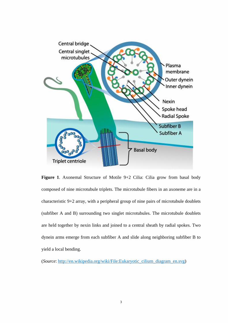
Figure 1. Axonemal Structure of Motile 9+2 Cilia: Cilia grow from basal body
composed of nine microtubule triplets. The microtubule fibers in an axoneme are in a
characteristic 9+2 array, with a peripheral group of nine pairs of microtubule doublets
(subfiber A and B) surrounding two singlet microtubules. The microtubule doublets
are held together by nexin links and joined to a central sheath by radial spokes. Two
dynein arms emerge from each subfiber A and slide along neighboring subfiber B to
yield a local bending.
(Source: http://en.wikipedia.org/wiki/File:Eukaryotic_cilium_diagram_en.svg)
3
diseases including polycystic kidney disease (PKD), retinal degeneration, anosmia,
laterality defects, chronic respiratory problems, situs inversus, hydrocephalus, or
infertility (17, 21-23) and other ailments such as Bardet–Biedl, Alstrom and Meckel
syndromes. These aliments extend the ciliary disorder to obesity, diabetes, liver
fibrosis, hypertension, heart malformations, skeletal anomalies (e.g. polydactyly),
cognitive impairment and developmental defects such as exencephaly (17, 21, 24-29).
Functions of Lung Epithelial Cilia and Related Diseases
The lung and respiratory passage are constantly exposed to respiratory pathogens,
toxins and foreign particles, and have evolved a very effective defense system. Lung
epithelium exerts protective effects through several ways: by providing a physical
barrier; through the secretion of factors that mediate immunity, inflammation, and
antioxidant defense; and through mucociliary clearance. Mucociliary clearance
plays an important role among these defense mechanisms for clearance of foreign
particles and prevention of bacterial colonization or viral spreading. The
pseudostratified columnar respiratory epithelium consists of ciliated,
mucus-producing and basal cells (30). Cilia line the nasal cavity, paranasal sinuses,
Eustachian tubes and middle ear of the upper respiratory tract. The lower respiratory
tract is lined by ciliated cells from the trachea to the terminal bronchioles. These
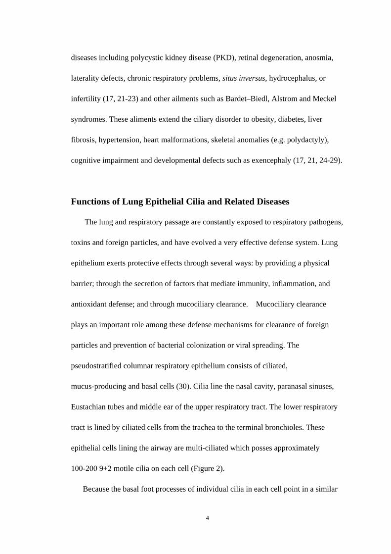
epithelial cells lining the airway are multi-ciliated which posses approximately
100-200 9+2 motile cilia on each cell (Figure 2).
Because the basal foot processes of individual cilia in each cell point in a similar
4
Figure 2. Scanning electron microscope image of lung trachea epithelium: These lung
epithelial cells are both ciliated and non-ciliated. The rod-like cilia on multicilated
cells is different in size from the sand-like microvilli on non-ciliated cell surface.
(Source: http://remf.dartmouth.edu/images/mammalianLungSEM/source/9.html)
5

direction (31), the effective ciliary stroke for each cell beats in a common direction.
Mucus secreted by respiratory epithelium acts as a physical barrier for capture of
inhaled pathogens and particles, while the tips of cilia interact with the mucus layer to
propel it along the airway surface from the lung and nasal cavities (32, 33).
Respiratory ciliary defects contribute to a wide range of human diseases such as
asthma, cystic fibrosis and chronic obstructive pulmonary disease. Primary respiratory
ciliary defects include primary ciliary dyskinesia (PCD) which results in chronic
sinusitis, bronchiectasis, rhinitis, otitis and “glue ear” with associated hearing loss (36,
37).
A variety of pathogens, toxins and trauma could cause the secondary ciliary
defects and abnormal functions. During acute viral infections such as coronavirus,
mucociliary clearance has been shown to be impaired (38), which might be due to the
cytopathic loss of epithelial cilia (38-42). When infected by various common cold
viruses or, RSV, a paramyxovirus, ciliated lung epithelial cells are lost. Cilia may also
become withdrawn into the cell body as was seen in the nasal epithelium during an
episode of coronavirus infection (43-46, 55, 56). Interestingly, infection of several
respiratory viruses is unique in ciliated and nonciliated airway epithelial cells (43). In
addition to viral infections, bacterial infections also compromise ciliated respiratory
epithelial cells. For example, when infecting the respiratory epithelium, Pseudomonas
aeruginosa will secrete a toxic substance named pyocyanin that will immobilize the
tracheal cilia (54). Infection with Mycoplasma pneumoniae, Bordetella pertussis or B.
bronchiseptica will cause derangement of ciliary attachment (50-52).
6
Mucociliary Differentiation of Human Airway Epithelial Cells
The commitment of an undifferentiated airway epithelial cell to a ciliated cell and
subsequent maturation to the ciliated phenotype occurs through undefined molecular
pathways. In the developing human lung, ciliated epithelial cells arise from
undifferentiated cells in the trachea during the canalicular stage, and, as lung
development proceeds, cilia progressively appear in large and then small airways (61).
Ciliogenesis in airway epithelial cells is a multistep process, in which precursor
structures called “amorphous bodies” give rise to centrioles that subsequently
transform to basal bodies. The basal bodies then migrate and dock at the apical
membrane where they give rise to axonemes on the ciliated cells (62, 63). Although
some regulators have been identified for the primary ciliogenesis (64), few molecular
regulators of ciliogenesis in motile ciliated cells have been identified.
Primary human bronchial epithelial cells (HBEC) can be harvested from lung
tissues and cultured in an air-liquid interface (ALI), which will promote their
mucociliary differentiation. In the first week of ALI culture, the cells form a stratified
squamous epithelial layer. By the end of the second week, those squamous cells are
no longer flattened in appearance but without other obvious morphological changes.
Although cilia could not be discerned on cells of this stage,α-tubulin are observed to
concentrate on the surface of individual cells, indicating that ciliary protein
constituents are being synthesized and transported, and that ciliogenesis has initiated.
In other cells in this culture mode, there is high concentration of MUC5B, which is
the main secreted mucin by secretory cells (57), suggesting the initiation of secretory
7
cell differentiation of non-ciliated cells. By the end of the third week, many of the
cells have arranged in the pseudostratified layer with a columnar morphology. At the
end of both third and fourth week, cilia are observed on the apical membrane surface
of the cells, and there is a large increase of acidic mucin on the surface of epithelium,
giving rise to the characteristic features of the predominant epithelial subpopulations
of ciliated and secretory cells in the airways (58, Figure 3).
Cilia Genes in Mucociliary Differentiation of HBEC Cells
Axonemes purified from primary ALI cultures of HBEC cells have been studied
by proteomic analysis, through which 200 proteins have been found (59). In another
study, changes in gene expression during differentiation of airway epithelial cells
have been examined by Affymetrix cDNA microarray analysis, and 1488 genes were
found to display two or more-fold increase at early stage of ciliogenesis. By
comparing the proteomics data with the microarray data, there are 37 overlapping
genes between these two data sets (Figure 4, 58). Thus these genes are very likely to
have potential roles in cilia formation or functions, and it is notable that most of them
have very similar expression profiles, with a dramatic increase between days 2 and 8
of ALI culture.
Among these 37 genes, a number of genes encode proteins that are components
of the ciliary axoneme, such as those encoding light, intermediate or heavy chains of
axonemal dyneins ( DNAH9, DNAI1, DNAI2, DNALI1), and tubulins (TUBA3,
TUBA4, TUBB2).
8

Figure 3. Mucociliary differentiation of HBEC cells during ALI culture on day 21.
Acetylated α-tubulin is immunostained to mark ciliary axonemes (red) and nuclei are
stained with DAPI (blue) respectively. Ciliated cells are denoted with red apical
staining to show axonemal structures.
9

A.
B.
Figure 4. A). This diagram shows the overlapping genes found to be regulated in
ciliogenesis by microarray analysis with proteins known to be found in the cilia
axoneme by proteomic analyses. The microarray analyses (Left) identified genes
displaying two or more fold increases during mucociliary differentiation of HBEC
cells. The proteomic analyses (Right) identified genes encoding proteins found in
purified ciliary axoneme. B). The list of the 37 overlapping genes and their encoding
proteins (Andrea J. Ross. etc. 2007. Am J Respir Cell Mol Biol.)
10

Another number of genes (TEK2, CENT2, SPAG6, NME5, SPA17, IFT57, IFT81,
and IFT88) encode proteins associated with cilia, flagella or microtubules. For
example, IFT57, IFT81, IFT88 are component proteins of the intraflagellar transport
machinery.
Calcium homeostasis is important for many aspects of ciliary and flagellar
functions, and proteins encoded by some genes (ENKURIN, CAPS, EFHC1, EFHC2)
bind calcium ions or are involved in calcium transport. Enkurin is a calmodulin
protein co-localizing with calcium channels, which is suggested to be an adaptor to
localize calcium signal transduction machinery to calcium channels (60). EFHC2
encodes a protein very similar to EFHC1 protein, and is proposed to be a calcium
protein associated with the axoneme and plays a role in cilial axoneme assembly and
stability.
In addition, several genes encode proteins with different functions. For example,
HSPA1A is a member of heat shock protein 70 family, and in conjunction with other
heat shock proteins, stabilizes existing proteins against aggregation to mediate the
folding of newly translated proteins in the cytosol and in organelles. It is also
involved in the ubiquitin –proteasome pathway (91). ROPN1L encodes a sperm
protein, which interacts with A-kinase anchoring protein AKAP3 and allows
ROPN1L to be targeted to specific subcellular compartments (92). MNS1 is a
meiosis-specific structural protein expressed at the pachytene stage during
spermatogenesis, and thought to be involved in the control of meiotic division and
germ cell differentiation through regulation of pairing and recombination during
11

meiosis (89, 90). Other genes (RABL4, RABL5, RIBC2) encode proteins with
unknown functions in cilia.
FOXJ1 Transcription Factor in Ciliogenesis
FOXJ1 (also called HFH-4) is a forkhead box (f-box) transcription factor
expressed in ciliated cells located in the upper and lower airway, choroid plexus,
ependyma, oviducts, testis and embryonic node (65-68). Colocalization of FOXJ1 and
β-tubulin IV but not Clara cell secretory protein confirmed that FOXJ1 is expressed in
ciliated epithelial cells rather than nonciliated columnar epithelial cells in the
respiratory tract (73). Much evidence indicates a direct relationship between Foxj1
expression and ciliogenesis: During the murine lung development, the expression of
Foxj1 immediately precedes the appearance of cilia in airway epithelial cells, which is
in the late pseudogladular stage at embryonic day 15.5 (65). In the infection of some
respiratory viruses such as the paramyxovirus respiratory syncytial virus (RSV), loss
of cilia is associated with loss of Foxj1 expression, and during airway repair, the
appearance of ultrastructural component of ciliogenesis is associated with
re-expression of Foxj1 (69, 74). It is noteworthy that interruption of Foxj1 gene in
genetically engineered mice leads to the loss of left-right dynein (Dnahc11)
expression and absence of cilia (66, 70). In airway cells that have been already
committed to ciliated cell phenotype, Foxj1 promotes the expression of capastatin, an
inhibitor of the protease calpain, leading to the stabilization of cytoskeletal anchoring
proteins ezrin and EBP-50. Ezrin and EBP50 both are associated with basal bodies of
12

cilia in the pulmonary epithelium. Thus, Foxj1 helps the docking of basal bodies to
the apical membrane and the induction of mechanisms of axoneme assembly (71, 72).
Although mutations in the FOXJ1 gene have not yet been identified in human
diseases with abnormalities of cilia formation (75), FOXJ1 is still be considered to be
a potential master regulator of ciliogenesis in airway epithelial cells.
ETS Transcription Factor Family
ETS is one of the largest families of transcription factors, and all members are
identified to have a highly conserved DNA binding domain, the ETS domain, a
winged helix-turn-helix structure that binds to DNA sites with a central GGA motif
sequence. As well as DNA binding functions, the ETS domain is also involved in
protein-protein interactions suggesting transcriptional co-regulation with other
transcription factors. ETS factors act as a transcriptional activator, repressor, or both
(78). There are 28 ETS genes in human and they are divided into 11 subfamilies
(Figure 5, [80]). The extensive conservation in DNA-binding ETS domain among
these genes leads to a substantial of redundancy in their DNA binding capacity. It is
thought that interactions with other proteins are one way in which specific binding to
DNA is achieved (79). There is limited similarity outside the ETS DNA binding
domain. This sequence divergence contributes to the individual properties of
ETS-domain transcription factors, altering their DNA binding specificity and
interactions with other transcription factors (76, 77). These unique properties, coupled
with distinct expression pattern, culminate in their distinct biological roles. The ETS
family is present through out the body and is involved in wide variety of functions
13

Figure 5. The members and their homologs of ETS family and they are divided into
11 sub-families. (Gutierrez-Hartman A.et.al. 2007. Trends Endocrinol Metab).
14

including the regulation of cellular differentiation, cell cycle control, cell
migration,cell proliferation, apoptosis and angiogenesis. Multiple Ets factors have
been reported to be associated with cancer, such as through gene fusion (81, 82). It is
worthy to mention that ETS family members have been proven to expressed in
stratified epithelial cells, including the lung epithelium (85, 86)
Motif discovery analysis of the promoters of mouse lung gene subsets as well as
their human homologues, which are found to transactivate several genetic programs
during lung embryogenesis and lung cell differentiation (88, 89), identified ETS
family members as candidate regulators in both sets of promoters. This analysis
suggests that ETS family potentially regulates crucial lung functions and development
(88).
RFX Transcription Factor Family
RFX proteins belong to the winged-helix family of transcription factors. They
share a characteristic 76-amino acid DNA binding domain that binds to an X-box
motif, and conserved in many eukaryotes (99, 100). Five RFX genes (RFX1,-2,-3,-4
and -5) have been identified in mice and human genome (101). RFX factors are
involved in the regulation of cell cycle (93, 94, 95), brain development and neuronal
functions (96, 97), and ciliogenesis (98).
In mice and humans, RFX-1 is the first gene found as a candidate major
histocompatibility complex (MHC) class II promoter binding protein (103). It also has
been proved to a transactivator of hepatitis B virus enhancer (104). It is primarily
found in the human brain with high expression in cerebral cortex and Purkinje cells
15

(105). RFX-2 and -4 both have highest expression in human testis (106, 107). In mice,
RFX-4 has been implicated in dorsal patterning of brain development and may
regulate circadian rhythm in human (112, 113). RFX-3 is expressed in ciliated cells
including pancreatic endocrine cells, ependymal cells and neuronal cells (108-110).
RFX-5 is a transcriptional activator of MHCII genes. It also mediates the formation of
an enhanceosome complex containing RFXANK, RFXAP, CREB, and CIITA (111).
The roles of RFX family in ciliogenesis of human airway epithelial cells remain
largely unknown. Similar to the discovery of ETS family members by motif discovery,
our lab has recently identified binding sites of RFX family members over-represented
in lung gene networks in mouse and in their human homologue promoters. These
findings predicate that RFX family members are candidate transcriptional regulators
in mammal lung development and functions (88).
General Features of Transcription Factor GATA2 and other GATA
factors
The GATA family of transcription factors controls multiple developmental
processes by regulating tissue-specific gene expression. The zinc finger DNA binding
domains bind to W(A/T)GATAR(A/G) motifs of gene promoter regulatory elements
( 118, 119).
GATA1, GATA2 and GATA3 regulate the development of different
hematopoietic lineages---erythroid, hematopoietic progenitor and T-lymphoid,
respectively (120-122). Similarly, GATA4, GATA5 and GATA6 have been shown to
be involved in cardiac, genitourinary, and multiple endodermal developmental events
16
(123-125).
GATA2 was first cloned from chicken reticulocyte and shown to be present all
along the development of erythoid cells (126). It is also expressed in other
hematopoietic cells, neurons and cells of developing heart, liver, pituitary and in
trophoblasts (127-132).
The roles of GATA transcription factors in human lung epithelial cells
differentiation and ciliogenesis remain elucidated. Our lab has shown that binding
motifs of GATA3 and GATA4 are over-represented in the gene promoters of mouse
and human lung genes, respectively (88). These findings predicate that there might be
multiple cis-regulatory elements of entire GATA family functioning in both
organisms, and GATA family may have roles in lung epithelial cell differentiation
and ciliogenesis.
Hypothesis and Rationale
The lung epithelial cells are highly specialized, differentiated cells originating
from common progenitors in the lung during development. They have unique
functions involved in airway host defense, gas exchange and ion transport (88, 136).
The lung epithelium is a pseudostratified layer consisting of basal cells, secretory
cells and columnar ciliated cells. Mucociliary clearance is an important host defense
mechanism, and defects in mucociliary clearance are associated with various
respiratory disorders and diseases.
Transcription of cilia genes is significantly increased during lung epithelial
differentiation and ciliogenesis. Transcription factor FOXJ1 has been identified as the
17

only one transcription factor during in vitro differentiation of air way epithelial cells.
However, direct transactivation of cilia genes by FOXJ1 was not elucidated. In
addition, through motif discovery and bioinformatics analyses, multiple binding sites
for other families of transcription factors, including RFX, ETS and GATA, were
located in many of the cilia gene proximal promoter regions.
In this thesis, we hypothesize that FOXJ1, ETS, RFX and GATA transcription
factors regulate the expressions of cilia genes in human bronchial epithelial cells,
alone or together.
To verify this, we address the following aims:
Aim 1: Analyze the expression patterns of ETS and RFX family transcription
factors during human bronchial epithelial cells differentiation and ciliogenesis, and
locate binding motifs of these transcription factors in the 5’ proximal promoters of
cilia genes.
Aim 2: Experimentally assess transactivation of distinct cilia genes by the
transcription factors FOXJ1, ETS, RFX and GATA2 through reporter assay, followed
by qRT-PCR detection of the changes of endogenous cilia genes.
Aim 3: Show the direct binding of FOXJ1 onto the promoters of selected cilia
genes.
18

Chapter 2: Materials and Methods
Cell Cultures
Cells were kept at 37°C with 5% CO2. Normal human bronchial epithelial (NHBE)
cells were purchased from Lonza and plated onto collage-coated 100mm dishes
(Becton-Dickinson, Biocoat) and grown in BEGM media (Lonza) to approximately
80% confluency (~4-5days). Then the cells were plated onto collagen coated transwell
inserts (Corning) with BEGM plus DMEM (Sigma-Aldrich) media in the basolateral
and apical chambers. NHBE cells are allowed to grow to 100% confluency (~3-7 days)
at which time the media in the chamber was removed and the cells were grown under
air-liquid interface (ALI) for differentiation (14-28 days). HBEC3KT, kindly
provided by Dr. John. D. Minna (137), were maintained in L-Glutamine containing
Keratinocyte-SFM medium (GIBCO, Invitrogen) in flask coated with FNC mix
(AthenaES™'s).
Bioinformatic Analysis of Cilia Gene Promoters
Proximal promoters of 37 cilia genes were acquired from human genome build 36
(v.1, March 2006) through UCSC genome browser. For each gene, the proximal
promoter was defined as 1000 base pairs upstream of transcriptional start site and the
downstream 5’UTR exon sequences were included to accommodate for the possible
existence of additional cis-regulatory elements.
The ExPlain tool of TRANSFAC Suite (BIOBASE Knowledge Library) was used
to predict transcription factor binding motifs on the cilia gene promoters. The profile
19

containing a matrix for the binding motifs of a set of transcription factors (ETS family
for example) was created by choosing them from existing matrices (Figure 6), and the
cut-offs value was set to be custom CSS (core similarity score) 0.75 and MSS (matrix
similarity score) 0.85. Potential binding motifs of specific transcription factors on
cilia gene promoters were analyzed and searched by uploading promoter sequences
from FASTA files, and match them with a specific matrix using the Match Workflow
Wizard.
Construction of Protein Expression Vectors
Total RNAs were isolated with TRIzol (Invitrogen) according to the protocol
provided by manufacturer. RNAs were extracted from NHBE cells under ALI culture
for 0, 2, 4 days and 3 weeks. Five hundred nanograms of total RNA were used to
synthesize the first-strand cDNA with Quanti-Tech Reverse Transcription Kit
(Qiagen).
Full length coding sequences of transcription factors FOXJ1, ETV1, ESE1, SPIC,
SPDEF, ETV5, RFX1, RFX5 and GATA2 were amplified and flag-tagged through
PCR with FastStart Taq DNA polymerase (Roche). The primers used are outlined in
Table 1 (restriction enzyme sites are shown in italics, and the antisense of FLAG-tag
coding sequences are underlined).
PCR products were cloned into pTarget T Vector (Promega). Competent NEB-5α
cells (NEB) were transformed with ligation products and selected through white/blue
colonies in Agar plates containing 100ug/ml Ampicillin. White colonies were cultured
20

a)
b)
Figure 6. Matrices of the binding sequences of a) ETS family (identifier: V$ETS_Q6)
and b) RFX family (identifier: V$RFX_Q6), for predicting their potential binding
sites on cilia gene promoters.
21

Table 1. PCR primers for amplifying cDNAs of transcription factors
Name Sequences FOXJ1 upstream 5’-aatggcggagagctggctgcg-3’ Downstream 5’-aaagcttttacttatcgtcgtcatccttgtaatccaagaaggcccccacgctggc-3’
(HindIII) GATA3 upstream 5’-aagatctatggaggtgacggcggacc-3’ (BglII) Downstream 5’-atctagattacttatcgtcgtcatccttgtaatcacccatggcggtgaccatgc-3’
(XbaI) ETV1 upstream 5’-ataccggtccatggatggattttatgaccagcaag-3’(AgeI) Downstream 5'-atctcgagttacttatcgtcgtcatccttgtaatcatacacgtagccttcgttgtagg-3'
(XhoI) ETV5 upstream 5'-ataccggtccatggacgggttttatgatcagcaag-3' (AgeI) Downstream 5'-atctcgagttacttatcgtcgtcatccttgtaatcgtaagcaaagccttcggcatagg-3'
(XhoI) ESE1 upstream 5'-ataccggtccatggctgcaacctgtgagattag-3' (AgeI) Downstream 5'-atatcgatttacttatcgtcgtcatccttgtaatcgttccgactctggagaacctcttc-3'
(ClaI) SPIC upstream 5'-ataccggtccatgacgtgtgttgaacaagacaag-3'(AgeI) Downstream 5'-atctcgagttacttatcgtcgtcatccttgtaatcgcaatcatggtgatttagctc-3'
(XhoI) SPDEF upstream 5'-ataccggtccatgggcagcgccagcccgggtctg-3' (AgeI) Downstream 5'-atatcgatttacttatcgtcgtcatccttgtaatcgatggggtgcacgaactggtag-3'
(ClaI) RFX1 upstream 5'-aaagcttatggcaacacaggcgtatac-3 ' (HindIII) Downstream 5'-atctagattacttatcgtcgtcatccttgtaatcgctggagggcagcgcctgcac-3'
(XbaI) RFX5 upstream 5'-aaagcttatggcagaagatgagcctg-3' (HindIII) Downstream 5'-atctagattacttatcgtcgtcatccttgtaatctgggggtgttgcttttgggtc-3'
(XbaI)
22

in 2ml 2xYT medium overnight at 37°C with shaking at 250rpm. Plasmids were
extracted with GenElute Plasmid Miniprep Kit (Sigma-Aldrich) according to the
manufacture’s protocol, and verified through restriction enzymatic cleavage analysis
and sequencing.
Construction of Luciferase-Reporter Plasmids
Genomic DNAs were extracted from A549 cells with TRIzol (Invitrogen)
according to the protocol provided by the manufacture and used as templates in the
PCR with primers to amplify proximal promoters (1000 bases upstream) and 5’ UTR
exon of cilia genes ENKURIN, EFHC2 and IFT57. The primers used are in Table 2
(restriction enzyme sites were shown in italics).
The PCR products were purified with QIAquick PCR Purification Kit (Qiagen)
and digested, then ligated into pGL3-Basic vectors (Promega) treated with
corresponding restriction enzymes. All the reporter plasmids were verified through
sequencing. Reporter plasmids containing truncated proximal promoters and 5’ UTR
of ENKURIN and EFHC2 were also constructed and were digested with NheI/HindIII
and BstEII/HindIII respectively. Two truncated fragments (distal 400bp and proximal
700bp) of ENKURIN promoters as well as proximal 600bp of EFHC2 promoters were
acquired (Figure 7). The cohesive end (BstEII) of proximal 600bp fragment of
EFHC2 promoter was converted to blunt end through T4 polymerase treatment before
HindIII digestion. Distal 400bp ENKURIN promoter, proximal 700bp ENKURIN
promoter, proximal 600bp EFHC2 promoters were inserted into pGL3-Basic vector
treated with NheI, NheI/HindIII, SmaI/HindIII respectively. Proximal 450bp and
23

Table 2. PCR primers for amplifying proximal promoter and 5’UTR
exon of cilia genes.
Name Sequence
ENKURIN upstream 5’-atctcgaggagtcccgacccggggaag-3’
(XhoI)
Downstream 5’-atagatctggccaccaaatgactcct-3’( BglII)
EFHC2 upstream 5’-actcgagaattataaaaacgacggctc-3’ (XhoI)
Downstream 5’-aaagcttacgttgcctggagacgggaag-3’
(HindIII)
IFT57 upstream 5’-acccgggcatccaccacttacctcatt-3’ (XmaI)
Downstream 5’-aagatctcgccgccagtacagccacgac-3’
(HindIII)
24

a)
b)
Figure 7. a) Promoter structures of ENKURIN in luciferase reporter vectors. b)
romoter structures of EFHC2 in luciferase reporter vectors. P
25

250bp ENKURIN promoter were further acquired by treating the reporter plasmid
containing proximal 700bp ENKURIN promoter with PvuII/HindIII, PstI/HindIII, then
ligated to pGL3-Basic vector treated with SmaI/HindIII. All reporter plasmids were
propagated in NEB-5α cells (NEB) and verified through restriction enzymatic
analysis and sequencing.
Maxiprep of Plasmids
Large amounts (~1-2μg/μl) of plasmids were acquired using Spin Doctor BAC
Prep Kit& Spin Doctor Super Clean BAC Prep Kit (Gerard Biotech). The procedure
was according to the protocol provided by the manufacturer with modifications as
follows: after the first centrifugation, 0.8ml endotoxin removal resin (Promega) was
added to supernatants and shaken for 10mins at room temperature. The resin was
removed by centrifuging at 4000rpm shortly. The DNA pellets in the last step was
re-suspended in 500μl Tris-HCl (pH8.0) and kept in 57� water bath. After
re-suspension, each tube was centrifuged at maximum speed for 5mins and transferred
to a new tube for subsequent transfections.
Transfection and Reporter Gene Assay
Immortalized hBEC3KT cells were seeded onto 6-well plate (2.5 x10⁵ cells per
well, for endogenous gene transcription assay) or 48-well plate (2x10⁴ cells per well,
for reporter assay) 24 h before the transfection. FuGENE HD (Roche) was used for
transfection according to the protocol provided by manufacturer. Typically, for each
well of 6-well plate, 250ng of expression vectors, and empty expression vectors
26

(CMV3) were added to a total 500ng in 100μl serum-free GIBCO Opti-MEM medium
( Invitrogen) with 2μl FuGENE HD. For each well of 48-well plate, 100 ng of
reporter plasmid, 25 ng of transcription factor expression vector, and empty
expression vector were added to a total 200ng in 17ul Opti-MEM medium with 1μl
FuGENE HD. Each transfection of a 48-well plate was carried out in duplicates.
Twenty-four hours after transfection, for 6-well plate, cells were washed cold PBS,
and RNA was isolated for RT and qRT-PCR analysis with TRIzol (Invitrogen)
according to the manufacturer’s protocol. For 48-well plate, 24 h after transfection,
cells were washed once with cold PBS followed by adding 100 μl of 1xlysis buffer
(Promega) per well, and kept at -70°C for 1 h. After 30 min of rotation at room
temperature, 5 μl of lysates was mixed with 25 μl of substrate (Promega) to detect
luciferase activity with Turner.
Quantitative Real-time PCR
Total RNAs isolated from transfected hBEC3KT and differentiated NHBE cells
on day 0, 2, 4 and three weeks were extracted and reverse transcribed as
aforementioned. In parallel, GAPDH CDS was amplified as an internal control with
primers GS (atcactgccacccagaagac) and primer GR (ttactccttggaggccatgtg). To detect
mRNA level of ETS family and RFX family, and their changes during NHBE cell
differentiation, qRT-PCR forward and reverse primers were designed. Besides, to
identify the effects of transcription factors on endogenous cilia gene expression after
transfection, qRT-PCR primers for cilia genes (ENKURIN, EFHC2, IFT57, HSPA1A,
MNS1, RIBC2 and ROPN1L) were also designed (Table 3). QuantiTect SYBR Green
27

PCR Kit (Qiagen) was used with forward and reverse primers (final concentration
300nM each ) according to the manufacturer’s protocol in an ABI 7300 real-time PCR
system (Applied Biosystem). The quantitative PCR results were analyzed using ABI’s
7300 System SDS software, RQ Study.
Chromatin Immunoprecipitation Assay
To verify whether FOXJ1 directly binds to the proximal 250bp ENKURIN
promoter and regulates its expression, chromatin immunoprecipitation assay was
performed with ChIP-IT Express Enzymatic Kit (Active Motif) according to the
manufacturer’s protocol. In brief, twelve wells of NHBE cells cultured for 3 weeks
were digested with 0.25% trypsin-EDTA solution and washed once with cold PBS,
then fixed with 1% formaldehyde for 10 min at room temperature. After
centrifugation and washing with cold PBS once, the residual formaldehyde was
neutralized with glycine (0.125 M). Released nuclei from collected cells were
enzymatically treated at 37 oC for 10 min in 400μl volume. 50μl of sheared
chromatins were used for immunoprecipitation in 500μl volume with normal goat
serum as a control, or polyclonal antibody against FOXJ1 (Santa Cruz). ENKURIN
proximal 250bp promoter was amplified with EPF (gcacagtcaacgacgcttagcaat) and
EPR (tctccgtccctttcttcactgctt). Surfactant protein D was used as a negative control to
verify the specific enrichment of ENKRUIN proximal promoter by FOXJ1 antibody,
and amplified with primers SPDF (tggaatgacagggcttgtggagaa) and primer SPDR
(aggatattggcagcatgagggtct). Specific enrichment of ENKURIN proximal promoter was
28

quantified with software described in qRT-PCR section, and normalized to
nonspecific precipitation of last exon of surfactant protein D.
Table 3. qRT-PCR primer sites for ETS, RFX family and cilia genes.
Name Sequence ELF1 upstream primer agctagaaccagtaccatgcagga downstream primer agacacaaccactggaacttgggt ELF2 upstream primer cctgaaggtgaaacagattgagcc downstream primer acatcgagaccatcctggctaaca ELF4 upstream primer actacctccaccatgctcgtctct downstream primer tggatgttgctgggcactgaaga GABP-α upstream primer gcctacgatgaactatgagaaactcagtcg downstream primer tgccttgaactttacaaatcatgtccc ELG upstream primer cgatcttcgtccagctaacaaacttgc downstream primer cagctccttggagaaatggctgat FLI1 upstream primer aggagtggatcaatcagccagtga downstream primer caccagcttgctgcatttgctaac FEV upstream primer tctcttcaaggacgggaagaaccc downstream primer agaaactgccacagctggatct ERF upstream primer gatgaattacgacaagctgagccg downstream primer gaaattgaacttgtaggtgaaccgt ETV3 upstream primer ccgcaggaaatgcaaaccaca downstream primer tgaaggatcctcttgttgtaatagtatctg ESE1 upstream primer acagcaacatgacctacgagaagc downstream primer ttgccaaacttgtagacgagtcgc ESE2 upstream primer agagtactaccctgcctttgagca downstream primer cacacatggcgcttagtccagtat ESE3 upstream primer tgccaagtgccacaccaaa downstream primer tcctgggttcttgtctgggttcaa ETS1 upstream primer ttatcagctggacaggagatggct downstream primer aggccacggctcagtttctcataa ETS2 upstream primer tgccagtcattcatcagctggact downstream primer gcttctcgtagttcatcttgggct
29

Table 3 (Cont.)
SPDEF upstream primer tcctcaaggagttgctactcaagc downstream primer acctgggctgagtcctcaattt ETV4 upstream primer atgaattacgacaagctgagccgc downstream primer aacttgtacacgtaacgctcaccagc ETV5 upstream primer atgaactatgacaagctgagccgc downstream primer agggcatctgggtcacagacaaat ETV1 upstream primer gataaacttagccgttcactccgc downstream primer gggcttctggatcacacacaaact ETV2 upstream primer tagcagctgcatccgttggact downstream primer aattcatgcccggctttctcttgc SPI upstream primer actggaggtgtctgacggcga downstream primer actggtacaggcggatcttcttct SPIB upstream primer tctcggacagcgagtcggatgag downstream primer aactggtacaggcgcagcttctt SPIC upstream primer gtacaacccactcttctccagcaa downstream primer atgccatctccggattatacaggg ELK1 upstream primer ttggaggcctgtctggaggctgaa downstream primer agctcttccgatttcaggtttggg ELK4 upstream primer ttatgacaaactcagccgagccct downstream primer tgtacacaaacttctgaccattcac ELK3 upstream primer tggagcagccttagtccagttgct downstream primer tggcatgtggccattaagcagtgt ETV6 upstream primer ccctgcgccactactacaaactaa downstream primer tcggccactcatgatttcatctgg ETV7 upstream primer gccaagatcttccgagttgtggat downstream primer ttataatagtggcgcagggcacga ERG upstream primer ctctccacggttaatgcatgctaga downstream primer ggcctagttgtaattctttgcgtagc RFX1 upstream primer tgccagccagtcttactctcaca downstream primer cctcagccgtctcatagttgtcca RFX2 upstream primer gatcgctgtgatgggagagttcaa downstream primer gctcatcgcccatgtcatctttgt RFX3 upstream primer agtggtcaacagacaggcacatct downstream primer ttctccaaactctggaagtgctcg RFX4 upstream primer actgtcagagaatactggacactg downstream primer gccgacaatgttcaccaccgt RFX5 upstream primer tcctgctacagcagcatctcatct downstream primer ttggtttagatgaccgttcccgag RFXANK upstream primer tcaccaccgaagccgactct downstream primer tggttctcgatcacctgttgcact
30
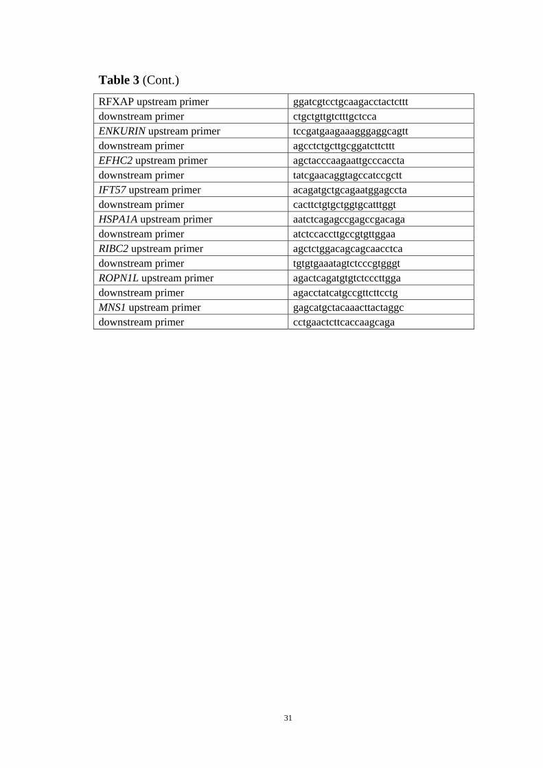
Table 3 (Cont.)
RFXAP upstream primer ggatcgtcctgcaagacctactcttt downstream primer ctgctgttgtctttgctcca ENKURIN upstream primer tccgatgaagaaagggaggcagtt downstream primer agcctctgcttgcggatcttcttt EFHC2 upstream primer agctacccaagaattgcccaccta downstream primer tatcgaacaggtagccatccgctt IFT57 upstream primer acagatgctgcagaatggagccta downstream primer cacttctgtgctggtgcatttggt HSPA1A upstream primer aatctcagagccgagccgacaga downstream primer atctccaccttgccgtgttggaa RIBC2 upstream primer agctctggacagcagcaacctca downstream primer tgtgtgaaatagtctcccgtgggt ROPN1L upstream primer agactcagatgtgtctcccttgga downstream primer agacctatcatgccgttcttcctg MNS1 upstream primer gagcatgctacaaacttactaggc downstream primer cctgaactcttcaccaagcaga
31

Chapter 3: Results
FOXJ1 in human airway epithelial differentiation and ciliogenesis
FOXJ1 is a master regulator during the ciliogenesis; however, few target genes of
FOXJ1 have been identified thus far. Mutation of the FOXJ1 gene is not correlated
with abnormality of ciliogenesis (75). The expression of thirty-seven cilia genes,
which encode axonemal components of cilia, increases significantly during human
lung epithelial ciliogenesis (58), corresponding to the increased expression of FOXJ1.
These genes are likely transcriptional targets of FOXJ1.
By quantitative RT-PCR, we assessed the relative mRNA levels of FOXJ1 as well
as several cilia genes (ENKURIN, EFHC2, IFT57) during differentiation of NHBE
cells under ALI cultures ( 0 day and the 21 day). We found that the expression of
FOXJ1 and these cilia genes increased significantly in NHBE cells at day 21 of
differentiation, when compared to undifferentiated NHBE cells lacking a ciliated cell
population on day 0 (Figure 8). It is noteworthy that FOXJ1 expression was highly
up-regulated.
To test whether ENKURIN, EFHC2 and IFT57 are transcriptional regulation
targets of FOXJ1, we constructed an expression vector of FOXJ1 through cloning of
coding sequence into pTarget expression plasmid; and reporter plasmids containing
the proximal promoters (1000 bases upstream of transcriptional start site and 5′ UTR
sequences) of ENKURIN, EFHC2 and IFT57 were constructed with pGL3 to direct
expression of the reporter gene luciferase. FOXJ1 expression vectors and the reporter
32

Figure 8. Expression of FOXJ1, EFHC2, ENKURIN and IFT57 were
up-regulated during lung epithelial differentiation and ciliogenesis. Fully
differentiation of NHBE cells cultured under ALI situation will achieved after 21 days.
Total RNAs were extracted from D0 and D21 ALI cells for quantitative RT-PCR
detection of the relative mRNA levels of FOXJ1 and three cilia genes, mRNA levels
of D0 were set as 1.
33

plasmids were cotransfected into HBEC3KT cells in 48-well plate and 24 hours later,
cellular lysates were mixed with luciferase substrates for reporter assay. Results
showed FOXJ1 can markedly transactivate the ENKURIN, EFHC2 and IFT57
promoters (Figure 9).
Overexpression of FOXJ1 in HBEC3KT cells is able to induce the expression of
cilial axoneme genes EFHC2, ENKURIN, RIBC2 and ROPN1L, while IFT57,
HSPA1A and MNS1 were not induced dramatically (Figure 10), indicating FOXJ1 can
only selectively transactivate the cilial axoneme genes EFHC2, ENKURIN, RIBC2
and ROPN1L. The inability of FOXJ1 to induce expression of endogenous IFT57
while capable of transactivating the IFT57 promoter will be discussed later.
To identify the FOXJ1 responsive cis-element(s) located in EFHC2 and
ENKURIN proximal promoters, we truncated their proximal promoters and cloned
them to pGL3 plasmid as described, and then cotransfected with FOXJ1 expression
plasmid in HBEC3KT cells. Cis-elements responsive to FOXJ1 transactivation in the
ENKURIN promoter were located in the proximal 250bp sequences, while the distal
400bp of the promoter didn’t contain cis-elements responsive to FOXJ1 (Figure 11).
Similarly, the proximal 600bp of EFHC2 promoter could be transactivated by FOXJ1
(Figure 12). These results indicate that the cis-elements responsive to FOXJ1
transactivation are located in the proximal region of ENKURIN and EFHC2
promoters.
Next, chromatin immunoprecipitation (ChIP) was employed to analyze if FOXJ1
could directly bind to the 250bp proximal region of ENKURIN promoter. Twelve
34

1
10
100
1000
IFT57 EFHC2 ENKURIN
Lu
cife
rase
Act
ivit
y (F
old
Ch
ang
es)
Figure 9. FOXJ1 transactivates IFT57, EFHC2 and ENKURIN promoters.
Reporter plasmids containing 1000bp proximal promoter and 5′ UTR of IFT57,
EFHC2 and ENKURIN were co-transfected with FOXJ1 expression plasmid
respectively. Luciferase activities were measured 24h later. Luciferase activity of
reporter plasmids co-transfected with control expression plasmids (empty CMV3) was
set as 1.
35

1
10
100
EF
HC
2
EN
KU
RI
N
HS
PA
1A
IFT
57
MN
S1
RIB
C2
RO
PN
1LR
elat
ive
mR
NA
leve
l (F
old
C
han
ges
)
Figure 10. Effects of FOXJ1 on endogenous cilia gene expression. Total RNAs
were extracted from HBEC3KT cells transfected with FOXJ1 expression vectors. The
mRNA levels of cilia genes (EFHC2,ENKURIN, HSPA1A, IFT57, MNS1, RIBC2 and
ROPN1L) were assayed by quantitative RT-PCR. Control groups were transfected
with empty CMV3 expression plasmid, whose mRNA levels of cilia genes were set as
1.
36

A)
B)
1
10
100
1000
ENKURIN ENKURIN P-700
ENKURIN D-400
ENKURIN P-450
ENKURIN P-250
Lu
cife
rase
cti
vity
(F
old
Ch
ang
es)
Figure 11. Cis-element(s) responsive for FOXJ1 transactivation were located in
the proximal promoter regions of ENKURIN. A) ENKURIN promoter (proximal
1000bp+5′UTR) were truncated at 700 bp, 450 bp, 250 bp proximal to the
transcriptional start site, and cloned into the luciferase reporter gene construct. In
addition the distal 400 bp of the proximal promoter was also cloned into the luciferase
expression construct. B) Subsequent cotransfection with FOXJ1 expression plasmid
was used to determine promoter activity as measured by luciferase activity assay.
37

A)
B)
1
10
100
EFHC2 EFHC2-P600
Lu
cife
rase
Act
ivit
y (F
old
Ch
ang
es)
Figure 12. Cis-element(s) responsive for FOXJ1 transactivation were located in
the proximal promoter regions of EFHC2. A) EFHC2 truncated proximal 600bp
fragment was cloned to pGL3 luciferase- reporter plasmid. B) Subsequent
co-transfection with FOXJ1 expression plasmid.
38

wells of NHBE cells under ALI culture for three weeks were treated as described for
ChIP analysis. Chromatin precipitates were enzymatically sheared into 200bp~400bp
pieces. Goat polyclonal antibody against FOXJ1 (Santa Cruz) was used for specific
enrichment of ENKURIN proximal promoter and non-immunized goat serum was
used as negative control. Quantitative PCR showed that the FOXJ1 polyclonal
antibody, but not the normal goat sera, specifically enriched the proximal ENKURIN
promoter (Figure 13), indicating ENKURIN is an authentic transcriptional target of
FOXJ1, and the cis-elements for FOXJ1 transactivation are located in the proximal
ENKURIN promoter.
ETS transcription factor family in human airway epithelial
differentiation and ciliogenesis
Although FOXJ1 was the only transcription factor identified by microarray to be
increased during the differentiation of NHBE cells (58), it failed to transactivate
several endogenous cilia genes. We analyzed the transcript levels of other
transcription family members such as ETS proteins, which are expressed in stratified
epithelium such as the pulmonary airways (85, 86). In lung epithelial cells, ETS
family members are involved in transcriptional regulation and differentiation of
several differentiated components of the airways (83, 84, 87). Previously, this
laboratory has discovered ETS binding motifs overrepresented in the promoters of
cilia genes. ETS binding motifs were found on promoters of mouse lung gene subsets
as well as their human homologues, regulating the lung cell differentiation (88, 89).
39

Figure 13. Direct binding of FOXJ1 to the proximal promoter of ENKURIN.
Sheared chromatins from NHBE cells of three-week ALI culture were
co-immunoprecipitated with goat polyclonal antibody against FOXJ1 or normal goat
serum. The specific enrichment of ENKURIN proximal promoter sequence was
quantified by quantitative PCR as detailed in Materials and Methods.
40

Thus ETS family members are potential transcriptional regulators involved in lung
epithelial ciliogenesis in addition to FOXJ1. Quantitatitive RT-PCR was employed to
detect the changes of expression patterns of all twenty-eight ETS family members in
differentiated, ciliated NHBE cells under ALI culture. Results showed that the
expression of ETV1, ETV2, ETV6, ETV7, ESE1, ESE2, ESE3, SPDEF and SPIC
were significantly increased during the differentiation, while the expression of ETV4
and ETV5 were decreased. The level of other ETS factors did not show significant
changes during ciliogenesis, and some, such as SPI, SPIB and ELG, were expressed
in very low level in both unciliated and ciliated NHBE cells, indicating they are not
likely involved in the lung epithelial ciliogenesis (Figure. 14).
The initial stage of ciliogenesis occurs between 2d and 4d apical upon exposure to the
air. To further define more acutely the temporal induction of ETS family members,
we assessed the expression level changes of ETS factors, during this stage to further
identify those that are most likely involved in regulation of ciliogenesis. As Figure 15
shows, by quantitative RT-PCR of identification of the eleven dramatically changed
ETS factors in Day 21 of ALI cells, the mRNA level of eight (ESE1, ETV1, ESE2,
ESE3, SPIC, ETV2, ETV6, SPDEF) were significantly up-regulated during the initial
stage of epithelial differentiation, while ETV5 significantly down-regulated. These
results suggest that these nine ETS factors, are most likely involved in regulation the
initiation of NHBE differentiation.
To identify transcription factors in regulation of cilial axoneme genes, motif
discovery analysis as detailed in Methods and Materials was utilized to examine the
41

Figure 14. Expression level changes of ETS transcription factors during lung
epithelial ciliogenesis. Total RNAs were extracted from unciliated (ALI Day 0) and
ciliated (ALI Day 21) NHBE cells. The mRNA levels of ETS transcription factors
were detected by quantitative RT-PCR. For comparison, the mRNA levels in
unciliated cells (D0) were set as 1.
42

Figure 15. Expression level changes of ETS factors during the initiation of NHBE
cell differentiation and ciliogenesis. Total RNAs were extracted from NHBE cells
under ALI culture at Day 0, Day 2 and Day 4, followed by quantitative RT-PCR to
detect the mRNA levels of chosen ETS genes (heatmap, deep red: mRNA levels
increased over 2 fold; deep green, mRNA levels decreased over 2 fold). Day 0 was set
as calibrator (black).
43

proximal promoter (proximal 1000bp plus 5′ UTR) of cilia genes to predict ETS
binding sites. As shown in Figure 16, eight and seven ETS binding motifs were
identified in ROPN1L and HSPA1A promoters among all cilia genes, respectively.
Four ETS sites were identified in the proximal promoter region of ENKURIN as well.
So we picked up ROPN1L, HSPA1A as well as four other cilia genes of interests
(ENKURIN, EFHC2, IFT57 and MNS1) for further analyses to test if their expressions
are regulated by the five ETS factors. HBEC3KT cells were transfected with
expression plasmids of ETV1, ESE1, SPDEF, SPIC and ETV5 in 6-well plates.
Twenty four hours after transfection, mRNA level of these cilia genes were quantified
by quantitative RT-PCR. We found that ETV1 and ESE1 were able to transactivate
endogenous EFHC2 and ENKURIN, and SPIC repressed the expression of EFHC2
(Figure 17). These five ETS factors had no effects on expression of endogenous
ROPN1L, HSPA1A, MNS1 and IFT57 (data not shown).
RFX transcription factor family in human airway epithelial
differentiation and ciliogenesis
The RFX family of transcription factors is associated with cilia formation in many
eukaryotic species including human being (102, 114-117). However, their roles in
ciliogenesis of lung epithelial cells remain largely unknown. Recently, our lab has
identified RFX binding motifs in promoters of human and mouse gene subsets that
are associated with fully differentiated lung epithelium, suggesting RFX factors are
possibly involved in lung epithelial differentiation (88). No information exists
regarding the expression or function of RFX family transcription factors in human
44

Figure 16. Numbers of ETS binding motifs in proximal promoters of cilia genes.
The numbers of ETS binding motifs on proximal 1000bp plus 5′ UTR region of cilia
gene promoters were predicted by motif discovery analysis as detailed in Methods and
Materials.
45
0
0.5
1
1.5
2
2.5
EFHC2 ENKURIN
Rel
ativ
e m
RN
A l
evel
(F
old
Ch
ang
es)
cmv3
ETV1
ETV5
ESE1
SPIC
SPDEF
Figure 17. ETV1 and ESE1 transactivate endogenous EFHC2 and ENKURIN,
while SPIC represses EFHC2. HBEC3KT cells were transfected with expression
plasmids of ETS factors (ETV1, ESE1, SPIC, SPDEF, ETV5). The effects of these
transcription factors on mRNA levels of EFHC2 and ENKURIN were analyzed by
quantitative RT-PCR. The mRNA levels in cells transfected with empty CMV3
plasmid was set as calibrator (whose signal was set as 1).
46
airway epithelium. Similar to ETS family of transcription factors, we first identified
RFX family members during the initial stage of differentiation and ciliogenesis of
NHBE cells by quantitative RT-PCR. As Figure 18 shows, RFX1, RFX3, RFXAP and
RFX5 were up-regulated significantly during differentiation and ciliogenesis, while
other RFX members did not show obvious changes, indicating these RFX factors may
be associated with the initiation of NHBE differentiation and ciliogenesis.
To identify the possible transcriptional regulation targets of RFX factors, we
analyzed the proximal promoters of cilia gene promoters for RFX binding motifs. The
numbers of binding motifs in seven cilia gene promoters (MNS1, RIBC2, ROPN1L,
HSPA1A, ENKURIN, EFHC2 and IFT57) are shown in Figure 19, in which MNS1
and RIBC2 have most among all cilia genes.
We tested effects of RFX1 and RFX5 on the endogenous expression of the seven
cilia genes. As showed in Figure. 20, RFX1 transactivated endogenous ENKURIN,
HSPA1A and ROPN1L, and RFX5 trans-activated endogenous EFHC2, HSPA1A and
RIBC2 (Figure 20). No obvious trans-activating effects were observed on other cilia
genes.
GATA2 in Human Lung Airway Epithelial Differentiation and
Ciliogenesis
GATA2 regulates the development of hematopoietic progenitor, and is expressed
in many cell types such as cells of developing heart, liver, pituitary and trophoblasts
(121, 127-132) and has been shown to be an important regulator of differentiated
47

Figure 18. Expression level changes of RFX factors during the initiation of
NHBE cell differentiation and ciliogenesis. Total RNAs were extracted from NHBE
cells under ALI culture at Day 0, Day 2 and Day 4. The mRNA levels of RFX factors
were quantified by quantitative RT-PCR (deep red: >2 fold upregulation; deep green:
>2 fold down-regulation). The mRNA level on Day 0 was set as calibrator (black).
48

Figure 19. Numbers of RFX binding motifs in proximal promoters of cilia genes.
RFX binding motifs in proximal 1000bp plus 5′ UTR region of the seven cilia gene
promoters were identified through motif discovery as detailed in Methods and
Materials.
49

0
0.5
1
1.5
2
2.5
3
3.5
EF
HC
2
EN
KU
RIN
HS
PA
1A
IFT
57
MN
S1
RIB
C2
RO
PN
1L
Rel
ativ
e m
RN
A l
evel
(F
old
Ch
ang
es)
CMV3
RFX1
RFX5
Figure 20. RFX1 transactivates endogenous ENKURIN, HSPA1A and ROPN1L;
and RFX5 transactivates endogenous EFHC2, HSPA1A and RIBC2 (over 1.5 fold
change). HBEC3KT cells were transfected with RFX1 and RFX5 expression
plasmids. Their effects on mRNA levels of the seven cilia genes (MNS1, RIBC2,
ROPN1L, HSPA1A, ENKURIN, EFHC2 and IFT57) were analyzed by quantitative
RT-PCR. The mRNA levels of the cells transfected with empty expression plasmid
(CMV3) was set as calibrator (whose signal was set as 1).
epithelium in embryogenesis. Its role in human airway epithelial cells differentiation
50

is still unclear. Previously, we have identified multiple GATA binding motifs in the
promoters of gene subsets associated with lung epithelial differentiation. To verify
this presumption, HBEC3KT cells were cotransfected with expression plasmids of
GATA2 and reporter plasmids containing ENKURIN or EFHC2 promoters directing
the repression of reporter gene luciferase. The reporter assay showed GATA2
transactivated both ENKURIN and EFHC2 promoters (Figure 21).
The GATA2 transactivation on the endogenous ENKURIN and EFHC2 gene
promoters was assessed in HBEC3KT cells following transfection of a GATA2
expression plasmid. The mRNA levels of ENKURIN and EFHC2 were detected 24
hours later. As showed in Figure. 22, both endogenous promoters were transactivated
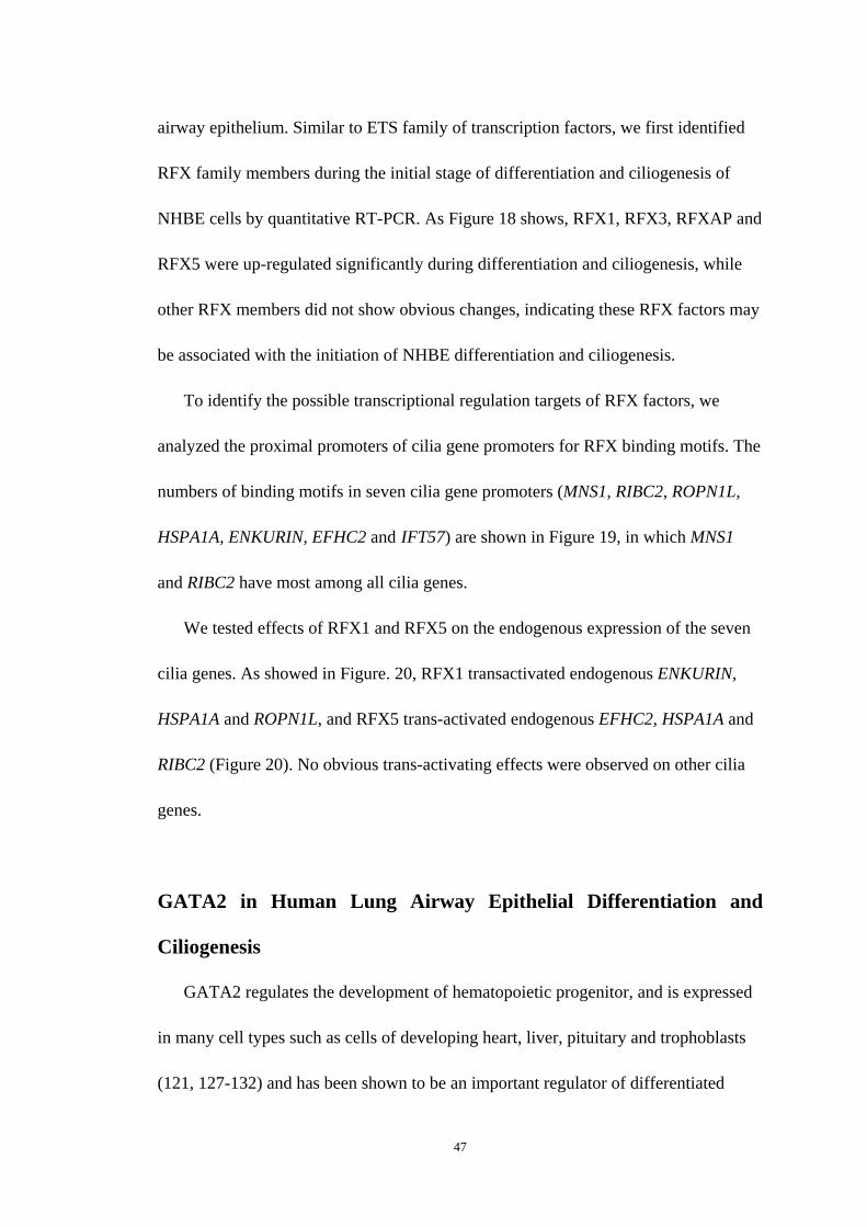
by GATA2. To further locate the cis-element(s) responsive to transactivation, GATA2
expression vector and reporter plasmids containing truncated promoter fragments of
ENKURIN and EFHC2 were cotransfected as aforementioned. Results showed that
GATA2 significantly trans-activated proximal ENKURIN (700bp and 400bp) and
EFHC2 (600bp) promoters, indicating the cis-element(s) responsive for
transactivation of GATA2 were located in the proximal promoter regions of the two
cilia genes (Figure 23).
51

0
2
4
6
8
10
12
14
16
EFHC2 ENKURIN
Lu
cife
rase
Act
ivit
y (F
old
Ch
ang
es)
CMV3
GATA2
Figure 21. GATA2 transactivates EFHC2 and ENKURIN promoters. Reporter
plasmids containing 1000bp proximal promoter and 5′ UTR of EFHC2 or ENKURIN
was co-transfected with GATA2 expression plasmid. Luciferase activity was
measured 24h after co-transfection. Reporter plasmid co-transfected with empty
CMV3 expression plasmids was set as control, whose activity was set as 1.
52

0
1
2
3
4
5
6
EFHC2 ENKURIN
Rel
ativ
e m
RN
A l
evel
(F
old
Ch
ang
es)
CMV3
GATA2
Figure 22. GATA2 transactivates endogenous EFHC2 and ENKURIN.
HBEC3KT cells were transfected with empty vector (CMV3) or GATA2 expression
vector respectively. The mRNA levels of EFHC2 or ENKURIN were quantified by
qRT-PCR. The mRNA levels in cells transfected with empty expression vector
(CMV3) was set as 1.
53
Figure 23. GATA2 transactivates proximal regions of ENKURIN and EFHC2
promoters. HBEC3KT cells was transfected with GATA2 expression vector and
reporter plasmids containing truncated distal 400bp, proximal 700bp, proximal 450bp
of ENKURIN promoter, as well as proximal 600bp of EFHC2 promoter. Luciferase
activities were measured 24 hours after transfection. Cells transfected with empty
CMV3 expression plasmid were set as value of one (1).
54
Chapter 4: Discussion
Ciliated cells in human airways are important components of host defense as part
of the mucociliary clearance mechanism. Ciliary defects are caused by an ever
growing recognition of genetic disorders and are often characterized by chronic or
recurrent infections of the respiratory tract. In addition, viral infections lead to
dysfunction of cilia which likely contributes in part to the overall presentation of lung
disease. The transcriptional regulation of human airway epithelial ciliogenesis is still
largely undefined. In this study, transcription factors that confer the coordinated
expression of genes that encode ciliary axoneme structural proteins in ciliogenesis are
identified and transcriptional targets of each transcription factor are defined.
FOXJ1 is a master regulator of motile cilia and is up-regulated dramatically
during differentiation of NHBE cells in vitro. Quantitative RT-PCR data showed that
the expression of several cilia genes (ENKURIN, EFHC2, IFT57, MNS1, RIBC2,
ROPN1L and HSPA1A) are markedly increased during differentiation of human
airway epithelia. We found that FOXJ1 transactivated the promoters of several cilia
genes in reporter gene assays, as well as transactivated the endogenous cilia genes of
human differentiated airway epithelial cells. Using studies of truncated promoters, the
cis-elements responsive for transactivation of FOXJ1 were located into the proximal
promoter regions of ENKURIN and EFHC2. Chromatin immunoprecipitation assays
provided further support that FOXJ1 binds an unknown cis-active element in
proximal promoter region of ENKURIN.
55

Although FOXJ1 endogenously transactivated the promoters of several cilia genes,
it failed to transactivate other cilia genes, such as MNS1 and IFT57. Previous studies
have indicated that overexpression of FOXJ1 alone does not confer ciliogenesis or
differentiation of unciliated lung epithelia. These findings suggest that other
transcription factors are also important in regulation of ciliogenesis of lung epithelial
cells. Through motif discovery, multiple binding sites for ETS, RFX and GATA
transcription factors were mapped in the promoters of a number of cilia genes,
indicating cilia genes are potential targets of these transcription factors.
The twenty-eight factors of ETS transcription family are involved in a variety of
mammalian developmental process at the cellular, tissue and organ level, including
hematopoiesis, immune function, lymph/angiogenesis, neurogenesis/neuromuscular
function, spermatogenesis, early embryonic patterning, and development of
extraembryonic tissues (138-149). All members of this transcription factor family
have the ETS DNA-binding domain which binds to sequences that contain a central
GGA motif. However, individual ETS domain protein can select nucleotides over an
11-base-pair sequences centered on this GGA motif. ETS transcription factors are
regulated, in part, by phosphorylation. Extracellular signals are transduced through a
series of mitogen-activated protein kinases (MAPKs) which result in activation of
downstream proteins such as ERK, JNK and p38, which in turn phosphorylate and
regulate the activity of ETS transcription factors (78). Although the overall functions
and roles of ETS family in lung epithelial differentiation is still unknown by now,
ETS family members have been shown to be involved in the transcriptional regulation
56

and differentiation of mammalian lung and its epithelium differentiation. For example,
aberrant expression of ELF5 disrupts lung branching and inhibits epithelial
differentiation (83); In adult mouse lung, ETV5 is expressed by alveolar epithelial
cells and regulates caveolin-1 transcription in mouse lung epithelial cell lines (84); In
human lung epithelial cells, ESE-1, -2, -3 have regulatory effects on expression of the
host defense genes lysozyme, chitinase and secretoglobins (87).
Many RFX factors are involved in cilia formations and functions in different
eukaryotes. For example, in C. elegans, the only known RFX factor to date regulates
daf-19 which is a key regulator of ciliogenesis (102). In zebra fish, rfx-2 is
specifically expressed in multiciliated cells of the pronephros and loss of its
expression results in cyst formation and loss of multicilia (114). Rfx2, foxj1 and
ift88 are recently shown to be regulated by FGF signaling which regulates cilia length
in diverse epithelial during embryo development (151). Most importantly, in mice,
RFX-3 functions in ciliogenesis are conserved. It controls the growth of mouse
embryonic node cilia, and loss of RFX-3 leads to significantly truncated but
functional node cilia (115). RFX-3 loss-of-function also results in hydrocephalus with
differentiation defects of ciliated ependymal cells of the choroid plexus and
subcommisural organ (116). Moreover, RFX-3 mutant mice show primary cilial
growth defects on islet cells and subsequent insulin secretion failure and impaired
glucose tolerance (117).
Although ETS and RFX families are associated with cilia formation in many cell
types and in many other simpler organisms, their roles in ciliogenesis of lung
57

epithelial cell subsets are still unknown and remain to be elucidated. We have
performed quantitative RT-PCR to determine the expression levels of all twenty eight
ETS factors and six RFX factors during the differentiation and ciliogenesis of NHBE
cells under ALI culture. Among the twenty-eight ETS family members, the
expression of nine (ETV1, ETV2, ETV6, ETV7, ESE1, ESE2, ESE3, SPDEF and
SPIC) were significantly increased during the differentiation, while ETV4 and ETV5
were decreased. ESE1, ETV1, SPIC and SPDEF were most significantly up-regulated
while ETV5 down-regulated most. RFX1 and RFX5 were the only RFX factors
dramatically up-regulated, RFX3 and RFXAP were also up-regulated to some extent,
while RFX2 and RFXANK slightly down-regulated. Based on these findings, we
focused our studies on the five ETS factors (ESE1, ETV1, SPIC, SPDEF, ETV5) and
the two RFX factors (RFX1 and RFX5) as putative transcriptional regulators in the
initiation of ciliogenesis. Motif discovery analysis of cilia gene promoters identified
ETS or RFX binding motifs in the proximal 1.2 kb region upstream of the first exon,
suggesting putative binding sites for these transcription factors. Among the 37 cilia
genes, promoters of MNS1, RIBC2 and ROPN1L have the most RFX binding motifs,
and promoters of ROPN1L, HSPA1A and ENKURIN had the most ETS binding motifs,
suggesting these gene promoters are most likely to have potential RFX and ETS
binding sites for transcription regulation. These five cilia genes as well as EFHC2 and
IFT57 were selected to assess the transactivation ability of ectopic ETS or RFX in
endogenous gene expression studies. Among the five ETS transcription factors (ETV1,
ESE1, SPIC, SPDEF, ETV5), ETV1 and ESE1 were shown to transactivate both
58

EFHC2 and ENKURIN expression, and SPIC repressed EFHC2 expression. None of
the ETS factors has obvious effects on the expression of ROPN1L, HSPA1A, MNS1
and IFT57. The transcriptional regulation of these ETS factors on the cilia genes may
be directly or indirectly, cooperative or independent, which requires further study.
Although HSPA1A and ROPN1L have most ETS binding motifs, these binding motifs
are not functional or they require other factors that are not yet apparent.
RFX1 and RFX5 overexpression was also assessed for the ability to regulate
endogenous cilia structural gene expression. ENKURIN, HSPA1A and ROPN1L were
transactivated by RFX1, and EFHC2, HSPA1A and RIBC2 by RFX5. IFT57 and
MNS1 were not regulated by either RFX1 or RFX5. Our findings provide the first
evidence for ETS- or RFX-mediated cilia gene induction in any metazoan system.
Our motif discovery also identified GATA elements in the promoters of ciliary
axoneme genes. Although GATA family members GATA4 and GATA6 are
involved in regulation of mammalian organogenesis by transactivating BMP-4 (150),
so far, GATA6 is the only known GATA factor important in lung-specific gene
expression and development (133-135). In this study, we investigate the effects of
GATA2 on cilia gene expressions. Over-expression of GATA2 in HBEC cells led to
the transactivation of ENKURIN and EFHC2, and this transactivation was located to
proximal promoter region of ENKURIN and EFHC2. Transactivation of endogenous
cilia genes in lung epithelial cells further supports GATA2 as a direct transactivator of
those cilia genes.
The genomic program conferring differentiation, development, or other complex
59

cell changes operates in part through the coordinated regulation of gene expression.
This program is executed by the recognitions of cis-regulatory DNAs by regulated
transcription factors and thus the control of specific gene expression. These regulatory
inputs and functional outputs of genes constitute a network-like architecture
commonly referred as transcriptional regulatory networks. Transcriptional regulatory
networks provide specific causal links between genomic expression patterns and
biological processes such as differentiation or development (152, 153). Many
transcription factors are expressed across multiple tissues and organs. For example, in
adult mammals, ETS family of transcription factors are widely expressed in a variety
of tissues including hematopoietic tissue, vasculature, brain and central nervous
system, mammary gland, endometrium, ovaries, testes, kidneys and lungs ( 140).
Responding to stimuli, these transcription factors turn on/off the transcription of the
appropriate genes which in turn allows changes of cell morphology or activity needed
for cell fate determination and cellular differentiation. Although most ETS-domain
proteins have been shown to be transcriptional activators, a considerable number have
been shown to be repressors, including ERF, YAN and TEL. For example, ETS factor
TEL1 has been shown to be a transcriptional activator and repressor, requiring
homo-dimerization or oligomerization (156). It also interacts with another ETS factor
Fli1, inhibiting Fli1 transactivation activity and thereby preventing the activation of
target genes (157,158). Therefore, the cellular specificity required for tissue specific
phenotypic of functional processes likely involves multiple, unique transcription
factor combinations to confer a coordinated gene expression network. Here we show
60

that multiple transcription factors from ETS (ETV1, ESE1, SPIC, SPDEF, ETV5),
RFX (RFX1, RFX5) and GATA (GATA2) family can regulate cilia gene expressions
together with FOXJ1 in ciliogenesis. A diagram illustrating the overlapping and
unique transcription factor-gene promoter pairs is depicted in Figure 24.
Besides transcriptional regulation, increasing evidence indicates that
post-transcriptional regulation plays a very important role in regulating steady-state
mRNA abundance or mRNA translation and, in turn, determining cell or tissue
specificity. After a gene is transcribed, the stability and distribution of the different
transcripts is regulated (post-transcriptional regulation) by means of RNA binding
proteins (RBP) that control the various steps and rates of the transcripts. Small RNA,
either encoded as polycistronic RNA elements expressed and processed into 21-24 bp
microRNAs (miRNAs), or small RNAs processed from the introns of expressed
protein-encoding genes is another type of post-transcriptional regulation of genomic
expression.
Much information regarding the expression of miRNA has been elucidated in
recent years. First, a primary miRNA transcript (pri-miRNA), which can be many
kilobases long and forms a stem-loop secondary structure, is transcribed from the
genome. This pri-miRNA is the recognized by a protein complex including the
enzyme Drosha in nucleus, and excised as a ~60nt hairpin called pre-miRNA. This
pre-miRNA is transported into cytoplasm and processed by an enzyme called Dicer,
leaving a 22nt double-stranded RNA duplex which is then taken up by a protein
complex RISC. Within RISC, the double strand RNA duplex will be separated, and a
61

guide miRNA strand will remain bound to RISC and be the element recognized by
target RNAs. Depending on the degree with which the guide miRNA strand matches
the target mRNA, the target RNA is inhibited through two different mechanisms. If
the match is imperfect, protein translation of that RNA will be inhibited by RISC
complex. On the other hand, if the match is perfect, the target RNA will be cleaved by
Aragonaute (Ago) protein which is a component of RISC complex. MiRNAs play key
roles in mammalian gene expression and cellular processes, especially differentiation
and development. In Dicer knock-out mice in which all miRNA productions are
inhibited, developing embryos die at an early embryonic stage and are depleted of
pluripotent stem cells (159),supporting miRNA is crucial in proper embryogenesis
and stem cell development. Conditional knockout of Dicer in a tissue or organ of
interests has demonstrated that miRNAs play roles in morphogenesis of several
organs including the lungs, limbs, and muscles, in T cell differentiation, and in skin
development (160-165). Considering the crucial roles of miRNAs in regulation of
gene expression and cellular differentiation, it is not surprising that misregulation of
miRNAs are linked with a variety of human diseases such as cancers. In cilia
dysfunction polycystic kidney disease (PKD), the functional patterns of predicted
miRNA targets and differentially expressed mRNAs are similar, suggesting an
important role of miRNAs in specific pathways underlying PKD (155). It is
noteworthy to mention that in mice within which Dicer is conditionally knocked out
in lung epithelium, normal branching is arrested and a few large epithelial pouches
appear in these mutant lungs. Significantly, these initial phenotypes are apparent
62
before an increase in epithelial cell death is observed, proposing that miRNAs play
specific roles in lung epithelial morphogenesis independent of its requirement in cell
survival (160). In the ciliogenesis of lung epithelial cells, some cilia genes may be
also regulated by post-transcriptional regulation, especially miRNA mechanism. In
the future study, techniques such as RNA immunoprecipitation on a chip can be
applied to investigate the expression patterns of predicted miRNAs targeting cilia
genes, and their role in regulating cilia gene expressions, which may shed lights on
elucidating the whole process of lung epithelial differentiation during development.
63

Figure 24. Transcriptional network in ciliogenesis: The transcriptional factors and
their cilia gene targets are connected. (arrow: transactivation; bar: inhibition)
64

Reference
1. Peter Satir, Soren Tvorup Chrsitensen. 2007. Overview of Structure and Function
of Mammalian Cilia. Annu. Rev. Physiol. 69:377-400
2. Wheatley DN. 1982. The Centriole: A Central Enigma of Cell Biology.
Amsterdam: Elsevier. 232 pp.
3. Fonte VG, Searls RL, Hilfer SR. 1971. The relationship of cilia with cell division
and differentiation. J. Cell Biol. 49:226–29
4. Schwartz EA, Leonard ML, Bizios R, Bowser SS. 1997. Analysis and modeling
of the primary cilium bending response to fluid shear. Am. J. Physiol.
272:F132–3
5. Praetorius HR, Spring KA. 2001. Bending the MDCK cell primary cilium
increases intracellular calcium. J. Membr. Biol. 184:71–79
6. Praetorius HR, Spring KA. 2003. Removal of the MDCK cell primary cilium
abolishes flow sensing. J. Membr. Biol. 191:69–76
7. Liu W, Murcia NS, Duan Y,Weinbaum S, Yoder BK, et al. 2005.
Mechanoregulation of intracellular Ca2+ concentration is attenuated in collecting
duct of monocilium-impaired orpk mice. Am. J. Physiol. Renal Physiol.
289:F978–88
8. Supp DM, Witte DP, Potter SS, Brueckner M. 1997. Mutation of an axonemal
dynein affects left-right asymmetry in inversus viscerum mice. Nature
389:963–96
65

9. Supp DM, Brueckner M, Kuehn MR,Witte DP, Lowe LA, et al. 1999. Targeted
deletion of theATPbinding domain of left-right dynein confirms its role in
specifying development of left-right asymmetries. Development 126:5495–504
10. Nonaka S, Shiratori H, Saijoh Y, Hamada H. 2002. Determination of left-right
patterning of the mouse embryo by artificial nodal flow. Nature 418:96–99
11. McGrath J, Somlo S, Makova S, Tian X, Brueckner M. 2003. Two populations of
node monocilia initiate left-right asymmetry in the mouse. Cell 114:61–73
12. Nakaya MA, Biris K, Tsukiyama T, Jaime S, Rawls JA, Yamaguchi TP. 2005.
Wnt3a links left-right determination with segmentation and anteroposterior axis
elongation. Development 132:5425–36
13. Tanaka Y, Okada Y, Hirokawa N. 2005. FGF-induced vesicular release of Sonic
hedgehog and retinoic acid in leftward nodal flow is critical for left-right
determination. Nature 435:172–77
14. Satir P. 1997. Cilia and related microtubular arrays in the eukaryotic cell. In
Handbook of Physiology, ed. JF Hoffman, JD Jamieson, pp. 787–817. New York:
Oxford Univ. Press
15. Okada Y,Takeda S,Tanaka Y, Belmonte JC, Hirokawa N. 2005. Mechanism of
nodal flow: a conserved symmetry breaking event in left-right axis determination.
Cell 121:633–44
16. Scholey, J.M. (2003) Intraflagellar transport. Annu. Rev. Cell Dev. Biol.19,
423–443
17. Ibanez-Tallon, I. et al. (2003) To beat or not to beat: roles of cilia in
66

development and disease. Hum. Mol. Genet. 12, 27–35
18. Cole, D.G. (2003) The intraflagellar transport machinery of
Chlamydomonas reinhardtii. Traffic 4, 435–442
19. Davenport, J.R. and Yoder, B.K. (2005) An incredible decade for the
primary cilium: a look at a once-forgotten organelle. Am. J. Physiol.
Renal Physiol. 289, 1159–1169
20. Pazour, G.J. and Witman, G.B. (2003) The vertebrate primary cilium is a sensory
organelle. Curr. Opin. Cell Biol. 15, 105–110
21. Pan, J. et al. (2005) Cilium-generated signaling and cilia-related
disorders. Lab. Invest. 85, 452–463
22. Afzelius, B.A. (2004) Cilia-related diseases. J. Pathol. 204, 470–477
23. Pazour, G.J. and Rosenbaum, J.L. (2002) Intraflagellar transport and
cilia-dependent diseases. Trends Cell Biol. 12, 551–555
24. Beales, P.L. (2005) Lifting the lid on Pandora’s box: the Bardet-Biedl
syndrome. Curr. Opin. Genet. Dev. 15, 315–323
25. Zhang, Q. et al. (2003) Loss of the Tg737 protein results in skeletal
patterning defects. Dev. Dyn. 227, 78–90
26. Ansley, S.J. et al. (2003) Basal body dysfunction is a likely cause of
pleiotropic Bardet–Biedl syndrome. Nature 425, 628–633
27. Hearn, T. et al. (2005) Subcellular localization of ALMS1 supports
involvement of centrosome and basal body dysfunction in the
pathogenesis of obesity, insulin resistance, and type 2 diabetes.
67

Diabetes 54, 1581–1587
28. Kulaga, H.M. et al. (2004) Loss of BBS proteins causes anosmia in
humans and defects in olfactory cilia structure and function in the
mouse. Nat. Genet. 36, 994–998
29. Nishimura, D.Y. et al. (2004) Bbs2-null mice have neurosensory
deficits, a defect in social dominance, and retinopathy associated
with mislocalization of rhodopsin. Proc. Natl. Acad. Sci. U. S. A.
101, 16588–16593
30. Nettesheim, P., J.S. Koo, and T. Gray. 2000. Regulation of differentiation of the
tracheobronchial epithelium. J. Aerosol. Med. 13:207–218.
31. Sleigh, M.A., J.R. Blake, and N. Liron. 1988. The propulsion
of mucus by cilia. Am. Rev. Respir. Dis. 137:726–741.
32. Knowles, M.R., and R.C. Boucher. 2002. Mucus clearance
as a primary innate defense mechanism for mammalian airways. J. Clin. Invest.
109:571–577.
33. Widdicombe, J.H. 2002. Regulation of the depth and composition of airway
surface liquid. J. Anat. 201: 313–318.
34. Schidlow, D.V. 1994. Primary ciliary dyskinesia (the immotile cilia syndrome).
Ann. Allergy 73:457–470.
35. Camner, P., B. Mossberg, and B.A. Afzelius. 1983. Measurements
of tracheobronchial clearance in patients with immotile-cilia syndrome and its
value in differential diagnosis. Eur. J. Respir. Dis. Suppl. 127:57–63.
68

36. W. Stannard, M.B. Ch.B. 2006. Ciliary Functions and the Role of Cilia in
Clearance. Journal of Aerosol Medicine. 19: 110-115
37. Afzelius, B.A., B. Mossberg, and S. Bergstrom. 2001. Immotile cilia syndrome
(primary ciliary dyskinesia), including Kartagener syndrome. In The metabolic
and molecular bases of inherited disease. C.R. Scriver, A.L. Beaudet, W.S. Sly,
and D. Valle, eds. New York: McGraw Hill, 4817–4827.
38. Pedersen, M., Y. Sakakura, B. Winther, et al. 1983. Nasal mucociliary transport,
number of ciliated cells, and beating pattern in naturally acquired common colds.
Eur. J. Respir. Dis. Suppl. 128:355–365.
39. Wilson, R., E. Alton, A. Rutman, et al. 1987. Upper respiratory tract viral
infection and mucociliary clearance. Eur. J. Respir. Dis. 70:272–279.
40. Chilvers, M.A., M. McKean, A. Rutman, et al. 2001. The effects of coronavirus
on human nasal ciliated respiratory epithelium. Eur. Respir. J. 18:965–970.
41. Carson, J.L., A.M. Collier, and S.S. Hu. 1985. Acquired ciliary defects in nasal
epithelium of children with acute viral upper respiratory infections. N. Engl. J.
Med. 312:463–468.
42. Rautiainen, M., J. Nuutinen, H. Kiukaanniemi, et al. 1992. Ultrastructural changes
in human nasal cilia caused by the common cold and recovery of ciliated
epithelium. Ann. Otol. Rhinol. Laryngol. 101:982–987.
43. Thompson C, Barclay W, Zambon M, et al. 2006. Infection of Human Airway
Epithelium by Human and Avian Strains of Influenza A Virus. Journal of
Virology. 80: 8060-8068
69
44. D. Look, M. Walter, M. Williamson, et al. 2001. Effects of Paramyxoviral
Infection on Airway Epithelial Cell Foxj1 Expression, Ciliogenesis, and
Mucociliary Function. American Journal of Pathology. 159: 2055-2069.
45. J. Nicholls, L. Poon, K. Lee, et al. 2003. Lung pathology of fatal severe acute
respiratory syndrome. Lancet. 361: 1773–78.
46. S. Philippou · P. Otto · P. Reinhold, et al. 1999. Respiratory syncytial
virus-induced chronic bronchiolitis in experimentally infected calves.
Virchows Arch. 436:617–621
50. Carson JL, Collier AM, Hu DC. 1980. Ultrastructural observations on
cellular and subcellular aspects of experimental Mycoplasma pneumoniae disease.
Infect Immun. 29: 11 117–11 124.
51. Muse K. 1980. Host cell membrane perturbations during experimental
Bordetella pertussis infection. Proc 7th Eur Conf El Micr Haag. 2: 432–433.
52. Bemis DA, Wilson SA.1985. Influence of potential virulence determinants on
Bordetella bronchiseptica induces ciliostasis. Infect
Immun. 50: 3–42.
54. Reimer A, Klementsson K, Ursing J, Wretlind B. 1980. The mucociliary
activity of the respiratory tract. Acta Otolaryngol. 90:462–469.
55. Rautiainen M, Nuutinen J, Kiukaanniemi H, Collan Y. 1992. Ultrastructural
changes in human nasal cilia caused by the common cold and recovery of
ciliated epithelium. Ann Otol Rhinol Laryngol . 101: 982–987.
56. Afzelius BA. 1994. Ultrastructure of human nasal epithelium during an episode
70

of coronavirus infection. Virchows Arch. 424: 295–300.
57. Bernacki SH, Nelson AL, Abdullah L, Sheehan JK, Harris A, Davis
CW, Randell SH. 1999. Mucin gene expression during differentiation of human
airway epithelia in vitro:Muc4 and muc5b are strongly induced. Am J Respir
Cell Mol Biol. 20:595–604.
58. Andrea J. Ross, Lisa A Dailey, Luisa E Brighton, etc. 2007. Transcriptional
Profiling of Mucociliary Differentiation in Human Airway Epithelial Cells. Am J
Respir Cell Mol Biol. 37: 169-185.
59. Ostrowski LE, Blackburn K, Radde KM, Moyer MB, Schlatzer DM, Moseley A,
Boucher RC. (2002). A proteomic analysis of human cilia: Identification of
novel components. Mol Cell Proteomics.1:451–465.
60. Sutton KA, Jungnickel MK, Wang Y, Cullen K, Lambert S, Florman HM. 2004.
Enkurin is a novel calmodulin and trpc channel binding protein in sperm. Dev
Biol. 274:426–435.
61. Gaillard DA, Lallement AV, Petit AF, and Puchelle ES. 1989. In vivo
ciliogenesis in human fetal tracheal epithelium. Am J Anat 185: 415–428.
62. Dirksen ER. 1991. Centriole and basal body formation during ciliogenesis
revisted. Biol Cell 72: 31–38.
63. Sorokin SP. 1968. Reconstructions of centriole formation and ciliogenesis in
mammalian lungs. J Cell Sci 3: 207–230.
64. Pazour GJ, Dickert BL, Vucica Y, Seeley ES, Rosenbaum JL, Witman GB, and
Cole DG. 2000. Chlamydomonas IFT88 and its mouse homologue, polycystic
71

kidney disease gene tg737, are required for assembly of cilia and flagella. J Cell
Biol 151: 709–718.
65. Blatt EN, Yan XH, Wuerffel MK, Hamilos DL, and Brody SL. 1999. Forkhead
transcription factor HFH-4 expression is temporally related to ciliogenesis. Am J
Respir Cell Mol Biol 21: 168–176.
66. Brody SL, Yan XH, Wuerffel MK, Song SK, and Shapiro SD. 2000.
Ciliogenesis and left-right axis defects in forkhead factor HFH-4-null mice. Am J
Respir Cell Mol Biol 23: 45–51.
67. Hackett BP, Brody SL, Liang M, Zeitz ID, Bruns LA, and Gitlin JD. 1995.
Primary structure of hepatocyte nuclear factor/forkhead homologue 4 and
characterization of gene expression in the developing respiratory and
reproductive epithelium. Proc Natl Acad Sci USA 92: 4249–4253.
68. Lim L, Zhou H, and Costa RH. 1997. The winged helix transcription factor
HFH-4 is expressed during choroid plexus epithelial development in the mouse
embryo. Proc Natl Acad Sci USA 94: 3094–3099.
69. Look DC, Walter MJ, Williamson MR, Pang L, You Y, Sreshta JN,
Johnson JE, Zander DS, and Brody SL. 2001. Effects of paramyxoviral
infection on airway epithelial cell Foxj1 expression, ciliogenesis, and
mucociliary function. Am J Pathol 159: 2055–2069.
70. Chen J, Knowles HJ, Hebert JL, and Hackett BP. 1998. Mutation of the
mouse hepatocyte nuclear factor/forkhead homologue 4 gene results in an
absence of cilia and random left-right asymmetry. J Clin Invest 102:1077–1082.
72

71.Yingjian Y, Tao H, Edward J R, Steven L B. 2004. Role of f-box factor foxj1 in
differentiation of ciliated airway epithelial cells. Am J Respir Cell Mol Biol 286:
650-657.
72. Brigitte N G, Xiulan G, Brian H. 2004. Foxj1 regulates basal body anchoring to
the cytoskeleton of ciliated pulmonary epithelial cells. J Cell Sci 117:
1329-1337
73. Tichelaar, J. W., S. E. Wert, R. H. Costa, S. Kimura, and J. A. Whitsett. 1999.
HNF-3/forkhead homologue-4 (HFH-4) is expressed in ciliated epithelial cells
in the developing mouse lung. J. Histochem. Cytochem.47:823–831.
74. Tristam DA, Hicks WJ, Hard R. 1998. Respiratory syncytial virus and human
bronchial epithelium. Arch Otolaryngol Head Neck Surg. 124:777–783.
75. Maiti AK, Bartoloni L, Mitchison HM, Meeks M, Chung E, Spiden S, Gehrig
C, Rossier C, DeLozier-Blanchet CD, Blouin J, et al. 2000. No deleterious
mutations in the Foxj1 (alias HFH-4) gene in patients with primary ciliary
dyskinesia (PCD). Cytogenet Cell Genet. 90:119–122.
76. Shore, P. et al. 1996. Determinants of DNA-binding specificity of
ETS-domain transcription factors. Mol. Cell. Biol. 16, 3338–3349 .
77. Fitzsimmons, D. et al. 1996. Pax-5 (BSAP) recruites Ets protooncogene
family proteins to form functional ternary complexes on a B-cell-specific
promoter. Genes Dev. 10, 2198–2211.
78. Sharrocks AD. 2001. The ETS-domain transcription factor family. Nat Rev
Mol Cell Biol 2 (11): 827–37.
73
79. Verger A, Duterque-Coquillaud M. 2002. When Ets transcription factors meet
their partners. Bioessays 24 (4): 362–70.
80. Gutierrez-Hartman A, Duval DL, Bradford AP. 2007. ETS transcription factors
in endocrine systems. Trends Endocrinol Metab 18 (4): 150–8.
81. Ida K, Kobayashi S, Taki T, Hanada R, Bessho F, Yamamori S, Sugimoto T,
Ohki M, Hayashi Y. 1995. EWS-FLI-1 and EWS-ERG chimeric mRNAs in
Ewing's sarcoma and primitive neuroectodermal tumor. Int J Cancer 63 (4):
500–4.
82. Peeters P, Raynaud SD, Cools J, Wlodarska I, Grosgeorge J, Philip P,
Monpoux F, Van Rompaey L, Baens M, Van den Berghe H, Marynen P. 1997.
Fusion of TEL, the ETS-variant gene 6 (ETV6), to the receptor-associated
kinase JAK2 as a result of t(9;12) in a lymphoid and t(9;15;12) in a myeloid
leukemia. Blood 90 (7): 2535–40.
83. Metzger DE, Stahlman MT, Shannon JM. 2008. Misexpression of ELF5
disrupts lung branching and inhibits epithelial differentiation. Dev Biol.
1;320(1):149-60.
84. Kathuria H, Cao Y, Hinds A, Ramirez MI, Williams MC.2007. ERM is
expressed by alveolar epithelial cells in adult mouse lung and regulates
caveolin-1 transcription in mouse lung epithelial cell lines. J Cell Biochem.
102(1):13-27.
85. Kola,I. et al. 1993. The Ets1 transcription factor is widely expressed during
74

murine embryo development and is associated with mesodermal cells involved
in morphogenetic processes such as organ formation. Proc. Natl Acad. Sci.
USA, 90, 7588–7592.
86. Kathuria,H. et al. 2004. Transcription of the caveolin-1 gene is differentially
regulated in lung type I epithelial and endothelial cell lines: a role for ETS
proteins in epithelial cell expression. J. Biol. Chem. 279, 30028–30036.
87. W. Lei, K. Harrod. et al. 2007. Transactivation of lung lysozyme expression by
Ets family member ESE-1. Am J Physiol Lung Cell Mol Physiol 293:
1359–1368.
88. Martinez. MJ, Smith .AD, Harrod. KS. 2007. Computational prediction of
novel components of lung transcriptional networks. Bioinformatics. 23(1): 21-9.
89. Furukawa K, Hotta Y. et al. 1994. cDNA cloning and functional
characterization of a meiosis-specific protein (MNS-1) with apparent nuclear
association. Chrosome Res. 2(2): 99-113.
90. Furukawa K, Hotta Y. et al. 1995. Meiosis specific transcription and functional
proteins. Adv Biophys. 31: 101-15
91. “Entrez Gene: HSPA1A heat shock 70kDa protein 1A.”
92. “Entrez Gene: ROPN1L ropporin 1-like.”
93. Wu, S. Y., and McLeod, M. 1995. The sak1+ gene of Schizosaccharomyces
pombe encodes an RFX family DNA-binding protein that positively regulates
cyclic AMP-dependent protein kinase-mediated exit from the mitotic cell cycle.
75
Mol. Cell. Biol 15, 1479–1488.
94. Huang, M., Zhou, Z., and Elledge, S. J. 1998. The DNA replication and damage
checkpoint pathways induce transcription by inhibition of the Crt1 repressor.
Cell 94, 595–605.
95. Otsuki, K., Hayashi, Y., Kato, M., Yoshida, H., and Yamaguchi, M. 2004.
Characterization of dRFX2, a novel RFX family protein in Drosophila. Nucleic
Acids Res 32, 5636–5648.
96. Ma, K., Zheng, S., and Zuo, Z. 2006. The transcription factor regulatory factor
X1 increases the expression of neuronal glutamate transporter type 3. J. Biol.
Chem 281, 21250–21255.
97. Zhang, D., Stumpo, D. J., Graves, J. P., DeGraff, L. M., Grissom, S. F., Collins,
J. B., Li, L., Zeldin, D. C., and Blackshear, P. J. 2006. Identification of potential
target genes for RFX4_v3, a transcription factor critical for brain development.
J. Neurochem 98, 860–875.
98. Senti G, Swoboda P. 2008. Distinct isoforms of the RFX Transcription factor
DAF-19 regulate ciliogenesis and maintenance of synaptic activity. Mol Biol of
the Cell. 19 (12): 5517-28.
99. Zhang, D., Stumpo, D. J., Graves, J. P., DeGraff, L. M., Grissom, S. F., Collins,
J. B., Li, L., Zeldin, D. C., and Blackshear, P. J. 2006. Identification of potential
target genes for RFX4_v3, a transcription factor critical for brain development.
J. Neurochem 98, 860–875.
100. Gajiwala KS, Chen H, Cornille F, Roques BP, Reith W, Mach B, Burley SK.
76
2000. Structure of the winged-helix protein hRFX1 reveals a new mode of
DNA binding. Nature. 403:916-921.
101. Emery P, Durand B, Mach B, Reith W. 1996. RFX proteins, a novel family of
DNA binding proteins conserved in the eukaryotic
kingdom. Nucleic Acids Res. 24:803-807.
102. Swoboda P, Adler HT, Thomas JH. 2000. The RFX-type transcription factor
DAF-19 regulates sensory neuron cilium formation in C. elegans. Mol Cell.
5:411-421.
103. Reith W, Barras E, Satola S, Kobr M, Reinhart D, Sanchez CH, Mach B. 1989.
Cloning of the major histocompatibility complex class II promoter binding
protein affected in a hereditary defect in class II gene regulation. Proc Natl
Acad Sci U S A. 86(11):4200-4204.
104. Siegrist CA, Durand B, Emery P, David E, Hearing P, Mach B, Reith W. 1993.
RFX1 is identical to enhancer factor C and functions as a transactivator of the
hepatitis B virus enhancer. Mol Cell Biol. 13:6375-6384.
105. Ma K, Zheng S, Zuo Z. 2006. The transcription factor regulatory factor X1
increases the expression of neuronal glutamate transporter type 3. J Biol Chem.
281(30):21250-21255.
106. Wolfe SA, van Wert J, Grimes SR. 2006. Transcription factor RFX2 is
abundant in rat testis and enriched in nuclei of primary spermatocytes where it
appears to be required for transcription of the testis-specific histone H1t gene. J
Cell Biochem. 99(3):735-746.
77

107. Morotomi-Yano K, Yano K, Saito H, Sun Z, Iwama A, Miki Y. 2002. Human
regulatory factor X 4 (RFX4) is a testis-specific dimeric DNA-binding protein
that cooperates with other human RFX members. J Biol Chem. 277(1):836-842.
108. Morotomi-Yano K, Yano K, Saito H, Sun Z, Iwama A, Miki Y. 2002. Human
regulatory factor X 4 (RFX4) is a testis-specific dimeric DNA-binding protein
that cooperates with other human RFX members. J Biol Chem. 277(1):836-842.
109. Blackshear PJ, Graves JP, Stumpo DJ, Cobos I, Rubenstein JL, Zeldin DC.
2003. Graded phenotypic response to partial and complete deficiency of a
brain-specific transcript variant of the winged helix transcription factor RFX4.
Development. 130(19):4539-4552.
110. Ait-Lounis A, Baas D, Barras E, Benadiba C, Charollais A, Nlend
Nlend R, Liegeois D, Meda P, Durand B, Reith W. 2007. Novel function of
the ciliogenic transcription factor RFX3 in development of the endocrine
pancreas. Diabetes. 56(4):950-959.
111. Reith W, Mach B. 2001. The bare lymphocyte syndrome and the regulation of
MHC expression. Annu Rev Immunol. 19:331-373.
112. Blackshear PJ, Graves JP, Stumpo DJ, Cobos I, Rubenstein JL, Zeldin DC.
2003.Graded phenotypic response to partial and complete deficiency of a
brain-specific transcript variant of the winged helix transcription factor RFX4.
Development. 130:4539-4552.
113. Araki R, Takahashi H, Fukumura R, Sun F, Umeda N, Sujino M, Inouye ST,
78

Saito T, Abe M. 2004. Restricted expression and photic induction of a novel
mouse regulatory factor X4 transcript in the suprachiasmatic nucleus. J Biol
Chem. 279:10237-10242.
114. Liu Y, Pathak N, Kramer-Zucker A, Drummond IA. 2007. Notch signaling
controls the differentiation of transporting epithelia and
multiciliated cells in the zebrafish pronephros. Development. 134:1111-1122.
115. Bonnafe E, Touka M, AitLounis A, Baas D, Barras E, Ucla C, Moreau A,
Flamant F, Dubruille R, Couble P, et al.2004. The transcription factor RFX3
directs nodal cilium development and left-right asymmetry specification. Mol
Cell Biol. 24:4417-4427.
116. Baas D, Meiniel A, Benadiba C, Bonnafe E, Meiniel O, Reith W,
Durand B. 2006. A deficiency in RFX3 causes hydrocephalus associated with
abnormal differentiation of ependymal cells. Eur J Neurosci. 24:1020-1030.
117. Ait-Lounis A, Baas D, Barras E, Benadiba C, Charollais A, Nlend
Nlend R, Liegeois D, Meda P, Durand B, Reith W. 2007. Novel function of
the ciliogenic transcription factor RFX3 in development of the endocrine
pancreas. Diabetes. 56:950-959.
118. Bresnick, E. H., Martowicz, M. L., Pal, S., and Johnson, K. D. 2005.
Developmental control via GATA factor interplay at chromatin domains. J. Cell.
Physiol. 205, 1-9
119. Molkentin, J. D. 2000. The zinc finger-containing transcription factors GATA-4,
-5, and -6. Ubiquitously expressed regulators of tissue-specific gene
79

expression.J. Biol. Chem. 275, 38949-38952
120. Pevny, L., Simon, M. C., Robertson, E., Klein, W. H., Tsai, S. F., D'Agati, V.,
Orkin, S. H., and Costantini, F. 1991. Erythroid differentiation in chimaeric
mice blocked by a targeted mutation in the gene for transcription factor
GATA-1. Nature 349, 257-260
121. Tsai, F. Y., Keller, G., Kuo, F. C., Weiss, M., Chen, J., Rosenblatt, M., Alt, F.
W., and Orkin, S.H. 1994. An early haematopoietic defect in mice lacking the
transcription factor GATA-2. Nature 371, 221-226
122. Pandolfi, P. P., Roth, M. E., Karis, A., Leonard, M. W., Dzierzak, E., Grosveld,
F. G., Engel, J.D., and Lindenbaum, M. H. 1995. Targeted disruption of the
GATA3 gene causes severe abnormalities in the nervous system and in fetal
liver haematopoiesis. Nat. Genet. 11, 40-44
123. Molkentin, J. D., Lin, Q., Duncan, S. A., and Olson, E. N. 1997. Requirement
of the transcription factor GATA4 for heart tube formation and ventral
morphogenesis. Genes Dev. 11, 1061-1072
124. Molkentin, J. D., Tymitz, K. M., Richardson, J. A., and Olson, E. N. 2000.
Abnormalities of the genitourinary tract in female mice lacking GATA5. Mol.
Cell. Biol. 20, 5256-5260
125. Morrisey, E. E., Tang, Z., Sigrist, K., Lu, M. M., Jiang, F., Ip, H. S., and
Parmacek, M. S. 1998. GATA6 regulates HNF4 and is required for
differentiation of visceral endoderm in the mouse embryo. Genes Dev. 12,
80

3579-3590
126. Ng, Y. K., George, K. M., Engel, J. D., and Linzer, D. I. 1994. GATA factor
activity is required for the trophoblast-specific transcriptional regulation of the
mouse placental lactogen I gene. Development 120, 3257-3266
127. Yamamoto, M., Ko, L. J., Leonard, M. W., Beug, H., Orkin, S. H., and Engel, J.
D. 1990. Activity and tissue-specific expression of the transcription factor
NF-E1 multigene family. Genes Dev. 4, 1650-1662
128. Craven, S. E., Lim, K. C., Ye, W., Engel, J. D., de Sauvage, F., and Rosenthal,
A. 2004. Gata2 specifies serotonergic neurons downstream of sonic hedgehog.
Development 131, 1165-1173.
129. Minegishi, N., Ohta, J., Yamagiwa, H., Suzuki, N., Kawauchi, S., Zhou, Y.,
Takahashi, S., Hayashi, N., Engel, J. D., and Yamamoto, M. 1999. The mouse
GATA-2 gene is expressed in the para-aortic splanchnopleura and aorta-gonads
and mesonephros region. Blood 93, 4196-4207
130. Nardelli, J., Thiesson, D., Fujiwara, Y., Tsai, F. Y., and Orkin, S. H. 1999.
Expression and genetic interaction of transcription factors GATA-2 and
GATA-3 during development of the mouse central nervous system. Dev. Biol.
210, 305-321
131. Orlic, D., Anderson, S., Biesecker, L. G., Sorrentino, B. P., and Bodine, D. M.
1995. Pluripotent hematopoietic stem cells contain high levels of mRNA for
c-kit, GATA-2, p45 NF-E2, and c-myb and low levels or no mRNA for c-fms
and the receptors for granulocyte colony-stimulating factor and interleukins 5
81

and 7. Proc. Natl. Acad. Sci. U S A 92, 4601-4605
132. Steger, D. J., Hecht, J. H., and Mellon, P. L. 1994. GATA-binding proteins
regulate the human gonadotropin alpha-subunit gene in the placenta and
pituitary gland. Mol. Cell. Biol. 14, 5592-5602
133. Bruno,M.D. et al. 2000. GATA-6 activates transcription of surfactant protein A.
J. Biol. Chem. 275, 1043–1009.
134. Yang,H. et al. 2002. GATA6 regulates differentiation of distal lung epithelium.
Development. 129, 2233–2246.
135. Liu,C. et al. 2003. Inhibition of alveolarization and altered pulmonary
mechanics in mice expressing GATA-6. Am. J. Physiol. Lung Cell. Mol.
Physiol. 285. L1246–L1254.
136. Perl,A.K. et al. 2002. Conditional gene expression in the respiratory epithelium
of the mouse. Transgenic Res. 11, 21–29.
137. Ramirez RD, Sheridan S, Girard L, et al. 2004. Immortalization of human
bronchial epithelial cells in the absence of viral oncoproteins. Cancer Res.
64:9027–34.
138. Jedlicka P, Sui X, Sussel L, Gutierrez-Hartmann A. 2009. Ets Transcription
Factors Control Epithelial Maturation and Transit and Crypt-Villus
Morphogenesis in the Mammalian Intestine. Am J Pathol. 174.4
139. Oikawa T, Yamada T. 2003. Molecular biology of the Ets family of transcription
factors. Gene. 303:11–34
140. Maroulakou IG, Bowe DB. 2000. Expression and function of Ets transcription
82

factors in mammalian development: a regulatory network.
Oncogene.19:6432–6442
141.Chen C, Ouyang W, Grigura V, Zhou Q, Carnes K, Lim H, Zhao GQ, Arber S,
Kurpios N, Murphy TL, Cheng AM, Hassell JA, Chandrashekar V, Hofmann
MC, Hess RA, Murphy KM. 2005. ERM is required for transcriptional control of
the spermatogonial stem cell niche. Nature. 436:1030–1034
142. Donnison M, Beaton A, Davey HW, Broadhurst R, L’Huillier P, Pfeffer PL.
2005.Loss of the extraembryonic ectoderm in Elf5 mutants leads to defects in
embryonic patterning. Development, 132:2299–2308
143. Georgiades P, Rossant J. 2006. Ets2 is necessary in trophoblast for normal
embryonic anteroposterior axis development. Development,
133:1059–1068
144. Hippenmeyer S, Huber RM, Ladle DR, Murphy K, Arber S. 2007. ETS
transcription factor Erm controls subsynaptic gene expression in skeletal muscles.
Neuron, 55:726–740
145. Livet J, Sigrist M, Stroebel S, De Paola V, Price SR, Henderson CE,
Jessell TM, Arber S. 2002. ETS gene Pea3 controls the central position and
terminal arborization of specific motor neuron pools. Neuron, 35:877–892
146. O’Leary DA, Noakes PG, Lavidis NA, Kola I, Hertzog PJ, Ristevski S.
2007.Targeting of the ETS factor GABPalpha disrupts neuromuscular junction
synaptic function. Mol Cell Biol, 27:3470–3480
147. Ristevski S, O’Leary DA, Thornell AP, Owen MJ, Kola I, Hertzog PJ. 2004. The
83

ETS transcription factor GABPalpha is essential for early embryogenesis. Mol
Cell Biol, 24:5844–5849
148. Rosmarin AG, Resendes KK, Yang Z, McMillan JN, Fleming SL. 2004.
GA-binding protein transcription factor: a review of GABP as an integrator of
intracellular signaling and protein-protein interactions. Blood Cells Mol Dis,
32:143–154
149. Zhou J, Chehab R, Tkalcevic J, Naylor MJ, Harris J, Wilson TJ, Tsao S, Tellis I,
Zavarsek S, Xu D, Lapinskas EJ, Visvader J, Lindeman GJ, Thomas R, Ormandy
CJ, Hertzog PJ, Kola I, Pritchard MA. 2005. Elf5 is essential for early
embryogenesis and mammary gland development during pregnancy and lactation.
EMBO J, 24:635–644
150. Nemer G, Nemer M. 2003. Transcriptional activation of BMP-4 and regulation
of mammalian organogenesis by GATA-4 and -6. Dev Biol. 254(1):131-48
151. Neugebauer JM, Amack JD, Peterson AG, Bisgrove BW, Yost HJ. 2009. FGF
signalling during embryo development regulates cilia length in diverse epithelia.
Nature. 10.1038.
152. Levine M, Davidson EH. 2005. Gene regulatory networks for development. Proc
Natl Acad Sci USA 102:4936–4942.
153. Oliveri P, Tu Q, Davidson EH. 2008. Global regulatory
logic for specification of an embryonic lineage. Proc Natl Acad Sci USA
105:5955–5962.
154. Whitsett,J.A. 2002. Intrinsic and innate defenses in the lung: intersection of
84
pathways regulating lung morphogenesis, host defense, and repair. J. Clin. Invest.
109, 565–569.
155. Pandev P, Brors B, Srivastava PK, etc. 2008. Microarray-based approach
identifies microRNAs and their target functional patterns in polycystic kidney
disease. BMC Genomics. 9: 624.
156. Lopez RG, Carron C, Oury C, Gardellin P, Bernard O and Ghysdael J. 1999.
TEL is a sequence-specific transcriptional repressor. J Biol Chem 274 (42):
30132-8
157. Bassuk AG and Leiden JM. 1995. A direct physical association between ETS and
AP-1 transcription factors in normal human T cells. Immunity 3(2):223-37
158. Kwiatkowski BA, Bastian LS, Bauer Jr TR, Tsai S, Zielinska-Kwiatkowska AG
and Hickstein DD. 1998. The ets family member Tel binds to the Fli-1
oncoprotein and inhibits its transcriptional activity. J Biol Chem 273 (28):
17525-30
159. Bernstein E, Kim SY, Carmell MA, Murchison EP, Alcorn H, Li
MZ, et al. 2003. Dicer is essential for mouse development. Nat Genet 35:215-7.
160. Harris KS, Zhang Z, McManus MT, Harfe BD, Sun X. 2006. Dicer
function is essential for lung epithelium morphogenesis. Proc
Natl Acad Sci USA 103:2208-13.
161. Harfe BD, McManus MT, Mansfield JH, Hornstein E, Tabin CJ.2005. The
RNaseIII enzyme Dicer is required for morphogenesis but not patterning of the
vertebrate limb. Proc Natl Acad Sci USA;102:10898-903.
85

86
162. O’Rourke JR, Georges SA, Seay HR, Tapscott SJ, McManus MT,
Goldhamer DJ, et al. 2007. Essential role for Dicer during skeletal
muscle development. Dev Biol;311:359-68.
163. Muljo SA, Ansel KM, Kanellopoulou C, Livingston DM, Rao A,
Rajewsky K. 20005.Aberrant T cell differentiation in the absence of
Dicer. J Exp Med;202:261-9.
164. Andl T, Murchison EP, Liu F, Zhang Y, Yunta-Gonzalez M, Tobias JW, et al.
2006. The miRNA-processing enzyme dicer is essential for the morphogenesis
and maintenance of hair follicles. Curr Biol;16:1041-9.
165. Yi R, O’Carroll D, Pasolli HA, Zhang Z, Dietrich FS, Tarakhovsky
A, et al. 2006. Morphogenesis in skin is governed by discrete sets of
differentially expressed microRNAs. Nat Genet;38: 356-62.