Embed Size (px)
Citation preview

2014
http://informahealthcare.com/bmgISSN: 1040-9238 (print), 1549-7798 (electronic)
Editor: Michael M. CoxCrit Rev Biochem Mol Biol, 2014; 49(2): 164–177
! 2014 Informa Healthcare USA, Inc. DOI: 10.3109/10409238.2014.887051
REVIEW ARTICLE
Transcriptome-wide studies uncover the diversity of modes of mRNArecruitment to eukaryotic ribosomes
Ivan N. Shatsky1, Sergey E. Dmitriev1,2, Dmitri E. Andreev1, and Ilya M. Terenin1,2
1Belozersky Institute of Physico-Chemical Biology, Lomonosov Moscow State University, Moscow, Russia and 2Engelhardt Institute of Molecular
Biology, Russian Academy of Sciences, Moscow, Russia
Abstract
The conventional paradigm of translation initiation in eukaryotes states that the cap-bindingprotein complex eIF4F (consisting of eIF4E, eIF4G and eIF4A) plays a central role in therecruitment of capped mRNAs to ribosomes. However, a growing body of evidence indicatesthat this paradigm should be revised. This review summarizes the data which have been mostlyaccumulated in a post-genomic era owing to revolutionary techniques of transcriptome-wideanalysis. Unexpectedly, these techniques have uncovered remarkable diversity in therecruitment of cellular mRNAs to eukaryotic ribosomes. These data enable a preliminaryclassification of mRNAs into several groups based on their requirement for particularcomponents of eIF4F. They challenge the widely accepted concept which relateseIF4E-dependence to the extent of secondary structure in the 50 untranslated regions ofmRNAs. Moreover, some mRNA species presumably recruit ribosomes to their 50 ends withoutthe involvement of either the 50 m7G-cap or eIF4F but instead utilize eIF4G or eIF4G-likeauxiliary factors. The long-standing concept of internal ribosome entry site (IRES)-elements incellular mRNAs is also discussed.
Keywords
50 UTR secondary structure, cap-independenttranslation enhancer, cellular IRESs, DAP5,eIF4F, eIF4G, ribosome profiling,translation initiation factor eIF4E
History
Received 4 November 2013Revised 17 January 2014Accepted 21 January 2014Published online 13 February 2014
Introduction
Translational control mainly operates at the initiation step of
polypeptide synthesis. The basis of translation initiation in
eukaryotes was elucidated in the 1970 and 80s, starting with
the discovery of the mRNA scanning mechanism by Kozak
(1989). This important event occurred in a ‘‘pre-genomic
era’’ when the set of available model mRNAs was extremely
limited and was not representative of the entire diversity of
mRNA structures. These ‘‘single mRNA’’ studies resulted in
a widely recognized model of cap-dependent translation
initiation which is believed to be used by the overwhelming
majority of cellular mRNAs in all eukaryotes. Somewhat later
(in the 1990s), an alternative mechanism – internal initiation
of translation – was discovered (Jang et al., 1988; Pelletier &
Sonenberg, 1988) and dissected (Pestova et al., 1996a,b,
1998) for uncapped genomic RNAs from several viruses
replicating in the cytoplasm. According to this mechanism,
the ribosome recognizes the mRNA at a specific internal
ribosome entry site (IRES) within its 50 untranslated region
(50 UTR). Whether this alternative mechanism of ribosome
recruitment to mRNA is also used by cellular mRNAs, in
particular by mammalian mRNAs, is still a matter of debate
(see below).
With the beginning of the transcriptome-wide era
(a discussion of respective techniques can be found in
Kapeli & Yeo, 2012), we now have the ability to analyze
large numbers of structurally diverse mRNAs while manip-
ulating specific key components of the cell or signaling
pathways. Some of the emerging data cannot be explained on
the basis of either the standard model of mRNA recruitment
to ribosomes or with the help of IRES-elements. These data
suggest that something very important has been missed in the
past, presumably since a large variety of mRNA structures
was not taken into account. Without filling these gaps in our
ideas on the molecular mechanisms of recruitment of various
mRNAs to ribosomes, we cannot advance our studies of the
mechanisms of translational control.
The aim of this review is to share our observations while
mining the literature of the last decade. We decided to focus
on the very first events of mRNA recruitment onto ribosomes
(40S subunits) and will not discuss the subsequent steps of
translation initiation (such as ribosomal scanning, start codon
selection or subunit joining) since these issues have been
covered in a number of comprehensive recent reviews
(Alekhina & Vassilenko, 2012; Hinnebusch & Lorsch, 2012;
Sonenberg & Hinnebusch, 2009;). We will not describe all
known cases of translational control, either. Instead we wish
to highlight those studies which do not easily align to the
Address for correspondence: Dr. Ivan N. Shatsky, PhD, BelozerskyInstitute of Physico-Chemical Biology, Lomonosov Moscow StateUniversity, Leninskie gory 1, Khokhlov str., bldg. 40, Moscow119234, Russia. Tel: +7495 939 4857. E-mail: [email protected]
Cri
tical
Rev
iew
s in
Bio
chem
istr
y an
d M
olec
ular
Bio
logy
Dow
nloa
ded
from
info
rmah
ealth
care
.com
by
Uni
vers
ity o
f W
inds
or o
n 06
/05/
14Fo
r pe
rson
al u
se o
nly.

current paradigm of primary mRNA recognition by
ribosomes.
The conventional mechanism of mRNA recruitmentto ribosomes in eukaryotes and factors involved inthis process
The classical mechanism (Sonenberg & Hinnebusch, 2009)
states that translation initiation starts with the binding of
factor eIF4E to the cap of mRNA. In the mammalian cell,
eIF4E1 (a prevalent version of eIF4E) may be: (1) in a free
state, (2) in a complex with its repressor, eIF4E-Binding
Protein (4E-BP), or (3) part of an active heterotrimer, termed
as eIF4F (eIF4E�eIF4G�eIF4A). eIF4G plays the role of a
scaffold in the trimer eIF4F. This large multifunctional
protein (M.W. �175 kDa) interacts with several ligands
involved in the accommodation of mRNA on the 40S
ribosomal subunit (Figure 1A). For mammalian eIF4G,
these include interactions with: eIF4E (which holds the
mRNA by the cap), eIF3 (which bridges the complex to the
40S subunit), eIF4A (an ATP-dependent RNA helicase), and
PABP (Poly(A) Binding Protein) bound to the mRNA
poly(A)-tail. In addition, eIF4G can also bind the mRNA
itself, contacting not only its 50 UTR but in some cases the 30
UTR (Park et al., 2011a), by means of distinct RNA-binding
sites. Yeast eIF4G has at least three sites that interact with
mRNA (Berset et al., 2003; Park et al., 2011a), while
mammalian eIF4G has two known RNA-binding sites (Prevot
et al., 2003). No information is available about the nucleotide
sequence preferences of eIF4G for RNA binding.
All these interactions result in the mRNA pseudo-
circularization which is thought to reinforce interactions that
help keeping eIF4F on the mRNA. Indeed, the interaction of
eIF4G with PABP increases the affinity of eIF4E for the cap
(Borman et al., 2000). However, our knowledge about its
Figure 1. eIF4G and eIF4G-like protein families. (A) Some characterized representatives of eIF4G family. The identified structural domains aredesignated inside the boxes whereas the binding sites for initiation factors, PABP and Mnk1 and 2 kinases are shown above the diagrams. Thehorizontal arrows denote alternative translation starts in eIF4G1 of Homo sapiens. The horizontal brackets delimit either sequences missing in someisoforms or C-terminal truncated ones. The vertical long arrows under the boxes denote cleavage sites for picornavirus proteases 2A (2A-pro) and L(L-pro), caspase 3 (Casp3) and HIV-1/2 proteases. The sites of interaction with RNA are indicated by arrows above the diagrams. (B) MIF4G domaincontaining proteins that have been shown to participate in the mRNA recruitment to ribosomes. The binding sites for particular initiation factors areshown with the same color as in (A). The designations for conserved domains are: MIF4 for ‘‘Middle domain of eIF4G’’ (pfam02854); MA3 for‘‘Domain in DAP-5, eIF4G, MA-3 and other proteins’’ (pfam02847); W2 for ‘‘C-terminal domain of eIF4-gamma/eIF5/eIF2b-epsilon’’ (pfam02020);KH for ‘‘hnRNP K homology RNA-binding domain, type I’’ (pfam00013).
DOI: 10.3109/10409238.2014.887051 Transcriptome-wide studies of mRNA binding to ribosomes 165
Cri
tical
Rev
iew
s in
Bio
chem
istr
y an
d M
olec
ular
Bio
logy
Dow
nloa
ded
from
info
rmah
ealth
care
.com
by
Uni
vers
ity o
f W
inds
or o
n 06
/05/
14Fo
r pe
rson
al u
se o
nly.
contribution to the translation of individual natural mRNAs,
i.e. possessing distinct 50 and 30 UTRs, is still very limited
(Kopeina et al., 2008; Park et al., 2011b).
The activity of helicase eIF4A is important for both
accommodation of the 43S preinitiation complex (43S PIC) at
the 50 end of mRNA and subsequent mRNA scanning assisted
by eIF4B (Parsyan et al., 2011). eIF4B is indispensable for
eIF4A functioning during scanning of the 50 UTRs that have
even minor base-pairings within their sequences (Dmitriev
et al., 2003). It is possible that the primary accommodation of
the scanning apparatus on some 50 UTRs that have highly
stable stems at their 50 termini is also promoted by additional
DEAD-box helicases, e.g. DDX3 (Ded1p in yeast) or
DDX9/RHA (for review, see Marintchev, 2013; Soto-Rifo
et al., 2012). Unfolding of especially stable stem-loops during
the scanning may also require DHX29 (Pisareva et al., 2008).
For some mRNAs, eIF4B may be substituted by its homolog
eIF4H (for review, see Parsyan et al., 2011).
The chain of interactions eIF4E–eIF4G–eIF3–40S is one
of the principal targets for translational regulation. Although a
direct interaction of eIF4G with eIF3 has not been found in
yeast, it may be mediated by some other translation compo-
nents, e.g. by eIF5 or eIF4B. Yeast also demonstrate a less
pronounced dependence on eIF4G for mRNA recruitment
in vitro, with eIF3 playing the major role (Jivotovskaya et al.,
2006; Mitchell et al., 2010). However, the importance
of eIF4G for increasing the rate and extent of mRNA
recruitment in yeast is nevertheless in no doubt (Mitchell
et al., 2010).
Regulation of eIF4F integrity by eIF4E-bindingproteins and mTOR signaling
It should be mentioned that though eIF4E and eIF4G form a
complex, their individual concentrations in proliferating cells
are quite different. eIF4E1 is a very abundant protein, and
even when its concentration in mammalian cells was
decreased 10-fold by RNA interference, no significant
reduction in global protein synthesis was observed
(Yanagiya et al., 2012). This high molar excess of eIF4E is
partially neutralized in cells by multiple specific repressors
4E-BPs (see below). Therefore, it is the ratio of eIF4E1 to
active (hypophosphorylated) 4E-BPs what should be con-
sidered (‘‘active’’ eIF4E1 concentration) rather than the
absolute concentration of the factor (Alain et al., 2012).
In higher eukaryotes, the global translational regulation of
eIF4E is thought to be based on 4E-BPs 1, 2 and 3. These
small proteins harbor the eIF4E-binding motif YXXXXL�(where X is any amino acid and � is a hydrophobic residue)
that interacts with the same dorsal surface of eIF4E respon-
sible for its interaction with eIF4G (Mader et al., 1995).
In a hypophosphorylated (active) state, 4E-BPs compete with
eIF4G for eIF4E binding to sequester eIF4E from the eIF4F
complex (for review, see Roux & Topisirovic, 2012).
On receiving a proliferative signal, these eIF4E repressors
are multiply phosphorylated by the complex C1 of mam-
malian Target Of Rapamycin kinase (mTORC1) and revers-
ibly inactivated. As a consequence, eIF4E is liberated,
associates with eIF4G-eIF4A (thereby forming eIF4F),
and directs the mRNA to the 40S ribosomal subunit.
mTORC1 also activates the ribosomal protein S6 kinases
p70S6K1/2, which subsequently phosphorylate eIF4B and
Programmed Cell Death 4 (PDCD4), an inhibitor of eIF4A
that is targeted to proteolysis after the phosphorylation
(for review, see Roux & Topisirovic, 2012). Although
direct effects of eIF4B modifications are open to question
(reviewed by Shagam et al., 2012), both eIF4B and PDCD4
phosphorylation are necessary in vivo for the manifestation of
mTOR activation effects (Dennis et al., 2012). mTOR can
directly bind eIF3 and positively affect the eIF4G–eIF3
interaction (Harris et al., 2006). Its indirect targets also
include eIF4G, translation elongation factor eEF2 and ribo-
somal protein S6. mTOR also controls the transcription of
rRNAs and tRNAs (Laplante & Sabatini, 2012). Such a
multiplicity of complex events caused by activation of this
huge protein kinase may diminish to some extent a direct role
of 4E-BPs in response to proliferative signals. It is important
to note that overexpression of a completely non-phosphor-
ylatable mutant of 4E-BP1 in cells inhibited translation of
a reporter mRNA by only �60% and overexpression of
wt 4E-BP1 inhibited it by only 20% (Mothe-Satney et al.,
2000). Similarly, modest effects of recombinant 4E-BP1
addition were also observed in vitro (Andreev et al., 2009;
Haghighat et al., 1995).
mTOR is a downstream effector in the PI3K/AKT pathway
that has been linked to cancer, obesity, diabetes, neuro-
degeneration and aging (Laplante & Sabatini, 2012).
A number of other signaling cascades that translate external
signals into the cell converge on this pathway, thus affecting
mTOR activity: these include special GTPases that sense
amino acid availability, receptors for growth factors
(e.g. insulin, EGF and interferons) or various molecules
responsible for energy supply. A detailed description of
mTOR complexes Q1 and Q2 and their functions, as well as
implication of other signaling pathways in translation regu-
lation can be found in (Laplante & Sabatini, 2012; Roux &
Topisirovic, 2012; Wang & Proud, 2011).
All eukaryotes express proteins that can repress the
eIF4E–eIF4G interaction thus regulating large number of
mRNAs. Examples in yeast are Caf20p and Eap1p with MWs
of 18 and 70 kDa, respectively (Cridge et al., 2010). Except
for the eIF4E interaction motif, they are quite distinct from
4E-BPs of higher eukaryotes. They interact with eIF4E while
being associated with 30 UTRs of the mRNAs they regulate.
It has been shown that Caf20p and Eap1p together control
translation of �1000 mRNAs and each repressor regulates
its own class of mRNAs. Apart from general regulators of
eIF4E activity, eukaryotic cells also contain eIF4E-binding
proteins that regulate translation of subsets of mRNAs.
Among them, it is worth mentioning Cup in Drosophila,
Maskin in oocytes of Xenopus laevis and Neuroguidin
in neurons (for references see reviews by Darnell &
Richter, 2012; Lasko, 2012; Richter, 2007). However, this
mode of regulation requires the formation of complexes
of eIF4E-binding proteins with additional protein factors
which in turn recognize specific RNA-motives within 30
UTRs of respective mRNAs. This leads to pseudo-
circularization of mRNA, and therefore resembles the afore-
mentioned mechanism of action proposed for yeast Caf20p
and Eap1p.
166 I. N. Shatsky et al. Crit Rev Biochem Mol Biol, 2014; 49(2): 164–177
Cri
tical
Rev
iew
s in
Bio
chem
istr
y an
d M
olec
ular
Bio
logy
Dow
nloa
ded
from
info
rmah
ealth
care
.com
by
Uni
vers
ity o
f W
inds
or o
n 06
/05/
14Fo
r pe
rson
al u
se o
nly.

Large variations in eIF4E-dependence for variousindividual mRNAs
The current literature, as a rule, proposes only two modes of
mRNA recruitment in eukaryotes – the classical mechanism
just described and the cap-independent IRES-driven mode of
initiation (Figure 2A and B). Until recently, no other
mechanisms of cellular mRNAs binding with 40S ribosomal
subunits were known. For the classical mechanism, the
interaction of the cap with eIF4E (mostly its eIF4E1 isoform
in mammals) is regarded as an obligatory first step in
recruitment of mRNAs to 40S ribosomes for the overwhelm-
ing majority of cellular mRNAs. Nevertheless, there is a
general agreement that the eIF4E dependence varies among
individual mRNAs, as some mRNAs still continue to bind
ribosomes under stress conditions when eIF4E1 is inactivated
by 4E-BPs and the scaffold factor eIF4G is cleaved by virus
proteases or caspases (Figure 1A). Such stress-resistant
cellular mRNAs are believed to harbor IRES-elements
within their 50 UTRs (for review, see Ruggero, 2013).
Conversely, another subset of mRNAs is believed to have
an increased requirement for eIF4E. Until recently this
property has been ascribed to those mRNAs that have long
and highly structured 50 UTRs (Gingras et al., 1999) and this
opinion still predominates (for references, see Jia et al., 2012).
Originally it came from two well-known reports where
mRNAs containing artificial perfect stem-loops of different
length in their 50 UTR were shown to require more eIF4E and
eIF4A for efficient translation (Koromilas et al., 1992; Svitkin
et al., 2001). Indeed, it is logical to assume that the structured
50 UTRs are more dependent on eIF4E since they may need
more helicase eIF4A for unwinding their stem-loop structures
and eIF4A is delivered to the 50 terminus through the
eIF4E�eIF4G complex. The recent unexpected finding that
eIF4E binding to eIF4G not only brings the complex to the 50
cap, but also stimulates in vitro the eIF4A-helicase activity in
a cap-independent manner, which has been presented as a fact
in favor of this hypothesis (Feoktistova et al., 2013). This is an
important point since highly structured 50 UTRs are charac-
teristic of many mRNAs encoding oncogenic growth and
transcription factors or regulatory protein kinases and these
considerations have been employed to explain why many
cancer cells (�30%) have elevated levels of eIF4E. The same
arguments have been used to explain why some specific
mRNAs with highly structured 50 UTRs are activated after the
knock-out of 4E-BPs in mice (Colina et al., 2008; Gkogkas
et al., 2013). Nevertheless, this concept should be revised: as
we shall see below, recent transcriptome-wide data do not
support it.
The initial testing of the correlation between the eIF4E
dependence and the length and degree of secondary structure
in natural 50 leaders was performed in 2009 (Andreev et al.,
2009). The analysis was carried out both in a cell-free system
and in RNA-transfected cells. It should be emphasized that for
in vitro assays a cytoplasmic extract from cultured mamma-
lian cells was used since the conventional rabbit reticulocyte
lysate was shown to be inadequate for translation initiation
studies of mRNAs possessing complex 50 leaders (Dmitriev
et al., 2009). Similarly, the RNA transfection technique was
chosen to avoid artifacts associated with DNA-encoded
reporters (Dmitriev et al., 2007; see also below). This study
showed that the 50 cap stimulated translation of mRNAs with
short and less structured 50 UTRs much more than some
mRNAs with long and highly structured 50 leaders. These
results were confirmed further by in vitro experiments
with 4E-BP1. Among the tested mRNAs, the lowest cap-
dependence, and therefore eIF4E-dependence, was the 50
UTR of Apaf-1 mRNA which possesses four highly structured
domains (though no IRES activity was detected within its
sequence) (Andreev et al., 2009).
Transcriptome-wide identification of mRNAs withincreased requirement for eIF4E
What classes of natural mRNAs are particularly sensitive to
the ‘‘active’’ eIF4E concentration? Or do 4E-BPs regulate
global protein synthesis? Of earlier reports on this point one
should mention the paper by (Mamane et al., 2007) where the
authors carried out a microarray analysis of polysomal mRNA
from an eIF4E-inducible NIH 3T3 cell line. Among 294
transcripts that shifted upon eIF4E induction to the heavier
polysome fractions, were mRNAs coding for components of
the translational machinery (ribosomal proteins, translation
factors, etc.) These mRNAs are known to possess relatively
short and low structured 50 UTRs.
The decisive contribution to clarifying this issue has been
done by recent transcriptome-wide studies by Thoreen et al.
(2012) and Hsieh et al. (2012). These authors treated mouse
p53 �/� embryonic fibroblasts (MEF) or human p53 �/�prostate cancer cells PC3, respectively, with inhibitors of the
ATP binding site of mTOR, thorin 1 and PP242. Both drugs
inhibit the catalytic subunit of mTOR and activate 4E-BP’s
(see above) much more efficiently than the well-known drug
rapamycin that acts allosterically. Changes in the translational
activity of individual mRNAs were assessed by ribosome
profiling, a novel powerful method of genome wide analysis
of mRNA activities in living cells (Ingolia et al., 2012).
A positive aspect in the design of both studies was a
short-time interval between the drug addition and the
moment when the cells were harvested, lysed and used in
ribosome profiling. In other words, the authors analyzed an
immediate response. This is an obvious advantage over the
experiments with 4E-BP knockout murine cells (see above)
where essential adaptive changes during development of
knockout murine stem cells (MSC) into embryos cannot be
excluded.
Surprisingly, the inhibition of eIF4E activity resulted in a
strong suppression of translation (from 4-to-8-fold) of only
120–150 mRNAs. The authors confirmed that this inhibition
was largely due to dephosphorylated 4E-BPs. The affected
mRNAs mostly coded for ribosomal proteins, translation
factors and proteins indispensable for growth rather than
oncogenic kinases, receptors or transcription factors. In fact,
this result is not as surprising as it seems at first glance: one of
the principal hallmarks of cancer cells is their enhanced
ability to grow and proliferate and mTOR is the one of the
main downstream regulators that governs these features. One
should bear in mind that growth and proliferation first
necessitate the synthesis of more ribosomes and translation
factors since the translational capacity of any cell is limited
DOI: 10.3109/10409238.2014.887051 Transcriptome-wide studies of mRNA binding to ribosomes 167
Cri
tical
Rev
iew
s in
Bio
chem
istr
y an
d M
olec
ular
Bio
logy
Dow
nloa
ded
from
info
rmah
ealth
care
.com
by
Uni
vers
ity o
f W
inds
or o
n 06
/05/
14Fo
r pe
rson
al u
se o
nly.

Fig
ure
2.
Div
erse
mo
del
so
fre
cru
itm
ent
of
euk
aryo
tic
40
Sri
bo
som
esto
mR
NA
s.(A
)T
he
can
on
ical
eIF
4E
-dep
end
ent
mec
han
ism
of
mR
NA
bin
din
gby
40
Sri
boso
mal
sub
un
its.
(B)
Th
ein
tern
alen
try
of
40
Sri
bo
som
alsu
bu
nit
so
nto
50 U
TR
so
fm
RN
As.
Her
e,th
eIR
ES
-dri
ven
mo
de
isex
emp
lifi
edby
the
mec
han
ism
use
dby
pic
orn
avir
us
IRE
S-e
lem
ents
.It
requ
ires
ast
able
bin
din
go
fa
key
mR
NA
recr
uit
ing
fact
or
(e.g
.eI
F4
G)
toa
spec
ific
stru
ctu
ral
elem
ent
wit
hin
the
IRE
S.
An
oth
erp
uta
tive
elem
ent(
s)o
fth
eIR
ES
may
pro
mo
teth
eac
com
mo
dat
ion
of
mR
NA
inte
rnal
seg
men
tin
the
mR
NA
bin
din
gch
ann
elo
f4
0S
rib
oso
me(
som
eIR
ES
sfr
om
oth
erv
iru
sfa
mil
ies
do
no
tre
qu
ire
scan
nin
g).
(C)
Th
e50 C
ITE
-dri
ven
mR
NA
recr
uit
men
t.T
his
mec
han
ism
also
requ
ires
ast
ruct
ura
lel
emen
tw
ith
anin
crea
sed
affi
nit
yto
akey
mR
NA
recr
uit
men
tfa
cto
r.H
ow
ever
,th
isel
emen
tis
no
tca
pab
leo
fp
laci
ng
the
adja
cen
tse
qu
ence
of
mR
NA
into
the
mR
NA
bin
din
gch
ann
el.
Th
eref
ore
,th
ere
cruit
men
tca
nb
ein
itia
ted
excl
usi
vel
yat
the
mR
NA
50
end
.(D
)T
he
30
CIT
E-d
irec
ted
mR
NA
recr
uit
men
tem
plo
yed
by
som
ep
lan
tv
iru
sR
NA
s(s
eeth
ete
xt)
.
168 I. N. Shatsky et al. Crit Rev Biochem Mol Biol, 2014; 49(2): 164–177
Cri
tical
Rev
iew
s in
Bio
chem
istr
y an
d M
olec
ular
Bio
logy
Dow
nloa
ded
from
info
rmah
ealth
care
.com
by
Uni
vers
ity o
f W
inds
or o
n 06
/05/
14Fo
r pe
rson
al u
se o
nly.

and determined to a large extent by translation apparatus
availability. Conversely, mTOR inhibition immediately stops
the formation and function of protein synthesizing factories as
the most resource and energy consuming process. That is why
mTOR activity is linked to all or almost all other signal
pathways sensing environmental changes. In the case of
prostate cancer PC3, a strong decrease of translation was also
noted for four mRNAs that determined mesenchymal charac-
teristics of PC3 cells and their capability of metastasizing
(vimentin, YB-1, MTA1, CD44) (Hsieh et al., 2012).
Remarkably, the 50 UTRs of the majority of these highly
eIF4E-dependent mRNAs did not demonstrate an increased
length and/or GC-content. Just the opposite, they revealed
some slight bias to a shorter length and a lower base-pairing.
Their common distinctive feature was the presence of a 50
terminal oligopyrimidine tract (50 TOP). This feature is
known to determine the ease with which mRNAs are recruited
to polysomes after transferring starved cells to rich medium.
Strikingly, the authors found no more than 30% of the total
number (about 5000) of examined individual mRNAs which
were sensitive to some extent to mTOR inhibition. The
translation of other mRNAs either did not change at all or
were even stimulated, presumably due to less competition for
ribosomes from the very abundant mRNAs encoding compo-
nents of the translational apparatus. Among the stimulated
mRNAs there were some that mediate cell survival after drug
treatment (XIAP, Bcl2), proliferation (c-Myc) or apoptosis
(Apaf-1). All these transcripts have long and highly structured
50 UTRs but do not pass through stringent criteria for IRES-
containing mRNAs (reviewed by Shatsky et al., 2010, see also
below). Thus, translation of the majority of mRNAs in the
investigated cells depended very little on the concentration of
‘‘free’’ eIF4E1, and is unlikely to be directly controlled by
mTOR kinase. Moreover, the activity of some of them,
e.g. the stress-induced translation of Hsp70 mRNA, has been
reported to be negatively regulated by PI3K–mTORC1
signaling (Sun et al., 2011).
As the function of eIF4E in the mRNA recruitment into
48S complex may be realized exclusively via its interaction
with eIF4G, it is logical to assume that the translation of
mRNAs coding for components of translational apparatus
should be highly dependent on eIF4G, as well. Indeed,
polysome analysis of mRNAs after eIF4G knockdown showed
(Thoreen et al., 2012) that the translation of mRNAs highly
sensitive to mTOR inhibition (mainly 50 TOP and 50 TOP-like
mRNAs) is also sensitive to eIF4GI depletion. Thus, the
translation of mRNAs directly controlled by 4E-BPs depends
both on eIF4E (mostly eIF4E1) and eIF4G1, and, presumably,
on eIF4G3, the minor eIF4G variant with a domain organ-
ization identical to that of eIF4G1.
The mRNAs with a relaxed requirement for eIF4E butwith a strong dependence on eIF4G
It is well known that the binding of eIF4G to mRNA
dramatically enhances the binding of eIF4E to the 50 cap
(Haghighat & Sonenberg, 1997). In fact, some natural
mRNAs even possess special RNA structures within their 50
UTRs that facilitate recruitment of eIF4E�eIF4G complexes
(Wallace et al., 2010). It is easy to assume, therefore, that
different RNA sequences at the 50 termini of mRNAs would
have different affinity to eIF4G and thus could modulate the
requirement of the mRNA for eIF4E. Indeed, there are many
mRNAs whose translation is only marginally affected by
eIF4E depletion but strongly requires eIF4G. Many breast
cancer cells manifest an enhanced expression of eIF4GI and
concentration of this factor is critical for their survival after
irradiation therapy (Badura et al., 2012). eIF4G1 depletion
results in a significant drop in the survival of these tumor cells
(unlike knockdown of the less abundant eIF4G3) and in
growth arrest. Using microarray profiling of polysomal
mRNAs, a set of mRNAs was identified that were particularly
dependent on eIF4GI levels. They coded for survivin, XIAP,
HIF1a, GADD45a, p53, ATM, Chk1 and many other protein
factors involved in the DNA damage response (Badura et al.,
2012). eIF4E knockdown does not cause a strong effect on the
expression of these proteins (a maximum of 2-fold).
Conversely, eIF4G1 overexpression strongly improved sur-
vival, promoted assembly of the DNA Damage Response
complex (DDR-complex), suppressed apoptosis and overcame
autophagy. As shown in the same paper (Badura et al., 2012),
these properties were not accounted for by the presence of
IRES-elements within the corresponding 50 UTRs.
These data demonstrate that some mRNAs can do without
eIF4E, that is, they retain substantial translational activity
when it is deficient. A cellular mRNA that is independent of
the 50 cap and eIF4E, at least under stress conditions, but
nevertheless uses a 50 end-dependent mode of translation
initiation has been identified (Andreev et al., 2012). The
occurrence of mRNAs with such properties was additionally
supported by creating an artificial 50 UTR possessing such
properties (Terenin et al., 2013).
Other members of eIF4E and eIF4G families and theirpossible functions
The existence of homologues of both eIF4E and eIF4G
additionally expands the number of essential modifications of
the conventional mechanism of mRNA recruitment to 40S
ribosomes. Eukaryotic organisms have multiple eIF4E homo-
logues but the function of most of them is unknown (Joshi
et al., 2004, 2005; Rhoads, 2009). As a rule, one of them is
the most abundant and ubiquitous in every organism.
In mammalian cells this form is represented by eIF4E1.
As described above, it is mostly responsible for growth and
proliferation. It may be used by many, if not all mRNAs but
with different impacts on translation activity. Other forms are
presumably implicated in some specialized functions and
expressed at specific steps of development and differentiation.
For instance, Drosophila has eight eIF4E isoforms. One of
them, eIF4E2 (d4EHP), specifically interacts with bicoid
(Bcd) to suppress the caudal (cad) translation during devel-
opment. This translational suppression occurs via the Bcd
binding region (BBR) located in the cad 30 UTR.
Simultaneous interactions of eIF4E2 with the cap structure
and of Bcd with BBR renders cad mRNA translationally
inactive (Cho et al., 2005). Drosophila eIF4E3 (not to be
confused with mammalian eIF4E3) has been recently shown
to be essential for spermatogenesis (Hernandez & Vazquez-
Pianzola, 2005; Hernandez et al., 2012; Lasko, 2012).
DOI: 10.3109/10409238.2014.887051 Transcriptome-wide studies of mRNA binding to ribosomes 169
Cri
tical
Rev
iew
s in
Bio
chem
istr
y an
d M
olec
ular
Bio
logy
Dow
nloa
ded
from
info
rmah
ealth
care
.com
by
Uni
vers
ity o
f W
inds
or o
n 06
/05/
14Fo
r pe
rson
al u
se o
nly.

Mammalian cells have three major forms of eIF4E:
eIF4E1, eIF4E2 (4EHP) and eIF4E3 (Joshi et al., 2004,
2005; Rhoads, 2009). Like in Drosophila, mammalian eIF4E2
does not interact with eIF4G and therefore is regarded as a
competitor for eIF4E1. Alone, it possesses a lower affinity for
the cap than eIF4E1 but we may not rule out that its
complexes with specific protein partners may modify this
affinity: a good example of this kind is the enhancement of
the cap-eIF4E interaction by eIF4G (Haghighat & Sonenberg,
1997; von Der Haar et al., 2000). Mammalian eIF4E2
interacts with the GIGYF2 protein and the zinc finger protein
598. This complex directs the inhibitory action of eIF4E2 to
specific mRNA targets to repress translation of a subset of
mRNAs during embryonic development (Morita et al., 2012).
The mechanism of repression is presumably similar to that
just described for eIF4E2 from Drosophila.
eIF4E3 forms a complex with eIF4G but does not bind
4E-BPs (Joshi et al., 2004). Its overexpression inhibits the
eIF4E1-cap interaction and therefore suppresses cellular
growth. The structural organization of its cap-binding surface
is strikingly distinct from that for eIF4E1 and 2 (Joshi et al.,
2004; Osborne et al., 2013). eIF4E3 is expressed only in some
tissues suggesting a role in translation of some specific
individual mRNAs rather than regulation of eIF4E1 – cap
recognition. It was found to be frequently deleted in oral
squamous cell carcinoma (Cha et al., 2011). We should note
again that the positive or inhibitory action of a particular
eIF4E homologue may be determined by protein partners with
which it interacts. For instance, eIF4E2 has recently been
shown to promote, rather than to inhibit, the translation
initiation of VEGF and other proteins under hypoxia (Uniacke
et al., 2012). To do this, eIF4E2 binds within the 30 UTRs of
corresponding mRNAs with assistance of auxiliary proteins,
one of which recognizing a specific nucleotide sequence. To
the best of our knowledge, this is thus far the only report on a
positive function of eIF4E2 in translation.
eIF4G also has several homologues (Figure 1A).
In different organisms, from yeast to mammals, there are
two homologous forms of this factor encoded by separate
genes, e.g. eIF4G1 (TIF4631) and eIF4G2 (TIF4632) in yeast
and eIF4G1 (eIF4GI) and eIF4G3 (eIF4GII) in vertebrates.
In addition, each has several variants arising from alternative
splicing, use of alternative promoters or variations in the
position of transcription starts (Byrd et al., 2005; Coldwell &
Morley, 2006; Coldwell et al., 2012). Interestingly,
Caenorhabditis elegans has a single gene for IFG-1, an
eIF4G homologue: the diversity of isoforms is maintained by
an alternative splicing and up to five isoforms have been
suggested to be synthesized. Importantly, the second major
isoform lacks a putative eIF4E-binding site and thus cannot be
purified via cap-binding chromatography (Contreras et al.,
2008). This raises the possibility that such truncated IFG-1
represents an ancient form of unusual eIF4G variant DAP5 in
mammalian cells (or eIF4G2, see below).
In general, various variants of eIF4G are able to function-
ally replace each other, at least partially. They are apparently
completely interchangeable in yeast (Clarkson et al., 2010).
In mammalian cells, the longest form of eIF4G3 is also
capable of substituting for eIF4G1 (Coldwell et al., 2012).
However, it seems very likely that specific combinations of
eIF4G and eIF4E forms do exist in higher eukaryotes.
Whether properties of eIF4F depend on which variants of
eIF4E and eIF4G form a complex is an intriguing question.
A Drosophila eIF4G variant, eIF4G-2, plays a critical role in
spermatocyte meiosis (Baker & Fuller, 2007; Franklin-
Dumont et al., 2007). It has been hypothesized that eIF4G-2
forms with eIF4E-3, a specific variant of eIF4F operating in
spermatocytes during this process (Hernandez et al., 2012).
Finally, the specialization of eIF4G isoforms can be achieved
through their specific phosphorylation (see Dobrikov et al.,
2014; Srivastava et al., 2012; and references therein).
Phosphorylation of eIF4E (Ser 209 in human eIF4E) by
MNK1/2 kinases in metazoans is one more way to diversify
the function of eIF4F. MNKs are recruited to eIF4E through
an interaction with the C-terminal part of eIF4G (Figure 1A).
It is poorly understood how this phosphorylation modifies
properties of eIF4E. On the one hand, the oncogenic potential
of unphosphorylatable mutant of eIF4E dramatically drops.
On the other hand, the phosphorylation was shown to reduce
its affinity to the cap. In addition to variable effects on general
translation, the phosphorylation of eIF4E was shown to
selectively affect translation of a subset of mRNAs, i.e. it may
serve as a translational switch between distinct classes of
mRNAs (for references, see Roux & Topisirovic, 2012).
eIF4F-independent ways of mRNA-binding toeukaryotic ribosomes
The existence of eIF4F-independent mode(s) of mRNA
recruitment to ribosomes has been strongly supported by an
identification of mRNAs that continue to be translated in
poliovirus infected cells (Macejak & Sarnow, 1991). The
poliovirus infection is known to produce the viral protease 2A
that rapidly cleaves eIF4G1 between amino acid 681 and 682
(and with a much slower kinetics also eIF4G3) (Figure 1A).
The cleavage disconnects the eIF4E binding site of eIF4G
from those for eIF4A and eIF3, thereby abolishing the
recruitment of mRNA to the 40S subunit by a cap-dependent
way. It has been thought for a long time that the continued
translation of some mRNAs in poliovirus-infected cells is
indicative of an IRES-driven mode (Figure 2B) of translation
initiation. More recent data strongly suggest that 50 UTRs of
some mammalian mRNAs can do without the cap (and
therefore without eIF4E) even if they have no IRES (Andreev
et al., 2009, 2012; Terenin et al., 2013).
Mammalian eIF4G2 (other names: Death Associated
Protein 5 (DAP5), Novel APOBEC Target1 (NAT1) or p97;
not to be confused with eIF4GII, or eIF4G3) deserves special
attention (Figure 1A). This mysterious version of eIF4G was
discovered simultaneously in 1997 by four separate labora-
tories even though each group worked in quite different fields
(Imataka et al., 1997; Levy-Strumpf et al., 1997; Shaughnessy
et al., 1997; Yamanaka et al., 1997). eIF4G2 (MW 97 kDa)
appears to be an N-terminal truncated form of eIF4G which
lacks binding sites for eIF4E and PABP. At least in higher
eukaryotes its coding sequence invariably starts with a non-
AUG codon (always GUG in vertebrates) (Takahashi et al.,
2005). eIF4G2 demonstrates a non-continuous distribution
among Metazoans, being present in all Chordata and some
invertebrates (insects, sea urchins, mollusks and some types
170 I. N. Shatsky et al. Crit Rev Biochem Mol Biol, 2014; 49(2): 164–177
Cri
tical
Rev
iew
s in
Bio
chem
istr
y an
d M
olec
ular
Bio
logy
Dow
nloa
ded
from
info
rmah
ealth
care
.com
by
Uni
vers
ity o
f W
inds
or o
n 06
/05/
14Fo
r pe
rson
al u
se o
nly.

of worms – but unexpectedly absent from C. elegans where it
may be replaced by a truncated variant of eIF4G, see above).
Such a complex distribution suggests a role for eIF4G2 in
some higher functions associated with multicellularity. Since
it cannot associate with eIF4E, eIF4G2 has long been
regarded as a negative regulator of cap-dependent translation
which is transformed into a positive regulator after cleavage
with Caspase-3 into p86 under apoptotic conditions (Marash
& Kimchi, 2005). It is believed to serve some cellular mRNAs
that harbor IRES-elements (Figure 2B) operating under stress
conditions. However, this hypothesis still requires experi-
mental support (see next section about cellular IRESs).
Surprisingly, apart from apoptosis (Marash & Kimchi, 2005;
Ozpolat et al., 2008), eIF4G2 has been implicated in the
opposite responses, e.g. in cell survival during mitosis and
differentiation (Liberman et al., 2009; Marash et al., 2008).
Null mutation of eIF4G2 in mice leads to an embryonic
lethality since the factor is essential for gastrulation. The
expression of retinoic acid-responsive genes, such as the cell-
cycle inhibitor p21 (WAF1), is also selectively impaired in
eIF4G2 (�/�) cells (Yamanaka et al., 2000). Knockdown of
eIF4G2 expression by siRNAs suppresses all-trans-retinoic
acid-induced granulocytic differentiation (Ozpolat et al.,
2008).
Overall, the data obtained in the first 10 years after the
discovery of eIF4G2 gave the impression that this factor is
implicated in translation of a rather small subset of mRNAs
involved in highly specialized functions. However, later
reports, including transcriptome-wide studies, suggest that
eIF4G2 governs the translation of a larger set of mRNAs and
may be required by cells growing under normal conditions
(Lee & McCormick, 2006; Nousch et al., 2007; Ramirez-
Valle et al., 2008). eIF4G2 was found to be associated with
the polysome fraction in actively proliferative cells (Nousch
et al., 2007). Remarkably, depletion on eIF4G1 alone by
siRNAs resulted in only 20–30% inhibition of total protein
synthesis, whereas simultaneous depletion of both eIF4G1
and eIF4G2 leads to 60% inhibition and cessation of cell
division. Moreover, overexpression of eIF4G2 does not
compensate the eIF4G1 depletion indicating that these two
factors direct translation of distinct sets of mRNAs (Ramirez-
Valle et al., 2008). Like all other members of the eIF4G
family, eIF4G2 contains the MIF4G domain that binds
helicase eIF4A albeit with a lower affinity than the respective
domain from eIF4G1 (Virgili et al., 2013). The mechanism of
functioning and partners of eIF4G2 are not known. It is
ubiquitous and relatively abundant in both cultured cells and
normal tissues (Geiger et al., 2013; Schwanhausser et al.,
2011) supporting the view that this factor is a translational
activator of many mRNAs even under normal conditions.
The eIF4G2 is not the only unusual homologue of eIF4G1/
3 with the MIF4G domain. In recent years, a number of other
MIF4G domain – containing proteins have been further
investigated (Figure 1B). They seem to escort eukaryotic
PolII-transcripts through all the steps of RNA metabolism:
starting from transcription and splicing (CBP80, NOM1/
SGD1, CWC22), through nuclear export and mRNA quality
control (all the above plus GLE1 and UPF2) to translation in
the cytoplasm (CBP80, GLE1, PAIP1, CTIF and SLIP1) and
to mRNA decay (NOT1). However, not all of them may be
regarded as factors involved in mRNA recruitment to
ribosomes, i.e. the process discussed in this review. Other
MIF4G proteins may be implicated in later steps of translation
initiation or its regulation (see, for example, Bolger & Wente,
2011). Complex of CBP80 and nuclear cap-binding protein
CBC20 is thought to be involved in the pioneer round of
translation of mRNAs (Maquat et al., 2010) exported out of
the nucleus. The mRNA recruitment to ribosomes with this
factor is assisted by CTIF (CBP80/20-dependent Translation
Initiation Factor) and eIF3g (Choe et al., 2012; Kim et al.,
2009). However, it is quite possible that CBP80/20 complex is
also involved in translation of a specific set of mRNAs, e.g.
the mRNAs activated under stress conditions (Sharma et al.,
2012). Its yeast functional analog cbc1 plays an active role in
translation: it is necessary for rapid reprograming of trans-
lation after hyper-osmotic shock (Garre et al., 2012). Paip1
stabilizes the interaction between eIF4G and PABP by
binding to eIF3, which in turn binds to eIF4G and the
40S ribosome (Martineau et al., 2008). The factor SLIP1
is needed for translation initiation on some replication-
dependent histon mRNAs which are not polyadenylated and
instead have a specific stem-loop structure at the 30 end
(Cakmakci et al., 2008; Neusiedler et al., 2012; von Moeller
et al., 2013).
Hernandez et al. have recently reported the discovery of
Mextli (Mxt), a novel eIF4E-binding protein in Drosophila
that promotes translation (Hernandez et al., 2013). It contains
(Figure 1B) a MIF4G domain, an hnRNP K homology
RNA-binding domain and a consensus eIF4E-binding site.
Strikingly, its eIF4E binding site is located at the C-terminus
of the factor. Mxt is expressed at high levels in ovarian
germline stem cells and early-stage cystocytes and forms a
complex with both eIF4E1 and eIF4E3. Thus, Mxt is a novel
type of scaffolding protein that binds RNA, eIF3 and eIF4Es.
Interestingly, Mxt interacts with a DEAH-box helicase
CG3225 (ortholog of mammalian DHX35) instead of
‘‘canonical’’ eIF4A. Although Mxt was not found in mam-
malian cells, its discovery is a very important event since it
hints at the existence of other non-canonical eIF4G analogs in
eukaryotic cells.
An even more intriguing question concerns the possible
participation of ‘‘alternative’’ cap-binding complexes in
translation initiation under certain conditions. For example,
a proteomic study of m7G-associated proteins from quiescent
plant cells revealed an unexpected set of polypeptides where
eIF4A was completely absent but several other DEAD-box
proteins (plant-specific or homologous to mammalian
DDX3X, DDX3Y or DDX16) appeared, together with nuclear
cap-binding protein CBP20 (Bush et al., 2009). In another
study, translation induction of a specific mRNA set encoding
enzymes of the pentose phosphate pathway was detected after
glucose depletion conditions that induced eIF4A loss from
eIF4F (Castelli et al., 2011).
The possibility of eIF4F- and eIF4G-independent modes of
translation initiation in eukaryotic cells is indirectly supported
by experiments with eIF4G depletion. As mentioned above,
its knock-down in mammalian cells resulted in a surprisingly
small effect. It strongly affected only 20% of cellular mRNAs
(Ramirez-Valle et al., 2008). Although the eIF4G depletion in
yeast cells was eventually lethal, it had a small effect on the
DOI: 10.3109/10409238.2014.887051 Transcriptome-wide studies of mRNA binding to ribosomes 171
Cri
tical
Rev
iew
s in
Bio
chem
istr
y an
d M
olec
ular
Bio
logy
Dow
nloa
ded
from
info
rmah
ealth
care
.com
by
Uni
vers
ity o
f W
inds
or o
n 06
/05/
14Fo
r pe
rson
al u
se o
nly.

translational potential of most mRNAs (Park et al., 2011b).
Depleting eIF4G just narrowed the range of translational
efficiencies transcriptome-wide: the yeast mRNAs with better
than average efficiency were translated relatively worse,
whereas the mRNAs with lower than average efficiency were
translated relatively better. Importantly, the mRNAs most
dependent on eIF4G showed an average 50 UTR length at or
below the mean for all yeast genes. The authors even
concluded that eIF4G (eIF4GI + eIF4GII) is not essential for
translation of most mRNAs but enhances the differentiation of
translational efficiencies transcriptome-wide (Park et al.,
2011b).
The concept of cellular IRES-elements twodecades later
Many researchers, in particular those who are not specialized
in translation, believe that the cap-independent mode of
translation initiation is synonymous to IRES-driven initiation.
The data discussed in the previous section of this review
strongly contradict this view. Nevertheless, the concept of
cellular IRESs as a principal cap-independent mechanism of
translation initiation in eukaryotes is still dominant since it
explains the deviations from the standard mechanism in a
plausible, simple and hence a very attractive way.
Virus IRES-elements are no longer a scientific hypothesis.
Their existence is a firmly established fact. Some IRESs from
viruses with a positive RNA genome are characterized in
detail (for recent review see, Jackson, 2013) though several
important gaps still remain. Based on their structures, factor
requirements and modes of function, the virus IRESs are even
amenable to some classification. In contrast, in spite of the
22-years long history (Macejak & Sarnow, 1991) of cellular
IRESs, we do not know yet what they look like, how they
work and even whether they exist. The existence of none of
them has been proven with the use of a set of rigorous control
experiments. Most of them have been proposed on the basis of
cell transfection experiments with dicistronic DNA construc-
tions, i.e. by an approach extremely prone to unavoidable
artifacts (reviewed by Shatsky et al., 2010). Recent work by
Lemp et al. (2012) who detected cryptic transcripts from an
ubiquitous plasmid origin of replication used in an over-
whelming majority of reporter plasmids has finally high-
lighted the severe short-comings of using DNA-based
constructs for IRES analysis. A set of strict criteria that
should be met and a set of control experiments that should be
performed before researchers confirm the existence of a
cellular IRES are presented by Andreev et al. (2009) and
Jackson (2013). One of the most important criterion, the ratio
of absolute translation efficiencies of a 50 UTR in the capped
monocystronic context versus dicistronic position, is routinely
absent from published reports. As a result, readers cannot
judge whether the authors deal with a physiologically relevant
IRES or with a background activity.
Another widely held opinion, which is used to support the
concept of cellular IRESs, is that the scanning 50 end-
dependent mechanism cannot operate in cells with an
inactivated eIF4E or cleaved eIF4G1 can also be challenged
(Andreev et al., 2012; and see the text above). Finally, an
mRNA with a low cap-dependence that still requires the 50
end has been recently demonstrated by our lab to be highly
translationally active. To this end, we created a model mRNA
that had such properties: its translation depended very little on
the cap both in vitro and in transfected cells and occurred via
a 50 end-dependent scanning mechanism (Terenin et al.,
2013). Additionally, this report indicated that the presence
within the 50 UTR of a high affinity binding site for a key
translation initiation component (factor) was necessary but
not sufficient for internal initiation. Indeed, the well-
characterized virus IRESs appear to harbor, along with such
an affinity site, additional specific domains that promote
placing the initiation region of the mRNA into the
mRNA-binding channel of the ribosome. The best studied
example is domain II of HCV IRES (Filbin & Kieft, 2011;
Malygin et al., 2013). Otherwise, if such an accommodation
‘‘device’’ is lacking, the only point at which the process of
mRNA ‘‘threading’’ into the 40S subunit channel can be
initiated is the very 50 end of the mRNA (Figure 2). The
requirement for a free 50 terminus is not determined
exclusively by the cap and eIF4E. Even more it is dictated
by a great preference that the scanning machinery has for an
unpaired mRNA end to start unwinding stem-loops within 50
UTRs of mRNAs (Rajagopal et al., 2012 and references cited
there in). The only exclusion from this general rule may be
A- or U-rich nucleotide tracts: their lower base-pairing
potential may enable them to spontaneously occupy the
mRNA binding groove of the 40S ribosome without assist-
ance of the RNA-unwinding machinery (Gilbert et al., 2007;
Terenin et al., 2005).
Finally, compelling evidence that the initiation from a free
50 end is only partially determined by recognition of the
cap by eIF4E is offered by RNAs from some plant viruses.
The mRNAs of viruses from the Potyviridae, Comoviridae,
Tombusviridae and Luteoviridae families and the
Sobemovirus and Umbravirus genera lack the m7GpppN-
cap structure. These viruses employ special elements termed
Cap Independent Translation Enhancers (CITE) (Figures 2C
and D) (reviewed by Miller et al., 2007). The majority of
CITEs are located in the 30 UTRs (30 CITEs). They are
thought to recruit components of the translational apparatus
(e.g. eIF4E or eIF4G) and then deliver them to the 50 end of
mRNA through long distance base pairing between 50 and 30
UTRs (Nicholson et al., 2010; Stupina et al., 2008; Treder
et al., 2008). The CITE’s can also function when placed
within 50 UTRs of mRNAs (Guo et al., 2000) (Figure 2C).
The initiation on these plant virus RNAs starts at the 50 end
and employs a scanning mechanism. We have suggested that a
similar mechanism may also be used by mRNAs from animal
cells (Shatsky et al., 2010) and demonstrated its feasibility
(Terenin et al., 2013).
Thus, there may be two quite different cap-independent
ways of mRNA recruitment onto the 40S ribosomal subunit in
mammalian cells, an IRES-driven mode and a 50 CITE-based
mechanism (Figure 2B and C). They are easy to distinguish:
the latter mode should manifest a very low translation activity
when the corresponding nucleotide sequence is placed
into the intercistronic position of a dicistronic mRNA in
comparison with that in a natural capped monocistronic
context. For a true IRES-element, these activities should be
quite similar. This test (capped monocistronic versus
172 I. N. Shatsky et al. Crit Rev Biochem Mol Biol, 2014; 49(2): 164–177
Cri
tical
Rev
iew
s in
Bio
chem
istr
y an
d M
olec
ular
Bio
logy
Dow
nloa
ded
from
info
rmah
ealth
care
.com
by
Uni
vers
ity o
f W
inds
or o
n 06
/05/
14Fo
r pe
rson
al u
se o
nly.

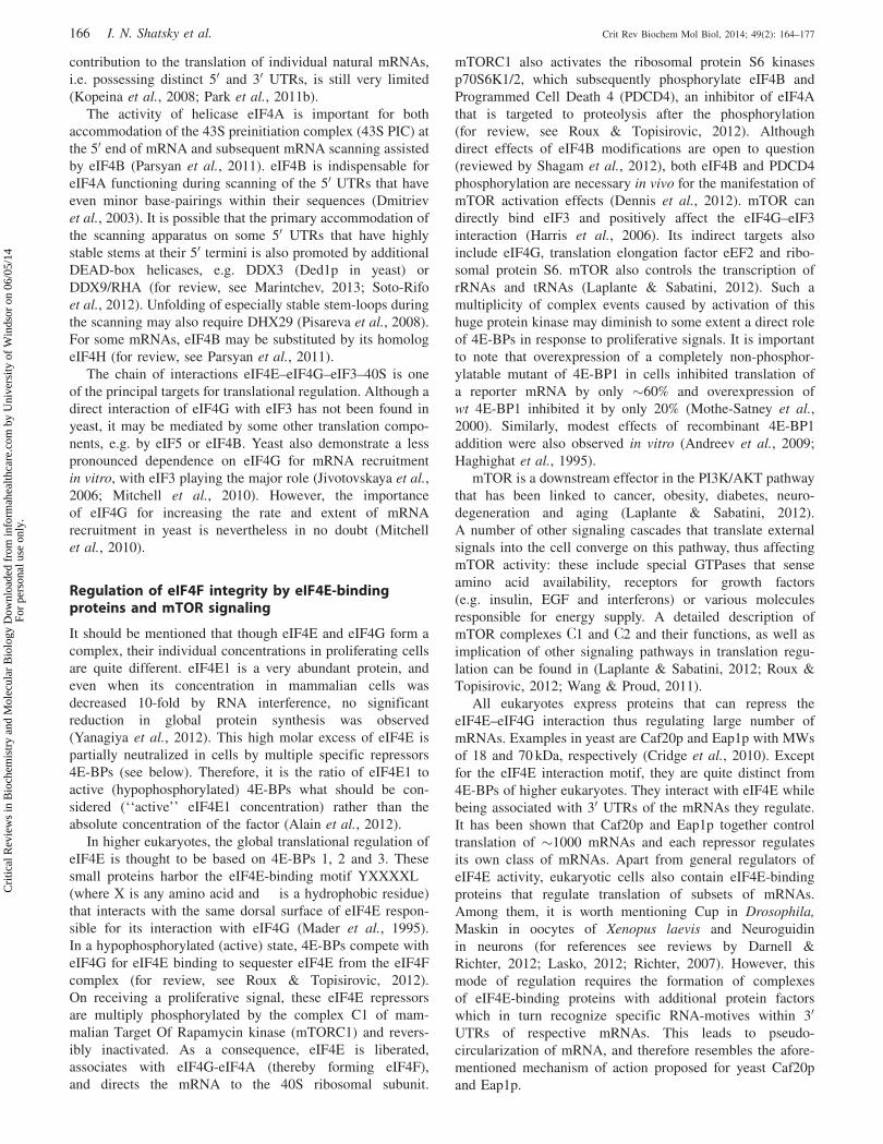
Figure 3. Speculative modes of mRNA recruitment to ribosomes used by distinct classes of cellular mRNAs. (A) The mRNAs that are highlydependent on eIF4E availability: blocking of eIF4E by dephosphorylated 4E-BPs results in dissociation of eIF4G + eIF4A from such mRNAs and theirtranslational inactivation. (B) The mRNAs which are less dependent on the eIF4E – m7G cap interaction and therefore less sensitive to mTORinhibition. These mRNAs may have a higher affinity of eIF4G + eIF4A to their 50 UTRs (may have CITEs) and remain active in the translationinitiation even under complete inhibition of eIF4E. (C) Specialized combinations of eIF4E and eIF4G. Specific variants of these factors are denoted aseIF4Ex and eIF4Gx. The corresponding forms of eIF4F may bind to particular mRNAs and activate them for translation during development anddifferentiation. Some of these combinations may be refractory to standard 4E-BPs. (D) The mRNAs that use eIF4G2 or eIF4G-like factors rather thaneIF4F. These factors may recognize the corresponding 50 UTRs with the help of auxiliary mRNA binding proteins (denoted with ‘‘Y’’). Such mRNAsare not necessarily cap-independent: they may use non-canonical cap-binding molecules (denoted as ‘‘X’’) which communicate with eIF4G-like factorsthrough specific mRNA-binding proteins. Alternatively, such mRNAs may employ IRES- or CITE-based mechanisms. All the modes of mRNArecruitment which are illustrated in this figure require a free 50 terminus and involve scanning from the 50 end. The IRES-driven translation initiation isdepicted in Figure 2(B).
DOI: 10.3109/10409238.2014.887051 Transcriptome-wide studies of mRNA binding to ribosomes 173
Cri
tical
Rev
iew
s in
Bio
chem
istr
y an
d M
olec
ular
Bio
logy
Dow
nloa
ded
from
info
rmah
ealth
care
.com
by
Uni
vers
ity o
f W
inds
or o
n 06
/05/
14Fo
r pe
rson
al u
se o
nly.

intercistronic) should be performed both under normal and
stress conditions, to check whether or not the putative IRES is
induced by stress.
Concluding remarks and hypotheses
A growing body of evidence hints at the existence of
significant modifications in the standard mechanism of
mRNA recruitment to eukaryotic ribosomes. They may
differ in the set of recruitment factors or their concentrations.
In addition, subsets of mRNAs may employ, at least under
some specific conditions, mechanisms that do not involve
eIF4E or even eIF4F. We do not exclude that IRES-elements
may also be implicated in the translation initiation of cellular
mRNAs. We only stress that all putative IRESs should be
verified and carefully characterized using stringent criteria to
show that internal ribosome binding activities do have
reasonable and physiologically justified values. These
values should be at least comparable with those of the 50
end-dependent initiation for the same 50 UTRs. On the
basis of information available to date, we can propose
several speculative modes of mRNA recruitment onto 40S
ribosomes that are used by distinct classes of eukaryotic
mRNAs:
Class 1. Highly eIF4E- and eIF4G-dependent mRNAs
(Figure 3A): The number of such mRNAs is relatively small
but they make up a substantial portion of the total amount of
mRNA within polysomes of proliferating cells. Their trans-
lation rapidly responds to changes in nutrient availability and
growth factors. They are strongly and directly controlled by
mTORC1, mostly via phosphorylation of 4E-BPs and are
represented by mRNAs responsible for growth and prolifer-
ation. Interestingly, restriction of their translation extends life
span (Harrison et al., 2009; Syntichaki et al., 2007; Zid et al.,
2009). They are overwhelmingly represented by mRNAs that
encode components of translational apparatus (ribosomal
proteins, some initiation factors, elongation factors eEF1 and
eEF2, PABP, etc.) as well as some factors participating in cell
cycle and epithelial-mesenchymal transition. All 50 TOP
mRNAs belong to this class. Their translation initiation is
strongly dependent on the concentration of free eIF4E, but
also dependent on eIF4G1/3. Importantly, eIF4G alone is not
able to support their efficient recruitment to 40S ribosomes,
presumably, because of a low affinity to the corresponding
UTRs. That is why disruption of the interaction of eIF4E with
eIF4G upon 4E-BPs activation results in a rapid dissociation
of eIF4G and eIF4A from such mRNAs. This process may be
additionally promoted by 50 TOP-binding proteins, TIA-1 and
TIAR (Damgaard & Lykke-Andersen, 2011).
Class 2. eIF4G-controlled mRNAs (Figure 3B): mRNAs of
this class may contain within their 50 UTRs sites with a high
affinity to eIF4G. Such mRNAs require less eIF4E or can
even do without this factor (e.g. when they harbor CITEs),
especially in non-dividing cells where the competition for
ribosomes with mRNAs of class 1 is low or even absent. It is
not excluded that a more stable and specific binding of eIF4G
to a 50 UTR may produce conformational changes in this
factor which in turn enable a more stable binding of eIF4E.
In this case, these mRNAs might successfully compete with
4E-BPs for eIF4E binding. It is worth mentioning that the
translation of the mRNAs which code for all eIF4G variants is
resistant to mTOR inhibition (4E-BPs activation) (Thoreen
et al., 2012).
Class 3. mRNAs controlled by specific variants of eIF4E,
eIF4G and their combinations (Figure 3C): As noted above,
the use of alternative forms of eIF4E and eIF4G during
mitosis and development is well documented, especially in
Drosophila. It is logical to predict the existence of their
specific combinations operating with particular mRNAs.
However, this issue has not been explored and we do not
know how these alternative forms and combinations are
selected by specific mRNAs. It may well be that this selection
is assisted by other auxiliary factors or mRNA-binding
proteins.
Class 4. mRNAs that do not use eIF4F (Figure 3D): They
may utilize a truncated form of eIF4G1, eIF4G2 (DAP5) or
other functional analogs of eIF4G1 (eIF4G-like proteins). The
mRNAs controlled by such factors are not necessary cap-
independent. They may use a cap-binding protein that
communicates with an eIF4G-like protein via a specific
mRNA-binding protein. Alternatively, DAP5 or similar
proteins may be a critical factor that governs internal
initiation on some cellular IRESs.
Finally, we have no idea about the factor requirement for
ribosome recycling through preformed polysomes. It may
well be that polysomes assembled on some mRNAs no longer
need eIF4E, eIF4G or even eIF4A for new rounds of
translation. This could be an alternative explanation why
eIF4G depletion produced a relatively minor effect on
eukaryotic translation. Some fragmentary data on insensitiv-
ity of preformed polysomes to a cap-analog (Amrani et al.,
2008) or non-hydrolysable derivative of ATP (Kopeina et al.,
2008) should encourage further experiments in this direction.
Certainly, there may be many more specific mechanisms
that determine translation initiation on eukaryotic mRNA.
This may be an exciting direction for further studies
of eukaryotic translation and its regulation. In addition,
knowledge of these mechanisms would be useful if we want
to design drugs targeting the translation of specific classes
of mRNAs in particular cells. To succeed in this tremendous
and very ambitious task, we should intensify not only
transcriptome-wide studies of translation but also focus
special attention on developing advanced biochemical tech-
niques and sophisticated in vitro approaches to study the
underlying mechanisms in detail. Otherwise, our knowledge
will remain just a myriad of effects, observations and their
arbitrary interpretations. Development of such techniques
certainly lags behind genome- and transcriptome-wide
studies.
Acknowledgements
We apologize to our colleagues for not being able to discuss
other important contributions due to space constraints. The
authors are very grateful to Gary Loughran (Cork Institute,
Ireland) for critical reading of the manuscript and valuable
suggestions.
Declaration of interest
The authors report no declaration of interest.
174 I. N. Shatsky et al. Crit Rev Biochem Mol Biol, 2014; 49(2): 164–177
Cri
tical
Rev
iew
s in
Bio
chem
istr
y an
d M
olec
ular
Bio
logy
Dow
nloa
ded
from
info
rmah
ealth
care
.com
by
Uni
vers
ity o
f W
inds
or o
n 06
/05/
14Fo
r pe
rson
al u
se o
nly.

We acknowledge financial support from the Russian
Foundation of Basic Researches to I.N.S. (grant N 11-04-
01010) and S.E.D. (grant N 12-04-33196).
References
Alain T, Morita M, Fonseca BD, et al. (2012). eIF4E/4E-BP ratiopredicts the efficacy of mTOR targeted therapies. Cancer Res 72:6468–76.
Alekhina OM, Vassilenko KS. (2012). Translation initiation in eukary-otes: versatility of the scanning model. Biochemistry (Mosc) 77:1465–77.
Amrani N, Ghosh S, Mangus DA, Jacobson A. (2008). Translationfactors promote the formation of two states of the closed-loop mRNP.Nature 453:1276–80.
Andreev DE, Dmitriev SE, Terenin IM, et al. (2009). Differentialcontribution of the m7G-cap to the 50 end-dependent translationinitiation of mammalian mRNAs. Nucleic Acids Res 37:6135–47.
Andreev DE, Dmitriev SE, Zinovkin R, et al. (2012). The 50 untranslatedregion of Apaf-1 mRNA directs translation under apoptosis conditionsvia a 50 end-dependent scanning mechanism. FEBS Lett 586:4139–43.
Badura M, Braunstein S, Zavadil J, Schneider RJ. (2012). DNA damageand eIF4G1 in breast cancer cells reprogram translation for survivaland DNA repair mRNAs. Proc Natl Acad Sci USA 109:18767–72.
Baker CC, Fuller MT. (2007). Translational control of meiotic cell cycleprogression and spermatid differentiation in male germ cells by anovel eIF4G homolog. Development 134:2863–9.
Berset C, Zurbriggen A, Djafarzadeh S, et al. (2003). RNA-bindingactivity of translation initiation factor eIF4G1 from Saccharomycescerevisiae. RNA 9:871–80.
Bolger TA, Wente SR. (2011). Gle1 is a multifunctional DEAD-boxprotein regulator that modulates Ded1 in translation initiation. J BiolChem 286:39750–9.
Borman AM, Michel YM, Kean KM. (2000). Biochemical characterisa-tion of cap-poly(A) synergy in rabbit reticulocyte lysates: theeIF4G-PABP interaction increases the functional affinity of eIF4Efor the capped mRNA 50-end. Nucleic Acids Res 28:4068–75.
Bush MS, Hutchins AP, Jones AM, et al. (2009). Selective recruitment ofproteins to 50 cap complexes during the growth cycle in Arabidopsis.Plant J 59:400–12.
Byrd MP, Zamora M, Lloyd RE. (2005). Translation of eukaryotictranslation initiation factor 4GI (eIF4GI) proceeds from multiplemRNAs containing a novel cap-dependent internal ribosome entry site(IRES) that is active during poliovirus infection. J Biol Chem 280:18610–22.
Cakmakci NG, Lerner RS, Wagner EJ, et al. (2008). SLIP1, a factorrequired for activation of histone mRNA translation by the stem-loopbinding protein. Mol Cell Biol 28:1182–94.
Castelli LM, Lui J, Campbell SG, et al. (2011). Glucose depletioninhibits translation initiation via eIF4A loss and subsequent 48Spreinitiation complex accumulation, while the pentose phosphatepathway is coordinately up-regulated. Mol Biol Cell 22:3379–93.
Cha JD, Kim HJ, Cha IH. (2011). Genetic alterations in oral squamouscell carcinoma progression detected by combining array-basedcomparative genomic hybridization and multiplex ligation-dependentprobe amplification. Oral Surg Oral Med Oral Pathol Oral RadiolEndod 111:594–607.
Cho PF, Poulin F, Cho-Park YA, et al. (2005). A new paradigm fortranslational control: inhibition via 50-30 mRNA tethering by Bicoidand the eIF4E cognate 4EHP. Cell 121:411–23.
Choe J, Oh N, Park S, et al. (2012). Translation initiation on mRNAsbound by nuclear cap-binding protein complex CBP80/20 requiresinteraction between CBP80/20-dependent translation initiation factorand eukaryotic translation initiation factor 3g. J Biol Chem 287:18500–9.
Clarkson BK, Gilbert WV, Doudna JA. (2010). Functional overlapbetween eIF4G isoforms in Saccharomyces cerevisiae. PLoS One 5:e9114.
Coldwell MJ, Morley SJ. (2006). Specific isoforms of translationinitiation factor 4GI show differences in translational activity. MolCell Biol 26:8448–60.
Coldwell MJ, Sack U, Cowan JL, et al. (2012). Multiple isoforms of thetranslation initiation factor eIF4GII are generated via use of
alternative promoters, splice sites and a non-canonical initiationcodon. Biochem J 448:1–11.
Colina R, Costa-Mattioli M, Dowling RJ, et al. (2008). Translationalcontrol of the innate immune response through IRF-7. Nature 452:323–8.
Contreras V, Richardson MA, Hao E, Keiper BD. (2008). Depletionof the cap-associated isoform of translation factor eIF4Ginduces germline apoptosis in C. elegans. Cell Death Differ 15:1232–42.
Cridge AG, Castelli LM, Smirnova JB, et al. (2010). Identifying eIF4E-binding protein translationally-controlled transcripts reveals links tomRNAs bound by specific PUF proteins. Nucleic Acids Res 38:8039–50.
Damgaard CK, Lykke-Andersen J. (2011). Translational coregulation of50TOP mRNAs by TIA-1 and TIAR. Genes Dev 25:2057–68.
Darnell JC, Richter JD. (2012). Cytoplasmic RNA-binding proteins andthe control of complex brain function. Cold Spring Harb Perspect Biol4:a012344.
Dennis MD, Jefferson LS, Kimball SR. (2012). Role ofp70S6K1-mediated phosphorylation of eIF4B and PDCD4proteins in the regulation of protein synthesis. J Biol Chem 287:42890–9.
Dmitriev SE, Andreev DE, Adyanova ZV, et al. (2009). Efficient cap-dependent translation of mammalian mRNAs with long and highlystructured 50-untranslated regions in vitro and in vivo. Mol Biol(Mosk) 43:108–13.
Dmitriev SE, Andreev DE, Terenin IM, et al. (2007). Efficienttranslation initiation directed by the 900-nucleotide-long and GC-rich 50 untranslated region of the human retrotransposon LINE-1mRNA is strictly cap dependent rather than internal ribosome entrysite mediated. Mol Cell Biol 27:4685–97.
Dmitriev SE, Terenin IM, Dunaevsky YE, et al. (2003). Assembly of 48Stranslation initiation complexes from purified components withmRNAs that have some base pairing within their 50 untranslatedregions. Mol Cell Biol 23:8925–33.
Dobrikov MI, Shveygert M, Brown MC, Gromeier M. (2014). MitoticPhosphorylation of eukaryotic initiation factor 4G1 (eIF4G1) atSer1232 by Cdk1: cyclin B inhibits eIF4A helicase complex bindingwith RNA. Mol Cell Biol 34:439–51.
Feoktistova K, Tuvshintogs E, Do A, Fraser CS. (2013). Human eIF4Epromotes mRNA restructuring by stimulating eIF4A helicase activity.Proc Natl Acad Sci USA 110:13339–44.
Filbin ME, Kieft JS. (2011). HCV IRES domain IIb affects theconfiguration of coding RNA in the 40S subunit’s decoding groove.RNA 17:1258–73.
Franklin-Dumont TM, Chatterjee C, Wasserman SA, Dinardo S. (2007).A novel eIF4G homolog, Off-schedule, couples translational controlto meiosis and differentiation in Drosophila spermatocytes.Development 134:2851–61.
Garre E, Romero-Santacreu L, De Clercq N, et al. (2012). Yeast mRNAcap-binding protein Cbc1/Sto1 is necessary for the rapid reprogram-ming of translation after hyperosmotic shock. Mol Biol Cell 23:137–50.
Geiger T, Velic A, Macek B, et al. (2013). Initial quantitative proteomicmap of 28 mouse tissues using the SILAC mouse. Mol CellProteomics 12:1709–22.
Gilbert WV, Zhou K, Butler TK, Doudna JA. (2007). Cap-independenttranslation is required for starvation-induced differentiation in yeast.Science 317:1224–7.
Gingras AC, Raught B, Sonenberg N. (1999). eIF4 initiation factors:effectors of mRNA recruitment to ribosomes and regulators oftranslation. Annu Rev Biochem 68:913–63.
Gkogkas CG, Khoutorsky A, Ran I, et al. (2013). Autism-related deficitsvia dysregulated eIF4E-dependent translational control. Nature 493:371–7.
Guo L, Allen E, Miller WA. (2000). Structure and function of a cap-independent translation element that functions in either the 30 or the 50
untranslated region. RNA 6:1808–20.Haghighat A, Mader S, Pause A, Sonenberg N. (1995). Repression of
cap-dependent translation by 4E-binding protein 1: competition withp220 for binding to eukaryotic initiation factor-4E. EMBO J 14:5701–9.
Haghighat A, Sonenberg N. (1997). eIF4G dramatically enhances thebinding of eIF4E to the mRNA 50-cap structure. J Biol Chem 272:21677–80.
DOI: 10.3109/10409238.2014.887051 Transcriptome-wide studies of mRNA binding to ribosomes 175
Cri
tical
Rev
iew
s in
Bio
chem
istr
y an
d M
olec
ular
Bio
logy
Dow
nloa
ded
from
info
rmah
ealth
care
.com
by
Uni
vers
ity o
f W
inds
or o
n 06
/05/
14Fo
r pe
rson
al u
se o
nly.

Harris TE, Chi A, Shabanowitz J, et al. (2006). mTOR-dependentstimulation of the association of eIF4G and eIF3 by insulin. EMBO J25:1659–68.
Harrison DE, Strong R, Sharp ZD, et al. (2009). Rapamycin fed late inlife extends lifespan in genetically heterogeneous mice. Nature 460:392–5.
Hernandez G, Han H, Gandin V, et al. (2012). Eukaryotic initiationfactor 4E-3 is essential for meiotic chromosome segregation,cytokinesis and male fertility in Drosophila. Development 139:3211–20.
Hernandez G, Miron M, Han H, et al. (2013). Mextli is a noveleukaryotic translation initiation factor 4E-binding protein that pro-motes translation in Drosophila melanogaster. Mol Cell Biol 33:2854–64.
Hernandez G, Vazquez-Pianzola P. (2005). Functional diversity of theeukaryotic translation initiation factors belonging to eIF4 families.Mech Dev 122:865–76.
Hinnebusch AG, Lorsch JR. (2012). The mechanism of eukaryotictranslation initiation: new insights and challenges. Cold Spring HarbPerspect Biol 4:a011544.
Hsieh AC, Liu Y, Edlind MP, et al. (2012). The translational landscapeof mTOR signalling steers cancer initiation and metastasis. Nature485:55–61.
Imataka H, Olsen HS, Sonenberg N. (1997). A new translationalregulator with homology to eukaryotic translation initiation factor 4G.EMBO J 16:817–25.
Ingolia NT, Brar GA, Rouskin S, et al. (2012). The ribosome profilingstrategy for monitoring translation in vivo by deep sequencing ofribosome-protected mRNA fragments. Nat Protoc 7:1534–50.
Jackson RJ. (2013). The current status of vertebrate cellular mRNAIRESs. Cold Spring Harb Perspect Biol 5:a011569.
Jang SK, Krausslich HG, Nicklin MJ, et al. (1988). A segment of the 50
nontranslated region of encephalomyocarditis virus RNA directsinternal entry of ribosomes during in vitro translation. J Virol 62:2636–43.
Jia Y, Polunovsky V, Bitterman PB, Wagner CR. (2012). Cap-dependenttranslation initiation factor eIF4E: an emerging anticancer drug target.Med Res Rev 32:786–814.
Jivotovskaya AV, Valasek L, Hinnebusch AG, Nielsen KH. (2006).Eukaryotic translation initiation factor 3 (eIF3) and eIF2 can promotemRNA binding to 40S subunits independently of eIF4G in yeast. MolCell Biol 26:1355–72.
Joshi B, Cameron A, Jagus R. (2004). Characterization of mammalianeIF4E-family members. Eur J Biochem 271:2189–203.
Joshi B, Lee K, Maeder DL, Jagus R. (2005). Phylogenetic analysis ofeIF4E-family members. BMC Evol Biol 5:48.
Kapeli K, Yeo GW. (2012). Genome-wide approaches to dissect the rolesof RNA binding proteins in translational control: implications forneurological diseases. Front Neurosci 6:144.
Kim KM, Cho H, Choi K, et al. (2009). A new MIF4G domain-containing protein, CTIF, directs nuclear cap-binding protein CBP80/20-dependent translation. Genes Dev 23:2033–45.
Kopeina GS, Afonina ZA, Gromova KV, et al. (2008). Step-wiseformation of eukaryotic double-row polyribosomes and circulartranslation of polysomal mRNA. Nucleic Acids Res 36:2476–88.
Koromilas AE, Lazaris-Karatzas A, Sonenberg N. (1992). mRNAscontaining extensive secondary structure in their 50 non-coding regiontranslate efficiently in cells overexpressing initiation factor eIF-4E.EMBO J 11:4153–8.
Kozak M. (1989). The scanning model for translation: an update. J CellBiol 108:229–41.
Laplante M, Sabatini DM. (2012). mTOR signaling in growth controland disease. Cell 149:274–93.
Lasko P. (2012). mRNA localization and translational control inDrosophila oogenesis. Cold Spring Harb Perspect Biol 4:a012294.
Lee SH, McCormick F. (2006). p97/DAP5 is a ribosome-associatedfactor that facilitates protein synthesis and cell proliferation bymodulating the synthesis of cell cycle proteins. EMBO J 25:4008–19.
Lemp NA, Hiraoka K, Kasahara N, Logg CR. (2012). Cryptic transcriptsfrom a ubiquitous plasmid origin of replication confound tests for cis-regulatory function. Nucleic Acids Res 40:7280–90.
Levy-Strumpf N, Deiss LP, Berissi H, Kimchi A. (1997). DAP-5, a novelhomolog of eukaryotic translation initiation factor 4G isolated as aputative modulator of gamma interferon-induced programmed celldeath. Mol Cell Biol 17:1615–25.
Liberman N, Marash L, Kimchi A. (2009). The translation initiationfactor DAP5 is a regulator of cell survival during mitosis. Cell Cycle8:204–9.
Macejak DG, Sarnow P. (1991). Internal initiation of translationmediated by the 50 leader of a cellular mRNA. Nature 353:90–4.
Mader S, Lee H, Pause A, Sonenberg N. (1995). The translationinitiation factor eIF-4E binds to a common motif shared by thetranslation factor eIF-4 gamma and the translational repressors4E-binding proteins. Mol Cell Biol 15:4990–7.
Malygin AA, Kossinova OA, Shatsky IN, Karpova GG. (2013). HCVIRES interacts with the 18S rRNA to activate the 40S ribosomefor subsequent steps of translation initiation. Nucleic Acids Res41:8706–14.
Mamane Y, Petroulakis E, Martineau Y, et al. (2007). Epigeneticactivation of a subset of mRNAs by eIF4E explains its effects on cellproliferation. PLoS One 2:e242.
Maquat LE, Hwang J, Sato H, Tang Y. (2010). CBP80-promoted mRNPrearrangements during the pioneer round of translation, nonsense-mediated mRNA decay, and thereafter. Cold Spring Harb Symp QuantBiol 75:127–34.
Marash L, Kimchi A. (2005). DAP5 and IRES-mediated translationduring programmed cell death. Cell Death Differ 12:554–62.
Marash L, Liberman N, Henis-Korenblit S, et al. (2008). DAP5 promotescap-independent translation of Bcl-2 and CDK1 to facilitate cellsurvival during mitosis. Mol Cell 30:447–59.
Marintchev A. (2013). Roles of helicases in translation initiation: amechanistic view. Biochim Biophys Acta 1829:799–809.
Martineau Y, Derry MC, Wang X, et al. (2008). Poly(A)-binding protein-interacting protein 1 binds to eukaryotic translation initiation factor3 to stimulate translation. Mol Cell Biol 28:6658–67.
Miller WA, Wang Z, Treder K. (2007). The amazing diversity of cap-independent translation elements in the 30-untranslated regions ofplant viral RNAs. Biochem Soc Trans 35:1629–33.
Mitchell SF, Walker SE, Algire MA, et al. (2010). The50-7-methylguanosine cap on eukaryotic mRNAs serves both tostimulate canonical translation initiation and to block an alternativepathway. Mol Cell 39:950–62.
Morita M, Ler LW, Fabian MR, et al. (2012). A novel 4EHP-GIGYF2translational repressor complex is essential for mammalian develop-ment. Mol Cell Biol 32:3585–93.
Mothe-Satney I, Yang D, Fadden P, et al. (2000). Multiple mechanismscontrol phosphorylation of PHAS-I in five (S/T)P sites that governtranslational repression. Mol Cell Biol 20:3558–67.
Neusiedler J, Mocquet V, Limousin T, et al. (2012). INT6 interacts withMIF4GD/SLIP1 and is necessary for efficient histone mRNAtranslation. RNA 18:1163–77.
Nicholson BL, Wu B, Chevtchenko I, White KA. (2010). Tombusvirusrecruitment of host translational machinery via the 30 UTR. RNA 16:1402–19.
Nousch M, Reed V, Bryson-Richardson RJ, et al. (2007). The eIF4G-homolog p97 can activate translation independent of caspase cleavage.RNA 13:374–84.
Osborne MJ, Volpon L, Kornblatt JA, et al. (2013). eIF4E3 actsas a tumor suppressor by utilizing an atypical mode of methyl-7-guanosine cap recognition. Proc Natl Acad Sci U S A 110:3877–82.
Ozpolat B, Akar U, Zorrilla-Calancha I, et al. (2008). Death-associatedprotein 5 (DAP5/p97/NAT1) contributes to retinoic acid-inducedgranulocytic differentiation and arsenic trioxide-induced apoptosis inacute promyelocytic leukemia. Apoptosis 13:915–28.
Park EH, Walker SE, Lee JM, et al. (2011a). Multiple elements in theeIF4G1 N-terminus promote assembly of eIF4G1*PABP mRNPsin vivo. EMBO J 30:302–16.
Park EH, Zhang F, Warringer J, et al. (2011b). Depletion of eIF4G fromyeast cells narrows the range of translational efficiencies genome-wide. BMC Genomics 12:68.
Parsyan A, Svitkin Y, Shahbazian D, et al. (2011). mRNA helicases:the tacticians of translational control. Nat Rev Mol Cell Biol 12:235–45.
Pelletier J, Sonenberg N. (1988). Internal initiation of translation ofeukaryotic mRNA directed by a sequence derived from poliovirusRNA. Nature 334:320–5.
Pestova TV, Hellen CU, Shatsky IN. (1996a). Canonical eukaryoticinitiation factors determine initiation of translation by internalribosomal entry. Mol Cell Biol 16:6859–69.
176 I. N. Shatsky et al. Crit Rev Biochem Mol Biol, 2014; 49(2): 164–177
Cri
tical
Rev
iew
s in
Bio
chem
istr
y an
d M
olec
ular
Bio
logy
Dow
nloa
ded
from
info
rmah
ealth
care
.com
by
Uni
vers
ity o
f W
inds
or o
n 06
/05/
14Fo
r pe
rson
al u
se o
nly.

Pestova TV, Shatsky IN, Fletcher SP, et al. (1998). A prokaryotic-likemode of cytoplasmic eukaryotic ribosome binding to the initiationcodon during internal translation initiation of hepatitis C and classicalswine fever virus RNAs. Genes Dev 12:67–83.
Pestova TV, Shatsky IN, Hellen CU. (1996b). Functional dissection ofeukaryotic initiation factor 4F: the 4A subunit and the central domainof the 4G subunit are sufficient to mediate internal entry of 43Spreinitiation complexes. Mol Cell Biol 16:6870–8.
Pisareva VP, Pisarev AV, Komar AA, et al. (2008). Translation initiationon mammalian mRNAs with structured 50UTRs requires DExH-boxprotein DHX29. Cell 135:1237–50.
Prevot D, Decimo D, Herbreteau CH, et al. (2003). Characterization of anovel RNA-binding region of eIF4GI critical for ribosomal scanning.EMBO J 22:1909–21.
Rajagopal V, Park EH, Hinnebusch AG, Lorsch JR. (2012). Specificdomains in yeast translation initiation factor eIF4G strongly bias RNAunwinding activity of the eIF4F complex toward duplexes with 50-overhangs. J Biol Chem 287:20301–12.
Ramirez-Valle F, Braunstein S, Zavadil J, et al. (2008). eIF4GI linksnutrient sensing by mTOR to cell proliferation and inhibition ofautophagy. J Cell Biol 181:293–307.
Rhoads RE. (2009). eIF4E: new family members, new binding partners,new roles. J Biol Chem 284:16711–15.
Richter JD. (2007). CPEB: a life in translation. Trends Biochem Sci 32:279–85.
Roux PP, Topisirovic I. (2012). Regulation of mRNA translation bysignaling pathways. Cold Spring Harb Perspect Biol 4:a012252.
Ruggero D. (2013). Translational control in cancer etiology. Cold SpringHarb Perspect Biol 5:a012336.
Schwanhausser B, Busse D, Li N, et al. (2011). Global quantification ofmammalian gene expression control. Nature 473:337–42.
Shagam LI, Terenin IM, Andreev DE, et al. (2012). In vitro activity ofhuman translation initiation factor eIF4B is not affected byphosphomimetic amino acid substitutions S422D and S422E.Biochimie 94:2484–90.
Sharma A, Yilmaz A, Marsh K, et al. (2012). Thriving under stress:selective translation of HIV-1 structural protein mRNA during Vpr-mediated impairment of eIF4E translation activity. PLoS Pathog 8:e1002612.
Shatsky IN, Dmitriev SE, Terenin IM, Andreev DE. (2010). Cap- andIRES-independent scanning mechanism of translation initiation as analternative to the concept of cellular IRESs. Mol Cells 30:285–93.
Shaughnessy Jr JD, Jenkins NA, Copeland NG. (1997). cDNA cloning,expression analysis, and chromosomal localization of a gene with highhomology to wheat eIF-(iso)4F and mammalian eIF-4G. Genomics39:192–7.
Sonenberg N, Hinnebusch AG. (2009). Regulation of translationinitiation in eukaryotes: mechanisms and biological targets. Cell136:731–45.
Soto-Rifo R, Rubilar PS, Limousin T, et al. (2012). DEAD-box proteinDDX3 associates with eIF4F to promote translation of selectedmRNAs. EMBO J 31:3745–56.
Srivastava T, Fortin DA, Nygaard S, et al. (2012). Regulation ofneuronal mRNA translation by CaM-kinase I phosphorylation ofeIF4GII. J Neurosci 32:5620–30.
Stupina VA, Meskauskas A, Mccormack JC, et al. (2008). The 30
proximal translational enhancer of Turnip crinkle virus binds to 60Sribosomal subunits. RNA 14:2379–93.
Sun J, Conn CS, Han Y, et al. (2011). PI3K-mTORC1 attenuates stressresponse by inhibiting cap-independent Hsp70 translation. J BiolChem 286:6791–800.
Svitkin YV, Pause A, Haghighat A, et al. (2001). The requirement foreukaryotic initiation factor 4A (elF4A) in translation is in directproportion to the degree of mRNA 50 secondary structure. RNA 7:382–94.
Syntichaki P, Troulinaki K, Tavernarakis N. (2007). eIF4E function insomatic cells modulates ageing in Caenorhabditis elegans. Nature445:922–6.
Takahashi K, Maruyama M, Tokuzawa Y, et al. (2005). Evolutionarilyconserved non-AUG translation initiation in NAT1/p97/DAP5(EIF4G2). Genomics 85:360–71.
Terenin IM, Andreev DE, Dmitriev SE, Shatsky IN. (2013). Anovel mechanism of eukaryotic translation initiation that isneither m7G-cap-, nor IRES-dependent. Nucleic Acids Res 41:1807–16.
Terenin IM, Dmitriev SE, Andreev DE, et al. (2005). A cross-kingdominternal ribosome entry site reveals a simplified mode of internalribosome entry. Mol Cell Biol 25:7879–88.
Thoreen CC, Chantranupong L, Keys HR, et al. (2012). A unifyingmodel for mTORC1-mediated regulation of mRNA translation. Nature485:109–13.
Treder K, Kneller EL, Allen EM, et al. (2008). The 30 cap-independenttranslation element of Barley yellow dwarf virus binds eIF4F via theeIF4G subunit to initiate translation. RNA 14:134–47.
Uniacke J, Holterman CE, Lachance G, et al. (2012). An oxygen-regulated switch in the protein synthesis machinery. Nature 486:126–9.
Virgili G, Frank F, Feoktistova K, et al. (2013). Structural analysis of theDAP5 MIF4G domain and its interaction with eIF4A. Structure 21:517–27.
Von Der Haar T, Ball PD, McCarthy JE. (2000). Stabilization ofeukaryotic initiation factor 4E binding to the mRNA 50-Cap bydomains of eIF4G. J Biol Chem 275:30551–5.
Von Moeller H, Lerner R, Ricciardi A, et al. (2013). Structural andbiochemical studies of SLIP1-SLBP identify DBP5 and eIF3g asSLIP1-binding proteins. Nucleic Acids Res 41:7960–71.
Wallace A, Filbin ME, Veo B, et al. (2010). The nematode eukaryotictranslation initiation factor 4E/G complex works with a trans-splicedleader stem-loop to enable efficient translation of trimethylguanosine-capped RNAs. Mol Cell Biol 30:1958–70.
Wang X, Proud CG. (2011). mTORC1 signaling: what we still don’tknow. J Mol Cell Biol 3:206–20.
Yamanaka S, Poksay KS, Arnold KS, Innerarity TL. (1997). A noveltranslational repressor mRNA is edited extensively in livers contain-ing tumors caused by the transgene expression of the apoB mRNA-editing enzyme. Genes Dev 11:321–33.
Yamanaka S, Zhang XY, Maeda M, et al. (2000). Essential role ofNAT1/p97/DAP5 in embryonic differentiation and the retinoic acidpathway. EMBO J 19:5533–41.
Yanagiya A, Suyama E, Adachi H, et al. (2012). Translationalhomeostasis via the mRNA cap-binding protein, eIF4E. Mol Cell46:847–58.
Zid BM, Rogers AN, Katewa SD, et al. (2009). 4E-BP extends lifespanupon dietary restriction by enhancing mitochondrial activity inDrosophila. Cell 139:149–60.
DOI: 10.3109/10409238.2014.887051 Transcriptome-wide studies of mRNA binding to ribosomes 177
Cri
tical
Rev
iew
s in
Bio
chem
istr
y an
d M
olec
ular
Bio
logy
Dow
nloa
ded
from
info
rmah
ealth
care
.com
by
Uni
vers
ity o
f W
inds
or o
n 06
/05/
14Fo
r pe
rson
al u
se o
nly.