Embed Size (px)
Citation preview

RESEARCH ARTICLE Open Access
Transcriptomic analysis of wound xylemformation in Pinus canariensisV. Chano1, C. Collada1,2 and A. Soto1,2*
Abstract
Background: Woody plants, especially trees, usually must face several injuries caused by different agents duringtheir lives. Healing of injuries in stem and branches, affecting the vascular cambium and xylem can take severalyears. In conifers, healing takes place mainly from the remaining vascular cambium in the margin of the wound.The woundwood formed in conifers during healing usually presents malformed and disordered tracheids as well asabundant traumatic resin ducts. These characteristics affect its functionality as water conductor and itstechnological properties.
Results: In this work we analyze for the first time the transcriptomic basis of the formation of traumatic wood inconifers, and reveal some differences with normal early- and late-wood. Microarray analysis of the differentiatingtraumatic wood, confirmed by quantitative RT-PCR, has revealed alterations in the transcription profile of up to1408 genes during the first period of healing. We have grouped these genes in twelve clusters, according to theirtranscription profiles, and have distinguished accordingly two main phases during this first healing.
Conclusions: Wounding induces a complete rearrangement of the transcriptional program in the cambial zoneclose to the injuries. At the first instance, radial growth is stopped, and a complete set of defensive genes, mostlyrelated to biotic stress, are induced. Later on, cambial activity is restored in the lateral borders of the wound, evenat a high rate. During this second stage certain genes related to early-wood formation, including genes involved incell wall formation and transcription factors, are significantly overexpressed, while certain late-wood related genesare repressed. Additionally, significant alterations in the transcription profile of abundant non annotated genes arereported.
Keywords: Wound, Healing, Conifers, Transcriptome, Wood, Pinus canariensis
BackgroundOrganisms usually suffer injuries throughout their life.In multicellular organisms these injuries can cause thedamage or loss of differentiated tissues or organs, andease the entry and spread of pathogens. The analysis ofthe similarities and differences in the regenerationprocess in animals and plants has been on the spotlightin recent years [1–3]. Animals can often regeneratethese damaged tissues and even, in certain cases, the lostorgans, and due to constant regeneration of certain tis-sues such as skin, eventually no signal of the injury re-mains after some time.
On their side, plants do not regenerate continuouslytheir tissues. Proliferation is usually limited to certainniches: the root and shoot apical meristems (includingaxillary buds) and the vascular cambium and the phello-gen in woody plants. If damage occurs, the plant gener-ates new tissues and organs from these meristems oreventually develops new meristematic niches from livingcells, usually parenchymatic ones [4, 5]. This is the caseof the traumatic periderms developed, for instance, fromcortical parenchyma or from mesophyll to seal a woundin a young stem or a leave.When a woody branch or stem suffers a deep wound,
affecting the secondary xylem, the vascular cambiummust be restored. In certain angiosperms proliferationfrom xylem parenchyma or from immature xylem con-ducting elements has been described, as in Tilia [6],Eucommia [7] or Populus [8]. These cells can reverse
* Correspondence: [email protected]. Genética, Fisiología e Historia Forestal. ETSI Montes, Forestal y del MedioNatural. Dpto. Sistemas y Recursos Naturales, Universidad Politécnica deMadrid, Ciudad Universitaria s, /n 28040 Madrid, Spain2Unidad Mixta de Genómica y Ecofisiología Forestal INIA/UPM, Madrid, Spain
© The Author(s). 2017 Open Access This article is distributed under the terms of the Creative Commons Attribution 4.0International License (http://creativecommons.org/licenses/by/4.0/), which permits unrestricted use, distribution, andreproduction in any medium, provided you give appropriate credit to the original author(s) and the source, provide a link tothe Creative Commons license, and indicate if changes were made. The Creative Commons Public Domain Dedication waiver(http://creativecommons.org/publicdomain/zero/1.0/) applies to the data made available in this article, unless otherwise stated.
Chano et al. BMC Plant Biology (2017) 17:234 DOI 10.1186/s12870-017-1183-3

their differentiation pathway and divide profusely, givingrise to a parenchymatic callus. Later on, a new, trau-matic vascular cambium differentiates within this callus,and new secondary xylem and phloem are produced.On the contrary, this proliferation from (partially) dif-
ferentiated cells is not usual in conifers. In these specieshealing takes place mainly from the remaining vascularcambium in the margins of the wound, as described re-cently for Pinus canariensis [9].Anyway, the traumatic wood formed this way can be
easily distinguished from normal wood. Traumatic woodusually presents malformed tracheary elements and fi-bers, with altered lignification patterns, and with a highproportion of parenchymatic cells. Orientation of theseelements is also very often distorted [9–12], probablydue to altered hormonal flux, and also to altered mech-anical signals, as suggested by Chano et al. [9]. This dis-organized xylem implies an evident disadvantage forwater and nutrient transport [10]. In the case of conifers,especially Pinaceae, traumatic wood also presents a veryhigh proportion of resin ducts, as described for Cedruslibani [13], Larix decidua [14, 15], Picea abies [15, 16],Pinus nigra [17] or Pinus pinaster [18]. Actually, forma-tion of traumatic resin ducts is the basis of traditionalresin exploitation, very common in the past for severalspecies of Mediterranean pines, and with increasinginterest in the last years [18].Since plants do not renew their secondary xylem, but
generate new sheets of xylem, centrifugally, year afteryear, traumatic xylem also remains in the damagedbranch or stem, leaving a “scar” in the wood. These scarshave proven to be very useful, for instance, for dendro-chronology studies [15, 19]. However, traumatic woodpresents undesirable characteristics from a technologicalpoint of view. Although the higher density due to the in-crease in resin content can improve certain mechanicalqualities of wood it also causes problems at machiningand blunting [20]. In addition, disordered and not prop-erly formed traumatic tracheids contribute to alter thephysico-mechanical properties of wood. Therefore, lum-ber dealers consider traumatic wood as a defect, lower-ing the price and reducing the applicability of woodpieces with important scars.Several works have focused in the consequences of
traumatisms on wood development in conifers: early-latewood ratio, ring width, formation of traumatic resinducts (f.i., [10, 21–24]), and a few others in the descrip-tion of the healing process from an anatomical point ofview [9, 25–27]. However, although the molecular as-pects of the response to traumatism has been analysedin different angiosperms (f. i., [4, 28–31]), the processhas been less studied in gymnosperms, where mostworks have focused in the induction of traumatic resinducts by traumatism, insect attack or fungal infections
[32–35]. In this work we focus on the molecular basis oftraumatic wood formation in a gymnosperm, P. canar-iensis, known for its extraordinary healing ability. Forthis purpose we have performed deep wounds in thestem of P. canariensis trees, affecting the vascular cam-bium, and have assessed the transcriptomic profile dur-ing the healing process and traumatic wood growth.
Results and discussionIdentification of genes induced and repressed in responseto woundingIn order to analyse the transcriptomic response in thecambial zone and differentiating xylem in the borders ofdeep wounds performed in the stem of pine trees, wehybridized a 60K two-color cDNA microarray (Agilent,USA) which includes genes involved in P. canariensisxylogenesis [36]. Samples were collected at three datesduring wound response: i) H1 was collected seven daysafter wounding, ii) H2 after 75 days, when developmentof traumatic wood was evident and while the trees out-side the wound area were still forming early wood, andfinally iii) H3 92 days after wounding, when the treeswere already forming late wood. Controls for each sam-ple were collected at the same sampling dates frombranches distant from the wound, in order to distinguishlocal effects caused by wound response from constitutivechanges in gene expression during the vegetative season.Figure 1 shows the distribution of genes selected as
over- and underexpressed at each sampling point. Weidentified 1408 differentially expressed genes (DEG),genes significantly overexpressed or repressed comparedto normal wood formation. Table 1 shows a selection of91 DEGs with the strongest response (induction or re-pression), grouped following the functional processesthey are presumably related to, according to their topBLASTx hit, as previously described [36]. The completetable can be found in a supplementary table (Additionalfile 1).Immediate response H1 included 837 DEGs; 619
DEGs were detected for H3, while only 336 DEGs weredetected at H2. Just 69 genes were identified as DEG forthe three sampling dates, H1, H2 and H3. Moreover, just87 genes were identified as DEGs exclusively for H2,while 348 were exclusive DEGs for H3 and up to 658 forH1 (Fig. 2).Enrichment analysis of DEGs pointed out an increase
of mRNA levels for the categories “defense response”,“response to stress” and different forms of “response tostimulus”, as “response to abiotic stimulus” or “responseto biotic stimulus”, among others, in the BiologicalProcess (BP) category. As well, other enriched GO termswere “nucleic acid binding transcription factor activity”and “sequence-specific DNA binding transcription factoractivity”, for the Molecular Function (MF) category, and
Chano et al. BMC Plant Biology (2017) 17:234 Page 2 of 16

“extracellular region”, “cell wall”, “cell periphery” and“external encapsulating structure” GO terms of the Cel-lular Component category.
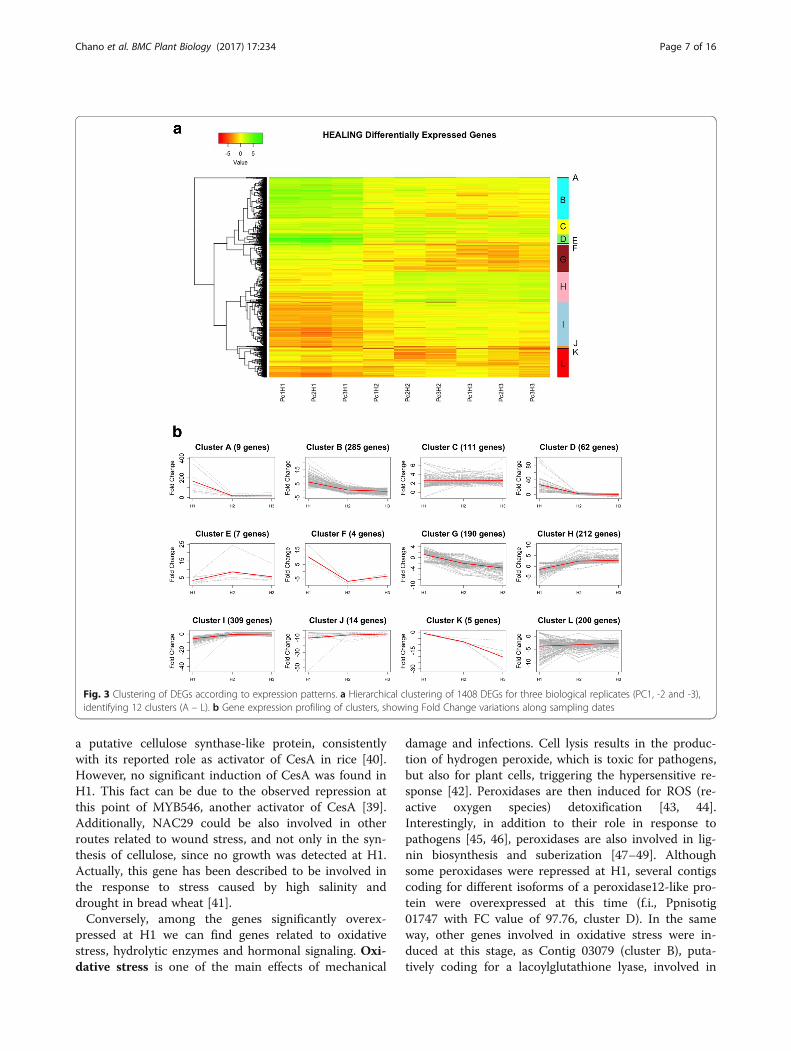
Hierarchical clustering of DEGsTwelve clusters were established according to the tran-scription patterns detected for DEGs throughout H1, H2and H3 (Fig. 3).Cluster A includes genes clearly induced at H1, which
keep high transcription levels during H2 and H3, al-though at a minor degree. Genes included in clusters B,D and F were also overexpressed at H1, but later ontheir transcription levels decrease, being even repressedat H2 and/or H3. Genes in cluster C show a faint over-expression throughout the three phases, while clusters Eand H show an increasing overexpression at H2 and H3.The opposite pattern is reflected by clusters G and K,with an increasing repression at H2 and H3. Genes incluster I show a significant repression at H1, followed bya recovery of transcription to normal levels at H2 andH3. Finally, clusters J and L are characterized by a gen-eral repression during the three phases.Clustering of samples revealed consistency among bio-
logical replicates, as shown in a supplementary figure(Additional file 2). Samples harvested at H1 clustered to-gether and separated from the other sampling dates; ontheir side, H2 and H3 samples were included in thesame group. Slight irregularities (for instance, samplePc3H2 is closer to H3 samples than to the other H2ones) can be due to the genetic variability among trees.This result supports the differentiation of two majorphases (H1 and H2/H3) in the response to wounding, asdiscussed below.
To validate the reliability of the transcription profilesobtained from microarray hybridization, we selected 12genes for qRT-PCR analysis, covering the main tenden-cies described above and the putative function of thegenes. Thus, we selected three genes directly involved incell growth and cell wall formation, as an expansin(Contig 03225, cluster H), a CeSA-like (Contig 00654,cluster L) and a CCoAOMT (Contig 06476, cluster G),transcription factors also involved in xylogenesis, as aMYB46-like (Contig 12050, cluster L), a WOX4-like(Contig 06813, cluster H), a bHLH35-like (Contig 05923,cluster C)an ATHB13-like (Contig 20304, cluster B), aNAC2-like (Contig 00787, cluster B), and a WRKY51-like (Contig 05551, cluster L). Finally, we also analysed agene coding for a PAL protein (Contig 20555, cluster B),involved in salicylic acid biosynthesis and presumably re-lated to defense, an EXORDIUM-like protein (Contig09007, cluster G), presumably involved in cell prolifera-tion, and a Major Allergen Pru AR1-like (Contig 22185,cluster A), putatively involved in defensive response.Profiles obtained by qRT-PCR for these genes match
the ones obtained from microarray hybridization, withhigh correlation coefficients, thus validating the gen-eral tendencies described above for microarray ana-lysis (Fig. 4).Detailed analysis of the transcription patterns of the
differentially expressed genes (DEGs) leads to the identi-fication of two major phases in the response towounding.
H1. Immediate responseA complete rearrangement of the transcriptional pro-gram takes place as immediate response to wounding.
Fig. 1 MA plot of microarray normalized data during wound-response. X-axis: Log2 of microarray signals; Y-axis: Log2 of Fold Change values;Green dots: probes selected as overexpressed (FC > 2, FDR < 0.05, between treatment and control RNA samples); Red dots: probes selected asunderexpressed (FC < -2, FDR < 0.05 between treatment and control RNA samples)
Chano et al. BMC Plant Biology (2017) 17:234 Page 3 of 16

Table 1 Selected wound-responsive DEGs, grouped according to their putative role and their expression profile clustering
Cluster ID Seq. Description H1 H2 H3
FC FDR FC FDR FC FDR
Defense and stress response genes
A Contig18804 disease resistance response protein 206-like 395.78 0.00 37.14 0.00 52.52 0.00
A Contig19053 pathogenesis-related protein pr-4b-like 96.55 0.00 23.91 0.00 19.77 0.00
A Contig22185 major allergen pru ar 1-like 138.29 0.00 13.75 0.00 4.94 0.01
A Contig22375 pathogenesis-related protein pr-4-like 343.24 0.00 7.69 0.00 7.38 0.00
A Ppnisotig12265 antimicrobial peptide 1-like 72.52 0.00 16.88 0.00 12.84 0.00
A Ppnisotig13133 pathogenesis-related protein pr-4-like 206.36 0.00 6.43 0.00 5.13 0.00
A Ppnisotig13431 disease resistance response protein 206-like 133.78 0.00 24.75 0.00 31.68 0.00
B Contig00602 defensin ec-amp-d2-like 11.27 0.00 2.07 0.12 -1.42 0.65
B Contig02906 nematode resistance hspro2-like 7.81 0.01 -1.23 0.94 -2.12 0.11
B Contig03079 lactoylglutathione lyase glyoxalase i family protein 3.96 0.04 1.97 0.20 -1.36 0.75
B Contig09180 thioredoxin h-type 6.82 0.01 1.52 0.44 1.39 0.55
B Contig19857 nematode resistance hspro2-like 9.88 0.00 -1.33 0.83 -2.56 0.06
B Contig20555 phenylalanine ammonia-lyase-like 14.41 0.00 3.19 0.03 -1.08 1.11
C Contig13499 (-)-camphene tricyclene chloroplastic-like 2.17 0.17 2.14 0.12 2.81 0.01
D Contig00126 basic endochitinase a-like 15.25 0.00 -1.20 0.55 0.34 1.13
D Contig10307 endochitinase a-like 65.05 0.00 3.40 0.02 1.49 0.47
D Contig17617 defensin ec-amp-d2-like 44.89 0.00 1.32 0.40 -1.23 0.69
D Contig21216 endochitinase a-like 92.45 0.00 2.28 0.08 2.51 0.02
D Contig23442 chitinase 1-like 29.31 0.00 2.12 0.37 -1.65 0.41
D Ppnisotig00751 endochitinase pr4-like 85.39 0.00 3.16 0.02 1.28 0.78
D Ppnisotig01747 peroxidase 12-like 97.76 0.00 1.74 0.28 1.53 0.37
D Ppnisotig06171 glutathione s-transferase f9-like 41.49 0.00 2.48 0.05 1.55 0.36
D Ppnisotig08058 endochitinase pr4-like 89.60 0.00 1.15 1.04 1.12 1.08
H Contig03270 geranylgeranyl pyrophosphate chloroplastic-like 1.61 0.46 1.48 0.45 2.57 0.03
H Contig08417 abietadienol abietadienal oxidase-like 1.10 1.12 2.08 0.11 2.64 0.02
H Ppnisotig10634 geranylgeranyl pyrophosphate chloroplastic-like -1.26 1.00 2.54 0.04 2.92 0.01
Cell-wall matrix development and/or carbohydrates metabolism
G Contig06476 caffeoyl- o-methyltransferase -1.27 0.97 -3.59 0.04 -1.44 0.61
G Contig15857 cellulose synthase-like protein d3 1.38 0.78 -1.14 1.08 -3.00 0.04
G Contig17013 probable xyloglucan endotransglucosylase hydrolase protein 23 -1.24 0.93 -6.29 0.00 -7.64 0.00
G Contig21865 galactinol–sucrose galactosyltransferase-like 2.27 0.18 -2.03 0.21 -3.39 0.03
H Contig00603 beta-xylosidase alpha-l-arabinofuranosidase 2-like -0.57 1.10 2.46 0.07 2.58 0.03
H Contig03225 expansin alpha -2.10 0.20 2.64 0.04 1.99 0.13
H Contig05066 probable pectate lyase 15-like -5.21 0.01 2.08 0.20 3.24 0.01
H Contig05424 probable xyloglucan endotransglucosylase hydrolase protein 8-like -1.78 0.37 2.55 0.04 2.18 0.06
H Contig09907 probable xyloglucan endotransglucosylase hydrolase protein 32 -1.86 0.32 2.14 0.09 2.95 0.01
H Contig10173 endoglucanase 24-like -3.26 0.05 1.58 0.46 2.58 0.03
H Contig13281 probable pectinesterase 68-like -1.56 0.60 2.18 0.09 3.40 0.01
H Contig18777 endoglucanase 24-like -1.69 0.41 2.54 0.04 5.32 0.00
H Contig18811 expansin alpha -1.59 0.58 3.42 0.01 4.98 0.00
I Contig00766 xyloglucan endotransglucosylase hydrolase protein 9-like -17.49 0.00 -1.36 0.72 -1.24 0.78
I Contig02447 caffeoyl- o-methyltransferase-like -6.41 0.01 1.17 0.75 -1.58 0.34
Chano et al. BMC Plant Biology (2017) 17:234 Page 4 of 16
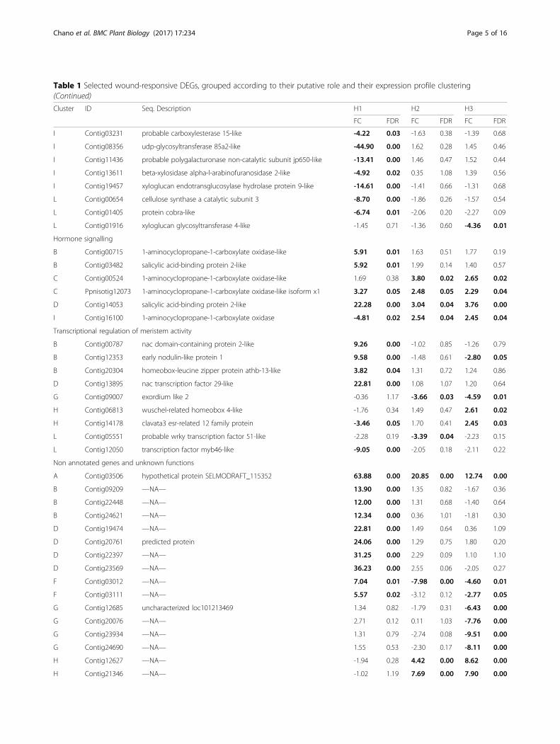
Table 1 Selected wound-responsive DEGs, grouped according to their putative role and their expression profile clustering(Continued)
Cluster ID Seq. Description H1 H2 H3
FC FDR FC FDR FC FDR
I Contig03231 probable carboxylesterase 15-like -4.22 0.03 -1.63 0.38 -1.39 0.68
I Contig08356 udp-glycosyltransferase 85a2-like -44.90 0.00 1.62 0.28 1.45 0.46
I Contig11436 probable polygalacturonase non-catalytic subunit jp650-like -13.41 0.00 1.46 0.47 1.52 0.44
I Contig13611 beta-xylosidase alpha-l-arabinofuranosidase 2-like -4.92 0.02 0.35 1.08 1.39 0.56
I Contig19457 xyloglucan endotransglucosylase hydrolase protein 9-like -14.61 0.00 -1.41 0.66 -1.31 0.68
L Contig00654 cellulose synthase a catalytic subunit 3 -8.70 0.00 -1.86 0.26 -1.57 0.54
L Contig01405 protein cobra-like -6.74 0.01 -2.06 0.20 -2.27 0.09
L Contig01916 xyloglucan glycosyltransferase 4-like -1.45 0.71 -1.36 0.60 -4.36 0.01
Hormone signalling
B Contig00715 1-aminocyclopropane-1-carboxylate oxidase-like 5.91 0.01 1.63 0.51 1.77 0.19
B Contig03482 salicylic acid-binding protein 2-like 5.92 0.01 1.99 0.14 1.40 0.57
C Contig00524 1-aminocyclopropane-1-carboxylate oxidase-like 1.69 0.38 3.80 0.02 2.65 0.02
C Ppnisotig12073 1-aminocyclopropane-1-carboxylate oxidase-like isoform x1 3.27 0.05 2.48 0.05 2.29 0.04
D Contig14053 salicylic acid-binding protein 2-like 22.28 0.00 3.04 0.04 3.76 0.00
I Contig16100 1-aminocyclopropane-1-carboxylate oxidase -4.81 0.02 2.54 0.04 2.45 0.04
Transcriptional regulation of meristem activity
B Contig00787 nac domain-containing protein 2-like 9.26 0.00 -1.02 0.85 -1.26 0.79
B Contig12353 early nodulin-like protein 1 9.58 0.00 -1.48 0.61 -2.80 0.05
B Contig20304 homeobox-leucine zipper protein athb-13-like 3.82 0.04 1.31 0.72 1.24 0.86
D Contig13895 nac transcription factor 29-like 22.81 0.00 1.08 1.07 1.20 0.64
G Contig09007 exordium like 2 -0.36 1.17 -3.66 0.03 -4.59 0.01
H Contig06813 wuschel-related homeobox 4-like -1.76 0.34 1.49 0.47 2.61 0.02
H Contig14178 clavata3 esr-related 12 family protein -3.46 0.05 1.70 0.41 2.45 0.03
L Contig05551 probable wrky transcription factor 51-like -2.28 0.19 -3.39 0.04 -2.23 0.15
L Contig12050 transcription factor myb46-like -9.05 0.00 -2.05 0.18 -2.11 0.22
Non annotated genes and unknown functions
A Contig03506 hypothetical protein SELMODRAFT_115352 63.88 0.00 20.85 0.00 12.74 0.00
B Contig09209 —NA— 13.90 0.00 1.35 0.82 -1.67 0.36
B Contig22448 —NA— 12.00 0.00 1.31 0.68 -1.40 0.64
B Contig24621 —NA— 12.34 0.00 0.36 1.01 -1.81 0.30
D Contig19474 —NA— 22.81 0.00 1.49 0.64 0.36 1.09
D Contig20761 predicted protein 24.06 0.00 1.29 0.75 1.80 0.20
D Contig22397 —NA— 31.25 0.00 2.29 0.09 1.10 1.10
D Contig23569 —NA— 36.23 0.00 2.55 0.06 -2.05 0.27
F Contig03012 —NA— 7.04 0.01 -7.98 0.00 -4.60 0.01
F Contig03111 —NA— 5.57 0.02 -3.12 0.12 -2.77 0.05
G Contig12685 uncharacterized loc101213469 1.34 0.82 -1.79 0.31 -6.43 0.00
G Contig20076 —NA— 2.71 0.12 0.11 1.03 -7.76 0.00
G Contig23934 —NA— 1.31 0.79 -2.74 0.08 -9.51 0.00
G Contig24690 —NA— 1.55 0.53 -2.30 0.17 -8.11 0.00
H Contig12627 —NA— -1.94 0.28 4.42 0.00 8.62 0.00
H Contig21346 —NA— -1.02 1.19 7.69 0.00 7.90 0.00
Chano et al. BMC Plant Biology (2017) 17:234 Page 5 of 16

At H1, a general repression of genes involved in the nor-mal development of early wood is detected. In particular,genes related to meristematic activity, cell division orsynthesis of cell wall show their transcription levels sig-nificantly lowered. This is consistent with anatomicalobservations: Chano et al. [9] described how cambial ac-tivity is stopped in response to a recent wound, and nogrowth in the cambial zone is further detected up to ap-proximately 4 weeks after wounding. On the contrary,numerous genes putatively involved in the defenseagainst stress (including biotic stress) are significantly in-duced at this point, serving as a defense against oppor-tunistic pathogens infecting the wound. Noteworthy,
many of these genes were reported to show theirtranscript maximum in normal xylogenesis during latewood formation [36]. Expression of genes related tostress and defense processes in differentiating latewood has also been reported in other species. For in-stance, Mishima et al. [37] described the abundanceof “defense mechanism genes” in the “cessation ofgrowth clusters” obtained from cambial zone and dif-ferentiating xylem in Cryptomeria japonica In normallate wood formation, these genes could act as a pre-ventive defense against putative pathogens infectingthe tree just before dormancy, since dormancy couldhamper the display of an induced response duringwinter. Later on, well differentiated late wood constitutesa barrier against eventual infections that could take placeduring winter, as described by the CODIT(Compartmentalization Of Decay In Trees) model [38].Thus, among the genes involved in cell wall develop-
ment typically overexpressed during early wood devel-opment and repressed at H1 we can find transcriptionfactors such as the HD-ZIP class III family memberATHB15-like or a MYB46-like transcription factor, re-ported to be involved in cell wall biosynthesis in Arabi-dopsis [39], included in clusters I and L, respectively(Fig. 3). Other genes directly involved in the cell wallbiosynthesis and repressed at this stage, were someCAZymes (f.i., Contigs 03231, 11436, 13611, 19457,00766, or 08356), COBRA or KORRIGAN endogluca-nase (Contigs 01405, or 10173, as well as a CCoAOMT(Contig 02447), involved in lignin biosynthesis and de-position. As well, a homologous of the rice NAC29 tran-scription factor (Contig 13895) has been found to belocally induced at H1 (FC=22.81). This gene was previ-ously reported to participate in normal late-wood devel-opment in the Canary Island pine [36], coexpressed with
Table 1 Selected wound-responsive DEGs, grouped according to their putative role and their expression profile clustering(Continued)
Cluster ID Seq. Description H1 H2 H3
FC FDR FC FDR FC FDR
I Contig02729 —NA— -11.85 0.00 2.03 0.19 1.70 0.23
I Contig13781 —NA— -12.25 0.00 -1.08 1.01 1.09 0.88
I Contig16419 —NA— -13.45 0.00 1.55 0.31 -0.45 0.83
I Contig19504 —NA— -12.31 0.00 -1.05 1.03 1.13 0.90
L Contig02798 uncharacterized loc101210414 -1.25 0.99 -1.78 0.29 -5.06 0.01
L Contig10360 —NA— -9.23 0.00 -2.42 0.12 -1.69 0.36
L Contig12514 —NA— -9.36 0.00 -2.14 0.17 -1.35 0.33
L Contig14134 —NA— -1.64 0.55 -9.50 0.00 -3.12 0.04
L Contig14477 —NA— -10.08 0.00 -2.38 0.12 -2.15 0.12
L Contig20478 —NA— -1.33 0.86 -5.43 0.01 -2.76 0.04
L Contig34794 PREDICTED: uncharacterized protein LOC101509257 -10.94 0.00 -3.20 0.04 -2.59 0.10
FC: Fold-change. FDR: adjusted p-value by False Discovery Rate. In bold, statistically significant values
Fig. 2 Differentially expressed genes during first healing. Venn’sdiagram of wound-responsive DEGs at 7 (H1), 78 (H2) and 92 (H3)days after wounding
Chano et al. BMC Plant Biology (2017) 17:234 Page 6 of 16
a putative cellulose synthase-like protein, consistentlywith its reported role as activator of CesA in rice [40].However, no significant induction of CesA was found inH1. This fact can be due to the observed repression atthis point of MYB546, another activator of CesA [39].Additionally, NAC29 could be also involved in otherroutes related to wound stress, and not only in the syn-thesis of cellulose, since no growth was detected at H1.Actually, this gene has been described to be involved inthe response to stress caused by high salinity anddrought in bread wheat [41].Conversely, among the genes significantly overex-
pressed at H1 we can find genes related to oxidativestress, hydrolytic enzymes and hormonal signaling. Oxi-dative stress is one of the main effects of mechanical
damage and infections. Cell lysis results in the produc-tion of hydrogen peroxide, which is toxic for pathogens,but also for plant cells, triggering the hypersensitive re-sponse [42]. Peroxidases are then induced for ROS (re-active oxygen species) detoxification [43, 44].Interestingly, in addition to their role in response topathogens [45, 46], peroxidases are also involved in lig-nin biosynthesis and suberization [47–49]. Althoughsome peroxidases were repressed at H1, several contigscoding for different isoforms of a peroxidase12-like pro-tein were overexpressed at this time (f.i., Ppnisotig01747 with FC value of 97.76, cluster D). In the sameway, other genes involved in oxidative stress were in-duced at this stage, as Contig 03079 (cluster B), puta-tively coding for a lacoylglutathione lyase, involved in
Fig. 3 Clustering of DEGs according to expression patterns. a Hierarchical clustering of 1408 DEGs for three biological replicates (PC1, -2 and -3),identifying 12 clusters (A – L). b Gene expression profiling of clusters, showing Fold Change variations along sampling dates
Chano et al. BMC Plant Biology (2017) 17:234 Page 7 of 16

the glutathione-based detoxification [50] and in the re-sponse to drought and cold stress [51]. In the same way,a glutathione-S-transferase (Ppnisotig 06171 found incluster D) or a thioredoxin (Contig 09180, cluster B),also involved in the anti-oxidative plant defense [52],were also overexpressed at H1.Another important group of genes induced at this
stage are those coding for hydrolytic enzymes that at-tack pathogen cell wall. Among them we find Contig18804 and Ppnisotig 13431 in cluster A, homologous toPI206, a disease resistance response protein firstly de-scribed in Pisum sativum, where is induced after inocu-lation with Fusarium solani [53, 54]. In the same way,and also in cluster A, the putative PR-4-like proteinsContig 22375, Ppnisotig 13133 and Contig 19053 arehighly induced at H1 (FC values of 343.24, 206.36 and
96.55, respectively). PR-4 protein was first described inSolanum tuberosum [55], named also win-1 and win-2for “wound-inducible genes”. In Capsicum chinense L.,PR-4 was found to have both RNAse and DNAse activityin the extracellular space during stress conditions [56].Other putative PR4-like proteins with endochitinase ac-tivity [57], as Ppnisotig 08058 and Ppnisotig 00751, werealso induced at H1 and found in cluster D. This clusteralso includes other chitinases, such as endochitinase a-like proteins (Contig 21216, Contig 10307 and Contig00126), and chitinase 1-like protein (Contig 23442). Amajor allergen pru ar1 homologous (Contig 22185) wasalso found in cluster A, strongly induced at H1 (FC138.29). This protein was first described in Prunus arme-niaca during ripening and annotated as a pathogenesis-related protein [58]. Ppnisotig 12265, found in cluster G,
Fig. 4 qRT-PCR validation of microarray transcription profiles. X-axis: sampled times; Y-axis: normalized gene expression values of selected DEGsfor qRT-PCR (bars) validation of microarray expression profiling (continuous lines)
Chano et al. BMC Plant Biology (2017) 17:234 Page 8 of 16

corresponds to a putative antimicrobial peptide 1,which are widely present in living organisms, andpossess antifungal and antibacterial properties [59].Finally, we can also mention Contig 00602 (cluster B)and Contig 17617 (cluster D), coding for two defen-sins, the most abundant antimicrobial peptides inplants, involved in defense-related processes, bioticstress response and plant development [60], whichwere also reported to be expressed during normal latewood differentiation in P. canariensis [36].In the first stage after wounding the plant displays an
extensive hormonal signaling. For instance, Contig00715 and Ppnisotig 12073, included in clusters B and C,respectively, encode for putative 1-aminocyclopropane-1-carboxilate oxidase (ACO) proteins, involved in the syn-thesis of ethylene, known to be involved in differentstress- and defense-related processes [61, 62].Jasmonic acid (JA) is known to trigger a complex sig-
naling network, both locally, activating the expression ofwound-induced genes, and systemically, via the systeminpeptide [63], mediated by ethylene [64]. However, wehave not detected any DEG related to JA biosynthesis.The restrictive criteria used in this work to select DEGscan account for this result. Additionally, a local repres-sion of the JA-dependent pathway by ethylene produc-tion has been reported in Arabidopsis [65], where theexistence of an additional JA-independent pathway hasalso been described. This could also be the case forPinus canariensis.Two genes coding for salicylic acid-binding protein 2-
like (SABP2-like) proteins were overexpressed at H1(Contig 03482, cluster B) and at H1 and H2 (Contig14053, cluster D). These proteins are involved in theplant immune response, through their salicylic acid(SA)-stimulated lipase activity [66]. SA is also involvedin the expression of plant pathogenesis-related genes[67], and is thought to be an antagonistic of JA [68],blocking its synthesis [69, 70]. This would also be con-sistent with the lack of detection of DEGs related to theJA-dependent wound response pathway in this work.Several non-annotated genes differentially overex-
pressed at H1 showed high levels of overexpression, spe-cifically 63 DEGs in cluster B, 11 in cluster D, as manyof the DEGs related to defense and stress mentionedabove, and 2 in cluster F (f.i., Contigs 09209, 22448 or24621 in cluster B, Contigs 23569, 22397 or 19474 incluster D, with FC values over 20, or Contigs 03012 and03111 in F). In addition, other non-annotated DEGswere repressed at H1, mainly grouped in clusters I (86DEGs, with FC values below -10 for Contigs 02729,13781, 19504 and 16419), and L (30 DEGs, with FCvalues close to -10 for Contigs 10360, 12514 or 14477).Additionally, other poorly annotated genes can be foundamong the H1-related DEGs. For instance, Contig 03506
was strongly induced at H1, with a FC value of 63.88,and kept overexpressed in H2 and H3. This sequencewas annotated as homologous of the hypothetical pro-tein SELMODRAFT_115352, predicted in the clubmossSelaginella moellendorfii [71]. As well, other remarkablecontigs poorly annotated were Contig 20761 (cluster D),a predicted protein with FC value of 24.06 in H1, andContig 34794 (cluster L), repressed to -10.94 at H1 andannotated as homologous to chickpea uncharacterizedprotein LOC101509257 [72].
H2/H3: Development of traumatic wood.Chano et al. [9] described that noticeable formation oftraumatic xylem begins 4 weeks after wounding. Accord-ingly, we collected traumatic wood samples 11 weeksafter wounding (H2), when traumatic growth was visibleat healing borders. At this date, early wood was still be-ing formed [36]. Two weeks later, when the trees werealready differentiating late wood [36], another samplesof traumatic wood were collected in independentwounds (H3).As expected, after the first phase, characterised by the
cessation of growth and by the expression of defensivegenes, cambial activity resumes at the wound marginsand development of traumatic wood is evident. Consist-ently, genes related to cell proliferation and cell wallbiosynthesis are expressed. Thus, transcription patternsare more similar between H2 and H3, and differ morefrom H1.While most of the genes involved in xylogenesis dur-
ing latewood formation do not change their normaltranscription patterns, and therefore are not detected asDEG at H2 or H3, several genes characteristic of earlywood formation appear as overexpressed at these phases.This is the case for Contig 06813 (cluster H), encodingfor a WOX4-like transcription factor. WOX4 belongs tothe WUSCHEL-related HOMEOBOX (WOX) family,which is involved in the differentiation in the organizingcenter of the apical shoot [73], in procambial and cam-bial growth with function in vascular bundles develop-ment [74, 75] and in the regulation of proliferation fromstem cells niches in root and shoot meristems after em-bryogenesis [76] together with CLAVATA (CLV; [77]).Moreover, a homologous of the clavata3-like protein(CLV3) was found to be induced at H3 as well (Contig14178, cluster H), suggesting similar combined roles inresponse to wounding and meristematic activity duringtissue regeneration and traumatic wood development. Inthe same way, homologues of two expansins (Contigs03225 and 18811), two KORRIGAN endoglucanases (f.i.,Contigs 18777 and 10173) or several CAZymes (f.i.,Contigs 00603, 13281, 09907, 05424, or 05066), typicallyexpressed during early wood formation in P. canariensis[36], are overexpressed at H3, when late wood is already
Chano et al. BMC Plant Biology (2017) 17:234 Page 9 of 16

differentiating in other parts of the stem. On the con-trary, other CAZymes (Contigs 01916, 17013 or 21865)or a cellulose synthase (Contig 15857), typicallyexpressed during late wood formation in P. canariensis[36], are repressed at H2 and H3, in the same way as anearly wood induced CCoAOMT (Contig 06476), crucialin lignin biosynthesis. Repression and overexpression ofputative early and late wood genes during H1, H2 andH3 are summarized in Fig. 5.These results are consistent with anatomical observa-
tions. As shown in Fig. 6 not a clear difference betweenearly and late wood is observed in the traumatic woodgrown during 18 months after wounding. On the con-trary, a high number of resin ducts appear in this trau-matic wood, as already reported by Chano et al [9].Accordingly, several genes related to resin synthesis havebeen detected as overexpressed at H2 and H3. Oleo-resins are one of the main conifer defenses against path-ogens, avoiding the spread of infections. In this work wehave detected DEGs encoding geranyl diphosphate syn-thase and geranylgeranyl diphosphate synthase (Contig03270 and Ppnisotig 10634, respectively; cluster H), in-volved in the synthesis of mono and diterpenes, inducedat H3. In the same way, in the same cluster, overex-pressed at H3, appears Contig 08417, encoding an
abietadienol/abietadienal oxidase–like protein, whichcatalyzes several oxidative steps in diterpenol biosyn-thesis [78]. Contig 13499, encoding a (-)-camphene tri-cyclene synthase-like is also induced at H3, appearing incluster C. This monoterpene synthase is involved in thesynthesis of different monoterpenes, as camphene, tricy-clene, limonene or myrcene [79].Also induced at H2/H3 appear several genes pre-
sumably involved in ethylene synthesis, although thishormone is supposed to act in the first steps of theresponse [62]. This is the case of ACS (1-aminocyclo-propane-1-carboxylic acid synthase) or ACO (1-ami-nocyclopropane-1-carboxilate oxidase). In spruce andDouglas fir, for instance, multiple ACS genes and asingle ACO gene were found to be induced duringthe immediate response to wounding [80]. Conversely,we found multiple ACO genes overexpressed duringthe whole response (from H1 to H3), such as Contig00524 (cluster C) and Contig 16100 (cluster I). Thisresult suggests a different response in P. canariensiscompared to Picea and Pseudotsuga, which could berelated with the efficient healing response of CanaryIsland pine.On the contrary, transcript levels of many other de-
fensive genes overexpressed at H1 decrease to normal
Fig. 5 Expression of xylogenesis-related genes during wound response. Venn’s diagrams of wound-induced DEGs and early-(a) and late-wood (b)genes, and of wound-repressed DEGs and early-(c) and late-wood (d) genes
Chano et al. BMC Plant Biology (2017) 17:234 Page 10 of 16

levels at H2/H3, or are even repressed at H3, when late-wood is forming in other parts of the tree. During late-wood formation defensive genes are expressed, aspreviously reported in P. canariensis [36] or C. japonica[37]. This constitutive upregulation of these genes couldaccount for the comparatively lower expression levelsdetected for traumatic wood formation. This is the caseof two HSPRO genes (Contigs 02906 and 19857), in-duced at H1 and related to nematode resistance, or anoduline (Contig 12353), presumably involved in plant-microbe interactions, which show lower transcriptionlevels at H2/H3 in the healing borders than in controls.As exposed previously for the immediate response,
other non-annotated DEGs were significantly overex-pressed at H2 and/or H3. Thus, 26 non-annotatedDEGs were included in cluster C, and 80 more incluster H. For instance, Contig 21346, with FC valuesover 7 in H2 and H3, or Contig 12627, with a FCvalue of 4.42 in H2 and 8.62 in H3). Important num-bers of non-annotated sequences were found in clus-ters G and L, where 58 and 70 DEGs, respectively,showed underexpression for H2 and/or H3 (f.i., Con-tigs 20076, 23934 or 24690 in cluster G were stronglyrepressed at H3, with FC values close to -10, or Con-tigs 14134 and 20478, repressed for H2 and H3 withFC values from -9.5 to -3.12 and from -5.43 to -2.76,respectively). As well, some poorly annotated contigswere remarkably repressed. For instance, Contig02798 (cluster L) and Contig 12685 (cluster G), hom-ologous to uncharacterized LOC101210414, andLOC101213469 from Cucumis sativus [81], showedFC values of -5.06 and -6.43 at H3, respectively.
ConclusionsWounding induces a complete rearrangement of thetranscriptional programme in the cambial zone close tothe injuries. In particular, a considerable percentage ofgenes presumably involved in xylogenesis show an al-tered transcription pattern in response to wound andduring healing.At the first instance, radial growth is stopped in the
vicinity of the wound, and a complete set of defensivegenes, mostly related to biotic stress, are induced, as abarrier against opportunistic pathogens. Interestingly,some of these genes have also been reported to be pref-erentially transcribed in differentiating late wood. Lateron, cambial activity is restored in the lateral borders ofthe wound, even at a higher rate than in other parts ofthe stem. This fast growth, which is dependent on thegeneral health and reserves of the tree, eventually leadsto the complete healing of the wound and restoration ofthe cambial ring. Anatomically, we have not detected awell-defined contrast among early and late wood in thetraumatic wood formed during 18 months after wound-ing. During this period, most of the genes preferentiallyexpressed during normal late wood development do notchange their expression pattern, described in Chanoet al. [36]. However, a subset of genes shows their tran-scription levels significantly altered by wound and heal-ing. Among them, it is noteworthy the presence of genesinvolved in cell wall formation. Thus, genes coding forCAZymes and cellulose synthases overexpressed in nor-mal late wood formation are comparatively repressed intraumatic wood. Conversely, similar genes typical ofearly wood keep their high transcription levels in trau-matic wood, even at the moment of late wood forma-tion. On the contrary, an early wood CCoAOMT,involved in lignin biosynthesis, is also repressed in trau-matic wood. These genes, together with many othersnon-annotated yet, but showing similarly modified tran-scription patterns in healing tissue, probably underliethe anomalous characteristics of traumatic wood. In thesame way, we cannot discard that other genes not de-tected as DEG due to the restrictive criteria used in thiswork could still play a biologically significant role in thewound wood formation process.Our results suggest that the tree, after the synthesis of
defensive molecules against eventual pathogens, andonce cambial activity is restored at the wound borders,produces a fast growing traumatic wood. This tissue, inwhich annual rings are not clearly distinguished, at leastthe first year, could be less efficient as preventive barrierthan normal late wood regarding secondary wall lignifi-cation, but it presents a high proportion of resin ducts,and also provides a good way to heal the wound in theshortest possible time. Further investigations are neededto clarify this point.
Fig. 6 Micrograph of wound-wood. Bright-field microscopy pictureof a 20 μm thick cross section of traumatic xylem 18 monthsafter wounding
Chano et al. BMC Plant Biology (2017) 17:234 Page 11 of 16

MethodsPlant material and woundingFor this work we used 3 Pinus canariensis trees, 5 yearsold. Pines were grown in greenhouse, using 650 ml con-ical containers with 3:1 (v/v) peat:vermiculite. After thefirst year, trees were transferred to soil in experimentalgarden at UPM facilities, and grown under environmen-tal conditions. At the moment of the beginning of thisexperiment, trees were approximately 2 m high and 7-10cm diameter at the base. Using a sterile scalpel, we per-formed two wounds, removing bark, phloem, vascularcambium and first rows of xylem from a rectangularwindow 10 cm high and spanning half the circumferenceof the stems (Fig. 7). Wounds were performed in oppos-ite sides of the stem and with an interval of approxi-mately three wound heights.Samples were collected according to the described sea-
sonal growth and healing patterns described previouslyfor the species [9, 36] Wounding was performed onApril 9th, when cambial activity was ongoing. One weeklater we collected a frame of tissue from the wound mar-gins in both wounds (H1); at this moment formation ofa first traumatic tissue can be expected [9]. On June25th, when the callous tissue was emerging from themargins of wounds and the trees were at the end of theearly-wood development period, we collected the tissuesgrowing in the margins of one wound per tree (H2).Later on, on July 9th, concurring with the late-wood de-velopment period [36], we collected the thick callous tis-sues growing in the frame of remaining wounds (H3).For each sample, controls were collected at the same
sampling dates, from branches away from the wounds,in order to distinguish transcriptomic changes of thevegetative growth of those caused by wounding. The tis-sue collected for control samples included bark, phloem,
vascular cambium and the most external layers of xylem.Collected samples were processed individually, immedi-ately frozen in liquid nitrogen and stored a -80°C.
RNA isolation and sequencingTotal RNA was isolated from each sample, using theCTAB-LiCl precipitation method [82], and purified withthe RNeasy Plant Mini Kit (Qiagen, CA, USA). Quantityof total RNA for each sample was measured with Nano-drop model ND-1000 (Thermo Scientific, MA, USA),and RNA quality was checked using Experion Bioanaly-zer (Bio-Rad, CA, USA).
Microarray analysisA set of 15266 contigs involved in meristematic activityof Pinus canariensis, selected from a previous work [36],was used for the design of a two-color 60K microarray(Agilent, USA). Furthermore, we added 2303 contigsfrom other cDNA libraries of P. pinea, as well ESTs andsequences of the loblolly pine from the Pine Gene IndexDatabase (http://www.mgel.msstate.edu/dna_libs.htm).For each contig, one 60 bp long probe was designed andspotted at least 3 times on the slide. Probes designed forPopulus, mouse and human ESTs available in public da-tabases were included as negative controls.For each sampling point (H1, H2, H3), the three
biological replicates were hybridized (wound vs. con-trol) following the two-color protocol provided by themanufacturer (Agilent Technologies, CA, USA), andimages were captured with a GenePix 4000B (Axon,CA, USA), and spots were quantified using the Gene-Pix software (Axon, CA, USA). Microarray data wasuploaded to the NCBI’ Gene Expression Omnibusand are accessible through the GEO series accession
Fig. 7 Wounded stem of P. canariensis at the sampling dates. a H1: 7 days after wounding; b H2: 78 days after wounding; c: 92 daysafter wounding
Chano et al. BMC Plant Biology (2017) 17:234 Page 12 of 16

number GSE102275 (https://www.ncbi.nlm.nih.gov/geo/query/acc.cgi?acc=GSE102275).Background correction and normalization of expres-
sion data were performed using LIMMA (Linear Modelsfor Microarray Data; [83]). For local background correc-tion and normalization, the methods “normexp” and“loess” in LIMMA were used, respectively. To achievesimilar distribution across arrays and consistency amongarrays, log-ratio values were scaled using the median-absolute value as scale estimator.The non-parametric algorithm “Rank Products”,
available as a package for Bioconductor in R [84–86],was used for evaluation of Differentially ExpressedGenes (DEGs). This method detects genes that areconsistently high ranked in a number of replicatedexperiments independently of their numerical inten-sities. Results are provided in the form of P adjustedby False Discovery Rate (FDR), defined as the prob-ability of a given gene is ranked in the observed pos-ition by chance.
Those probes with a FC above 2 and below -2, with asignificance level FDR below 0.05, were selected as dif-ferentially expressed. Thus, technical replicates weremerged into one value per contig, and a datamatrixformed by ratios between experimental and control mea-surements for selected Differentially Expressed Genes(DEGs), including time sampled and biological replicate,was created. Clustering was performed in R, and theheatmap was plotted using the heatmap.2 function ofthe gplots package [87]. Enrichment analysis of DEGswas performed using Blast2GO v.2.7.2 as well.
qRT-PCR validationThe expression patterns of 12 DEGs covering the mainprofiles obtained from microarrays were confirmed byqRT-PCR using the same RNA employed for microarrayhybridizations. First strand cDNA synthesis was per-formed using SuperScript™ III reverse transcriptase(Invitrogen, USA) following manufacturer’s instructionsand using 4 μgr of total RNA and random hexamers.
Table 2 Primers used for qRT-PCR
Contig name Oligo name Description bp Tm GC% Sequence (5’-3’)
Contig00654 Pc_00654_CESA_F1 cellulose synthase a-like protein Forward 20 63.0 55 GGACCACACTCCTCATTCCT
Pc_00654_CESA_R1 Reverse 20 63.0 45 ACCCCATGACTGAAATCCAT
Contig12050 Pc_12050_MYB_F1 MYB46-like protein Forward 20 62.8 45 ATTCCCAACATGGAAGAAGC
Pc_12050_MYB_R1 Reverse 20 63.7 50 CTGCATCACCATCACACTCA
Contig20304 Pc_20304_ATHB13_F1 ATHB13-like protein Forward 20 63.2 50 CCCATTCTCATGATGTCTGC
Pc_20304_ATHB13_R1 Reverse 20 63.1 50 CAGAACTGCCTTCACTTCCA
Contig00787 Pc_00787_NAC_F1 NAC2-like prtoein Forward 20 62.5 45 CTAAATGGCCCTGGGTAAAA
Pc_00787_NAC_R1 Reverse 20 62.8 50 CCCCTTCTTCTTACCAACCA
Contig20555 Pc_20555_PAL_F1 phenylalanine ammonia-lyase-like protein Forward 20 63.1 50 GAATTGACGTCCTGGTTGTG
Pc_20555_PAL_R1 Reverse 20 62.7 50 CAGCCTGGACTATGGTTTCA
Contig03225 Pc_03225_EXPANSIN_F1 α-expansin-like protein Forward 20 62.8 45 AAGCGGAGCTGATTCTTGAT
Pc_03225_EXPANSIN_R1 Reverse 20 63.1 60 CTCAGAGCCACAGAGACGAG
Contig05551 Pc_05551_WRKY_F1 WRKY51-like protein Forward 20 62.5 45 ACGCAGAGGGGAATAAGAAA
Pc_05551_WRKY_R1 Reverse 20 63.2 50 CAGAAAACGTTCACCCACAG
Contig06476 Pc_06476_CCoAOMT_F1 CCoAOMT-like protein Forward 20 64.0 50 GATTGAACAACCGAGGTGCT
Pc_06476_CCoAOMT_R1 Reverse 20 63.6 45 TGCAACACCTGAATTCCAAC
Contig06813 Pc_06813_WOX_F1 WOX4-like protein Forward 20 63.1 50 TCTCGGCTCATGTTCACTTC
Pc_06813_WOX_R1 Reverse 20 63.1 50 TACCAGTGGTTGCAGGTGTT
Contig09007 Pc_09007_EXO_F1 exordium 2-like protein Forward 20 62.9 45 TACCCGATCATGCAAGACAT
Pc_09007_EXO_R1 Reverse 20 62.7 55 GCGCCTAAATCTACCTGCTC
Contig05923 Pc_05923_bHLH_F1 bHLH35-like protein Forward 20 63.9 45 GTGCGAATAGAGGGCAAAAA
Pc_05923_bHLH_R1 Reverse 20 64.1 45 CGAAGCAGCAGATGTTTGAA
Contig22185 Pc_22185_PR_F1 Major allergen PRU-like protein Forward 20 65.0 60 GTGGAGGCAAGGAGACTGTG
Pc_22185_PR_R1 Reverse 19 64.9 63.2 CTGCCTACGCCTCCATCTC
House-keeping Ri18S_FW 18S ribosomal Forward 19 62.4 53 GCGAAAGCATTTGCCAAGG
Ri18S_RV Reverse 21 62.4 48 ATTCCTGGTCGGCATCGTTTA
Tm: Melting temperature. GC%: guanine-cytosine content
Chano et al. BMC Plant Biology (2017) 17:234 Page 13 of 16

Gene specific primers were designed for twelve selectedDEGs (Table 2) using the Primer3 software [88], with amelting temperature between 60 and 65° C, and produ-cing amplicons between 80 and 120 bp. qRT-PCR wasperformed in a CFX96™ Real-Time PCR Detection Sys-tem (Biorad, USA), using the SsoFast™ EVAgreen® Super-mix (Biorad, USA), according to manufacturer’sprotocol, and following the standard thermal profile: 95° Cfor 3 min, 40 cycles of 95° C for 10 s and 60° C for 10 s. Inorder to compare data from different qRT-PCR runs, theCT values were normalized using the Ri18S as housekeep-ing gene, whose specific primers were FW 5’-GCGAAAGCATTTGCCAAGG-3’ and REV 5’-ATTCCTGGTCGGCATCGTTTA-3’. This genes has been previously provedto be useful for this purpose in pine species (f.i., see Perdi-guero et al. [89]). The expression ratios were then ob-tained using the delta-delta-CT method corrected for thePCR efficiency for each DEG [90].
Additional files
Additional file 1: Wound-responsive differentially expressed genes. FC:fold change. FDR: adjusted p-value by False Discovery Rate. In bold,statistically significant values. (XLSX 151 kb)
Additional file 2: Hierarchical clustering of samples. (PNG 22 kb)
AcknowledgementsAuthors thank the editor and the two anonymous referees for their helpfulcomments and suggestions. This work has been funded through theprojects AGL2009-10606 (Spanish Ministry of Science and Innovation) andSPIP2014-01093 (Spanish National Parks Agency, Ministry of Agriculture). VChad a pre-doctoral fellowship granted by the Spanish Ministry of Scienceand Innovation.
Availability of data and materialsThe datasets generated during the current study are available in the NCBI’Gene Expression Omnibus and are accessible through the GEO seriesaccession number GSE102275 (https://www.ncbi.nlm.nih.gov/geo/query/acc.cgi?acc=GSE102275).
Authors’ contributionsCC and AS designed and supervised the experiment. VC and AS performedthe wounds and prepared the samples. VC carried out the expressionanalysis. VC and AS drafted the manuscript. All the authors read andapproved the final manuscript.
Competing interestsThe authors declare that they have no competing interest.
Publisher’s NoteSpringer Nature remains neutral with regard to jurisdictional claims inpublished maps and institutional affiliations.
Received: 2 August 2017 Accepted: 22 November 2017
References1. Birnbaum KD, Alvarado AS. Slicing across Kingdoms: Regeneration in Plants
and Animals. Cell. 2008:697–710.2. Sugimoto K, Gordon SP, Meyerowitz EM. Regeneration in plants and
animals: Dedifferentiation, transdifferentiation, or just differentiation? Trendsin Cell Biology. 2011:212–8.
3. Sánchez Alvarado A, Yamanaka S. Rethinking differentiation: Stem cells,regeneration, and plasticity. Cell. 2014:110–9.
4. Sena G, Wang X, Liu H-Y, Hofhuis H, Birnbaum KD. Organ regeneration doesnot require a functional stem cell niche in plants. Nature. 2009;457:1150–3.
5. Sena G, Birnbaum KD. Built to rebuild: In search of organizing principles inplant regeneration. Current Opinion in Genetics and Development. 2010:460–5.
6. Stobbe H. Developmental Stages and Fine Structure of Surface CallusFormed after Debarking of Living Lime Trees (Tilia sp.). Ann Bot. 2002;89:773–82.
7. Pang Y, Zhang J, Cao J, Yin SY, He XQ, Cui KM. Phloem transdifferentiationfrom immature xylem cells during bark regeneration after girdling inEucommia ulmoides Oliv. J Exp Bot. 2008;59:1341–51.
8. Zhang J, Gao G, Chen JJ, Taylor G, Cui KM, He XQ. Molecular features ofsecondary vascular tissue regeneration after bark girdling in Populus. NewPhytol. 2011;192:869–84.
9. Chano V, López R, Pita P, Collada C, Soto Á. Proliferation of axialparenchymatic xylem cells is a key step in wound closure of girdled stemsin Pinus canariensis. BMC Plant Biol. 2015;15:64.
10. Arbellay E, Stoffel M, Sutherland EK, Smith KT, Falk DA. Changes in tracheidand ray traits in fire scars of North American conifers and theirecophysiological implications. Ann Bot. 2014;114:223–32.
11. Zajaczkowska U. Regeneration of Scots pine stem after wounding. IAWA J.2014;35:270–80.
12. Zajaczkowska U. Overgrowth of Doublas fir (Pseudotsuga menziensii Franco)stumps with regenerative tissue as an example of cell ordering and tissuereorganization. Planta. 2014;240:1203–11.
13. Fahn A, Werker E, Ben-Tzur P. Seasonal effects of wounding and growthsubstances on development of traumatic resin ducts in Cedrus libani. NewPhytol. 1979;82:537–44.
14. Bollschweiler M, Stoffel M, Schnewly D, Bourqui K. Traumatic resin ducts inLarix decidua stems impacted by debris flows. Tree Physiol. 2008;28:255–63.
15. Stoffel M. Dating past geomorphic processes with tangential rows oftraumatic resin ducts. Dendrochronologia. 2008;26:53–60.
16. Nagy NE, Franceschi VR, Solheim H, Krekling T, Christiansen E. Wound-induced traumatic resin duct development in stems of Norway spruce(Pinaceae): Anatomy and cytochemical traits. Am J Bot. 2000;87:302–13.
17. Luchi N, Ma R, Capretti P, Bonello P. Systemic induction of traumatic resinducts and resin flow in Austrian pine by wounding and inoculation withSphaeropsis sapinea and Diplodia scrobiculata. Planta. 2005;221:75–84.
18. Rodríguez-García A, López R, Martín JA, Pinillos F, Gil L. Resin yield in Pinuspinaster is related to tree dendrometry, stand density and tapping-inducedsystemic changes in xylem anatomy. For Ecol Manage. 2014;313:47–54.
19. Stoffel M, Klinkmüller M. 3D analysis of anatomical reactions in conifers aftermechanical wounding: first qualitative insights from X-ray computedtomography. Trees. 2013;27:1805–11.
20. García-Iruela A, Esteban L, de Palacios P, García-Fernández F, de MiguelTorres Á, Vázquez-Iriarte E, Simón C. Resinous wood of Pinus pinaster Ait.:physico-mechanical properties. Bioresources. 2016;11:5230–41.
21. Gärtner H, Heinrich I. The formation of traumatic rows of resin ducts in Larixdecidua and Picea abies (Pinaceae) as a result of wounding experiments inthe dormant season. IAWA J. 2009;30:199–215.
22. Schneuwly DM, Stoffel M, Dorren LKA, Berger F. Three-dimensional analysisof the anatomical growth response of European conifers to mechanicaldisturbance. Tree Physiol. 2009;29:1247–57.
23. Schneuwly DM, Stoffel M, Bollschweiler M. Formation and spread of callustissue and tangential rows of resin ducts in Larix decidua and Picea abiesfollowing rockfall impacts. Tree Physiol. 2009;29:281–9.
24. Ballesteros JA, Stoffel M, Bodoque JM, Bollschweiler M, Hitz O, Díez-HerreroA. Changes in Wood Anatomy in Tree Rings of Pinus pinaster Ait. FollowingWounding by Flash Floods. Tree-Ring Res. 2010;66:93–103.
25. Mullick DB. A new tissue essential to necrophylactic periderm formation inthe bark of fou conifers. Can J Bot. 1975;53:2443–57.
26. Oven P, Torelli N. Wound response of the bark in healthy and decliningsilver firs (Abies alba). IAWA J. 1994;15:407–15.
27. Oven P, Torelli N. Response of the cambial zone in conifers to wounding.Phyton-Ann. REI Bot. 1999;39:133–7.
28. Xu J, Hofhuis R, Sauer M, Friml J, Scheres B. A molecular framework for plantregeneration. Science. 2006;311:386–8.
29. Sugimoto K, Jiao Y, Meyerowitz EM. Arabidopsis regeneration from multipletissues occurs via a root development pathway. Dev Cell. 2010;18:463–71.
Chano et al. BMC Plant Biology (2017) 17:234 Page 14 of 16
30. Wan X, Landhäusser SM, Lieffers VJ, Zwiazek JJ. Signals controlling rootsuckering and adventitious shoot formation in aspen (Populus tremuloides).Tree Physiol. 2006;26:681–7.
31. Asahina M, Azuma K, Pitaksaringkarn W, Yamazaki T, Mitsuda N, Ohme-Takagi M, Yamaguchi S, Kamiya Y, Okada K, Nishimura T, Koshiba T, YokotaT, Kamada H, Satoh S. Spatially selective hormonal control of RAP2.6L andANAC071 transcription factors involved in tissue reunion in Arabidopsis. ProcNatl Acad Sci U S A. 2011;108:16128–32.
32. Fäldt J, Martin D, Miller B, Rawat S, Bohlmann J. Traumatic resin defense inNorway spruce (Picea abies): Methyl jasmonate-induced terpene synthasegene expression, and cDNA cloning and functional characterization of (+)-3-carene synthase. Plant Mol Biol. 2003;51:119–33.
33. Krokene P, Nagy NE, Solheim H. Methyl jasmonate and oxalic acidtreatment of Norway spruce: anatomically based defense responses andincreased resistance against fungal infection. Tree Physiol. 2008;28:29–35.
34. Mckay SAB, Hunter WL, Godard K, Wang SX, Martin DM, Bohlmann J, PlantAL. Insect attack and wounding induce traumatic resin duct developmentand gene expression of (-)-pinene synthase in Sitka spruce. Plant Physiol.2003;133:368–78.
35. Zulak KG, Bohlmann J. Terpenoid biosynthesis and specialized vascular cellsof conifer defense. Journal of Integrative Plant Biology. 2010:86–97.
36. Chano V, López de Heredia U, Collada C, Soto Á: Transcriptomic analysis ofjuvenile wood formation during the growing season in Pinus canariensis.Holzforschung 2017, aop:1–19. doi: 10.1515/hf-2017-0014
37. Mishima K, Fujiwara T, Iki T, Kuroda K, Yamashita K, Tamura M, Fujisawa Y,Watanabe A. Transcriptome sequencing and profiling of expressed genes incambial zone and differentiating xylem of Japanese cedar (Cryptomeriajaponica). BMC Genomics. 2014;15:219.
38. Shigo AL. Compartmentalization: A Conceptual Framework forUnderstanding How Trees Grow and Defend Themselves. Annu RevPhytopathol. 1984;22:189–214.
39. Zhong R, Richardson EA, Ye ZH. The MYB46 transcription factor is a directtarget of SND1 and regulates secondary wall biosynthesis in Arabidopsis.Plant Cell. 2007;19:2776–92.
40. Huang D, Wang S, Zhang B, Shang-Guan K, Shi Y, Zhang D, Liu X, Wu K, XuZ, Fu X, Zhou Y, Gibberellin-Mediated A. DELLA-NAC Signaling CascadeRegulates Cellulose Synthesis in Rice. Plant Cell. 2015;27:1681–96.
41. Xu Z, Gongbuzhaxi, Wang C, Xue F, Zhang H, Ji W: Wheat NAC transcriptionfactor TaNAC29 is involved in response to salt stress. Plant Physiol Biochem2015, 96:356–363.
42. Levine A, Tenhaken R, Dixon R, Lamb C. H2O2 from the oxidative burstorchestrates the plant hypersensitive disease resistance response. Cell. 1994;79:583–93.
43. Diehn SH, Burkhart W, Graham JS. Purification and partial amino acid sequenceof a wound-inducible, developmentally regulated anionic peroxidase fromsoybean leaves. Biochem Biophys Res Commun. 1993;195:928–34.
44. Mohan R, Vijayan P, Kolattukudy PE. Developmental and tissue-specificexpression of a tomato anionic peroxidase (TAP1) gene by a minimalpromoter, with wound and pathogen induction by an additional 5’-flankingregion. Plant Mol Biol. 1993;22:475–90.
45. Gunnar Fossdal C, Sharma P, Lönneborg A. Isolation of the first putativeperoxidase cDNA from a conifer and the local and systemic accumulationof related proteins upon pathogen infection. Plant Mol Biol. 2001;47:423–35.
46. Wang JE, Liu KK, Li DW, Zhang YL, Zhao Q, He YM, Gong ZH. A novelperoxidase CanPOD gene of pepper is involved in defense responses toPhytophtora capsici infection as well as abiotic stress tolerance. Int J Mol Sci.2013;14:3158–77.
47. Hiraga S, Sasaki K, Ito H, Ohashi Y, Matsui H. A Large Family of Class III PlantPeroxidases. Plant Cell Physiol. 2001;42:462–8.
48. Valério L, De Meyer M, Penel C, Dunand C. Expression analysis of theArabidopsis peroxidase multigenic family. Phytochemistry. 2004;65:1331–42.
49. Passardi F, Cosio C, Penel C, Dunand C. Peroxidases have more functionsthan a Swiss army knife. Plant Cell Rep. 2005;24:255–65.
50. Thornalley PJ. Glutathione-dependent detoxification of α-oxoaldehydes bythe glyoxalase system: involvement in disease mechanisms andantiproliferative activity of glyoxalase I inhibitors. Chem Biol Interact. 1998;111–112:137–51.
51. Seki M, Narusaka M, Abe H, Kasuga M, Yamaguchi-Shinozaki K, Carninci P,Hayashizaki Y, Shinozaki K. Monitoring the expression pattern of 1300Arabidopsis genes under drought and cold stresses by using a full-lengthcDNA microarray. Plant Cell. 2001;13:61–72.
52. Meyer Y, Siala W, Bashandy T, Riondet C, Vignols F, Reichheld JP.Glutaredoxins and thioredoxins in plants. Biochim Biophys Acta. 2008;1783:589–600.
53. Riggelman R, Fristensky B, Hadwiger L. The disease resistance response inpea is associated with increased levels of specific mRNAs. Plant Mol Biol.1985;4:81–6.
54. Culley DE, Horovitz D, Hadwiger LA. Molecular characterization of disease-resistance response gene DDR206-d from Pisum sativum (L.). Plant Physiol.1995;107:301–2.
55. Stanford A, Bevan M, Northcote D. Differential expression within a family ofnovel wound-induced genes in potato. Mol Gen Genet. 1989;215:200–8.
56. Guevara-Morato MÁ, De Lacoba MG, García-Luque I, Serra MT.Characterization of a pathogenesis-related protein 4 (PR-4) induced inCapsicum chinense L3 plants with dual RNase and DNase activities. J ExpBot. 2010;61:3259–71.
57. Seo PJ, Lee AK, Xiang F, Park CM. Molecular and functional profiling ofArabidopsis pathogenesis-related genes: Insights into their roles in saltresponse of seed germination. Plant Cell Physiol. 2008;49:334–44.
58. Mbéguié-A-Mbeguié D, Gomez RM, Fils-Lycaon B: Sequence of an allergen-,stress-, and pathogenesis-related protein from apricot fruit (Accession No.U93165). Gene expression during fruit ripening (PGR 97-180). Plant physiol1997, 115:1730.
59. Castro MS, Fontes W. Plant defense and antimicrobial peptides. Protein PeptLett. 2005;12:11–6.
60. Tam JP, Wang S, Wong KH, Tan WL. Antimicrobial peptides from plants.Pharmaceuticals. 2015:711–57.
61. Yuan S, Wang Y, Dean JFD. ACC oxidase genes expressed in the wood-forming tissues of loblolly pine (Pinus taeda L.) include a pair of nearlyidentical paralogs (NIPs). Gene. 2010;453:24–36.
62. Hudgins JW, Ralph SG, Franceschi VR, Bohlmann J. Ethylene in inducedconifer defense: cDNA cloning, protein expression, and cellular andsubcellular localization of 1-aminocyclopropane-1-carboxylate oxidase inresin duct and phenolic parenchyma cells. Planta. 2006;224:865–77.
63. Rojo E, León J, Sánchez-Serrano JJ. Cross-talk between wound signallingpathways determines local versus systemic gene expression in Arabidopsisthaliana. Plant J. 1999;20:135–42.
64. O’Donnell PJ, Calvert C, Atzorn R, Wasternack C, Leyser HMO, Bowles DJ.Ethylene as a Signal Mediating the Wound Response of Tomato Plants.Science. 1996;274:1914–7.
65. León J, Rojo E, Sánchez-Serrano JJ. Wound signalling in plants. J Exp Bot.2001;52:1–9.
66. Kumar D, Klessig DF. High-affinity salicylic acid-binding protein 2 is requiredfor plant innate immunity and has salicylic acid-stimulated lipase activity.Proc Natl Acad Sci. 2003;100:16101–6.
67. Ward E, Uknes S, Williams S, Dincher S, Wiederhold D, Alexander D, Ahl-GoyP, Metraux J, Ryals J. Coordinate Gene Activity in Response to Agents ThatInduce Systemic Acquired Resistance. Plant Cell. 1991;3:1085–94.
68. Vidhyasekaran P. Salicyclic acid signalling in plant innate immunity. In: PlantHormone Signalling Systems in Plant Innate Immunity. Dordrecht: Springer;2015. p. 27–122.
69. Pena-Cortés H, Albrecht T, Prat S, Weiler EW, Willmitzer L. Aspirin preventswound-induced gene expression in tomato leaves by blocking jasmonicacid biosynthesis. Planta. 1993;191:123–8.
70. Spoel SH, Koornneef A, Claessens SMC, Korzelius JP, Van Pelt JA, Mueller MJ,Buchala AJ, Métraux J-P, Brown R, Kazan K, Van Loon LC, Dong X, PieterseCMJ. NPR1 modulates cross-talk between salicylate- and jasmonate-dependent defense pathways through a novel function in the cytosol. PlantCell. 2003;15:760–70.
71. Banks JA, Nishiyama T, Hasebe M, Bowman JL, Gribskov M, dePamphilis C,Albert VA, Aono N, Aoyama T, Ambrose BA, Ashton NW, Axtell MJ, Barker E,Barker MS, Bennetzen JL, Bonawitz ND, Chapple C, Cheng C, Correa LGG,Dacre M, DeBarry J, Dreyer I, Elias M, Engstrom EM, Estelle M, Feng L, FinetC, Floyd SK, Frommer WB, Fujita T, et al. The Selaginella genome identifiesgenetic changes associated with the evolution of vascular plants. Science.2011;332:960–3.
72. Varshney RK, Song C, Saxena RK, Azam S, Yu S, Sharpe AG, Cannon S, Baek J,Rosen BD, Tar’an B, Millan T, Zhang X, Ramsay LD, Iwata A, Wang Y, NelsonW, Farmer AD, Gaur PM, Soderlund C, Penmetsa R V, Xu C, Bharti AK, He W,Winter P, Zhao S, Hane JK, Carrasquilla-Garcia N, Condie JA, Upadhyaya HD,Luo MC, et al.: Draft genome sequence of chickpea (Cicer arietinum)provides a resource for trait improvement. Nat Biotechnol 2013, 31:240–246.
Chano et al. BMC Plant Biology (2017) 17:234 Page 15 of 16
73. Mayer K, Schoof H, Haecker A, Lenhard M, Jürgens G, Laux T. Role ofWUSCHEL in regulating stem cell fate in the Arabidopsis shoot meristem.Cell. 1998;95:805–15.
74. Ji J, Strable J, Shimizu R, Koenig D, Sinha N, Scanlon MJ. WOX4 PromotesProcambial Development. Plant Physiol. 2010;152:1346–56.
75. Ji J, Shimizu R, Sinha N, Scanlon MJ. Analyses of WOX4 transgenics providefurther evidence for the evolution of the WOX gene family during theregulation of diverse stem cell functions. Plant Signal Behav. 2010;5:916–20.
76. Haecker A, Gross-Hardt R, Geiges B, Sarkar A, Breuninger H, Herrmann M,Laux T. Expression dynamics of WOX genes mark cell fate decisions duringearly embryonic patterning in Arabidopsis thaliana. Development. 2004;131:657–68.
77. Miwa H, Kinoshita A, Fukuda H, Sawa S. Plant meristems: CLAVATA3/ESR-related signaling in the shoot apical meristem and the root apical meristem.J Plant Res. 2009;122:31–9.
78. Ro D-K, Arimura G-I, Lau SYW, Piers E, Bohlmann J. Loblolly pineabietadienol/abietadienal oxidase PtAO (CYP720B1) is a multifunctional,multisubstrate cytochrome P450 monooxygenase. Proc Natl Acad Sci U S A.2005;102:8060–5.
79. Falara V, Akhtar TA, Nguyen TTH, Spyropoulou EA, Bleeker PM, SchauvinholdI, Matsuba Y, Bonini ME, Schilmiller AL, Last RL, Schuurink RC, Pichersky E.The Tomato Terpene Synthase Gene Family. Plant Physiol. 2011;157:770–89.
80. Ralph SG, Hudgins JW, Jancsik S, Franceschi VR, Bohlmann J.Aminocyclopropane carboxylic acid synthase is a regulated step inethylene-dependent induced conifer defense. Full-length cDNA cloning of amultigene family, differential expression, and cellular and subcellularlocalization in spruce and Douglas fir. Plant Physiol. 2007;143:410–24.
81. Huang S, Li R, Zhang Z, Li L, Gu X, Fan W, Lucas WJ, Wang X, Xie B, Ni P,Ren Y, Zhu H, Li J, Lin K, Jin W, Fei Z, Li G, Staub J, Kilian A, van der VossenE a G, Wu Y, Guo J, He J, Jia Z, Ren Y, Tian G, Lu Y, Ruan J, Qian W, Wang M,et al.: The genome of the cucumber, Cucumis sativus L. Nat Genet 2009, 41:1275–1281.
82. Chang S, Puryear J, Cairney J. A simple and efficient method for isolatingRNA from pine trees. Plant Mol Biol Report. 1993;11:113–6.
83. Smyth GK: Linear models and empirical bayes methods for assessingdifferential expression in microarray experiments. Stat Appl Genet Mol Biol2004, 3:Article3.
84. Hong F, Breitling R, McEntee CW, Wittner BS, Nemhauser JL, Chory J.RankProd: A bioconductor package for detecting differentially expressedgenes in meta-analysis. Bioinformatics. 2006;22:2825–7.
85. Gentleman RC, Carey VJ, Bates DM, Bolstad B, Dettling M, Dudoit S, Ellis B,Gautier L, Ge Y, Gentry J, Hornik K, Hothorn T, Huber W, Iacus S, Irizarry R,Leisch F, Li C, Maechler M, Rossini AJ, Sawitzki G, Smith C, Smyth G, TierneyL, Yang JYH, Zhang J. Bioconductor: open software development forcomputational biology and bioinformatics. Genome Biol. 2004;5:R80.
86. R Core Team: R: A Language and Environmental for Statistical Computing. RFoundation for Statistical Computing, Vienna (Austria) 2013.
87. Warnes GR, Bolker B, Bonebakker L, Gentleman R, Liaw WHA, Lumley T,Maechler M, Magnusson A, Moeller S, Schwartz M, Venables B: gplots: Various RProgramming Tools for Plotting Data. R package version 3.0.1, 2016.
88. Untergasser A, Cutcutache I, Koressaar T, Ye J, Faircloth BC, Remm M, RozenSG. Primer3-new capabilities and interfaces. Nucleic Acids Res. 2012;40:e115.
89. Perdiguero P, Barbero MDC, Cervera MT, Collada C, Soto A. Molecularresponse to water stress in two contrasting Mediterranean pines (Pinuspinaster and Pinus pinea). Plant Physiol Biochem. 2013;67:199–208.
90. Pfaffl MW. A new mathematical model for relative quantification in real-timeRT-PCR. Nucleic Acids Res. 2001;29:e45.
• We accept pre-submission inquiries
• Our selector tool helps you to find the most relevant journal
• We provide round the clock customer support
• Convenient online submission
• Thorough peer review
• Inclusion in PubMed and all major indexing services
• Maximum visibility for your research
Submit your manuscript atwww.biomedcentral.com/submit
Submit your next manuscript to BioMed Central and we will help you at every step:
Chano et al. BMC Plant Biology (2017) 17:234 Page 16 of 16