Embed Size (px)
Citation preview

Copyright 0 1996 by the Genetics Society of America
A Transgene-Induced Mitotic Arrest Mutation in the Mouse Allelic With Oligosyndactylism
Dimitrina D. Pravtcheva and Thomas L. Wise
Department of Pediatrics, Pediatric Research Institute, Saint Louis University Health Sciences Center, St. Louis, Missouri 63110
Manuscript received June 18, 1996 Accepted for publication September 3, 1996
ABSTRACT Oligosyndactylism (Us) is a radiation-induced mutation on mouse chromosome 8 associated with early
postimplantation lethality in homozygotes and abnormal development of the limbs and kidneys in heterozy- gotes. The recessive lethal effect of Us is due to a mitotic block of the embryonic cells that becomes apparent at the blastocyst stage, but it is not known if the heterozygous effect of Us is due to haploinsufficiency of the gene responsible for the mitotic arrest, or is due to mutation(s) of other gene(s). We have recently described a transgene-induced recessive mutation, 94A/K, that results in early postimplantation death of the embryos, and we have mapped this mutation to the same region of chromosome 8 where Us has been assigned. On the basis of complementation tests between transgenic and U s / + mice, in vitro growth characteristics and increased mitotic index of 94A/K embryos, and molecular structural analysis of 94A and 94K transgenic and Us/+ mice, we conclude that the 94A/K mutation represents a new allele of Os. This insertional mutation should facilitate the isolation of a mammalian gene essential for normal progression of the cell cycle beyond metaphase.
K NOWLEDGE of the molecular mechanisms under- lying the cell division cycle is important for under-
standing both normal and deregulated cell prolifera- tion. Genetic analysis, mostly in lower eukaryotes, has contributed significantly to our current understanding of the components of the cell division machinery and of cell cycle control (MCINTOSH and KOONCE 1989; MURRAY 1995a). Fewer mutants that affect cell cycle progression are known in mammalian cells, however, most of them have been identified through their effects on cells cultured in vitro or through their altered re- sponse to DNA damage and involvement in tumorigen- esis ( MCINTOSH and KOONCE 1989; HARTWELL and KAS TAN 1994; MURRAY 1995a). One developmental mouse mutation that is known to affect the progression through the cell cycle is Oligosyndactylism. Os is a radia- tion-induced mutation that shows an early, recessive lethal phenotype in homozygotes (VAN VALEN 1966) and dominant developmental defects in heterozygotes. In heterozygotes Os causes fusion of the second and third digits on all four limbs (GR~NEBERG 1956, 1961), abnormal tendon attachments (-AM 1962), a strain- dependent reduction in kidney size, and diabetes insip- idus (FALCONER 1964; STEWART and STEWART 1969). Homozygous Os embryos die soon after implantation (VAN VALEN 1969). The lethal effect of Os is due to the inability of the mutant embryonic cells to complete mitosis, with progressive accumulation and subsequent degeneration of cells arrested in metaphase, beginning
Cmespondingauthw: Dimitrina Pravtcheva, Pediatric Research Insti- tute, 3662 Park Ave., St. Louis, MO 63110. E-mail: [email protected]
Genetics 144: 1747-1756 (December, 1996)
on day 4 of gestation (the blastocyst stage) (PATERSON 1979; MACNUSON and EPSTEIN 1984). The mutant cells may survive to this stage because they utilize a stable maternal product supplied by the egg, or because the function of Os may not be required in the preblastocyst stage embryo (YEE et al. 1987). The defect in Os/Os embryos is cell autonomous, since the mutant cells can- not be rescued in aggregation chimeras (YEE et al. 1987). It is not known if the heterozygous and the ho- mozygous effects of Os are due to defects in the same or different genes.
We have recently described a transgene-induced re- cessive lethal mutation associated with early postimplan- tation lethality in two transgenic lines that carry extra copies of the mouse phosphoglycerate kinase 1 (Pglzl) gene (PRAVTCHEVA et al. 1991; PRAVTCHEVA and WISE 1995). These lines, designated 94A and 94K, were de- rived from a common founder, but differ in the number of tandem transgene repeats and in the position of the transgene-cellular DNAjunctions. Molecular analysis of the 94A and 94K insertion sites indicated that the transgenes are associated with overlapping but non- identical deletions of endogenous DNA, possibly as a result of rearrangement of the transgene locus very early in the development of the founder (PRAVTCHEVA and WISE 1995). Mice with two copies of the transgene (A/A, A/K, or K/K) die between implantation and day 7 (we will use the designation 94A/K for transgenic mice that contain any combination of the two transgene forms). The limbs of heterozygous 94A and 94K mice are normal and there are no obvious differences in kidney size between transgenic and nontransgenic mice, although precise measurements have not been
1748 D. D. Pravtcheva and T. L. Wise
carried out. Mapping of probes from the 9 4 A and 9 4 K transgene flanks with a BSS mouse backcross panel (from the JACKSON Laboratory) placed the transgene insertion in the subcentral region of mouse chromo- some 8 (PRAVTCHEVA and WISE 1995). The assignment of Os to the same chromosome region (CECI 1994), and the death of 94A/K and Os/Os embryos at approxi- mately the same embryonic age, prompted us to investi- gate the relationship between the 94A/K mutation and Os. The results of this investigation indicate that the 94- A/K insertional mutation is a new allele of Os.
MATERIALS AND METHODS
Mice: The production of the 94A and 94K transgenic mice has been described (PRAVTCHEVA et al. 1991). These mice contain several copies of a complete genomic clone of the mouse Pgkl gene. Although originally the transgene was placed on a mixed genetic background (CDl, B6D2F1), the transgenic lines have been maintained by backcrossing to FVB/N mice for >4 years. ROP/GnLe mice were obtained from the JACK~ON Laboratory. They segregate the dominant (semidominant) mutations Rugged (Ru), Os and Pintail (Pt) . All Os/+ mice that were used in the crosses with 94A mice were wild type (wt) with respect to Ru and A .
DNA analysis: DNA was isolated from mouse tails using described procedures (SAMBROOK et nl. 1989; HOGAN et al. 1994). Restriction digests were performed under conditions recommended by the manufacturer. Southern analysis was performed using standard techniques (SAMBROOK et al. 1989). Genotyping of mice with respect to their transgenic status was carried out with a probe from the 3’ end of the Pgkl transgene, which detects junction fragments after EcoRI di- gests in both 94A and 94K mice (probe C in Figure 1 and Figure 2B in PRAVTCHEVA and WISE 1995). Since the 5’ geno- mic flank of the 94K transgene included in phage 5’K-3-3-4 contains highly repeated sequences, the probe from this phage (Figure 4D, this report) that detects a novel fragment in Os mice was preannealed with mouse repetitive (Cot-1) DNA before hybridization, under conditions recommended by the supplier (GIBCO/BRL). DNA from inbred strains 129/ SvJ, A/HeJ, AKR/J, BALB/cJ, C3H/HeJ, CBA/J and SJL/J was received from theJACKSON Laboratory. DNA from mouse strain 101/RI was a gift from Dr. LIANE B. RUSSELL..
Culture of embryos: Blastocyst-stage embryos were flushed from the uteri on day 4 of gestation (day 1 = day of plug) in Dulbecco’s medium containing 15% fetal calf serum (D-FCS). The embryos were treated for 15 min at room temperature with M2 medium (HOGAN et al. 1994) containing 0.5% pro- nase and 0.5% polyvinylpyrrolidone (PW), followed by three rinses in M2 medium containing 0.5% PW. The treatment with pronase did not always result in the removal of the zona pellucida, but nevertheless facilitated hatching. The blasto- cysts were placed in individual drops of D-FCS under oil and cultured at 37” in a COY atmosphere for 5 additional days. The drops were numbered and the embryos were photo- graphed with an inverted phase contrast microscope on each consecutive day. This provided a record of the development of each individual embryo and allowed retrospective identifi- cation of homozygous transgenic embryos before the manifes- tation of their mutant phenotype.
Cytologic preparations and mitotic indices of early em- bryos: These were performed as described (TARKOWSKI 1966; MAGNUSON and EPsTElN 1984) with small modifications. Blas- tocysts were removed on day 4 as described in the previous paragraph, and cultured in individual drops of medium (D- FCS) under oil for 2 additional days. On the second day of
in vitro culture they were removed from the medium with a fine drawn pipet and transferred to individual wells of a 2 4 well Corning dish containing 1 ml of 0.9% sodium citrate on ice. This and the subsequent manipulations were monitored on a dissecting microscope. After a hypotonic treatment of 20-50 min each individual embryo was transferred with a fine pipet to a separate precleaned glass slide in a volume of hypotonic solution of <5 yl. The embryos were fixed on the slide by adding three 20-pl drops of methanol/acetic acid (’73) . After the fixative had spread on the slide, but before it had dried, a small drop of an acetic acid/lactic acid (80% syrup)/water mixture (5:1:4) was placed on top of the embryo with a fine pipet. The dissociation of the embryo cells was monitored on the microscope and was aided by dragging the acetic acid/lactic acid/water drop on the surface of the slide with the tip of the pipet. The slides were air-dried and stained with a 10% Giemsa solution for 10 min. Mitotic indices were determined by counting cells in mitosis and dividing the num- ber by the total number of cells.
Structural analysis of the 94A and 94K insertion sites: The production of the genomic libraries from 94A and 94K mice and from mice that do not carry either transgene has been described (PRAVTCHEVA and WISE 1995). Phage isolation and restriction mapping were carried out by standard techniques (SAMBROOK et al. 1989). Phage 5‘K-3-3-4, which includes the 5’junction fragment of the 94K transgene, was isolated with a probe from the Pgkl transgene (probe B in Figure 1 of PRAVlY:HEVA and WISE 1995). Phage 27-1 and 43-2-1 were isolated in a chromosome walk initiated from the 3‘ flank of the 94K transgene. Fragments devoid of repeats, used as probes for hybridization with genomic or phage DNA, were identified by hybridizing phage or plasmid digests with total mouse genomic DNA.
RESULTS
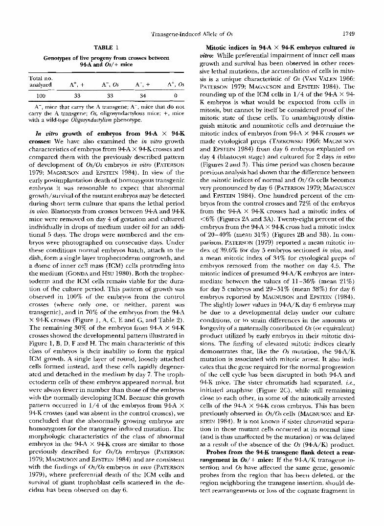
Complementation analysis of Os/+ and 94A mice: We crossed 9 4 A transgenic mice with Os/+ mice of the ROP/GnLe strain (from the JACKSON laboratory) or with Os/+ mice generated in the laboratory from the crosses between 9 4 A and ROP Os/+ mice. ROP/GnLe mice segregate three unlinked dominant (or semidomi- nant) mutations: Ragged on chromosome 2, Os on chro- mosome 8, and Pintail on chromosome 4. The Os/+ mice used in our crosses carried wild-type alleles of Ra and Pt. If the Os and 9 4 A mutations affect different genes, they should be able to complement each other and Os/+ progeny that carry the 9 4 A transgene should be viable. The Os phenotype was scored by inspecting the digits of the progeny, and the presence of the transgene was detected by Southern blotting using probes that can distinguish the transgene from the en- dogenous Pgkl gene. The results of these crosses are shown in Table 1. None of the 100 mice we have scored for the Os phenotype and the presence of the 9 4 A transgene were simultaneously Os and transgenic. The ratios of the mice in the three groups (Os, non- transgenic; wt for Os, transgenic; wt for Os, non- transgenic) closely match the expected ratios of surviv- ing progeny if the Os/transgenic class is embryonic lethal. The absence of live transgenic progeny with the Os phenotype indicates that 9 4 A is unable to comple- ment the recessive lethal effect of Os.
Transgene-Induced Allele of Os 1749
TABLE 1
Genotypes of live progeny from crosses between 94A and Os/+ mice
Total no. analyzed A+, + A-, Os A-, + A', Os
100 33 33 34 0
A', mice that carry the A transgene; A-, mice that do not carry the A transgene; Os, oligosyndactylous mice; +, mice with a wild-type Oligosyndactylism phenotype.
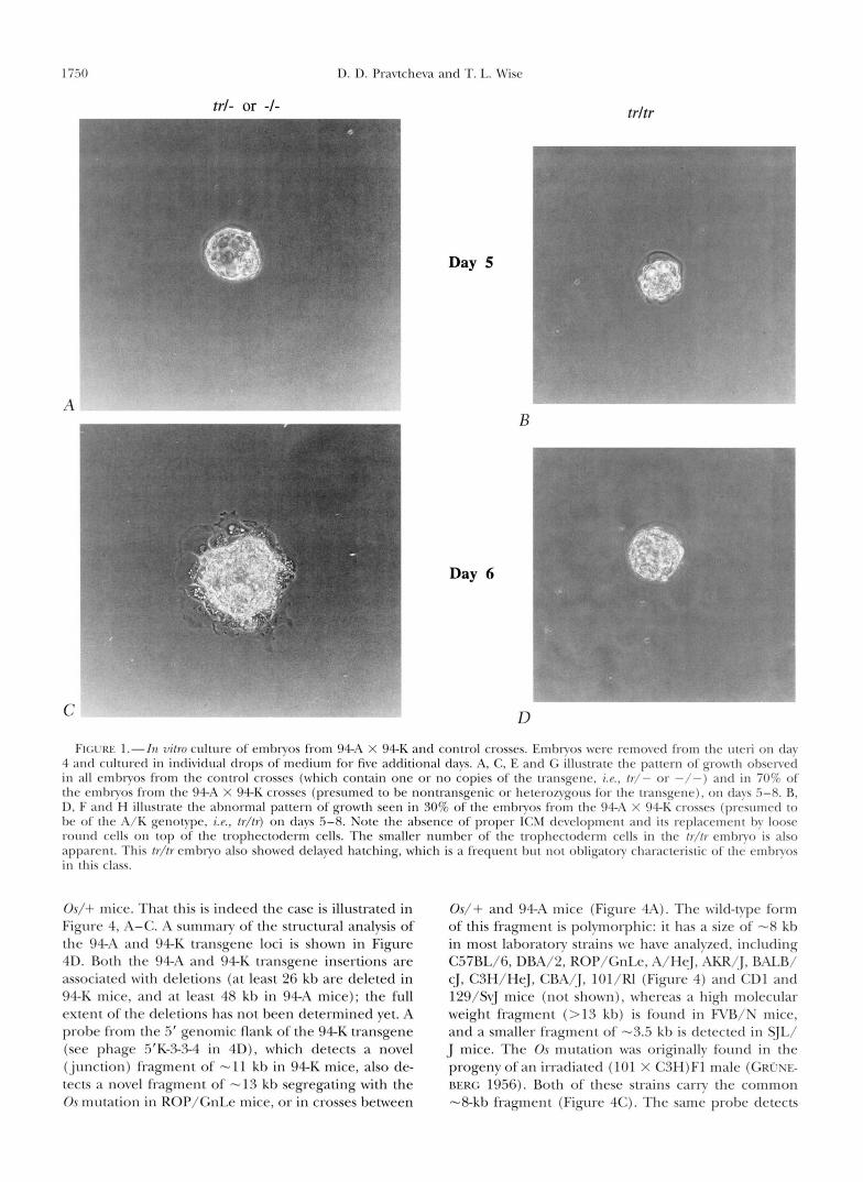
In vitro growth of embryos from 9 4 A X 94-K crosses: We have also examined the in vitro growth characteristics of embryos from 94A X 94K crosses and compared them with the previously described pattern of development of Os/Os embryos in vitro (PATERSON 1979; MAGNUSON and EPSTEIN 1984). In view of the early postimplantation death of homozygous transgenic embryos it was reasonable to expect that abnormal growth/survival of the mutant embryos may be detected during short term culture that spans the lethal period in vivo. Blastocysts from crosses between 94A and 94K mice were removed on day 4 of gestation and cultured individually in drops of medium under oil for an addi- tional 5 days. The drops were numbered and the em- bryos were photographed on consecutive days. Under these conditions normal embryos hatch, attach to the dish, form a single layer trophectoderm outgrowth, and a dome of inner cell mass (ICM) cells protruding into the medium (GONDA and HSU 1980). Both the trophec- toderm and the ICM cells remain viable for the dura- tion of the culture period. This pattern of growth was observed in 100% of the embryos from the control crosses (where only one, or neither, parent was transgenic), and in 70% of the embryos from the 94A X 94K crosses (Figure 1, A, C , E and G, and Table 2). The remaining 30% of the embryos from 94A X 94K crosses showed the developmental pattern illustrated in Figure 1, B, D, F and H. The main characteristic of this class of embryos is their inability to form the typical ICM growth. A single layer of round, loosely attached cells formed instead, and these cells rapidly degener- ated and detached in the medium by day 7. The troph- ectoderm cells of these embryos appeared normal, but were always fewer in number than those of the embryos with the normally developing ICM. Because this growth pattern occurred in 1/4 of the embryos from 94A X 94K crosses (and was absent in the control crosses), we concluded that the abnormally growing embryos are homozygotes for the transgene induced mutation. The morphologic characteristics of the class of abnormal embryos in the 94A X 94K cross are similar to those previously described for Os/Os embryos (PATERSON 1979; MAGNUSON and EPSTEIN 1984) and are consistent with the findings of Os/Os embryos in vivo (PATERSON 1979), where preferential death of the ICM cells and survival of giant trophoblast cells scattered in the de- cidua has been observed on day 6.
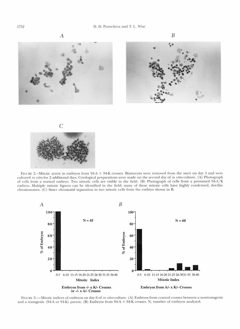
Mitotic indices in 94-A x 9 4 K embryos cultured in vitro: While preferential impairment of inner cell mass growth and survival has been observed in other reces- sive lethal mutations, the accumulation of cells in mito- sis is a unique characteristic of Os (VAN VALEN 1966; PATERSON 1979; MACNUSON and EPSTEIN 1984). The rounding up of the ICM cells in 1/4 of the 94A X 9 4 K embryos is what would be expected from cells in mitosis, but cannot by itself be considered proof of the mitotic state of these cells. To unambiguously distin- guish mitotic and nonmitotic cells and determine the mitotic index of embryos from 94A X 94K crosses we made cytological preps (TARKO~SKI 1966; MACNUSON and EPSTEIN 1984) from day 6 embryos explanted on day 4 (blastocyst stage) and cultured for 2 days in vitro (Figures 2 and 3). This time period was chosen because previous analysis had shown that the difference between the mitotic indices of normal and Os/Os cells becomes very pronounced by day 6 (PATERSON 1979; MAGNUSON and EPSTEIN 1984). One hundred percent of the em- bryos from the control crosses and 72% of the embryos from the 94A X 94K crosses had a mitotic index of <6% (Figures 2A and 3A). Twenty-eight percent of the embryos from the 94A X 94K cross had a mitotic index of 20-40% (mean 31%) (Figures 2B and 3B). In com- parison, PATERSON (1979) reported a mean mitotic in- dex of 39.6% for day 5 embryos sectioned in vivo, and a mean mitotic index of 34% for cytological preps of embryos removed from the mother on day 4.5. The mitotic indices of presumed 94A/K embryos are inter- mediate between the values of 11-36% (mean 21%) for day 5 embryos and 29-51% (mean 38%) for day 6 embryos reported by MAGNUSON and EPSTEIN (1984). The slightly lower values in 94A/K day 6 embryos may be due to a developmental delay under our culture conditions, or to strain differences in the amounts or longevity of a maternally contributed Os (or equivalent) product utilized by early embryos in their mitotic divi- sions. The finding of elevated mitotic indices clearly demonstrates that, like the Os mutation, the 94A/K mutation is associated with mitotic arrest. It also indi- cates that the gene required for the normal progression of the cell cycle has been disrupted in both 94A and 9 4 K mice. The sister chromatids had separated, i.e., initiated anaphase (Figure 2C), while still remaining close to each other, in some of the mitotically arrested cells of the 94A X 94K cross embryos. This has been previously observed in Os/Os cells (MAGNUSON and EP- STEIN 1984). It is not known if sister chromatid separa- tion in these mutant cells occurred at its normal time (and is thus unaffected by the mutation) or was delayed as a result of the absence of the Os (94A/K) product.
Probes from the 94-K transgene flank detect a rear- rangement in Os/+ mice: If the 94A/K transgene in- sertion and Os have affected the same gene, genomic probes from the region that has been deleted, or the region neighboring the transgene insertion, should de- tect rearrangements or loss of the cognate fragment in
1 i.50 D. D. Pravtcheva and T. L. Wise
trl- or -1- a
Day 5
trltr
C D FI(;rRE I.-In vi/ro culture of embryos from 94-A X 94-K and control crosses. Embryos were removed from the uteri o n clay
4 and cultured in individual drops of medium for five additional days. A, C, E and G illustrate the pattern of growth obsenerl in all embryos from the control crosses (which contain one or no copies of the transgrnr, i .~ . , / r / - o r - / - ) a n r l in 70% of the embryos from the 9 4 A X 94-K crosses (presumed to be nontransgenic or heterozygous for the transgene), o n days .5-8. R, D, F antl H illustrate the almormal pattern of growth seen in 30% of the embryos from the 94-11 X 94-E; crossrs (prcsumctl to be o f the A/K genotvpe, i . ~ . , /r / /r) on days .5-8. Note the absence of proper ICM development antl its replacemrnt by loose round cells on top of the trophectoderm cells. The smaller number of the trophectotlerm cells i n the / r / / r rmbryo is also apparent. This /r//rernbryo also showed delayed hatching, which is a frequent but not obligatory characteristic of the embryos i n this class.
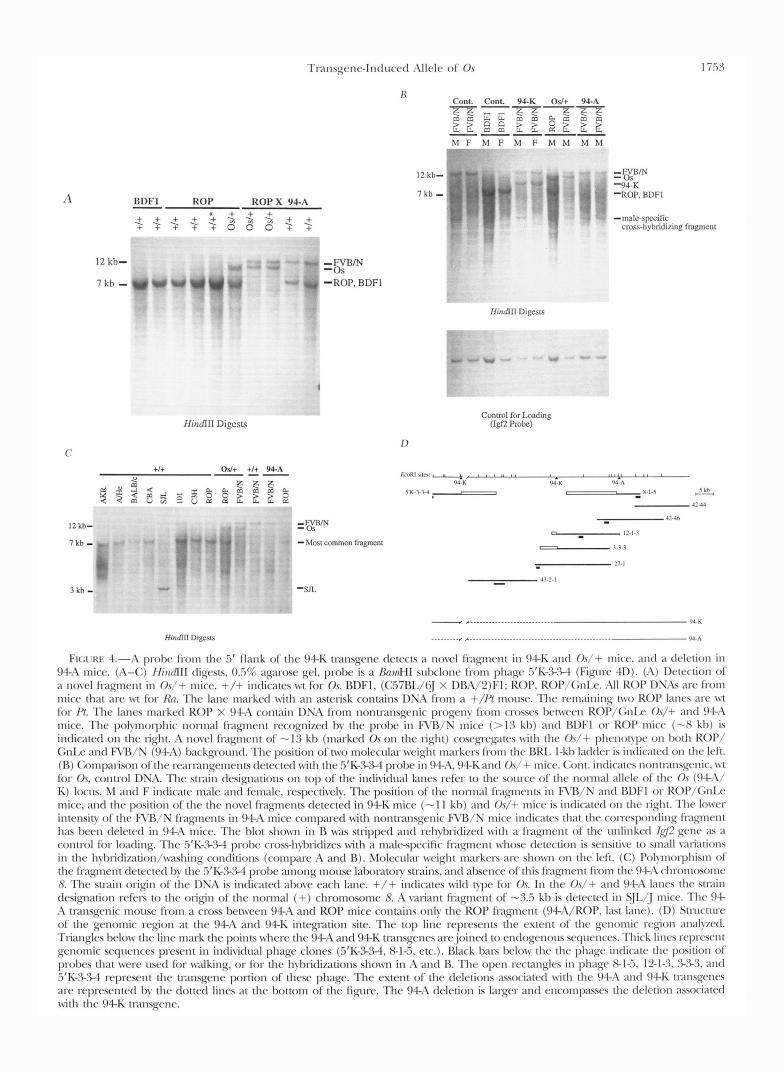
O v / + mice. That this is indeed the case is illustrated in Figure 4, A-C. A summary of the structural analysis of the 94-A and 94-K transgene loci is shown in Figure 4D. Both the 94-A and 94-K transgene insertions are associated with deletions (at least 26 kh are deleted in 94-K mice, and at least 48 kh in 94-A mice); the full extent of the deletions has not been determined yet. A probe from the .if genomic flank of the 94-K transgene (see phage 5’K-S.cs-4 in 4D), which detects a novel (junction) fragment of - 1 1 kh in 9 4 K mice, also de- tects a novel fragment of -13 kh segregating with the Os mutation in ROP/GnLe mice, o r in crosses between
Os/+ and 94-A mice (Figure 4A). The wild-type form of this fragment is polymorphic: i t has a size of -8 kh in most laboratory strains we have analyzed, including C57BL/6, DBA/2, ROP/GnLde, A/HqJ, AKR/J, BALB/ cJ, CSH/HeJ, CRA/J, 101/RI (Figure 4) and CD1 and 129/SvJ mice (not shown), whereas a high molecular weight fragment (>13 kh) is found in FVR/N mice, and a smaller fragment of - 3 3 kh is detected in SJL/ J mice. The 0 . ~ mutation was originally found in the progeny of an irradiated (101 X C3H)Fl male ( G R ~ s E - BERG 1956). Both of these strains carry the common -8-kh fragment (Figure 4C). The same probe detects
Transgene-Induced Allele of Os 1 7.5 1
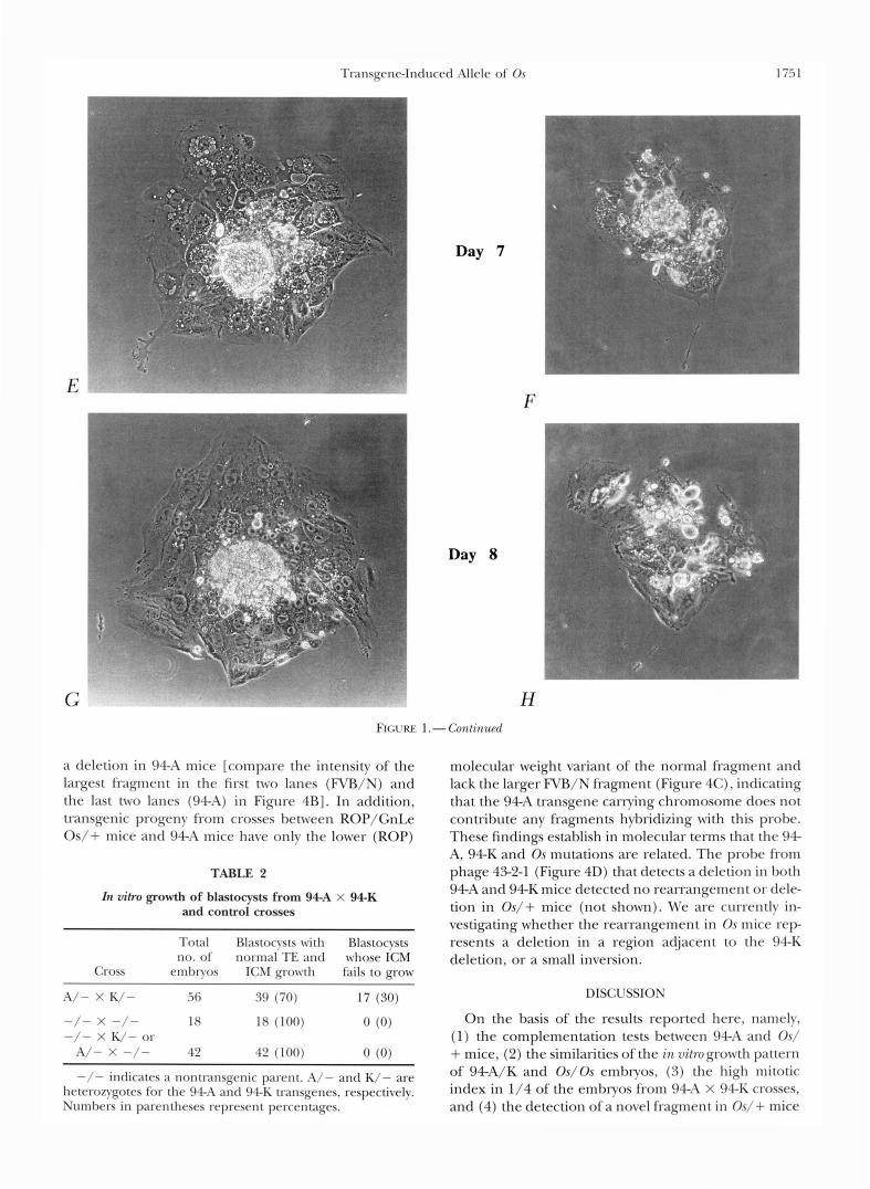
Day 7
E
Day 8
F
FIGURE 1 .- Conlinzcd
a deletion in 94-A mice [compare the intensity of the largest fragment in the first two lanes (FVB/N) and the last two lanes (94-A) in Figure 4B]. In addition, transgenic progeny from crosses between ROP/GnLe Os/+ mice and 94A mice have only the lower (ROP)
TABLE 2
In vihp growth of blastocysts from 9 4 A X 9 4 K and control crosses
Total Blastocysts with Blastocvsts no. of normal TE and whose ICM
Cross embryos ICM growth fails to grow
molecular weight variant of the normal fragment and lack the larger FVB/N fragment (Figure 4C), indicating that the 94A transgene carrying chromosome does not contribute any fragments hybridizing with this probe. These findings establish in molecular terms that the 94- A, 94K and Os mutations are related. The probe from phage 43-2-1 (Figure 4D) that detects a deletion in both 94A and 94K mice detected no rearrangement or dele- tion in Os/+ mice (not shown). We are currently in- vestigating whether the rearrangement in Os mice r e p resents a deletion in a region adjacent to the 94-K deletion, or a small inversion.
A/- X K/- 56 39 (70) 17 ( S O ) DISCUSSION
-/- x -/- 18 18 (100) 0 (0) On the basis of the results reported here, namelv, -/- X K/- or ( 1 ) the complementation tests between 94-A and Os/
-/- indicates a nontransgenic parent. A/- and K/- are of 94A/K and Os/Os embryos, (3) the high mitotic heterozygotes for the 94-A and 94-K transgenes, respectively. index in 1/4 of the embryos from 94A X 94-K crosses, Numbers in parentheses represent percentages. and (4) the detection of a novel fragment i n Os/+ mice
A/- X -/- 42 42 (100) 0 ( 0 ) + mice, (2) the similarities of the in 7drogrowth pattern
D. D. Pravtchera antl T. I,. \Vise
A - c
C
B
FIGI'RE 2.-Mitotic arrest in embryos from 94-A X 94-K crosses. Blastocysts were removed from the uteri on day 4 and were cultured in 7~itro for 2 additional days. Cytological preparations were made on the second day of in vitro culture. (A) Photograph of cells from a normal embryo. Two mitotic cells are visible in the field. (R) Photograph of cells from a presumed W A / K embryo. Multiple mitotic figures can be identified in the field; many o f these mitotic cells have highly condensed, dot-like chromosomes. (C) Sister chromatid separation in two mitotic cells from the embryo shown in B.
A B
N =60 80
0-5 6-10 11-15 16-20 21-25 26-30 31-35 36-40 0-5 6-10 11-15 16-2021-25 26-3031-3s 3640
Mitotic Index Mitotic Index
Embryos from -I- x W- Crosses or -I- x AI- Crosses
Embryos from AI- x W- Crosses
FIGLKE 3.-Mitotic indices of embryos on day 6 of in vitro culture. (A) Embryos from control crosses between a nontransgenic antl a transgenic (94-A or 94-K) parent. (R) Embryos from 94-A X 94-K crosses. N, number of embryos analyzed.
T ~ l ~ l s ~ c ~ l ~ - I ~ l t l u c r t l ; \ I Ic Iv o f 0 , s 1 i.5 3
. . . . . . . . . . . I-. -.- ~~~~ ~~~~, ~~~~~~~~~~~~~~~~ ~~~~~~ ~~~~~~~ ~ ~~ ~ ~~ . ..
FK;I‘KI< 4.-;\ p d x from the .5’ f l m k o f the 94-I( tl-ansgcnc tlctc~ts a novc.l fqmcvlt i n 94-I( and Or/+ mice. ;lnd :I t l r l r t i o n i n 94-A mice. (I\-(:) /findIII tligvsts, 03% ;1prose gel, probe is a BmnHI sulxhnc~ from p h q c .5’1<->34 (Fiqqurc 41)). (I\) 1)ctcction of a n o \ ~ h l fragment i n O s / + mice. +/+ indicates w t for Or BDFl, (C.57BL/6] X DRA/2)Fl; KOP, ROI’/<;nI.(~. A l l ROP 1)S;ls m-e from mice that m-c w t for /h. The lane m;1rkerl w i t h an asterisk contains DNA from a +//? m o ~ ~ s e . The remaining t w o KOP Imrs arc wt for /’/. The lanes m;u-krtl ROP X 94-A cont;1in DNA from nonttxnsgenic progeny from crosses Ix~twcwl ROl’/(;nIx O s / + and <)+I\
mice. The polynorphic nonnal fklqncmt recognized by the probe i n R’R/S mice (> 1 3 kl)) and RDFI or KOP mice (-X k l ~ ) is indicated o n the light. A novel fkigment o f - 1 3 kh (marked 0 , s on the light) cosrgl-c’ptc-s w i t h thc O s / + phrnol!l~r on both KoP/ G n k and FVIlB/N (!)4-;\) I m A q w u l t l . Thc position of two molecular weight 1n;lrkrls from the RRI, I-kb Ixldcr is intlicatrtl o n the left. (R) (:omp;wison of the r ~ ~ ~ l r ~ ~ i ~ l g e ~ n ~ ~ ~ l t s dctrctctl with the .5’I(-%.W probc i n 9 4 4 , 94-I( antl O s / + nlicc.. (:ant. intlicatcs nontl;~nsg~~~lic. w t for O.s, control DNA. The stlain designations on top of the indi\itlu;d I;Ines rcfcr t o the source of the nom1al ; d l r l r of the O r ( 9 - l - 1 b ‘
K) locrn. M antl F intlicate male ant1 female, respectively. The position of the nonn;ll f1xgments i n RB/S antl IWF1 o r R<)l’/GnIx. mice, antl t h e position of the the novc4 fragments drtected in 94-I( mice (- 1 1 kh) antl O s / + mice is indicated on the right. Thr lo\vcr intensity of t h e R’B/N f1-agmcnts i n 94-A mice compared nith nontnnsgenic F\rR/N mice indicates that thc con-cyx)nding fl:lp1el1t h;ls hccn tlrlctcd i n 94-A mice. The I d o t shown i n R was stripped antl rchylxitlizctl w i t h a lixgncnt of t h e unlinked (42 g e n ~ 21s ;I
contn>l for loading. The .5’K->%l prol~c cross-hybridizes with :I malc-specific frqmrnt whose detection is sensitive t o small variations i n thc h~I~~i t l i~~~t ion/~v;~shing conditions (compare I\ and R). Molccul;u- weight m;ukrn :~rc shown on thc left. (C) Polynoq>hism o f the fragtncnt drtectcrl by t h r .5‘I(->3-4 probe among mouse lalmntoy stlains. a n d ;d~srncc. of this l i p ~ c n t from the 94.A chronlosomc 8 Thc stnin origin of the I)?&\ is indicated above each lane. +/+ indicates w i l d t y p e fior O s . I n the Or/+ and 94-A lancs the stlain designation rcf(w t o the origin of the nonnal (+) chromosome 8. A \;lIiant f1apnent of - 3 3 kl, is t l c~c ted i n S]I,[j mice. The 94- h t1xnsgenic mousc from a cross Ix-tween 94-.L\ and ROP mice contains only the ROP f1agncnt (94-,-\/ROP, last lane). (I)) Stnlcture of thr genomic ~ ~ g i o n a t the !)+A and 94-I( intep-ation site. The top line I-cpresmts the extent of the genomic region ; ln: l lycd.
Trianglrs I x h v t h r line mark t h e points where the 94-A and 94-K transgenes are.joinctl t o cnrlogcnous scqucncrs. Thick linrs rrprcsc11t gcnonlic scqwnces present i n intli\itlual phage clones (3‘I(->.W, 8-1-5, etc.). Black han I M ~ W the the phage intlic;ltc the position of prohcs that w c w usctl for walking, o r for the I1ybridi;rAons show1 i n A and R. The open rectanglcs i n phage 8 - 1 4 . 12-13, 3-%3, and .5‘K->.M represent the transgene portion of these phage. The extent of the deletions aswciatcd w i t h t h e 944 ;und 94-I( tlilnsgt‘11es arc ~rprescntctl by thc clotted lines a t the bottom o f the figure. The 94-A dcletion is larger antl cncompasscs the tlrlrtion ;asociated with the 94-K t~-ansgcnr.

1754 D. D. Pravtcheva and T. L. Wise
by a probe from the 5' flank of the 94K transgene, we conclude that the insertional mutation in 94A/K mice has affected the same gene that is responsible for the mitotic arrest and early postimplantation lethality of Os/Os embryos. The preferential involvement of ICM cells in 94A/K and Os/Os embryos would be attributed to the continuous mitotic activity of these cells and their need for the 94A/K (Os) gene product, whereas troph- ectoderm cells, which undergo endoreduplication with- out mitosis, would be able to survive without it. The absence of limb abnormalities in the 94A and 94K mice is most easily explained by assuming that the radia- tion-induced Os mutation has affected more than one gene (i .e. , through a deletion or an inversion), and that the limb and kidney abnormalities and the postimplan- tation lethality are due to defects in different genes. Further molecular characterization of Oligosyndactylism will determine if this is the case.
Studies by MAGNUSON and EPSTEIN (1984) on the nature of the mitotic block in Os/Os cells have deter- mined that these cells are unable to make the meta- phase/anaphase transition (as indicated by the chro- mosome alignment on the metaphase plate). In normal cells metaphase is followed by disjoining of the sister chromatids and their movement toward the poles (ana- phase A) and elongation of the mitotic spindle (ana- phase B) . The metaphase-anaphase transition repre- sents one of the checkpoints in the cell cycle (HARTWELL and WEINERT 1989; HOW et al. 1991; LI and MURRAY 1991; MURRAY 1992; WELLS 1996). Its function is to assess the formation of proper microtubule/kinet- ochore attachments and chromosome alignment, and to prevent progress in the cell cycle (and the ensuing aneuploidy) if improperly attached, misaligned, lagging chromosomes are detected in the cell (CAMPBELL and GORESKY 1995; LI and NICKLAS 1995; RIEDER et al. 1995). A variety of factors that affect the dynamics of microtu- bule assembly/disassembly and interaction with the ki- netochore may be able to activate this checkpoint and delay or block exit from metaphase. The mitotic spin- dle, centrosomes, and kinetochores are apparently nor- mal in Os/ Os cells (MAGNUSON and EPSTEIN 1984). Sim- ilarly, no abnormalities were detected in CY and /3 tubulin, and the spindle microtubules showed normal sensitivity to cold temperatures and depolymerizing agents (MAGNUSON and EPSTEIN 1984). Thus obvious abnormalities in the structural components of the mi- totic apparatus are unlikely to be the reason for the mitotic arrest of Os/Os cells. It is known, however, that drugs that induce mitotic arrest by affecting microtu- bule stability are able to block cells in mitosis even at concentrations that have little visible effect on the struc- ture of the mitotic spindle (JORDAN et al. 1992). The existence of such subtle alterations in the components of the microtubule/kinetochore interaction in Os/ Os (94A/K) cells at present cannot be ruled out. It is therefore possible that the role of the Os (94A/K) gene product may be in normal microtubule/kinetochore
attachment and dynamics, and its absence may prevent the cells from passage through the metaphase/ana- phase checkpoint.
Alternatively, the Os/Os (94A/K) embryo cells may lack a product required to activate and carry out the complex series of events that are initiated upon suc- cessful passage through this checkpoint. A crucial event in this cell cycle transition is the degradation of cyclin B and the ensuing inactivation of the matura- tion-promoting factor, a complex of cyclin B with p34'd'2 (NURSE 1990; KING et al. 1994). Cyclin B degra- dation is mediated by polyubiquitination and requires the normal function of a multiprotein complex (ana- phase-promoting complex) that includes the products of the budding yeast genes CDCl6, CDC23 and CDC27, or their homologues in other species (LAMB et al. 1994; IRNICER et al. 1995; KING et al. 1995; MURRAY 199513; TUGENDREICH et al. 1995). Activation of the spindle assembly checkpoint results in sustained high activity of MPF (HUNT et al. 1992; MINSHULL et al. 1994). In addition, species differences in the duration of the cell cycle block imposed by the spindle assembly checkpoint correlate with the ability to maintain high MPF activity (KUNG et al. 1990). Thus genetic defects that prevent the normal decline in activity of MPF would be expected to result in a mitotic block. How- ever, maintaining high levels of cyclin B (and MPF activity) through overproduction of the protein, or through use of nondegradable cyclin, delays chromo- some decondensation, nuclear membrane reconstitu- tion, and cytokinesis, but has no effect on sister chro- matid separation, which proceeds on an unaltered schedule to form two separate clusters at the opposite poles of the spindle (HOLLOWAY et al. 1993; SURANA et al. 1993; IRNIGER et al. 1995). Introducing nondegrad- able cyclin into human HeLa cells also did not prevent the movement of the chromosomes away from the metaphase plate (GALLANT and NIGG 1992). On the other hand, sister chromatid separation and anaphase movement were prevented by introducing an N-termi- nal fragment of cyclin B, which is believed to interfere with the degradation of an yet unknown protein that may act as an inhibitor of the normal process of sister chromatid separation (HOLLOWAY et al. 1993). At pres- ent we do not know if sister chromatid separation in Os/Os (94A/K) embryos is delayed (suggesting a possi- ble defect that slows down the degradation of the pro- tein maintaining sister chromatid cohesion), or takes place at its normal time. The metaphase arrest of Os/ Os embryo cells, however, suggests a mitotic block at an earlier stage than the mitotic block observed in the presence of high cyclin B levels and MPF activity.
The transition from metaphase to anaphase is accom- panied by marked changes in the degree of phosphory- lation of a large number of cellular proteins. The mono- clonal antibody MPM2 recognizes a phosphorylated epitope shared by at least 16 different proteins located on the chromosomes or in the cytoplasm of mitotic
Transgene-Induced Allele of Os 1755
cells in a wide variety of species (DAVIS et al. 1983). In normal cells the MPM2 reactivity appears with entry into mitosis and is lost as cells exit from metaphase. Cells arrested in metaphase by Nocodazole (a treatment that results in microtubule depolymerization and activa- tion of the metaphase-anaphase transition checkpoint) maintain their high level of MPM2 staining for as long as 18 hr (VANDRE and BORIW 1989). In this respect it is interesting to note that some mitotically arrested cells in Os/Os embryos have been found to exhibit no fluo- rescence (i.e., to have a postmetaphase type staining) with the MPM2 antibody (HIRAOKA et al. 1989). This may point to some defect in the process of phosphoryla- tion of the MPM2 epitope (which accompanies entry into mitosis) in Os/Os cells (HIRAOKA et al. 1989). Alternatively, the absence of MPM2 staining in some Os/ Os mitotic cells may be the result of dephosphoryla- tion that normally takes place at the metaphase/ana- phase transition. In this case, the absence of staining in these cells will simply reflect a more advanced stage in the cell cycle that they have been able to reach in the absence of the Os (94A/K) gene product. The sepa- ration of sister chromatids in fission yeast requires the action of phosphatases (OHKURA et al. 1989). The nor- mal course of anaphase in Aspergzllus nidulans (DOONAN and MORRIS 1989), Drosophila (MAYER~AEKEL et al. 1993), and mammals (VANDRE and WILLS 1992) also requires the activity of protein phosphatases (PP) . In A. nidulans, PP1 is required for the removal of the MPM2 epitope (DOONAN and MORRIS 1989). In this regard it would be interesting to determine if the cells in which the sister chromatids have already been separated (as in Figure 2C) are the ones that have lost the phosporylated epitope recognized by MPM2.
Studies on mitosis in frog egg extracts have led to the suggestion that poleward movement of sister chro- matids is an automatic consequence of the loss of cohe- sion between the sister chromatids: no change in the force applied to the kinetochore has been noted at the transition from metaphase to anaphase (HOLLOWAY et al. 1993). If this is so in mammalian cells, the absence of anaphase movement of sister chromatids that have clearly been separated from each other in 94A/K (Fig- ure 2C) and in Os/ Os embryos (MAGNUSON and EPSTEIN 1984) is unexplained. In normal cells this chromosome movement is initially mainly the result of depolymeriza- tion of the plus (kinetochore) end of the kinetochore fibers (microtubules connecting the kinetochore and the spindle pole) and appears to require the presence of motor proteins that can couple microtubule depoly- merization and chromosome movement (MITCHISON et ul. 1986; GORBSKY et al. 1987; DESAI and MITCHISON 1995). Separation of sister chromatids without pole- ward movement has been observed in some cells treated with microtubule depolymerizing agents (RIEDER and PALAzzO 1992), where it can be explained by the micro- tubule effects of these agents. The limited studies on the mitotic spindle in Os/Os embryos do not preclude
the existence of subtle alterations that prevent the pole- ward separation of the sister chromatids. However, the observation of premature centromere division and sis- ter chromatid separation without poleward movement as an age related, or inherited, cytogenetic abnormality in humans suggests the possibility that rather than be- ing an automatic consequence of loss of sister chroma- tid cohesion, anaphase movement of sister chromatids is itself an independently regulated cell cycle event (FITZGERALD et al. 1975; RUDD et al. 1983). Elucidation of the molecular nature of the Os (94A/K) mutation may bring about a better understanding of the consecu- tive steps in the metaphase/anaphase transition.
In summary, we have characterized an insertional mutation in transgenic mice and have determined that it represents a new allele of Oligosyndactylism. Os affects a cellular process that is essential for the normal transi- tion of the cell division cycle from metaphase to ana- phase. The insertional mutation will facilitate the isola- tion and characterization of the affected gene and will allow a better understanding of the sequence of molec- ular and cytologically observable events (dephosphory- lation, protein degradation, sister chromatid separa- tion) at the metaphase-anaphase transition.
We thank Dr. LIANE B. RUSSEIL for the gift of mouse strain 101/ Rl DNA. We acknowledge the excellent technical assistance of SARA SEEMATTER. This work was supported by the Pediatric Research Insti- tute of Cardinal Glennon Children’s Hospital New Project Develop- ment Fund, and by a Fleurde-Lis grant to D.P.
LITERATURE CITED
CAMPBELL, M. S., and G. J. GORBSKY, 1995 Microinjection of mitotic cells with the 3F3/2 anti-phosphoepitope antibody delays the onset of anaphase. J. Cell Biol. 129: 1195-1204.
CECI, J. D., 1994 Mouse chromosome 8. Mamm. Genome 5: 5124- 5138.
DAVIS, F. M., T. Y. TSAO, S. K. FOWLER and P. N. RAO, 1983 Mono- clonal antibodies to mitotic cells. Proc. Natl. Acad. Sci. USA 80:
DESAI, A,, and T. J. MITCHISON, 1995 A new role for motor proteins as couplers to depolymerizing microtubules. J. Cell Biol. 128: 1- 4.
DOONAN, J. H., and N. R. MORRIS, 1989 The bimC gene of Aspergil- lus nidulans required for completion of anaphase encodes a homolog of mammalian phosphoprotein phosphatase 1. Cell 57:
FALCONER, D. S., M. LATYSZEWSKI and J. H. ISAACSON, 1964 Diabetes insipidus associated with Olzgosynductylism in the mouse. Genet. Res. 5: 473-488.
FITZGERALD, P. H., A. F. PICKERING, J. M. MERCER and P. M. MIETHKE, 1975 Premature centromere division: a mechanism of nondis-
junction causing X chromosome aneuplioidy in somatic cells. Ann. Hum. Genet. 38: 417-428.
GALLANT, P., and E. A. NIGG, 1992 Cyclin B2 undergoes cell cycle- dependent nuclear translocation and, when expressed as a non-
Biol. 117: 213-224. destructible mutant, causes mitotic arrest in HeLa cells. J. Cell
GONDA, M. A., and Y.-C. HSU, 1980 Correlative scanning electron, transmission electron, and light microscope studies of the in uitro development of mouse embryos on a plastic substrate at the implantation stage. J. Emblyol. Exp. Morphol. 5 6 23-39.
GORBSKY, G. J., P. J. SA”AK and G. G. BORISY, 1987 Chromosomes move poleward in anaphase along stationary microtubules that coordinately disassemble from their kinetochore ends. J. Cell Biol. 104 9-18.
GR~NEBERG, H., 1956 Genetical studies on the skeleton of the
2926-2930.
987-996.
1756 D. D. Pravtcheva and T. L. Wise
mouse. XVIII. Three genes for syndactylism. J. Genet. 54: 113- 145.
GR~NEBERG, H., 1961 Genetical studies on the skeleton of the mouse. XXVII. The development of Oligosyndactylism. Genet. Res.
H A R T W ~ I . , L. H., and M. B. KASTAN, 1994 Cell cycle control and cancer. Science 266: 1821-1828.
HARTWELI., L. H., and T. A. WEINERT, 1989 Checkpoints: controls that ensure the order of cell cycle events. Science 246: 629-634.
HIRAOKA, L., W. GOLDEN and T. MAGNUSON, 1989 Spindle pole organization during early mouse development. Dev. Biol. 133: 24-36.
HOGAN, B., R. BEDDINGTON, F. COSTANTINI and E. LAW, 1994 M u n i p - lating the Mouse Emblyo: A Laboratoly Manual, Ed. 2. Cold Spring Harbor Laboratory Press, Cold Spring Harbor, NY.
HOI.I.OMJ.Y, S. L., M. GLOTZER, R. W. KING and A. W. MURRAY, 1993 Anaphase is initiated by proteolysis rather than by the inactiva- tion of maturation-promoting factor. Cell 73: 1393-1402.
Hour, M. A., L. TOTIS and B. T. ROBERTS, 1991 S. rmruisiae genes required for cell cycle arrest in response to loss of microtubule function. Cell 66: 507-517.
HL~NT, T., F. C. Luciz and J. V. RLWERMAN, 1992 The requirements for protein synthesis and degradation, and the control of destruc- tion of cyclins A and B in the meitotic and mitotic cell cycles of the clam embryo. J. Cell Biol. 116: 707-724.
IKNIGER, S., S. PIATTI, C. MICHAEIJS and K. NASMYTH, 1995 Genes involved in sister chromatid separation are needed for &type cyclin proteolysis in budding yeast. Cell 81: 269-277.
JORDAN, M. A,, D. THROWER and L. WILSON, 1992 Effects ofvinblas- tine, podophyllotoxin, and nocodazole on mitotic spindles. Im- plications for the role of microtubule dynamics in mitosis. J. Cell Sci. 102: 401-416.
KAIIAM, K. M., 1962 Genetical studies on the skeleton of the mouse. XXXI. The muscular anatomy of .syndac[ylism and Oligoryndactyl- i5m. Genet. Res. 3: 139-156.
KING, R. W., P. K. JACK~ON and M. W. URSCHNER, 1994 Mitosis in transition. Cell 79: 563-571.
KING, R. W., J.-M. PKTERS, S. TUGENDKEICH, M. ROL.FE, P. HIETER el a/., 1995 A 20s complex containing CDC27 and CDC16 cata- lyzes the mitosis-specific conjugation of ubiquitin to cyclin B. Cell 81: 279-288.
KUNG, A. L., S. W. SHERWOOD and R. T. SCHIMKE, 1990 Cell line- specific differences in the control of cell cycle progression in the absence of mitosis. Proc. Natl. Acad. Sci. USA 87: 9553- 9557.
LAMR, J. R., W. A. MICHAUD, R. S. SI~~ORSKI and P. HIETER, 1994 Cdcl6p, Cdc23p, and Cdc27p form a complex essential for mito- sis. EMBO J. 13: 4321-43'28.
LI, R., and A. W. MURKAY, 1991 Feedback control of mitosis in bud- ding yeast. Cell 66: 519-531.
LI, X., and R. B. NIC:KIAS, 1995 Mitotic forces control a cell cycle checkpoint. Nature 373: 630-632.
MAC;NIISON, T., and C. J. EPRI'EIN, 1984 Oligosyndactyly: a lethal muta- tion in the mouse that results in mitotic arrest very early in development. Cell 38: 823-833.
MAYER~AEKEI., R. E., H. OHKUKA, R. GOMES, C. E. SUNKEI., S. BAUM- (;,wrNk:R et al., 1993 The 55 kd regulatory subunit of drosophila protein phosphatase 2A is required for anaphase. Cell 72: 621- 633.
MCINTOSH, J. R., and M. P. K o o ~ c e , 1989 Mitosis. Science 246 622-628.
MINSHUIJ., J., H. SUN, N. K. TONKS and A. W. MURRAY, 1994 A MAP kinasedependent spindle assembly checkpoint in Xenopus egg
2: 33-42.
extracts. Cell 79: 475-486.
MITCHISON, T., L. EVANS, E. SCHLLZE and M. KIRSCHNER, 1986 Sites of microtubule assembly and disassembly in the mitotic spindle. Cell 45: 515-527.
MURRAY, A. W., 1992 Creative blocks: cell cycle checkpoinB and feedback controls. Nature 359: 599-602.
MURRAY, A. W., 1995a The genetics of cell cycle checkpoints. Curr. Opin. Genet. Dev. 5: 5-11.
MURRAY, A. W., 1995b Cyclin ubiqnitination: the destructive end of mitosis. Cell 81: 149-152.
Nt:Ksk., P., 1990 Universal control mechanism regulating onset of metaphase. Nature 344: 503-508.
OIIKL'RA, H., N. KINOSHITA, S. MIYATANI, T. TOIM and M. YAN.M;IIIA, 1989 The fission yeast dZs2+ gene required for chromosome disjoining encodes one of two putative type 1 protein phospha-
PATERSON, H. F., 1979 In vivo and in vitro studies on the early ern- hryonic lethal Oligosyndactylism (Os) i n the mouse. J. Embryol. Exp. Morphol. 52: 115-125.
PRAVKHEVA, D. D., and T. 1,. WISE, 1995 A postimplantation lethal mutation induced by transgene insertion on mouse chromosome 8. Genornics 30: 529-544.
PRAVTC:FIEVA, D. D., C. N. ADRA and F. H. RUDDI.E, I991 Timing of paternal Pgkl expression in embryos of transgenic mice. Develop- ment 111: 1109-1120.
RIEDER, C . L., and R. E. PAIALZO, 1992 Colcemid and the mitotic cycle. J. Cell Sci. 102: 387-392.
RIEDER, C. L., R. W. COLE, A. K H O Q ~ K O V and G. SLUDER, 1995 The checkpoint delaying anaphase in response to chromosome mo- noorientation is mediated by an inhibitory signal produced by unattached kinetochores. J. Cell Biol. 1 3 0 941 -948.
Rrlnn, N. I.., 1. E. TESIIIMA, R. H. MARTIN, J. E. SISKEN and R. WEKS BERG, 1983 A dominantly inherited cytogenetic anomaly. A pos- sible cell division mutant. Hum. Genet. 65: 117-121.
SAMBROOK, J., E. F. FRITSCH and T. MANWTIS, I989 Molecular C l m - zng. A Labomtory Manual, Ed. 2. Cold Spring Harbor Laboratory Press, Cold Spring Harbor, NY.
S r w , w r , A. D., and J. STEWAR'I', 1969 Studies on syndrome of.diabe- tes insipidus associated with O/igosyndactylism in mice. Am. J. Phys- iol. 217: 1191-1198.
SUKANA, U., A. AMOX, C. DOWER, J. MC:C;REW, B. BYERS rt ul., 1993 Destruction of CDC28/CLB mitotic kinase is not required for the metaphase to anaphase transition in budding yeast. EMBO
TARKOWSKI, A. K., 1966 An air-drying method for chromosome preparations from mouse eggs. Cytogenet. 5: 394-400.
T o ~ ~ ~ l ~ ~ l ; l c : r ~ , S., J. TOMKIEI., W. EARNSHAW and P. HIKI'ER, 1995 CDC27Hs colocalizes with CDC16Hs to the centrosome and mi- totic spindle and is essential for the metaphase to anaphase tran- sition. Cell 81: 261-268.
VAN VAIXN, P., 1966 Oligosynductylism, an early embryonic lethal in the mouse. J. Embryol. Exp. Morphol. 1 5 119-124.
VANL)RE, D. D., and G. G. BORIS,, 1989 Anaphase onset and dephos- phorylation of mitotic phosphoproteins occur concomitantly. ,J. Cell Sci. 94: 245-258.
VANIMW, D. D., and V. L,. WIILS, 1992 Inhibition of mitosis by oka- daic acid: possible involvement of a protein phosphatase 2A in the transition form metaphase to anaphase. J. Cell Sci. 101: 79- 91.
WEI.I.S, W. A. E., 1996 The spindle assembly checkpoint: aiming for a perfect mitosis, every time. Trends Cell Biol. 6: 228-234.
YEE, D,, W. GOLDEX, S. DKHROT and T. MAGNI:SON, 1987 Short term rescue by RNA injection of a mitotic arrest mutation that affects the preimplantation mouse embryo. Dev. Biol. 122: 256-261.
tases. (:ell 57: 997- 1007.
J. 12: 1969-1978.
Communicating editor: N. A. JtNKINS