Embed Size (px)
Citation preview
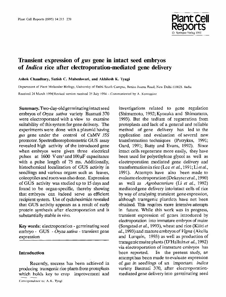
Plant Cell Reports (1995) 14:215-220 Plant Cell Reports �9 Springer-Verlag 1995
Transient expression of gus gene in intact seed embryos of lndica rice after electroporation-mediated gene delivery
Ashok Chaudhury, Satish C. Maheshwari, and Akhilesh K. Tyagi
Department of Plant Molecular Biology, University of Delhi South Campus, Benito Juarez Road, New Delhi-ll0021, India
Received 24 March 1994/Revised version received 25 July 1994- Communicated by A. Komamine
Summary. Two-day-old germinating intact seed embryos of Oryza sativa variety Basmati 370 were electroporated with a view to examine suitability of this system for gene delivery. The experiments were done with a plasmid having gus gene under the control of CaMV 35S promoter. Spectrofluorophotometric GUS assay revealed high activity of the introduced gene when embryos were given three electrical pulses at 1600 V cm 1 and 100/zF capacitance with a pulse length of 75 ms. Additionally, histochemical localization of GUS activity in seedlings and various organs such as leaves, coleoptiles and roots was also done. Expression of GUS activity was studied up to 15 days and found to be organ-specific, thereby showing that embryos can indeed serve as efficient recipient system. Use of cycloheximide revealed that GUS activity appears as a result of early protein synthesis after electroporation and is substantially stable in vivo.
Key words: electroporation - germinating seed embryo - GUS - Oryza sativa - transient gene expression
Introduct ion
Recently, success has been achieved in producing transgenic rice plants from protoplasts which holds key to crop improvement and
Correspondence to: A.K. Tyagi
investigations related to gene regulation (Shimamoto, 1992; Kyozuka and Shimamoto, 1993). But the tedium of regneration from protoplasts and lack of a general and reliable method of gene delivery has led to the application and evaluation of several new transformation techniques (Potrykus, 1991; Oard, 1991; Batty and Evans, 1992). Since intact cells regenerate more easily, they have been used for polyethylene glycol as well as electroporation mediated gene delivery and transformation in rice (Lee et aL, 1991; Li et aL, 1991). Attempts have also been made to evaluate electroporation (Dekeyser et aL, 1990) as well as Agrobacterium (Li et aL, 1992) mediated gene delivery into intact cells of rice by way of analysing transient gene expression, although transgenic plantlets have not been obtained. This requires more intensive attempts in future. While this work was in progress, transient expression of genes introduced by electroporation into immature embryos of maize (Songstad et al., 1993), wheat and rice (K16ti et al., 1993) and mature embryos of Vigna (Akella and Lurquin, 1993) as well as production of transgenic maize plants (D'Halluin et aL, 1992) via electroporation of immature embryos has been reported. In the present study, an attempt has been made to evaluate expression of gus in seedlings of an important indica variety Basmati 370, after electroporation- mediated gene delivery into germinating seed

216
embryos as a prelude to develop an efficient, simple and general transformation system in rice.
Materials and Methods
Plasmid, electroporation of seed embryos and culture. The plasmid pBI 221 (Clontech Laborator ies , Inc.USA) having the /~- glucuronidase (gus) gene under the control of CaMV 35S promoter and nopaline synthase gene polyadenylation sequences was isolated according to the alkaline lysis method of Birnboim and Doly (1979) and purified by cesium chloride buoyant density gradient centrifugation.
Seeds of indica rice variety Basmati 370 were obtained from Division of Genetics, Indian Agricultural Research Institute, New Delhi. The embryos were dissected out from the dehusked seeds and were surface-sterilized by immersing in 2.5% (w/v) sodium hypochlorite for 10 min and subsequently rinsed three times with sterilized distilled water. These were cultured on MS medium (Murashige and Skoog, 1962) for 48 h to initiate germination. Lots of fifty embryos were rinsed, once in filter-sterilized electroporation buffer (8 mM HEPES, 272 mM glucose, 200 mM mannitol, 4 mM CaC12 and 1 mM MgC12, pH 7.4, Li et aL, 1991) and then incubated in 2.7 ml of electroporation buffer on ice for 15 min. The electroporation cuvette, made of Plexiglass and having stainless steel electrodes 3 mm apart, was sterilized in 70% ethanol and dried in a laminar flow hood prior to use. Sterilized plasmid DNA and calf thymus DNA (20 and 30#g per ml, respectively) found to be optimal by Chaudhury et al. (1993) were added to the embryo suspension and after incubation on ice for 15 min it was transferred to electroporation cuvette. Three pulses at 100#F and 1600 Vcm -1 were applied, from a capacitor-discharge apparatus, which decayed exponentially (Chaudhuryet al., 1993). After electroporation, embryos were incubated on ice for 15 min and then inoculated on MS medium in 90 mm Petri
dishes and cultured in 16 h light, provided by fluorescent tubes (Philips TL W/54, or TL 65- 80 W/54) at a photon flux density of 24 #mole m -z s "a at 25+2 ~ Non-electroporated embryos as also embryos dectroporated in the absence of DNA served as controls.
The effect of inhibition of protein synthesis on gene expression was studied by inoculating seed embryos on MS medium containing 2 mM cycloheximide (Sigma Chemical Co., USA) at 0, 1 and 4 days after electroporation and growing for various durations. Total seedlings and different parts (excised shoot, root and endosperm)were frozen in liquid nitrogen before GUS assay.
GUS assay. Fluorometric assay for GUS was performed according to Jefferson et aL (1987) and the relative fluorescence was measured by using a Shimadzu spectrofluorophotometer Model RF 540 after 15 h of incubation. GUS specific activityvalues in the figures represents average of at least two independent experiments from which any activity (the maximum ever obtained being 330 pmoles 4- MU per mg protein per h) obtained in controls, i.e. embryos electroporated without DNA, has been deducted. Bars represent the extent of absolute variation observed in two experiments.
Histochemical localization of GUS activity in seedlings and various organs was done according to Jefferson et aL (1987), after 4 days of electroporation and incubation of tissues in GUS staining solution having 1 mM 5-bromo- 4-chloro-3 indolyl-/3 glucuronide (X-gluc), obtained from Clontech Laboratories, USA, and 0.1% Triton X-100 at 37 ~ for 15 h. Green tissues were cleared of chlorophyll by suspending in acetone and ethanol 1:3 (v/v) for 2-3 h prior to GUS assay.
Results and Discussion
Suitability of embryos for electroporation-mediated gene delivery and la'netics of gus expression
The suitability of electroporation as a gene
217
3000
2500
500 ii
0 ~ 1
Seedling [ ~ Endosperm ~ ] Shoot ~ Root
3500 I "
~, 2000
~ 1500
6 7 2 3
~ I000
4 5 i
8 15 Culture Period (Days)
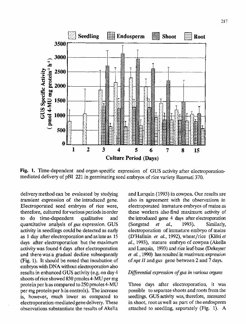
Fig. 1. Time-dependent and organ-specific expression of GUS activity after electroporation- mediated delivery of pBI 221 in germinating seed embryos of rice variety Basmati 370.
delivery method can be evaluated by studying transient expression of the introduced gene. Electroporated seed embryos of rice were, therefore, cultured for various periods in order to do time-dependent qualitative and quantitative analysis of gus expression. GUS activity in seedlings could be detected as early as 1 day after electroporation and as late as 15 days after electroporation but the maximum activity was found 4 days after electroporation and there was a gradual decline subsequently (Fig. 1). It should be noted that incubation of embryos with DNA without electroporation also results in enhanced GUS activity (e.g. on day 4 shoots of rice showed 830 pmoles 4-MU per mg protein per h as compared to 250 pmoles 4-MU per mg protein per h in controls). The increase is, however, much lower as compared to electroporation-mediated gene delivery. These observations substantiate the results of Akella
and Lurquin (1993) in cowpea. Our results are also in agreement with the observations in electroporated immature embryos of maize as these workers also find maximum activity of the introduced gene 4 days after electroporation (Songstad et aL, 1 9 9 3 ) . Similarly, electroporation of immature embryo of maize (D'Halluin et aL, 1992), wheat/rice (K16ti et aL, 1993), mature embryo of cowpea (Akella and Lurquin, 1993) and rice leaf-base (Dekeyser et aL, 1990) has resulted in maximum expression of npt H and gus gene between 2 and 7 days.
Differential expression of gus in various organs
Three days after electroporation, it was possible to separate shoots and roots from the seedlings. GUS activity was, therefore, measured in shoot, root as well as part of the endosperm attached to seedling, separately (Fig. 1). A
218
differential expression pattern ofgus gene was observed as the shoot expressed a higher GUS specific activity than root; shoots showed a 2-6 folds higher activity than roots depending on period of growth. The endosperm also showed GUS activity which could also be contributed by the adhering portion of shoot and root bases in close proximity to the endosperm. Interestingly, the pattern of organ-specific transient gene expression under the control of CaMV 35S promoter is similar to that in transgenic rice (Battraw and Hall, 1990).
Effect of cycIoheximide on expression of GUS
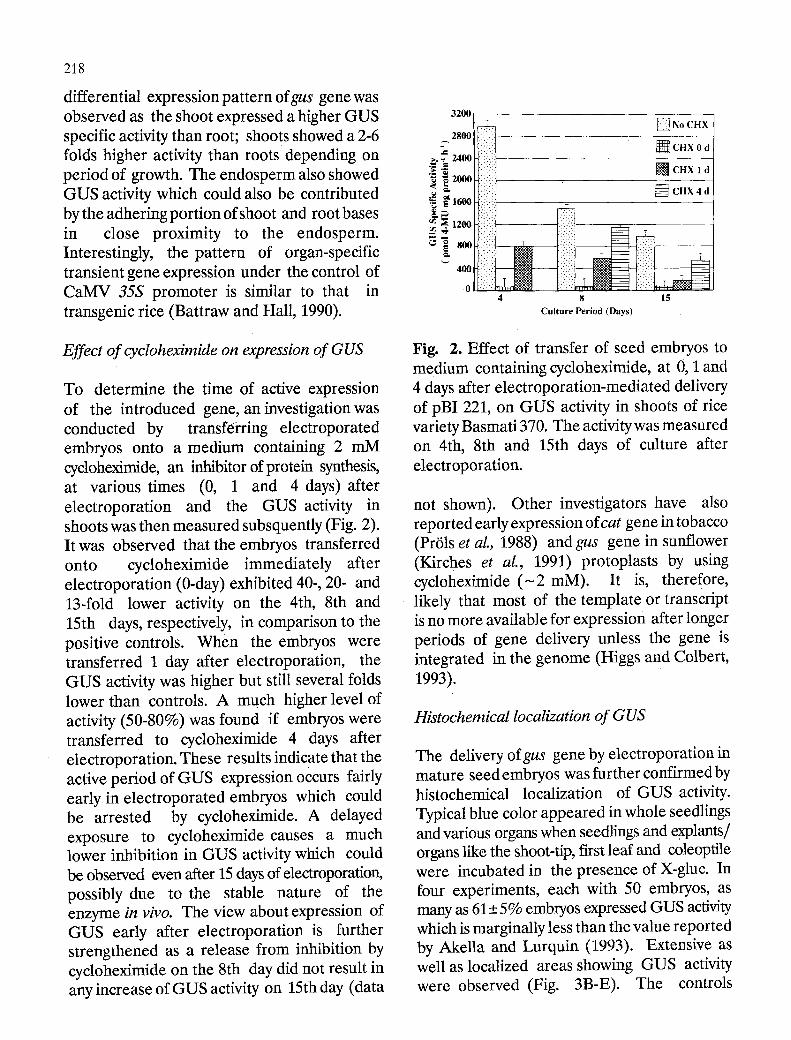
To determine the time of active expression of the introduced gene, an investigation was conducted by transf6rring electroporated embryos onto a medium containing 2 mM cycloheximide, an inhibitor of protein synthesis, at various times (0, 1 and 4 days) after electroporation and the GUS activity in shoots was then measured subsquently (Fig. 2). It was observed that the embryos transferred onto cycloheximide immediately after electroporation (0-day) exhibited 40-, 20- and 13-fold lower activity on the 4th, 8th and 15th days, respectively, in comparison to the positive controls. When the embryos were transferred 1 day after electroporation, the GUS activity was higher but still several folds lower than controls. A much higher level of activity (50-80%) was found if embryos were transferred to cycloheximide 4 days after electroporation. These results indicate that the active period of GUS expression occurs fairly early in electroporated embryos which could be arrested by cycloheximide. A delayed exposure to cycloheximide causes a much lower inhibition in GUS activity which could be observed even after 15 days of electroporation, possibly due to the stable nature of the enzyme in vivo. The view about expression of GUS early after electroporation is further strengthened as a release from inhibition by cycloheximide on the 8th day did not result in any increase of GUS activity on 15th day (data
3200,
"~ 2000[ [ ] CHX l d
r g~ .~ ~ 1600[ ~ CHX 4 d
m ,12001
0 -'.:i ~ . 4 8 15
Culture Period (Days)
Fig. 2. Effect of transfer of seed embryos to medium containing cycloheximide, at 0, I and 4 days after electroporation-mediated delivery of pBI 221, on GUS activity in shoots of rice variety Basmati 370. The activity was measured on 4th, 8th and 15th days of culture after electroporation.
not shown). Other investigators have also reported early expression of cat gene in tobacco (PrOls et al., 1988) and gus gene in sunflower (Kirches et aI., 1991) protoplasts by using cycloheximide ( - 2 mM). It is, therefore, likely that most of the template or transcript is no more available for expression after longer periods of gene delivery unless the gene is integrated in the genome (Higgs and Colbert, 1993).
Histochemical localization of GUS
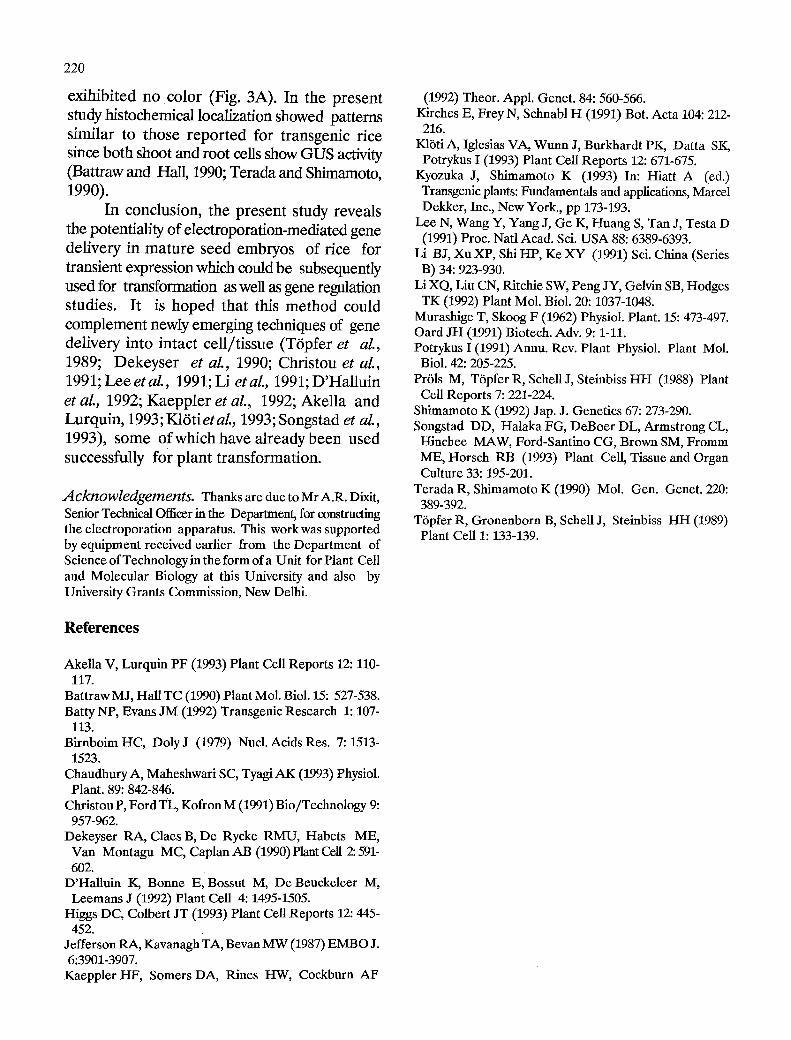
The delivery ofgus gene by electroporation in mature seed embryos was further confirmed by histochemical localization of GUS activity. Typical blue color appeared in whole seedlings and various organs when seedlings and explants/ organs like the shoot-tip, first leaf and coleoptile were incubated in the presence of X-gluc. In four experiments, each with 50 embryos, as many as 61+_ 5% embryos expressed GUS activity which is marginally less than the value reported by Akella and Lurquin (1993). Extensive as well as localized areas showing GUS activity were observed (Fig. 3B-E). The controls

219
Fig. 3. Histochemical localization of GUS activity in intact seedling and various explants after electroporation-mediated gene (gus) deliveryin germinating seed embryos of rice variety Basmati 370. Seedlings from seed embryos electroporated in the absence (A) and presence (B) of plasmid DNA and cultured for 4 days. Various explants i.e. Shoot (C), First leaf (D), and Coleoptile (E) from seedlings electroporated in the presence of plasmid DNA. For C, embryos were cultured for 2 days and for D and E, embryos were cultured for 4 days.
220
exihibited no color (Fig. 3A). In the present study histochemical localization showed patterns similar to those reported for transgenic rice since both shoot and root cells show GUS activity (Battraw and Hall, 1990; Terada and Shimamoto, 1990).
In conclusion, the present study reveals the potentiality of electroporation-mediated gene delivery in mature seed embryos of rice for transient expression which could be subsequently used for transformation as well as gene regulation studies. It is hoped that this method could complement newly emerging techniques of gene delivery into intact cell/tissue (T6pfer et al., 1989; Dekeyser et aL, 1990; Christou et al., 1991; LeeetaL, 1991; Li etal., 1991; D'Halluin et aL, 1992; Kaeppler et aL, 1992; Akella and Lurquin, 1993; K16tietal., 1993; Songstad et aL, 1993), some of which have already been used successfully for plant transformation.
Acknowledgements. Thanks are due to Mr A.R. Dixit, Senior Technical Officer in the Department, for constructing the electroporation apparatus. This work was supported by equipment received earlier from the Department of Science of Technologyin the form of a Unit for Plant Cell and Molecular Biology at this University and also by University Grants Commission, New Delhi.
References
Akella V, Lurquin PF (1993) Plant Cell Reports 12: 110- 117.
Battraw MJ, Hall TC (1990) Plant Mol. Biol. 15: 527-538. Batty NP, Evans JM (1992) Transgenic Research 1: 107-
113. Birnboim HC, DolyJ (1979) Nucl. Acids Res. 7: 71513-
1523. Chaudhury A, Maheshwari SC, Tyagi AK (1993) Physiol. Plant. 89: 842-846.
Christou P, Ford TL, Kofron M (1991) Bio/Technology 9: 957-962.
Dekeyser RA, Claes B, De Rycke RMU, Habets ME, Van Montagu MC, Caplan AB (1990) Plant Cell 2: 591- 602;
D'Halluin K, Bonne E, Bossut M, De Beuckeleer M, Leemans J (1992) Plant Cell 4: 1495-1505.
Higgs DC, Colbert JT (1993) Plant Cell Reports 12: 445- 452.
Jefferson RA, Kavanagh TA, Bevan MW (1987) EMBO J. 6:3901-3907.
Kaeppler HF, Somers DA, Rines HW, Cockburn AF
(1992) Theor. Appl. Genet. 84: 560-566. Kirches E, Frey N, Schnabl H (1991) Bot. Acta 104: 212- 216.
KlSti A, Iglesias VA, Wunn J, Burkhardt PK, Datta SK, Potrykus I (1993) Plant Cell Reports 12: 671-675.
Kyozuka J, Shimamoto K (1993) In: Hiatt A (ed.) Transgenic plants: Fundamentals and applications, Marcel Dekker, Inc., New York., pp 173-193.
Lee N, Wang Y, Yang J, Ge K, Huang S, Tan J, Testa D (1991) Proc. Natl Acad. Sci. USA 88: 6389-6393.
Li B J, Xu XP, Sift HP, Ke XY (1991) Sci. China (Series B) 34: 923-930.
Li XQ, Liu CN, Ritchie SW, Peng JY, Gelvin SB, Hodges TK (1992) Plant Mol. Biol. 20: 1037-1048.
Murashige T, Skoog F (1962) Physiol. Plant. 15: 473-497. Oard JH (1991) Biotech. Adv. 9: 1-11. Potrykus I (1991) Annu. Rev. Plant Physiol. Plant Mol. Biol. 42: 205-225.
Pr61s M, T6pfer R, Schell J, Steinbiss HH (1988) Plant Cell Reports 7: 221-224.
Shimamoto K (1992) Jap. J. Genetics 67: 273-290. Songstad DD, Halaka FG, DeBoer DL, Armstrong CL, Hinchee MAW, Ford-Santino CG, Brown SM, Fromm ME, Horsch RB (1993) Plant Cell, Tissue and Organ Culture 33: 195-201.
Terada R, Shimamoto K (1990) Mol. Gen. Genet. 220: 389-392.
T6pfer R, Gronenborn B, ScheU J, Steinbiss HH (1989) Plant Cell 1: 133-139.