Embed Size (px)
Citation preview

13
Transitioning Newborns from NICU to Home
Family Information PacketYour Health Coach has prepared this information packet for your family to help explain the medical needs of your newborn as you prepare to leave the hospital. A Health Coach helps families/caregivers adjust to working directly with the health care providers as well as increasing your ability and confidence to care for your infant.
When infants are born before their due date or with health problems, families often need help managing their baby’s health care after leaving the hospital. Since they spend the first part of their lives in the hospital, the babies and their families may find it helpful to have extra support.
Your Health Coach will work with your family to teach you to care for your infant, connect with the right doctors and specialists for treatment, and find resources in your area. The role of the Health Coach is as an educator, not as a caregiver.
Planning must start before hospital discharge and continue through followup with the primary care provider. After discharge from the hospital, your infant’s care must be well coordinated and clearly communicated to avoid medical errors that could harm the baby.
After your infant is discharged, your Health Coach should follow up with you by phone within a few days to make sure the transition is going well. Your Health Coach will want to know how your baby is doing, whether you have any questions or concerns, if your infant has been to the scheduled appointments, etc.
This information packet contains tips for getting care, understanding signs and symptoms of illness, medicines and immunizations, managing breathing problems, and feeding.
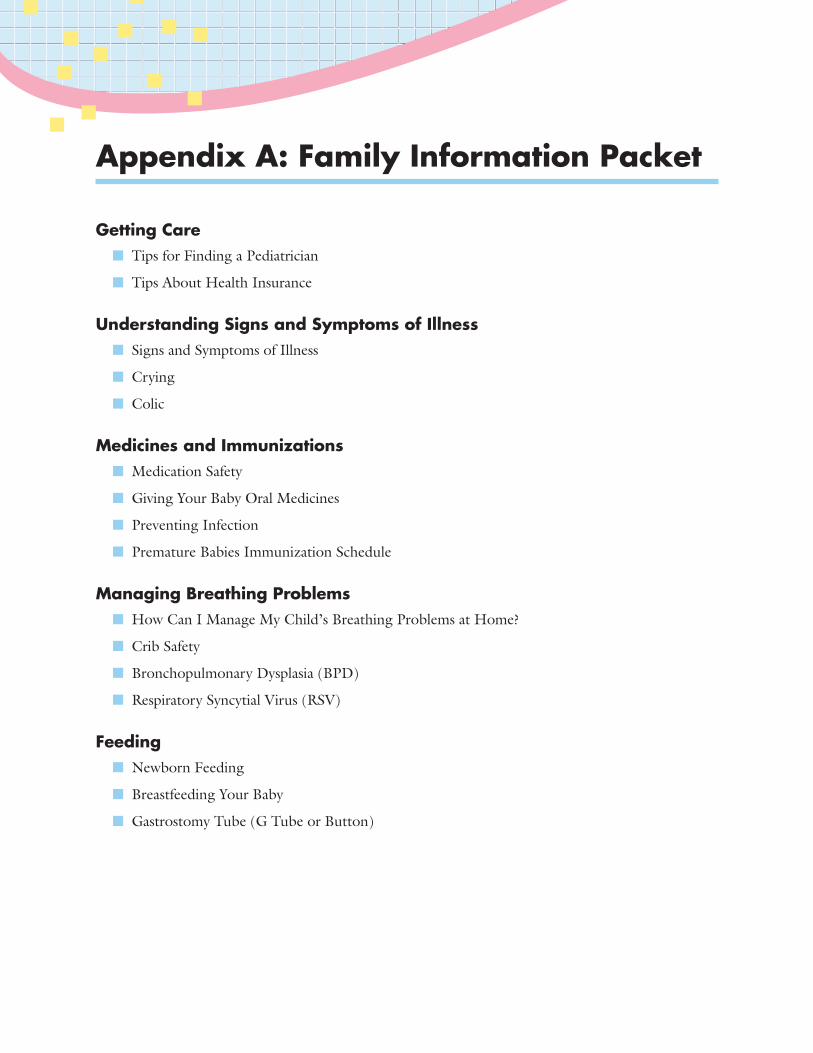
Appendix A: Family Information Packet
Getting Care ■ Tips for Finding a Pediatrician
■ Tips About Health Insurance
Understanding Signs and Symptoms of Illness ■ Signs and Symptoms of Illness
■ Crying
■ Colic
Medicines and Immunizations ■ Medication Safety
■ Giving Your Baby Oral Medicines
■ Preventing Infection
■ Premature Babies Immunization Schedule
Managing Breathing Problems ■ How Can I Manage My Child’s Breathing Problems at Home?
■ Crib Safety
■ Bronchopulmonary Dysplasia (BPD)
■ Respiratory Syncytial Virus (RSV)
Feeding ■ Newborn Feeding
■ Breastfeeding Your Baby
■ Gastrostomy Tube (G Tube or Button)

Tips for Finding a Pediatrician
■ Your family will need to visit a pediatrician within the first week after leaving the hospital.
■ Plan ahead to avoid the pressure and frustration of finding a pediatrician right before your baby is discharged from the hospital.
■ Ask your Health Coach or social worker if you need help finding a doctor for your baby.
■ Decide whether the pediatrician’s office needs to be close to your home, job, or day care.
■ Call the pediatrician’s office to ask if they accept your current health insurance and if they are accepting new patients.
■ Ask the pediatrician’s office what your co-payment and responsibilities are.
■ Ask the pediatrician’s office which doctor your baby will see at every visit.
■ Ask the pediatrician’s office if a doctor is on-call 24 hours a day for emergencies.

Tips About Health Insurance
■ Your baby will need to visit the doctor regularly after birth.
■ You have only 30 days to add your newborn to your insurance plan. Call your current insurance company as soon as possible to add your newborn.
■ Ask your social worker if you qualify for Medicaid, and apply while your baby is still in the hospital.
■ Ask about Children’s Medicaid (CHIP), Project Medical Home, and other low-cost health care benefits if you do not qualify for Medicaid.

Signs and Symptoms of Illness
Call the doctor if your baby:
■ Has a fever more than 100 degrees Fahrenheit.
■ Has diarrhea for more than one day.
■ Is throwing up forcefully.
■ Is not eating well.
■ Has not had at least six to eight wet diapers per day.
■ Has very yellow eye color or skin color.
■ Is hard to wake up.
■ Has fast or difficult breathing.
■ Makes jerking movements.
■ Is not looking well or acting well.

Crying
Common Causes ■ Hunger
■ Pain
■ Sleepy
■ Clothes too tight
■ Soiled diaper
■ Too hot or too cold
Call your doctor if: ■ Your baby looks or acts sick (is throwing up, appears to be in pain).
■ You are exhausted from the crying and fear you might hurt your baby.
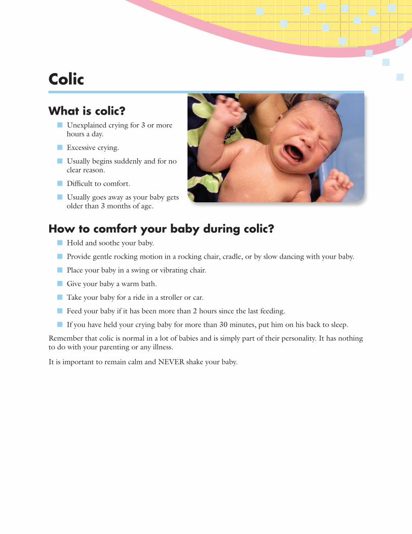
Colic
What is colic? ■ Unexplained crying for 3 or more hours a day.
■ Excessive crying.
■ Usually begins suddenly and for no clear reason.
■ Difficult to comfort.
■ Usually goes away as your baby gets older than 3 months of age.
How to comfort your baby during colic? ■ Hold and soothe your baby.
■ Provide gentle rocking motion in a rocking chair, cradle, or by slow dancing with your baby.
■ Place your baby in a swing or vibrating chair.
■ Give your baby a warm bath.
■ Take your baby for a ride in a stroller or car.
■ Feed your baby if it has been more than 2 hours since the last feeding.
■ If you have held your crying baby for more than 30 minutes, put him on his back to sleep.
Remember that colic is normal in a lot of babies and is simply part of their personality. It has nothing to do with your parenting or any illness.
It is important to remain calm and NEVER shake your baby.
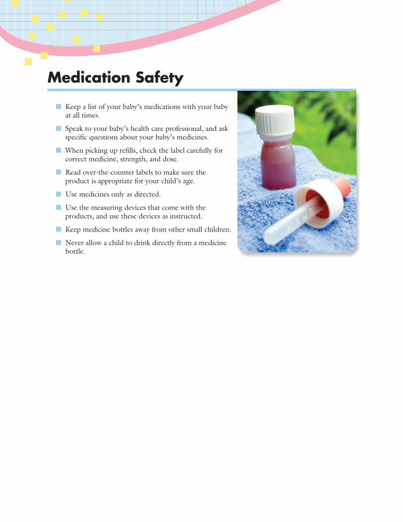
Medication Safety
■ Keep a list of your baby’s medications with your baby at all times.
■ Speak to your baby’s health care professional, and ask specific questions about your baby’s medicines.
■ When picking up refills, check the label carefully for correct medicine, strength, and dose.
■ Read over-the-counter labels to make sure the product is appropriate for your child’s age.
■ Use medicines only as directed.
■ Use the measuring devices that come with the products, and use these devices as instructed.
■ Keep medicine bottles away from other small children.
■ Never allow a child to drink directly from a medicine bottle.
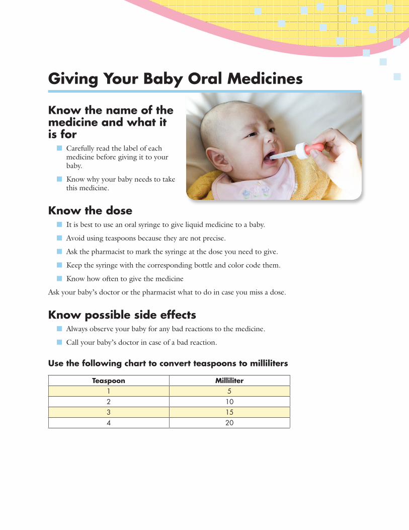
Giving Your Baby Oral Medicines
Know the name of the medicine and what it is for
■ Carefully read the label of each medicine before giving it to your baby.
■ Know why your baby needs to take this medicine.
Know the dose ■ It is best to use an oral syringe to give liquid medicine to a baby.
■ Avoid using teaspoons because they are not precise.
■ Ask the pharmacist to mark the syringe at the dose you need to give.
■ Keep the syringe with the corresponding bottle and color code them.
■ Know how often to give the medicine
Ask your baby’s doctor or the pharmacist what to do in case you miss a dose.
Know possible side effects ■ Always observe your baby for any bad reactions to the medicine.
■ Call your baby’s doctor in case of a bad reaction.
Use the following chart to convert teaspoons to milliliters
Teaspoon Milliliter1 52 103 154 20
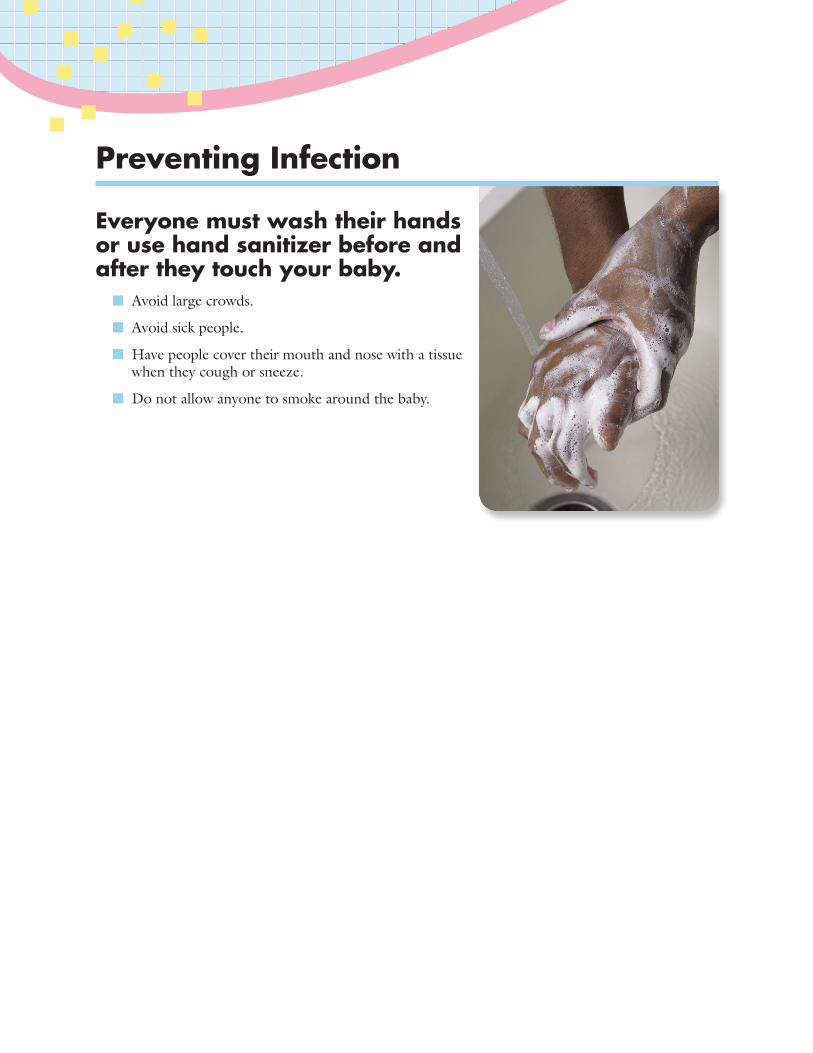
Preventing Infection
Everyone must wash their hands or use hand sanitizer before and after they touch your baby.
■ Avoid large crowds.
■ Avoid sick people.
■ Have people cover their mouth and nose with a tissue when they cough or sneeze.
■ Do not allow anyone to smoke around the baby.

Premature Babies Immunization Schedule
What is an immunization?Immunizations protect us from getting sick with serious diseases. Often, they are given by a shot, but sometimes they are given by mouth or nose.
Premature babiesImmunizations are given to premature babies at the same age as full-term infants.
At birth ■ Hepatitis B Vaccine
– First dose is usually given at birth before leaving the hospital.
– Should complete the doses by 6-18 months of age.
6-8 weeks old ■ Diphtheria and tetanus and pertussis vaccine (DTaP)
■ Haemophilus influenzae vaccine (Hib)
■ Pneumococcal vaccine
■ Poliovirus vaccine (IPV)
Respiratory Syncytial Virus (RSV)Your child may be eligible for a treatment to prevent RSV, a virus that can cause serious lung infections. Ask your baby’s doctor.
Benefits of vaccines ■ Vaccines prepare your baby’s body to fight illness.
■ Vaccines protect you and others around you.
■ It cost less to prevent a disease than to treat it.
Are Your child’s immunizations up to date?You can find out more about which immunizations babies need on the Centers for Disease Control and Prevention’s (CDC’s) Web site http://www.cdc.gov/vaccines/.
If you have any concerns about vaccines, ask the doctor caring for your baby.
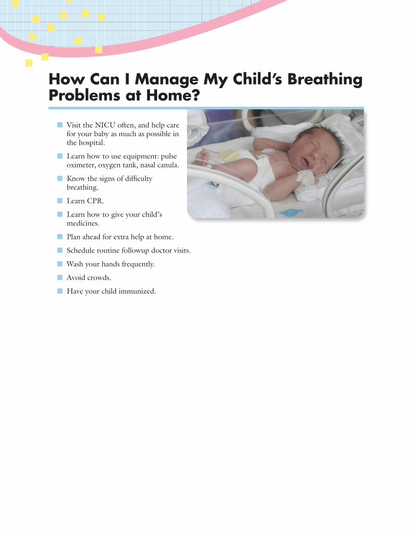
How Can I Manage My Child’s Breathing Problems at Home?
■ Visit the NICU often, and help care for your baby as much as possible in the hospital.
■ Learn how to use equipment: pulse oximeter, oxygen tank, nasal canula.
■ Know the signs of difficulty breathing.
■ Learn CPR.
■ Learn how to give your child’s medicines.
■ Plan ahead for extra help at home.
■ Schedule routine followup doctor visits.
■ Wash your hands frequently.
■ Avoid crowds.
■ Have your child immunized.
Crib Safety
What is SIDS? ■ “SIDS” stands for Sudden Infant Death Syndrome.
■ Sudden, unexplained death of an infant less than 1 year old.
What should I know about SIDS? ■ Babies sleep safer on their backs.
■ Babies should be placed on a firm sleep surface.
What can I do to lower my baby’s risk of SIDS?
■ Babies should be put on their backs to sleep for nap and at night.
■ Keep toys, objects, and loose bedding out of your baby’s sleep area.
■ Do not allow smoking around your baby.
■ Do not allow your baby to overheat during sleeping.
What about “tummy time”? ■ Daily tummy time is necessary for normal development.
■ Make sure your baby spends several hours on their tummy when they are awake and someone is watching.
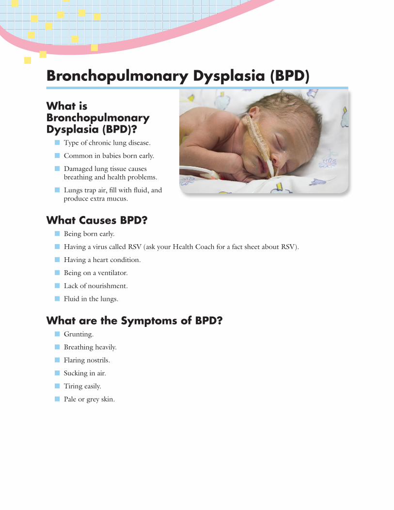
Bronchopulmonary Dysplasia (BPD)
What is Bronchopulmonary Dysplasia (BPD)?
■ Type of chronic lung disease.
■ Common in babies born early.
■ Damaged lung tissue causes breathing and health problems.
■ Lungs trap air, fill with fluid, and produce extra mucus.
What Causes BPD? ■ Being born early.
■ Having a virus called RSV (ask your Health Coach for a fact sheet about RSV).
■ Having a heart condition.
■ Being on a ventilator.
■ Lack of nourishment.
■ Fluid in the lungs.
What are the Symptoms of BPD? ■ Grunting.
■ Breathing heavily.
■ Flaring nostrils.
■ Sucking in air.
■ Tiring easily.
■ Pale or grey skin.

How is BPD Diagnosed? ■ If your baby still needs oxygen at 36 weeks old.
■ If your baby has been on a ventilator.
How is BPD Treated? ■ BPD is treated with oxygen to control fluid in the body and medicine to relax the airway.
■ Treatment does not cure BPD.
■ Treatment helps your baby breathe better.
■ Lungs will eventually heal.
■ Your baby needs nutrients for healthy growth.

Respiratory Syncytial Virus (RSV)
What is Respiratory Syncytial Virus (RSV)?
■ A common virus that affects babies and infants.
■ Leading cause of two lung infections: pneumonia and bronchitis.
Symptoms of RSV ■ Starts out like a cold with fever or runny nose.
■ Can also include: – Coughing. – Problems breathing. – Fast breathing.
– Not eating well.
How is RSV Spread? ■ Contact with someone who is coughing or sneezing.
■ Enters the body through the eyes, nose, or mouth.
How to Prevent the Spread of RSV? ■ Wash your hands before and after handling a baby.
■ Avoid exposing your baby to others with cold symptoms.
■ Cover coughs/sneezes and throw away used tissues.
■ Keep your baby away from crowded areas.
■ If your baby is at high risk for RSV, talk to your doctor about a monthly shot that can help lower the risk of a baby getting severe RSV.
■ Your baby may be eligible for palivizumab (brand name: Synagis®), a treatment given to prevent and reduce RSV.
■ The shot, given monthly during RSV season, reduces the chance of your baby getting pneumonia and bronchitis.
■ Babies may still get RSV but will be less sick.
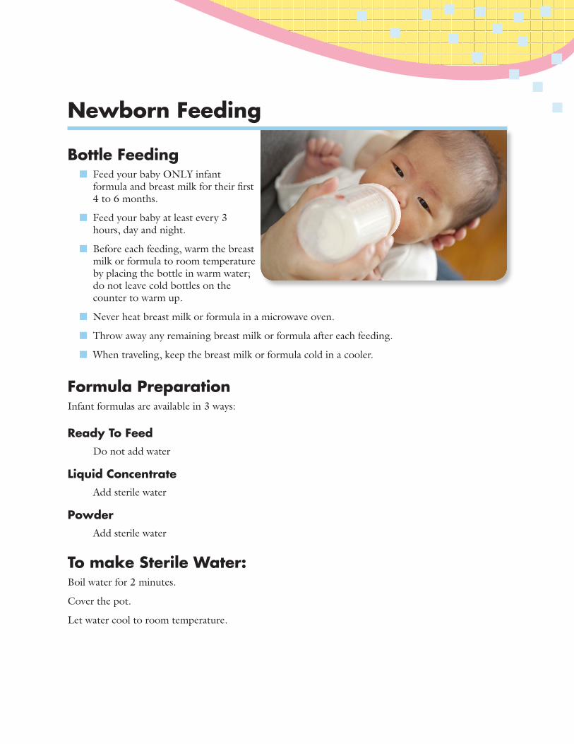
Newborn Feeding
Bottle Feeding ■ Feed your baby ONLY infant formula and breast milk for their first 4 to 6 months.
■ Feed your baby at least every 3 hours, day and night.
■ Before each feeding, warm the breast milk or formula to room temperature by placing the bottle in warm water; do not leave cold bottles on the counter to warm up.
■ Never heat breast milk or formula in a microwave oven.
■ Throw away any remaining breast milk or formula after each feeding.
■ When traveling, keep the breast milk or formula cold in a cooler.
Formula PreparationInfant formulas are available in 3 ways:
Ready To FeedDo not add water
Liquid ConcentrateAdd sterile water
PowderAdd sterile water
To make Sterile Water:Boil water for 2 minutes.
Cover the pot.
Let water cool to room temperature.

Formula Storage and UseStore prepared formula in a refrigerator.
Use formula in 24 to 48 hours.
Bottle Cleaning Clean bottles and nipples by washing with hot, soapy water or on top rack of dishwasher.
Allow bottles and nipples to air dry.

Breastfeeding Your Baby
Breastfed babies have: ■ Fewer ear infections.
■ Lower chance of asthma, food allergies, and dental cavities.
■ Protection against diarrhea, stomach, and lung infections.
■ Better nervous system development and higher IQ levels.
■ Lower risk of some childhood cancers.
■ Lower chance of becoming over weight.
Mothers who breastfeed have: ■ Lower risk of pre-menopausal breast cancer.
■ Lower chance of osteoporosis later in life.
■ Quicker return to pre-pregnancy weight.
■ Food source for their babies even during emergencies.
■ Lower chance of becoming pregnant before menstruation returns.
Gastrostomy Tube (G Tube or Button)
Giving medicines and feeding if your baby has a gastrostomy tube:
■ Clear the G tube or button as your health care provider showed you.
■ Check for placement of the G tube or button.
■ Slowly push in liquid medicine or feeding with a syringe.
■ If the pharmacist says it is ok, pills and capsules may be dissolved in 10 to 20 cc of warm tap water.
■ All medicines and feedings should be flushed in with 5 to 10 cc of warm tap water.
■ Ask your baby’s doctor, nurse, or pharmacist how to measure the tap water.
It is important to use the specific tube adapter made by the manufacturer of your button.
■ In fluid restricted babies flush medicines with ONLY 1 to 5 cc of warm tap water.
■ Vent the tube after feeding to remove excess air or fluid and reduce leaking.
Protecting the G Tube or Button ■ Snap t-shirts and onsies work best to prevent babies from pulling on the tube or button.
■ You may also use a sticky wrap or stretchy dressing.
Appendix B: Clinical Materials to Share With Primary Care Providers
Diagnoses and Conditions ■ Anemia of Prematurity
■ Apnea of Prematurity
■ Bronchopulmonary Dysplasia
■ Gastroesophageal Reflux
■ Nephrocalcinosis
■ Patent Ductus Arteriosus
■ Short Bowel Syndrome
■ Vision Screening and Retinopathy of Prematurity
Care, Treatment, and Development ■ Medications
■ Neurodevelopment
■ Ostomy Care
■ Sleep in Preterm Infants
■ Tracheostomies
Feeding ■ Breast Milk
■ Feeding Tubes
■ Formula Feedings
■ Growth
■ Weaning

Anemia Of Prematurity
Characteristics ■ Occurs during the normal developmental switch from fetal to adult hemoglobin synthesis.
■ Immediately after birth, the increase in blood oxygen content results in the downregulation of erythropoietin.
■ Once hemoglobin level decreases enough, tissue oxygen needs are greater than oxygen delivery, and erythropoietin production increases.
■ Anemia is more profound and occurs earlier in premature infants due to:
– Blood loss from sampling.
– Short survival time of RBC’s (40-60 days compared to adult 120 days, and term infant 60-80 days).
– Suboptimal erythropoietic response.
– Relatively more rapid rate of growth than in term infants.
■ Usually reach nadir of 7-10 g/dL at 4-8 weeks of life.
■ Normocytic, normochromic, hypoproliferative anemia.
■ Premature infants may have lower iron stores despite iron supplementation; consider iron deficiency anemia as an etiology for persistent or progressive anemias.
Monitoring ■ May check hematocrit and reticulocyte count periodically, but most babies can be managed by watching for symptoms (tachycardia, tachypnea, apnea and bradycardia, poor weight gain, oxygen requirement, diminished activity, pallor, poor feeding).
Treatment ■ Healthy asymptomatic newborns will self correct, provided their iron intake is adequate.
■ Transitional formulas and fortified breast milk provide approximately 2 mg/kg/day of iron.
■ Iron administration before 10-14 weeks of age does not reduce the nadir, but iron is stored for later use.
■ Full-term infants should receive 1 mg/kg/day of iron supplementation from age 4 months to 1 year.

■ Premature or low-birthweight infants (<2500g) should receive 2 mg/kg/day from 2 months to 1 year (from iron enriched formula or 1 mL multi-vitamin with iron).
■ No strong evidence to favor use of erythropoietin.
■ Infants with significant respiratory disease or congenital heart disease may need a hematocrit maintained >40 gm%.
■ Infants with a hematocrit <25 gm% and symptoms may require red blood cell transfusion.
■ For infants requiring blood transfusion:
– [Insert contact information for appropriate specialist or department.]
– Order: PRBC, leukocyte poor, irradiated, CMV negative, 15 ml/kg.
– If the baby is very fluid sensitive (e.g., chronic lung disease), IV lasix 1 mg/kg immediately after transfusion may be indicated.

Apnea Of Prematurity
Characteristics ■ Respiratory pause for at least 20 seconds, or a pause that is accompanied by bradycardia (heart rate <100 bpm), cyanosis, or pallor in an infant <37 weeks postmenstrual age (PMA).
■ Types of apnea:
– Central: the respiratory center in the immature brain stem does not trigger inspiration; frequently responds to methylxanthine treatment.
– Obstructive: due to impaired tone of larynx/pharynx.
– Mixed: combination of the two; most common type found in neonates.
■ Can persist for many weeks in very preterm infants, although resolution typically occurs by 37-43 weeks PMA (resolution tends to be later for extremely low-birthweight infants).
■ Periodic breathing: recurrent sequences of pauses in respiration lasting 5-10 seconds, followed by 10-15 seconds of rapid breathing without bradycardia or other symptoms.
■ Benign apnea: isolated apneas of 5-10 seconds, without bradycardia or color change; resolve spontaneously.
Home Treatment ■ Most infants still having apnea are kept hospitalized; few may be discharged home.
■ Methylxanthines: act primarily on the brainstem respiratory structures, producing a central stimulatory effect.
– Caffeine is most commonly used due to its broader therapeutic index and slower excretion rate than theophylline.
Cardiorespiratory Home Monitors ■ Infants are generally monitored for a 5- to 7-day apnea-free period after discontinuing methylxanthine therapy before sending an infant home without a monitor.
■ Home monitors are rarely indicated for the detection of apnea, because infants with immature respiratory systems are generally still hospitalized until they are no longer at risk for apnea of prematurity.
■ Any home cardiorespiratory monitor used must have download capability.
■ Monitoring may be prescribed for some preterm infants with an unusually prolonged course of recurrent, extreme apnea.
■ Monitoring should be limited to approximately 43 weeks’ PMA or after the cessation of extreme episodes, whichever comes last.
Additional Factors ■ Preterm infants are known to be at higher risk for SIDS; high-risk period lasts up to 10 months.
■ As “a causal relationship between prolonged apnea and SIDS has not been established,” home cardiorespiratory monitoring should not be used to prevent SIDS (AAP Task Force on Prolonged Infantile Apnea, 2003).
■ Premature infants beyond the immediate neonatal period can experience apnea following ketamine sedation or general anesthesia; they should have cardiorespiratory monitoring in hospital for 24 hours after surgery.

Bronchopulmonary Dysplasia
Characteristics ■ Need for supplemental oxygen at 36 weeks postmenstrual age, with radiographic changes on chest x-ray (bilateral, diffuse hazy lungs; interstitial thickening; increased lung inflation).
■ Symptoms: tachypnea, increased work of breathing, chronic wheeze or cough, oxygen dependence, +/- ventilator dependence, hypercarbia with compensated respiratory acidosis.
■ Course characterized by acute exacerbations of respiratory distress.
■ Overall incidence is unchanged due to increased survival of extremely premature infants.(~7500 new cases/year) but is decreased with surfactant and early extubation to NCPAP.
■ Risk factors: premature birth, respiratory failure, O2 supplementation, mechanical ventilation, air leaks, pulmonary edema, PDA ligation, infection causing lung injury, lung hypoplasia.
Treatment ■ Oxygen (see home oxygen topic sheet).
■ Fluid restriction and diuretics:
– Use of diuretics is reserved for severe BPD and evidence of diuretic responsive disease.
– Furosemide, chlorothiazide, spironolactone (see medication sheet for dosing).
– May need KCl or NaCl supplements to correct electrolyte deficiencies.
– Chronic furosemide use improves both oxygenation and lung compliance, but has not decreased length of hospital stay or need for oxygen therapy.
■ Inhaled bronchodilators or corticosteroids (see medication sheet for dosing).
– Only for infants that show evidence of reversible airway obstruction.
– Only patients that have a family history of asthma and have episodic attacks similar to asthma have been shown to have any benefit from inhaled steroids.
■ RSV immunoprophylaxis; influenza and pneumococcus vaccinations.
■ Higher caloric intake needed to account for increased work of breathing, as well as ongoing tissue repair and tissue deposition; due to fluid restrictions, these patients may need fortified feeds to maintain an adequate growth rate.
Monitoring ■ Monitor O2 saturations, resting respiratory rate and effort, presence of retractions, and color.
■ Watch for adequate weight gain (15-30 g/day).
– If inadequate, may be due to insufficient caloric intake or hypoxemia.
■ Watch for excessive fluid retention (tachypnea, retractions, rales, excessive weight gain, enlarged liver, poor feeding, O2 saturation <92%).
■ Watch neurodevelopment closely.
Outcomes ■ 50% with CLD require rehospitalization in the first year, and 37% in the second year of life.
■ More prone to: frequent lower respiratory tract illnesses, feeding difficulties, growth failure, rehospitalization during infancy.
■ Despite treatment, approximately 10% of these children die in the first year of life.
■ Increased risk for cognitive, motor, and language impairment, hearing loss, and poor academic performance.
■ Lung function generally remains reduced until adolescence, at which point it approaches normal for age; in some, pulmonary function will again deteriorate in adulthood.
■ Children with CLD are more likely to develop asthma and require bronchodilator therapy.

Gastroesophageal Reflux
Characteristics ■ Common problem in premature infants.
– Lower esophageal sphincter hypotonia.
– Transient relaxation of the esophageal sphincter.
– Less frequent esophageal peristaltic activity.
– Delayed gastric emptying.
– Decreased gastric compliance.
– Neurologic impairment.
■ Is a physiologic event; it is important to distinguish between physiologic and pathologic GER (GERD, or GER disease) before beginning any therapeutic interventions.
■ Failure to thrive is usually NOT associated with physiologic GER in premature infants.
Symptoms Of Pathologic GER ■ Failure to thrive due to malnourishment.
■ Frequent respiratory problems due to aspiration.
■ Esophagitis with or without stricture formation.
■ Growth delay, poor feeding, irritability.
■ Post-prandial vomiting, gagging, coughing, arching, fussiness, feeding refusal.
■ Some infants may experience aspiration, cyanosis, and vomiting.
■ Sandifer syndrome: excessive hiccups, sleep disturbances, and arching.
■ Uncommon symptoms: intermittent stridor, hoarse voice, acute episodes of spasmodic croup, bronchospasm, pneumonia.
■ Data and research supporting a causal relationship between GER and apnea is lacking.

Diagnosis Of Pathologic GER ■ History.
■ Barium swallow.
■ pH probe.
Treatment ■ Medical or surgical therapy is indicated only for pathological GER.
■ Therapies may improve symptoms, but generally reflux is not entirely eliminated.
■ GER usually self-improves within 6 months of life due to maturation, sitting upright, and intake of more solid foods.
■ Antireflux wedge (elevates head by 30 degrees).
■ Postprandial prone positioning when awake and under supervision, or left lateral positioning.
■ Thickened feeds (rice cereal, 2-3 tsp/oz).
■ Concentrating milk for decreased volume of feeds.
■ More frequent, smaller volume feeds.
■ H2 blockers or proton pump inhibitors.
■ Prokinetics.
■ Nissen fundoplication is the last resort for selected severe and intractable cases, or for those with severe neurological impairment in whom aspiration is a real risk.

Nephrocalcinosis
Characteristics ■ Renal lithiasis in which calcium deposits form in the renal parenchyma and result in reduced kidney function and hematuria.
■ Seen with renal ultrasound, or occasionally on plain radiographs of the kidneys.
■ Results from an imbalance of stone-promoting and stone-inhibiting factors.
■ Incidence in very low-birthweight infants (BW<1500g) ranges from 16-64%.
■ Risk factors in at risk infants:
– Medications with hypercalciuric effects (furosemide, corticosteroids, aminoglycosides).
– Metabolic acidosis.
– Hypocitraturia.
– Hypercalciuria.
– High urinary oxalate:creatinine ratios.
– High urate:creatinine ratios.
Effects ■ Glomerular function: Mean GFR and microalbuminuria in preterm infants is slightly worse than in healthy infants.
■ Proximal tubule function: Phosphate reabsorption is significantly lower in patients with nephrocalcinosis, but plasma phosphate levels are within reference limits; no firm evidence for proximal tubular dysfunction.
■ Distal tubule function: Urine anion gap of infants with nephrocalcinosis is high, and serum bicarbonate levels are low, indicating distal tubular dysfunction.
■ Blood pressure: Not higher in infants with nephrocalcinosis, but overall higher in preterm infants than in healthy term infants.
■ Hypercalciuria: Significantly more hypercalciuria in infants with nephrocalcinosis.

Monitoring/Treatment ■ If associated with a medication that causes hypercalciuria, consider trying to stop the medication and monitor for resolution.
■ Resolves in 75% by 7 years of age.
■ Does not appear to have any long-lasting significant health effects.
■ Should have:
– Blood pressure checked at every visit.
– Yearly renal ultrasounds until resolution occurs.
– Electrolytes and BUN/Cr checked every 2 years.
■ Patients do not need to be followed by renal team unless other issues arise.
■ UTI and urolithiasis do not occur any more frequently than in general population.
■ Do NOT stop the use of high-calcium containing infant formulas.

Patent Ductus Arteriosus
Characteristics ■ A persistent open connection beyond 3 months of age between the pulmonary artery and the aorta with blood flow from the aorta to the pulmonary artery.
■ An open ductus may lead to:
– Congestive heart failure.
– Pulmonary hypertension.
– Increased risk of bacterial endocarditis.
Followup ■ A high rate of spontaneous closure occurs during the first 2 years of life.
■ If the ductus is open at the time of hospital discharge, followup with a cardiologist should occur within 2 months of discharge to assess well-being and the presence/absence of congestive heart failure.
– Earlier followup should occur if poor weight gain, difficulty feeding, and tachypnea develop.
– Generally do not need a followup echocardiogram, as anatomy is already known.
Management ■ If spontaneous closure does not occur, closure should be performed to:
– Eliminate pulmonary overcirculation.
– Eliminate risk of endocarditis.
■ If symptomatic:
– Initially, try diuretics and maximize caloric intake to 140 kcal/kg/day.
– If still symptomatic despite optimal medical management and:
• >2.4 kg, surgical ligation or percutaneous closure should be considered.
• <2.4 kg, surgical ligation should be considered.
■ If asymptomatic:
– Wait until patient weighs between 10-12 kg, or is close to 2 years of age, then attempt percutaneous closure with Amplatzer occlude or coils.
– Surgery if percutaneous attempt is unsuccessful.

Short Bowel Syndrome
Pathophysiology ■ Functional disorder caused by alterations of normal intestinal anatomy and physiology.
■ Shortened bowel combined with malabsorption; dependent on parenteral nutrition >3 months.
■ May result from: necrotizing enterocolitis, congenital bowel atresia, volvulus, gastroschisis, Hirschsprung’s disease.
■ After resection, the residual small bowel undergoes intestinal adaptation, stimulated by hormones and oral nutrients:
– Mucosal hyperplasia.
– Villus lengthening and increased crypt depth.
– Bowel dilatation.
■ Jejunum: long villi and large absorptive surface with high concentration of enzymes; site of greatest nutrient absorption.
– If resected, will have transient or permanent nutrient losses.
– Ileum can develop the absorptive capacity of the jejunum for various nutrients.
■ Ileum: shorter villi, more lymphoid tissue, tighter epithelium; effective absorption of fluid and electrolytes; responsible for absorption of vitamin B12 and bile salts through receptors.
– Other bowel will never develop ability to absorb B12 and bile salts.
– Resection may impair bowel motility (many GI hormones produced in ileum).
– Intestinal adaptation after massive ileal resection is more difficult than after jejunal.
■ Malabsorption of rapidly digested carbohydrates produces osmotic diarrhea.
■ Fat soluble vitamins may also be inadequately absorbed.
■ No absolute number can be placed on the length of remaining bowel necessary for successful enteral nutrition; remaining bowel may be damaged and act dysfunctional.
■ Best prognosis over time is for infants in whom the duodenum, distal ileum, and ileocecal valve can be preserved.
Complications ■ Long term TPN use
– Hepatobiliary disease.
– Catheter-associated sepsis.
– Fluid and electrolyte imbalance.
– Bacterial intestinal overgrowth.
■ Malnutrition.
■ Failure to thrive.
■ Dumping syndrome: post-prandial tachycardia, diaphoresis, lethargy, watery diarrhea.
– Marked increase in stools may indicate poor absorption, and enteral feedings should not be advanced.
– If significant volume loss, rehydration therapy and electrolyte replacement will be needed.
■ Mortality of 30-40% from sepsis or liver failure.
Management/Feedings ■ Recommend managing with consulting services in pediatric surgery, GI (and possibly the liver team), and neonatal dietitian.
■ Attempt to wean off TPN as soon as possible.
■ Enteral nutrition should be started promptly to promote intestinal adaptation.
■ Usually started on elemental/semi-elemental diets containing free amino acids or small peptides, or breast milk.
– There are no elemental preterm formulas.
– Once they tolerate feeds, can gradually introduce a portion of preterm formula, fortifier, or supplements to improve the mineral and protein content of a term formula.
■ These infants may have “leaky gut” with high rates of sensitization to cow’s milk or soy protein.
■ High proportion of fat in long-chain fatty acids promotes more mucosal adaptation; but those with ileal resection may not be able to absorb these and may need more medium-chain fats.
■ Feedings are started slowly and continuously to maximally saturate carrier proteins.
■ Higher concentrated formulas may cause osmotic diarrhea.
■ Oral feedings should be initiated promptly; may have solids at 4 months CGA (high in protein and fat and low in carbohydrate is preferred).
■ For high stomas, may be beneficial to re-feed the proximal stoma effluent through the mucous fistula for additional nutrient and fluid absorption and to stimulate distal gut.
■ For bacterial overgrowth, may need intermittent dosing of antibiotics that affect anaerobic bacteria.
■ Infants with ileal resection are at risk for B12 deficiency; may need parenteral B12 every 1-3 months.
■ Children with enterostomies or diarrhea are at risk for zinc deficiency and may need supplementation:
– Poor growth, diarrhea, impaired wound healing, perianal and perioral skin rash, alopecia.
■ Gastric acid hypersecretion:
– More common in larger bowel resections and initiation of enteral feedings.
– Can cause secretory diarrhea.
– Ranitidine may help.
Surgical Treatments ■ Goals of surgery:
– Slow intestinal transit.
– Increase mucosal surface area.
– Improve peristaltic function.
– Increase intestinal length.
■ May eventually require intestinal +/- liver transplant.

Vision Screening and Retinopathy of Prematurity
Visual Deficits Seen in Preterm Infants ■ High-risk infants are more likely to have permanent visual deficits and/or show a delay in visual development that persists until adolescence.
■ Deficits in the ability to perform visual discrimination tasks is one of the most striking, persistent deficits.
■ Reduced visual fields, amblyopia, myopia (associated with BPD, seizures and asphyxia), and strabismus (associated with intraventricular hemorrhage (IVH), bronchopulmonary dysplasia (BPD), necrotizing enterocolitis (NEC), periventricular leukomalacia (PVL)).
Retinopathy of Prematurity ■ Abnormal blood vessel growth in the incompletely vascularized retinas of premature infants.
■ Incidence increases with lower gestational age and birthweight.
■ Classification by:
– Zones 1-3: locates the disease from most posterior (Zone 1) to most anterior (Zone 3).
– Stages 1-5: degree of vasculopathy at the vascular-avascular transition (Stage 5 is most severe and involves complete retinal detachment).
– Plus disease: occurs when arrested blood vessel growth resumes abnormally, with tortuous vessels piling up within the retina, forming a thick ridge of tissue.
■ Neovascular tissue (i.e., plus disease) may contract and form a scar, which then pulls and distorts the retina, resulting in retinal detachment, especially if ROP is in zone 1.
Screening/Monitoring ■ All infants <1500 grams or <30 weeks estimated gestational age (EGA) should be screened prior to discharge.
■ If retinal vasculature is completely mature, no further exams are needed.
■ Followup is dictated by degree of maturation, severity of ROP, and ophthalmologist.

■ Patients with ROP requiring laser therapy, and grade III/IV IVH, PVL, HIE, or hydrocephalus requiring shunting should be seen again at 9-12 months postnatal age by ophthalmology.
■ A pediatric ophthalmologic assessment for glasses and possible strabismus or amblyopia therapy should be obtained in the first year of life.
Treatment/Outcomes ■ Mild ROP (stage I or II without plus disease) have a somewhat higher incidence of myopia, strabismus, and amblyopia.
■ Threshold ROP (residual scar without retinal detachment):
– Associated with severe myopia, glaucoma.
– At risk for slowly progressive retinal degenerations that can lead to retinal detachments and acuity loss in later decades.
■ Total retinal detachment equates to no useful vision in that eye, even when vitrectomy is performed to reattach the retina.
■ Criteria for laser therapy or cryotherapy:
– Zone 2: plus disease with stage 2 or 3 ROP.
– Zone 1: plus disease with any stage ROP.
– Zone 1: stage 3 ROP with no plus disease.

Med
ication
Indic
ation
Usu
al D
osi
ng
Reg
imen
Rec
om
men
ded
M
onitori
ng
Side
Effe
cts
When
To S
top
Alb
uter
ol (V
ento
lin
HFA
®) o
r Lev
albu
tero
l (X
open
ex H
FA®
)
Reac
tive
airw
ay (R
A)
dise
ase
asso
ciat
ed w
ith
bron
chop
ulm
onar
y dy
spla
sia
(BPD
)
2 pu
ffs in
hale
d by
M
DI e
very
4-6
hou
rs
as n
eede
d
Non
eTa
chyc
ardi
a, e
xace
rbat
ion
of tr
ache
omal
acia
, ag
itatio
n
Whe
n w
heez
ing
epis
odes
ar
e no
long
er p
rese
nt
and,
in R
A, t
aper
and
di
scon
tinue
.
Inha
led
becl
omet
haso
ne
(QVA
R®)
Reac
tive
airw
ay
dise
ase
asso
ciat
ed w
ith
BPD
1 pu
ff in
hale
d q1
2 ho
urs
Non
eRe
duce
d lin
ear g
row
th,
thru
shW
hen
whe
ezin
g ep
isod
es
are
no lo
nger
pre
sent
an
d, in
RA
, tap
er a
nd
disc
ontin
ue.
Caf
fein
e ci
trate
(C
afci
t®)
Apn
ea o
f pre
mat
urity
5-10
mg/
kg/d
ay P
O
daily
mai
nten
ance
do
sing
(20
mg/
kg
load
ing
dose
)
Non
eTa
chyc
ardi
a, G
I ups
et,
agita
tion
Usu
ally
can
dis
cont
inue
be
twee
n 34
and
35
wee
ks
postm
enstr
ual a
ge, o
r afte
r 5-
7 da
ys fr
ee o
f apn
ea.
Hom
e ca
rdio
resp
irato
ry
mon
itorin
g af
ter d
isch
arge
m
ay b
e pr
escr
ibed
for
som
e pr
eter
m in
fant
s w
ith
an u
nusu
ally
pro
long
ed
cour
se o
f rec
urre
nt, e
xtre
me
apne
a.
Chl
orot
hiaz
ide
(Diu
ril®
)Fl
uid
sens
itive
BPD
; in
hibi
ts so
dium
and
ch
lorid
e re
abso
rptio
n in
di
stal t
ubul
e
20-4
0 m
g/kg
/day
PO
div
ided
BID
; m
axim
um 3
75 m
g/da
y
Onc
e on
sta
ble
dose
, che
ck
elec
troly
tes
once
a
mon
th.
If do
se
chan
ges,
che
ck
elec
troly
tes
2-3
days
late
r.
Hyp
okal
emia
, hy
pona
trem
ia,
hypo
mag
nese
mia
, hy
poch
lore
mic
alk
alos
is
Afte
r inf
ant i
s sta
ble
on R
A,
allo
w to
out
grow
diu
retic
do
se b
y 50
% b
efor
e sto
ppin
g.
Furo
sem
ide
(Las
ix®
)Fl
uid
sens
itive
BPD
; in
hibi
ts so
dium
and
ch
lorid
e re
abso
rptio
n in
th
e as
cend
ing
loop
of
Hen
le
1-4
mg/
kg/d
ay
PO o
r IV
daily
, or
divi
ded
BID
Onc
e on
sta
ble
dose
, che
ck
elec
troly
tes
once
a
mon
th.
If do
se
chan
ges,
che
ck
elec
troly
tes
2-3
days
late
r.
Hyp
okal
emia
, hy
pona
trem
ia,
hypo
calc
emia
, hy
perc
alci
uria
, ne
phro
calc
inos
is,
met
abol
ic a
lkal
osis
, ph
otos
ensi
tivity
Afte
r inf
ant i
s sta
ble
on R
A,
allo
w to
out
grow
diu
retic
do
se b
y 50
% b
efor
e sto
ppin
g.
Med
ications
Con
tinue
d

Med
ication
Indic
ation
Usu
al D
osi
ng
Reg
imen
Rec
om
men
ded
M
onitori
ng
Side
Effe
cts
When
To S
top
Phen
obar
bita
lSe
izur
es3-
6 m
g/kg
PO
da
ily o
r div
ided
BID
m
aint
enan
ce d
osin
g;
(load
ing
dose
20
mg/
kg, m
ay re
peat
on
ce)
Targ
et tr
ough
leve
ls ar
e 20
-40
mcg
/mL
CN
S de
pres
sion
, co
gniti
ve d
efici
tsW
ean
grad
ually
as
per
neur
olog
y re
com
men
datio
ns
if se
izur
e-fre
e.
Rani
tidin
e (Z
anta
c®)
GER
D, g
astri
c hy
pers
ecre
tory
sta
tes
2-4
mg/
kg/d
ay P
O
divi
ded
q8-1
2 h;
m
axim
um 6
mg/
kg/
day
Non
eA
bdom
inal
pai
n, n
ause
a,
vom
iting
, dia
rrhe
aW
hen
sym
ptom
s of
reflu
x im
prov
e
Prev
acid
GER
D0.
5-1
mg/
kg P
O q
da
y, m
axim
um 7
.5
mg/
day
Non
eA
bdom
inal
pai
n, n
ause
a,
vom
iting
, dia
rrhe
aW
hen
sym
ptom
s of
reflu
x im
prov
e
Ferr
ous
sulfa
te
supp
lem
ents
(Fer
-In-
Sol®
)
Nut
ritio
nal s
uppl
emen
t, iro
n-de
ficie
ncy
anem
ia2-
4 m
g el
emen
tal
Fe/k
g PO
q d
ay;
max
imum
15
mg
elem
enta
l Fe/
day
Non
eG
I ups
et, c
onsti
patio
n,
naus
eaA
t 1 y
ear o
f age
, unl
ess
iron-
defic
ienc
y an
emia
pr
esen
t.
Vita
min
D (
D-V
i-Sol
®,
Tri-V
i-Sol
® o
r Pol
y-Vi
-So
l®)
Nut
ritio
nal s
uppl
emen
t: Vi
tam
in D
inta
ke o
f at
leas
t 400
IU/d
ay is
re
com
men
ded
1 m
l PO
q d
ayN
one
GI u
pset
Pota
ssiu
m c
hlor
ide
supp
lem
ents
Elec
troly
te re
plac
emen
t w
hile
on
diur
etic
s1-
2 m
Eq/k
g/da
y PO
di
vide
d 2-
4 tim
es
daily
; mix
qua
ntity
in
with
feed
s
Onc
e on
sta
ble
dose
, che
ck
elec
troly
tes
once
a
mon
th.
If do
se
chan
ges,
or d
iure
tic
clas
s ch
ange
s,
chec
k el
ectro
lyte
s 2-
3 da
ys la
ter.
Abd
omin
al p
ain,
di
arrh
ea, n
ause
a,
vom
iting
, hyp
erka
lem
ia
As
diur
etic
wea
ns o
ff, w
ill
need
to d
ecre
ase
dose
/
disc
ontin
ue K
Cl s
uppl
emen
ts an
d m
onito
r ele
ctro
lyte
s cl
osel
y
Sodi
um c
hlor
ide
supp
lem
ents
Elec
troly
te re
plac
emen
t w
hile
on
diur
etic
s1-
6 m
Eq/k
g/da
y PO
di
vide
d 2-
4 tim
es
daily
; mix
qua
ntity
in
with
feed
s
Onc
e on
sta
ble
dose
, che
ck
elec
troly
tes
once
a
mon
th.
If do
se
chan
ges,
or d
iure
tic
clas
s ch
ange
s,
chec
k el
ectro
lyte
s 2-
3 da
ys la
ter.
Abd
omin
al p
ain,
di
arrh
ea, n
ause
a,
vom
iting
, hyp
erna
trem
ia
As
diur
etic
wea
ns o
ff,
will
nee
d to
dec
reas
e do
se /
dis
cont
inue
NaC
l su
pple
men
ts an
d m
onito
r el
ectro
lyte
s cl
osel
y

Neurodevelopment
Risk Factors for Poor Neurodevelopmental Outcome ■ Developmental progress may be variable for motor and language skills.
■ BPD, even in the absence of overt brain injury, is a risk factor for neurodevelopmental delay.
■ Peri- or intraventricular hemorrhage, periventricular leukomalacia, necrotizing enterocolitis, retinopathy of prematurity, sepsis, complex congenital heart disease, and hearing loss have a negative effect on neurodevelopment.
■ Post-discharge social and environmental conditions significantly influence cognitive outcome.
■ Infants with low birth weight who live in an underprivileged (i.e., low socioeconomic) environment have the greatest risk of poor neurodevelopmental outcome.
Testing ■ Adjust an infant’s age for prematurity until he/she is 2 years adjusted age.
■ Include developmental screening as a regular component of well-child care.
■ Use standardized screening tests to assure consistency and accuracy in the identification of infants who may need a more comprehensive developmental evaluation.
■ Repeat screening tests at each well-child visit in order to follow their developmental trajectory.
■ If an evaluation is abnormal or parents express concerns, a specific intervention needs to be started within 2 months.
■ Parental report of current skills is predictive of developmental delay.
■ Parental concerns about language, fine-motor, cognitive, and emotional-behavioral development are highly predictive of true problems.
■ Parent test instruments have excellent psychometric properties and are comparable to screening tests administered by medical staff. These include:
– Parent’s Evaluation of Developmental Status.
– Ages and Stages Questionnaires.
– Child Development Inventories.
■ Screening tests requiring direct examination of the infant:
– Denver-II Screening Test.
– Bayley Infant Neurodevelopmental Screener.
– Battelle Developmental Inventory.
– Early Language Milestone Scale.
■ Screening tests for behavioral and psychosocial problems:
■ Temperament and Atypical Behavior Scale.
■ Child Behavioral Checklist.
■ The Carey Temperament Scales.
■ Eyberg Child Behavior Inventory.
■ Family Psychosocial Screening.
Behavioral Issues ■ Infants <1500g have an increased risk of behavioral, social, and emotional problems in early childhood, and these may persist.
– Behavioral: peer conflict; aggressiveness; hyperactivity; less adaptability, activity, attention, and persistence.
– Social: lower levels of social competence and poorly developed social skills.
– Emotional: increased risk of developing ADHD.
Referrals/Early Intervention Programs ■ Recommend management along with developmental pediatricians, early childhood intervention programs, and possible neurology.
■ Questionable or borderline evaluations necessitate early and frequent repeat assessments to detect trends in the child’s development.
■ Err on the side of caution, and refer the infant for formal testing early rather than waiting.
■ Identifying a child as needing a more complete evaluation may provoke anxiety in a parent but should not be avoided even in cases of mildly delayed children.
■ Intervention referrals should be made early for infants or children younger than 3 years who are at risk for, or experiencing, developmental delay.
■ Early intervention programs provide, at no cost to families:
– Family education and counseling, home visits, parent support groups, speech pathology, audiology, occupational therapy, physical therapy, psychological services, nursing services, nutritional services, social work, vision services, access to assistive technology devices.
OutcomesPoor head growth by 8 months corrected age is a strong predictor of poor cognitive function, poor academic achievement, and behavioral issues at 8-9 years of age.
Cerebral palsy, a nonprogressive, chronic disorder of posture and movement is:
– 28 times more frequent than in term infants.
– Most often manifests as spasticity in the lower extremities (spastic diplegia).
– Characterized as hypotonia in infancy followed by spasticity later, increased deep tendon reflexes, clonus, abnormal postural reflexes, “commando” creeping.
Among adolescents who were former VLBW infants:
30-50% have poor academic achievement.
20-30% have ADHD.
25-30% have psychiatric disorders.
Premature infants:
■ 40-80% may have transient tone abnormalities that usually resolve by 12 months corrected age. These include increased or decreased muscle tone, poor head control, jerky movements, tremors, asymmetry, and delayed development of autonomic reactions.
■ 30% of infants born <1000g with “normal” head ultrasound at discharge have cerebral palsy or other severe developmental delay at followup.
■ Frequently exhibit a delay in expressive language over the first 2 years of age.
■ Have a high rate of impaired visual-motor integration and neuromotor and cognitive functions; problems with behavior and temperament may occur in early childhood and can persist.
■ 45-50% with a birth weight <1000g require special educational services.
Ostomy Care
Ostomy Basics ■ An ostomy is usually temporary and created as part of a staged surgical approach to divert fecal matter, allow for resolution of inflammation, and to provide time for infant to grow and stabilize.
■ Often has 2 parts: proximal ostomy and distal mucous fistula for distal bowel decompression.
■ Premature infants have specific issues:
– Epidermal barrier is far less well-developed, with only 2-3 layers of stratum corneum, compared to term infant’s 10-20 layers.
– Dermal-epidermal junction has fewer anchoring elements to bond the epidermis to the underlying dermis, so skin is more susceptible to friction injury and blistering.
Care ■ Skin must be protected from effluent (i.e., low pH, liquid, and contains enzymes).
■ Peristomal skin must be protected from mechanical trauma due to use of aggressive adhesives, improper adhesive removal, or inappropriate cleansing.
■ Moldable barrier or caulking strips can be used to fill in skin creases or scars.
■ After applying barrier, warm with hand to mold to contours of abdomen and increase adhesion.
■ Attach pouch. It should be clear to allow inspection of stoma and effluent, and the pouch opening should be no more than 1/8 inch in diameter larger than stoma.
■ Vent frequently to prevent gas pressure from compromising the seal.
■ To remove pouch, use a soft cloth dampened with warm water to loosen barrier adhesive.
■ Soap may be helpful for particularly adherent barriers, but be sure to completely rinse it away.
■ The soiled pouch should be removed by gently pushing down on skin to separate it from the wafer while lifting up on the pouch.
■ Commercial adhesive removers should not be used as they contain alcohol, which may be absorbed and cause neurologic toxicity.
■ Use only alcohol-free skin sealant, a plasticizing agent applied to the skin to create a barrier.
■ Gently clean peristomal skin with water between pouch applications.

Troubleshooting ■ Peristomal skin damage: loss of epidermis results in “weeping” of serous exudate, and fluid accumulation can decrease adhesion of pouch.
– Apply skin barrier powder to “weepy” areas prior to pouching.
■ Denuded peristomal skin:
– May need period of non-pouching.
– Protect peristomal skin from effluent with an occlusive barrier ointment. (Do not use a cream, as they are water soluble).
– Place fluffed gauze or diaper around stoma to absorb effluent.
■ Fungal infections: can occur in the warm, moist environment beneath the barrier and erode skin.
– Apply nystatin powder to involved area; gently brush away excess powder before applying the barrier and pouch.
– May “seal” nystatin powder by patting it with a damp finger after application and allow it to dry before reattempting barrier placement.
■ Liquid stool pooling around stoma and degrading barrier is common in ileostomies and jejunostomies.
– Use cotton or gauze to wick the fluid away from the stoma toward the bag.

Sleep in Preterm Infants
SIDS ■ Preterm infants are known to be at higher risk for SIDS; high-risk period lasts up to 10 months.
■ To reduce risk:
– Use supine positioning on a firm mattress with no fluffy or loose bedding.
– Be sure the infant’s face remains uncovered.
– Do not allow smoking in the household.
– Support breastfeeding through the first year of life.
– Consider offering a pacifier to infants >1 month of age at naptime.
Sleep States ■ Active sleep
– Respiration is uneven and primarily costal in nature; REM occurs intermittently.
– Sporadic motor movements may occur with low muscle tone between these movements.
– May have a functional role in stimulating the nervous system, encouraging growth and maturation.
■ Quiet sleep
– Respiration is regular and abdominal in nature.
– Maintains tonic level of muscle tone.
– Motor activity is limited to occasional startles, sighs, or other brief movements.
Development ■ As development progresses:
– Awake time and quiet sleep time increases, and active sleep without REM decreases.
■ Newborn infant spends 2/3 of each day asleep; by 6 months only half of each day is spent asleep.
– Sleep consolidation is an important aspect of the infant’s sleep development.

■ Infants after 28 weeks corrected postmenstrual age may benefit from circadian lighting cycle.
– Studies have shown that these benefits are most pronounced after discharge and persist for several months:
• More organized and longer sleep cycles.
• Improvements in weight gain.
Sleep Patterns and Cognitive Development ■ Development of sleep-wake cycles likely has major impact on development of most infant behaviors.
■ Term infants who sleep for long, quiet periods early in the night and infants who sleep for longer periods uninterruptedly are more likely to obtain high scores on the Bayley mental scale at 6 months and 1 year of age.
■ Sleep problems are more common in preterm infants, especially in the early weeks at home.
– Caused by: immature sleep-wake cycles, disruptions in sleep patterns caused by the need to give medications, and heightened parental concerns about infant’s vulnerability.
– If severe, may benefit from sleep training or referral to sleep specialist.
■ To help the infant’s state regulation, swaddle the infant, keeping the infant’s arms and legs close to the body, and avoid sudden movements.

Tracheostomies
Tracheostomy Basics ■ Indications: upper airway obstruction, long term ventilation, ease of pulmonary toilet, to protect the airway.
■ Tube selection based on:
– Indication for tracheostomy.
– Tracheal width/length/shape.
– Upper airway resistance.
– Lung mechanics.
– Needs of the child for speech/ventilation/airway clearance.
■ Tube diameter:
– Outer diameter of the tube should not exceed 2/3 of the tracheal diameter.
– Use the largest size possible to reduce airway resistance, keep work of breathing low, reduce the risk of aspiration, and allow efficient pulmonary toilet.
■ Length of tube:
– Should extend 2 cm distal to the stoma, but be no closer than 1-2 cm from the carina.
– Should be 3-6 cm (neonatal length tube) for children under 1 year.
■ Curvature of tube:
– Distal end of tube should be concentric and co-linear with the trachea.
■ Tracheostomy tube connector:
– Needs standard 15 mm connector at upper end to allow connection to a bag or ventilator.
Types of Tubes ■ PVC: Softens at body temperature but becomes more rigid with use and may develop cracks.
– Shiley, Portex
■ Silicone: Less rigid, naturally soft, does not stiffen with repeated use, may be an option in children in whom standard PVC tube does not provide a satisfactory fit.
– Bivona
■ Cuffed: Indicated to prevent chronic aspiration, or when ventilating with high pressures.
– May cause tracheal erosion unless the cuff is regularly deflated.
■ Fenestrated: Allows the patient to breathe around and through the tube; may promote translaryngeal airflow and enhance secretion clearance.
■ Speaking valves: Have thin silicone membrane that allows inspiration via the tracheostomy tube and direct expiratory flow around the tube up to the vocal cords.
– If expiratory flow is obstructed, this may cause overinflation of the lungs.
– These should not be used in very small or very sick infants due to the superimposed increased work of breathing.
Care ■ Stomal care:
– Clean skin around the neck with a damp cloth.
– Encrusted secretions under neck plate can be removed with a cotton wool swab and normal saline twice a day and as needed.
• Always remove secretions by rolling from the stoma outward.
– Skin around stoma should be kept dry; a partially slit dressing made of non-fluffy, water-permeable material may help.
■ Humidity: Lack of humidity in inhaled air will cause pathological changes in structure and function of airway, as well as thickening of secretions and increased risk of mucous plugging.
– Always add warm humidified oxygen to ventilator or tubing.
– Passive humidifier (artificial nose, Heat-Moisture-Exchanger): picks up heat and moisture during exhalation and partially returns it during inspiration.
• Cannot be used with speaking valves, and creates extra resistance.
■ Suctioning
– Best performed on as-needed basis.
– Indications: Oxygen desaturation, patient anxiety or restlessness, visible secretions in tube, or increasing respiratory distress with noisy respirations or tachypnea.
– Set suction pressure between 80-100 mmHg.
– Shallow suctioning: Only insert catheter to depth where side-holes reach tip of tube.
– Deep suctioning: Insert catheter until resistance met, apply suction on withdrawal
– This technique should not routinely be used due to risk of mucosal damage.
– Suction rapidly (<5 sec) to prevent development of atelectasis.
– On withdrawal of catheter, twist between fingers to clear secretions as much as possible.
– American Thoracic Society does not recommend any use of saline during suctioning.
– Use clean technique (reuse catheters cleaned with alcohol and soapy water) at home.
■ Changing out tracheostomy
– Change the tube every few weeks; more frequently if develops inspissated secretions.
– Replacement tube, smaller tube, and self-inflating bag should all be in easy reach.
– Position infant with blanket under shoulders to extend neck slightly for visualization.
– Use an obturator (the curved plastic device that fits inside trach tube) to place tube, with a downward curving motion, then immediately remove obturator once tube is in place.
– Secure trach with twill tapes tied by triple square knots to both sides of neck flange.
– Tapes should be tight enough to prevent accidental decannulation, but loose enough to allow for change in neck size during activity.
– Correct tension is when one finger can be slipped beneath tape at back of flexed neck.
■ Speech development
– Crucial factors affecting speech and language are the age at and duration of cannulation.
– Use speaking valve (e.g., Passy-Muir valve), if tolerated, in infants as young as 2 weeks.
– If not tolerated, alternative methods of communication should be introduced.
– All patients with a tracheostomy should be followed by speech therapist.
■ Additional concerns:
– When continuous presence of a competent caregiver is not available, child should always be connected to a reliable monitoring device.
– Oral feedings should always be supervised to prevent aspiration through tracheostomy.
– Bath water level should not be higher than the abdomen; constant supervision and care are necessary to prevent aspiration of splash water.
– Clothing should not have furry or fluffy materials; high necklines should be avoided.
– Avoid exposure to feathery or furry pets, garden sprinklers, fountains, swimming pools, sand pits, powdery building or cleaning materials, smoke, household sprays.
– Smoking should be forbidden around a patient who has a tracheostomy.
■ Emergency package should be with child at all times and contain:
– Spare tracheostomy tube of same size and one size smaller, scissors, ties, suction catheter, normal saline, gloves, self-inflating bag with appropriate sized mask.
– Information card with details on brand, size, and length of tube; catheter insertion depth; reason for tracheostomy; potential individual risks; names and telephone numbers of physicians, therapists, nurses, and service and maintenance companies.
Complications ■ Mortality directly associated with tracheostomy ranges between 0.5% and 3%.
– Most result from accidental decannulation or blockage of tracheostomy tube.
■ Granuloma formation is the most frequent complication.
– Usually occurs just superior to the internal stoma site on the anterior tracheal wall.
– Large granulomas may cause bleeding, aphonia, and accidental decannulation.
■ Accidental decannulation: Emergency kit described above should always be at hand.
■ Blockage of the tracheostomy tube will cause sudden deterioration in a child with a tracheostomy.
– Suction, and give positive pressure breaths with the self-inflating bag.
– If no response, change the tracheostomy tube.
– If tube cannot be replaced, bag-mask ventilate while occluding stoma.
■ Bleeding
– Tip of tube may irritate tracheal wall, causing irritation, inflammation, and ulceration.
– Anterior wall erosion can invade innominate artery, causing catastrophic hemorrhage.
■ Infection
– Bronchitis/tracheitis may cause increase in purulent secretions with an increased risk of plugging.
– Periostomal infection may cause erythema or drainage around stomal site; treat with topical antimicrobials, more frequent tube changes and cleanings.
■ Tracheo-esophageal fistula may occur as a result of posterior wall erosion.
■ Aspiration is a risk associated with fractured tracheostomy tubes or other foreign objects.
■ Granulation tissue that grows around the external stoma causing partial occlusion may be treated with silver nitrate application.
■ Suprastomal collapse
– Pressure on tracheal cartilage just superior to the stoma may cause chondritis and weakening of the cartilage.
■ Subglottic stenosis may be caused by intraoperative damage to cricoid, tracheostomy placed too high, mucosal trauma, or low-grade chronic inflammation.
Followup and Decannulation ■ Once stable, patients should be seen every 1-3 months by ENT; initially they should be seen more frequently.
■ Imaging is not routinely required, only when complications develop.
■ Routine endoscopic evaluation by flexible bronchoscopy on a 6- to 12-month basis to assess airway pathology and readiness for decannulation.
■ Decannulation:
– Most children are able to be decannulated within 2 years.
– Tracheostomy tube is serially downsized, and when the child tolerates it, capped.
– Stoma usually closes within few hours of tracheostomy tube’s removal.
– Removal creates an increase in dead space, resulting in an increased work of breathing.
■ Persistent tracheocutaneous fistula
– Occurs in up to 40% of children post-decannulation.
– May signal an underlying airway obstruction; assess airway patency prior to closure.
Breast Milk
Benefits ■ Optimal food for almost all infants regardless of gestational age.
■ Has been shown to improve subsequent mental development in term/preterm infants as well as provide enhanced protection against infection.
■ Premature infants with BW<1800-2000g are supplemented with human milk fortifier while hospitalized to provide adequate calories, protein, and nutrients for growth and development.
■ Human milk fortifier is not recommended after discharge.
■ Mothers of premature babies produce milk with significantly higher concentrations of lipids, proteins, sodium chloride, iron, anti-infective properties (e.g., IgA), and neuroprotective properties for approximately 4-6 weeks following birth.
■ Nutritional composition of breast milk varies greatly among mothers.
Recommendations ■ Slow feeding and easy fatigue are common problems after discharge.
■ Feed on demand every 1.5-3 hours; preterm infants may require a schedule for feedings (e.g., every 3 hours).
■ If supplementation is required at discharge due to prematurity, poor growth, inadequate volume intake, or fluid restriction:
– 2-4 feedings per day with premature transitional formula (22kcal/oz), and remainder as breastfeeding (preferred method); OR
– Add premature transitional formula powder (Enfamil EnfaCare Lipil 22 or Similac NeoSure 22) to expressed breast milk to make 24-30 kcal/oz (call dietitian for recommendations and recipe).
■ Infants <34 weeks or BW<1800g should receive a multivitamin (1mL/day) and an iron supplement (2 mg/kg/day) for the first year of life (can be given as 1mL of MVI with Fe).
■ All term breastfeeding infants should receive Vitamin D (400 IU/day) as 1mL of MVI.
■ Infants who have birthweight <1500g and are discharged on unfortified human milk may be at risk for nutritional insufficiency (growth failure and metabolic bone disease).
– Evaluate at 2-4 weeks post-discharge and as needed thereafter: weight, length, FOC, serum phosphorous and alkaline phosphatase activity.

– If poor growth or abnormal laboratory results, recommend neonatal dietitian consult to assess need for supplementation or further evaluation.
Growth Outcomes ■ Very low-birthweight infants fed human milk initially have slower early growth, but have improved Bayley mental developmental index scores.
■ Early catch-up growth for length (<9 months) and head circumference (<4 months).
Feeding Tubes
Types of Devices ■ Nasogastric (NG) tube: non-weighted, polyurethane tube for use more than 10 days.
■ Gastrostomy tube: tube protrudes from anterior abdominal wall; most common initial gastrostomy device.
■ Button/skin-level gastrostomy: access device that is flush to the skin.
– Mushroom tip: mushroom or wing-tip portion to secure the tube to the stomach wall.
– Balloon tip: port on external portion of catheter to inflate or deflate the balloon securing the tube to the stomach wall.
Care ■ Recommend management with consultation from GI and pediatric surgery.
■ Check NG tube position by aspiration of gastric fluid before each feeding, or if child has been retching/vomiting/coughing.
■ NG or G-tube/button care:
– Assess skin around tube site for redness and skin breakdown with every feeding.
– Use ordinary soap and water to clean around the tube site to prevent build-up of debris.
– Keep the tube open for a short time after feedings, to allow infant to “burp.”
– Flush tube with water before and after feedings or medications to prevent occlusion.
– Check external tube length daily to make sure tube has not migrated, and adjust as needed.
■ Check G-button balloon volume 1-2 times per week, and re-inflate as needed to initial volume.
■ Button devices typically last several months; replace when the valve fails and/or the tube leaks.
Complications ■ NG tubes:
– Easily displaced.
– Risk of malplacement/pulmonary aspiration.
– Gastric or esophageal trauma during placement.
■ Gastrostomies:
– Granulation tissue (67%).
• Bleeding (28%).
• Pain (65%).
• Requiring silver nitrate (56%).
– Broken or leaky tube (56%).
– Dressing needed on a regular basis to control drainage (60%).
– Stomal infection requiring antibiotics (45%).
– Drainage of pus or clear fluid from the stoma (82%).
– Leakage of tube feed at least 1-2 times per month (49%), daily (25%).
Troubleshooting
Complication Causes How to avoid/prevent/cure
Displaced tube ■ Inadequate seal ■ Check tube length, balloon volume regularly.
■ If tube comes out before tract has formed (6-12 weeks), must be reinserted by a physician.
■ If replacement tube or button does not fit, place one size smaller or place catheter to maintain patency of tract until smaller tube or button is available (tract will close visibly in 12-24 hours); then gradually up-size.
Granulation tissue ■ Leakage ■ Improper fit ■ Instability of tube
■ Treat leakage promptly. ■ Ensure proper tube fit and stabilize against
skin. ■ Apply petroleum to surrounding normal
skin, then apply silver nitrate to granulation tissue; may require multiple applications.
Leaking tube ■ Poor intragastric seal
■ Poor transabdominal seal (tube passing through abdominal wall)
■ Check volume of water used to inflate balloon.
■ Add water to balloon (do not exceed maximum manufacturer’s recommended volume).
■ Change leaking mushroom tip for a balloon tip catheter.
■ Ensure tube is perpendicular to abdominal wall.
■ May require up- or down-sizing the G-tube.

Complication Causes How to avoid/prevent/cure
Tube occlusion ■ Build-up of residue from feed or medication
■ Flush with water before/after each feed or medication (1-5 mL).
■ Try flushing with carbonated water.Pain or discomfort at periostomal site
■ Incorrect insertion ■ Infection ■ Failure of fixation ■ Leakage of gastric
acid
■ Check that external fixation disc is not too tight or loose.
■ Apply gentle traction to ensure correct position of internal anchoring device.
■ Send swab for culture and treat with appropriate topical or systemic antibiotics.
■ Consider analgesics. ■ Consider acid suppressant. ■ Protect skin from excoriation. ■ Check gastric residual volume before
feeding.
Complication Causes How to avoid/prevent/cure
Regurgitation, nausea or vomiting
■ Gastroparesis/gastric stasis
■ Infusion rate too high
■ Inappropriate patient positioning
■ Constipation
■ Adjust infusion rate. ■ Position child at 30-45° angle during
feeding. ■ Alleviate constipation. ■ Consider prokinetic drugs. ■ Consider antiemetics.
Diarrhea ■ Antibiotic therapy ■ Infusion rate/
volume too high ■ Gastroenteritis ■ Low serum
albumin ■ Carbohydrate
intolerance ■ Malabsorption ■ Dumping
syndrome
■ Ask caregiver to record output. ■ Review antibiotic therapy. ■ Obtain stool sample for culture. ■ Ask dietician to review feed and rate. ■ Consider reducing feeding volume or
replacing losses with rehydration fluid. ■ Check stool for reducing substances. ■ Consider continuous feedings.
Constipation ■ Low residue feed ■ Dehydration ■ GI motility
disorder
■ Discuss with dietician feedings with fiber. ■ Consider laxatives. ■ Review total fluid intake.
Table adapted from Khair J, Br J Community Nurs, 2003;8(3):116-126.
Formula Feedings
We recommend breastfeeding for all infants when possible. However, when breastfeeding is unavailable or undesired, the following formula recommendations apply.
Benefits ■ Premature transitional formulas have higher contents of protein, minerals, trace elements, and long-chain polyunsaturated fats, as well as providing higher caloric intake (e.g., 22kcal/oz), compared to term formula.
■ Increased energy density may be required to optimize growth in the presence of fluid restriction.
■ Soy formulas are not recommended for preterm infants due to an increased incidence of osteopenia and poorer protein bioavailability.
Recommendations ■ Infants with birth weight <1800g or poor growth history, fluid restriction, or abnormal laboratory indices may be discharged on premature transitional formula until 9 months corrected age.
■ Healthy, larger preterm infants may demonstrate catch-up growth more quickly and uncommonly require the use of transitional formula.
■ Former very low-birthweight infants should receive formula with arachidonic acid (ARA) and docosahexaenoic acid (DHA).
■ Preterm infants on iron fortified premature transitional formula should be given multivitamin (0.5 mL/day) until weight is >5 kg.
■ Preterm infants receiving an iron fortified term formula should be given multivitamin (0.5mL/day) until weight is >3 kg.
Growth Outcomes ■ Premature infants fed formula supplemented with long-chain polyunsaturated fats (ARA and DHA) have shown improved growth and psychomotor development.
■ Transitional formula may result in greater weight gain, linear growth, and bone mineralization than term infant formula.
Growth
General Growth ■ Use growth charts made specifically for premature infants.
■ After infant reaches term corrected gestational age, a standard growth chart may be used.
■ Use corrected postmenstrual age until 2 years of age.
■ Interpretations of catch-up growth vary. Catch-up growth usually occurs when an infant reaches between 5th to 10th percentile on growth chart. This may not be the goal for all infants, especially those born IUGR or SGA (goal may be to simply follow their own curve below the standardized curves).
– Catch-up growth minimizes the differences between term and preterm infant, usually by 12-18 months of age, but may continue for up to 5-7 years.
– Most significant growth occurs between 36-40 weeks postmenstrual age.
– Attainment of catch-up growth affected by birth weight, gestational age at birth, genetic potential, and continuing morbidity.
■ Premature infants who are IUGR or infants who are SGA demonstrate less catch-up growth and higher rates of poor growth.
■ Extremely low-birthweight infants commonly demonstrate growth that is close to or below the 5th percentile; if their growth runs parallel to the normal curve, however, this is usually a healthy growth pattern.
■ Otherwise healthy preterm infants first catch up in head circumference, then weight and length.
Weight ■ Weight gain of 20-30 g/day is desirable in preterm infants (>2 kg).
Length ■ Increases by approximately 1.1 cm/wk until term.
■ From term-3 months, 0.75 cm/wk.
■ From 3-6 months, 0.5 cm/wk.

Head Circumference ■ Head growth is approximately 0.5 cm/wk until 3 months of age.
■ From 3-6 months, slows to 0.25 cm/wk.
■ Infants with head growth >1.25 cm/wk should be evaluated for hydrocephalus.
Abnormal Growth ■ Infants whose growth curve plateaus or growth trajectory falls off, and those who lose weight after discharge or fall below the 5th percentile warrant further evaluation to assess energy intake.
■ Consider consultation with neonatal dietitian, gastroenterologist.
Growth rates of preterm infants through 18 months of age
Age (months) Weight (g/day) Length (cm/month) Head Circumference (cm/month)
1 26–40 3–4.5 1.6–2.5
4 15–25 2.3–3.6 0.8–1.4
8 12–17 1–2 0.3–0.8
12 9–12 0.8–1.5 0.2–0.4
18 4–10 0.7–1.3 0.1–0.4
In Kelly (2006), adapted from Verma, Sridhar, and Spitzer (2003)
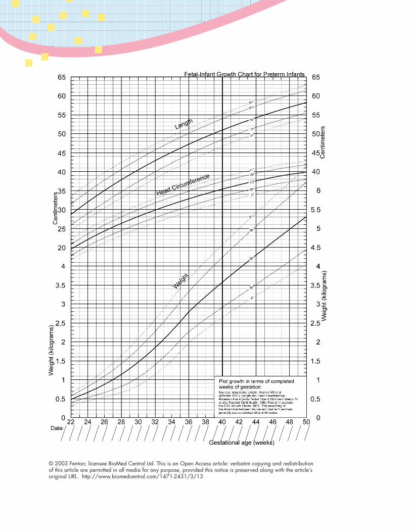
© 2003 Fenton; licensee BioMed Central Ltd. This is an Open Access article: verbatim copying and redistribution of this article are permitted in all media for any purpose, provided this notice is preserved along with the article’s original URL. http://www.biomedcentral.com/1471-2431/3/13
Weaning
Description ■ The introduction of solid food feedings and the gradual replacement of milk by solid food as the main source of energy and nutrients.
■ Shared pleasure in eating and feeding enhances the relationship between infants and their caregivers.
■ Signs of readiness for solid foods:
– Adequate head control.
– Interest in feedings.
– Decrease in tongue thrust.
– Ability to sit with support at 60-90 degrees.
Considerations ■ Very low-birthweight infants are at risk for developing aversions to feeding.
– May have more gagging when initiating solid feeding.
■ Water and juices should not be offered prior to 6 months of age.
■ Whole milk should be offered at ≥12 months of age.
■ Weight gain may be jeopardized by adding poor caloric solid foods in place of breast milk or formula.
■ Premature infants may feed more slowly due to weaker global tone, limited energy, and a lack of fully coordinated suck and swallow reflexes and oromotor tone.
Recommendations ■ AAP recommends solid foods be introduced at 4-6 months corrected age.
■ Use preterm formula milk to liquefy dried cereal foods until 9 months corrected age.
■ Introduce rice cereal first, then vegetables or fruits.
■ Introduce single-ingredient baby foods one at a time, and continue for 5-7 days before introducing an additional new food.

Appendix C: NICU Needs Assessment
Infant’s Name: Date of Birth:
ID#: Male Female Bed#:
Caregiver’s Contact #: Address:
Pediatrician Name and Number: Birthweight Current Weight
Primary Diagnosis
Newborn Blood Screening Date: _______________________________________
Newborn Blood Screening Results
Immunizations Current? ■ No ■ Yes
RSV Prophylaxis Given? ■ No ■ Yes Date: _____________________________
Feeding: ■ Breast milk ■ Formula
Tobacco Use In Home? ■ No ■ Yes
Social Worker Referral Needed? ■ No ■ Yes
Transportation Needs? ■ No ■ Yes
Car Seat? ■ No ■ Yes
Car Seat Education? ■ No ■ Yes Date: _____________________________
Car Seat Test? ■ No ■ Yes Date: _____________________________
CPR Education? ■ No ■ Yes Date: _____________________________
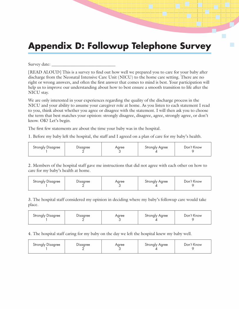
Appendix D: Followup Telephone Survey
Survey date: ______________________________
[READ ALOUD] This is a survey to find out how well we prepared you to care for your baby after discharge from the Neonatal Intensive Care Unit (NICU) to the home care setting. There are no right or wrong answers, and often the first answer that comes to mind is best. Your participation will help us to improve our understanding about how to best ensure a smooth transition to life after the NICU stay.
We are only interested in your experiences regarding the quality of the discharge process in the NICU and your ability to assume your caregiver role at home. As you listen to each statement I read to you, think about whether you agree or disagree with the statement. I will then ask you to choose the term that best matches your opinion: strongly disagree, disagree, agree, strongly agree, or don’t know. OK? Let’s begin.
The first few statements are about the time your baby was in the hospital.
1. Before my baby left the hospital, the staff and I agreed on a plan of care for my baby’s health.
Strongly Disagree 1
Disagree 2
Agree 3
Strongly Agree 4
Don’t Know 9
2. Members of the hospital staff gave me instructions that did not agree with each other on how to care for my baby’s health at home.
Strongly Disagree 1
Disagree 2
Agree 3
Strongly Agree 4
Don’t Know 9
3. The hospital staff considered my opinion in deciding where my baby’s followup care would take place.
Strongly Disagree 1
Disagree 2
Agree 3
Strongly Agree 4
Don’t Know 9
4. The hospital staff caring for my baby on the day we left the hospital knew my baby well.
Strongly Disagree 1
Disagree 2
Agree 3
Strongly Agree 4
Don’t Know 9
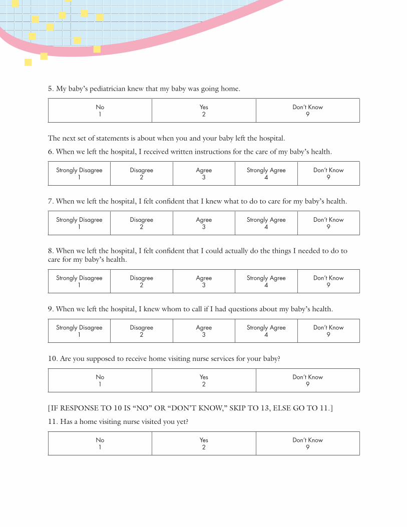
5. My baby’s pediatrician knew that my baby was going home.
No 1
Yes 2
Don’t Know 9
The next set of statements is about when you and your baby left the hospital.
6. When we left the hospital, I received written instructions for the care of my baby’s health.
Strongly Disagree 1
Disagree 2
Agree 3
Strongly Agree 4
Don’t Know 9
7. When we left the hospital, I felt confident that I knew what to do to care for my baby’s health.
Strongly Disagree 1
Disagree 2
Agree 3
Strongly Agree 4
Don’t Know 9
8. When we left the hospital, I felt confident that I could actually do the things I needed to do to care for my baby’s health.
Strongly Disagree 1
Disagree 2
Agree 3
Strongly Agree 4
Don’t Know 9
9. When we left the hospital, I knew whom to call if I had questions about my baby’s health.
Strongly Disagree 1
Disagree 2
Agree 3
Strongly Agree 4
Don’t Know 9
10. Are you supposed to receive home visiting nurse services for your baby?
No 1
Yes 2
Don’t Know 9
[IF RESPONSE TO 10 IS “NO” OR “DON’T KNOW,” SKIP TO 13, ELSE GO TO 11.]
11. Has a home visiting nurse visited you yet?
No 1
Yes 2
Don’t Know 9
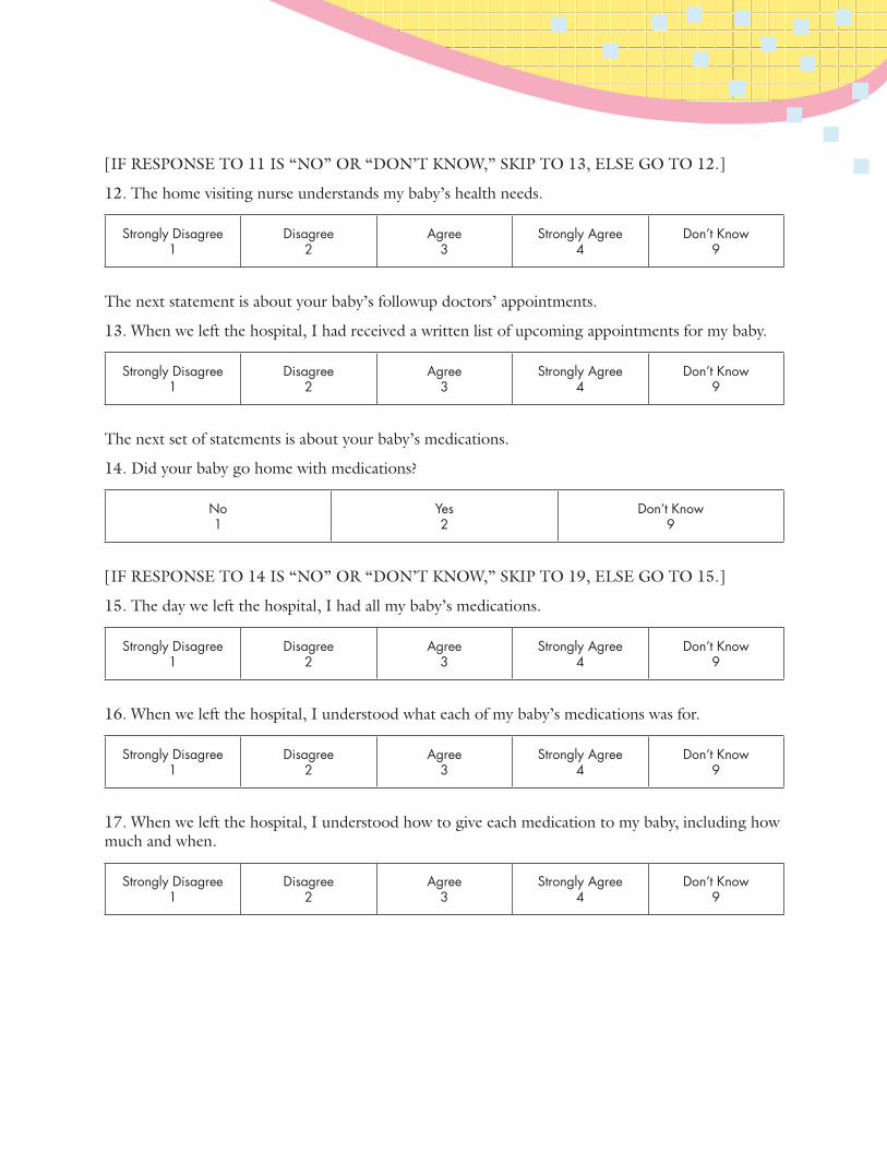
[IF RESPONSE TO 11 IS “NO” OR “DON’T KNOW,” SKIP TO 13, ELSE GO TO 12.]
12. The home visiting nurse understands my baby’s health needs.
Strongly Disagree 1
Disagree 2
Agree 3
Strongly Agree 4
Don’t Know 9
The next statement is about your baby’s followup doctors’ appointments.
13. When we left the hospital, I had received a written list of upcoming appointments for my baby.
Strongly Disagree 1
Disagree 2
Agree 3
Strongly Agree 4
Don’t Know 9
The next set of statements is about your baby’s medications.
14. Did your baby go home with medications?
No 1
Yes 2
Don’t Know 9
[IF RESPONSE TO 14 IS “NO” OR “DON’T KNOW,” SKIP TO 19, ELSE GO TO 15.]
15. The day we left the hospital, I had all my baby’s medications.
Strongly Disagree 1
Disagree 2
Agree 3
Strongly Agree 4
Don’t Know 9
16. When we left the hospital, I understood what each of my baby’s medications was for.
Strongly Disagree 1
Disagree 2
Agree 3
Strongly Agree 4
Don’t Know 9
17. When we left the hospital, I understood how to give each medication to my baby, including how much and when.
Strongly Disagree 1
Disagree 2
Agree 3
Strongly Agree 4
Don’t Know 9
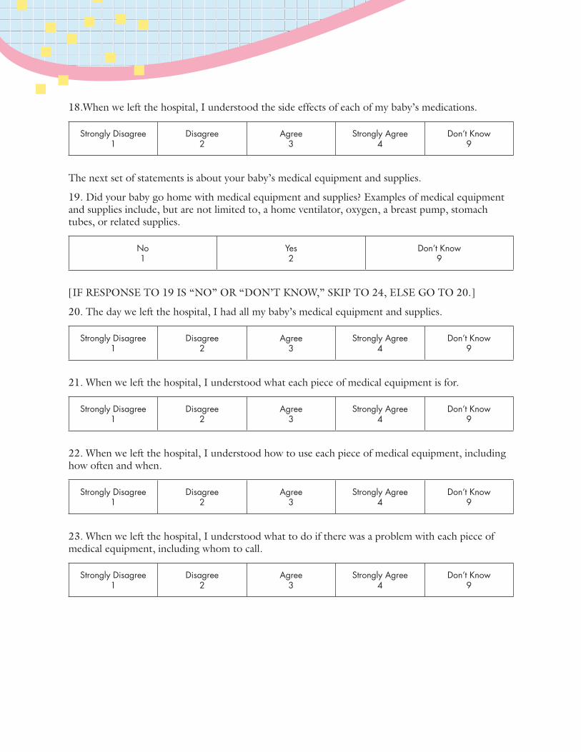
18.When we left the hospital, I understood the side effects of each of my baby’s medications.
Strongly Disagree 1
Disagree 2
Agree 3
Strongly Agree 4
Don’t Know 9
The next set of statements is about your baby’s medical equipment and supplies.
19. Did your baby go home with medical equipment and supplies? Examples of medical equipment and supplies include, but are not limited to, a home ventilator, oxygen, a breast pump, stomach tubes, or related supplies.
No 1
Yes 2
Don’t Know 9
[IF RESPONSE TO 19 IS “NO” OR “DON’T KNOW,” SKIP TO 24, ELSE GO TO 20.]
20. The day we left the hospital, I had all my baby’s medical equipment and supplies.
Strongly Disagree 1
Disagree 2
Agree 3
Strongly Agree 4
Don’t Know 9
21. When we left the hospital, I understood what each piece of medical equipment is for.
Strongly Disagree 1
Disagree 2
Agree 3
Strongly Agree 4
Don’t Know 9
22. When we left the hospital, I understood how to use each piece of medical equipment, including how often and when.
Strongly Disagree 1
Disagree 2
Agree 3
Strongly Agree 4
Don’t Know 9
23. When we left the hospital, I understood what to do if there was a problem with each piece of medical equipment, including whom to call.
Strongly Disagree 1
Disagree 2
Agree 3
Strongly Agree 4
Don’t Know 9
The final set of statements is about your baby’s feeding.
24. The day we left the hospital, I had what I needed to feed my baby.
Strongly Disagree 1
Disagree 2
Agree 3
Strongly Agree 4
Don’t Know 9
25. When we left the hospital, I understood how to feed my baby, including how much and how often.
Strongly Disagree 1
Disagree 2
Agree 3
Strongly Agree 4
Don’t Know 9
26. When we left the hospital, I felt confident in my ability to feed my baby.
Strongly Disagree 1
Disagree 2
Agree 3
Strongly Agree 4
Don’t Know 9
Thank You!

80
AHRQ Pub. No. 12(14)-0054-EF December 2013
U.S. Department of Health and Human ServicesAgency for Healthcare Research and Quality 540 Gaither Road Rockville, Maryland 20850