Embed Size (px)
Citation preview
Transjugular Intrahepatic Portosystemic Shunt: Decoding the TIPS Procedure, Appearance, and
Complications for the Diagnostic Radiologist Laura Eisenmenger MD
Paul Nikolaidis MD
Thomas Winter MD
Eugene Huo MD
2393384University of Utah Health Sciences Center
Salt Lake City, [email protected]: None
Learning Objectives
• Target audience: Diagnostic radiologists interpreting imaging on patients post-TIPS
• Understand the clinical indications for TIPS
• Identify the appearance of a typical TIPS procedure
• Learn the appropriate follow-up findings
• Identify variations of a normal TIPS
• Identify alternatives to a TIPS
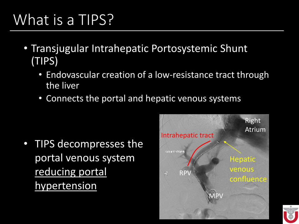
What is a TIPS?
• Transjugular Intrahepatic Portosystemic Shunt (TIPS)• Endovascular creation of a low-resistance tract through
the liver
• Connects the portal and hepatic venous systems
• TIPS decompresses the portal venous system reducing portal hypertension
Right Atrium
MPV
RPV
Intrahepatic tract
Hepatic venous confluence
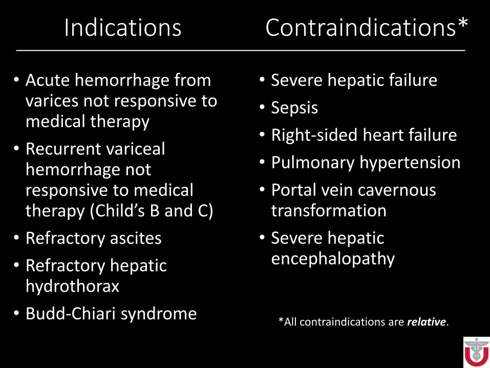
Indications
• Acute hemorrhage from varices not responsive to medical therapy
• Recurrent variceal hemorrhage not responsive to medical therapy (Child’s B and C)
• Refractory ascites
• Refractory hepatic hydrothorax
• Budd-Chiari syndrome
Contraindications*
• Severe hepatic failure
• Sepsis
• Right-sided heart failure
• Pulmonary hypertension
• Portal vein cavernous transformation
• Severe hepatic encephalopathy
*All contraindications are relative.
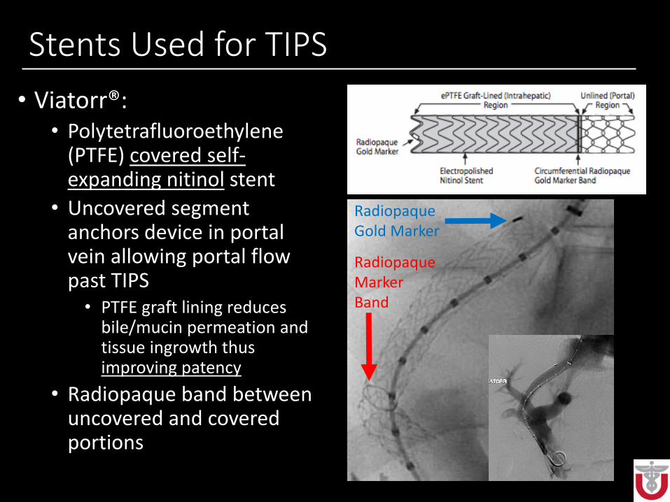
Stents Used for TIPS
• Viatorr®:• Polytetrafluoroethylene
(PTFE) covered self-expanding nitinol stent
• Uncovered segment anchors device in portal vein allowing portal flow past TIPS• PTFE graft lining reduces
bile/mucin permeation and tissue ingrowth thus improving patency
• Radiopaque band between uncovered and covered portions
Radiopaque Gold Marker
Radiopaque Marker Band
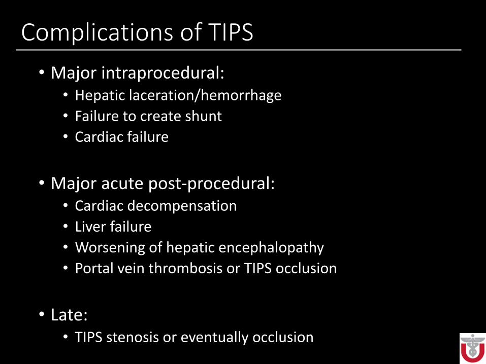
Complications of TIPS
• Major intraprocedural:• Hepatic laceration/hemorrhage
• Failure to create shunt
• Cardiac failure
• Major acute post-procedural: • Cardiac decompensation
• Liver failure
• Worsening of hepatic encephalopathy
• Portal vein thrombosis or TIPS occlusion
• Late: • TIPS stenosis or eventually occlusion

Typical TIPS Placement
• Ideal/most common approach:• Right hepatic vein to right portal vein
• Portal end should be at least 1 cm from main PV bifurcation
• Anatomic variants/hepatic or portal vein occlusion may require alternatives: • Middle hepatic to left or right portal vein
• Left hepatic to left portal vein
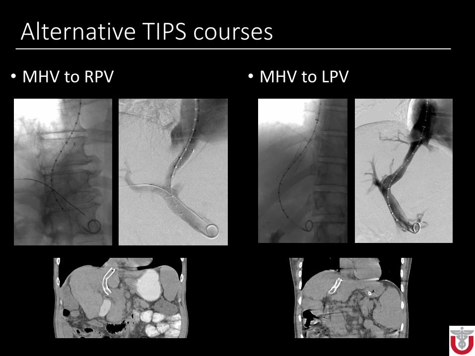
Alternative TIPS courses
• MHV to RPV • MHV to LPV
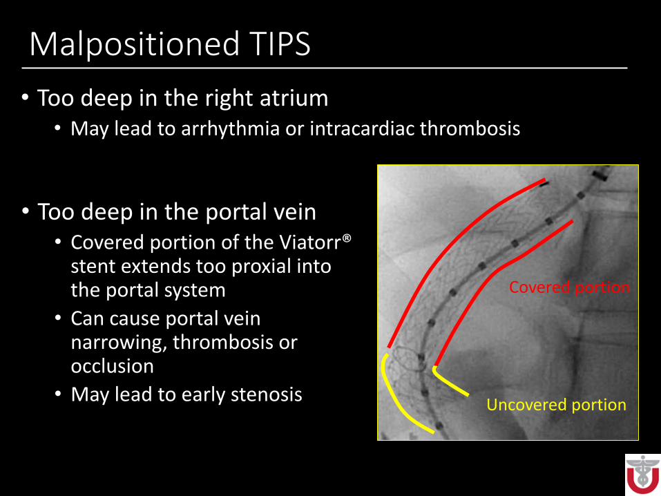
Malpositioned TIPS
• Too deep in the right atrium• May lead to arrhythmia or intracardiac thrombosis
• Too deep in the portal vein• Covered portion of the Viatorr®
stent extends too proxial into the portal system
• Can cause portal vein narrowing, thrombosis or occlusion
• May lead to early stenosis
Covered portion
Uncovered portion
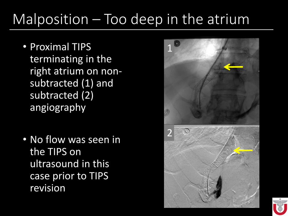
Malposition – Too deep in the atrium
• Proximal TIPS terminating in the right atrium on non-subtracted (1) and subtracted (2) angiography
• No flow was seen in the TIPS on ultrasound in this case prior to TIPS revision
2
1
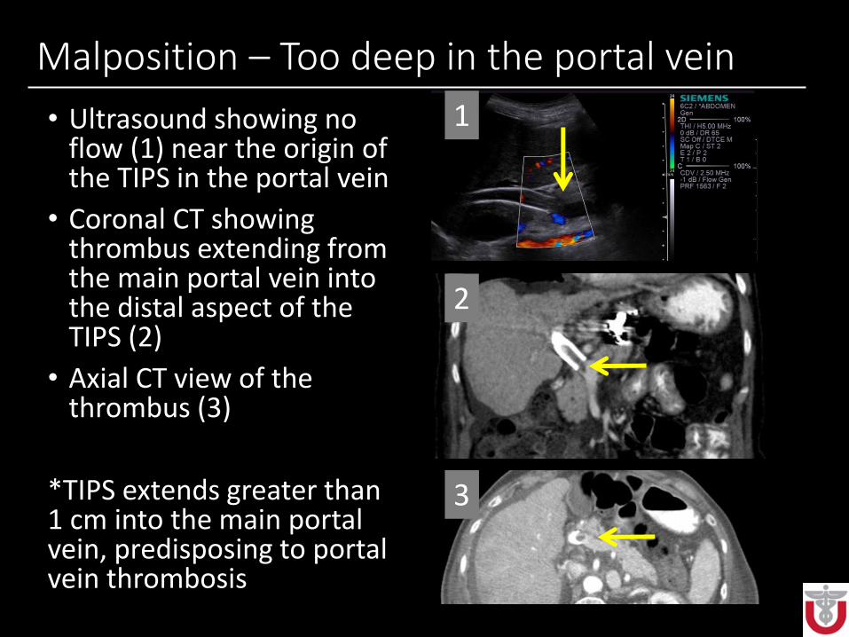
Malposition – Too deep in the portal vein
• Ultrasound showing no flow (1) near the origin of the TIPS in the portal vein
• Coronal CT showing thrombus extending from the main portal vein into the distal aspect of the TIPS (2)
• Axial CT view of the thrombus (3)
*TIPS extends greater than 1 cm into the main portal vein, predisposing to portal vein thrombosis
1
2
3
Post-procedure Follow-up
• Doppler ultrasound• Air trapped within the expanded PTFE layers of the
Viatorr® can cause shadowing • Can take a week to dissipate
• Baseline exam around 1 week after placement
• CT with contrast:• Can show shunt patency, insensitive for stenosis
• Repeat ultrasound at 3 month, then 6 month intervals
TIPS ultrasound evaluation
• Normal:• Pulsatile flow in shunt
• Peak systolic shunt velocities: 90-190 cm per second
• Velocity at portal and hepatic ends of shunt similar
• Hepatofugal flow in the nonshunted PV branch
• Hepatopedal flow in portal and splenic
• Portal vein velocity: At least 40 cm/sec
TIPS ultrasound evaluation• Abnormal:
• Abnormal shunt peak systolic velocities: <90 cm/sec or >190 cm/sec
• Change from baseline post-TIPS shunt velocities (>40 cm/sec decrease or >60 cm/sec increase in fasting patient)
• Increase in velocity from one point to another in the shunt of >100 cm/sec
• Visible shunt narrowing
• Non-pulsatile shunt flow
• Decrease in portal vein velocity from post-procedure baseline
• Portal vein velocity less than 30 cm/sec
• Hepatofugal or to-and-fro main portal or splenic vein flow
• Change from hepatofugal to hepatopedal flow in the non-shunted portal vein
• Absence of flow in shunt
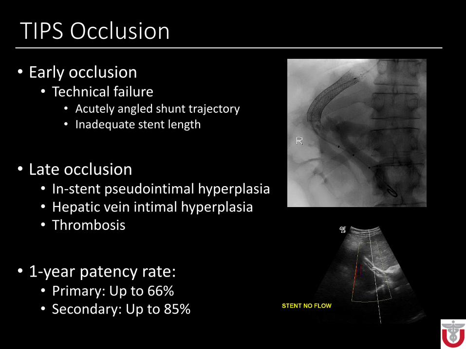
TIPS Occlusion
• Early occlusion• Technical failure
• Acutely angled shunt trajectory• Inadequate stent length
• Late occlusion• In-stent pseudointimal hyperplasia• Hepatic vein intimal hyperplasia• Thrombosis
• 1-year patency rate:• Primary: Up to 66%• Secondary: Up to 85%
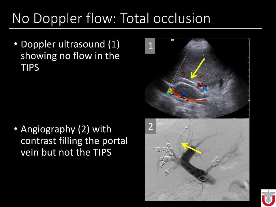
No Doppler flow: Total occlusion
• Doppler ultrasound (1) showing no flow in the TIPS
• Angiography (2) with contrast filling the portal vein but not the TIPS
1
2

No Doppler flow: Total occlusion
• Angioplasty of the occluded TIPS with a waist in the balloon at level of occlusion (1)
• Post-angioplasty evaluation of the TIPS demonstrates patency of the TIPS (2)
2
1
2
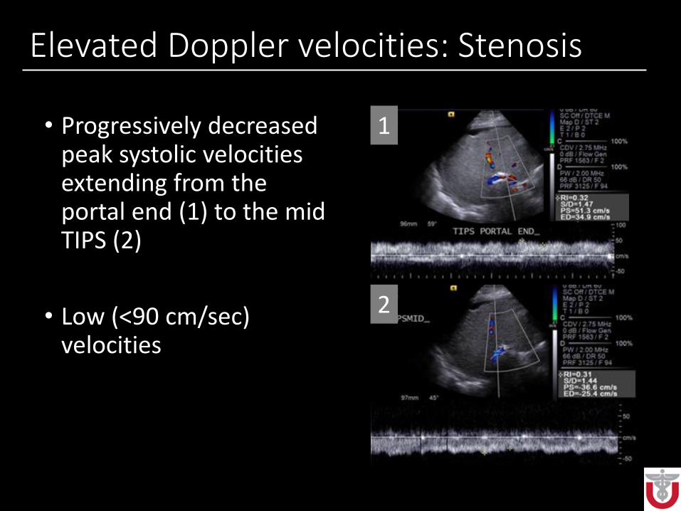
Elevated Doppler velocities: Stenosis
• Progressively decreased peak systolic velocities extending from the portal end (1) to the mid TIPS (2)
• Low (<90 cm/sec) velocities
1
2
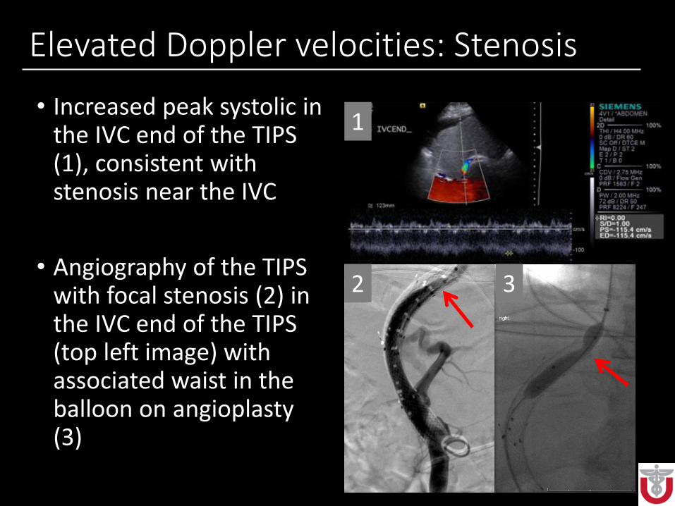
Elevated Doppler velocities: Stenosis
• Increased peak systolic in the IVC end of the TIPS (1), consistent with stenosis near the IVC
• Angiography of the TIPS with focal stenosis (2) in the IVC end of the TIPS (top left image) with associated waist in the balloon on angioplasty (3)
32
1
Elevated Doppler velocities: Stenosis
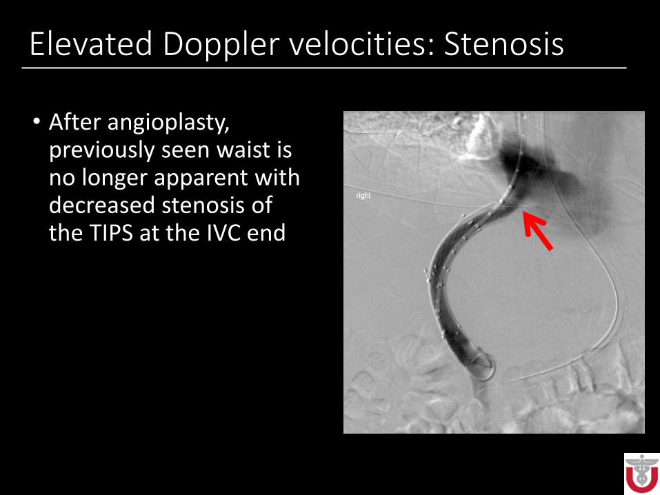
• After angioplasty, previously seen waist is no longer apparent with decreased stenosis of the TIPS at the IVC end
In-stent Stenosis
• Pseudointimal or hepatic vein intimal hyperplasia
• Most common ultrasound findings:• Abnormal shunt peak systolic velocities: <90 cm/sec or >220
cm/sec• Change from baseline post-TIPS shunt velocities (>40 cm/sec
decrease or >60 cm/sec increase in fasting patient)• Increase in velocity from one point to another in the shunt of >100
cm/sec
• Less common findings:• Visible shunt narrowing• Non-pulsatile shunt flow• Decrease in portal vein velocity from post-procedure baseline• Portal vein velocity less than 30 cm/sec• Hepatofugal or to-and-fro main portal or splenic vein flow• Change from hepatofugal to hepatopedal flow in portal vein
branches distal to shunt• Absence of flow in shunt
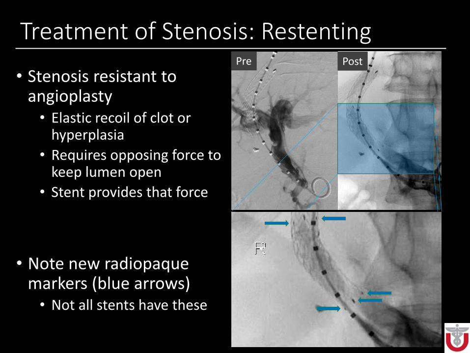
Treatment of Stenosis: Restenting
• Stenosis resistant to angioplasty• Elastic recoil of clot or
hyperplasia
• Requires opposing force to keep lumen open
• Stent provides that force
• Note new radiopaque markers (blue arrows)• Not all stents have these
Pre Post
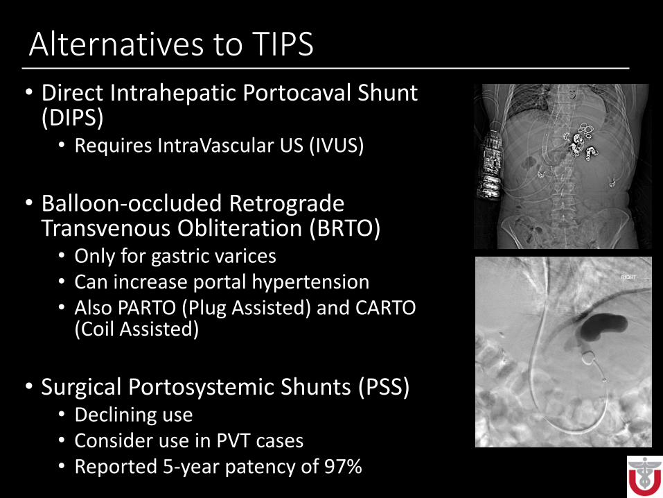
Alternatives to TIPS• Direct Intrahepatic Portocaval Shunt
(DIPS)• Requires IntraVascular US (IVUS)
• Balloon-occluded Retrograde Transvenous Obliteration (BRTO)• Only for gastric varices• Can increase portal hypertension• Also PARTO (Plug Assisted) and CARTO
(Coil Assisted)
• Surgical Portosystemic Shunts (PSS)• Declining use• Consider use in PVT cases • Reported 5-year patency of 97%
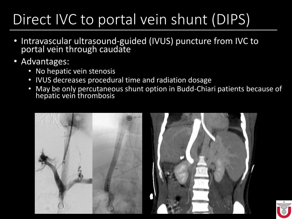
Direct IVC to portal vein shunt (DIPS)• Intravascular ultrasound-guided (IVUS) puncture from IVC to
portal vein through caudate
• Advantages:• No hepatic vein stenosis• IVUS decreases procedural time and radiation dosage• May be only percutaneous shunt option in Budd-Chiari patients because of
hepatic vein thrombosis

PARTO – Bleeding gastric varices
• Catheter in splenorenalshunt showing gastric varices (1)
• Amplatzer plug (2) occlusion of splenorenal shunt, embolization of varices performed
• Complete occlusion of the splenorenal shunt (3)
• Amplatzer plug (4) at the origin of the splenorenal shunt on CT
1 2
3 4

Congenital “TIPS”
• Congenital vascular malformation connecting the portal venous system to the systematic venous system
• Can cause progressively worsening hepatic encephalopathy later in life
• Situation may be complicated by the presence of portal hypertension, which may worsen if congenital shunt is occluded

Conclusions
• With increasing rates of chronic liver disease and portal hypertension, the incidence of TIPS placement will continue to increase
• Accurate identification of normal versus abnormal TIPS can help guide patient care and allow for early intervention
• Familiarity with the intraprocedural imaging and identification of technical variations can be helpful to correlate with follow-up imaging
References• Cura M, Cura A, Suri R, El-merhi F, Lopera J, Kroma G. Causes of TIPS dysfunction. AJR Am J
Roentgenol. 2008;191(6):1751-7.
• Chong WK. Abdominal Ultrasound, An Issue of Ultrasound Clinics. Elsevier Health Sciences; 2014.
• Saad WE. Balloon-occluded retrograde transvenous obliteration of gastric varices: concept, basic techniques, and outcomes. Semin Intervent Radiol. 2012;29(2):118-28.
• Saad WE, Darwish WM, Davies MG, Waldman DL. Stent-grafts for transjugular intrahepatic portosystemic shunt creation: specialized TIPS stent-graft versus generic stent-graft/bare stent combination. J Vasc Interv Radiol. 2010;21(10):1512-20.
• Haskal ZJ, Pentecost MJ, Soulen MC, Shlansky-goldberg RD, Baum RA, Cope C. Transjugularintrahepatic portosystemic shunt stenosis and revision: early and midterm results. AJR Am J Roentgenol. 1994;163(2):439-44.
• Gur I, Diggs BS, Orloff SL. Surgical portosystemic shunts in the era of TIPS and liver transplantation are still relevant. HPB (Oxford). 2014;16(5):481-93.
• Fidelman N, Kwan SW, Laberge JM, Gordon RL, Ring EJ, Kerlan RK. The transjugular intrahepatic portosystemic shunt: an update. AJR Am J Roentgenol. 2012;199(4):746-55.
For further questions, please contact:
Eugene Huo