Embed Size (px)
Citation preview
From
THE DEPARTMENT OF MICROBIOLOGY, TUMOR AND CELL BIOLOGY Karolinska Institutet, Stockholm, Sweden
TRANSLATIONAL REGULATION IN PLASMODIUM FALCIPARUM
Sherwin Chun Leung Chan
Stockholm 2017

Cover illustration: Merozoites and an amino acid roulette, on top of a background of codon codes. Designed by Madle Sirel, all rights reserved . All previously published papers were reproduced with permission from the publisher. Published by Karolinska Institutet. Printed by AJ E-print AB © Sherwin Chan, 2017 ISBN 978-91-7676-719-1

Translational Regulation in Plasmodium falciparum THESIS FOR DOCTORAL DEGREE (Ph.D.) By
Sherwin Chun Leung Chan Principal supervisor: Professor Mats Wahlgren Karolinska Institutet Department of Microbiology, Tumor and Cell Biology Co-supervisor: Professor Björn Andersson Karolinska Institutet Department of Cell and Molecular Biology
Opponent: Professor Karine Le Roch University of California, Riverside Department of Cell Biology and Neuroscience Examination Board: Professor Pedro Gil Karolinska Institutet Department of Physiology and Pharmacology Dr. Gerald McInerney Karolinska Institutet Department of Microbiology, Tumor and Cell Biology Dr. Alexey Amunts Stockholm University Department of Biochemistry and Biophysics

It is better to light a candle than to curse the darkness Anonymous

ABSTRACT Plasmodium falciparum is the causative agent of the most malignant form of human malaria, which remains as one of the most devastating infectious diseases. In face of a continuous international effort to eliminate the disease, the parasite not only has evaded a total obliteration, but has now evolved resistance to many of the available drugs. Next generation rational drug design is in urgent need and the key of such will lie on the successful identification of the parasite’s ‘Achilles heel’. While many existing and outstanding drugs have shown the promises of targeting the parasite translation machinery, the translation dynamics as well as the translational regulatory mechanisms are poorly understood. The studies described in this thesis aim to further our understanding on the translational regulation in P. falciparum, at both the global and gene-specific levels. Pregnancy associated malaria (PAM) is commonly seen with excessive sequestration of infected red blood cells in the placenta, the phenomenon is widely considered as the result of the specific ligand-receptor binding between the parasite derived PfEMP1-VAR2CSA proteins and the CSA proteoglycans. Translation of VAR2CSA protein is repressed by an upstream open reading frame, and a predicted trans factor is required for de-repression of var2csa translation. By using a spontaneously derived mutant that fails to efficiently translation the VAR2CSA proteins, we identified PTEF (Plasmodium translation enhancing factor) as the putative trans acting factor that allows efficient VAR2CSA translation. PTEF binds to the ribosomes and can enhance translation in a E. coli system. Importantly, higher PTEF expression was invariably observed to be associated with PAM in previous studies. Furthermore, PTEF function requires the processing by a calpain protease, blockage of the processing abolishes PTEF function in a reporter assay. Our data strongly suggest PTEF is an important regulator of PAM and raises potential therapeutic opportunity. It has been well described that codon usage bias could have a profound effect on translation efficiency. Codon usage is extremely biased in P. falciparum and cumulated to frequent insertions of asparagine homorepeats in up to one fourth of the proteome. However, the biological effect of this codon usage bias has not been studied. By using rationally recodonized GFP sequences, we showed that the increased use of GU wobble codon could reduce translation efficiency. We also demonstrated that the GU wobble-rich codon context underlying the asparagine homorepeats could impart significant influence on the translational output and transcript stability of the host gene. Despite this, GU wobble codons are overrepresented in the genome. Bioinformatics analyses suggested the high content of GU wobble codon might serve as a global regulatory mechanism. We thus offered new insight on the genome evolution of the parasite. RIFIN is the largest variable surface antigen family in P. falciparum. Its research profile has been much uplifted recently, as report showed that it might have a crucial link with severe malaria. While there is a sufficient interest to investigate the regulatory mechanisms associated with the RIFIN family, functional study of RIFIN is often marred by the lack of robustly verified reagents. By using RNA-sequencing and ultra-dense peptide microarray, we were able to authenticate specific RIFIN antibodies that exhibit some degree of intra-family cross-reactivity but minimal non-specific reactivity with other antigens. The derivation of these reagents will be important for future studies.


LIST OF PUBLICATIONS This thesis is based on the following papers:
I. Chan S, Frasch A, Mandava CS, Ch’ng JH, Quintana MdelP, Vesterlund M, Ghorbal M, Joannin N, Franzén O, Lopez-Rubio JJ, Barbieri S, Lanzavecchia A, Sanyal S, Wahlgren M. Regulation of PfEMP1-VAR2CSA translation by a Plasmodium translation-enhancing factor. Nature Microbiology [In Press]
II. Chan S#, Ch’ng JH, Wahlgren M, Thutkawkorapin J. Frequent GU wobble pairings reduce translation efficiency in Plasmodium falciparum. Sci Rep 2017 Apr 7; 7(1):723
III. Ch’ng JH, Sirel M*, Zandian A*, Quintana MdelP*, Chan SCL*, Moll K*, Tellgren-Roth A*, Nilsson I, Nilsso P, Qundos U, Wahlgren M. Epitopes of anti-RIFIN antibodies and characterization of rif-expressing Plasmodium falciparum parasites by RNA sequencing. Sci Rep 2017 Feb 24; 7:43190 # Corresponding author * Equal contribution
The following publications were obtained during the course of the PhD studies but are not included in this thesis:
I. Nunes-Silva S, Gangnard S, Vidal M, Vuchelen A, Dechavanne S, Chan S, Pardon E, Steyaert J, Ramboarina S, Chêne A, Gamain B. Llama immunization with full-length VAR2CSA generates cross-reactive and inhibitory single-domain antibodies anainst the DBL1X domain. Sci Rep. 2014 Dec 9: 4:7373
II. Geislinger TM, Chan S, Moll K, Wixforth A, Wahlgren M, Franke T. Label-free microfluidic enrichment of ring-stage Plasmodium falciparum-infected red blood cells using non-inertial hydrodynamic lift. Malar J. 2014 Sep 20; 13:375
III. Ch’ng JH, Moll K*, Quintana Mdel P*, Chan SC*, Masters E*, Liu J, Eriksson AB, Wahlgren M. Rosette-disrupting effect of an anti-plasmodial compound for the potential treatment of Plasmodium falciparum malaria complications. Sci Rep 2016 Jul 11; 6:29317 * Equal contribution


CONTENTS 1 INTRODUCTION ....................................................................................... 1 1.1 Malaria and global health ................................................................. 1 1.2 Malaria parasites and the life cycle .................................................. 3 1.3 Malaria pathogenesis ........................................................................ 4 1.3.1 General pathogenesis in uncomplicated malaria ................... 4 1.3.2 Severe malaria ....................................................................... 5 Cerebral malaria .................................................................... 5 Pregnancy assoicated malaria ............................................... 6 1.4 Antigenic variation and associated virulence ................................... 6 1.4.1 var genes and PfEMP1 .......................................................... 7 1.4.2 RIFIN and STEVOR ............................................................. 8 1.4.3 Cytoadhesion and Rosetting .................................................. 8 1.5 P. falciparum genome and its regulation ......................................... 9 1.5.1 General features of P. falciparum genome ........................... 9 1.5.2 Genome regulation ................................................................ 9 Nuclear architecture and higher order chromatin structure .. 9 Epigenetic regulation .......................................................... 11 Transcriptional regulation ................................................... 14 The non-coding Transcriptomes ......................................... 15 Post-Transcriptional regulation ........................................... 16 Translational regulation ...................................................... 18 Post-Translational regulation .............................................. 21 2 SCOPE OF THE THESIS ...................................................................... 24 3 EXPERIMENTAL PROCEDURES ...................................................... 25 4 RESULTS AND DISCUSSION ............................................................ 30 4.1 Paper I ............................................................................................ 30 4.2 Paper II ............................................................................................... 33 4.3 Paper III .............................................................................................. 35 5 CONCLUDING REMARKS AND FURTURE PERSPECTIVES ....... 38 6 Acknowledgements .................................................................................... 40 7 References .................................................................................................. 44

LIST OF ABBREVIATIONS CDS Coding DNA sequence CM Cerebral malaria CSA Chondroitin sulfate A CTD C-terminal domain DC Domain cassette ES Expansion segment FACS Fluorescence-activated cell sorting FISH Fluorescence in situ hybridization GTF General transcription factor HAT Histone acetyltransferase Hb Hemoglobin HDAC Histone deacetylase HDM Histone demethylase HMT Histone methyltransferase IDC Intraerythrocytic developmental cycle IE Infected erythrocyte IFA Immunofluorescence assay IPTp Intermittent preventive treatment in pregnancy KO Knockout LD Linker domain miRNA microRNA ncRNA Non coding RNA NES Nuclear export signal NLS Nuclear localization signal NMD Nonsense mediated decay NPC Nuclear pore complex NTD N-terminal domain PAM Pregnancy associated malaria PEXEL Protein export element PfEMP1 Plasmodium falciparum erythrocyte membrane protein 1 PTEF Plasmodium translation enhancing factor PV Parasitophorous vacuole PVM Parasitophorous vacuole membrane RBC Red blood cell RBP RNA binding protein rDNA Ribosomal DNA RIFIN Repetitive interspersed RPKM Read per RTTF Reconstituted transcription translation and folding

SAM Sterile alpha motif STEVOR Subtelomeric variable open reading frame TARE Telomere associated repeat element TERRA Telomeric repeat-containing RNA TPE Telomere position effect tRNA Transfer RNA TSS Transcription start site uORF upstream Open reading frame UTR Untranslated region WHO World Health Organization


1
1 INTRODUCTION 1.1 Malaria and global health
The word ‘Malaria’ originates from the Italian word mala aria, meaning ‘bad air’, which justly reflects how this deadly disease had instilled fear from people in the medieval time. We now know that human malaria can be caused by at least five parasite species from the Apicomplexa phylum; Plasmodium falciparum, Plasmodium vivax, Plasmodium ovale, Plasmodium malariae and Plasmodium knowlesi. Malaria is an archaic disease. While the origin of the human malarial parasites is still debated, early studies once proposed that P. falciparum could have diverged from a chimpanzee parasite, P. reichenowi, since the origin of the hominids, and was closely associated with the divergence of hominids and chimpanzee at almost 5 million years ago (1, 2). However, it was later suggested that P. falciparum has only undergone a rapid expansion from a severe bottleneck population within the past 6000 years, dubbed the ‘Malaria’s Eve’, that have likely defined the limited genetic structure of the contemporary parasite population (3, 4). Recent reports added to the complexity by pointing to possible lateral transfer events of human malarial parasites from other primate hosts at some point, in particular P. falciparum was found to be most related to Plasmodium spp. that infect gorilla, but not the chimpanzee (5). Regardless of the time of the origin, malaria has left an unmistakable trail during human evolution, on both the cultural and biological context (6, 7). Today, malaria remains one of the most devastating infectious diseases and is still endemic in 91 mid and low-‐income countries, holding tight onto its reputation as a poverty-‐associated disease. At the conclusion of the Millennium Development Goal in 2015, WHO reported an annual 212 million clinical cases of malaria, resulting in the loss of 429 000 human lives and of which 70% are children under the age of 5 (WHO malaria report 2016). Worse still, mortality and morbidity figures of WHO are, suggested from a few studies, disputably underestimating the level of malaria endemicity (8, 9). Yet, it is undeniable that more than a decade of intensified control intervention and investment, boosted by a shared commitment among the international community, had turned the tides against this deadly disease into a favorable one (10). Mortality and incidence continue to move steadily along a downward projectile, accompanied by an ever-‐shrinking malaria map. This achievement is attributed to the scaling up of various control measurements, including an increased coverage of vector control measures through the use of insecticide-‐treated net and indoor residual spraying, improved availability of diagnostic and surveillance tools, improved anti-‐malarial drug distribution and treatment regimes, as well as a continuous economic development that has reduced poverty at an unprecedented rate (11-‐13). In 2016, the WHO put forth the ‘Global Technical Strategy for Malaria’ that aimed to ambitiously reduce global incidence and mortality by 90% in 2030, effectively stressing a global roadmap from disease control to elimination. Meanwhile, at the dawn of this inter-‐phase, challenges lie ahead. Malaria epidemiology in many regions is now adopting a changing dynamics (14, 15). While many regions begin to eliminate the disease, transmission has mostly been reduced to low

2
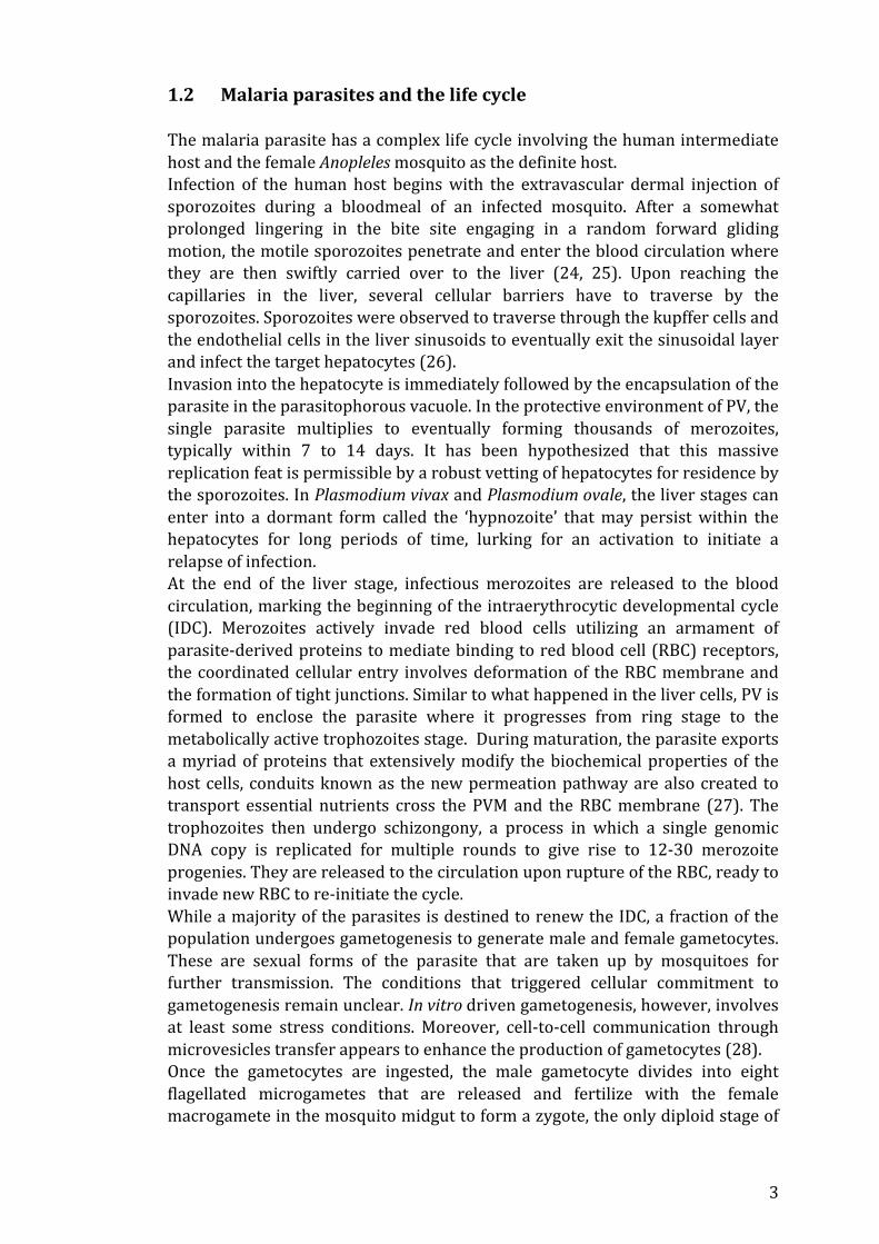
Figure 1. A reducing malaria endemicity. Upper panel shows a shrinking malaria map in Africa between (a) 2000 and (b) 2015. Heat map of Plasmodium falciparum parasite rate in children of age 2-‐10. Lower panel shows a reducing population in risk of high transmission area. (Upper: adopted from S. Bhatt et al. 2017, Lower: adopted from AM Noor et al. 2014, with permission to reproduce) intensity, and the remaining parasite reservoir is increasingly present at low density that often eludes detection by traditional microscopy techniques. Furthermore, asymptomatic adults have replaced children as the major parasite carriers. These new circumstances render traditional intervention strategies increasingly less cost-‐effective, which would potentially melt away financial interests as well as political commitments. Furthermore, artemisinin resistance has emerged and is gradually gaining a foothold in the Southeast Asia (16). The ongoing trend, aided by increased international travel, poises to spread the resistance to neighboring India and sub-‐Saharan Africa, of which occurrence would cast a dooming spell across the continent (17). Therefore, as the arms race between humans and parasites continues to rage, novel and innovative strategies would be the beacons for future control. Promising new generations of drugs and vaccine are now available on the shelf or in the late stage of developmental pipeline (18, 19), as well as diagnostic tools with increased sensitivity. At the same time, vector control can now be implemented through environmental management (20), the use of biologically modified vectors or through manipulating vector behavior (21, 22). Seasonal malaria chemoprevention can also be administered to interrupt transmission (23).
3
1.2 Malaria parasites and the life cycle The malaria parasite has a complex life cycle involving the human intermediate host and the female Anopleles mosquito as the definite host. Infection of the human host begins with the extravascular dermal injection of sporozoites during a bloodmeal of an infected mosquito. After a somewhat prolonged lingering in the bite site engaging in a random forward gliding motion, the motile sporozoites penetrate and enter the blood circulation where they are then swiftly carried over to the liver (24, 25). Upon reaching the capillaries in the liver, several cellular barriers have to traverse by the sporozoites. Sporozoites were observed to traverse through the kupffer cells and the endothelial cells in the liver sinusoids to eventually exit the sinusoidal layer and infect the target hepatocytes (26). Invasion into the hepatocyte is immediately followed by the encapsulation of the parasite in the parasitophorous vacuole. In the protective environment of PV, the single parasite multiplies to eventually forming thousands of merozoites, typically within 7 to 14 days. It has been hypothesized that this massive replication feat is permissible by a robust vetting of hepatocytes for residence by the sporozoites. In Plasmodium vivax and Plasmodium ovale, the liver stages can enter into a dormant form called the ‘hypnozoite’ that may persist within the hepatocytes for long periods of time, lurking for an activation to initiate a relapse of infection. At the end of the liver stage, infectious merozoites are released to the blood circulation, marking the beginning of the intraerythrocytic developmental cycle (IDC). Merozoites actively invade red blood cells utilizing an armament of parasite-‐derived proteins to mediate binding to red blood cell (RBC) receptors, the coordinated cellular entry involves deformation of the RBC membrane and the formation of tight junctions. Similar to what happened in the liver cells, PV is formed to enclose the parasite where it progresses from ring stage to the metabolically active trophozoites stage. During maturation, the parasite exports a myriad of proteins that extensively modify the biochemical properties of the host cells, conduits known as the new permeation pathway are also created to transport essential nutrients cross the PVM and the RBC membrane (27). The trophozoites then undergo schizongony, a process in which a single genomic DNA copy is replicated for multiple rounds to give rise to 12-‐30 merozoite progenies. They are released to the circulation upon rupture of the RBC, ready to invade new RBC to re-‐initiate the cycle. While a majority of the parasites is destined to renew the IDC, a fraction of the population undergoes gametogenesis to generate male and female gametocytes. These are sexual forms of the parasite that are taken up by mosquitoes for further transmission. The conditions that triggered cellular commitment to gametogenesis remain unclear. In vitro driven gametogenesis, however, involves at least some stress conditions. Moreover, cell-‐to-‐cell communication through microvesicles transfer appears to enhance the production of gametocytes (28). Once the gametocytes are ingested, the male gametocyte divides into eight flagellated microgametes that are released and fertilize with the female macrogamete in the mosquito midgut to form a zygote, the only diploid stage of

4
the parasite. The zygote then becomes motile and transforms into an ookinete and transverses across the midgut. The ookinete subsequently establishes as an oocyst after migration. The established oocyst generates a large number of sporozoites, which migrate to the salivary gland and completing the life cycle. While all the five human malaria parasites have the same life cycle, there are marked differences in the biology of some of the developmental stages. Most notably is the different duration of the IDC. Tertian malaria includes P. falciparum, P. vivax and P. ovale, which have a 48-‐hour IDC. Whereas P. knowlesi and P. malariae are known as quotidian and quartan malaria, having a signature IDC duration of 24 hours and 72 hours respectively.
Figure 2. (A-‐E) depicts the complete life cycle of P. falciparum. (Adopted from AF Cowman et al. 2016, with permission to reproduce) 1.3 Malaria pathogenesis 1.3.1 General pathogenesis in uncomplicated malaria The clinical symptoms of malaria appear when the parasites enter the IDC developmental stage. Of the five species causing human malaria, P. falciparum is overwhelmingly blamed as the major contributor of morbidity and mortality. Though with an improved efficiency in disease surveillances, it is increasingly

5
understood that P. vivax and P. knowlesi could also cause severe clinical symptoms (29, 30). The typical non-‐specific symptoms are usually systemic, including flu-‐like manifestations, fever, muscle ache, diarrhea, lethargy and nausea. Fever is notoriously known as the malarial paroxysm, and is characterized by bouts of sudden onset of shivering and cold sensation amid an elevated body temperature that can last for a few hours. This periodicity is apparently associated with the synchronized destruction of RBCs when merozoites are released at the end of the IDC, triggering an intense ‘cytokine storm’ mounted by the host innate immune response (31). The mass destruction of RBC, on the other hand, can cause hemolytic anemia. Splenomegaly is also a common feature in malaria, with extreme cases of spleen rupture were reported. It is because the bio-‐physiological properties of the parasitized RBCs are altered by the parasites, most notably with a reduced deformability, and the spleen therefore traps and destroys these pRBC as a defense mechanism (32, 33). Overloading of the spleen by recurring infections and high parasitemia thus can cause splenomegaly. Spleen size in children was historically used as an indicator of transmission intensity before the introduction of modern molecular techniques. 1.3.2 Severe malaria Severe malaria refers to the progression from general non-‐specific clinical symptoms to the exhibition of more severe and specific complications, usually with the risk of fatal outcome if left untreated. Severe malaria can be categorized into cerebral malaria (CM), severe anemia, acute respiratory distress syndrome and pregnancy associated malaria (PAM). The pathogenesis and causes of these severe complications are not totally clear, with opinions mainly divided into two schools of thought, one school claiming these complications to be associated directly with parasite sequestration and the other adopts a more cytokine-‐centric view. Cerebral malaria CM is a neurological manifestation of severe malaria, it is defined as a clinical syndrome in patients with unarousable coma and P. falciparum parasitemia in the peripheral blood, in which the coma cannot be explained by another cause. In high transmission area, coma can befall children with sudden onset of seizure following 1-‐3 day of fever. Symptoms can include brain swelling, intracranial hypertension and abnormal posture that indicates brainstem damage. Death is invariable without treatment and with a 15-‐20% fatality rate even if treatment is provided, survivors are also more prone to neurological squeals (34, 35). A common feature of CM is the sequestration of parasite in the cerebral microvasculature (36), this is proposed to cause occlusion that can impair blood perfusion and create a hypoxic microenvironment. Hypoxia induces ischemic injury and an increased blood flow ensues to compensate the metabolic necessities, which in turn causes hypertension. From a cytokine-‐centric perspective, increased TNF production can be seen as trigger of endothelial activation, which up-‐regulates ICAM1 expression and further reinforces parasite

6
sequestration. Finally, vascular injury can eventually lead to disruption of blood brain barrier, inducing a cascade of intense pro-‐inflammatory responses (37). Pregnancy associated malaria While most severe malaria complications are incidentally associated with young children that have less adaptive immunity against the parasites, PAM is an out-‐group that affects only pregnant women. It is estimated that 50 millions pregnancies occur annually in area of stable malaria transmission, putting a huge risk group to PAM (38). Despite the semi-‐immune status acquired through repeatedly exposure to malaria, primigravid women are very susceptible to PAM. PAM is associated with poor birth outcomes, including low birth weigh, preterm delivery and an increased risk of prenatal and neonatal mortality. Mortality to the pregnant women can also be attributed to an increased risk of maternal anemia (39). A hallmark of PAM is usually the excessive sequestration of parasites in the placenta. This specific sequestration is mediated by the binding of parasite VAR2CSA protein to the glycosaminoglycan chondroitin sulfate A (CSA) and will be detailed in this chapter later. This binding property, which is central to PAM, explains why successive pregnancies can gradually lead to acquisition of a semi-‐immune status. The binding through a relatively conserved parasite protein also promises the development of a vaccine. In general, the increased parasite biomass in the placenta is countered by host defense mechanism and results in an accumulation of immune cells, most notably macrophages (40). Besides the infiltration of immune cells, a pro-‐inflammatory cytokines profile can also be seen (41). Together, it is suggested that an enhanced complement activation and hemozoin deposition in the intervillous fibrin due to phagocytosis of parasitized cells can contribute to the adverse outcome of PAM (42-‐44). In many endemic area, PAM is managed by Intermittent Presumptive Treatment (IPTp), which is a mass drug administration strategy targeted to pregnant women using single dose of sulfadoxine-‐pyrimethamine both during early second and third trimester (45). 1.4 Antigenic variation and associated virulence Antigenic variation is a common strategy adopted by many pathogens. By constantly varying the surface landscape, it allows the pathogen to discontinue the exposure of antigens that are targeted by the host adaptive immunity and effectively evade destruction as well as exhausting host immune mechanism. Antigenic variation is likely an important evolutionary trait that are selected on the population level, because it permits the pathogen to maintain a persistent chronic infection, to easily transmit within a larger effective naïve host population as well as to allow repeated infections in the same host. In eukaryotic parasites, Typanosoma brucei (46), Giardia lamblia (47) and Plasmodium are well known for exhibiting different degrees of antigenic variation. In P. falciparum, antigen variation has been shown to involve variable surface antigens, invasion antigens and solute transporters (48). The dynamics governing antigenic variation is though to be host immune-‐modulated, as
7
parasites in splenectomized individuals have been found to behave profoundly different in this dynamics (49, 50). However, recent study has challenged this notion, as an apparently hard-‐wired antigenic variation program still happened in parasites infecting immuno-‐compromised mice (51). 1.4.1 var genes and PfEMP1 var gene family, and the encoded Plasmodium falciparum erythrocyte membrane protein 1 (PfEMP1) (52), is indisputably the most studied gene family in P. falciparum. Each haploid genome of the parasite contains around 60 copies of var gene. A typical var gene contains two exons separated by a small conserved intron, they can be divided into four distinct types depending on the sequence of their upstream elements, and are also defined by their orientations. UpsA genes are found exclusively in the subtelomeric regions and transcribed towards the telomere, upsB are also found in the subtelomeric but some upsB var genes can be found in the central region of the chromosome, in which all UpsC are invariably located. UpsE sequence exclusively flanks the relatively conserved var2csa (53). Importantly, the ‘vardom’ between parasite strains and isolates are highly polymorphic, and recombinations happen frequently between var genes of the same ups group to further generate genetic diversity (54, 55). Var gene is under the control of a strict program of mutual exclusive regulation. Though at times disputed, current opinion is that only one member is expressed in a parasite at a time (56-‐58), and its periodic switching to another var member almost defines the central thesis of antigenic variation in P. falciparum. Switching between members occurs at around 2% per generation, but can be higher depending on the genetic background (59), the switching appears not programmed but are also suggested to follow some undefined rules, as any disturbance to the mutual exclusive regulation frequently resulted in the on-‐switching of the upsE var2csa (60). The regulation of the mutual exclusive expression is transcription dependent and is at least dependent on sequence elements found on the promoter and the conserved intron, and that the paring of the two elements are required for a ‘gene-‐counting’ mechanism (61-‐63). When and how a var gene is decided to be transcribed and the eventual expression of the protein are now increasingly appreciated to involve practically all levels of the central dogma, some of these will be discussed later in this chapter. Var genes encode PfEMP1 proteins, which are large multi-‐domain proteins. A typical PfEMP1 is consist of an N-‐terminal sequence (NTS), multiple Duff Binding Like (DBL) domains, a cysteine-‐rich interdomain region (CIDR) and a transmembrane region followed by a relatively conserved acidic terminal sequence (ATS). Large-‐scale survey was able to classify, by sequence similarity, six DBL types and five CIDR types. PfEMP1s can vary in both the number and the order of the domains in their overall architecture, creating variable mosaic patterns as well as intra-‐domain sequence diversity (53). Interestingly, many domain cassettes (DC) with defined domain types and orders can also be classified, suggesting possible functional constraints underlying these domain cassettes. A major function of PfEMP1 is to mediate cytoadhesion, a process in which infected RBCs sequester to the endothelial lining.

8
1.4.2 RIFIN and STEVOR Also found in P. falciparum genome are >150 copies of rif and 30-‐40 copies of stevor genes, that encode the RIFIN (repetitive interspersed family) and the STEVOR (sub-‐telomeric variable open reading frame) proteins respectively. Unlike PfEMP1, they are typically 30-‐50 kDa and their gene structures invariably contain two exons. RIFIN can be further divided into group A and group B, with group A proteins retaining a conserved internal indel of 25 amino acids (64). Both gene families appear to exhibit some degree of mutual exclusive expression and property of antigenic variation (65, 66). Their peak expression are generally at late stages of the asexual cycle, though expression in other stages were noted (67, 68). While all other Plasmodium spp lack PfEMP1-‐like proteins, some can still sequester to vessels. The relatively small size of RIFINs and STEVORs, therefore, make them the more comparable entities to the variable surface antigens found in other Plasmodium spp, fuelling speculation that they maybe also mediating cytoadhesion. Supporting this, RIFINs were found to be target of naturally acquired antibodies during malaria infection and functional protection of these antibodies were reported (69, 70). 1.4.3 Cytoadhesion and Rosetting Cytoadhesion is an ubiquitous feature of P. falciparum, it refers to the sequestration of parasitized RBC to the endothelial cells that line the microvasculatures. The more specific occurrence of aggregation of uninfected RBCs centering a parasitized RBC is commonly termed as ‘Rosetting’. Cytoadhesion and rosetting primarily serve as adaptive mechanism for the parasites to prevent destruction by the spleen, as parasitized RBCs have reduced deformability and would be sidelined from the circulation for destruction when they pass through the spleen. Secondary, the effect of cytoadhesion and rosetting can be associated with disease severity (71). A wealth of literatures has established strong association between these phenomena and the expression of PfEMP1s. Different PfEMP1s variants have been demonstrated to bind a number of receptor ligands that are found on endothelial cells and RBCs. The current known ‘interactome’ of PfEMP1 includes CD36, ICAM-‐1, EPCR, PECAM1, Heparan sulphate, CSA, P-‐selectin, Thrombospoindin, CR1 and Blood group A, (see reviews (72, 73)). Given the huge diversity of PfEMP1 variants, this ‘interactome’ is likely to be further expanded. Of particular interest is the apparent association of some PfEMP1 variants and severe disease outcomes. Variants consisting of DC8 and DC13 can bind EPCR and are associated with severe malaria (likely CM) incidence (36, 74-‐76). Another classical example is the almost predictive expression of VAR2CSA in PAM (77). The relatively conserved VAR2CSA is the only variant known to bind to chondroitin sulfate A, which is a proteoglycan found on syndecan-‐1 proteins expressed on the surface of syncytiotrophoblast microvillous cells (78). Furthermore, rosetting can also be mediated by RIFIN and STEVOR variants (79, 80). In particular, RIFIN preferentially binds to blood group A and aggravates rosetting phenotypes, to an extent that it has been suggested as a driving force for the purifying selection of blood group A allele in African populations (79, 81).
9
1.5 P. falciparum genome and its regulation 1.5.1 General features of P. falciparum genome The genome sequence of P. falciparum was first reported in 2002 from the parasite clone 3D7 (82). The genome consists of a ~23Mb nuclear genome organized into 14 linear chromosomes ranging from ~0.6 to 3.3Mb, a 35kb circular apicoplast plastid and a 6kb mitochondrial genome. The nuclear genome has an AT content considered to be the highest among all sequenced genomes, averaging at 81%, and spiking to ~90% in non-‐coding regions. Similar to many unicellular organisms, gene density is relatively high, ~50% of the genome sequencing is predicted to be protein coding. More than half of the ~5300 protein coding genes contain intron, and the average gene length is much longer than that of other organisms. Notably, initial assignment showed that up to 60% of genes encode proteins of unknown function, effectively sharing no sequence homology to any known protein. While gene prediction by sequence homology can sometimes be confounded by the high genomic AT content, it reflects the very limited knowledge in our understanding of the parasite genome. 1.5.2 Genome regulation The regulation of genome activity in P. falciparum has been studied and described on all levels of the central dogma of molecular biology; this section will discuss some mechanisms shown to be important for the eventual function modulation of the genome, either through functional studies or system biology analyses. Nuclear architecture and High order chromatin structure The organization of chromosomes and the dynamics of their physical localization into nuclear sub-‐compartments are increasingly appreciated as important regulatory mechanisms of the genome activities. The majority of the chromosomal regions of P. falciparum can be seen in electron microscopy to be predominantly maintained in decondensed euchromatin, which usually defines transcriptionally permissive sites. However, telomeric regions are tethered to the nuclear periphery, forming transcriptionally repressive heterochromatin (83). Four to seven clustered nuclear foci containing the 28 telomeric ends of the chromosomes were visible in the nuclear periphery in early FISH experiments (84). It is now clear that chromosome-‐end plays an important role in gene regulation. P. falciparum telomeres contain tandem GGGTT(T/C)A repeats and are organized into non nucleosomal structure in the most distal region. The telomeric region is followed by a subtelomeric region containing non-‐coding elements that include the telomere-‐associated-‐repetitive elements (TARE 1-‐6), and is adjoined by a region with coding-‐genes mostly of variable surface antigens (82, 84, 85). While a telosome complex is expected to bind the telomere, experimental data suggests a very different components as compared to other eukaryotes (86). So far, a number of proteins have been

10
shown to bind telomeric sequence or colocalized in these telomeric foci, including the histone deacetylase PfSir2A (87) and the cooperatively bound PfORC1 (88), PfHP1 that binds the repressive methylated H3K9 marks (89), the histone H3K4 methyltransferase PfSET10 (90), DNA-‐binding domain containing PfAlba3 (91) and PfSIP2 which binds to the SPE2 elements that are mostly scattered in the subtelomeric regions (92). PfTRZ, which contains a C2H2 zinc finger domain, has also been shown to bind directly to the telomeric repeats (93). Most recently, the atypical AP2 domain-‐containing PfAP2Tel was co-‐pulldown with the telomere repeat sequences together with a number of novel factors and was demonstrated to cluster with the telomeres in vivo (94). This has expanded the repertoire of potential telosome-‐forming proteins. The effect of telomeric clustering appears to affect var genes on the genomic and transcriptional level. Clustering of the telomere brings together var genes on different chromosomes and may contribute to the increased recombination frequency between different var genes and generates sequence diversity (54, 55). The silencing and activation of var genes are also closely related to telomeric clustering. The activated var gene, while still localizes in the nuclear periphery, has been demonstrated to delocalize with the silenced var gene members, a mechanism known as telomere positioning effect (TPE) (83). The mechanism governing the observed TPE and the activation and silencing remains unclear, but it was reported that a sequence element within the var intron serves as the platform for scaffolding a nuclear protein complex that recruits the actin protein complex, and the disruption of any of these elements derail the normal regulation of TPE and thus the mutual exclusive expression of the var gene family (95). Besides var genes, rDNA genes are loci that are also positioned to the nuclear periphery, and colocalize with PfNOP1, a nucleolus marker (96). All rDNA genes were once believed to colocalize in a perinuclear focus and then dispersed to multiple foci upon DNA replication stage, resulted in decreased transcriptional activities (96). However, recent advances using chromosome conformation capture and next generation sequencing techniques suggested direct chromosomal contact between the active rDNA loci but not the silenced counterparts (97, 98). The centromeres, which are stretches of 2-‐3kb extremely AT rich sequence, also focalize in the perinucleus (99). In P. falciparum, a nucleosome unit is defined by the wrapping of 155bp, instead of the usual 147bp, of DNA to the histone core (100). Nucleosome positioning has important implication on the transcriptional status of the genes. Regions of nucleosome depletion promote formation of open chromatin and accessibility to the transcriptional machinery. Due to the technical challenges presented by the extreme AT richness of the genome, reports on nucleosome occupancy in P. falciparum were sometimes disputed (101-‐104). However, in general, most reports suggested that lower nucleosome occupancy was observed in the promoter regions that mark the transcriptional start sites, and nucleosome depletion is usually more prominent in actively transcribed genes, thus establishing a correlation between low nucleosome occupancy and transcriptional activities. A generally lower occupancy in the intergenic regions, while disputed (101, 104), could be a result of the high AT content in these regions, which would present a reduced binding stability to histones (105).
11
Interestingly, global dynamic changes of nucleosome occupancy, instead of a more targeted fashion, appear to be coupled with the transcriptional activities of the developmental stages, with trophozoite stages showing lower global nucleosome occupancy when compared to ring and schizont stages (101). The stage-‐specific open chromatin structure reflects the high transcriptional activities, or perhaps also facilitates the assembly of pre-‐replication complexes. This was further supported by the observation of less intrachromosomal contacts in the trophozoites stage, corroborating the existence of a relaxed chromatin structure (97, 98). Plasmodium histones have considerably diverged sequences as compared with other eukaryotes. The genome harbors a lineage specific H2B variant (H2B.Z), but lacks the linker histone H1, in addition to all canonical histone units (106). Nucleosome sub-‐structures characterized by differential assembly of histone units was reported and has association with the transcriptional status in the parasite. H2A.Z and H2B.Z were preferentially enriched in intergenic regions and most markedly deposited in the promoters of actively transcribed genes (107). Most distinctly, only the single active var gene is deposited with these histone variants (108, 109). However, there was no evidence to suggest their fluctuation during the asexual cycle. These data may point to their role in establishing cellular memory. Another H3 variant, PfCENH3, is enriched preferentially in nucleosomes wrapped by the centromeres (99). Epigenetic regulation Epigenetic is the study of stable heritable traits that cannot be explained by changes in DNA sequences. It is commonly referring to the study of post-‐translational histone modifications and DNA methylation, in addition to regulation of chromatin structure. In P. falciparum, histone modifications capture most of the spotlight in this regard. Global proteomic studies by several independent research groups established an extensive library of histone modifications in P. falciparum, many of these appeared to be unique for this species. While most identified histone marks have yet to be defined functionally, some investigated histone marks were shown to denote conserved function as in other eukaryotes. For example, enrichment in the intergenic region with H3K4 methylation and acetylation in various histone H3 residues are associated with the euchromatin regions and thus positively associated with transcriptional activities, whereas, H3K9 methylation and histone hypoacetylations are generally localized in the nuclear periphery, effectively demarcating the repressive heterochromatin regions (110-‐112). Importantly, var regulation implicitly involves the dynamic interplay of these histone marks, the single active var gene is deposited with H3K4me3 and H3K9ac, while all the silent var genes are marked repressive H3K9me3 (112). On the other hand, at least some important histone marks have functionally departed in P. falciparum, such as methylations of H3K36, which generally marks coding regions of transcriptionally active loci, were found to be associated with gene repression in the parasite (113), specifically, H3K36me3 is distributed on all var genes regardless of the transcriptional status. Furthermore, the important repressive H3K27 methylation marks were reportedly absent in the parasites in multiple
12
studies (110, 113-‐115), although a recent study specifically detected the presence of this mark almost exclusively in sexual stage parasites (116), which may suggest a role in global reprogramming transcription during differentiation. Histone modifications are dynamically deposited and removed by chromatin-‐modifying enzymes. These enzymes include histone acetyl transferases (HAT), histone deacetylase (HDAC), histone methyl transfeases (HMT) and histone demethylase (HDM). P. falciparum genomes retain an extensive panel of these enzymes, including ten SET-‐domain containing proteins that mediate histone lysine methylation, three HDMs harboring either the LSD or JmjC domains (117), eight HATs, one class I HDAC, as well as two of each class II and III HDACs. A few of these chromatin-‐modifying enzymes have been functionally characterized and, together with the reversible histone modifications, they regulate diverse processes. PfGCN5 and PfMYST are HATs that preferentially acetylate various lysine residues of histone H3 and H4 respectively. Inhibition of PfGCN5 induced cell-‐cycle arrest (118), whereas PfMYST is refractory to gene disruption and that overexpression resulted in reduction in cell proliferation and increased sensitivity to DNA damages (119). These indicate HATs and dynamic histone acetylation are essential for the asexual stage. PfSIR2A and PfSIR2B of class III NAD+ dependent HDAC are important regulators of the subtelomeric heterochromatin, deletion of either gene resulted in the abolition of mutual exclusive var expression (120), PfSIR2A was further shown to regulate the transcription of rDNA and also to be important for telomere length homeostasis and may promote inter-‐chromosome recombinations (120-‐122). Since all these elements localize in the nuclear periphery, PfSIR2A is likely to be instrumental in the maintenance of the transcription repressive center underlying this nuclear subcompartment. Depletion of the class II HDAC PfHda2 at the post-‐translational level was reported to also abolish the global silencing effect of var loci (123). Moreover, increased gametogenesis was observed as a result of transcriptional activation of AP2-‐g, silencing of which is normally mediated by PfHda2 deacetylation. PfSET2 (PfSETvs), a primate specific SET-‐domain containing protein, mediates trimethylation of H3K36 residue. H3K36me3 appears to be restricted to the var genes coding region regardless of the transcriptional status, and knockout of PfSET2 resulted in the simultaneous activation of all var genes (124). Recruitment of PfSET2 to the var loci is thought to be dependent on the unphosphorylated form of RNA polymerase II, and that the disruption of this binding phenocopies the effect of PfSET2 knockout (125). Another functional study on PfSET10 showed that this HKMT is responsible for H3K4 methylation and exclusively colocalizes with the active var gene, but not the silent var. Interestingly, PfSET10 interacts with PfActin, potentially implicated in the TPE and thus var switching mechanism (90). Biochemical characterization of PfSET7 also suggested methyltranserase activities towards H3K4, although the same enzyme can also methylate the antagonistic H3K9 residue (126). In addition to chromatin modifiers, numerous ‘histone code’ readers are known. Domains such as the chromo-‐domain and the bromo-‐domain which bind to methylated and acetylated lysine residue respectively are also present in the Plasmodium genomes. One classical example is the chromodomain-‐containing PfHP1 (heterochromatin protein 1). PfHP1 recognizes and binds to trimethylated H3K9, it is believed that the homodimerization of PfHP1 results in

13
Figure 3. A general view of epigenetic regulation in P. falciparum. (A) depicts the typical nuclear architecture of asexual stage P. falciparum, The perinucleus contains the repressive heterchromatin center where the telomeres are clustered, the heterochromatin is enriched with repressive histone marks such H3K9me3 and is hypoacetylated. The single active var gene and the active rDNA genes are also sequestered in the perinuclear but delocalized with the repressive center. The major part of the chromosomes are maintained as euchromatin, characterized by high H3K4me3 and hyperactylation status. (B) During the progression of the asexual stage, there is a gradual decrease in nucleosome occupancy across the genome, correlate with increased global transcriptional activities. There is also an increase of nuclear pores to enhance trafficking of mRNA. After schizogony, higher nucleosome occupancy indicates a more compact chromatin in the parasite nucleus. (Adopted from F Ay et al. 2015, in agreement with the creative commons attribution license)

14
the condensation of nucleosomes and thus the formation and propagation of heterchromatin (89). Strikingly, PfHP1 knockdown-‐associated phenotypes include the dysregulation of mutual var expression, inhibition of schizogony and induced commitment to gametogenesis (127). On the other hand, bromodomain-‐containing PfBDP1 binds to acetylated H3 and cooperatively regulates the expression of many invasion essential genes with PfBDP2 (128). This data complements previous results that pointed to the involvement of epigenetic regulation on the switching of alternative invasion pathways. DNA methylation of cytosine is conventionally considered at promoter CpG islands in eukaryotes. Due to the AT rich genome, detection of DNA methylation in P. falciparum has been challenging. Only one study has so far successfully identified that presence of methylate cytosine in the parasite genome using bisulfite sequencing (129). Unlike model eukaryotes, DNA methylation occurs within the exon regions and preferentially in CHH motifs, reinforcing the presence of plant features in P. falciparum genome. Although intragenic hypomethylation is associated with high transcriptional activites as does conventionally, the importance of DNA methylation in the parasite as well as to how DNA methylation marks in the asymmetric CHH motifs can be propagated after DNA synthesis remain unclear. Transcriptional regulation The speculation that gene regulation was developmentally regulated preceded the advent of system biology methodologies. When the genome sequence of P. falciparum was first available, it was immediately clear that the genome harbors a paucity of transcription factors, not only that of specific transcription factors (82), but also as a failure to identify a complete registry of known eukaryotic general transcription factors (GTF) through simple sequence homology prediction (130). Although extensive in silico approaches were able to then identify almost all GTFs that are shared between eukaryotes (131), this implies that many Plasmodial GTFs have extensively divergent sequences. Regulation on the transcriptional level remains enigmatic, with a number of transcriptomic analyses concluded a transcriptional cascade that is dependent on the developmental stage. Most genes have a cyclic fluctuation of transcript abundance with typically one expression peak (132, 133). Perturbation of the transcriptional cascade could be observed when parasite cultures were metabolically stressed or when incubated with artesunate or small compounds, with reports showing that the transcription of more than half of the expressed genes could be affected (134, 135). However, this mechanism cannot be generalized, as treatments with an experimental anti-‐folate drug and chloroquine resulted in limited, largely hardwired, transcriptional responses (136, 137). The phasic nature of gene expression is also tightly correlated with the stage-‐dependent recruitment and phosphorylation status of RNA polymerase II (138, 139). Moreover, global proteomic analyses suggest a somewhat delayed but correlated expression of many proteins to their respective transcript abundance (140). Overall, a ‘just-‐in-‐time’ induction model is proposed whereby most genes are transcribed just at the time of need.

15
Due to the high AT content, identification of sequence motifs remain a challenge. A recent profiling of global transcriptional start sites using 5’ cap sequencing revealed multiple transcriptional start sites (TSS) for many genes and a changing dynamic throughout the asexual stage. Interestingly, the study found that transcription preferentially initiates with a T-‐A dinucleotide context and may sometimes be initiated downstream of the putative start codons (141). Many DNA binding domains known in eukaryotes, such as the homeobox domain, bZip and bHLH are lacking in Plasmodium. Only few specific transcription factors are found (82). While a few transcription factors have been characterized before (142-‐144), none has generated the same enthusiasm as the AP2 transcription factors do. AP2 (Apetala2)-‐integrase DNA binding domain is typically found in plant genomes, but has been expanded to 26 copies in P. falciparum genome (145). In vitro and in silico analyese on the binding specificity suggested the protein family could potentially regulate genes involved in all developmental stages (146, 147). Subsequently, a few members of the AP2 family were found to regulate the differentiation of various developmental stages. AP2-‐g and AP2-‐2g were associated with gametogenesis (148-‐150), four AP2s were implicated in ookinete development (151, 152), three AP2s were implicated in sporozoites differentiation (152, 153), AP2-‐L was found to be associated with the vitality of the liver stage (154). And many more of the family members were implicated in the regulation of cell cycle progression during the asexual stage in a major knockout functional screen (152). Furthermore, PfSIP2, also an AP2 family member, colocalizes with the HP1 proteins on the telomere ends and could be involved in the silencing of var genes in the subtelomeric region (92). Also PfAP2Tel, which contain an atypical AP2 domain, has been recently illustrated to bind directly to the telomere repeats (94). Despite the mounting evidence of AP2 in regulating the transition of developmental stages, probably by reprogramming the global transcription profiles, there has been no report of specific induction of any AP2, or indeed any transcription factor, upon metabolic stimulation or drug treatment. Perhaps, an exception to this does not involve the usual realm of RNA polymerase II mediated transcription; MAF1 represses RNA polymerase III and thus the transcription of tRNAs, and was demonstrated to be important for nutrient sensing or response to amino acid starvation (155). Furthermore, PfTRZ, has been shown to regulate 5S rDNA genes, which also implicated the involvement of RNA polymerase III (93). The non-‐coding transcriptome The non-‐coding transcriptome collectively includes rRNA, tRNA, snoRNA, miRNA and emerging classes of ncRNA (156). Aided by the ever-‐improving next generation sequencing, novel non-‐coding transcripts are continuously found and described in P. falciparum (157-‐162). The different classes of non-‐coding RNA include: antisense non-‐coding RNA (160), telomeric repeat containing lncRNA (TERRA) (158), centromeric ncRNA (163), GC-‐rich ncRNA (164) and circular RNA (161). While most of the ncRNAs are not functionally characterized, a few of them have shown to be involved or associated with regulatory processes.
16
The most classical ncRNAs studied in P. falciparum are the sense and anti-‐sense non-‐coding RNA originated from the bi-‐directional var intron promoter. These ncRNAs are long (∼1.7kb) and RNA-‐FISH indicated perinuclear localization similar to var gene, suggesting these ncRNAs associated with chromatin around the var loci (165). It was subsequently showed that the expression of the anti-‐sense ncRNA was associated with an active var promoter and that complementing the anti-‐sense ncRNA can activate the corresponding var promoter in a sequence-‐dependent manner (166). The monoallelic regulation of var gene was further implicated by GC-‐rich ncRNAs, where the loci are tightly associated with the central var genes. Essentially, these ncRNAs also localize in the perinuclear and their overexpression led to the simultaneous activation of some var genes (164). TERRA is long ncRNAs originated from the TARE regions of 15 subtelomeric regions (158). They are maximally expressed in schizonts and could bind histones when incubated with nuclear extract (167). miRNA is an important class of ncRNA that has pivotal role in post-‐transcriptional regulation. Plasmodium, however, do not have the orthologues for the biogenesis of miRNA (168). Interestingly, it was demonstrated that host miRNAs could be translocated into the cytoplasm of the parasite and fuse with transcripts of parasite origin. Incorporation of the miRNA reduces the ribosome loading potential of these transcripts and directly causing a reduction of protein output (169). Notably, because these miRNA species are enriched in HbAS and HbSS erythrocytes, this has presented a potential molecular link between sickle cell anemia and malaria resistance trait. Post-‐transcriptional regulation The perception that P. falciparum modulates gene expression heavily on the post-‐transcriptional level was initially fuelled by the revelation that the global proteomic profiles generally lagged behind the transcriptional profiles, in particular, ring and trophozoite-‐stage proteomes were respectively more closely correlated with the transcriptomes of merozoites and ring stage, than the transcriptomes of their corresponding stages (140, 170). Moreover, a significant proportion of transcripts display discordancy with the appearance of their respective proteins (140, 171). Moreover, the choice between alternative invasion pathways appeared to be ad hoc decision made independent of transcriptional regulation, meaning the expression of invasion related antigens are likely controlled post-‐transcriptionally (172). Large-‐scale bioinformatics analysis has consolidated this speculation, up to 18% of all coding-‐genes were predicted to be potential RNA binding domain-‐containing proteins (RBP) (173). This number is hypothetically higher given an abundance of functionally unknown genes in the genome. Here, post-‐transcriptional regulation is referred to the maturation, export and localization, as well as the global and specific turnover rate of mRNA transcripts. Once nascent transcripts are generated as pre-‐mRNA, splicing event ensues to remove the introns. Multicomponent ribonucleoprotein complexes, which are collectively called the spliceosome, execute the splicing process. P. falciparum retains the canonical 5’ and 3’ splice site, but demonstrates a weak conservation

17
in the 5’ consensus motif, thus there are discrepancies between cDNA library data and predicted splice sites (174). Alternative splicing is a major mechanism to diversify the proteome in eukaryotes, therefore, there were attempts to obtain an accurate mapping of all splice sites in the asexual stages using next generation sequencing techniques (175, 176). However, despite more than half of the genes containing an intron structure, alternative splicing events are not as prevalent as in other eukaryotes, occurring in maybe less than 5% of the genes. However, cases describing functional disparity between splice isoforms have been reported (177). Further, PfSR1 has been demonstrated to bind specific SR1 binding motif found on a subset of transcripts and regulate the alternative splicing as well as the steady state mRNA level of these transcripts. Over-‐expression of pfsr1 reduced in vitro growth and changes the splicing pattern of some transcripts that are bound by PfSR1 in vivo (178, 179). In addition, PfCELF1 was shown to localize in the nucleus and bind RNAs with U-‐rich consensus sequences containing UG repeats. As UG repeats commonly occur in intron regions, a function involving pre-‐mRNA processing has been suggested (180). All in all, it is probably interesting to also investigate global splice isoforms in between different developmental stages. After transcript maturation, mRNAs have to be exported to the cytosol to access to the translation machinery. Very few studies have focused on mRNA export in P. falciparum. The only indication that this process can be regulated comes from two studies demonstrating the dynamic reorganization of the nuclear pore complex (NPC) during the progression of the asexual stages. Only a few NPCs appeared to condense into polarized foci in the ring stage, and the number of NPCs progressively increased as they were redistributed to the whole nuclear envelope during trophozoite stage (181, 182). This dynamics intuitively associate the active transcriptional status of the trophozoites with the necessity of more active mRNA trafficking. mRNA turnover is governed both by degradation through general decapping and deadenylation dynamics as well as mRNA surveillance mechanisms that are translationally coupled. Regulated transcript turnover rate is a common strategy for eukaryotes to control the steady state mRNA level. Meanwhile, mRNA surveillance mechanisms bestow a quality control checkpoint for removing erroneous mRNA generated during transcription and splicing, thus prevent the synthesis of truncated proteins. Known mRNA survelliance mechanisms include non-‐sense mediated decay (NMD), no-‐go decay and non-‐stop decay, and specially deal with scenarios involving the presence of premature termination codon, ribosome stalling and the lack of a stop codon (183). Interestingly, while a repertoire of factors involved in the formation of decapping and deadenylation complexes can be found in P. falciparum, only a few homologues specific for NMD are predicted. Nevertheless, regulated mRNA decay appears to be a prominent mechanism in the asexual stages. Assessment of the global transcript decay rate in parasites transcriptionally blocked by actinomycinD treatment revealed a general trend of transcript stabilization during progression from ring to late stages, moreover, differential transcript stability can be observed between genes of distinct functional categories (184). Furthermore, knockout of a protein involved in regulating mRNA decay, in this case CCR4-‐associated factor 1 (CAF1), resulted in the mistimed expression of 1031 genes, including a
18
significant number of invasion related genes. The mutant exhibited major phenotypes in growth, most characterized by impaired schizogony and egress (185). More recently, a chromatin-‐associated exoribonuclease PfRNase II has been implicated in the silencing of var genes. In particular, allelic exchange with a defective PfRNase II gene resulted in de-‐repression of, most prominently, upsA var genes and the antisense ncRNA generated from their intron promoters (186). Given the exonuclease activity related to PfRNase II, the authors proposed a mechanism in which nascent transcripts generated from upsA var loci are normally removed by PfRNase II to achieve pan var silencing. Translational regulation Translational regulation is modulated both on a global systemic level and on target specific level that typically involves trans acting RBPs. Global translational dynamics can be evaluated by system biology approaches, most typically by polysome profiling that couples polysome purification with RNA sequencing. Since actively translated transcripts are likely to be loaded with higher ribosome density, this will allow the profiling of actively translated transcriptome. A polysome profiling study performed on the asexual stage of P. falciparum suggested a significant fraction of genes (18.4%) exhibited a delayed ribosome loading profile comparing to their moment of peak transcription (187), reinforcing proteomic studies that showed protein peak delay in many genes. In particular, many of these delayed genes are translated early in the cell cycle after re-‐invasion while transcriptionally peak at schizonts stage, and thus likely under robust translational regulation to achieve ‘just-‐in-‐time’ translation. Moreover, the study also revealed a significantly higher read coverage on 5’UTR, 3’UTR and intronic region, higher 5’UTR coverage is more commonly associated with genes with predicted uORF, suggesting uORFs may trap ribosomes. It is, however, possible that these transcripts may represent ribosome loaded ncRNA or unproductive transcript isoforms, and so confounding the accurate measurement of the translation dynamics of the putative coding transcripts. Alternatively, ribosome profiling was also performed on asexual stage parasites (188). Ribosome profiling differed with polysome profiling in which polysomes fraction are first digested to retain only the monosome protected fragments (∼30 nt), thus sequencing of these fragments provides positional information on translating ribosomes. The study found that, in contrary to the polysome profiling, translation dynamics are generally not delayed, while identifying a subset of invasion related genes that exhibited low translation efficiency. Surprisingly, the study observed widespread ribosome occupancy in the 5’UTR that did not correspond to predicted uORF. The major discrepancy between the two studies may arise due to technical issue, the short fragments generated in ribosome profiling rendered many reads to be unmappable in the AT rich genome. As a result, occupancy in the non-‐coding regions, which were highly covered in the polysome profiling, maybe unintentionally removed during data filtering. Overall, systemic approaches have generated puzzling new understanding on the global translation dynamics. Consistently, 5’UTR and uORF were suggested to play important role in governing this dynamics. A global survey, indeed,
19
reveals an average of 4 uORFs within 350bp upstream of the start codon of all predicted coding DNA sequences (189). One functionally characterized uORF lies 269bp upstream of the var2csa CDS. The 360nt uORF represses the translation of var2csa, translational derepression could only be achieved via a re-‐initiation mechanism that requires a predicted trans factor expressed in CSA binding parasites (190, 191). This trans factor is now identified in Paper I. The potential of 5’ UTR elements regulating translation of the CDS was further demonstrated in at least two more genes (192, 193). Given that 5’ UTR or uORF can potentially affect start codon recognition of the scanning 40S ribosome, the relative importance of the Kozak context on start codon recognition and translation initiation has so far been investigated in one study only. The study concluded that the general Kozak consensus sequence seen in other eukaryotes also underlies efficient start codon recognition in the parasite (194). In other eukaryotes, the mTOR signaling pathways regulate global translation under stress conditions by concertedly suppresses translation initation, elongation and ribosome biogenesis (195). mTOR and many of its target effectors, however, are not found in Plasmodium. Regulated translation upon stress responses in the parasite is mediated through regulating the phosphorylation status of eIF2α. eIF2α forms a ternary complex with GTP and initiator Met-‐tRNAiMet and recruits it to the 40S ribosome for translation initiation upon GTP hydrolysis. After each round of initiation, eIF2α is recycled through exchanging the GDP with eIF2B. Phosphorylation of eIF2α can block this exchange process and results in global downregulation of translation initiation. In Plasmodium, three kinases are known to phosphorylate eIF2α. PfPK4 is predominantly expressed in asexual stage and was shown to be essential for the life cycle completion (196, 197). IK2, on the other hand, is expressed mostly in salivary gland sporozoites. IK2 mediated eIF2α phosphorylation is required for preventing premature transition from sporozoite to liver stage in rodent malaria model, and the timely transition is coordinated with the antagonistic PP1 phosphatase (198, 199). The physiological signals that stimulate these kinases are, however, unclear. The amino acid sensing GCN2 homologue, PfeIK1, meanwhile was demonstrated to phosphorylate eIF2α upon amino acid starvation and resulting in a cytostatic status during the progression of the asexual stage (200). Interestingly, PfeIK1 KO mutant could still mount an effective response upon starvation, without any changes in the phosphorylation status of eIF2α. This implied the presence of an unknown alternative sensing mechanism (201). Modulation of global translation initiation can also be achieved by sequestering the cap-‐binding eIF4E. PfDZ50, which is a DDX6/DHH1-‐like RNA helicase (alias DOZI), was shown to bind eIF4E in vitro and reduce translation in a reticulocyte system (202). However, the in vivo importance was not studied. Given the average long length of Plasmodium genes, translation elongation is likely to play an important role in governing the global translation landscape. Codon usage, which is well known for its effect on elongation efficiency, has only been addressed in silico (203, 204). Notably, asparagine homorepeats are found in as many as 25% of all proteins (205). The rapid expansion of these repeats are puzzling and are not necessarily related to protein function, as the specific deletion of the homorepeat in the essential Rpn6 proteasome subunit gave no

20
phenotype (206). Interestingly, despite the increased requirement for decoding the asparagine codons, the genome of P. falciparum does not harbor redundant tRNA gene copy. It was therefore proposed that asparagine homorepeats might function as sponges to slow down elongating ribosomes and allow proper protein folding (207). Recently, new studies have shed light on possible translational regulation through modulating tRNA levels. One study shows that the parasite imports host tRNAs to cope with the translational needs through the lineage-‐specific tRip protein (208). Another study showing the parasite can response to amino acid through downregulation of RNA polymerase III by the transcriptional repressor MAF1, complementing an alternative sensing mechanism (155). Furthermore, cleaved tRNA species was also detected in the parasites, suggesting the presence of the rapid tRNA decay pathway, and perhaps also a regulatory role of these tRNA fragments (209). In Paper II, we have generated new data to fill some of the knowledge gap in codon usage. Instead of tandemly arrayed identical rRNA genes, all Plasmodium genomes contain only a few non-‐identical copies of rRNA gene scattered in different chromosomes (82). It was demonstrated that each of these genes was expressed on a stage-‐dependent manner, categorically asexual or sexual stage specific (A or S-‐type) (210, 211). This observation prompted the speculation of the existence of structurally or functionally distinct ribosome populations. Indeed, the different rRNA sequences have at least difference in the GTPase domain of the LSU and reportedly gave different phenotypes when complemented in yeast, in which the expression of purely S-‐type ribosomes was found lethal (212, 213). However, in vivo studies on rodent models showed conflicting conclusion that suggesting both an equivalent and a non-‐interchangeable functions between A and S-‐type rRNAs, dependent on the experiment designs (214, 215). One interesting hypothesis is to test whether the different types of rRNA can offer better responsiveness and adaptability, especially when the parasites are under specific types of stress associated with the corresponding developmental stages. Moreover, while the core of the ribosomes is structurally well conserved, large expansion segments (ES) were also seen in three independent studies, some of these ES are solvent exposed (216-‐218). While the features of the ES have not been systematically compared between the different rRNA types, it maybe possible that these ES can act as scaffold to recruit different ribosome-‐associated proteins and form compositionally distinct ribosome populations. In this regard, Paper I has offered new insight in the ‘specialized ribosome’ theory. Another unique feature is the obvious lack of RACK1 protein on the ribosome, despite the protein being co-‐sedimented with the polysome fractions and that it is essential for in vitro growth (173, 187, 219). In addition to global translation regulation, individual transcripts can also be regulated by trans acting RBPs through specific cis-‐elements. Though, only a few RBPs have been described so far in Plasmodium spp, some have prominent functions. The earliest report of gene specific translational regulation was associated with the translational up-‐regulation of DHFR-‐TS enzyme upon challenging parasites with the antifolate pyrimethamine (220). DHFR-‐TS was shown to bind to its own cognate mRNA and the effect of translational up-‐regulation was believed to be the perturbation of this interaction (221). Another early example of RBP is
21
PfIRPa, which was reported to bind the consensus iron responsive element in vitro, but its in vivo importance is yet to be clarified (222, 223). Transcript storing for timely mRNA translation is a common feature that coordinate oocyte-‐to-‐embryonic transition in vertebrate (224), This property that underlies the transition from gamete to post-‐fertilized stages in P. falciparum appears to be regulated by DOZI (development of zygote inhibited, a homologue of Dhh1 RNA helicases) and CITH (homolog of worm CAR-‐I and fly Trailer Hitch) (225, 226). In rodent parasites, the two proteins, together with 14 other proteins, were found to form mRNA ribonucleoprotein (mRNP) complex that stores translationally silent transcripts and protecting them from degradation. RNA immunoprecipitation (RIP) suggested at 731 mRNA species could potentially associate with this mRNP complex (227). Deletion of either gene generated mutant permissive of fertilization but failed to subsequently transform to ookinetes A concomitant reduction of a subset of transcripts in gametocyte, including transcripts for the highly abundant ookinete P25 and P28 proteins was observed, suggesting these mRNAs have instead been diverted to degradation (225, 226). Meanwhile, two homologues of the Pumilio protein family, Puf1 and Puf2, are expressed in gametocyte and sporozoites stages. While Puf1 is dispensable for all developmental stages examined, deletion of the gene in rodent parasite resulted in lower viable gametocytes, in particular a reduced female gametocyte ratio (228). Puf2 regulates both gametogenesis and sporozoites infectivities (229-‐233). Opposite to the phenotype of Puf1 KO, deletion of PfPuf2 promoted gametocyte formation, in particular stimulated male gametocyte differentiation (233). In the sporozoites stage of P. berghei, Puf2 KO mutant has lower transcript level of the aforementioned IK2, and a similar resultant phenotype was observed (234). At least four Alba (Acetylation lowers binding affinity) family proteins are identified in Plasmodium, these proteins were shown to possess DNA/RNA binding capacity (235). Using a combination of in vitro RNA binding, RIP and in vivo over-‐expression methods, a study found that PfAlba1 could bind to at least 105 mRNA species. Binding of Alba1 to these transcripts result in translational repression, and in order for translation to proceed requires dissociation of these mRNA with Alba1. Notably, overexpression of Alba1 inhibited growth with expression changes in a rather significant number of genes (236). Therefore, Alba1 is proposed to be an important regulator for the ‘just-‐in-‐time’ translation model. Lastly, the gametocyte expressed Pfg27/25 antigen was also experimentally and structurally shown to bind RNA via the sterile alpha motif-‐like (SAM-‐like) domain, albeit in a non-‐specific manner (237, 238). And despite being one of the earliest identified proteins, its function remains unknown. In Paper I, we functionally characterized the PTEF protein, which shares homology with Pfg27/25. Post-‐translation regulation Post-‐translation regulation refers to all types of enzymatic modification, usually covalent addition of moiety, on proteins after they are translated. phospho-‐proteomes have been profiled for multiple stages of the asexual stages
22
Table 1. A list of reported RBPs in P. falciparum * The motif was identified from differentially expressed genes in DOZI-‐KO (239), it is, however, notable that many differentially expressed genes in DOZI-‐KO and CITH-‐KO do not overlap (225, 226), a later RIP chip experiment could not find any common motif (227). # PfPuf2 binding motif is the same as the consensus found in eukaryotes (233). Given the importance of reversible protein phosphorylation in many cellular processes, this particular modification has been extensively studied. The global and have since identified more than 6000 phosphorylation sites in more than 1000 proteins (240-‐245). The phosphor-‐proteome includes proteins that are involved in DNA replication, transcription, metabolism and invasion to name a few. Kinases are also identified as phosphoprotein, resembling features of signaling cascades. Comparison between stages suggested many phosphorylation sites are dynamics during cell cycle progression (244). Phosphorylation and dephosphorylation are catalyzed by kinases and phosphatase respectively. At least 86 kinases were identified in Plasmodium, 65 are related to eukaryotic protein kinase family, though sequences of these kinases are significantly divergent with the host. P. falciparum also harbors a lineage-‐specific 21-‐member FIKK kinase family, so called because of a conserved phenylalanine-‐isoleucine-‐lysine-‐lysine motif. The genome also lacks any of the tyrosine kinase, though tyrosine phosphorylations are evident, they are likely phosphorylated by the tyrosine-‐like kinase (246). On the other hand, 30 phosphatases have been identified (247). System-‐wide screening on all kinases and phosphatases revealed at least 22 kinases and 15 phosphatases were essential in blood stage development and that kinases are organized into functional clusters for other developmental stages (247-‐249). Methylation and acetylation are commonly investigated for their role in histone-‐associated epigenetic regulation, however, these modifications were also found in non-‐histone proteins (245, 250). Furthermore, lipid modifications including N-‐myristorylation (251), palmitoylation (252), GPI anchor (253) and polypeptide addition such as ubiquitylation (254) and sumoylation (255) have all been described. Finally, targeting post-‐translational modification (PTM) pathways could be the harbinger of next generation drug discovery. For example, the molecular mechanism of arteminsinin resistant has recently been shown to involve at least the ubiquitylation of PI3K (256), implicating at least two PTM pathways.
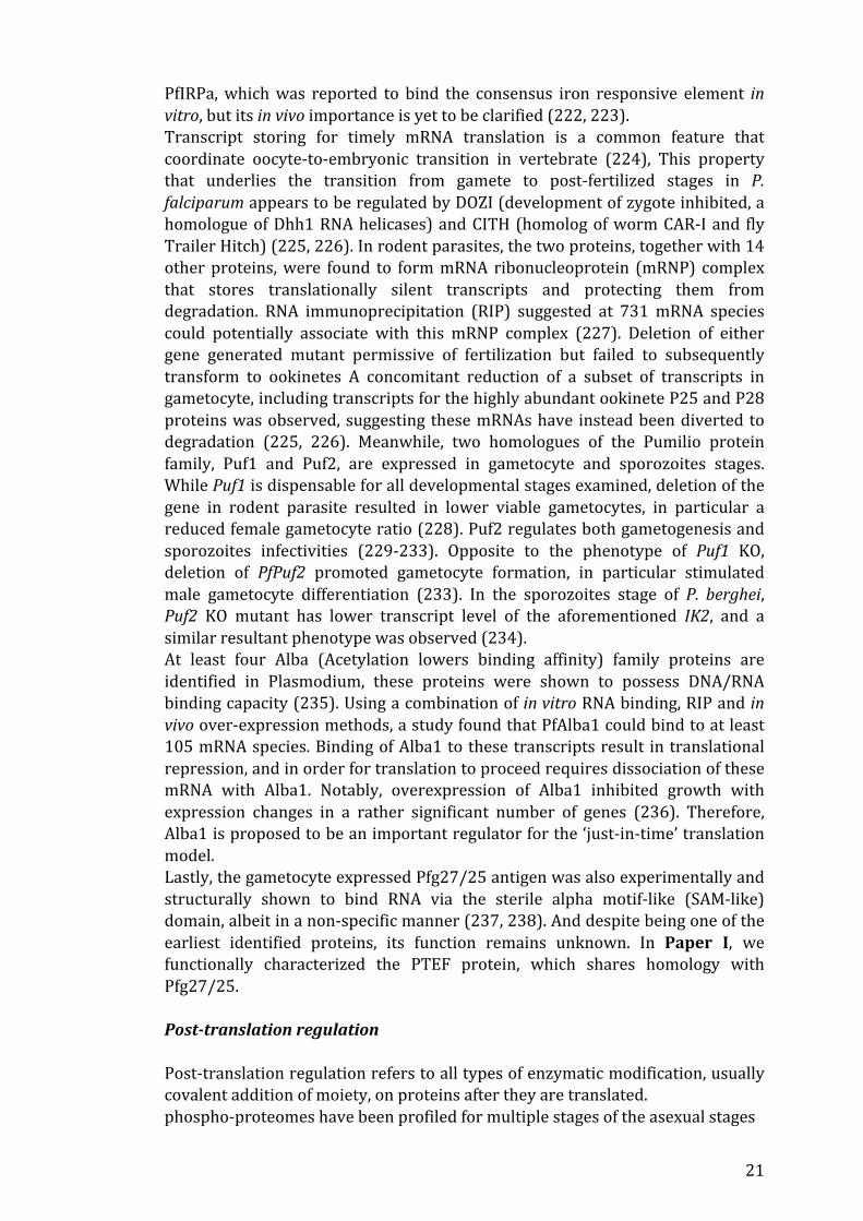
RBPs Expression stages studied Evidence for binding Binding specificity DHFR-‐TS Asexual stage In vitro cognate dhfs-‐ts mRNA IRPa Asexual stage In vitro Iron response element DOZI and CITH gametocyte, ookinete In vivo Motif specific (*) Puf1 and Puf2 Gametocyte, sporozoite In vitro, in vivo Motif specific (#) Alba1 Asexual stage In vitro, in vivo ND Pfg27/25 Gametocyte In vitro, structural ND

23
Technically, proteolytic cleavage is also considered, to some degree, a mechanism of post-‐translational regulation, such as activation of pro-‐enzymes. Current information on this in P. falciparum is mostly on a gene-‐by-‐gene basis, making it hard to approach in a systemic level. An exception is the targeted co-‐translational processing of the PEXEL motif present in many exported proteins, it has been shown that the cleavage of the conserved arginine or lysine residue in the PEXEL motif by plasmepsin V is required for protein export into the cytoplasm of the host cell (257).
24
2 SCOPE OF THE THESIS As mentioned in chapter 1, gene regulation in Plasmodium falciparum appears to be heavily dependent on post-‐transcriptional and translational levels. In particular, the translation machinery of Plasmodium falciparum possesses many atypical features when compared to the human host. While these features are rarely explored, their uniqueness confers much potential for deriving new generation of therapeutic opportunities against the deadly parasites. The overall aim of this thesis is to further our understanding on the translation dynamics of P. falciparum by pondering on these features, as well as to investigate and identify potential translational regulatory mechanisms on both specific and systemic levels. Specific Aims Paper I To investigate the mechanism underlying the translational regulation of PfEMP1-‐VAR2CSA expression and to explore features permissible for therapeutic targeting. Paper II To characterize the effect of codon usage bias in P. falciparum, as well as to elucidate the relationship between the extreme codon usage bias and the evolution of the genome. Paper III To derive and authenticate essential reagents necessary for future study on the RIFIN family of proteins.

25
3 EXPERIMENTAL PROCEDURES Material and method protocols are detailed in each constituent papers included in this thesis. This chapter will briefly describe the methods that result in the generation of conclusive evidence for each individual study. In vitro culture and transfection of P. falciparum 3D7S8.4.2, NF54CSA, FCR3, FCR3S1.2, IT4CD36ICAM1, R29 and PAvarO are P. falciparum strains used in the generation of the thesis. All the parasites were continuously growth in RPMI medium supplemented with O+ red blood cell and 10% A+ human serum. The hematocrit was maintained at 3% to support high parasitemia. 5% sorbitol was routinely used to achieve tight stage synchrony of the parasites by selective killing of the late-‐stage parasites. To maintain the CSA binding phenotype, NF54CSA was regularly panned on petri dish coated overnight with 100 µg/ml chondroitin 4-‐sulfate from bovine trachea. Mid-‐to-‐late stage infected erythrocytes (IE) were resuspended in growth medium and incubated for 30 min on the coated dish which was pre-‐blocked by 3% BSA/ PBS. Subsequently, the petri dish was wash extensively until no IE was bound to the uncoated area. Transfection protocol was used in Paper I and II (258). 150 µg Plasmid DNA was resuspended in 400 µl cytomix and preloaded into 400 µl fresh RBC by electroporation with an exponential decay program set to 0.31kV and 950μF. Schizont-‐infected IEs were inoculated to the transfected RBC. Either 50 nM pyrimethamine, 1.5 µM DSM1 or 2.5 µg/ml blasticidin was applied to the culture 24 hours post transfection depending on the drug selection cassette. 40 µM 5-‐Fluorocytosine was used for the selection of ptef-‐knockout parasite. RNA extraction, cDNA synthesis and quantitative PCR analysis qPCR method was used in all the constituent papers to evaluate transcription level of genes of interest. Total RNA was extracted from tightly synchronized parasites using Trizol reagent at a ratio of 10:1 pellet v/v ratio, subsequently followed the manufacturer’s recommendations. cDNA was synthesized from 500 ng -‐ 1 µg total RNA using iscript reverse transcriptase. qPCR was performed with iQ SYBR Green Supermix. All reactions were run for 40 cycles of 95°C for 10 s and at 60°C for 1 min. Data analysis was done with 2−ΔCt or 2−ΔΔCt method. Whole-‐genome sequencing and assembly 3D7S8.4.2 genomic DNA was sequenced on the Roche 454 FLX Titanium platform using standard protocols in Paper I (259, 260). Raw sequences of up to 25x genome coverage were obtained and low-‐quality nucleotides were trimmed from the reads using Trimmomatic (261). The sequences were mapped to the 3D7 reference genome using Bowtie (262). Artemis/ACT software program was used to visualize the pairwise alignment with the reference genome. Raw reads for 3D7S8.4.2 whole genome sequencing were deposited in SRA (Accession: PRJNA377901).
26
RNA sequencing RNAseq data was used in all constituent papers for global quantitative transcripts profiling. Extracted total RNA was first run on a Bio-‐analyzer microchip for quality and quantity assessment. TruSeq Stranded mRNA Library Prep kit was used to construct sequence libraries, in which mRNA was enriched from 2 µg of total RNA by PolyA selection and fragmented before cDNA synthesis. Sequencing was performed on the Illumina Hiseq 2000 platform by multiplexing up to 20 indexed sequence libraries to obtain 2x 100 bp paired-‐end reads. Raw reads were aligned to the applicable reference genomes using Star and Htseq (263). None uniquely mapped reads, but not duplicated reads, were removed before calculating the RPKM (Reads Per Kilobase of transcript per Million mapped reads) values. 3D7S8.4.2 and NF54CSA RNA sequencing reads were deposited in SRA (Accession: PRJNA377896 and PRJNA374979). Recombinant protein synthesis Recombinant CTD and SAM-‐like domain proteins were synthesized for antibodies generation and the RTTF assay in Paper I. The CDS was codon optimized for E. coli expression and fused with 6X His tag at the C-‐terminus. XL-‐10 gold E. coli was used for plasmid transformation. Bacteria culture was growth to reach an OD 600 of 0.6 and immediately induced with 0.25 mM IPTG for 3 hours at 37°C. Culture was pelleted and sonicated in lysis buffer (10 mM hepes pH7.5, 10% glycerol, 150 mM NaCl, and protease inhibitor cocktail). The lysate was incubated for 3 hours at 4°C with cobalt resins and then washed with 500 mM NaCl and 20 mM sodium phosphate buffer. Up to 150 mM imidazole was used to elute the recombinant proteins, which was dialyzed for the removal of imidazole. Generation of rat antibodies and recombinant PAM1.4 In-‐house antibodies were generated in Paper I, in full compliant of the relevant ethical consideration. Immunization protocols were outsourced to Agrisera AB, Sweden. Recombinant CTD protein and a KLH conjugated synthetic peptide from the NTD were used to immunize six rats using Freund’s incomplete adjuvants. The sera were collected after four immunizations. Recombinant PAM1.4 antibody was generated using sequence retrieved from a previously described VSAPAM-‐specific monoclonal IgG1 antibody (264). cDNA was synthesized from selected B cell cultures and the sequence of both the heavy chain and the light chain variable regions (VH and VL) were obtained (265). These sequences were then cloned into human IgG1 and Igκ expression vectors and then transiently transfected in Expi293F cells for expression. The antibody was affinity purified by protein A chromatography from culture supernatant. Immunofluorescence Assay (IFA) IFA was conducted in Paper I and III to understand the cellular localization patterns of the proteins of interest. IEs at the required developmental stages were attached to poly-‐L-‐lysine treated glass slides. PBS solution with 4% paraformaldehyde was applied for 10 min for antigen fixation. Antigen fixation procedure was omitted in Paper III, instead the slide was left to air dry. The attached IEs were then incubated in PBS containing 0.25% Triton X-‐100 for 10

27
minutes for permeabilizing the membrane and were subsequently washed with PBS three times. Afterwards, 10% goat serum was used for blocking overnight at 4°C and then incubated with the corresponding primary antibodies diluted in PBST for 1 hour at room temperature. After washing in PBS, the slide was incubated for 30 min with the compatible secondary antibodies. Nikon Eclipse 80i fluorescence microscope was then used to detect the fluorescent signals. Western blot analysis Western blotting was performed in all constituent papers as a major molecular technique used for semi-‐quantitation of proteins of interest. Briefly, late stages parasites were first treated with 0.1% saponin and then washed three times with PBS to remove majority of hemoglobin in the host red cells. The parasite pellet was then solubilized in NuPAGE LDS loading buffer supplemented with reducing agent and boiled for 10 min. Protein gel electrophoresis were performed with NuPAGE Novex 4-‐12% Bis-‐Tris gels in MOPS running buffer. The resolved proteins were transferred to membrane in Tris-‐glycine transfer buffer (25 mM Tris, 192 mM glycine, 20% methanol, 0.025% SDS), and was blocked overnight at 4°C with a commercial western blocking reagent. Primary antibodies were diluted in the same blocking buffer, and then the membrane was incubated with the diluted antibodies for 1 hour at room temperature. After three washes in TBST, HRP-‐conjugated secondary antibodies were incubated with the membrane for 45 minutes at room temperature. We used ECL prime western blotting detection reagent and ECL hyperfilm for signal development and detection. In Paper I, we have also carried out a cellular fractionation protocol coupled subsequently with western blotting, the protocol was adopted and modified from previous report (266). Blue native gel electrophoresis Native gel electrophoresis allowed us to assess the native complex formation of protein of interest and was performed in Paper I. Similar to sample preparation in standard western blot. Parasites were first treated with 0.1% saponin. The parasites pellet was then lysed in hypertonic cytoplasmic lysis buffer for 10 minutes on ice. Lysates were diluted in NativePAGE sample buffer and loaded onto a NativePAGE Novex Bis-‐Tris gel. Running buffers for the electrophoresis included a separate anode buffer (50 mM Bis-‐Tris and pH7.0) and cathode buffer (50 mM Tricine, 15 mM Bis-‐Tris, and 0.02% Coomassie blue G250). After the electrophoresis, the gel was soaked in transfer buffer added with 1% β-‐mercaptoethanol for 30 min. The gel was subsequently transferred to a PVDF membrane for immunodetection mentioned in section 3.8. Co-‐immunoprecipitation Co-‐immunoprecipitation was used to identify physically interacting protein partners, and this protocol was used in Paper I. Immunoprecipitation was performed using 3D7S8.4.2 transfected with GFP or CTD-‐GFP. IEs were treated with 0.1% saponin and then lysed in IP lysis buffer at a v/v ratio of 1:10 (50 mM Tris-‐HCl, 150 mM NaCl, 1% NP40, 0.5% deoxycholate, and protease inhibitor). The lysate suspension was allowed to stand on ice for 20 minutes to maximize
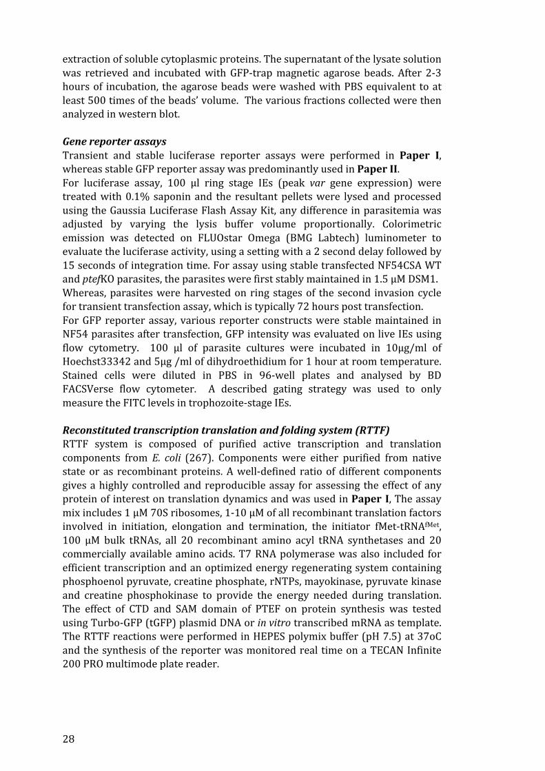
28
extraction of soluble cytoplasmic proteins. The supernatant of the lysate solution was retrieved and incubated with GFP-‐trap magnetic agarose beads. After 2-‐3 hours of incubation, the agarose beads were washed with PBS equivalent to at least 500 times of the beads’ volume. The various fractions collected were then analyzed in western blot. Gene reporter assays Transient and stable luciferase reporter assays were performed in Paper I, whereas stable GFP reporter assay was predominantly used in Paper II. For luciferase assay, 100 µl ring stage IEs (peak var gene expression) were treated with 0.1% saponin and the resultant pellets were lysed and processed using the Gaussia Luciferase Flash Assay Kit, any difference in parasitemia was adjusted by varying the lysis buffer volume proportionally. Colorimetric emission was detected on FLUOstar Omega (BMG Labtech) luminometer to evaluate the luciferase activity, using a setting with a 2 second delay followed by 15 seconds of integration time. For assay using stable transfected NF54CSA WT and ptefKO parasites, the parasites were first stably maintained in 1.5 µM DSM1. Whereas, parasites were harvested on ring stages of the second invasion cycle for transient transfection assay, which is typically 72 hours post transfection. For GFP reporter assay, various reporter constructs were stable maintained in NF54 parasites after transfection, GFP intensity was evaluated on live IEs using flow cytometry. 100 µl of parasite cultures were incubated in 10µg/ml of Hoechst33342 and 5µg /ml of dihydroethidium for 1 hour at room temperature. Stained cells were diluted in PBS in 96-‐well plates and analysed by BD FACSVerse flow cytometer. A described gating strategy was used to only measure the FITC levels in trophozoite-‐stage IEs. Reconstituted transcription translation and folding system (RTTF) RTTF system is composed of purified active transcription and translation components from E. coli (267). Components were either purified from native state or as recombinant proteins. A well-‐defined ratio of different components gives a highly controlled and reproducible assay for assessing the effect of any protein of interest on translation dynamics and was used in Paper I, The assay mix includes 1 µM 70S ribosomes, 1-‐10 µM of all recombinant translation factors involved in initiation, elongation and termination, the initiator fMet-‐tRNAfMet, 100 µM bulk tRNAs, all 20 recombinant amino acyl tRNA synthetases and 20 commercially available amino acids. T7 RNA polymerase was also included for efficient transcription and an optimized energy regenerating system containing phosphoenol pyruvate, creatine phosphate, rNTPs, mayokinase, pyruvate kinase and creatine phosphokinase to provide the energy needed during translation. The effect of CTD and SAM domain of PTEF on protein synthesis was tested using Turbo-‐GFP (tGFP) plasmid DNA or in vitro transcribed mRNA as template. The RTTF reactions were performed in HEPES polymix buffer (pH 7.5) at 37oC and the synthesis of the reporter was monitored real time on a TECAN Infinite 200 PRO multimode plate reader.

29
Peptide array analysis In Paper III, a peptide array experiments were performed to map the epitopes and to determine the specificity of the RIFIN antibodies. The array was fabricated by Roche-‐Nimblegen and it was customized to include 175000 peptides of 12-‐residue length, representing primary sequence of selected members from 2TM, PHISTs, RIFINs, STEVORs, SURFINs and PfEMP1 protein families. The array was designed so that any neighboring peptides will span an 11-‐residue overlapping region for improved differentiation between background and epitopes, which should constitute clustered reactive neighboring peptides.
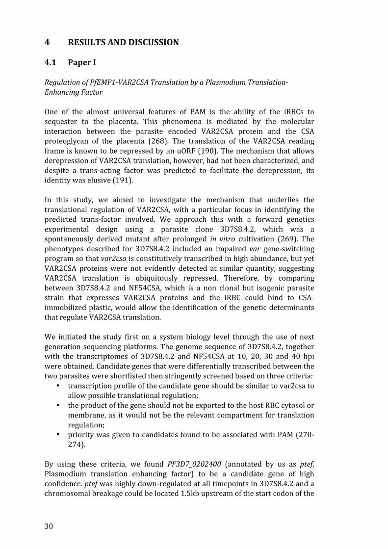
30
4 RESULTS AND DISCUSSION 4.1 Paper I Regulation of PfEMP1-‐VAR2CSA Translation by a Plasmodium Translation-‐Enhancing Factor One of the almost universal features of PAM is the ability of the iRBCs to sequester to the placenta. This phenomena is mediated by the molecular interaction between the parasite encoded VAR2CSA protein and the CSA proteoglycan of the placenta (268). The translation of the VAR2CSA reading frame is known to be repressed by an uORF (190). The mechanism that allows derepression of VAR2CSA translation, however, had not been characterized, and despite a trans-‐acting factor was predicted to facilitate the derepression, its identity was elusive (191). In this study, we aimed to investigate the mechanism that underlies the translational regulation of VAR2CSA, with a particular focus in identifying the predicted trans-‐factor involved. We approach this with a forward genetics experimental design using a parasite clone 3D7S8.4.2, which was a spontaneously derived mutant after prolonged in vitro cultivation (269). The phenotypes described for 3D7S8.4.2 included an impaired var gene-‐switching program so that var2csa is constitutively transcribed in high abundance, but yet VAR2CSA proteins were not evidently detected at similar quantity, suggesting VAR2CSA translation is ubiquitously repressed. Therefore, by comparing between 3D7S8.4.2 and NF54CSA, which is a non clonal but isogenic parasite strain that expresses VAR2CSA proteins and the iRBC could bind to CSA-‐immobilized plastic, would allow the identification of the genetic determinants that regulate VAR2CSA translation. We initiated the study first on a system biology level through the use of next generation sequencing platforms. The genome sequence of 3D7S8.4.2, together with the transcriptomes of 3D7S8.4.2 and NF54CSA at 10, 20, 30 and 40 hpi were obtained. Candidate genes that were differentially transcribed between the two parasites were shortlisted then stringently screened based on three criteria:
• transcription profile of the candidate gene should be similar to var2csa to allow possible translational regulation;
• the product of the gene should not be exported to the host RBC cytosol or membrane, as it would not be the relevant compartment for translation regulation;
• priority was given to candidates found to be associated with PAM (270-‐274).
By using these criteria, we found PF3D7_0202400 (annotated by us as ptef, Plasmodium translation enhancing factor) to be a candidate gene of high confidence. ptef was highly down-‐regulated at all timepoints in 3D7S8.4.2 and a chromosomal breakage could be located 1.5kb upstream of the start codon of the

31
gene. As per criterion, the expression of the gene was always found to be associated with PAM in multiple studies. Furthermore, the primary sequence of the encoded protein does not contain the PEXEL motif that drives protein export to the host RBC. Upon extensive bioinformatic analysis, we demarcated the encoded protein into three domains: an N-‐terminal domain (NTD), a low complexity linker domain (LD) and a C-‐terminal domain (CTD). Interestingly, conflicting localization signals were predicted, with a nuclear localization signal (NLS) found on the NTD and a nuclear export signal (NES) on the CTD. Intrigued by the observation, we generated antibodies in rats using a peptide from the NTD and recombinant expressed CTD proteins. Subsequently, the reduced expression of PTEF in 3D7S8.4.2 was confirmed at the protein level using western blot analysis. Additionally, the results suggested the presence of fragments containing only the CTD, and that the generation of the fragments is dependent on cysteine protease. We observed a potent inhibition of the process when the parasites were treated with a calpain inhibitor (ALLN). The CTD contains a Pfg27 domain. Pfg27 domain was first identified in the gametocyte Pfg25/27 antigen, of which function is still unknown. It was, however, demonstrated that the Pfg27 domain has a non-‐specific RNA binding capacity, which was experimentally and structurally validated (237, 238). We realized that the cleaved CTD fragments, now retaining only the NES, would potentially localize to the parasite cytoplasm and regulate translation through the Pfg27 domain. The cytoplasmic localization of the CTD was addressed by a three-‐pronged approach. First, we performed immunofluorescence assays using both antibodies against the NTD and CTD, diffused signal patterns were observable in both cases that imply PTEF localizing in multiple cellular compartments, yet conclusive evidence could not be yielded to support the cytoplasmic presence of the CTD. Second, we performed live imaging on parasites transfected with GFP protein fused to different domains or sequence motifs found in PTEF. Importantly, the CTD-‐GFP fusion protein was exclusively localized in the cytoplasm. The cytoplasmic localization of CTD was then unambiguously revealed in cellular fractionation experiments, where the CTD was found preferentially localized in the cytoplasmic fractions while the full-‐length proteins were mostly in the nuclear fraction. Therefore, the expression of PTEF is tempo-‐spatial relevant to translation regulation of var2csa and the CTD has the contextual association with such function. We next investigated the function of PTEF. In vitro RNA binding assays were first performed using parasite total RNA and recombinant CTD proteins. The results suggested CTD could bind RNA in a non-‐specific manner, expectedly similar to what was described for the Pfg27 domain. Results from RNA immunoprecipitation were also congruent. Interestingly, CTD co-‐migrated with a high molecular weight complex, which appeared to be highly abundant in protein content but was also readily destabilized by RNase. We sought to

32
identify the complex by excising gel bands and performed mass spectrometry, the results strongly point to the ribosomes. A physical interaction between the CTD and the ribosomes were further illustrated by the co-‐immunoprecipitation of the CTD with the ribosomal RPS14 protein. To determine if PTEF could be involved in var2csa regulation, we generated a PTEF knockout parasite from NF54CSA using the recently available CRIPSR-‐Cas9 system (275). The VAR2CSA protein level in the knockout parasite was clearly reduced in western blot analysis, while the var2csa transcript level was not altered. Strikingly, VAR2CSA protein level was abundantly detected when the knockout parasite was complemented with the CTD, indicating a phenotype rescue by the CTD. Likewise, luciferase reporter gene under the control of the 5’ UTR of var2csa was also inefficiently expressed in the knockout parasite. Notably, inhibition of calpain activities in wildtype parasites stably transfected with the luciferase reporter would also drastically reduce the luciferase activities. These functional assays firmly established the role of PTEF in regulating var2csa translation. As PTEF affects translation without apparently recognizing any sequence motif and that it can interact with the ribosomes. Since ribosomes contain evolutionary conserved elements, we took a radical approach and tested the recombinant CTD proteins in a E. coli RTTF assay (267). Surprisingly, the recombinant CTD protein was able to enhance translation even in a heterologous translation system, suggesting PTEF may interact with a highly conserved element of the translation machinery, such as the rRNA. We conclude that PTEF can interact with the ribosomes and enhance the general translation efficiency. The study has identified an important regulator for placental malaria. The ability of iRBCs to sequester in the placenta is not only dependent on the switching to var2csa expression, but is also dependent on the expression level of PTEF that fine-‐tunes the protein level of VAR2CSA and thus the binding capacity in the placenta. This added new perspectives to the widely accepted view that var genes and the encoded PfEMP1s expression, or more generally the variable surface antigens, is mostly a function of passive immuno-‐modulated selection processes. Yet, this doctrine has recently been challenged with a new study showing antigenic variation occurred even in mice lacking adaptive immunity (51), which has also refueled the long puzzling observation that antigenic variation does also happen in vitro. In this study, our data suggests the parasite also has the capability to autonomously modulate the expression of VAR2CSA. It is unknown to how wide this could be extended to other variable surface antigens. It is, however, likely that the process of antigenic variation is a function of interplay between autonomous induction events and passive selection processes. Mechanistically, PTEF provides convincing evidence to the long speculated ‘specialized ribosome’ theory. This theory builds on the existence of compositionally heterogeneous ribosome populations that have different

33
functional modules (276). Plasmodium spp. is known to harbor only a few copies of non-‐identical rDNA genes in their genomes (82), but previous attempts were inconclusive in assessing the potential presence of heterogeneous ribosomes (214, 215). Our data suggests PTEF, as an additional component to the ribosomes, may form a ribosome subpopulation with enhanced translation efficiency. It will be interesting to further determine if PTEF can enhance translation of uORF-‐containing transcripts, which are believed to be quite prevalent (277). Further, this study also opens new therapeutic opportunities for the management of PAM, as we provided strong evidence that PTEF is an important regulator of placental binding and thus the pathogenesis of PAM. Notably, calpain processing is essential in order for PTEF to exert its function. This will add merit to use proteases of the parasite as new druggable targets. Paper II Frequent GU wobble pairings reduce translation efficiency in Plasmodium falciparum The genome of P. falciparum is strikingly AT biased. One result of this extreme evolution is the extremely biased codon usage. The lysine encoding AAA and the asparagine encoding AAT codons, for example, account for more than 20% of the total codon used in the genome. Importantly, this extreme codon usage has to be coped by a minimum set of 45 nuclear tRNA genes. Despite this obvious discordancy, it has mostly escaped attention and has been addressed by only a few in silico studies previously (203, 278). Our interest was further prompted by a surprising observation during the generation of Paper I, where we found that the expression of the CTD in the parasite was more efficient if the sequence was codon optimized for E.coli, than if we used the native endogenous sequence. The aim of the study was to investigate the effect of codon usage on translation by employing GFP reporter assays. Using GFP reporter assays confer numerous advantages:
• The use of GFP to assess codon effects can eliminate potential confounding effects due to functional selection
• The GFP reporter gives lysis-‐free and highly sensitive readouts • GFP allows accurate stage-‐specific flow cytometry analysis when
compared with luciferase-‐based assay We started by first tallying the genomic codon usage frequency of P. falciparum, and then carefully characterized the anticodon of all the nuclear tRNA genes by aligning the universally conserved T33 as well as through sequence homology prediction. After that, all possible codon: anticodon-‐pairing dynamics were mapped. Our analysis showed that the genome predominantly used codons with highly skewed A+T content, which faithfully reflects the A+T rich genome. Interestingly, we also found that P. falciparum genome frequently uses wobble codons, with up to one third of all codons used in the genome contributed by the

34
eight GU wobble codons. GU wobbling refers to codons that have a T in the third position that are necessarily paired with a G in the wobble position of the anticodon during translation. Puzzled by the findings, we sought to recodonize the GFP reporter in four different ways: an AT rich sequence with synonymous codon usage reminiscent of the genome of P. falciparum-‐GFPPF, a sequence with GC rich codons-‐GFPGC, a sequence with high wobble codon content-‐GFPwob and one that has a low wobble codon content-‐GFPno wob. All sequences were transfected into NF54 parasites for assessing the translation efficiency by flow cytometry. Interestingly, the FACS results consistently demonstrated lower fluorescence intensity in GFPno wob. qPCR detection, meanwhile, suggested no difference in the transcript level of GFPno wob. This indicated a specific effect on translation. To facilitate our analysis, we established a panel of parameters that can comprehensively reflect the rareness of the codons, the nucleotide compositions, as well as the wobble codon contents in each of the recodonized sequences. These parameters were individually tested for their effect on translation efficiency. In support of our initial observation, these analyses revealed a strong negative association between high GU-‐wobble content and translation efficiency. No association was, however, seen between non GU-‐wobble codon content and translation efficiency. In other eukaryotes, this negative association was also supported in studies using reporter assays as well as ribosome profiling (279, 280). As aforementioned, this study was inspired by the fact that the expression of PTEF-‐CTD in the P. falciparum was more efficient when the sequence was codon optimized for E. coli expression. We re-‐characterized this observation in a systematic way and found that the sequence that was codon optimized for E. coli expression had, indeed, a lower GU wobble content. This provided further evidence for our conclusion that the GU wobble content negatively correlates with translation efficiency. One of the most frequently used codon in the genome is the asparagine encoding AAT codon, which is a GU wobble codon. The high usage frequency of this codon most likely leads to frequent insertion of asparagine homorepeat in many P. falciparum proteins (281). The rapid expansion of these repeats in P. falciparum has so far defied understanding. Deletion of the asparagine repeat in the essential proteasome component, Rpn6, resulted in no phenotypic changes, suggesting the expansion of asparagine homorepeats maybe functionally neutral (206). Prompted by our data, we hypothesized that the locally clustered GU wobble codon context underlying these asparagine homorepeats would have major influence on the translation dynamics of the host proteins. We addressed this by inserting AAT codon repeats in the GFPno wob and investigated the effect of the insertion on GFP production. Expectedly, AAT codon insertion reduced GFP production, when compared to the synonymous AAC codons insertion and the
35
control insertion. However, we repeatedly found a reduced equilibrium transcript level when the GFP was inserted with AAT codons. By blocking transcription with ActinomycinD, such an effect appeared to be attributed to accelerated transcript turnover rates. So far, all experimental data converge to support the use of GU wobble codon can negatively influence protein output. To complement this experimental data, we performed bioinformatic analyses and were able to show that:
• Highly expressed genes (top 5% percentile of expression) use significantly less GU wobble codons
• Genes encoding ribosomal proteins have drastically reduced GU wobble content when compared to the genome average
• From previously reported data (187), transcripts that are efficiently loaded on polysomes also have lower GU wobble content when compared to transcripts that are poorly loaded, inferring that a high GU wobble content reduces translation efficiency as could be seen by less loaded ribosomes
These analyses further strengthen the study conclusion. During further investigation, we discovered that genes with high GU wobble content are preferentially enriched in genes that have a regulatory role or that are involved in nitrogen metabolism (transcription and translation etc). In particular, the AP2 transcription factors are commonly high in GU wobble content. Meanwhile, genes encoding surface antigens, such as the rif genes, have much lower GU wobble content. This observation may imply the use of GU wobble codons as a possible gene regulatory mechanism, being in place to complement the obvious lack of transcriptional regulation. Additionally, we found a marked increase in GU wobble content and in the frequency of asparagine homorepeat insertion in single-‐exon genes when comparing to multi-‐exon genes. Single-‐exon genes are generally more vulnerable to transcriptional errors, because the non-‐sense mediated decay (NMD) mechanism which can promptly remove transcript with premature termination codon is only efficiently triggered by the exon junction complex. Therefore, our results gives rise to an interesting proposal that, perhaps, single-‐exon genes may have evolved to harbor more asparagine homorepeats so as to achieve a rapid transcript turnover and prevent the accumulation of defective transcripts. This study represents the first in vitro study that addresses codon usage in P. falciparum. The results have provided new insights on gene regulation and genome evolution of this parasite. Paper III RIFIN has recently emerged as an important variable surface antigen family, both because of being verified as a new ligand that mediates binding to the A-‐blood group antigen as well as being identified as targets of naturally occurring
36
broadly neutralizing antibodies (70, 79). Despite these new promises, there is an apparent lack of studies on the 160-‐member RIFIN family. Regulation of rif and RIFIN expression have not been investigated nor described at any level of the central dogma. This scarcity is likely obscured by the promiscuity of many antibody reagents that are at times giving inconsistent conclusions in different experiments. The study aims to generate antibody reagents that are thoroughly validated for use in subsequent studies to elucidate the function of RIFIN and its regulatory mechanism. To start with, we immunized ten rabbits with peptides encompassing regions of different RIFIN proteins as well as immunized one goat with a recombinant RIFIN protein. In brief, four rabbits were immunized with peptides generated from the C-‐terminus of two A-‐RIFIN proteins (PF3D7_0100400 for RαRIFC and R2αRIFC, PF3D7_0223100 for R3αRIFC and R4αRIFC), whereas four other rabbits were immunized with peptides generated from the C-‐terminus of two B-‐RIFIN proteins (PF3D7_0900500 for R5αRIFC and R6αRIFC, 3D7_0223200 for R7αRIFC and R8αRIFC), the remaining two rabbits were immunized with a peptide generated from the A-‐RIFIN specific indel sequence of PF3D7_0100400 (RαRIFI and R2αRIFI). Since amino acid sequences in the C-‐terminus of are relatively conserved for both A-‐ and B-‐RIFINs, while the indel sequence is A-‐RIFINs specific (64). This immunization strategy was designed to ideally generate antibodies that would recognize either all RIFINs or A-‐RIFINs only. Meanwhile, an A-‐RIFIN (PF3D7_0100400) recombinant protein was synthesized and subsequently used to immunize a goat (GαRIF). We subsequently tested all the antibodies in traditional antibody-‐based molecular techniques, i.e. western blotting and immunofluorescence assays. Despite the broad panel of generated antibodies, only RαRIFC, RαRIFI and GαRIF could detect the proteins of expected size in a few parasite strains in western blot analysis. However, all the three antibodies gave varying or inconsistent localization pattern in immunofluorescence assays. To validate the specificity of the antibodies, we sought to map the epitopes in an ultra-‐dense peptide array. The peptide array was coated with 12-‐residue peptides representing surface antigens from PfMC-‐2TM, PHIST, RIFIN, STEVOR, SURFIN and PfEMP1 protein families. Adjacent peptides are laterally shifted by one residue and thus any adjacent pair of peptides would contain an 11-‐residue overlapped region. The results from the peptide array experiment suggest: 1) RαRIFC and RαRIFI exhibit good degree of cross-‐reactivity with a third to a half of all RIFINs, 2) GαRIF can also cross-‐react with one fourth of all RIFIN, 3) epitopes recognized by GαRIF cluster in five regions which may represent immuno-‐dominant region, 4) R5αRIFC and R6αRIFC have poor specificity against RIFINs, 5) Non RIFIN cross-‐reactivity is most commonly seen against STEVORs, which form a super-‐family with RIFINs. RNA sequencing was performed to fully characterize the rif transcription profiles of the parasite strains tested prior in western blot analysis. Base on the
37
transcription profiles, it could be concluded that the ability of RαRIFC, RαRIFI and GαRIF to detect RIFIN in each parasite strain could be predicted by whether the dominant or highly expressed rif genes would encode epitopes recognized by each antibodies, as illustrated in the peptide array experiment. Through this study, robustly validated RIFIN antibodies were obtained for use in future studies. This study also provided an example of the use of peptide array to authenticate antibody reagents. Notably, our result implies that any conclusion made by immunofluorescence assays is highly dependent and sensitive to the epitopes recognized by the antibodies used. It is thus advised to use alternative strategies such as GFP-‐tagging, or to use multiple antibodies in order to make concrete conclusion. In cohesion with this, these strategies were employed in Paper I.
38
5 CONCLUDING REMARKS AND FUTURE PERSPECTIVES A decade of global commitment in malaria control has been rewarded with remarkable achievements and the laying of encouraging milestones. On the crossroad where passive control is to be replaced by proactive elimination initiatives, the disturbing uprising of artemisinin-‐resistant parasites in pockets of the endemic regions can potentially reverse the course, leading to catastrophic consequences. Studies looking at new and old antimalarial drugs converge to illuminate the promises of targeting the translation machineries of the parasites as a multistage solution (218, 282-‐288). Regrettably, the global translation dynamics as well as specific translation regulation have not been sufficiently investigated. The studies presented in this thesis have advanced some of the current understanding on the translational regulation in P. falciparum: I) The translation of the PfEMP1-‐VAR2CSA protein, which mediates binding to
placental CSA ligand, is regulated by PTEF (Plasmodium Translation Enhancing Factor). Unlike many known trans acting translation regulators, PTEF enhances translation rather than represses translation. In the case of VAR2CSA, the enhancement leads to the relief of translational repression by the bicistronic uORF. While exhibiting promiscuous cellular localization patterns, full length PTEF is imported into the nucleus of the parasite, processed by PfCALPAIN to generate a C-‐terminal domain containing fragment. The CTD contains an RNA-‐binding Pfg27 domain and interacts with the ribosome. Interestingly, PTEF enhances translation also in the heterologous E. coli model. Although the mechanism of the translational regulation by PTEF is not yet determined, it is plausible that the association of PTEF with the ribosome constitutes a ‘specialized ribosome’ population. Importantly, high ptef transcript and protein levels, similar to VAR2CSA, were previously observed and associated with PAM patient isolates. Therefore, it may represent a master regulator of PAM pathogenesis and a potential therapeutic target for this subgroup of clinical malaria.
II) We showed that the pervasive use of GU wobble codons in P. falciparum is
associated with reduced protein output, probably affecting the efficiency of translation elongation. Although results are concluded from experiments using GFP reporter, global bioinformatics analyses suggested the effect could be system-‐wide. Highly expressed genes harbor reduced GU wobble codon content with concomitant increase in the usage of the synonymous codons. The GU wobble AAT codon is frequently organized in clusters that encode asparagine homorepeats. It is found that the insertion of clustered AAT codons can destabilize the host transcripts. Interestingly, single exon genes have higher GU wobble content and more frequent asparagine homorepeat insertion, suggesting a mechanism to compensate for the inefficiency of non-‐sense mediated decay mechanism on intronless genes. Furthermore, higher GU wobble content is also commonly found in regulatory genes. Overall, the
39
widespread use of GU wobble codon may evolve as an important gene regulatory mechanism.
III) Immunization of peptide and recombinant protein generated from selected
A-‐RIFIN can elicit specific antibody responses against a limited subset of RIFIN proteins. This study provides proof of concept for the potential of generating cross-‐reactive RIFIN antibodies as well as validated RIFIN reagents for use in later studies. It also shows the rationale of using peptide array to authenticate antibodies before important conclusion should be made.
40
6 ACKNOWLEDGEMENTS What was supposed to be a four-‐year PhD has eventually become an extended journey for me. It is hard not try to sometimes look back and imagine how I will be, had I not made the decision to come to Stockholm, but then I will immediately reckon that there is no use and no need for this, because I have had gotten so much during my time in Stockholm, in both a career and personal perspectives. I don’t think there is ‘very important decision’ in life, my many decisions were usually made in disproportionally short time relative to their seeming importance. The decision that landed me to Stockholm was of no exception. It all happened on a normal day when I was almost finishing my master program, at that time I was doing stem cell related research in the University of Hong Kong, focusing on Hox gene regulation. Hox genes are important body patterning genes, so at the end the day it is like asking why our head grows like a head and why our hand has five digits etc (of course much simplified). I was, however, uninspired and unmotivated. Then, in a kind of ‘Fika’ setting, I got a glimpse of PhD life in KI from an alumni who completed hers under the supervision of my now supervisor Mats Wahlgren. Already catalyzed by some African-‐themed movies at the time, I decided that I would like to change to the malaria field. My first impression of Mats was from email communication. Since I was cold calling, I spent a week to familiarize myself with literatures and wrote a long organized email (maybe 1200 words) to Mats asking for PhD opportunity. ‘Sent’ and 3 days went-‐by with no news, hope was fading. Then on the fourth day, I got a short email answer, with a word-‐to-‐word conversion rate of 300 : 1. Yes…it contained only four words! That if one was not careful, would have easily overlooked and thought it was ‘Dear Mr. Sherwin Chan’. Hilarious as it was, it was also difficult to interpret on its own – ‘What’s your time aspect?’ as it was. I called a family meeting and, well, at least it helped to prevent unequal division of labor, as we are four exactly. Due to limited information, we have to approach it by guessing the motives; was he interested? Maybe he had taken time to thoroughly think about the possibility, but why took him four days to then wrote a four-‐word email. Anyway, I replied and Mats invited me for the interview and then I was here. To Mats, contrary to the number of words I usually see from your emails, you are truly a person of a lot. I am extraordinary thankful for you giving me the chance to work in the lab and all the supervision you have given me during the years. Your omnipresent support, and sometimes tolerance, to me has allowed me to focus on the research. I am even more grateful that you have given me such a high degree of autonomy, allowing ideas to flow freely and flourish, and also to allow no barrier and hierarchy to stand between so that I can always feel free to discuss and sometimes disagree. I definitely think if any of these elements was taken away from me, the outcome would have been very different. So thank you! and maybe you are convinced that it is a good idea to give Hong Kong people more autonomy by now? Stockholm is a beautiful city, but one may need a bit of time to understand this. Same is to me. During the first few months in Stockholm, I wasn’t happy. Skatteverket didn’t allow me register despite no illusion that tax money has been taken away from me, and as a result bank didn’t allow me to open an account,
41
and I have to constantly worry about accommodation, worst off 2010 saw a truly bad winter. Luckily, the lab environment was really amazing and welcoming, and it had quickly become my refuge. At times, I honestly feel very relax in the lab, so much that it feels like home. Indeed, on a statistical point of view, I might have gone to the lab on 99.5% of all the days I have spent in Stockholm (excluding away-‐from-‐Stockholm days). So I guess it is not significantly different than home. What have made the lab amazing is not because it has good equipment, but because it has the best people in it. I see three generations of people movement during my time here and all that I have enjoyed spending time with. I would like to express my thankfulness to all the people who have helped me: For the early generation, Pilar Quintana, thank you very much for the friendship and for helping me when I was stuck and so much more thanks for being my ‘secretary’, don’t you know that I am disoriented amid all these things since that you have left. I enjoyed all the activities and the good time outside the lab and still vividly remember the near miss hitting a koala. I have lost the race to PhD completion to you, but I don’t think I can complain on this because your planning and organizational skills are exceptional. I hope one day we can go to see the tall palm tree or to Lake Titicaca together. By the way, you may want to train up your physicals to get rid of ‘the weak’ tag. Sriwipa Chuangchaiya ‘Jeab’, the mama, even though you have left the lab a few years back, I still miss the time. You may not be the most logical person I know, but you can make people feel very relax and also a very loyal friend. Thanks for all this and also for coming all the way to Bangkok when we were in Thailand, though it might be better if you could also know the way to navigate around………... and sorry for letting you to return to Stockholm prematurely because of my ever-‐delaying defense!! Hodan Ismail, ‘The sick’ one of the trio, thanks for being my first translator and driver. I enjoyed you expressive personality (a stark contrast to me) and me actually miss teasing you. Your well written thesis has also help in the generation of this book J; Nicolas Joannin for starting such an interesting project and also fooling me into buying a very expensive jacket, which maybe only usable in Siberia. Also to Kirsten Moll, Davide Angeletti, Letusa Albrecht, Karin Blomqvist, Jana Busch, Mia Palmqvist and Sandra Nilsson who have very nicely introduced me into the malaria field and whom I shared many of my first Swedish experiences with. In particular, the annual anticipated Julbord!!! For the inter-‐generation, Daisy Hjelmqvist, thank you for always helping me with almost everything, being my second-‐generation translator, stocking up the desk with essential food supplies and haribos. Also for of course helping me to take care of mochi it is needed. Though your presence has completely disrupted my financial planning, because of all the lunch eating out. Zulkarnian MD Idris for all the fun you have given with your delightful personality, I will not forget watching the match between ‘Lin and Lee’ together in the office, and cheering for our own team, it would have been an atmosphere as good as watching live in the stadium, if only had you not ‘fxxted’………………Junhong Ch’ng for saving me the need to enter the ‘lion pit’, and also enlightening me with all the philosophy on how to deal with situations and people and life in general. I hope to visit you in Singapore sometime. And thanks to Xiaogang Feng for giving me enjoyable ‘Chinese moment’ in the lab, and also for always trying your best to help me

42
when I am in need. Chim Chan, my flatmate for two years, you are the type who always work quietly but are very reliable. I will remember your smiles, because they may not come so easy J. Alejandra Frasch for always helping me to deal with the works when I was totally frustrated and letting me pinch a bit of Benicio. Maryam Imam for sometimes sharing your soul-‐transcending meditation philosophy, though I am more interested in your cooking in general. Martina Jalava, thanks for getting things so nicely sorted and organized all the time, your kind and reliable helps have really saved me a lot of worries. For the new generation, Ulf Ribacke, though you are also the pre-‐generation, I wish you would have come earlier during my PhD, I have now find an outlet to discuss hardcore molecular biology. Atiqul Islam, the destroyer in the ping-‐pong table, I sincerely wish you all the best for the future, though doesn’t mean that Pontus Hdeberg and I will show you mercy on the table… and also to Madle Sirel and Viktor Persson for creating a good office vibe in the room. And I would want to give a special thanks to Madle Sirel for also designing the cover illustration of this thesis book as well! I also need to thank all the senior members in the malaria corridor for sometimes guidance and also interesting discussion during the Friday meetings: Susanne Nylen, Kristina Persson, Akira Kaneko, Anders Bjorkman and Manuel Patarroyo. As Caroline Rönnberg has once commented that I had have almost become a piece of furniture in the lab, tactfully implying that I have stayed a long time in the lab. I like it in such a creative way, but will feel more honored if I could be referred to as a piece of equipment. Or at least, I will not want to be my own desk………Spending such a long time in the lab, I have seen many people coming by and going, despite at times I have become desensitized about people coming and going, I enjoyed the interactions I had with almost everyone ever stayed sometime in the lab. These include: Ulrika Morris, Irina Jovel-‐Dalmau, Maria Pena Fransch, Juliana Inoue, Berit Schmidt, Mubasher Mohammed, Casja Classon, Reetesh Akhouri, Suchi Goel, Allan Lugaajju, Shuhan Xu, Pablo Gusti, Sreenivasulu Reddy, Muyideen Kolapo Tijani , Mattias Westman, Anna Jötten, Thomas Geislinger, John Nyberg, Maria Hesselman and Adrian Luscombe. I would also like to acknowledge all of my co-‐authors and co-‐supervisor Björn Andersson. In particular, Suparna Sanyal and Chandra Mandava for the stimulating discussion and for the important contribution. Jose Juan Rubio-‐Lopez, even though I have only spent a few days in your lab, I am so inspired by your spirit in science and fascinated by your life philosophy. SCILIFE lab for the sequencing, and Thomas Källman from BILS for the bioinformatics support. To my friends outside the lab, Paola, Kiang and Atticus, for all the fun time together, bbq, sporting activities and board gaming outside the lab, though not so much for the dancing and Karaoke. Kiang, also thanks for helping me so many times and so promptly on bioinformatics issues, amid a very busy schedule of your own.

43
And also to my many friends in Hong Kong who have come all the way to Stockholm to visit me during my time here. Zen, Vanir, TK, YY, Petra, Keith and Tommy, I really enjoy waking up in the morning and seeing 200 whatsapp messages in a roll, though they are totally irrelevant to me, they make me feel so close to home and lend me heartfelt warmth in the otherwise cold Nordic weather. And of course, my PhD would not have been even started if not of the life-‐long influence and support from my mother and father. I remember when I first told you about the possibility of going to Sweden to do malaria research, I understand it would have essentially translated as ‘It is very unlikely I can repatriate to the finance-‐dominated society of Hong Kong’ and would not have been so easily taken by any parents. Still you have supported and respected my decision, as always that you have given me all the liberty to choose my way even at your huge expense. And also to my sister Edwina, whose supports and encouragements have always been unlimited and unconditional. I have not been expressive enough to let you guys know, but you are always in my heart and mind!! Sichao, my wife to-‐be, I am extremely lucky to have met you in Stockholm, and I believe it was destiny because if I would have come one month earlier or later, I might have missed the chance to even meet you. Unlike my feeling to Stockholm, yours was at first sight. All this time would be very difficult, if not impossible, if you have not been by my side, supporting, helping, understanding and more importantly for tolerating all my bad habits and behaviors!! Let’s continue to explore the world, both on land and underwater, for all the years to come. Thank you and I love you!!! So here is the end of my PhD journey. But it is only the end of the beginning.
44
7 REFERENCES 1. Escalante AA, Ayala FJ. Phylogeny of the malarial genus Plasmodium, derived from rRNA gene sequences. Proc Natl Acad Sci U S A. 1994;91(24):11373-‐7. Epub 1994/11/22. 2. Escalante AA, Barrio E, Ayala FJ. Evolutionary Origin of Human and Primate Malarias -‐ Evidence from the Circumsporozoite Protein Gene. Molecular biology and evolution. 1995;12(4):616-‐26. 3. Rich SM, Light MC, Hudson RR, Ayala FJ. Malaria's eve: Evidence of a recent population bottleneck throughout the world populations of Plasmodium falciparum. P Natl Acad Sci USA. 1998;95(8):4425-‐30. 4. Joy DA, Feng XR, Mu JB, Furuya T, Chotivanich K, Krettli AU, et al. Early origin and recent expansion of Plasmodium falciparum. Science. 2003;300(5617):318-‐21. 5. Liu WM, Li YY, Learn GH, Rudicell RS, Robertson JD, Keele BF, et al. Origin of the human malaria parasite Plasmodium falciparum in gorillas. Nature. 2010;467(7314):420-‐U67. 6. Carter R, Mendis KN. Evolutionary and historical aspects of the burden of malaria. Clinical microbiology reviews. 2002;15(4):564-‐+. 7. Hawass Z, Gad YZ, Ismail S, Khairat R, Fathalla D, Hasan N, et al. Ancestry and pathology in King Tutankhamun's family. Jama. 2010;303(7):638-‐47. Epub 2010/02/18. 8. Dhingra N, Jha P, Sharma VP, Cohen AA, Jotkar RM, Rodriguez PS, et al. Adult and child malaria mortality in India: a nationally representative mortality survey. Lancet. 2010;376(9754):1768-‐74. Epub 2010/10/26. 9. Murray CJL, Rosenfeld LC, Lim SS, Andrews KG, Foreman KJ, Haring D, et al. Global malaria mortality between 1980 and 2010: a systematic analysis. Lancet. 2012;379(9814):413-‐31. 10. Pigott DM, Atun R, Moyes CL, Hay SI, Gething PW. Funding for malaria control 2006-‐2010: a comprehensive global assessment. Malar J. 2012;11:246. Epub 2012/07/31. 11. Tusting LS, Willey B, Lucas H, Thompson J, Kafy HT, Smith R, et al. Socioeconomic development as an intervention against malaria: a systematic review and meta-‐analysis. Lancet. 2013;382(9896):963-‐72. 12. Gething PW, Patil AP, Smith DL, Guerra CA, Elyazar IRF, Johnston GL, et al. A new world malaria map: Plasmodium falciparum endemicity in 2010. Malaria J. 2011;10. 13. Bhatt S, Weiss DJ, Cameron E, Bisanzio D, Mappin B, Dalrymple U, et al. The effect of malaria control on Plasmodium falciparum in Africa between 2000 and 2015. Nature. 2015;526(7572):207-‐+. 14. Cotter C, Sturrock HJW, Hsiang MS, Liu J, Phillips AA, Hwang J, et al. The changing epidemiology of malaria elimination: new strategies for new challenges. Lancet. 2013;382(9895):900-‐11. 15. Noor AM, Kinyoki DK, Mundia CW, Kabaria CW, Mutua JW, Alegana VA, et al. The changing risk of Plasmodium falciparum malaria infection in Africa: 2000-‐10: a spatial and temporal analysis of transmission intensity. Lancet. 2014;383(9930):1739-‐47. 16. Woodrow CJ, White NJ. The clinical impact of artemisinin resistance in Southeast Asia and the potential for future spread. FEMS microbiology reviews. 2017;41(1):34-‐48. Epub 2016/09/11.

45
17. Ashley EA, Dhorda M, Fairhurst RM, Amaratunga C, Lim P, Suon S, et al. Spread of Artemisinin Resistance in Plasmodium falciparum Malaria. New Engl J Med. 2014;371(5):411-‐23. 18. Sulyok M, Ruckle T, Roth A, Murbeth RE, Chalon S, Kerr N, et al. DSM265 for Plasmodium falciparum chemoprophylaxis: a randomised, double blinded, phase 1 trial with controlled human malaria infection. The Lancet Infectious diseases. 2017. Epub 2017/04/02. 19. Olotu A, Fegan G, Wambua J, Nyangweso G, Leach A, Lievens M, et al. Seven-‐Year Efficacy of RTS, S/AS01 Malaria Vaccine among Young African Children. New Engl J Med. 2016;374(26):2519-‐29. 20. Keiser J, Singer BH, Utzinger J. Reducing the burden of malaria in different eco-‐epidemiological settings with environmental management: a systematic review. The Lancet Infectious diseases. 2005;5(11):695-‐708. Epub 2005/10/29. 21. Lees RS, Gilles JRL, Hendrichs J, Vreysen MJB, Bourtzis K. Back to the future: the sterile insect technique against mosquito disease vectors. Curr Opin Insect Sci. 2015;10:156-‐62. 22. Emami SN, Lindberg BG, Hua S, Hill SR, Mozuraitis R, Lehmann P, et al. A key malaria metabolite modulates vector blood seeking, feeding, and susceptibility to infection. Science. 2017;355(6329):1076-‐80. Epub 2017/02/12. 23. Nonvignon J, Aryeetey GC, Issah S, Ansah P, Malm KL, Ofosu W, et al. Cost-‐effectiveness of seasonal malaria chemoprevention in upper west region of Ghana. Malaria J. 2016;15. 24. Sidjanski S, Vanderberg JP. Delayed migration of Plasmodium sporozoites from the mosquito bite site to the blood. The American journal of tropical medicine and hygiene. 1997;57(4):426-‐9. Epub 1997/11/05. 25. Amino R, Thiberge S, Martin B, Celli S, Shorte S, Frischknecht F, et al. Quantitative imaging of Plasmodium transmission from mosquito to mammal. Nat Med. 2006;12(2):220-‐4. Epub 2006/01/24. 26. Mota MM, Pradel G, Vanderberg JP, Hafalla JC, Frevert U, Nussenzweig RS, et al. Migration of Plasmodium sporozoites through cells before infection. Science. 2001;291(5501):141-‐4. Epub 2001/01/06. 27. Ginsburg H, Krugliak M, Eidelman O, Cabantchik ZI. New permeability pathways induced in membranes of Plasmodium falciparum infected erythrocytes. Molecular and biochemical parasitology. 1983;8(2):177-‐90. Epub 1983/06/01. 28. Mantel PY, Hoang AN, Goldowitz I, Potashnikova D, Hamza B, Vorobjev I, et al. Malaria-‐Infected Erythrocyte-‐Derived Microvesicles Mediate Cellular Communication within the Parasite Population and with the Host Immune System. Cell host & microbe. 2013;13(5):521-‐34. 29. Anstey NM, Douglas NM, Poespoprodjo JR, Price RN. Plasmodium vivax: Clinical Spectrum, Risk Factors and Pathogenesis. Adv Parasit. 2012;80:151-‐201. 30. Setiadi W, Sudoyo H, Trimarsanto H, Sihite BA, Saragih RJ, Juliawaty R, et al. A zoonotic human infection with simian malaria, Plasmodium knowlesi, in Central Kalimantan, Indonesia. Malaria J. 2016;15. 31. Clark IA, Alleva LM, Budd AC, Cowden WB. Understanding the role of inflammatory cytokines in malaria and related diseases. Travel medicine and infectious disease. 2008;6(1-‐2):67-‐81. Epub 2008/03/18. 32. del Portillo HA, Ferrer M, Brugat T, Martin-‐Jaular L, Langhorne J, Lacerda MVG. The role of the spleen in malaria. Cellular microbiology. 2012;14(3):343-‐+.

46
33. Sponaas AM, do Rosario APF, Voisine C, Mastelic B, Thompson J, Koernig S, et al. Migrating monocytes recruited to the spleen play an important role in control of blood stage malaria. Blood. 2009;114(27):5522-‐31. 34. Idro R, Marsh K, John CC, Newton CR. Cerebral malaria: mechanisms of brain injury and strategies for improved neurocognitive outcome. Pediatric research. 2010;68(4):267-‐74. Epub 2010/07/08. 35. Birbeck GL, Molyneux ME, Kaplan PW, Seydel KB, Chimalizeni YF, Kawaza K, et al. Blantyre Malaria Project Epilepsy Study (BMPES) of neurological outcomes in retinopathy-‐positive paediatric cerebral malaria survivors: a prospective cohort study. The Lancet Neurology. 2010;9(12):1173-‐81. Epub 2010/11/09. 36. Turner L, Lavstsen T, Berger SS, Wang CW, Petersen JEV, Avril M, et al. Severe malaria is associated with parasite binding to endothelial protein C receptor. Nature. 2013;498(7455):502-‐+. 37. Hunt NH, Grau GE. Cytokines: accelerators and brakes in the pathogenesis of cerebral malaria. Trends Immunol. 2003;24(9):491-‐9. 38. Fried M, Duffy PE. Malaria during Pregnancy. Cold Spring Harbor perspectives in medicine. 2017. Epub 2017/02/19. 39. Rahman MM, Abe SK, Rahman MS, Kanda M, Narita S, Bilano V, et al. Maternal anemia and risk of adverse birth and health outcomes in low-‐ and middle-‐income countries: systematic review and meta-‐analysis. Am J Clin Nutr. 2016;103(2):495-‐504. 40. Muehlenbachs A, Fried M, Lachowitzer J, Mutabingwa TK, Duffy PE. Genome-‐wide expression analysis of placental malaria reveals features of lymphoid neogenesis during chronic infection. J Immunol. 2007;179(1):557-‐65. 41. Nmorsi OPG, Isaac C, Ohaneme BA, Obiazi HAK. Pro-‐inflammatory cytokines profiles in Nigerian pregnant women infected with Plasmodium falciparum malaria. Asian Pac J Trop Med. 2010;3(9):731-‐3. 42. McGready R, Brockman A, Cho T, Levesque MA, Tkachuk AN, Meshnick SR, et al. Haemozoin as a marker of placental parasitization. Transactions of the Royal Society of Tropical Medicine and Hygiene. 2002;96(6):644-‐6. Epub 2003/03/11. 43. Conroy AL, Silver KL, Zhong K, Rennie M, Ward P, Sarma JV, et al. Complement activation and the resulting placental vascular insufficiency drives fetal growth restriction associated with placental malaria. Cell host & microbe. 2013;13(2):215-‐26. Epub 2013/02/19. 44. Khattab A, Kremsner PG, Meri S. Complement activation in primiparous women from a malaria endemic area is associated with reduced birthweight. Placenta. 2013;34(2):162-‐7. 45. Walker PG, Floyd J, Ter Kuile F, Cairns M. Estimated impact on birth weight of scaling up intermittent preventive treatment of malaria in pregnancy given sulphadoxine-‐pyrimethamine resistance in Africa: A mathematical model. PLoS medicine. 2017;14(2):e1002243. Epub 2017/03/01. 46. Horn D. Antigenic variation in African trypanosomes. Molecular and biochemical parasitology. 2014;195(2):123-‐9. Epub 2014/05/27. 47. Prucca CG, Slavin I, Quiroga R, Elias EV, Rivero FD, Saura A, et al. Antigenic variation in Giardia lamblia is regulated by RNA interference. Nature. 2008;456(7223):750-‐4. 48. Cortes A, Carret C, Kaneko O, Yim Lim BY, Ivens A, Holder AA. Epigenetic silencing of Plasmodium falciparum genes linked to erythrocyte invasion. Plos Pathog. 2007;3(8):e107. Epub 2007/08/07.
47
49. Munasinghe A, Ileperuma M, Premawansa G, Handunnetti S, Premawansa S. Spleen modulation of cytoadherence properties of Plasmodium falciparum. Scandinavian journal of infectious diseases. 2009;41(6-‐7):538-‐9. Epub 2009/05/19. 50. Bachmann A, Esser C, Petter M, Predehl S, von Kalckreuth V, Schmiedel S, et al. Absence of erythrocyte sequestration and lack of multicopy gene family expression in Plasmodium falciparum from a splenectomized malaria patient. Plos One. 2009;4(10):e7459. Epub 2009/10/15. 51. Brugat T, Reid AJ, Lin JW, Cunningham D, Tumwine I, Kushinga G, et al. Antibody-‐independent mechanisms regulate the establishment of chronic Plasmodium infection. Nature microbiology. 2017;2:16276. Epub 2017/02/07. 52. Su XZ, Heatwole VM, Wertheimer SP, Guinet F, Herrfeldt JA, Peterson DS, et al. The Large Diverse Gene Family Var Encodes Proteins Involved in Cytoadherence and Antigenic Variation of Plasmodium-‐Falciparum-‐Infected Erythrocytes. Cell. 1995;82(1):89-‐100. 53. Rask TS, Hansen DA, Theander TG, Pedersen AG, Lavstsen T. Plasmodium falciparum Erythrocyte Membrane Protein 1 Diversity in Seven Genomes -‐ Divide and Conquer. Plos Comput Biol. 2010;6(9). 54. Kraemer SM, Kyes SA, Aggarwal G, Springer AL, Nelson SO, Christodoulou Z, et al. Patterns of gene recombination shape var gene repertoires in Plasmodium falciparum: comparisons of geographically diverse isolates. BMC genomics. 2007;8. 55. Bopp SER, Manary MJ, Bright AT, Johnston GL, Dharia NV, Luna FL, et al. Mitotic Evolution of Plasmodium falciparum Shows a Stable Core Genome but Recombination in Antigen Families. Plos Genet. 2013;9(2). 56. Joergensen L, Bengtsson DC, Bengtsson A, Ronander E, Berger SS, Turner L, et al. Surface Co-‐Expression of Two Different PfEMP1 Antigens on Single Plasmodium falciparum-‐Infected Erythrocytes Facilitates Binding to ICAM1 and PECAM1. Plos Pathog. 2010;6(9). 57. Brolin KJM, Ribacke U, Nilsson S, Ankarklev J, Moll K, Wahlgren M, et al. Simultaneous transcription of duplicated var2csa gene copies in individual Plasmodium falciparum parasites. Genome Biol. 2009;10(10). 58. Chen QJ, Fernandez V, Sundstrom A, Schlichtherle M, Datta S, Hagblom P, et al. Developmental selection of var gene expression in Plasmodium falciparum. Nature. 1998;394(6691):392-‐5. 59. Horrocks P, Pinches R, Christodoulou Z, Kyes SA, Newbold CI. Variable var transition rates underlie antigenic variation in malaria. P Natl Acad Sci USA. 2004;101(30):11129-‐34. 60. Ukaegbu UE, Zhang X, Heinberg AR, Wele M, Chen QJ, Deitsch KW. A Unique Virulence Gene Occupies a Principal Position in Immune Evasion by the Malaria Parasite Plasmodium falciparum. Plos Genet. 2015;11, e1005234(5). 61. Kyes S, Christodoulou Z, Pinches R, Kriek N, Horrocks P, Newbold C. Plasmodium falciparum var gene expression is developmentally controlled at the level of RNA polymerase II-‐mediated transcription initiation. Molecular microbiology. 2007;63(4):1237-‐47. 62. Calderwood MS, Gannoun-‐Zaki L, Wellems TE, Deitsch KW. Plasmodium falciparum var genes are regulated by two regions with separate promoters, one upstream of the coding region and a second within the intron. Journal of Biological Chemistry. 2003;278(36):34125-‐32. 63. Frank M, Dzikowski R, Costantini D, Amulic B, Berdougo E, Deitsch K. Strict pairing of var promoters and introns is required for var gene silencing in
48
the malaria parasite Plasmodium falciparum. Journal of Biological Chemistry. 2006;281(15):9942-‐52. 64. Joannin N, Abhiman S, Sonnhammer EL, Wahlgren M. Sub-‐grouping and sub-‐functionalization of the RIFIN multi-‐copy protein family. BMC genomics. 2008;9. 65. Wang CW, Magistrado PA, Nielsen MA, Theander TG, Lavstsen T. Preferential transcription of conserved rif genes in two phenotypically distinct Plasmodium falciparum parasite lines. Int J Parasitol. 2009;39(6):655-‐64. 66. Niang M, Yam XY, Preiser PR. The Plasmodium falciparum STEVOR Multigene Family Mediates Antigenic Variation of the Infected Erythrocyte. Plos Pathog. 2009;5(2). 67. Wang CW, Mwakalinga SB, Sutherland CJ, Schwank S, Sharp S, Hermsen CC, et al. Identification of a major rif transcript common to gametocytes and sporozoites of Plasmodium falciparum. Malaria J. 2010;9. 68. McRobert L, Preiser P, Sharp S, Jarra W, Kaviratne M, Taylor MC, et al. Distinct trafficking and localization of STEVOR proteins in three stages of the Plasmodium falciparum life cycle. Infect Immun. 2004;72(11):6597-‐602. 69. Abdel-‐Latif MS, Dietz K, Issifou S, Kremsner PG, Klinkert MQ. Antibodies to Plasmodium falciparum rifin proteins are associated with rapid parasite clearance and asymptomatic infections. Infect Immun. 2003;71(11):6229-‐33. 70. Tan J, Pieper K, Piccoli L, Abdi A, Foglierini M, Geiger R, et al. A LAIR1 insertion generates broadly reactive antibodies against malaria variant antigens. Nature. 2016;529(7584):105-‐+. 71. Doumbo OK, Thera MA, Kone AK, Raza A, Tempest LJ, Lyke KE, et al. High Levels of Plasmodium falciparum Rosetting in All Clinical Forms of Severe Malaria in African Children. American Journal of Tropical Medicine and Hygiene. 2009;81(6):987-‐93. 72. Hviid L, Jensen AT. PfEMP1-‐A Parasite Protein Family of Key Importance in Plasmodium falciparum Malaria Immunity and Pathogenesis. Advances in Parasitology, Vol 88. 2015;88:51-‐84. 73. Smith JD. The role of PfEMP1 adhesion domain classification in Plasmodium falciparum pathogenesis research. Molecular and biochemical parasitology. 2014;195(2):82-‐7. 74. Avril M, Tripathi AK, Brazier AJ, Andisi C, Janes JH, Soma VL, et al. A restricted subset of var genes mediates adherence of Plasmodium falciparum-‐infected erythrocytes to brain endothelial cells. P Natl Acad Sci USA. 2012;109(26):E1782-‐E90. 75. Claessens A, Adams Y, Ghumra A, Lindergard G, Buchan CC, Andisi C, et al. A subset of group A-‐like var genes encodes the malaria parasite ligands for binding to human brain endothelial cells. P Natl Acad Sci USA. 2012;109(26):E1772-‐E81. 76. Lavstsen T, Turner L, Saguti F, Magistrado P, Rask TS, Jespersen JS, et al. Plasmodium falciparum erythrocyte membrane protein 1 domain cassettes 8 and 13 are associated with severe malaria in children. P Natl Acad Sci USA. 2012;109(26):E1791-‐E800. 77. Salanti A, Staalsoe T, Lavstsen T, Jensen ATR, Sowa MPK, Arnot DE, et al. Selective upregulation of a single distinctly structured var gene in chondroitin sulphate A-‐adhering Plasmodium falciparum involved in pregnancy-‐associated malaria. Molecular microbiology. 2003;49(1):179-‐91. 78. Pereira MA, Clausen TM, Pehrson C, Mao Y, Resende M, Daugaard M, et al. Placental Sequestration of Plasmodium falciparum Malaria Parasites Is Mediated

49
by the Interaction Between VAR2CSA and Chondroitin Sulfate A on Syndecan-‐1. Plos Pathog. 2016;12(8). 79. Goel S, Palmkvist M, Moll K, Joannin N, Lara P, Akhouri RR, et al. RIFINs are adhesins implicated in severe Plasmodium falciparum malaria. Nat Med. 2015;21(4):314-‐+. 80. Niang M, Bei AK, Madnani KG, Pelly S, Dankwa S, Kanjee U, et al. STEVOR Is a Plasmodium falciparum Erythrocyte Binding Protein that Mediates Merozoite Invasion and Rosetting. Cell host & microbe. 2014;16(1):81-‐93. 81. Fry AE, Griffiths MJ, Auburn S, Diakite M, Forton JT, Green A, et al. Common variation in the ABO glycosyltransferase is associated with susceptibility to severe Plasmodium falciparum malaria. Hum Mol Genet. 2008;17(4):567-‐76. 82. Gardner MJ, Hall N, Fung E, White O, Berriman M, Hyman RW, et al. Genome sequence of the human malaria parasite Plasmodium falciparum. Nature. 2002;419(6906):498-‐511. 83. Ralph SA, Scheidig-‐Benatar C, Scherf A. Antigenic variation in Plasmodium falciparum is associated with movement of var loci between subnuclear locations. P Natl Acad Sci USA. 2005;102(15):5414-‐9. 84. Figueiredo LM, Freitas-‐Junior LH, Bottius E, Olivo-‐Marin JC, Scherf A. A central role for Plasmodium falciparum subtelomeric regions in spatial positioning and telomere length regulation. The EMBO journal. 2002;21(4):815-‐24. Epub 2002/02/16. 85. Figueiredo LM, Pirritt LA, Scherf A. Genomic organisation and chromatin structure of Plasmodium falciparum chromosome ends. Molecular and biochemical parasitology. 2000;106(1):169-‐74. 86. Figueiredo L, Scherf A. Plasmodium telomeres and telomerase: the usual actors in an unusual scenario. Chromosome Research. 2005;13(5):517-‐24. 87. Freitas-‐Junior LH, Hernandez-‐Rivas R, Ralph SA, Montiel-‐Condado D, Ruvalcaba-‐Salazar OK, Rojas-‐Meza AP, et al. Telomeric heterochromatin propagation and histone acetylation control mutually exclusive expression of antigenic variation genes in malaria parasites. Cell. 2005;121(1):25-‐36. Epub 2005/04/12. 88. Mancio-‐Silva L, Rojas-‐Meza AP, Vargas M, Scherf A, Hernandez-‐Rivas R. Differential association of Orc1 and Sir2 proteins to telomeric domains in Plasmodium falciparum. J Cell Sci. 2008;121(Pt 12):2046-‐53. Epub 2008/06/06. 89. Perez-‐Toledo K, Rojas-‐Meza AP, Mancio-‐Silva L, Hernandez-‐Cuevas N, Delgadillo DM, Vargas M, et al. Plasmodium falciparum heterochromatin protein 1 binds to tri-‐methylated histone 3 lysine 9 and is linked to mutually exclusive expression of var genes. Nucleic acids research. 2009;37(8):2596-‐606. 90. Volz JC, Bartfai R, Petter M, Langer C, Josling GA, Tsuboi T, et al. PfSET10, a Plasmodium falciparum Methyltransferase, Maintains the Active var Gene in a Poised State during Parasite Division. Cell host & microbe. 2012;11(1):7-‐18. 91. Goyal M, Alam A, Iqbal MS, Dey S, Bindu S, Pal C, et al. Identification and molecular characterization of an Alba-‐family protein from human malaria parasite Plasmodium falciparum. Nucleic acids research. 2012;40(3):1174-‐90. 92. Flueck C, Bartfai R, Niederwieser I, Witmer K, Alako BT, Moes S, et al. A major role for the Plasmodium falciparum ApiAP2 protein PfSIP2 in chromosome end biology. Plos Pathog. 2010;6(2):e1000784. Epub 2010/03/03. 93. Bertschi NL, Toenhake CG, Zou A, Niederwieser I, Henderson R, Moes S, et al. Malaria parasites possess a telomere repeat-‐binding protein that shares

50
ancestry with transcription factor IIIA. Nature microbiology. 2017;2:17033. Epub 2017/03/14. 94. Sierra-‐Miranda M, Vembar SS, Delgadillo DM, Avila-‐Lopez PA, Vargas M, Hernandez-‐Rivas R. PfAP2Tel, harbouring a non-‐canonical DNA-‐binding AP2 domain, binds to Plasmodium falciparum telomeres. Cellular microbiology. 2017. Epub 2017/04/05. 95. Zhang Q, Huang Y, Zhang Y, Fang X, Claes A, Duchateau M, et al. A critical role of perinuclear filamentous actin in spatial repositioning and mutually exclusive expression of virulence genes in malaria parasites. Cell host & microbe. 2011;10(5):451-‐63. Epub 2011/11/22. 96. Mancio-‐Silva L, Zhang Q, Scheidig-‐Benatar C, Scherf A. Clustering of dispersed ribosomal DNA and its role in gene regulation and chromosome-‐end associations in malaria parasites. Proc Natl Acad Sci U S A. 2010;107(34):15117-‐22. Epub 2010/08/11. 97. Lemieux JE, Kyes SA, Otto TD, Feller AI, Eastman RT, Pinches RA, et al. Genome-‐wide profiling of chromosome interactions in Plasmodium falciparum characterizes nuclear architecture and reconfigurations associated with antigenic variation. Molecular microbiology. 2013;90(3):519-‐37. 98. Ay F, Bunnik EM, Varoquaux N, Bol SM, Prudhomme J, Vert JP, et al. Three-‐dimensional modeling of the P. falciparum genome during the erythrocytic cycle reveals a strong connection between genome architecture and gene expression. Genome Res. 2014;24(6):974-‐88. 99. Hoeijmakers WAM, Flueck C, Francoijs KJ, Smits AH, Wetzel J, Volz JC, et al. Plasmodium falciparum centromeres display a unique epigenetic makeup and cluster prior to and during schizogony. Cellular microbiology. 2012;14(9):1391-‐401. 100. Silberhorn E, Schwartz U, Loffler P, Schmitz S, Symelka A, de Koning-‐Ward T, et al. Plasmodium falciparum Nucleosomes Exhibit Reduced Stability and Lost Sequence Dependent Nucleosome Positioning. Plos Pathog. 2016;12(12). 101. Kensche PR, Hoeijmakers WAM, Toenhake CG, Bras M, Chappell L, Berriman M, et al. The nucleosome landscape of Plasmodium falciparum reveals chromatin architecture and dynamics of regulatory sequences. Nucleic acids research. 2016;44(5):2110-‐24. 102. Bunnik EM, Polishko A, Prudhomme J, Ponts N, Gill SS, Lonardi S, et al. DNA-‐encoded nucleosome occupancy is associated with transcription levels in the human malaria parasite Plasmodium falciparum. BMC genomics. 2014;15. 103. Ponts N, Harris EY, Prudhomme J, Wick I, Eckhardt-‐Ludka C, Hicks GR, et al. Nucleosome landscape and control of transcription in the human malaria parasite. Genome Res. 2010;20(2):228-‐38. 104. Westenberger SJ, Cui L, Dharia N, Winzeler E, Cui LW. Genome-‐wide nucleosome mapping of Plasmodium falciparum reveals histone-‐rich coding and histone-‐poor intergenic regions and chromatin remodeling of core and subtelomeric genes. BMC genomics. 2009;10. 105. Russell K, Cheng CH, Bizzaro JW, Ponts N, Emes RD, Le Roch K, et al. Homopolymer tract organization in the human malarial parasite Plasmodium falciparum and related Apicomplexan parasites. BMC genomics. 2014;15. 106. Miao J, Fan Q, Cui L, Li JS, Li JY, Cui LW. The malaria parasite Plasmodium falciparum histones: Organization, expression, and acetylation. Gene. 2006;369:53-‐65.
51
107. Bartfai R, Hoeijmakers WAM, Salcedo-‐Amaya AM, Smits AH, Janssen-‐Megens E, Kaan A, et al. H2A.Z Demarcates Intergenic Regions of the Plasmodium falciparum Epigenome That Are Dynamically Marked by H3K9ac and H3K4me3. Plos Pathog. 2010;6(12). 108. Petter M, Lee CC, Byrne TJ, Boysen KE, Volz J, Ralph SA, et al. Expression of P. falciparum var Genes Involves Exchange of the Histone Variant H2A.Z at the Promoter. Plos Pathog. 2011;7(2). 109. Petter M, Selvarajah SA, Lee CC, Chin WH, Gupta AP, Bozdech Z, et al. H2A.Z and H2B.Z double-‐variant nucleosomes define intergenic regions and dynamically occupy var gene promoters in the malaria parasite Plasmodium falciparum. Molecular microbiology. 2013;87(6):1167-‐82. 110. Issar N, Ralph SA, Mancio-‐Silva L, Keeling C, Scherf A. Differential sub-‐nuclear localisation of repressive and activating histone methyl modifications in P. falciparum. Microbes and infection. 2009;11(3):403-‐7. 111. Lopez-‐Rubio JJ, Mancio-‐Silva L, Scherf A. Genome-‐wide Analysis of Heterochromatin Associates Clonally Variant Gene Regulation with Perinuclear Repressive Centers in Malaria Parasites. Cell host & microbe. 2009;5(2):179-‐90. 112. Lopez-‐Rubio JJ, Gontijo AM, Nunes MC, Issar N, Hernandez Rivas R, Scherf A. 5' flanking region of var genes nucleate histone modification patterns linked to phenotypic inheritance of virulence traits in malaria parasites. Molecular microbiology. 2007;66(6):1296-‐305. Epub 2007/11/22. 113. Karmodiya K, Pradhan SJ, Joshi B, Jangid R, Reddy PC, Galande S. A comprehensive epigenome map of Plasmodium falciparum reveals unique mechanisms of transcriptional regulation and identifies H3K36me2 as a global mark of gene suppression. Epigenet Chromatin. 2015;8. 114. Salcedo-‐Amaya AM, van Driel MA, Alako BT, Trelle MB, van den Elzen AMG, Cohen AM, et al. Dynamic histone H3 epigenome marking during the intraerythrocytic cycle of Plasmodium falciparum. P Natl Acad Sci USA. 2009;106(24):9655-‐60. 115. Saraf A, Cervantes S, Bunnik EM, Ponts N, Sardiu ME, Chung DWD, et al. Dynamic and Combinatorial Landscape of Histone Modifications during the Intraerythrocytic Developmental Cycle of the Malaria Parasite. J Proteome Res. 2016;15(8):2787-‐801. 116. Coetzee N, Sidoli S, van Biljon R, Painter H, Llinas M, Garcia BA, et al. Quantitative chromatin proteomics reveals a dynamic histone post-‐translational modification landscape that defines asexual and sexual Plasmodium falciparum parasites. Sci Rep. 2017;7(1):607. Epub 2017/04/06. 117. Cui LW, Fan Q, Cui L, Miao J. Histone lysine methyltransferases and demethylases in Plasmodium falciparum. Int J Parasitol. 2008;38(10):1083-‐97. 118. Fan Q, An LJ, Cui LW. Plasmodium falciparum histone acetyltransferase, a yeast GCN5 homologue involved in chromatin remodeling. Eukaryot Cell. 2004;3(2):264-‐76. 119. Miao J, Fan Q, Cui L, Li XL, Wang HY, Ning G, et al. The MYST family histone acetyltransferase regulates gene expression and cell cycle in malaria parasite Plasmodium falciparum. Molecular microbiology. 2010;78(4):883-‐902. 120. Tonkin CJ, Carret CK, Duraisingh MT, Voss TS, Ralph SA, Hommel M, et al. Sir2 Paralogues Cooperate to Regulate Virulence Genes and Antigenic Variation in Plasmodium falciparum. Plos Biol. 2009;7(4):771-‐88. 121. Mancio-‐Silva L, Lopez-‐Rubio JJ, Claes A, Scherf A. Sir2a regulates rDNA transcription and multiplication rate in the human malaria parasite Plasmodium falciparum. Nat Commun. 2013;4.

52
122. Merrick CJ, Jiang RH, Skillman KM, Samarakoon U, Moore RM, Dzikowski R, et al. Functional analysis of sirtuin genes in multiple Plasmodium falciparum strains. Plos One. 2015;10(3):e0118865. Epub 2015/03/18. 123. Coleman BI, Skillman KM, Jiang RHY, Childs LM, Altenhofen LM, Ganter M, et al. A Plasmodium falciparum Histone Deacetylase Regulates Antigenic Variation and Gametocyte Conversion. Cell host & microbe. 2014;16(2):177-‐86. 124. Jiang LB, Mu JB, Zhang QF, Ni T, Srinivasan P, Rayavara K, et al. PfSETvs methylation of histone H3K36 represses virulence genes in Plasmodium falciparum. Nature. 2013;499(7457):223-‐+. 125. Ukaegbu UE, Kishore SP, Kwiatkowski DL, Pandarinath C, Dahan-‐Pasternak N, Dzikowski R, et al. Recruitment of PfSET2 by RNA polymerase II to variant antigen encoding loci contributes to antigenic variation in P. falciparum. Plos Pathog. 2014;10(1):e1003854. Epub 2014/01/07. 126. Chen PB, Ding S, Zanghi G, Soulard V, DiMaggio PA, Fuchter MJ, et al. Plasmodium falciparum PfSET7: enzymatic characterization and cellular localization of a novel protein methyltransferase in sporozoite, liver and erythrocytic stage parasites. Sci Rep-‐Uk. 2016;6. 127. Brancucci NMB, Bertschi NL, Zhu L, Niederwieser I, Chin WH, Wampfler R, et al. Heterochromatin Protein 1 Secures Survival and Transmission of Malaria Parasites. Cell host & microbe. 2014;16(2):165-‐76. 128. Josling GA, Petter M, Oehring SC, Gupta AP, Dietz O, Wilson DW, et al. A Plasmodium Falciparum Bromodomain Protein Regulates Invasion Gene Expression. Cell host & microbe. 2015;17(6):741-‐51. 129. Ponts N, Fu LJ, Harris EY, Zhang J, Chung DWD, Cervantes MC, et al. Genome-‐wide Mapping of DNA Methylation in the Human Malaria Parasite Plasmodium falciparum. Cell host & microbe. 2013;14(6):696-‐706. 130. Coulson RM, Hall N, Ouzounis CA. Comparative genomics of transcriptional control in the human malaria parasite Plasmodium falciparum. Genome Res. 2004;14(8):1548-‐54. Epub 2004/07/17. 131. Bischoff E, Vaquero C. In silico and biological survey of transcription-‐associated proteins implicated in the transcriptional machinery during the erythrocytic development of Plasmodium falciparum. BMC genomics. 2010;11:34. Epub 2010/01/19. 132. Llinas M, Bozdech Z, Wong ED, Adai AT, DeRisi JL. Comparative whole genome transcriptome analysis of three Plasmodium falciparum strains. Nucleic acids research. 2006;34(4):1166-‐73. Epub 2006/02/24. 133. Bozdech Z, Llinas M, Pulliam BL, Wong ED, Zhu J, DeRisi JL. The transcriptome of the intraerythrocytic developmental cycle of Plasmodium falciparum. Plos Biol. 2003;1(1):E5. Epub 2003/08/21. 134. Hu G, Cabrera A, Kono M, Mok S, Chaal BK, Haase S, et al. Transcriptional profiling of growth perturbations of the human malaria parasite Plasmodium falciparum. Nature biotechnology. 2010;28(1):91-‐8. Epub 2009/12/29. 135. Natalang O, Bischoff E, Deplaine G, Proux C, Dillies MA, Sismeiro O, et al. Dynamic RNA profiling in Plasmodium falciparum synchronized blood stages exposed to lethal doses of artesunate. BMC genomics. 2008;9:388. Epub 2008/08/19. 136. Gunasekera AM, Myrick A, Le Roch K, Winzeler E, Wirth DF. Plasmodium falciparum: genome wide perturbations in transcript profiles among mixed stage cultures after chloroquine treatment. Experimental parasitology. 2007;117(1):87-‐92. Epub 2007/05/04.
53
137. Ganesan K, Ponmee N, Jiang L, Fowble JW, White J, Kamchonwongpaisan S, et al. A genetically hard-‐wired metabolic transcriptome in Plasmodium falciparum fails to mount protective responses to lethal antifolates. Plos Pathog. 2008;4(11):e1000214. Epub 2008/11/22. 138. Rai R, Zhu L, Chen H, Gupta AP, Sze SK, Zheng J, et al. Genome-‐wide analysis in Plasmodium falciparum reveals early and late phases of RNA polymerase II occupancy during the infectious cycle. BMC genomics. 2014;15:959. Epub 2014/11/07. 139. Sims JS, Militello KT, Sims PA, Patel VP, Kasper JM, Wirth DF. Patterns of gene-‐specific and total transcriptional activity during the Plasmodium falciparum intraerythrocytic developmental cycle. Eukaryot Cell. 2009;8(3):327-‐38. Epub 2009/01/20. 140. Le Roch KG, Johnson JR, Florens L, Zhou YY, Santrosyan A, Grainger M, et al. Global analysis of transcript and protein levels across the Plasmodium falciparum life cycle. Genome Res. 2004;14(11):2308-‐18. 141. Adjalley SH, Chabbert CD, Klaus B, Pelechano V, Steinmetz LM. Landscape and Dynamics of Transcription Initiation in the Malaria Parasite Plasmodium falciparum. Cell Rep. 2016;14(10):2463-‐75. Epub 2016/03/08. 142. Gissot M, Ting LM, Daly TM, Bergman LW, Sinnis P, Kim K. High mobility group protein HMGB2 is a critical regulator of Plasmodium oocyst development. Journal of Biological Chemistry. 2008;283(25):17030-‐8. 143. Gissot M, Briquet S, Refour P, Boschet C, Vaquero C. PfMyb1, a Plasmodium falciparum transcription factor, is required for intra-‐erythrocytic growth and controls key genes for cell cycle regulation. J Mol Biol. 2005;346(1):29-‐42. 144. Komaki-‐Yasuda K, Okuwaki M, Nagata K, Kawazu S, Kano S. Identification of a Novel and Unique Transcription Factor in the Intraerythrocytic Stage of Plasmodium falciparum. Plos One. 2013;8(9). 145. Balaji S, Babu MM, Iyer LM, Aravind L. Discovery of the principal specific transcription factors of Apicomplexa and their implication for the evolution of the AP2-‐integrase DNA binding domains. Nucleic acids research. 2005;33(13):3994-‐4006. 146. De Silva EK, Gehrke AR, Olszewski K, Leon I, Chahal JS, Bulyk ML, et al. Specific DNA-‐binding by apicomplexan AP2 transcription factors. Proc Natl Acad Sci U S A. 2008;105(24):8393-‐8. Epub 2008/06/11. 147. Campbell TL, De Silva EK, Olszewski KL, Elemento O, Llinas M. Identification and Genome-‐Wide Prediction of DNA Binding Specificities for the ApiAP2 Family of Regulators from the Malaria Parasite. Plos Pathog. 2010;6(10). 148. Sinha A, Hughes KR, Modrzynska KK, Otto TD, Pfander C, Dickens NJ, et al. A cascade of DNA-‐binding proteins for sexual commitment and development in Plasmodium. Nature. 2014;507(7491):253-‐7. Epub 2014/02/28. 149. Kafsack BF, Rovira-‐Graells N, Clark TG, Bancells C, Crowley VM, Campino SG, et al. A transcriptional switch underlies commitment to sexual development in malaria parasites. Nature. 2014;507(7491):248-‐52. Epub 2014/02/28. 150. Yuda M, Iwanaga S, Kaneko I, Kato T. Global transcriptional repression: An initial and essential step for Plasmodium sexual development. P Natl Acad Sci USA. 2015;112(41):12824-‐9. 151. Yuda M, Iwanaga S, Shigenobu S, Mair GR, Janse CJ, Waters AP, et al. Identification of a transcription factor in the mosquito-‐invasive stage of malaria parasites. Molecular microbiology. 2009;71(6):1402-‐14.
54
152. Modrzynska K, Pfander C, Chappell L, Yu L, Suarez C, Dundas K, et al. A Knockout Screen of ApiAP2 Genes Reveals Networks of Interacting Transcriptional Regulators Controlling the Plasmodium Life Cycle. Cell host & microbe. 2017;21(1):11-‐22. Epub 2017/01/13. 153. Yuda M, Iwanaga S, Shigenobu S, Kato T, Kaneko I. Transcription factor AP2-‐Sp and its target genes in malarial sporozoites. Molecular microbiology. 2010;75(4):854-‐63. Epub 2009/12/23. 154. Iwanaga S, Kaneko I, Kato T, Yuda M. Identification of an AP2-‐family Protein That Is Critical for Malaria Liver Stage Development. Plos One. 2012;7(11). 155. McLean KJ, Jacobs-‐Lorena M. Plasmodium falciparum Maf1 Confers Survival upon Amino Acid Starvation. Mbio. 2017;8(2). Epub 2017/03/30. 156. Raabe CA, Sanchez CP, Randau G, Robeck T, Skryabin BV, Chinni SV, et al. A global view of the nonprotein-‐coding transcriptome in Plasmodium falciparum. Nucleic acids research. 2010;38(2):608-‐17. Epub 2009/10/30. 157. Mourier T, Carret C, Kyes S, Christodoulou Z, Gardner PP, Jeffares DC, et al. Genome-‐wide discovery and verification of novel structured RNAs in Plasmodium falciparum. Genome Res. 2008;18(2):281-‐92. Epub 2007/12/22. 158. Broadbent KM, Park D, Wolf AR, Van Tyne D, Sims JS, Ribacke U, et al. A global transcriptional analysis of Plasmodium falciparum malaria reveals a novel family of telomere-‐associated lncRNAs. Genome Biol. 2011;12(6):R56. Epub 2011/06/22. 159. Lopez-‐Barragan MJ, Lemieux J, Quinones M, Williamson KC, Molina-‐Cruz A, Cui K, et al. Directional gene expression and antisense transcripts in sexual and asexual stages of Plasmodium falciparum. BMC genomics. 2011;12:587. Epub 2011/12/02. 160. Siegel TN, Hon CC, Zhang QF, Lopez-‐Rubio JJ, Scheidig-‐Benatar C, Martins RM, et al. Strand-‐specific RNA-‐Seq reveals widespread and developmentally regulated transcription of natural antisense transcripts in Plasmodium falciparum. BMC genomics. 2014;15. 161. Broadbent KM, Broadbent JC, Ribacke U, Wirth D, Rinn JL, Sabeti PC. Strand-‐specific RNA sequencing in Plasmodium falciparum malaria identifies developmentally regulated long non-‐coding RNA and circular RNA. BMC genomics. 2015;16. 162. Wei C, Xiao T, Zhang P, Wang Z, Chen X, Zhang L, et al. Deep profiling of the novel intermediate-‐size noncoding RNAs in intraerythrocytic Plasmodium falciparum. Plos One. 2014;9(4):e92946. Epub 2014/04/10. 163. Li F, Sonbuchner L, Kyes SA, Epp C, Deitsch KW. Nuclear non-‐coding RNAs are transcribed from the centromeres of plasmodium falciparum and are associated with centromeric chromatin. Journal of Biological Chemistry. 2008;283(9):5692-‐8. 164. Guizetti J, Barcons-‐Simon A, Scherf A. Trans-‐acting GC-‐rich non-‐coding RNA at var expression site modulates gene counting in malaria parasite. Nucleic acids research. 2016;44(20):9710-‐8. 165. Epp C, Li F, Howitt CA, Chookajorn T, Deitsch KW. Chromatin associated sense and antisense noncoding RNAs are transcribed from the var gene family of virulence genes of the malaria parasite Plasmodium falciparum. Rna. 2009;15(1):116-‐27. Epub 2008/11/28. 166. Amit-‐Avraham I, Pozner G, Eshar S, Fastman Y, Kolevzon N, Yavin E, et al. Antisense long noncoding RNAs regulate var gene activation in the malaria
55
parasite Plasmodium falciparum. Proc Natl Acad Sci U S A. 2015;112(9):E982-‐91. Epub 2015/02/19. 167. Sierra-‐Miranda M, Delgadillo DM, Mancio-‐Silva L, Vargas M, Villegas-‐Sepulveda N, Martinez-‐Calvillo S, et al. Two long non-‐coding RNAs generated from subtelomeric regions accumulate in a novel perinuclear compartment in Plasmodium falciparum. Molecular and biochemical parasitology. 2012;185(1):36-‐47. 168. Xue X, Zhang Q, Huang Y, Feng L, Pan W. No miRNA were found in Plasmodium and the ones identified in erythrocytes could not be correlated with infection. Malar J. 2008;7:47. Epub 2008/03/11. 169. LaMonte G, Philip N, Reardon J, Lacsina JR, Majoros W, Chapman L, et al. Translocation of Sickle Cell Erythrocyte MicroRNAs into Plasmodium falciparum Inhibits Parasite Translation and Contributes to Malaria Resistance. Cell host & microbe. 2012;12(2):187-‐99. 170. Reddy BP, Shrestha S, Hart KJ, Liang X, Kemirembe K, Cui L, et al. A bioinformatic survey of RNA-‐binding proteins in Plasmodium. BMC genomics. 2015;16:890. Epub 2015/11/04. 171. Foth BJ, Zhang N, Mok S, Preiser PR, Bozdech Z. Quantitative protein expression profiling reveals extensive post-‐transcriptional regulation and post-‐translational modifications in schizont-‐stage malaria parasites. Genome Biol. 2008;9(12). 172. Kuss C, Gan CS, Gunalan K, Bozdech Z, Sze SK, Preiser PR. Quantitative Proteomics Reveals New Insights into Erythrocyte Invasion by Plasmodium falciparum. Mol Cell Proteomics. 2012;11(2). 173. Bunnik EM, Batugedara G, Saraf A, Prudhomme J, Florens L, Le Roch KG. The mRNA-‐bound proteome of the human malaria parasite Plasmodium falciparum. Genome Biol. 2016;17. 174. Zhang XH, Tolzmann CA, Melcher M, Haas BJ, Gardner MJ, Smith JD, et al. Branch Point Identification and Sequence Requirements for Intron Splicing in Plasmodium falciparum. Eukaryot Cell. 2011;10(11):1422-‐8. 175. Sorber K, Dimon MT, DeRisi JL. RNA-‐Seq analysis of splicing in Plasmodium falciparum uncovers new splice junctions, alternative splicing and splicing of antisense transcripts. Nucleic acids research. 2011;39(9):3820-‐35. 176. Otto TD, Wilinski D, Assefa S, Keane TM, Sarry LR, Bohme U, et al. New insights into the blood-‐stage transcriptome of Plasmodium falciparum using RNA-‐Seq. Molecular microbiology. 2010;76(1):12-‐24. 177. Saenz FE, Balu B, Smith J, Mendonca SR, Adams JH. The Transmembrane Isoform of Plasmodium falciparum MAEBL Is Essential for the Invasion of Anopheles Salivary Glands. Plos One. 2008;3(5). 178. Eshar S, Altenhofen L, Rabner A, Ross P, Fastman Y, Mandel-‐Gutfreund Y, et al. PfSR1 controls alternative splicing and steady-‐state RNA levels in Plasmodium falciparum through preferential recognition of specific RNA motifs. Molecular microbiology. 2015;96(6):1283-‐97. 179. Eshar S, Allemand E, Sebag A, Glaser F, Muchardt C, Mandel-‐Gutfreund Y, et al. A novel Plasmodium falciparum SR protein is an alternative splicing factor required for the parasites' proliferation in human erythrocytes. Nucleic acids research. 2012;40(19):9903-‐16. 180. Wongsombat C, Aroonsri A, Kamchonwongpaisan S, Morgan HP, Walkinshaw MD, Yuthavong Y, et al. Molecular characterization of Plasmodium falciparum Bruno/CELF RNA binding proteins. Molecular and biochemical parasitology. 2014;198(1):1-‐10.

56
181. Guizetti J, Martins RM, Guadagnini S, Claes A, Scherf A. Nuclear Pores and Perinuclear Expression Sites of var and Ribosomal DNA Genes Correspond to Physically Distinct Regions in Plasmodium falciparum. Eukaryot Cell. 2013;12(5):697-‐702. 182. Weiner A, Dahan-‐Pasternak N, Shimoni E, Shinder V, von Huth P, Elbaum M, et al. 3D nuclear architecture reveals coupled cell cycle dynamics of chromatin and nuclear pores in the malaria parasite Plasmodium falciparum. Cellular microbiology. 2011;13(7):967-‐77. Epub 2011/04/20. 183. Shoemaker CJ, Green R. Translation drives mRNA quality control. Nat Struct Mol Biol. 2012;19(6):594-‐601. 184. Shock JL, Fischer KF, DeRisi JL. Whole-‐genome analysis of mRNA decay in Plasmodium falciparum reveals a global lengthening of mRNA half-‐life during the intra-‐erythrocytic development cycle. Genome Biol. 2007;8(7). 185. Balu B, Maher SP, Pance A, Chauhan C, Naumov AV, Andrews RM, et al. CCR4-‐Associated Factor 1 Coordinates the Expression of Plasmodium falciparum Egress and Invasion Proteins. Eukaryot Cell. 2011;10(9):1257-‐63. 186. Zhang QF, Siegel TN, Martins RM, Wang F, Cao J, Gao Q, et al. Exonuclease-‐mediated degradation of nascent RNA silences genes linked to severe malaria. Nature. 2014;513(7518):431-‐+. 187. Bunnik EM, Chung DWD, Hamilton M, Ponts N, Saraf A, Prudhomme J, et al. Polysome profiling reveals translational control of gene expression in the human malaria parasite Plasmodium falciparum. Genome Biol. 2013;14(11). 188. Caro F, Ahyong V, Betegon M, DeRisi JL. Genome-‐wide regulatory dynamics of translation in the Plasmodium falciparum asexual blood stages. Elife. 2014;3. 189. Kumar M, Srinivas V, Patankar S. Upstream AUGs and upstream ORFs can regulate the downstream ORF in Plasmodium falciparum. Malaria J. 2015;14. 190. Amulic B, Salanti A, Lavstsen T, Nielsen MA, Deitsch KW. An Upstream Open Reading Frame Controls Translation of var2csa, a Gene Implicated in Placental Malaria. Plos Pathog. 2009;5, e1000256(1). 191. Bancells C, Deitsch KW. A molecular switch in the efficiency of translation reinitiation controls expression of var2csa, a gene implicated in pregnancy-‐associated malaria. Molecular microbiology. 2013;90(3):472-‐88. 192. Hasenkamp S, Russell K, Ullah I, Horrocks P. Functional analysis of the 5 ' untranslated region of the phosphoglutamase 2 transcript in Plasmodium falciparum. Acta Trop. 2013;127(1):69-‐74. 193. Brancucci NMB, Witmer K, Schmid C, Voss TS. A var Gene Upstream Element Controls Protein Synthesis at the Level of Translation Initiation in Plasmodium falciparum. Plos One. 2014;9(6). 194. Patakottu BR, Singh PK, Malhotra P, Chauhan VS, Patankar S. In vivo analysis of translation initiation sites in Plasmodium falciparum. Mol Biol Rep. 2012;39(3):2225-‐32. 195. Nandagopal N, Roux PP. Regulation of global and specific mRNA translation by the mTOR signaling pathway. Translation. 2015;3(1). 196. Zhang M, Mishra S, Sakthivel R, Rojas M, Ranjan R, Sullivan WJ, et al. PK4, a eukaryotic initiation factor 2 alpha(eIF2 alpha) kinase, is essential for the development of the erythrocytic cycle of Plasmodium. P Natl Acad Sci USA. 2012;109(10):3956-‐61. 197. Mohrle JJ, Zhao Y, Wernli B, Franklin RM, Kappes B. Molecular cloning, characterization and localization of PfPK4, an eIF-‐2alpha kinase-‐related enzyme

57
from the malarial parasite Plasmodium falciparum. The Biochemical journal. 1997;328 ( Pt 2):677-‐87. Epub 1998/01/24. 198. Zhang M, Fennell C, Ranford-‐Cartwright L, Sakthivel R, Gueirard P, Meister S, et al. The Plasmodium eukaryotic initiation factor-‐2alpha kinase IK2 controls the latency of sporozoites in the mosquito salivary glands. The Journal of experimental medicine. 2010;207(7):1465-‐74. Epub 2010/06/30. 199. Zhang M, Mishra S, Sakthivel R, Fontoura BMA, Nussenzweig V. UIS2: A Unique Phosphatase Required for the Development of Plasmodium Liver Stages. Plos Pathog. 2016;12(1). 200. Fennell C, Babbitt S, Russo I, Wilkes J, Ranford-‐Cartwright L, Goldberg DE, et al. PfeIK1, a eukaryotic initiation factor 2alpha kinase of the human malaria parasite Plasmodium falciparum, regulates stress-‐response to amino-‐acid starvation. Malar J. 2009;8:99. Epub 2009/05/14. 201. Babbitt SE, Altenhofen L, Cobbold SA, Istvan ES, Fennell C, Doerig C, et al. Plasmodium falciparum responds to amino acid starvation by entering into a hibernatory state. P Natl Acad Sci USA. 2012;109(47):E3278-‐E87. 202. Tarique M, Ahmad M, Ansari A, Tuteja R. Plasmodium falciparum DOZI, an RNA helicase interacts with eIF4E. Gene. 2013;522(1):46-‐59. 203. Musto H, Romero H, Zavala A, Jabbari K, Bernardi G. Synonymous codon choices in the extremely GC-‐poor genome of Plasmodium falciparum: compositional constraints and translational selection. Journal of molecular evolution. 1999;49(1):27-‐35. Epub 1999/06/16. 204. Chanda I, Pan A, Dutta C. Proteome composition in Plasmodium falciparum: higher usage of GC-‐rich nonsynonymous codons in highly expressed genes. Journal of molecular evolution. 2005;61(4):513-‐23. Epub 2005/07/27. 205. Muralidharan V, Goldberg DE. Asparagine Repeats in Plasmodium falciparum Proteins: Good for Nothing? Plos Pathog. 2013;9, e1003488(8). 206. Muralidharan V, Oksman A, Iwamoto M, Wandless TJ, Goldberg DE. Asparagine repeat function in a Plasmodium falciparum protein assessed via a regulatable fluorescent affinity tag. P Natl Acad Sci USA. 2011;108(11):4411-‐6. 207. Filisetti D, Theobald-‐Dietrich A, Mahmoudi N, Rudinger-‐Thirion J, Candolfi E, Frugier M. Aminoacylation of Plasmodium falciparum tRNA(Asn) and Insights in the Synthesis of Asparagine Repeats. Journal of Biological Chemistry. 2013;288(51):36361-‐71. 208. Bour T, Mahmoudi N, Kapps D, Thiberge S, Bargieri D, Menard R, et al. Apicomplexa-‐specific tRip facilitates import of exogenous tRNAs into malaria parasites. P Natl Acad Sci USA. 2016;113(17):4717-‐22. 209. Galizi R, Spano F, Giubilei MA, Capuccini B, Magini A, Urbanelli L, et al. Evidence of tRNA cleavage in apicomplexan parasites: Half-‐tRNAs as new potential regulatory molecules of Toxoplasma gondii and Plasmodium berghei. Molecular and biochemical parasitology. 2013;188(2):99-‐108. 210. Waters AP, Syin C, Mccutchan TF. Developmental Regulation of Stage-‐Specific Ribosome Populations in Plasmodium. Nature. 1989;342(6248):438-‐40. 211. Li J, Gutell RR, Damberger SH, Wirtz RA, Kissinger JC, Rogers MJ, et al. Regulation and trafficking of three distinct 18 S ribosomal RNAs during development of the malaria parasite. J Mol Biol. 1997;269(2):203-‐13. 212. Rogers MJ, Gutell RR, Damberger SH, Li J, McConkey GA, Waters AP, et al. Structural features of the large subunit rRNA expressed in Plasmodium falciparum sporozoites that distinguish it from the asexually expressed subunit rRNA. Rna. 1996;2(2):134-‐45. Epub 1996/02/01.

58
213. Velichutina IV, Rogers MJ, McCutchan TF, Liebman SW. Chimeric rRNAs containing the GTPase centers of the developmentally regulated ribosomal rRNAs of Plasmodium falciparum are functionally distinct. Rna. 1998;4(5):594-‐602. 214. van Spaendonk RML, Ramesar J, van Wigcheren A, Eling W, Beetsma AL, van Gemert GJ, et al. Functional equivalence of structurally distinct ribosomes in the malaria parasite, Plasmodium berghei. Journal of Biological Chemistry. 2001;276(25):22638-‐47. 215. Qi YW, Zhu F, Eastman RT, Fu Y, Zilversmit M, Pattaradilokrat S, et al. Regulation of Plasmodium yoelii Oocyst Development by Strain-‐ and Stage-‐Specific Small-‐Subunit rRNA. Mbio. 2015;6(2). 216. Wong W, Bai XC, Brown A, Fernandez IS, Hanssen E, Condron M, et al. Cryo-‐EM structure of the Plasmodium falciparum 80S ribosome bound to the anti-‐protozoan drug emetine. Elife. 2014;3. 217. Sun M, Li W, Blomqvist K, Das S, Hashem Y, Dvorin JD, et al. Dynamical features of the Plasmodium falciparum ribosome during translation. Nucleic acids research. 2015;43(21):10515-‐24. 218. Wong W, Bai XC, Sleebs BE, Triglia T, Brown A, Thompson JK, et al. Mefloquine targets the Plasmodium falciparum 80S ribosome to inhibit protein synthesis. Nature microbiology. 2017;2:17031. Epub 2017/03/14. 219. Blomqvist K, DiPetrillo C, Streva VA, Pine S, Dvorin JD. Receptor for Activated C-‐Kinase 1 (PfRACK1) is required for Plasmodium falciparum intra-‐erythrocytic proliferation. Molecular and biochemical parasitology. 2017;211:62-‐6. 220. Nirmalan N, Sims PFG, Hyde JE. Translational up-‐regulation of antifolate drug targets in the human malaria parasite Plasmodium falciparum upon challenge with inhibitors. Molecular and biochemical parasitology. 2004;136(1):63-‐70. 221. Zhang K, Rathod PK. Divergent regulation of dihydrofolate reductase between malaria parasite and human host. Science. 2002;296(5567):545-‐7. 222. Loyevsky M, LaVaute T, Allerson CR, Stearman R, Kassim OO, Cooperman S, et al. An IRP-‐like protein from Plasmodium falciparum binds to a mammalian iron-‐responsive element. Blood. 2001;98(8):2555-‐62. 223. Hodges M, Yikilmaz E, Patterson G, Kasvosve I, Rouault TA, Gordeuk VR, et al. An iron regulatory-‐like protein expressed in Plasmodium falciparum displays aconitase activity. Molecular and biochemical parasitology. 2005;143(1):29-‐38. 224. Evsikov AV, de Evsikova CM. Gene Expression During the Oocyte-‐to-‐Embryo Transition in Mammals. Mol Reprod Dev. 2009;76(9):805-‐18. 225. Mair GR, Lasonder E, Garver LS, Franke-‐Fayard BMD, Carret CK, Wiegant JCAG, et al. Universal Features of Post-‐Transcriptional Gene Regulation Are Critical for Plasmodium Zygote Development. Plos Pathog. 2010;6(2). 226. Mair GR, Braks JAM, Garver LS, Wiegant LCAG, Hall N, Dirks RW, et al. Regulation of sexual development of Plasmodium by translational repression. Science. 2006;313(5787):667-‐9. 227. Guerreiro A, Deligianni E, Santos JM, Silva PAGC, Louis C, Pain A, et al. Genome-‐wide RIP-‐Chip analysis of translational repressor-‐bound mRNAs in the Plasmodium gametocyte. Genome Biol. 2014;15(11). 228. Shrestha S, Li XL, Ning G, Miao J, Cui LW. The RNA-‐binding protein Puf1 functions in the maintenance of gametocytes in Plasmodium falciparum. J Cell Sci. 2016;129(16):3144-‐52.
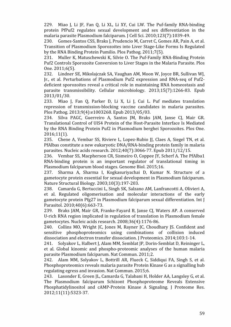
59
229. Miao J, Li JF, Fan Q, Li XL, Li XY, Cui LW. The Puf-‐family RNA-‐binding protein PfPuf2 regulates sexual development and sex differentiation in the malaria parasite Plasmodium falciparum. J Cell Sci. 2010;123(7):1039-‐49. 230. Gomes-‐Santos CSS, Braks J, Prudencio M, Carret C, Gomes AR, Pain A, et al. Transition of Plasmodium Sporozoites into Liver Stage-‐Like Forms Is Regulated by the RNA Binding Protein Pumilio. Plos Pathog. 2011;7(5). 231. Muller K, Matuschewski K, Silvie O. The Puf-‐Family RNA-‐Binding Protein Puf2 Controls Sporozoite Conversion to Liver Stages in the Malaria Parasite. Plos One. 2011;6(5). 232. Lindner SE, Mikolajczak SA, Vaughan AM, Moon W, Joyce BR, Sullivan WJ, Jr., et al. Perturbations of Plasmodium Puf2 expression and RNA-‐seq of Puf2-‐deficient sporozoites reveal a critical role in maintaining RNA homeostasis and parasite transmissibility. Cellular microbiology. 2013;15(7):1266-‐83. Epub 2013/01/30. 233. Miao J, Fan Q, Parker D, Li X, Li J, Cui L. Puf mediates translation repression of transmission-‐blocking vaccine candidates in malaria parasites. Plos Pathog. 2013;9(4):e1003268. Epub 2013/05/03. 234. Silva PAGC, Guerreiro A, Santos JM, Braks JAM, Janse CJ, Mair GR. Translational Control of UIS4 Protein of the Host-‐Parasite Interface Is Mediated by the RNA Binding Protein Puf2 in Plasmodium berghei Sporozoites. Plos One. 2016;11(1). 235. Chene A, Vembar SS, Riviere L, Lopez-‐Rubio JJ, Claes A, Siegel TN, et al. PfAlbas constitute a new eukaryotic DNA/RNA-‐binding protein family in malaria parasites. Nucleic acids research. 2012;40(7):3066-‐77. Epub 2011/12/15. 236. Vembar SS, Macpherson CR, Sismeiro O, Coppee JY, Scherf A. The PfAlba1 RNA-‐binding protein is an important regulator of translational timing in Plasmodium falciparum blood stages. Genome Biol. 2015;16. 237. Sharma A, Sharma I, Kogkasuriyachai D, Kumar N. Structure of a gametocyte protein essential for sexual development in Plasmodium falciparum. Nature Structural Biology. 2003;10(3):197-‐203. 238. Camarda G, Bertuccini L, Singh SK, Salzano AM, Lanfrancotti A, Olivieri A, et al. Regulated oligomerisation and molecular interactions of the early gametocyte protein Pfg27 in Plasmodium falciparum sexual differentiation. Int J Parasitol. 2010;40(6):663-‐73. 239. Braks JAM, Mair GR, Franke-‐Fayard B, Janse CJ, Waters AP. A conserved U-‐rich RNA region implicated in regulation of translation in Plasmodium female gametocytes. Nucleic acids research. 2008;36(4):1176-‐86. 240. Collins MO, Wright JC, Jones M, Rayner JC, Choudhary JS. Confident and sensitive phosphoproteomics using combinations of collision induced dissociation and electron transfer dissociation. J Proteomics. 2014;103:1-‐14. 241. Solyakov L, Halbert J, Alam MM, Semblat JP, Dorin-‐Semblat D, Reininger L, et al. Global kinomic and phospho-‐proteomic analyses of the human malaria parasite Plasmodium falciparum. Nat Commun. 2011;2. 242. Alam MM, Solyakov L, Bottrill AR, Flueck C, Siddiqui FA, Singh S, et al. Phosphoproteomics reveals malaria parasite Protein Kinase G as a signalling hub regulating egress and invasion. Nat Commun. 2015;6. 243. Lasonder E, Green JL, Camarda G, Talabani H, Holder AA, Langsley G, et al. The Plasmodium falciparum Schizont Phosphoproteome Reveals Extensive Phosphatidylinositol and cAMP-‐Protein Kinase A Signaling. J Proteome Res. 2012;11(11):5323-‐37.
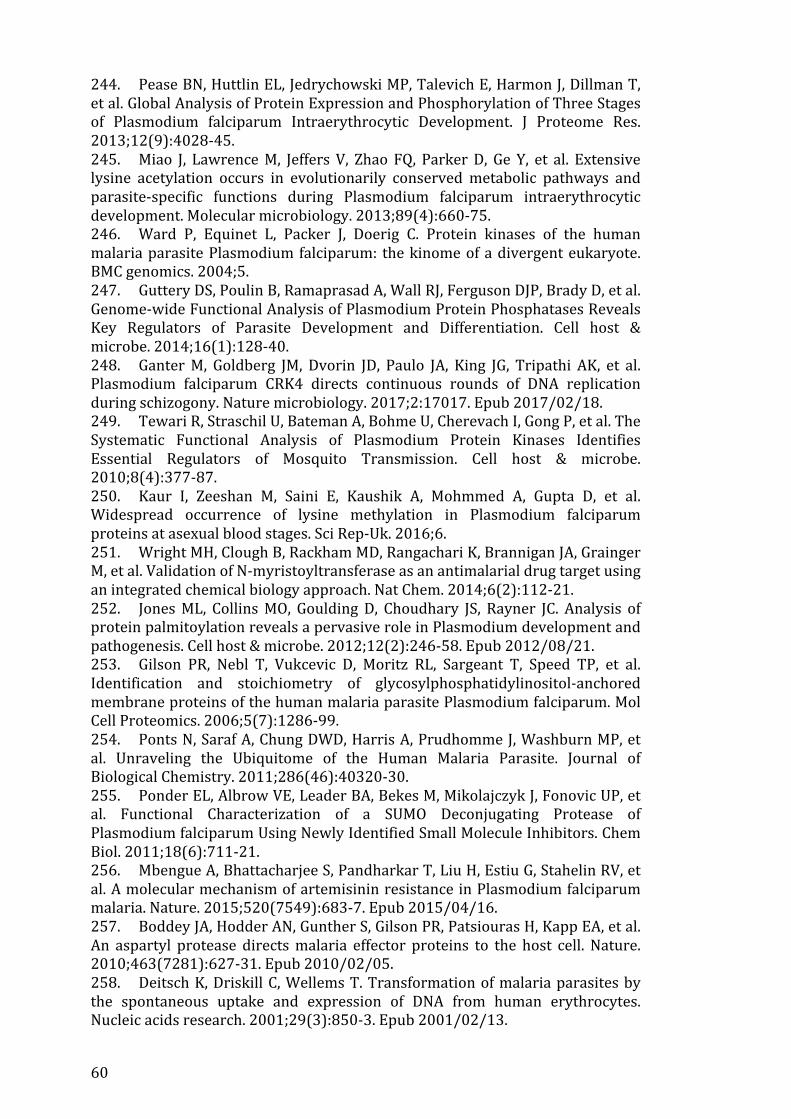
60
244. Pease BN, Huttlin EL, Jedrychowski MP, Talevich E, Harmon J, Dillman T, et al. Global Analysis of Protein Expression and Phosphorylation of Three Stages of Plasmodium falciparum Intraerythrocytic Development. J Proteome Res. 2013;12(9):4028-‐45. 245. Miao J, Lawrence M, Jeffers V, Zhao FQ, Parker D, Ge Y, et al. Extensive lysine acetylation occurs in evolutionarily conserved metabolic pathways and parasite-‐specific functions during Plasmodium falciparum intraerythrocytic development. Molecular microbiology. 2013;89(4):660-‐75. 246. Ward P, Equinet L, Packer J, Doerig C. Protein kinases of the human malaria parasite Plasmodium falciparum: the kinome of a divergent eukaryote. BMC genomics. 2004;5. 247. Guttery DS, Poulin B, Ramaprasad A, Wall RJ, Ferguson DJP, Brady D, et al. Genome-‐wide Functional Analysis of Plasmodium Protein Phosphatases Reveals Key Regulators of Parasite Development and Differentiation. Cell host & microbe. 2014;16(1):128-‐40. 248. Ganter M, Goldberg JM, Dvorin JD, Paulo JA, King JG, Tripathi AK, et al. Plasmodium falciparum CRK4 directs continuous rounds of DNA replication during schizogony. Nature microbiology. 2017;2:17017. Epub 2017/02/18. 249. Tewari R, Straschil U, Bateman A, Bohme U, Cherevach I, Gong P, et al. The Systematic Functional Analysis of Plasmodium Protein Kinases Identifies Essential Regulators of Mosquito Transmission. Cell host & microbe. 2010;8(4):377-‐87. 250. Kaur I, Zeeshan M, Saini E, Kaushik A, Mohmmed A, Gupta D, et al. Widespread occurrence of lysine methylation in Plasmodium falciparum proteins at asexual blood stages. Sci Rep-‐Uk. 2016;6. 251. Wright MH, Clough B, Rackham MD, Rangachari K, Brannigan JA, Grainger M, et al. Validation of N-‐myristoyltransferase as an antimalarial drug target using an integrated chemical biology approach. Nat Chem. 2014;6(2):112-‐21. 252. Jones ML, Collins MO, Goulding D, Choudhary JS, Rayner JC. Analysis of protein palmitoylation reveals a pervasive role in Plasmodium development and pathogenesis. Cell host & microbe. 2012;12(2):246-‐58. Epub 2012/08/21. 253. Gilson PR, Nebl T, Vukcevic D, Moritz RL, Sargeant T, Speed TP, et al. Identification and stoichiometry of glycosylphosphatidylinositol-‐anchored membrane proteins of the human malaria parasite Plasmodium falciparum. Mol Cell Proteomics. 2006;5(7):1286-‐99. 254. Ponts N, Saraf A, Chung DWD, Harris A, Prudhomme J, Washburn MP, et al. Unraveling the Ubiquitome of the Human Malaria Parasite. Journal of Biological Chemistry. 2011;286(46):40320-‐30. 255. Ponder EL, Albrow VE, Leader BA, Bekes M, Mikolajczyk J, Fonovic UP, et al. Functional Characterization of a SUMO Deconjugating Protease of Plasmodium falciparum Using Newly Identified Small Molecule Inhibitors. Chem Biol. 2011;18(6):711-‐21. 256. Mbengue A, Bhattacharjee S, Pandharkar T, Liu H, Estiu G, Stahelin RV, et al. A molecular mechanism of artemisinin resistance in Plasmodium falciparum malaria. Nature. 2015;520(7549):683-‐7. Epub 2015/04/16. 257. Boddey JA, Hodder AN, Gunther S, Gilson PR, Patsiouras H, Kapp EA, et al. An aspartyl protease directs malaria effector proteins to the host cell. Nature. 2010;463(7281):627-‐31. Epub 2010/02/05. 258. Deitsch K, Driskill C, Wellems T. Transformation of malaria parasites by the spontaneous uptake and expression of DNA from human erythrocytes. Nucleic acids research. 2001;29(3):850-‐3. Epub 2001/02/13.
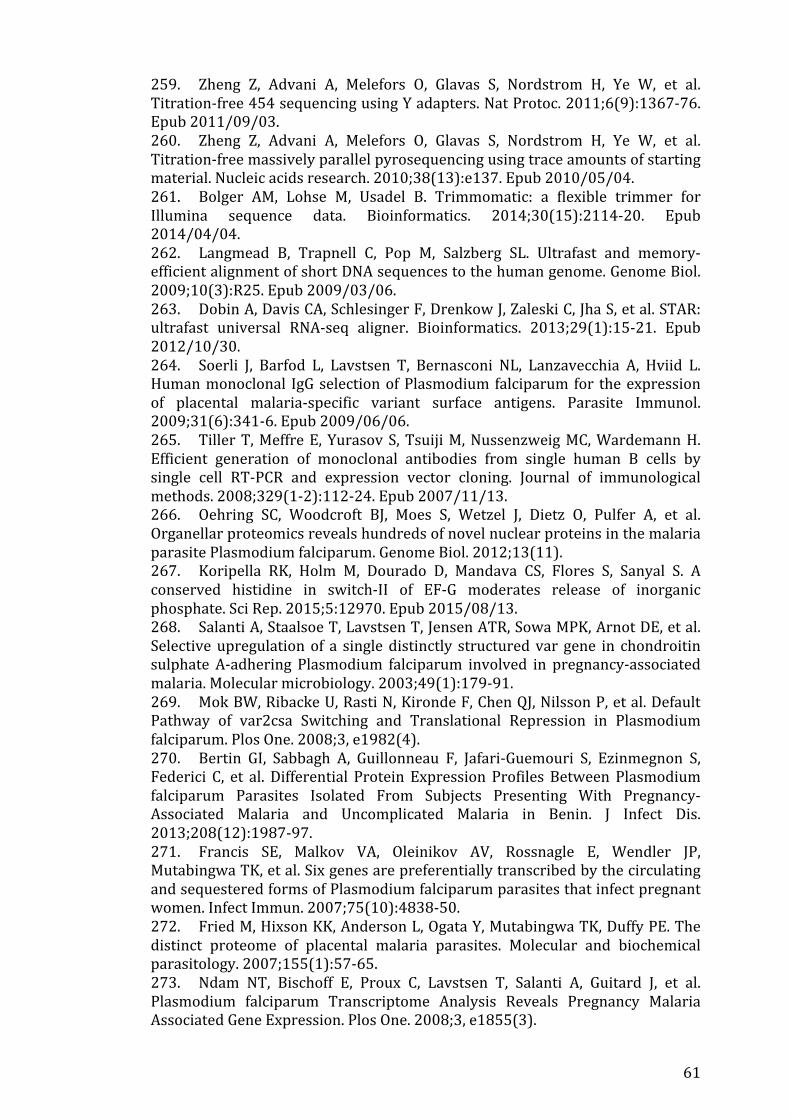
61
259. Zheng Z, Advani A, Melefors O, Glavas S, Nordstrom H, Ye W, et al. Titration-‐free 454 sequencing using Y adapters. Nat Protoc. 2011;6(9):1367-‐76. Epub 2011/09/03. 260. Zheng Z, Advani A, Melefors O, Glavas S, Nordstrom H, Ye W, et al. Titration-‐free massively parallel pyrosequencing using trace amounts of starting material. Nucleic acids research. 2010;38(13):e137. Epub 2010/05/04. 261. Bolger AM, Lohse M, Usadel B. Trimmomatic: a flexible trimmer for Illumina sequence data. Bioinformatics. 2014;30(15):2114-‐20. Epub 2014/04/04. 262. Langmead B, Trapnell C, Pop M, Salzberg SL. Ultrafast and memory-‐efficient alignment of short DNA sequences to the human genome. Genome Biol. 2009;10(3):R25. Epub 2009/03/06. 263. Dobin A, Davis CA, Schlesinger F, Drenkow J, Zaleski C, Jha S, et al. STAR: ultrafast universal RNA-‐seq aligner. Bioinformatics. 2013;29(1):15-‐21. Epub 2012/10/30. 264. Soerli J, Barfod L, Lavstsen T, Bernasconi NL, Lanzavecchia A, Hviid L. Human monoclonal IgG selection of Plasmodium falciparum for the expression of placental malaria-‐specific variant surface antigens. Parasite Immunol. 2009;31(6):341-‐6. Epub 2009/06/06. 265. Tiller T, Meffre E, Yurasov S, Tsuiji M, Nussenzweig MC, Wardemann H. Efficient generation of monoclonal antibodies from single human B cells by single cell RT-‐PCR and expression vector cloning. Journal of immunological methods. 2008;329(1-‐2):112-‐24. Epub 2007/11/13. 266. Oehring SC, Woodcroft BJ, Moes S, Wetzel J, Dietz O, Pulfer A, et al. Organellar proteomics reveals hundreds of novel nuclear proteins in the malaria parasite Plasmodium falciparum. Genome Biol. 2012;13(11). 267. Koripella RK, Holm M, Dourado D, Mandava CS, Flores S, Sanyal S. A conserved histidine in switch-‐II of EF-‐G moderates release of inorganic phosphate. Sci Rep. 2015;5:12970. Epub 2015/08/13. 268. Salanti A, Staalsoe T, Lavstsen T, Jensen ATR, Sowa MPK, Arnot DE, et al. Selective upregulation of a single distinctly structured var gene in chondroitin sulphate A-‐adhering Plasmodium falciparum involved in pregnancy-‐associated malaria. Molecular microbiology. 2003;49(1):179-‐91. 269. Mok BW, Ribacke U, Rasti N, Kironde F, Chen QJ, Nilsson P, et al. Default Pathway of var2csa Switching and Translational Repression in Plasmodium falciparum. Plos One. 2008;3, e1982(4). 270. Bertin GI, Sabbagh A, Guillonneau F, Jafari-‐Guemouri S, Ezinmegnon S, Federici C, et al. Differential Protein Expression Profiles Between Plasmodium falciparum Parasites Isolated From Subjects Presenting With Pregnancy-‐Associated Malaria and Uncomplicated Malaria in Benin. J Infect Dis. 2013;208(12):1987-‐97. 271. Francis SE, Malkov VA, Oleinikov AV, Rossnagle E, Wendler JP, Mutabingwa TK, et al. Six genes are preferentially transcribed by the circulating and sequestered forms of Plasmodium falciparum parasites that infect pregnant women. Infect Immun. 2007;75(10):4838-‐50. 272. Fried M, Hixson KK, Anderson L, Ogata Y, Mutabingwa TK, Duffy PE. The distinct proteome of placental malaria parasites. Molecular and biochemical parasitology. 2007;155(1):57-‐65. 273. Ndam NT, Bischoff E, Proux C, Lavstsen T, Salanti A, Guitard J, et al. Plasmodium falciparum Transcriptome Analysis Reveals Pregnancy Malaria Associated Gene Expression. Plos One. 2008;3, e1855(3).
62
274. Vignali M, Armour CD, Chen JY, Morrison R, Castle JC, Biery MC, et al. NSR-‐seq transcriptional profiling enables identification of a gene signature of Plasmodium falciparum parasites infecting children. Journal of Clinical Investigation. 2011;121(3):1119-‐29. 275. Wagner JC, Platt RJ, Goldfless SJ, Zhang F, Niles JC. Efficient CRISPR-‐Cas9-‐mediated genome editing in Plasmodium falciparum. Nat Methods. 2014;11(9):915-‐8. 276. Xue SF, Barna M. Specialized ribosomes: a new frontier in gene regulation and organismal biology. Nat Rev Mol Cell Bio. 2012;13(6):355-‐69. 277. Kumar M, Srinivas V, Patankar S. Upstream AUGs and upstream ORFs can regulate the downstream ORF in Plasmodium falciparum. Malar J. 2015;14(1):512. Epub 2015/12/23. 278. Peixoto L, Fernandez V, Musto H. The effect of expression levels on codon usage in Plasmodium falciparum. Parasitology. 2004;128(Pt 3):245-‐51. Epub 2004/04/13. 279. Spencer PS, Siller E, Anderson JF, Barral JM. Silent substitutions predictably alter translation elongation rates and protein folding efficiencies. J Mol Biol. 2012;422(3):328-‐35. Epub 2012/06/19. 280. Stadler M, Fire A. Wobble base-‐pairing slows in vivo translation elongation in metazoans. Rna. 2011;17(12):2063-‐73. 281. Muralidharan V, Goldberg DE. Asparagine Repeats in Plasmodium falciparum Proteins: Good for Nothing? Plos Pathog. 2013;9(8). 282. Hoepfner D, McNamara CW, Lim CS, Studer C, Riedl R, Aust T, et al. Selective and Specific Inhibition of the Plasmodium falciparum Lysyl-‐tRNA Synthetase by the Fungal Secondary Metabolite Cladosporin. Cell host & microbe. 2012;11(6):654-‐63. 283. Baragana B, Hallyburton I, Lee MCS, Norcross NR, Grimaldi R, Otto TD, et al. A novel multiple-‐stage antimalarial agent that inhibits protein synthesis. Nature. 2015;522(7556):315-‐+. 284. Herman JD, Pepper LR, Cortese JF, Estiu G, Galinsky K, Zuzarte-‐Luis V, et al. The cytoplasmic prolyl-‐tRNA synthetase of the malaria parasite is a dual-‐stage target of febrifugine and its analogs. Sci Transl Med. 2015;7(288). 285. Ahyong V, Sheridan CM, Leon KE, Witchley JN, Diep J, DeRisi JL. Identification of Plasmodium falciparum specific translation inhibitors from the MMV Malaria Box using a high throughput in vitro translation screen. Malaria J. 2016;15. 286. Baragana B, Norcross NR, Wilson C, Porzelle A, Hallyburton I, Grimaldi R, et al. Discovery of a Quinoline-‐4-‐carboxamide Derivative with a Novel Mechanism of Action, Multistage Antimalarial Activity, and Potent in Vivo Efficacy. J Med Chem. 2016;59(21):9672-‐85. 287. Kato N, Comer E, Sakata-‐Kato T, Sharma A, Sharma M, Maetani M, et al. Diversity-‐oriented synthesis yields novel multistage antimalarial inhibitors. Nature. 2016;538(7625):344-‐+. 288. Sonoiki E, Palencia A, Guo DH, Ahyong V, Dong C, Li XF, et al. Antimalarial Benzoxaboroles Target Plasmodium falciparum Leucyl-‐tRNA Synthetase. Antimicrob Agents Ch. 2016;60(8):4886-‐95.