Embed Size (px)
Citation preview
ARTICLE
Translocations Disrupting PHF21A in thePotocki-Shaffer-Syndrome Region Are Associatedwith Intellectual Disability and Craniofacial Anomalies
Hyung-Goo Kim,1,2,3,* Hyun-Taek Kim,4,27 Natalia T. Leach,5,27 Fei Lan,6,7,27 Reinhard Ullmann,3
Asli Silahtaroglu,8 Ingo Kurth,9,10 Anja Nowka,10 Ihn Sik Seong,1,11 Yiping Shen,1,6,12
Michael E. Talkowski,1,11,13 Douglas Ruderfer,1,13 Ji-Hyun Lee,1 Caron Glotzbach,14 Kyungsoo Ha,15
Susanne Kjaergaard,16 Alex V. Levin,17 Bernd F. Romeike,18 Tjitske Kleefstra,19 Oliver Bartsch,20
Sarah H. Elsea,21 Ethylin Wang Jabs,22,23 Marcy E. MacDonald,1,11,13 David J. Harris,24
Bradley J. Quade,25 Hans-Hilger Ropers,3 Lisa G. Shaffer,14 Kerstin Kutsche,10 Lawrence C. Layman,2
Niels Tommerup,8 Vera M. Kalscheuer,3 Yang Shi,6 Cynthia C. Morton,5,13,25 Cheol-Hee Kim,4
and James F. Gusella1,13,26
Potocki-Shaffer syndrome (PSS) is a contiguous gene disorder due to the interstitial deletion of band p11.2 of chromosome 11 and is
characterized by multiple exostoses, parietal foramina, intellectual disability (ID), and craniofacial anomalies (CFAs). Despite the iden-
tification of individual genes responsible for multiple exostoses and parietal foramina in PSS, the identity of the gene(s) associated with
the ID and CFA phenotypes has remained elusive. Through characterization of independent subjects with balanced translocations and
supportive comparative deletionmapping of PSS subjects, we have uncovered evidence that the ID and CFA phenotypes are both caused
by haploinsufficiency of a single gene, PHF21A, at 11p11.2. PHF21A encodes a plant homeodomain finger protein whose murine and
zebrafish orthologs are both expressed in amanner consistent with a function in neurofacial and craniofacial development, and suppres-
sion of the latter led to both craniofacial abnormalities and neuronal apoptosis. Along with lysine-specific demethylase 1 (LSD1),
PHF21A, also known as BHC80, is a component of the BRAF-histone deacetylase complex that represses target-gene transcription. In
lymphoblastoid cell lines from two translocation subjects in whom PHF21A was directly disrupted by the respective breakpoints, we
observed derepression of the neuronal gene SCN3A and reduced LSD1 occupancy at the SCN3A promoter, supporting a direct functional
consequence of PHF21A haploinsufficiency on transcriptional regulation. Our finding that disruption of PHF21A by translocations in
the PSS region is associated with ID adds to the growing list of ID-associated genes that emphasize the critical role of transcriptional regu-
lation and chromatin remodeling in normal brain development and cognitive function.
Introduction
In many regions of the genome, microdeletions1,2 or
balanced translocations3–6 are associated with phenotypic
abnormalities and are presumably caused by haploinsuffi-
ciency of the various genes involved. Potocki-Shaffer
syndrome (PSS [MIM 601224]) is a rare contiguous gene-
deletion syndrome caused by heterozygous interstitial
microdeletions of chromosomal region 11p11-p12 and is
1Center for Human Genetic Research, Massachusetts General Hospital, Bosto
Molecular Medicine and Genetics, Georgia Health Sciences University, August
Institute for Molecular Genetics, Ihnestraße 63–73, 14195 Berlin, Germany; 4
Korea; 5Department of Obstetrics, Gynecology, and Reproductive Biology,
MA 02115, USA; 6Department of Pathology, Harvard Medical School, 77 Aven
Department of Biology, 215 First Street, Cambridge, MA 02142, USA; 8Wilhelm
andMolecular Medicine, University of Copenhagen, DK-2200 Copenhagen, De
Germany; 10Institut fur Humangenetik, UniversitatsklinikumHamburg-Eppen
General Hospital, Boston, MA 02114, USA; 12Shanghai Children’s Medical Ce
China; 13Program in Medical and Population Genetics, Cambridge, MA
WA 99207, USA; 15Georgia Health Sciences University Cancer Center, August
Rigshospitalet, DK-2100 Copenhagen, Denmark; 17Pediatric Ophthalmology
Philadelphia, PA 19107, USA; 18Department of Neuropathology, Friedrich Sch
Radboud University Nijmegen Medical Centre, 6500 Nijmegen, The Nethe
Johannes Gutenberg-University Mainz, 55101 Mainz, Germany; 21Departmen
University School of Medicine, Richmond, VA 23298, USA; 22Department of G
NY 10029, USA; 23Institute of Genetic Medicine, The Johns Hopkins Universi
Boston and Harvard Medical School, Boston, MA 02115, USA; 25Department o
Boston, MA 02115, USA; 26Department of Genetics, Harvard Medical School,27These authors contributed equally to this work
*Correspondence: [email protected]
http://dx.doi.org/10.1016/j.ajhg.2012.05.005. �2012 by The American Societ
56 The American Journal of Human Genetics 91, 56–72, July 13, 2012
characterized by developmental defects that include
intellectual disability (ID), craniofacial anomalies (CFAs),
multiple exostoses (MIM 133701), and parietal foramina
(MIM 609597).7,8 Genes responsible for the latter two
phenotypes in this chromosomal region have been identi-
fied: Deletion of EXT2 (MIM 608210) causes multiple exos-
toses,9 and deletion of ALX4 (MIM 605420) causes parietal
foramina.10,11 However, the cause of the ID and abnor-
mal craniofacial development has remained uncertain.
n, MA 02114, USA; 2Department of Obstetrics & Gynecology, Institute of
a, GA 30912, USA; 3Department of Human Molecular Genetics, Max Planck
Department of Biology, Chungnam National University, Daejeon 305-764,
Brigham and Women’s Hospital and Harvard Medical School, Boston,
ue Louis Pasteur, Boston, MA 02115, USA; 7Constellation Pharmaceuticals,
Johannsen Centre for Functional Genome Research, Department of Cellular
nmark; 9Jena University Hospital, Institute of Human Genetics, 07743 Jena,
dorf, 20249 Hamburg, Germany; 11Department of Neurology, Massachusetts
nter, Shanghai Jiaotong University School of Medicine, Shanghai, 200127
02114, USA; 14Signature Genomic Laboratories, PerkinElmer, Spokane,
a, GA 30912, USA; 16Department of Clinical Genetics, University Hospital
and Ocular Genetics, Wills Eye Institute, Thomas Jefferson University,
iller University, 07747 Jena, Germany; 19Department of Human Genetics,
rlands; 20Institute of Human Genetics, University Medical Center of the
ts of Pediatrics and Human & Molecular Genetics, Virginia Commonwealth
enetics and Genomic Sciences, Mount Sinai School of Medicine, New York,
ty, Baltimore, MD 21287, USA; 24Division of Genetics, Children’s Hospital
f Pathology, Brigham and Women’s Hospital and Harvard Medical School,
Boston, MA 02114, USA
y of Human Genetics. All rights reserved.
Through identification of two independent subjects with
balanced translocations and support from a third pub-
lished translocation subject,12,13 we have uncovered
evidence that haploinsufficiency of a single gene, PHF21A
(also known as BHC80 [MIM 608325]), at 11p11.2 is
associated with ID and CFA phenotypes. This evidence
was complemented by comparative deletion mapping of
PSS subjects with diverse phenotypes that positioned
PHF21A within the critical region associated with ID and
CFAs, functional in vitro analysis of PHF21A in cells from
translocation subjects, and generation and rescue of zebra-
fish phenotypes through the suppression of phf21a with
morpholino oligonucleotides (MOs). PHF21A specifically
binds unmethylated histone H3 lysine 4 (H3K4me0) and
participates in the lysine-specific demethylase 1 (LSD1
[MIM 609132]) demethylase complex, implicating it as
a regulatory protein in histone-methylation dynamics14
and suggesting that disruption of this process might
underlie the ID and CFA phenotypes in these translocation
individuals.
Subjects and Methods
Human SubjectsDGAP012 and MCN1762 (Table 1) were ascertained through the
Developmental Genome Anatomy Project (DGAP) and Mendelian
Cytogenetic Network, respectively, and blood samples were
obtained for initiation of a lymphoblastoid cell line.17 PSS sub-
jects PSS02, PSS08, PSS10, and PSS-Romeike were previously
described.7,15,18 Where possible, all subjects were tested for copy-
number variants (CNVs) by array comparative genomic hybridiza-
tion (CGH) with the use of Agilent (Santa Clara, CA) 244K arrays as
described (Table 2).19 DGAP012 displayed one 156 kb deletion
CNV (chr13: 48,430,969–48,587,500; hg18) that is located at
13q14.2 and has not been reported previously and that involves
FNDC3A, which is expressed in spermatids and Leydig cells and
produces male sterility when homozygously inactivated.20 All
human studies were performed under informed-consent protocols
approved by the Partners HealthCare System Human Research
Committee.
DGAP012
At birth, this white male was small but normally proportioned
and suffered from supraventricular tachycardia, hyperbilirubine-
mia, and hypoglycemia, all of which resolved. Chromosome
analysis revealed an apparently balanced chromosomal transloca-
tion with the karyotype 46,XY,t(11;19)(p11.2;p13.3)dn, which
was revised by molecular analysis here to 46,XY,t(11;19)
(p11.2;p13.2)dn. The family history was unremarkable, and
parental chromosomes were normal. At 1 year of age, the subject
displayed bifrontal biparietal atrophy on a computed tomography
(CT) scan, and at 15 months of age, he showed significant global
developmental delay, digitalized thumbs, brachycephaly, micro-
cephaly (a head circumference of 46 cm; tenth percentile), a small
downturned mouth, mild midfacial hypoplasia, a flat midface,
a narrow nasal bridge, a very small nose, large ears, bilateral epible-
pharon (an extra skin fold was medially under each lower lid)
without trichiasis, small hands and feet, and an absence of
emotional expression. He also displayed hand flapping and had
feeding problems prior to the age of 3 years.
The
Amagnetic resonance image (MRI) at the age of 2 years revealed
prominent cerebrospinal fluid, which might represent volume
loss. At the age of 5 years, DGAP012 displayed hypotonia and dys-
tonic movement and could not walk and showed significant near-
sightedness and astigmatism (�3.00 þ3.00 3 90 in both eyes), as
well as some diffuse pigmentary mottling suggesting a possible
retinal dystrophy.
MCN1762
This white female, aged 42 years, was delivered at term after an
uneventful pregnancy but was lethargic and had feeding difficul-
ties. At age 3.5 years, her gross motor development was normal,
but her speech development was delayed, and she displayed
hyperactivity, poor concentration, and mild myopia (�2.00 [right
eye], �2.50 [left eye]). A verbal intelligence quotient (IQ) test
(Terman-Merril) indicated a delay of 1 year, and a nonverbal IQ
test (Leiter) was normal. The following dysmorphic features were
noted: brachycephaly, microcephaly, a long narrow nose, mild
midfacial hypoplasia, a downturned mouth, thin lips, and prom-
inent ear lobes and, in childhood only, downslanting palpebral
fissures and epicanthal folds. GTG-banded karyotyping revealed
an apparently balanced reciprocal translocation, 46,XX,t(1;11)
(p13;p11)dn, which was revised by molecular analysis to
46,XX,t(1;11)(p21.1p11.2)dn. Her parents and two siblings are
healthy, and parental karyotypes are normal. MCN1762 attended
a school for special needs and is able to read. Her linear growth
(to 173 cm) and pubertal development were normal, and she has
developed truncal obesity (see Figure 4C). She now has mild ID
but manages to live independently and have a sheltered part-
time job.
GM03316
This female Venezuelan subject was 3 years old when her blood
was submitted to the National Institute of General Medical
Sciences Human Genetic Cell Repository at the Coriell Institute
in January of 1978 (prior to identification of PSS) and showed an
apparently balanced translocation: t(X;11)(q11.1;p11.2)dn. The
clinical symptoms were listed as ID (quantitative intelligence
[Gq] ¼ 60) with a strikingly unusual dysmorphology syndrome
including epicanthus, hypertelorism, oblique palpebral fissures,
trigonocephaly, and micrognathia. At 5 years of age, her vocabu-
lary was progressing well, and she had a good memory, but her
Gq corresponded to that of a 3-year-old girl. Her principal problem
was an inability to concentrate. She could feed herself when she
wished and had gained control of her sphincters both day and
night by age 3.5 years. She was shy and easily frightened and
was clumsy with both hands and legs.
GC14361
This 2.25-year-old male from Bangladesh had a history of
static encephalopathy and developmental delay, which were first
noted when he was 6 months old. He displayed microcephaly,
short stature, a small phallus, a unilateral absent testis, and dys-
morphic features, including a short forehead, prominent biparie-
tal foramina, a midline parietal cortical defect, a flat midface,
a flat occiput, sensorineural hearing loss, epicanthal folds, protu-
berant ears, a bulbous nasal tip that continued below the colu-
mella, a depressed nasal root, a small mouth and small chin
(micrognathia), hypotonia, a slight pectus excavatum, recurrent
otitis media, and slender fingers.
Breakpoint Mapping, Cloning, and CharacterizationIn brief, bacterial artificial chromosomes (BACs), followed by
fosmid or cosmid probes chosen on the basis of the human
genome map, were used for fluorescence in situ hybridization
American Journal of Human Genetics 91, 56–72, July 13, 2012 57
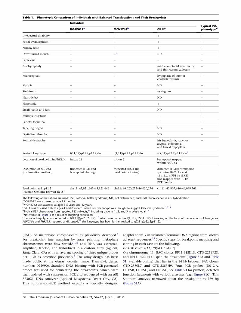
Table 1. Phenotypic Comparison of Individuals with Balanced Translocations and Their Breakpoints
IndividualTypical PSSphenotypedDGAP012a MCN1762b GILLEc
Intellectual disability þ þ þ þ
Facial dysmorphism þ þ þ þ
Narrow nose þ þ þ þ
Downturned mouth þ þe ND þ
Large ears þ � � þ
Brachycephaly þ þ mild craniofacial asymmetryand thin corpus callosum
þ
Microcephaly þ þ hypoplasia of inferiorcerebellar vermis
þ
Myopia þ þ ND þ
Strabismus þ � nystagmus þ
Heart defect þ � ND þ
Hypotonia þ þ þ þ
Small hands and feet þ � ND þ
Multiple exostoses � � � þ
Parietal foramina � � � þ
Tapering fingers � � ND þ
Digitalized thumbs þ � ND �
Retinal dystrophy þ � iris hypoplasia, superioratypical coloboma,and foveal hypoplasia
�
Revised karyotype t(11;19)(p11.2;p13.2)dn t(1;11)(p21.1;p11.2)dn t(X;11)(p22.2;p11.2)dnf
Location of breakpoint in PHF21A intron 14 intron 5 breakpoint mappedwithin PHF21A
Disruption of PHF21A(confirmation method)
truncated (FISH andbreakpoint cloning)
truncated (FISH andbreakpoint cloning)
disrupted (FISH); breakpoint-spanning BAC clone at11p11.2 is RP11-618K13;fine mapped with 10 kbPCR product
Breakpoint at 11p11.2(Human Genome Browser hg18)
chr11: 45,921,645–45,921,646 chr11: 46,020,273–46,020,274 chr11: 45,907,446–46,099,561
The following abbreviations are used: PSS, Potocki-Shaffer syndrome; ND, not determined; and FISH, fluorescence in situ hybridization.aDGAP012 was assessed at age 15 months.bMCN1762 was assessed at ages 3.5 years and 42 years.cGILLE was assessed only at ages 4 and 8 months when her phenotype was thought to suggest Gillespie syndrome.12,13dTypical PSS phenotypes from reported PSS subjects,15 including patients 1, 2, and 3 in Wuyts et al.16eNot visible in Figure 4 as a result of laughing expression.fThe initial karyotype was reported as t(X;11)(p22.32;p12),12 which was revised as t(X;11)(p22.3;p12). However, on the basis of the locations of two genes,ARHGAP6 and PHF21A, reported as disrupted,13 this karyotype has been further revised to t(X;11)(p22.2;p11.2).
(FISH) of metaphase chromosomes as previously described.6
For breakpoint fine mapping by array painting, metaphase
chromosomes were flow sorted,21,22 and DNA was extracted,
amplified, labeled, and hybridized to a custom array (Agilent,
Santa Clara, CA) with an average spacing of three unique probes
per 1 kb as described previously.4 The array design has been
made public at the eArray website (name: Translok4; design
number: 022990). Standard DNA blotting with PCR-generated
probes was used for delineating the breakpoints, which were
then isolated with suppression PCR and sequenced with an ABI
3730XL DNA Analyzer (Applied Biosystems, Foster City, CA).
This suppression-PCR method exploits a specially designed
58 The American Journal of Human Genetics 91, 56–72, July 13, 2012
adaptor to walk in unknown genomic DNA regions from known
adjacent sequences.23 Specific steps for breakpoint mapping and
cloning in each case are the following.
DGAP012 with t(11;19)(p11.2;p13.2)
On chromosome 11, BAC clones RP11-618K13, CTD-2254P23,
and RP11-142O14 all span the breakpoint (Figure S1A and Table
S1, available online) that lies in the 14 kb between BAC clones
CTD-2580L7 and CTD-2353H9. Four PCR probes (D012-A,
D012-B, D012-C, and D012-D; see Table S3 for primers) detected
junction fragments with various enzymes (e.g., Figure S1C). This
Southern analysis narrowed down the breakpoint to 729 bp
(Figure S1A).
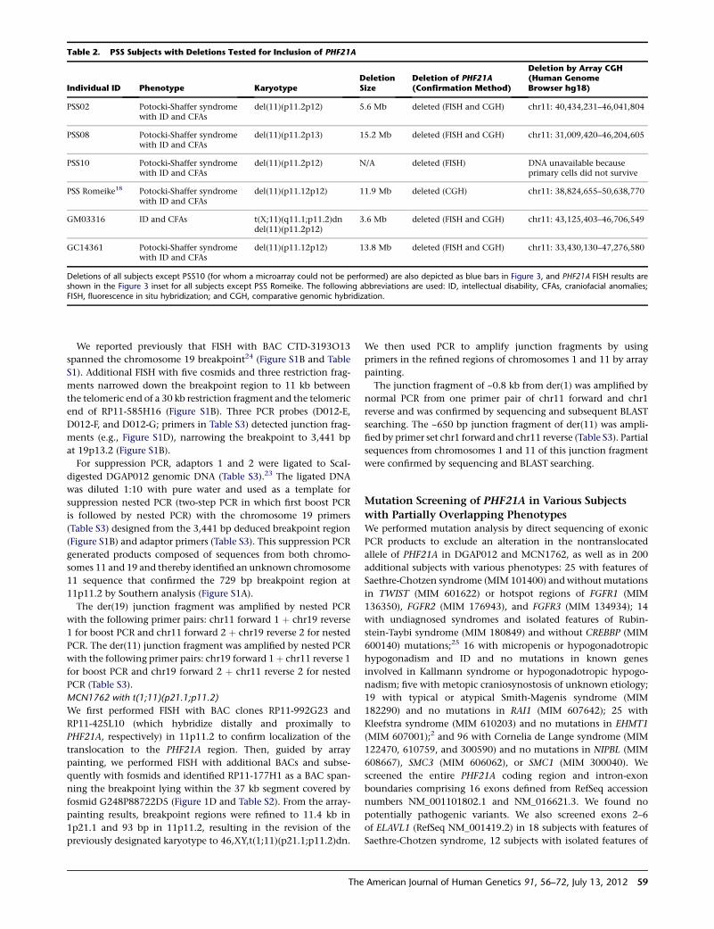
Table 2. PSS Subjects with Deletions Tested for Inclusion of PHF21A
Individual ID Phenotype KaryotypeDeletionSize
Deletion of PHF21A(Confirmation Method)
Deletion by Array CGH(Human GenomeBrowser hg18)
PSS02 Potocki-Shaffer syndromewith ID and CFAs
del(11)(p11.2p12) 5.6 Mb deleted (FISH and CGH) chr11: 40,434,231–46,041,804
PSS08 Potocki-Shaffer syndromewith ID and CFAs
del(11)(p11.2p13) 15.2 Mb deleted (FISH and CGH) chr11: 31,009,420–46,204,605
PSS10 Potocki-Shaffer syndromewith ID and CFAs
del(11)(p11.2p12) N/A deleted (FISH) DNA unavailable becauseprimary cells did not survive
PSS Romeike18 Potocki-Shaffer syndromewith ID and CFAs
del(11)(p11.12p12) 11.9 Mb deleted (CGH) chr11: 38,824,655–50,638,770
GM03316 ID and CFAs t(X;11)(q11.1;p11.2)dndel(11)(p11.2p12)
3.6 Mb deleted (FISH and CGH) chr11: 43,125,403–46,706,549
GC14361 Potocki-Shaffer syndromewith ID and CFAs
del(11)(p11.12p12) 13.8 Mb deleted (FISH and CGH) chr11: 33,430,130–47,276,580
Deletions of all subjects except PSS10 (for whom a microarray could not be performed) are also depicted as blue bars in Figure 3, and PHF21A FISH results areshown in the Figure 3 inset for all subjects except PSS Romeike. The following abbreviations are used: ID, intellectual disability, CFAs, craniofacial anomalies;FISH, fluorescence in situ hybridization; and CGH, comparative genomic hybridization.
We reported previously that FISH with BAC CTD-3193O13
spanned the chromosome 19 breakpoint24 (Figure S1B and Table
S1). Additional FISH with five cosmids and three restriction frag-
ments narrowed down the breakpoint region to 11 kb between
the telomeric end of a 30 kb restriction fragment and the telomeric
end of RP11-585H16 (Figure S1B). Three PCR probes (D012-E,
D012-F, and D012-G; primers in Table S3) detected junction frag-
ments (e.g., Figure S1D), narrowing the breakpoint to 3,441 bp
at 19p13.2 (Figure S1B).
For suppression PCR, adaptors 1 and 2 were ligated to ScaI-
digested DGAP012 genomic DNA (Table S3).23 The ligated DNA
was diluted 1:10 with pure water and used as a template for
suppression nested PCR (two-step PCR in which first boost PCR
is followed by nested PCR) with the chromosome 19 primers
(Table S3) designed from the 3,441 bp deduced breakpoint region
(Figure S1B) and adaptor primers (Table S3). This suppression PCR
generated products composed of sequences from both chromo-
somes 11 and 19 and thereby identified an unknown chromosome
11 sequence that confirmed the 729 bp breakpoint region at
11p11.2 by Southern analysis (Figure S1A).
The der(19) junction fragment was amplified by nested PCR
with the following primer pairs: chr11 forward 1 þ chr19 reverse
1 for boost PCR and chr11 forward 2 þ chr19 reverse 2 for nested
PCR. The der(11) junction fragment was amplified by nested PCR
with the following primer pairs: chr19 forward 1 þ chr11 reverse 1
for boost PCR and chr19 forward 2 þ chr11 reverse 2 for nested
PCR (Table S3).
MCN1762 with t(1;11)(p21.1;p11.2)
We first performed FISH with BAC clones RP11-992G23 and
RP11-425L10 (which hybridize distally and proximally to
PHF21A, respectively) in 11p11.2 to confirm localization of the
translocation to the PHF21A region. Then, guided by array
painting, we performed FISH with additional BACs and subse-
quently with fosmids and identified RP11-177H1 as a BAC span-
ning the breakpoint lying within the 37 kb segment covered by
fosmid G248P88722D5 (Figure 1D and Table S2). From the array-
painting results, breakpoint regions were refined to 11.4 kb in
1p21.1 and 93 bp in 11p11.2, resulting in the revision of the
previously designated karyotype to 46,XY,t(1;11)(p21.1;p11.2)dn.
The
We then used PCR to amplify junction fragments by using
primers in the refined regions of chromosomes 1 and 11 by array
painting.
The junction fragment of ~0.8 kb from der(1) was amplified by
normal PCR from one primer pair of chr11 forward and chr1
reverse and was confirmed by sequencing and subsequent BLAST
searching. The ~650 bp junction fragment of der(11) was ampli-
fied by primer set chr1 forward and chr11 reverse (Table S3). Partial
sequences from chromosomes 1 and 11 of this junction fragment
were confirmed by sequencing and BLAST searching.
Mutation Screening of PHF21A in Various Subjects
with Partially Overlapping PhenotypesWe performed mutation analysis by direct sequencing of exonic
PCR products to exclude an alteration in the nontranslocated
allele of PHF21A in DGAP012 and MCN1762, as well as in 200
additional subjects with various phenotypes: 25 with features of
Saethre-Chotzen syndrome (MIM 101400) and without mutations
in TWIST (MIM 601622) or hotspot regions of FGFR1 (MIM
136350), FGFR2 (MIM 176943), and FGFR3 (MIM 134934); 14
with undiagnosed syndromes and isolated features of Rubin-
stein-Taybi syndrome (MIM 180849) and without CREBBP (MIM
600140) mutations;25 16 with micropenis or hypogonadotropic
hypogonadism and ID and no mutations in known genes
involved in Kallmann syndrome or hypogonadotropic hypogo-
nadism; five with metopic craniosynostosis of unknown etiology;
19 with typical or atypical Smith-Magenis syndrome (MIM
182290) and no mutations in RAI1 (MIM 607642); 25 with
Kleefstra syndrome (MIM 610203) and no mutations in EHMT1
(MIM 607001);2 and 96 with Cornelia de Lange syndrome (MIM
122470, 610759, and 300590) and no mutations in NIPBL (MIM
608667), SMC3 (MIM 606062), or SMC1 (MIM 300040). We
screened the entire PHF21A coding region and intron-exon
boundaries comprising 16 exons defined from RefSeq accession
numbers NM_001101802.1 and NM_016621.3. We found no
potentially pathogenic variants. We also screened exons 2–6
of ELAVL1 (RefSeq NM_001419.2) in 18 subjects with features of
Saethre-Chotzen syndrome, 12 subjects with isolated features of
American Journal of Human Genetics 91, 56–72, July 13, 2012 59
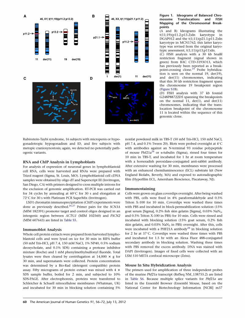
Figure 1. Ideograms of Balanced Chro-mosome Translocations and FISHMapping of the Chromosomal Break-points(A and B) Ideograms illustrating thet(11;19)(p11.2;p13.2)dn karyotype inDGAP012 and the t(1;11)(p21.1;p11.2)dnkaryotype in MCN1762; this latter karyo-type was revised from the original karyo-typic assessment, t(1;11)(p13;p11)dn.(C) FISH analysis with a 30 kb SnaBIrestriction fragment (signal shown ingreen) from BAC CTD-3193O13, whichhas previously been reported as a break-point-crossing clone.24 Probe hybridiza-tion is seen on the normal 19, der(19),and der(11) chromosomes, indicatingthat this 30 kb restriction fragment spansthe chromosome 19 breakpoint region(Figure S1B).(D) FISH analysis with 37 kb fosmidG248P88722D5 spanning the breakpointson the normal 11, der(1), and der(11)chromosomes, indicating that the trans-location breakpoint of the chromosome11 is located within the sequence of thisgenomic clone.
Rubinstein-Taybi syndrome, 16 subjects with micropenis or hypo-
gonadotropic hypogonadism and ID, and five subjects with
metopic craniosynostosis; again, we detected no potentially path-
ogenic variants.
RNA and ChIP Analysis in LymphoblastsFor analysis of expression of neuronal genes in lymphoblastoid
cell RNA, cells were harvested and RNAs were prepared with
Trizol reagent (Sigma, St. Louis, MO). Lymphoblastoid cell cDNA
samples were obtained by oligo dT and Superscript III (Invitrogen,
San Diego, CA) with primers designed to cross multiple introns for
the exclusion of genomic amplification. RT-PCR was carried out
for 34 cycles by annealing at 60�C for 30 s and elongation at
72�C for 30 s with Platinum PCR SuperMix (Invitrogen).
LSD1 chromatin immunoprecipitation (ChIP) experiments were
done as previously described.26,27 Primer pairs for the SCN3A
(MIM 182391) promoter target and control oligos designed in an
intergenic region between ACTG1 (MIM 102560) and FSCN2
(MIM 607643) are listed in Table S5.
Immunoblot AnalysisWhole cell protein extracts were prepared fromharvested lympho-
blastoid cells and were lysed on ice for 30 min in RIPA buffer
(50 mM Tris-HCl, pH 7.4, 150 mM NaCl, 1% NP40, 0.5% sodium
deoxycholate, and 0.1% SDS) containing a protease inhibitor
mixture (Roche) and 1 mM phenylmethylsulfonyl fluoride. Total
lysates were then cleared by centrifugation at 14,000 3 g for
30 min, and supernatants were collected. Protein concentration
was determined by a Bio-Rad (detergent compatible) protein
assay. Fifty micrograms of protein extract was mixed with 4 3
SDS sample buffer, boiled for 2 min, and subjected to 10%
SDS-PAGE. After electrophoresis, proteins were transferred to
Schleicher & Schuell nitrocellulose membranes (Whatman, UK)
and incubated for 30 min in blocking solution containing 5%
60 The American Journal of Human Genetics 91, 56–72, July 13, 2012
nonfat powdered milk in TBS-T (50 mM Tris-HCl, 150 mM NaCl,
pH 7.4, and 0.1% Tween 20). Blots were probed overnight at 4�Cwith antibodies against an N-terminal 93 residue polypeptide
of mouse Phf21a28 or a-tubulin (Sigma), rinsed four times for
10 min in TBS-T, and incubated for 1 hr at room temperature
with a horseradish peroxidase-conjugated anti-rabbit antibody.
After extensive washing for 30 min, membranes were processed
with an enhanced chemiluminescence (ECL) substrate kit (New
England Biolabs, Beverly, MA) and exposed to autoradiographic
film (Hyperfilm ECL, Amersham Bioscience, Piscataway, NJ).
ImmunostainingCells were grown on glass coverslips overnight. After being washed
with PBS, cells were fixed in 4% paraformaldehyde and 0.5%
Triton X-100 for 10 min. Coverslips were washed three times
with PBS and incubated in block-permeabilization solution (15%
goat serum [Sigma], 0.2% fish skin gelatin [Sigma], 0.03% NaN3,
and 0.5% Triton X-100 in PBS) for 10 min. Cells were rinsed and
incubated with blocking solution (15% goat serum, 0.2% fish
skin gelatin, and 0.03% NaN3 in PBS) overnight. After this, cells
were incubated with a PHF21A antibody28 in blocking solution
for 2 hr at 37�C. Coverslips were washed three times with PBS
and incubated for 1.5 hr with an Alexa Fluor 488-conjugated
secondary antibody in blocking solution. Washing three times
with PBS removed the excess antibody. DNA was stained with
DAPI (Invitrogen). Images of fixed cells were collected with an
LSM 510 META confocal microscope (Zeiss).
Mouse In Situ Hybridization AnalysisThe primers used for amplification of three independent probes
of the murine Phf21a transcript (RefSeq NM_138755.2) are listed
in Table S6. Because multiple splice variants for Phf21a are
listed in the Ensembl Browser (Ensembl Mouse, based on the
National Center for Biotechnology Information [NCBI] m37
mouse assembly), probes were designed to detect the majority of
the known isoforms. Probes were labeled with [a-35S]-UTP for
hybridization on 15 mm cryosections. The day of plug was not
counted for the specification of embryonic stages. No specific
signals were detected with the respective sense probes except as
indicated in the results section.
Fish Stocks and MaintenanceZebrafish were maintained at 28.5�C under a 14 hr light/10 hr
dark cycle in 1/3 Ringer’s solution. Transgenic fish Tg[flk1:GFP]
(kindly provided by Dr. Tao P. Zhong) and Tg[huC:EGFP]29 were
used in overexpression or knockdown experiments. Embryos
older than 24 hr postfertilization (hpf) were usually incubated in
0.003% 1-phenyl-2-thiourea (PTU, Sigma) for the inhibition of
pigmentation. Embryos at appropriate stages were fixed with 4%
paraformaldehyde in PBS.
Zebrafish phf21a ConstructsZebrafish phf21a was isolated from the 24 hpf zebrafish cDNA
library by RT-PCR and was first cloned in a pGEM-T easy vector
(Promega, Madison, USA) and then subcloned into the EcoRI
site in the pCS2þ multipurpose expression vector. For the
construction of the phf21a-RFP fusion reporter, specific enzyme-
linked primers were designed for PCR amplification. PCR primers
are listed in Table S7. PCR products were subcloned into the ClaI
site in a pCS2þ RFP vector.
Whole-Mount In Situ Hybridization and Alcian-Blue
and Acridine-Orange StainingAntisense digoxigenin-labeled RNA probes for dlx2a, ngn1, huC,
and phf21a were produced with a DIG-RNA labeling kit (Roche,
Germany) according to the manufacturer’s instructions. Whole-
mount in situ hybridization was performed with digoxigenin-
labeled probes as previously described.30 Cartilage staining was
carried out with Alcian blue.31 For the detection of apoptotic
cells, embryos were placed in 10 mg/ml acridine orange (Sigma)
for 30 min and were washed in egg water.
Microinjection of mRNA and Antisense MOsSynthetic capped mRNAs for PHF21A and phf21awere transcribed
in vitro with the linearized plasmid DNA as a template. mRNA
was dissolved in 0.2% phenol red (as a tracking dye) and then
microinjected into 1- to 2-cell-stage embryos. Antisense MOs for
phf21a MO 50-GCGTCATAAATGATATTTACCTGTG-30 and stan-
dard control MO 50-CCTCTTACCTCAGTTACAATTTATA-30 were
synthesized by Gene Tools (Corvallis, OR, USA). Each morpholino
was resuspended in 13Danieau buffer (58mMNaCl, 0.7mMKCl,
0.4 mM MgSO4, 0.6 mM Ca(NO3)2, 5.0 mM HEPES, and pH 7.6)
and injected into 1- to 2-cell-stage embryos at the concentration
of 5 ng/embryo.
Results
PHF21A Is Disrupted in Unrelated Subjects with
Chromosomal Translocations, ID, and CFAs
DGAP is a collaborative effort to identify genes of develop-
mental importance through the study of individuals with
apparently balanced chromosomal abnormalities and
developmental defects.32 Identification of multiple cases
in whom the same gene is disrupted in independent
The
subjects with de novo translocations and similar pheno-
types provides particularly strong evidence of the causative
nature of the lesion. ThroughDGAP, we identified a subject
(DGAP012) with an apparently balanced de novo translo-
cation between chromosomes 11 and 19; this transloca-
tion resulted in a 46,XY,t(11;19)(p11.2;p13.2)dn karyotype
(Figure 1A). A second subject, MCN1762 (MCN19730002-
227), identified through the Mendelian Cytogenetic
Network database, had an apparently balanced de novo
translocation between chromosomes 1 and 11; this was
initially reported as 46,XX,t(1;11)(p13;p11)dn (Figure 1B).
They both display evidence of ID with CFAs, as well as
other typical PSS features, except for multiple exostoses
and parietal foramina, as summarized in Table 1, suggest-
ing that the disruption in each case might affect the
same gene in or near the PSS region in 11p11.2.
To map precisely the translocation in DGAP012, we first
used FISH to bracket a candidate region and then to define
a breakpoint-crossing BAC (Figure 1C and Figures S1A and
S1B).24 After DNA blotting (Figures S1C and S1D) to refine
the breakpoints, we used suppression cloning23 and tar-
geted PCR for subsequent isolation and sequencing of
junction fragments. Full details of the breakpoint-cloning
steps are given in the Subjects and Methods section.
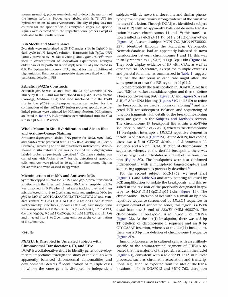
The chromosome 19 breakpoint lies within a SINE/Alu
sequence in intron 5 of ELAVL1, whereas the chromosome
11 breakpoint interrupts a LINE/L2 repetitive element in
intron 14 of PHF21A (Figure 2A). At the der(19) breakpoint,
there was a 5 nt CTCCT deletion of chromosome 11
sequence and a 5 nt TTCAG deletion of chromosome 19
sequence, whereas at the der(11) breakpoint, there was
no loss or gain of nucleotides as a result of the transloca-
tion (Figure 2C). The breakpoints were also confirmed
independently with a multiplexed targeted-capture and
sequencing approach as previously described.33
For the second subject, MCN1762, we used FISH
(Figure 1D and Table S2) and array painting followed by
PCR amplification to isolate the breakpoints, and this re-
sulted in the revision of the previously designated karyo-
type to 46,XY,t(1;11)(p21.1;p11.2)dn (Figure 1B). The
chromosome 1 breakpoint lies within a small 48 bp non-
repetitive sequence surrounded by LINE/L1 sequences in
a region devoid of annotated genes; this region is 635 kb
distal from the 50 end of PRMT6 (MIM 608274). The
chromosome 11 breakpoint is in intron 5 of PHF21A
(Figure 2B). At the der(1) breakpoint, there was a 2 bp
TT deletion of chromosome 1 sequence and an 8 bp
CTCCAAAT insertion, whereas at the der(11) breakpoint,
there was a 3 bp TTA deletion of chromosome 1 sequence
(Figure 2D).
Immunofluorescence in cultured cells with an antibody
specific to the amino-terminal segment of PHF21A re-
vealed that the majority of the protein resides in the nuclei
(Figure S3), consistent with a role for PHF21A in nuclear
processes, such as chromatin association and transcrip-
tional regulation. As expected from the sites of the trans-
locations in both DGAP012 and MCN1762, disruption
American Journal of Human Genetics 91, 56–72, July 13, 2012 61
Figure 2. Mapping of the Breakpoints in Two Balanced Translocations(A and B) Disruption of PHF21A by two translocations. A schematic diagram of the DGAP012 breakpoints (not to scale) shows, in blue,PHF21A on chromosome 11 (exons 1–18) and, in white, either (A) ELAVL1 on chromosome 19 (exons 1–6) or (B) chromosome 1. Allexons are depicted as vertical black boxes, and the start and direction of transcription are indicated by an arrow above the correspondingexons. The translocation occurred at the site of the red vertical line. In DGAP012, the translocation produced der(11) and der(19) chro-mosomes that encode potential fusion transcripts (A). Note that the stop codon of the potential PHF21ADex15-18/ELAVL1Dex1-5 fusiongene is at the same location as wild-type ELAVL1 because there is no frameshift, whereas ELAVL1Dex6/ PHF21ADex1-14 has a frameshiftwith a premature stop codon in the new exon 6 (equivalent to exon 15 of PHF21A). The translocation inMCN1762 does not predict anypotential fusion product because the breakpoint on chromosome 1 is located in a gene desert.(C and D) Genomic DNA sequence from the normal chromosomes and at the breakpoints on derivative chromosomes. In DGAP012, atthe der(19) breakpoint, there was a 5 nt CTCCT deletion of chromosome 11 sequence and a 5 nt TTCAG deletion of chromosome 19sequence, whereas at the der(11) breakpoint, there was no loss or gain of sequence (C). In MCN1762, the junction sequences revealeda 3 bp TTA deletion of chromosome 1 sequence on der(11) and a 2 bp TT deletion of chromosome 11 sequence and an 8 bp CTCCAAATinsertion on der(1). Details of the mapping of both breakpoints in DGAP012 and MCN1762 are described in Figure S1 as well as in theSubjects and Methods section.(E) Immunoblot analysis of PHF21A levels in DGAP012 and MCN1762 and in controls. The PHF21A antibody against an N-terminal93 residue polypeptide28 recognizes the ~92 kDa PHF21A in both female (‘‘F’’) and male (‘‘M’’) controls, as well as in DGAP012 andMCN1762 lymphoblastoid cell line extracts. It shows notably reduced protein levels due to disruption of PHF21A (arrow) in bothDGAP012 and MCN1762. A 73 kDa protein (arrowhead) was noted in DGAP012 and is likely to be a product of the PHF21ADex15-18/ELAVL1Dex1-5 fusion gene, which deletes the critical plant homeodomain (PHD) finger domain. a-tubulin was used as an internalloading control. The bar graph shows the mean and 5 standard deviations from three independent experiments (*p < 0.001, **p <0.0001). The subcellular localization of PHF21A with the same antibody is described in Figure S3.(F) PHF21A functional domains in wild-type and theoretical truncated proteins in two balanced translocation subjects. PHF21A containstwo leucine zipper domains (LZD1 and LZD2), one AT-hook domain, and one PHD zinc finger domain. The amino acid positions of alldomains are indicated as numbers below the domain structures. Note that if any protein were produced from the truncated PHF21A ofDGAP012 or MCN1762, it would lack the PHD finger domain essential for binding H3K4me0.14
of PHF21A resulted in reduced protein levels of full-length
PHF21A (as detected by immunoblot analysis) (Figure 2E).
In both subjects, the PHF21A promoter could theoretically
62 The American Journal of Human Genetics 91, 56–72, July 13, 2012
drive expression of a truncated PHF21A either alone or, in
the case of DGAP012, as part of a fusion protein (Figure S2).
However, in neither case would such a protein product
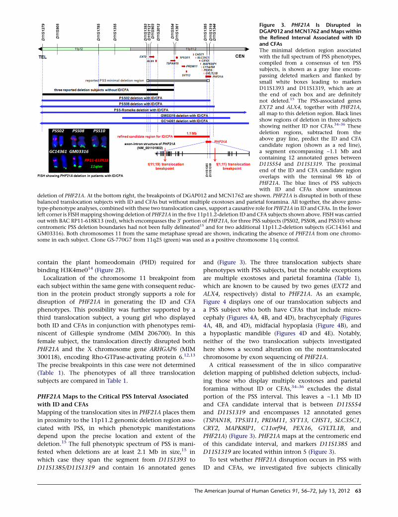
Figure 3. PHF21A Is Disrupted inDGAP012 andMCN1762 andMaps withinthe Refined Interval Associated with IDand CFAsThe minimal deletion region associatedwith the full spectrum of PSS phenotypes,compiled from a consensus of ten PSSsubjects, is shown as a gray line encom-passing deleted markers and flanked bysmall white boxes leading to markersD11S1393 and D11S1319, which are atthe end of each box and are definitelynot deleted.15 The PSS-associated genesEXT2 and ALX4, together with PHF21A,all map to this deletion region. Black linesshow regions of deletion in three subjectsshowing neither ID nor CFAs.34–36 Thesedeletion regions, subtracted from theabove gray line, predict the ID and CFAcandidate region (shown as a red line),a segment encompassing ~1.1 Mb andcontaining 12 annotated genes betweenD11S554 and D11S1319. The proximalend of the ID and CFA candidate regionoverlaps with the terminal 98 kb ofPHF21A. The blue lines of PSS subjectswith ID and CFAs show unanimous
deletion of PHF21A. At the bottom right, the breakpoints of DGAP012 and MCN1762 are shown. PHF21A is disrupted in both of thesebalanced translocation subjects with ID and CFAs but without multiple exostoses and parietal foramina. All together, the above geno-type-phenotype analyses, combined with these two translocation cases, support a causative role for PHF21A in ID and CFAs. In the lowerleft corner is FISHmapping showing deletion of PHF21A in the five 11p11.2-deletion ID and CFA subjects shown above. FISHwas carriedout with BAC RP11-618K13 (red), which encompasses the 30 portion of PHF21A, for three PSS subjects (PSS02, PSS08, and PSS10) whosecentromeric PSS deletion boundaries had not been fully delineated15 and for two additional 11p11.2-deletion subjects (GC14361 andGM03316). Both chromosomes 11 from the same metaphase spread are shown, indicating the absence of PHF21A from one chromo-some in each subject. Clone GS-770G7 from 11q25 (green) was used as a positive chromosome 11q control.
contain the plant homeodomain (PHD) required for
binding H3K4me014 (Figure 2F).
Localization of the chromosome 11 breakpoint from
each subject within the same gene with consequent reduc-
tion in the protein product strongly supports a role for
disruption of PHF21A in generating the ID and CFA
phenotypes. This possibility was further supported by a
third translocation subject, a young girl who displayed
both ID and CFAs in conjunction with phenotypes remi-
niscent of Gillespie syndrome (MIM 206700). In this
female subject, the translocation directly disrupted both
PHF21A and the X chromosome gene ARHGAP6 (MIM
300118), encoding Rho-GTPase-activating protein 6.12,13
The precise breakpoints in this case were not determined
(Table 1). The phenotypes of all three translocation
subjects are compared in Table 1.
PHF21A Maps to the Critical PSS Interval Associated
with ID and CFAs
Mapping of the translocation sites in PHF21A places them
in proximity to the 11p11.2 genomic deletion region asso-
ciated with PSS, in which phenotypic manifestations
depend upon the precise location and extent of the
deletion.15 The full phenotypic spectrum of PSS is mani-
fested when deletions are at least 2.1 Mb in size,15 in
which case they span the segment from D11S1393 to
D11S1385/D11S1319 and contain 16 annotated genes
The
and (Figure 3). The three translocation subjects share
phenotypes with PSS subjects, but the notable exceptions
are multiple exostoses and parietal foramina (Table 1),
which are known to be caused by two genes (EXT2 and
ALX4, respectively) distal to PHF21A. As an example,
Figure 4 displays one of our translocation subjects and
a PSS subject who both have CFAs that include micro-
cephaly (Figures 4A, 4B, and 4D), brachycephaly (Figures
4A, 4B, and 4D), midfacial hypoplasia (Figure 4B), and
a hypoplastic mandible (Figures 4D and 4E). Notably,
neither of the two translocation subjects investigated
here shows a second alteration on the nontranslocated
chromosome by exon sequencing of PHF21A.
A critical reassessment of the in silico comparative
deletion mapping of published deletion subjects, includ-
ing those who display multiple exostoses and parietal
foramina without ID or CFAs,34–36 excludes the distal
portion of the PSS interval. This leaves a ~1.1 Mb ID
and CFA candidate interval that is between D11S554
and D11S1319 and encompasses 12 annotated genes
(TSPAN18, TP53I11, PRDM11, SYT13, CHST1, SLC35C1,
CRY2, MAPK8IP1, C11orf94, PEX16, GYLTL1B, and
PHF21A) (Figure 3). PHF21A maps at the centromeric end
of this candidate interval, and markers D11S1385 and
D11S1319 are located within intron 5 (Figure 3).
To test whether PHF21A disruption occurs in PSS with
ID and CFAs, we investigated five subjects clinically
American Journal of Human Genetics 91, 56–72, July 13, 2012 63
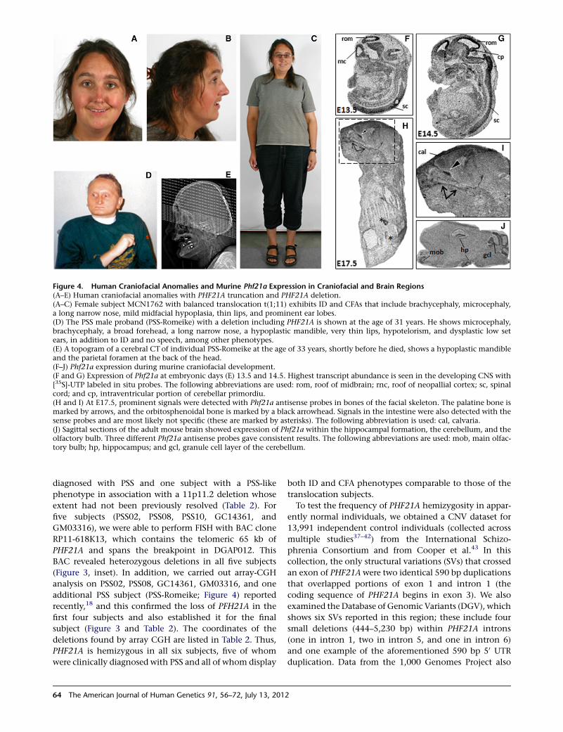
Figure 4. Human Craniofacial Anomalies and Murine Phf21a Expression in Craniofacial and Brain Regions(A–E) Human craniofacial anomalies with PHF21A truncation and PHF21A deletion.(A–C) Female subject MCN1762 with balanced translocation t(1;11) exhibits ID and CFAs that include brachycephaly, microcephaly,a long narrow nose, mild midfacial hypoplasia, thin lips, and prominent ear lobes.(D) The PSS male proband (PSS-Romeike) with a deletion including PHF21A is shown at the age of 31 years. He shows microcephaly,brachycephaly, a broad forehead, a long narrow nose, a hypoplastic mandible, very thin lips, hypotelorism, and dysplastic low setears, in addition to ID and no speech, among other phenotypes.(E) A topogram of a cerebral CT of individual PSS-Romeike at the age of 33 years, shortly before he died, shows a hypoplastic mandibleand the parietal foramen at the back of the head.(F–J) Phf21a expression during murine craniofacial development.(F and G) Expression of Phf21a at embryonic days (E) 13.5 and 14.5. Highest transcript abundance is seen in the developing CNS with[35S]-UTP labeled in situ probes. The following abbreviations are used: rom, roof of midbrain; rnc, roof of neopallial cortex; sc, spinalcord; and cp, intraventricular portion of cerebellar primordiu.(H and I) At E17.5, prominent signals were detected with Phf21a antisense probes in bones of the facial skeleton. The palatine bone ismarked by arrows, and the orbitosphenoidal bone is marked by a black arrowhead. Signals in the intestine were also detected with thesense probes and are most likely not specific (these are marked by asterisks). The following abbreviation is used: cal, calvaria.(J) Sagittal sections of the adult mouse brain showed expression of Phf21a within the hippocampal formation, the cerebellum, and theolfactory bulb. Three different Phf21a antisense probes gave consistent results. The following abbreviations are used: mob, main olfac-tory bulb; hp, hippocampus; and gcl, granule cell layer of the cerebellum.
diagnosed with PSS and one subject with a PSS-like
phenotype in association with a 11p11.2 deletion whose
extent had not been previously resolved (Table 2). For
five subjects (PSS02, PSS08, PSS10, GC14361, and
GM03316), we were able to perform FISH with BAC clone
RP11-618K13, which contains the telomeric 65 kb of
PHF21A and spans the breakpoint in DGAP012. This
BAC revealed heterozygous deletions in all five subjects
(Figure 3, inset). In addition, we carried out array-CGH
analysis on PSS02, PSS08, GC14361, GM03316, and one
additional PSS subject (PSS-Romeike; Figure 4) reported
recently,18 and this confirmed the loss of PFH21A in the
first four subjects and also established it for the final
subject (Figure 3 and Table 2). The coordinates of the
deletions found by array CGH are listed in Table 2. Thus,
PHF21A is hemizygous in all six subjects, five of whom
were clinically diagnosed with PSS and all of whom display
64 The American Journal of Human Genetics 91, 56–72, July 13, 2012
both ID and CFA phenotypes comparable to those of the
translocation subjects.
To test the frequency of PHF21A hemizygosity in appar-
ently normal individuals, we obtained a CNV dataset for
13,991 independent control individuals (collected across
multiple studies37–42) from the International Schizo-
phrenia Consortium and from Cooper et al.43 In this
collection, the only structural variations (SVs) that crossed
an exon of PHF21Awere two identical 590 bp duplications
that overlapped portions of exon 1 and intron 1 (the
coding sequence of PHF21A begins in exon 3). We also
examined the Database of Genomic Variants (DGV), which
shows six SVs reported in this region; these include four
small deletions (444–5,230 bp) within PHF21A introns
(one in intron 1, two in intron 5, and one in intron 6)
and one example of the aforementioned 590 bp 50 UTR
duplication. Data from the 1,000 Genomes Project also
shows multiple CNVs at each of the exon 1 and intron 5
locations, as well as a single 545 bp CNV in the 30 UTR.44
In addition to these presumably benign SVs, there is a
single report in the DGV of a large 75 kb deletion encom-
passing six genes, including a portion of PHF21A. We are
not in a position to validate that deletion or to confirm
the absence of phenotype; however, there is precedent
for even well-established disease-associated CNVs being
nonpenetrant in some individuals.
Murine Phf21a Is Expressed in the CNS and Cranial
Bones
To determine whether the pattern of expression of PHF21A
supports a role in craniofacial and neuronal development,
we performed in situ hybridization experiments for the or-
thologous mouse gene. Predominant expression of Phf21a
is detected in the developing CNS at early stages. At
mouse embryonic days (E) 13.5 and 14.5, the roof of the
neopallidal cortex and thus the developing cerebral cortex,
as well as the roof of the midbrain and the spinal cord,
showed the highest expression levels of Phf21a. The intra-
ventricular portion of the cerebellar primordium also ex-
pressed Phf21a at E14.5 (Figure 4G). At early embryonic
stages, facial bone and viscerocranial ossification initiates
and, with ongoing ossification, high levels of Phf21a
transcripts were found at E17.5 in the palatine bone
(Figure 4H and magnification in Figure 4I, marked by
arrows) and the orbitosphenoidal bone (Figure 4H, I black
arrowhead), as well as in the calvaria. Signals observed
in bone with Phf21a antisense probes were essentially
restricted to cranial bones, suggesting a particular func-
tion for Phf21a in craniofacial development. In the adult
mouse brain, the most abundant expression of Phf21a
was observed in the neuronal layers of the hippocampus,
the granule cell layer of the cerebellum, and the main
olfactory bulb (Figure 4J). All together, these findings indi-
cate that expression of Phf21a is consistent with an impor-
tant role in the CNS and craniofacial skeletal development
and in adult neuronal function. Interestingly, a single
report of a mouse knockout for Phf21a described no gross
morphological abnormality, although the potential for
CFAs was not specifically evaluated. Neonatal mice died
as a result of an inability to suckle properly; this inability
was interpreted as a likely defect in neuronal control of
milk-sucking behavior.45
Suppression of Zebrafish phf21a Expression Causes
CFAs and Neuronal Apoptosis
To directly test the developmental importance of PHF21A,
we isolated the zebrafish phf21a ortholog, examined
its expression pattern, and performed gain- and loss-of-
function experiments in this model organism. The zebra-
fish phf21a is highly related to human and mouse
PHF21A proteins: It exhibits an AT-hook domain, a PHD,
and two coiled-coil domains (data not shown). Using
RT-PCR, we confirmed that zebrafish phf21a showed
maternal and zygotic transcripts during embryonic devel-
The
opment (Figure S4A). Whole-mount in situ hybridization
analyses revealed phf21a transcripts ubiquitously distrib-
uted throughout the embryo during the stages of cleavage,
blastula, gastrula, and early segmentation. Expression in
the head region was increased from later somitogenesis
and continued to 24 and 48 hpf (Figures S4B–S4J).
To investigate the function of phf21a in zebrafish devel-
opment, we tested the effect of phf21a knockdown by
antisense MO. Injection of the phf21a MO, but not of
a standard control MO or no MO, caused a small-head
phenotype and facial dysmorphism with a pronounced
defect in growth of the lower jaw at 3 days postfertilization
(dpf); these features are reminiscent of the microcephaly
and dysmorphism seen in the translocation subjects
(Figures 5A–5C and 5Q).
We examined the head structure of phf21amorphants in
more detail by using Alcian blue to visualize the extent of
cartilage development in larval fishes. At 5 dpf, Meckel’s
and palatoquadrate cartilages were severely distorted in
their size and shape in phf21a morphants (Figures 5D–
5G). Such defects were already manifest during early
stages given that we also observed defects in dlx2a-positive
pharyngeal-arch-cartilage progenitor cells in phf21a mor-
phants at 2 dpf (Figures 5H and 5I). Defects in cranial-
cartilage formation were also observed for the zebrafish
headless mutation, which is known to be involved in
the signaling pathway of vertebrate head formation and
patterning.31 To investigate further whether these defects
also involve other arch-associated structures, we injected
the phf21a MO into flk1:GFP transgenic zebrafish, in
which the vascular endothelial cells were visualized by
green fluorescent protein (GFP) fluorescence.46 At 4 dpf,
the aortic arches of the phf21a-MO-injected flk1:GFP
transgenic embryos were found to be hypoplastic: They
showed poor development of capillary networks associated
with pharyngeal arches (Figures 5J and 5K). In vertebrates,
Meckel’s and palatoquadrate cartilages form the embry-
onic jaw apparatus.47,48 Thus, it would be interesting to
examine whether the Phf21a/Bhc80-deficient mice, which
display a failure to suckle,45 might also have a defect in
jaw structure.
We also examined the effects of gain or loss of phf21a
function on neuronal development but did not see any
prominent change (Figure S5). However, injection of the
phf21a MO, but not a standard control MO or PHF21a
mRNA, caused apoptosis in the developing brain region
at 36 hpf (Figures 5L–5N). Importantly, this apoptosis
and the small-head phenotype can be rescued by introduc-
tion of wild-type human PHF21A mRNA (Figure 5O), sug-
gesting that the ID phenotype in humans might be due
to a requirement for PHF21A in the function of neuronal
cell survival in the developing brain.
Overall, the phf21a MO caused craniofacial, morpholog-
ical, and growth defects in the developing zebrafish
embryo, as depicted by the notable ventral curvature of
the body and small-head phenotype (Figure 5Q) relative
to that of the control (Figure 5P). The body axis of the
American Journal of Human Genetics 91, 56–72, July 13, 2012 65
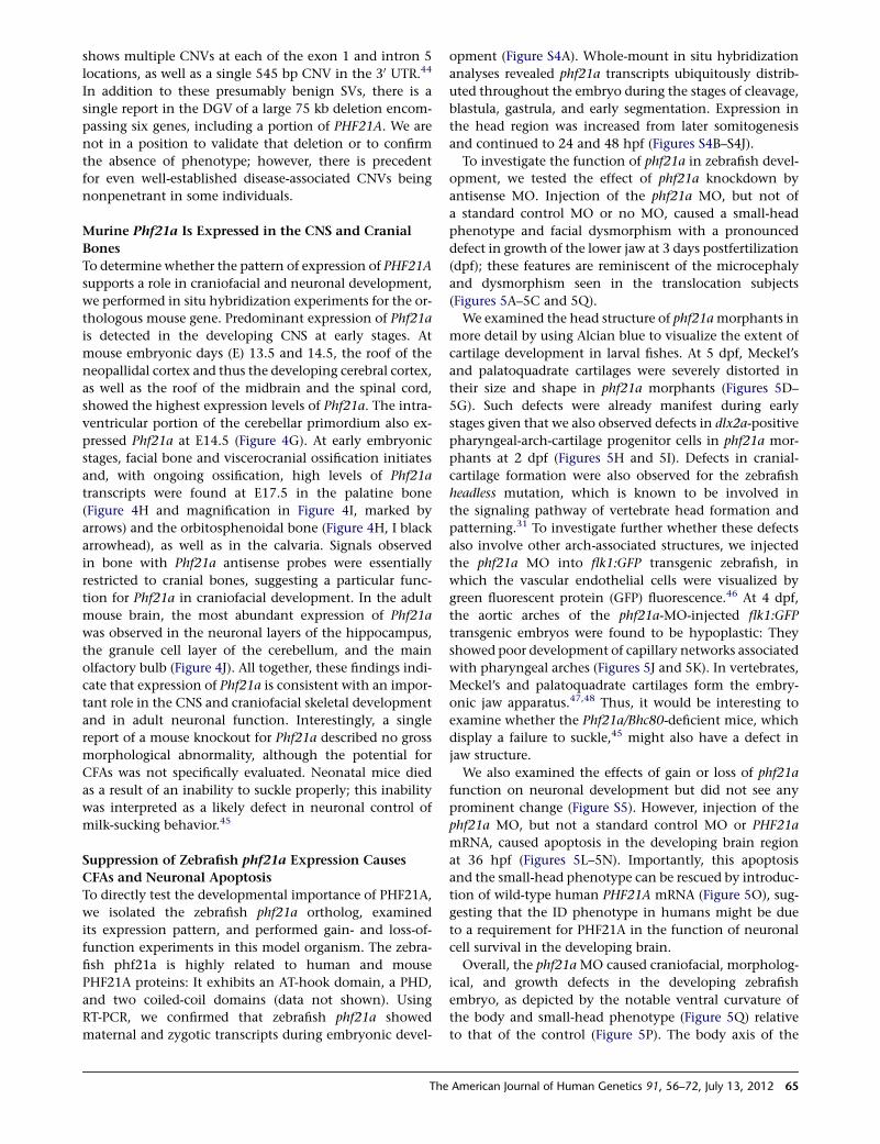
Figure 5. PHF21A Regulates Zebrafish Neuronal Cell Survival and Craniofacial Development(A–C) A noninjected control embryo (A), an embryo injected with a control MO (B), and an embryo injected with a phf21a MO (C).Knockdown of phf21a causes a reduction in head size, a change of head and face shape (arrowhead), and a major reduction of thejaw (arrow) at 3 dpf; these features are reminiscent of themicrocephaly and craniofacial dysmorphism seen in the translocation subjects.The scale bar in (A) represents 300 mm. The following abbreviations are used: ov, otic vesicle; and h, heart.(D–G) Cartilage staining of embryos injected with the control MO (D and F) or phf21aMO (E and G). Compared with that of the controlembryo, Meckel’s and palatoquadrate cartilage in the phf21a-MO-injected embryos are severely distorted in their size and shape. Five-day-old embryos are shown in ventral (D and E) or lateral (F and G) views. The following abbreviations are used: ch, ceratohyal; bh,basihyal cartilage; m, Meckel’s cartilage; pq, palatoquadrate cartilage; and cb 1–5, ceratobranchial cartilage 1–5.(H and I) dlx2a expression in the control (H) and the phf21a morphant (I) at 2 dpf. phf21a morphants fail to expand dlx2a-expressingpharyngeal-arch progenitor cells (arrow). The scale bar in (H) represents 300 mm, and the scale bar in (I) represents 230 mm. The followingabbreviation is used: PA 1–7, pharyngeal arches 1–7.(J and K) Formation and patterning of arch-associated blood vessels in the control (J) and the phf21amorphant (K). A lateral view of theflk1:GFP transgenic line at 4 dpf is shown. In phf21a-MO-injected embryos, aortic arches (arrowheads) form normally but fail to swing toa more anterior position (compare the position of arrows in J and K). Also, the vessels associated with gill filaments develop poorly inmorphants.(L–O) Effects on neuronal cell survival. Compared with the control MO (L) and PHF21A-mRNA-injected embryos (M), phf21a-MO-injected embryos (N) show a dramatically increased number of acridine-orange-positive apoptotic cells. The apoptotic phenotype ofthe phf21a morphant is rescued by coinjection of PHF21A mRNA (O).
66 The American Journal of Human Genetics 91, 56–72, July 13, 2012
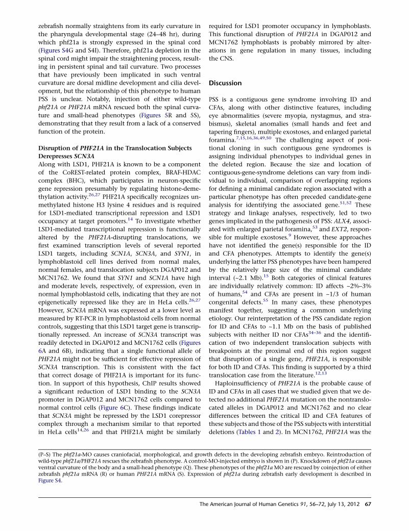
zebrafish normally straightens from its early curvature in
the pharyngula developmental stage (24–48 hr), during
which phf21a is strongly expressed in the spinal cord
(Figures S4G and S4I). Therefore, phf21a depletion in the
spinal cord might impair the straightening process, result-
ing in persistent spinal and tail curvature. Two processes
that have previously been implicated in such ventral
curvature are dorsal midline development and cilia devel-
opment, but the relationship of this phenotype to human
PSS is unclear. Notably, injection of either wild-type
phf21A or PHF21A mRNA rescued both the spinal curva-
ture and small-head phenotypes (Figures 5R and 5S),
demonstrating that they result from a lack of a conserved
function of the protein.
Disruption of PHF21A in the Translocation Subjects
Derepresses SCN3A
Along with LSD1, PHF21A is known to be a component
of the CoREST-related protein complex, BRAF-HDAC
complex (BHC), which participates in neuron-specific
gene repression presumably by regulating histone-deme-
thylation activity.26,27 PHF21A specifically recognizes un-
methylated histone H3 lysine 4 residues and is required
for LSD1-mediated transcriptional repression and LSD1
occupancy at target promoters.14 To investigate whether
LSD1-mediated transcriptional repression is functionally
altered by the PHF21A-disrupting translocations, we
first examined transcription levels of several reported
LSD1 targets, including SCN1A, SCN3A, and SYN1, in
lymphoblastoid cell lines derived from normal males,
normal females, and translocation subjects DGAP012 and
MCN1762. We found that SYN1 and SCN1A have high
and moderate levels, respectively, of expression, even in
normal lymphoblastoid cells, indicating that they are not
epigenetically repressed like they are in HeLa cells.26,27
However, SCN3A mRNA was expressed at a lower level as
measured by RT-PCR in lymphoblastoid cells from normal
controls, suggesting that this LSD1 target gene is transcrip-
tionally repressed. An increase of SCN3A transcript was
readily detected in DGAP012 and MCN1762 cells (Figures
6A and 6B), indicating that a single functional allele of
PHF21A might not be sufficient for effective repression of
SCN3A transcription. This is consistent with the fact
that correct dosage of PHF21A is important for its func-
tion. In support of this hypothesis, ChIP results showed
a significant reduction of LSD1 binding to the SCN3A
promoter in DGAP012 and MCN1762 cells compared to
normal control cells (Figure 6C). These findings indicate
that SCN3A might be repressed by the LSD1 corepressor
complex through a mechanism similar to that reported
in HeLa cells14,26 and that PHF21A might be similarly
(P–S) The phf21a-MO causes craniofacial, morphological, and growtwild-type phf21a/PHF21A rescues the zebrafish phenotype. A control-ventral curvature of the body and a small-head phenotype (Q). Thesezebrafish phf21a mRNA (R) or human PHF21A mRNA (S). ExpressiFigure S4.
The
required for LSD1 promoter occupancy in lymphoblasts.
This functional disruption of PHF21A in DGAP012 and
MCN1762 lymphoblasts is probably mirrored by alter-
ations in gene regulation in many tissues, including
the CNS.
Discussion
PSS is a contiguous gene syndrome involving ID and
CFAs, along with other distinctive features, including
eye abnormalities (severe myopia, nystagmus, and stra-
bismus), skeletal anomalies (small hands and feet and
tapering fingers), multiple exostoses, and enlarged parietal
foramina.7,15,16,36,49,50 The challenging aspect of posi-
tional cloning in such contiguous gene syndromes is
assigning individual phenotypes to individual genes in
the deleted region. Because the size and location of
contiguous-gene-syndrome deletions can vary from indi-
vidual to individual, comparison of overlapping regions
for defining a minimal candidate region associated with a
particular phenotype has often preceded candidate-gene
analysis for identifying the associated gene.51,52 These
strategy and linkage analyses, respectively, led to two
genes implicated in the pathogenesis of PSS: ALX4, associ-
ated with enlarged parietal foramina,53 and EXT2, respon-
sible for multiple exostoses.9 However, these approaches
have not identified the gene(s) responsible for the ID
and CFA phenotypes. Attempts to identify the gene(s)
underlying the latter PSS phenotypes have been hampered
by the relatively large size of the minimal candidate
interval (~2.1 Mb).15 Both categories of clinical features
are individually relatively common: ID affects ~2%–3%
of humans,54 and CFAs are present in ~1/3 of human
congenital defects.55 In many cases, these phenotypes
manifest together, suggesting a common underlying
etiology. Our reinterpretation of the PSS candidate region
for ID and CFAs to ~1.1 Mb on the basis of published
subjects with neither ID nor CFAs34–36 and the identifi-
cation of two independent translocation subjects with
breakpoints at the proximal end of this region suggest
that disruption of a single gene, PHF21A, is responsible
for both ID and CFAs. This finding is supported by a third
translocation case from the literature.12,13
Haploinsufficiency of PHF21A is the probable cause of
ID and CFAs in all cases that we studied given that we de-
tected no additional PHF21A mutation on the nontranslo-
cated alleles in DGAP012 and MCN1762 and no clear
differences between the critical ID and CFA features of
these subjects and those of the PSS subjects with interstitial
deletions (Tables 1 and 2). In MCN1762, PHF21A was the
h defects in the developing zebrafish embryo. Reintroduction ofMO-injected embryo is shown in (P). Knockdown of phf21a causesphenotypes of the phf21aMO are rescued by coinjection of eitheron of phf21a during zebrafish early development is described in
American Journal of Human Genetics 91, 56–72, July 13, 2012 67
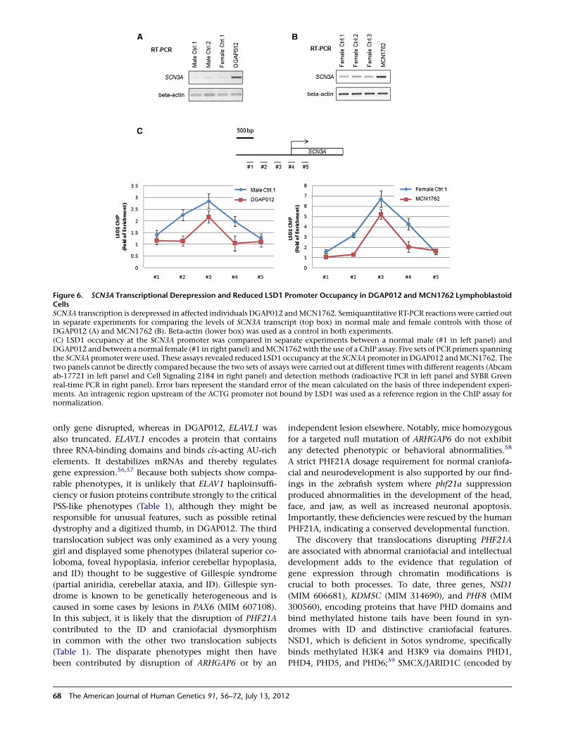
Figure 6. SCN3A Transcriptional Derepression and Reduced LSD1 Promoter Occupancy in DGAP012 and MCN1762 LymphoblastoidCellsSCN3A transcription is derepressed in affected individuals DGAP012 andMCN1762. Semiquantitative RT-PCR reactions were carried outin separate experiments for comparing the levels of SCN3A transcript (top box) in normal male and female controls with those ofDGAP012 (A) and MCN1762 (B). Beta-actin (lower box) was used as a control in both experiments.(C) LSD1 occupancy at the SCN3A promoter was compared in separate experiments between a normal male (#1 in left panel) andDGAP012 and between a normal female (#1 in right panel) andMCN1762with the use of a ChIP assay. Five sets of PCR primers spanningthe SCN3A promoter were used. These assays revealed reduced LSD1 occupancy at the SCN3A promoter in DGAP012 andMCN1762. Thetwo panels cannot be directly compared because the two sets of assays were carried out at different times with different reagents (Abcamab-17721 in left panel and Cell Signaling 2184 in right panel) and detection methods (radioactive PCR in left panel and SYBR Greenreal-time PCR in right panel). Error bars represent the standard error of the mean calculated on the basis of three independent experi-ments. An intragenic region upstream of the ACTG promoter not bound by LSD1 was used as a reference region in the ChIP assay fornormalization.
only gene disrupted, whereas in DGAP012, ELAVL1 was
also truncated. ELAVL1 encodes a protein that contains
three RNA-binding domains and binds cis-acting AU-rich
elements. It destabilizes mRNAs and thereby regulates
gene expression.56,57 Because both subjects show compa-
rable phenotypes, it is unlikely that ELAV1 haploinsuffi-
ciency or fusion proteins contribute strongly to the critical
PSS-like phenotypes (Table 1), although they might be
responsible for unusual features, such as possible retinal
dystrophy and a digitized thumb, in DGAP012. The third
translocation subject was only examined as a very young
girl and displayed some phenotypes (bilateral superior co-
loboma, foveal hypoplasia, inferior cerebellar hypoplasia,
and ID) thought to be suggestive of Gillespie syndrome
(partial aniridia, cerebellar ataxia, and ID). Gillespie syn-
drome is known to be genetically heterogeneous and is
caused in some cases by lesions in PAX6 (MIM 607108).
In this subject, it is likely that the disruption of PHF21A
contributed to the ID and craniofacial dysmorphism
in common with the other two translocation subjects
(Table 1). The disparate phenotypes might then have
been contributed by disruption of ARHGAP6 or by an
68 The American Journal of Human Genetics 91, 56–72, July 13, 2012
independent lesion elsewhere. Notably, mice homozygous
for a targeted null mutation of ARHGAP6 do not exhibit
any detected phenotypic or behavioral abnormalities.58
A strict PHF21A dosage requirement for normal craniofa-
cial and neurodevelopment is also supported by our find-
ings in the zebrafish system where phf21a suppression
produced abnormalities in the development of the head,
face, and jaw, as well as increased neuronal apoptosis.
Importantly, these deficiencies were rescued by the human
PHF21A, indicating a conserved developmental function.
The discovery that translocations disrupting PHF21A
are associated with abnormal craniofacial and intellectual
development adds to the evidence that regulation of
gene expression through chromatin modifications is
crucial to both processes. To date, three genes, NSD1
(MIM 606681), KDM5C (MIM 314690), and PHF8 (MIM
300560), encoding proteins that have PHD domains and
bind methylated histone tails have been found in syn-
dromes with ID and distinctive craniofacial features.
NSD1, which is deficient in Sotos syndrome, specifically
binds methylated H3K4 and H3K9 via domains PHD1,
PHD4, PHD5, and PHD6;59 SMCX/JARID1C (encoded by
KDM5C) of nonsyndromic XLID binds histone H3K9me3
via its PHD1 domain;60 and PHF8 of Siderius-Hamel
syndrome binds histone H3K4me3 via its PHD.61 At least
the latter two act as demethylases by targeting H3K4me2
and H3K4me3 in the case of SMCX/JARID1C60 and
H3K9me1/2 and H4K20me1 in the case of PHF8.61,62
Unlike the above proteins, PHF21A is neither a methyl-
transferase nor a demethylase but instead specifically binds
histone H3K4 when it is not methylated.14 This suggests
that both recognition of the unmodified state of histone
tails and binding of proteins to methylated histone tails
are critical for maintaining the appropriate balance and
control of particular chromatin modifications for the
support of normal intellectual and craniofacial develop-
ment. Although the PSS-associated ID and CFAs appear to
be due to haploinsufficiency of PHF21A, it has not been
possible to ascertain and screen a large series of nontranslo-
cation subjects with comparable phenotypes for PHF21A
mutations. Therefore, we cannot state with certainty that
missense, nonsense, splicing, or other mutations in
PHF21A would lead to the same ID and CFA phenotypes.
Indeed, it is conceivable that other types of genetic lesions
in PHF21A could actually be associated with other develop-
mental phenotypes; we were able to identify 200 individ-
uals with ID and/or CFAs but without the full constellation
of phenotypes exhibited by our translocation subjects, and
we performed a mutation screen of PHF21A. We did not
detect any truncating or missense mutations that have
implicated particular PHD domains in NSD1 in binding to
their methylated targets and that could thus aid in struc-
ture-function experiments. This is not surprising given
the frequency of these two major phenotypes and the
extent of genetic heterogeneity underlying each of them,
but more extensive mutation analysis of subjects with
various manifestations of ID, CFAs, and additional pheno-
types seen in our translocation subjects might prove valu-
able to understanding the functional domains of PHF21A.
PHF21A (BHC80) is known to participate in the six-
subunit BHC, which also comprises BRAF35 (MIM
605535), HDAC1 (MIM 601241), HDAC2 (MIM 605164),
CoREST (MIM 607675), and LSD1 (BHC110); the latter
is a histone demethylase that targets H3K4me2.26 This
complex interacts with the promoters of genes, such as
synapsin and sodium-channel genes, tomediate repression
of these neuron-specific genes through the cis-regulatory
element known as repressor element 1 or neural restric-
tive silencer (RE1/NRS).63 Specific binding of PHF21A to
H3K4me0 is required for optimal LSD1 promoter occu-
pancy in vivo and for LSD1-mediated gene repression.14
Our data showing derepression of the neural gene SCN3A
in lymphoblasts from the translocation subjects as a
consequence of reduced levels of PHF21A are consistent
with this role for the protein. Repression of neuronal-
specific genes is of fundamental importance in the devel-
opment of both neuronal and nonneuronal tissues,63 so
the failure of this particular function in the translocation
subjects might have contributed to their ID and CFAs.
The
Another interesting XLID candidate, ZMYM3 (MIM
300061), encodes a zinc finger protein that is predomi-
nantly expressed in the brain and that is a component of
transcriptional corepressor complexes that also contain
LSD1 (BHC110) and HDAC2.64,65 The 50 UTR of ZMYM3
is disrupted by a presumably balanced t(X;13) karyotype
in a female with ID and preferential inactivation of the
normal X chromosome.66 In addition, Kleefstra syndrome,
characterized by ID and CFAs comparable to PSS, has been
associated with disruption of EHMT1, encoding euchro-
matin histone methyltransferase 1, which acts as a methyl
transferase to modify H3K9 and has been reported as a
component of the E2F6 transcription repressor complex
and of a CtBP repressor complex that also contains
LSD1.2,67–69 The parallels between PHF21A, ZMYM3, and
EHMT1 suggest that other X-linked and autosomal loci
underlying ID and/or CFAs might encode proteins that
participate in complexes involving LSD1 or potentially
other demethylases or methyltransferases. Our finding
that decreased dosage of PHF21A, a histone-binding
protein that interacts with and is required for the
histone-demethylase activity of LSD1, leads to both ID
and CFAs provides the proof of principle for investigation
of other regulators of histone modification as genetic
factors in ID and/or CFAs. Indeed, the recent finding of
haploinsufficiency of ARID1B (MIM 614556), encoding
an E3-ubiquitin-ligase component that functions with
the chromatin-remodeling switch/sucrose nonferment-
able complex,70–74 suggests that genes involved with
other aspects of chromatin modification might also
contribute to ID and CFAs and that ultimately, human
mutations affecting both regulatory and enzymatic com-
ponents of histone-modification complexes might repre-
sent important tools for delineating the chromatin-regula-
tion features that are critical for normal craniofacial and
neurological development and cognitive function.
Supplemental Data
Supplemental Data include five figures and seven tables and can be
found with this article online at http://www.cell.com/AJHG.
Acknowledgments
We are grateful to DGAP012 and MCN1762, as well as their family
members, for their cooperation and participation in this study. We
are also indebted to Amy Bosco, Heather L. Ferguson, and Chantal
Kelly for obtaining informed consent and clinical information;
to Joanne Sutherland, genetic counselor at the Hospital for Sick
Children, Toronto, for her assistance in obtaining samples; to Shi-
geki Iwase and Tadashi Baba for the PHF21A antibody; to Mary
Anne Anderson and Tammy Gillis in the Center for Human
Genetic Research Tissue Culture Facility and Genomics Resource
for technical assistance; to Ian Krantz, Stephanie Seminara, and
Simeon A. Boyadjiev for providing samples of affected individuals;
and to Ines Muller and Corinna Menzel for technical assistance.
This work was supported by a grant from the Next-Generation
BioGreen 21 Program (PJ00812701 to C.H.K.), Republic of Korea,
American Journal of Human Genetics 91, 56–72, July 13, 2012 69
a grant from the Deutsche Forschungsgemeinschaft (KU 1240/5-1
to K.K.), the Danish National Research Foundation (N.T.), the
Lundbeck Foundation (N.T. and A.S.), National Institutes of
Health grants NCI118487 (to Y.S.) and RO1 GM071004 (to Y.S.),
and United States Public Health Service grants GM061354 (Devel-
opmental Genome Anatomy Project to C.C.M. and J.F.G.) and
HD065286 (to J.F.G.). Part of this work was financed by the Euro-
pean Union’s Seventh Framework Program under grant agreement
number 241995, project GENCODYS, and the German Federal
Ministry of Education and Research through the German Mental
Retardation Network (grant 01GS08161 to H.H.R.).
Received: December 27, 2011
Revised: March 18, 2012
Accepted: May 10, 2012
Published online: July 5, 2012
Web Resources
The URLs for data presented herein are as follows:
Agilent Technologies, https://earray.chem.agilent.com
Developmental Genome Anatomy Project, http://dgap.
harvard.edu
Mendelian Cytogenetics Network Online Database, http://www.
mcndb.org/index.jsp
NCBI and GenBank, http://www.ncbi.nlm.nih.gov
NIGMS Human Genetic Cell Repository, http://ccr.coriell.org/
nigms/
Online Mendelian Inheritance in Man (OMIM), http://www.
omim.org
UCSC Genome Browser, http://genome.ucsc.edu/
References
1. Koolen, D.A., Vissers, L.E., Pfundt, R., de Leeuw, N., Knight,
S.J., Regan, R., Kooy, R.F., Reyniers, E., Romano, C., Fichera,
M., et al. (2006). A new chromosome 17q21.31 microdeletion
syndrome associated with a common inversion polymor-
phism. Nat. Genet. 38, 999–1001.
2. Kleefstra, T., Brunner, H.G., Amiel, J., Oudakker, A.R., Nillesen,
W.M., Magee, A., Genevieve, D., Cormier-Daire, V., van Esch,
H., Fryns, J.P., et al. (2006). Loss-of-function mutations in
euchromatin histone methyl transferase 1 (EHMT1) cause
the 9q34 subtelomeric deletion syndrome. Am. J. Hum. Genet.
79, 370–377.
3. Kalscheuer, V.M., Tao, J., Donnelly, A., Hollway, G., Schwinger,
E., Kubart, S., Menzel, C., Hoeltzenbein, M., Tommerup, N.,
Eyre, H., et al. (2003). Disruption of the serine/threonine
kinase 9 gene causes severe X-linked infantile spasms and
mental retardation. Am. J. Hum. Genet. 72, 1401–1411.
4. Kalscheuer, V.M., FitzPatrick, D., Tommerup, N., Bugge, M.,
Niebuhr, E., Neumann, L.M., Tzschach, A., Shoichet, S.A.,
Menzel, C., Erdogan, F., et al. (2007). Mutations in autism
susceptibility candidate 2 (AUTS2) in patients with mental
retardation. Hum. Genet. 121, 501–509.
5. Kim, H.G., Ahn, J.W., Kurth, I., Ullmann, R., Kim, H.T., Kul-
harya, A., Ha, K.S., Itokawa, Y., Meliciani, I., Wenzel, W.,
et al. (2010). WDR11, a WD protein that interacts with tran-
scription factor EMX1, is mutated in idiopathic hypogonado-
tropic hypogonadism and Kallmann syndrome. Am. J. Hum.
Genet. 87, 465–479.
70 The American Journal of Human Genetics 91, 56–72, July 13, 2012
6. Kim, H.G., Kishikawa, S., Higgins, A.W., Seong, I.S., Donovan,
D.J., Shen, Y., Lally, E., Weiss, L.A., Najm, J., Kutsche, K., et al.
(2008). Disruption of neurexin 1 associated with autism spec-
trum disorder. Am. J. Hum. Genet. 82, 199–207.
7. Potocki, L., and Shaffer, L.G. (1996). Interstitial deletion of
11(p11.2p12): A newly described contiguous gene deletion
syndrome involving the gene for hereditary multiple exos-
toses (EXT2). Am. J. Med. Genet. 62, 319–325.
8. Shaffer, L.G., Hecht, J.T., Ledbetter, D.H., and Greenberg, F.
(1993). Familial interstitial deletion 11(p11.12p12) associated
with parietal foramina, brachymicrocephaly, and mental
retardation. Am. J. Med. Genet. 45, 581–583.
9. Stickens, D., Clines, G., Burbee, D., Ramos, P., Thomas, S.,
Hogue, D., Hecht, J.T., Lovett, M., and Evans, G.A. (1996).
The EXT2 multiple exostoses gene defines a family of putative
tumour suppressor genes. Nat. Genet. 14, 25–32.
10. Mavrogiannis, L.A., Antonopoulou, I., Baxova, A., Kutılek, S.,
Kim, C.A., Sugayama, S.M., Salamanca, A., Wall, S.A., Morriss-
Kay, G.M., and Wilkie, A.O. (2001). Haploinsufficiency of the
human homeobox gene ALX4 causes skull ossification
defects. Nat. Genet. 27, 17–18.
11. Wu, Y.Q., Badano, J.L., McCaskill, C., Vogel, H., Potocki, L.,
and Shaffer, L.G. (2000). Haploinsufficiency of ALX4 as
a potential cause of parietal foramina in the 11p11.2 contig-
uous gene-deletion syndrome. Am. J. Hum. Genet. 67,
1327–1332.
12. Dollfus, H., Joanny-Flinois, O., Doco-Fenzy, M., Veyre, L.,
Joanny-Flinois, L., Khoury, M., Jonveaux, P., Abitbol, M.,
and Dufier, J.L. (1998). Gillespie syndrome phenotype with
a t(X;11)(p22.32;p12) de novo translocation. Am. J. Ophthal-
mol. 125, 397–399.
13. Fantes, J.A., Boland, E., Ramsay, J., Donnai, D., Splitt, M.,
Goodship, J.A., Stewart, H., Whiteford, M., Gautier, P., Hare-
wood, L., et al. (2008). FISH mapping of de novo apparently
balanced chromosome rearrangements identifies characteris-
tics associated with phenotypic abnormality. Am. J. Hum.
Genet. 82, 916–926.
14. Lan, F., Collins, R.E., De Cegli, R., Alpatov, R., Horton, J.R., Shi,
X., Gozani, O., Cheng, X., and Shi, Y. (2007). Recognition of
unmethylated histone H3 lysine 4 links BHC80 to LSD1-medi-
ated gene repression. Nature 448, 718–722.
15. Wakui, K., Gregato, G., Ballif, B.C., Glotzbach, C.D., Bailey,
K.A., Kuo, P.L., Sue, W.C., Sheffield, L.J., Irons, M., Gomez,
E.G., et al. (2005). Construction of a natural panel of
11p11.2 deletions and further delineation of the critical
region involved in Potocki-Shaffer syndrome. Eur. J. Hum.
Genet. 13, 528–540.
16. Wuyts, W., Waeber, G., Meinecke, P., Schuler, H., Goecke,
T.O., Van Hul, W., and Bartsch, O. (2004). Proximal 11p
deletion syndrome (P11pDS): Additional evaluation of the
clinical and molecular aspects. Eur. J. Hum. Genet. 12,
400–406.
17. Anderson, M.A., and Gusella, J.F. (1984). Use of cyclosporin A
in establishing Epstein-Barr virus-transformed human lym-
phoblastoid cell lines. In Vitro 20, 856–858.
18. Romeike, B.F., and Wuyts, W. (2007). Proximal chromosome
11p contiguous gene deletion syndrome phenotype: Case
report and review of the literature. Clin. Neuropathol. 26,
1–11.
19. Miller, D.T., Shen, Y., and Wu, B.L. (2008). Oligonucleotide
microarrays for clinical diagnosis of copy number variation.
Curr. Protoc. Hum. Genet. 58, 8.12.1–8.12.17.
20. Obholz, K.L., Akopyan, A., Waymire, K.G., and MacGregor,
G.R. (2006). FNDC3A is required for adhesion between sper-
matids and Sertoli cells. Dev. Biol. 298, 498–513.
21. Arkesteijn, G., Jumelet, E., Hagenbeek, A., Smit, E., Slater, R.,
and Martens, A. (1999). Reverse chromosome painting for
the identification of marker chromosomes and complex trans-
locations in leukemia. Cytometry 35, 117–124.
22. Chen, W., Kalscheuer, V., Tzschach, A., Menzel, C., Ullmann,
R., Schulz, M.H., Erdogan, F., Li, N., Kijas, Z., Arkesteijn, G.,
et al. (2008). Mapping translocation breakpoints by next-
generation sequencing. Genome Res. 18, 1143–1149.
23. Siebert, P.D., Chenchik, A., Kellogg, D.E., Lukyanov, K.A.,
and Lukyanov, S.A. (1995). An improved PCR method for
walking in uncloned genomic DNA. Nucleic Acids Res. 23,
1087–1088.
24. Cheung, V.G., Nowak, N., Jang, W., Kirsch, I.R., Zhao, S.,
Chen, X.N., Furey, T.S., Kim, U.J., Kuo, W.L., Olivier, M.,
et al; BAC Resource Consortium. (2001). Integration of cytoge-
netic landmarks into the draft sequence of the human
genome. Nature 409, 953–958.
25. Bartsch, O., Schmidt, S., Richter, M., Morlot, S., Seemanova,
E., Wiebe, G., and Rasi, S. (2005). DNA sequencing of CREBBP
demonstrates mutations in 56% of patients with Rubinstein-
Taybi syndrome (RSTS) and in another patient with incom-
plete RSTS. Hum. Genet. 117, 485–493.
26. Shi, Y., Lan, F., Matson, C., Mulligan, P., Whetstine, J.R., Cole,
P.A., Casero, R.A., and Shi, Y. (2004). Histone demethylation
mediated by the nuclear amine oxidase homolog LSD1. Cell
119, 941–953.
27. Shi, Y.J., Matson, C., Lan, F., Iwase, S., Baba, T., and Shi, Y.
(2005). Regulation of LSD1 histone demethylase activity by
its associated factors. Mol. Cell 19, 857–864.
28. Iwase, S., Januma, A., Miyamoto, K., Shono, N., Honda, A.,
Yanagisawa, J., and Baba, T. (2004). Characterization of
BHC80 in BRAF-HDAC complex, involved in neuron-specific
gene repression. Biochem. Biophys. Res. Commun. 322,
601–608.
29. Park, H.C., Kim, C.H., Bae, Y.K., Yeo, S.Y., Kim, S.H., Hong,
S.K., Shin, J., Yoo, K.W., Hibi, M., Hirano, T., et al. (2000).
Analysis of upstream elements in the HuC promoter leads to
the establishment of transgenic zebrafish with fluorescent
neurons. Dev. Biol. 227, 279–293.
30. Kim, C.H., Ueshima, E., Muraoka, O., Tanaka, H., Yeo, S.Y.,
Huh, T.L., and Miki, N. (1996). Zebrafish elav/HuC homo-
logue as a very early neuronal marker. Neurosci. Lett. 216,
109–112.
31. Kim, C.H., Oda, T., Itoh, M., Jiang, D., Artinger, K.B., Chandra-
sekharappa, S.C., Driever, W., and Chitnis, A.B. (2000).
Repressor activity of Headless/Tcf3 is essential for vertebrate
head formation. Nature 407, 913–916.
32. Higgins, A.W., Alkuraya, F.S., Bosco, A.F., Brown, K.K., Bruns,
G.A., Donovan, D.J., Eisenman, R., Fan, Y., Farra, C.G., Fergu-
son, H.L., et al. (2008). Characterization of apparently
balanced chromosomal rearrangements from the develop-
mental genome anatomy project. Am. J. Hum. Genet. 82,
712–722.
33. Talkowski, M.E., Ernst, C., Heilbut, A., Chiang, C., Hanscom,
C., Lindgren, A., Kirby, A., Liu, S., Muddukrishna, B., Ohsumi,
T.K., et al. (2011). Next-generation sequencing strategies
enable routine detection of balanced chromosome rearrange-
ments for clinical diagnostics and genetic research. Am. J.
Hum. Genet. 88, 469–481.
The
34. Hall, C.R., Wu, Y., Shaffer, L.G., and Hecht, J.T. (2001).
Familial case of Potocki-Shaffer syndrome associated with
microdeletion of EXT2 and ALX4. Clin. Genet. 60, 356–359.
35. Mavrogiannis, L.A., Taylor, I.B., Davies, S.J., Ramos, F.J., Oli-
vares, J.L., andWilkie, A.O. (2006). Enlarged parietal foramina
caused by mutations in the homeobox genes ALX4 and
MSX2: From genotype to phenotype. Eur. J. Hum. Genet.
14, 151–158.
36. Wuyts, W., Di Gennaro, G., Bianco, F., Wauters, J., Morocutti,
C., Pierelli, F., Bossuyt, P., Van Hul, W., and Casali, C. (1999).
Molecular and clinical examination of an Italian DEFECT11
family. Eur. J. Hum. Genet. 7, 579–584.
37. Ferreira, M.A., O’Donovan, M.C., Meng, Y.A., Jones, I.R., Ru-
derfer, D.M., Jones, L., Fan, J., Kirov, G., Perlis, R.H., Green,
E.K., et al; Wellcome Trust Case Control Consortium. (2008).
Collaborative genome-wide association analysis supports
a role for ANK3 andCACNA1C in bipolar disorder. Nat. Genet.
40, 1056–1058.
38. O’Donovan, M.C., Craddock, N., Norton, N., Williams, H.,
Peirce, T., Moskvina, V., Nikolov, I., Hamshere, M., Carroll,
L., Georgieva, L., et al; Molecular Genetics of Schizophrenia
Collaboration. (2008). Identification of loci associated with
schizophrenia by genome-wide association and follow-up.
Nat. Genet. 40, 1053–1055.
39. Purcell, S.M., Wray, N.R., Stone, J.L., Visscher, P.M., O’Dono-
van, M.C., Sullivan, P.F., and Sklar, P.; International Schizo-
phrenia Consortium. (2009). Common polygenic variation
contributes to risk of schizophrenia and bipolar disorder.
Nature 460, 748–752.
40. Smith, E.N., Bloss, C.S., Badner, J.A., Barrett, T., Belmonte, P.L.,
Berrettini, W., Byerley, W., Coryell, W., Craig, D., Edenberg,
H.J., et al. (2009). Genome-wide association study of bipolar
disorder in European American and African American individ-
uals. Mol. Psychiatry 14, 755–763.
41. Sullivan, P.F., Lin, D., Tzeng, J.Y., van den Oord, E., Perkins,
D., Stroup, T.S., Wagner, M., Lee, S., Wright, F.A., Zou, F.,
et al. (2008). Genomewide association for schizophrenia in
the CATIE study: Results of stage 1. Mol. Psychiatry 13,
570–584.
42. International Schizophrenia Consortium. (2008). Rare chro-
mosomal deletions and duplications increase risk of schizo-
phrenia. Nature 455, 237–241.
43. Cooper, G.M., Coe, B.P., Girirajan, S., Rosenfeld, J.A., Vu, T.H.,
Baker, C.,Williams, C., Stalker, H., Hamid, R., Hannig, V., et al.
(2011). A copy number variation morbidity map of develop-
mental delay. Nat. Genet. 43, 838–846.
44. 1000 Genomes Project Consortium. (2010). A map of human
genome variation from population-scale sequencing. Nature
467, 1061–1073.
45. Iwase, S., Shono, N., Honda, A., Nakanishi, T., Kashiwabara, S.,
Takahashi, S., and Baba, T. (2006). A component of BRAF-
HDAC complex, BHC80, is required for neonatal survival in
mice. FEBS Lett. 580, 3129–3135.
46. Choi, J., Dong, L., Ahn, J., Dao, D., Hammerschmidt, M., and
Chen, J.N. (2007). FoxH1 negatively modulates flk1 gene
expression and vascular formation in zebrafish. Dev. Biol.
304, 735–744.
47. Schilling, T.F., Piotrowski, T., Grandel, H., Brand, M., Heisen-
berg, C.P., Jiang, Y.J., Beuchle, D., Hammerschmidt, M.,
Kane, D.A., Mullins, M.C., et al. (1996). Jaw and branchial
arch mutants in zebrafish I: Branchial arches. Development
123, 329–344.
American Journal of Human Genetics 91, 56–72, July 13, 2012 71
48. Wilkie, A.O., and Morriss-Kay, G.M. (2001). Genetics of
craniofacial development and malformation. Nat. Rev. Genet.
2, 458–468.
49. Bartsch, O., Wuyts, W., Van Hul, W., Hecht, J.T., Meinecke, P.,
Hogue, D., Werner, W., Zabel, B., Hinkel, G.K., Powell, C.M.,
et al. (1996). Delineation of a contiguous gene syndrome
with multiple exostoses, enlarged parietal foramina, craniofa-
cial dysostosis, and mental retardation, caused by deletions
in the short arm of chromosome 11. Am. J. Hum. Genet. 58,
734–742.
50. Yamamoto, T., Akaboshi, S., Ninomiya, H., and Nanba, E.
(2001). DEFECT 11 syndrome associated with agenesis of the
corpus callosum. J. Med. Genet. 38, E5.
51. Dode, C., Levilliers, J., Dupont, J.M., De Paepe, A., Le Du, N.,
Soussi-Yanicostas, N., Coimbra, R.S., Delmaghani, S., Com-
pain-Nouaille, S., Baverel, F., et al. (2003). Loss-of-function
mutations in FGFR1 cause autosomal dominant Kallmann
syndrome. Nat. Genet. 33, 463–465.
52. Vissers, L.E., van Ravenswaaij, C.M., Admiraal, R., Hurst, J.A.,
de Vries, B.B., Janssen, I.M., van der Vliet, W.A., Huys, E.H., de
Jong, P.J., Hamel, B.C., et al. (2004). Mutations in a new
member of the chromodomain gene family cause CHARGE
syndrome. Nat. Genet. 36, 955–957.
53. Wuyts, W., Cleiren, E., Homfray, T., Rasore-Quartino, A., Van-
hoenacker, F., and Van Hul, W. (2000). The ALX4 homeobox
gene is mutated in patients with ossification defects of the
skull (foramina parietalia permagna, OMIM 168500). J. Med.
Genet. 37, 916–920.
54. Gecz, J. (2004). The molecular basis of intellectual disability:
Novel genes with naturally occurring mutations causing
altered gene expression in the brain. Front. Biosci. 9, 1–7.
55. Tassabehji, M., Hammond, P., Karmiloff-Smith, A., Thomp-
son, P., Thorgeirsson, S.S., Durkin, M.E., Popescu, N.C., Hut-
ton, T., Metcalfe, K., Rucka, A., et al. (2005). GTF2IRD1 in
craniofacial development of humans and mice. Science 310,
1184–1187.
56. Ma, W.J., and Furneaux, H. (1997). Localization of the human
HuR gene to chromosome 19p13.2. Hum. Genet. 99, 32–33.
57. Ma, W.J., Cheng, S., Campbell, C., Wright, A., and Furneaux,
H. (1996). Cloning and characterization of HuR, a ubiqui-
tously expressed Elav-like protein. J. Biol. Chem. 271, 8144–
8151.
58. Prakash, S.K., Paylor, R., Jenna, S., Lamarche-Vane, N., Arm-
strong, D.L., Xu, B., Mancini, M.A., and Zoghbi, H.Y. (2000).
Functional analysis of ARHGAP6, a novel GTPase-activating
protein for RhoA. Hum. Mol. Genet. 9, 477–488.
59. Pasillas, M.P., Shah, M., and Kamps, M.P. (2011). NSD1 PHD
domains bind methylated H3K4 and H3K9 using interactions
disrupted by point mutations in human sotos syndrome.
Hum. Mutat. 32, 292–298.
60. Iwase, S., Lan, F., Bayliss, P., de la Torre-Ubieta, L., Huarte, M.,
Qi, H.H., Whetstine, J.R., Bonni, A., Roberts, T.M., and Shi, Y.
(2007). The X-linkedmental retardation gene SMCX/JARID1C
defines a family of histone H3 lysine 4 demethylases. Cell 128,
1077–1088.
61. Feng, W., Yonezawa, M., Ye, J., Jenuwein, T., and Grummt, I.
(2010). PHF8 activates transcription of rRNA genes through
H3K4me3 binding and H3K9me1/2 demethylation. Nat.
Struct. Mol. Biol. 17, 445–450.
62. Qi, H.H., Sarkissian, M., Hu, G.Q., Wang, Z., Bhattacharjee, A.,
Gordon, D.B., Gonzales, M., Lan, F., Ongusaha, P.P., Huarte,
72 The American Journal of Human Genetics 91, 56–72, July 13, 2012
M., et al. (2010). Histone H4K20/H3K9 demethylase PHF8
regulates zebrafish brain and craniofacial development.
Nature 466, 503–507.
63. Hakimi, M.A., Bochar, D.A., Chenoweth, J., Lane, W.S., Man-
del, G., and Shiekhattar, R. (2002). A core-BRAF35 complex
containing histone deacetylase mediates repression of
neuronal-specific genes. Proc. Natl. Acad. Sci. USA 99, 7420–
7425.
64. Hakimi, M.A., Dong, Y., Lane, W.S., Speicher, D.W., and Shie-
khattar, R. (2003). A candidate X-linked mental retardation
gene is a component of a new family of histone deacetylase-
containing complexes. J. Biol. Chem. 278, 7234–7239.
65. Scheer, M.P., van der Maarel, S., Kubart, S., Schulz, A., Wirth,
J., Schweiger, S., Ropers, H., and Nothwang, H.G. (2000).
DXS6673E encodes a predominantly nuclear protein, and its
mouse ortholog DXHXS6673E is alternatively spliced in
a developmental- and tissue-specific manner. Genomics 63,
123–132.
66. van der Maarel, S.M., Scholten, I.H., Huber, I., Philippe, C.,
Suijkerbuijk, R.F., Gilgenkrantz, S., Kere, J., Cremers, F.P., and
Ropers, H.H. (1996). Cloning and characterization of
DXS6673E, a candidate gene for X-linked mental retardation
in Xq13.1. Hum. Mol. Genet. 5, 887–897.
67. Kleefstra, T., Smidt, M., Banning, M.J., Oudakker, A.R., Van
Esch, H., de Brouwer, A.P., Nillesen, W., Sistermans, E.A., Ha-
mel, B.C., de Bruijn, D., et al. (2005). Disruption of the gene
Euchromatin Histone Methyl Transferase1 (Eu-HMTase1) is
associated with the 9q34 subtelomeric deletion syndrome. J.
Med. Genet. 42, 299–306.
68. Ogawa, H., Ishiguro, K., Gaubatz, S., Livingston, D.M., and
Nakatani, Y. (2002). A complex with chromatin modifiers
that occupies E2F- and Myc-responsive genes in G0 cells.
Science 296, 1132–1136.
69. Shi, Y., Sawada, J., Sui, G., Affar, B., Whetstine, J.R., Lan, F.,
Ogawa, H., Luke, M.P., Nakatani, Y., and Shi, Y. (2003). Coor-
dinated histone modifications mediated by a CtBP co-
repressor complex. Nature 422, 735–738.
70. Halgren, C., Kjaergaard, S., Bak, M., Hansen, C., El-Schich, Z.,
Anderson, C., Henriksen, K., Hjalgrim, H., Kirchhoff, M.,
Bijlsma, E., et al. (2011). Corpus callosum abnormalities, intel-
lectual disability, speech impairment, and autism in patients
with haploinsufficiency of ARID1B. Clin. Genet.
71. Li, X.S., Trojer, P., Matsumura, T., Treisman, J.E., and Tanese,
N. (2010). Mammalian SWI/SNF—a subunit BAF250/ARID1
is an E3 ubiquitin ligase that targets histone H2B. Mol. Cell.
Biol. 30, 1673–1688.
72. Hoyer, J., Ekici, A.B., Endele, S., Popp, B., Zweier, C., Wiesener,
A.,Wohlleber, E., Dufke, A., Rossier, E., Petsch, C., et al. (2012).
Haploinsufficiency of ARID1B, a member of the SWI/SNF-a
chromatin-remodeling complex, is a frequent cause of intel-
lectual disability. Am. J. Hum. Genet. 90, 565–572.
73. Santen, G.W., Aten, E., Sun, Y., Almomani, R., Gilissen, C.,
Nielsen, M., Kant, S.G., Snoeck, I.N., Peeters, E.A., Hilhorst-
Hofstee, Y., et al. (2012). Mutations in SWI/SNF chromatin re-
modeling complex gene ARID1B cause Coffin-Siris syndrome.
Nat. Genet. 44, 379–380.
74. Tsurusaki, Y., Okamoto, N., Ohashi, H., Kosho, T., Imai, Y.,
Hibi-Ko, Y., Kaname, T., Naritomi, K., Kawame, H., Wakui,
K., et al. (2012). Mutations affecting components of the
SWI/SNF complex cause Coffin-Siris syndrome. Nat. Genet.
44, 376–378.