Embed Size (px)
Citation preview

Transmission Imaging in Lymphoscintigraphy with a 153GdFlood Source
Frank P. DiFilippo, Richard C. Brunken, and Donald R. Neumann
Department of Nuclear Medicine, Cleveland Clinic, Cleveland, Ohio
Lymphoscintigraphy uses intradermal or interstitial injections of99mTc-labeled tracers to produce images of focal lymph nodes.Because there is little or no anatomic information in the 99mTcimages, a 57Co flood source is sometimes used to providetransmission data along with the emission data. The anatomicshadow from the transmission scan generally improves inter-pretation and surgical planning. However, the 57Co transmis-sion photons contribute to background on the 99mTc images,reducing contrast and signal-to-noise ratio (SNR). SNR is re-lated to lesion detection, and some lymph nodes that wouldbe detected in an emission-only scan might not be detectedif acquired with a 57Co flood source. An alternative to a 57Coflood source is a 153Gd flood source, which has primary photonemissions well below the 99mTc emission window, allowing theshadow to be acquired in a separate transmission window. Sig-nificantly smaller crosstalk from 153Gd should improve SNRand therefore would be expected to improve lymph node de-tection. We hypothesized that the use of a 153Gd flood sourcewould reduce background and improve SNR for these studies.Methods: Phantom studies simulating lymphoscintigraphywere performed to compare performance with a 153Gd floodsource, a 57Co flood source, and no flood source. SNR in the99mTc emission images was measured using a water phantomto simulate patient body and point sources of various activitiesto simulate nodes and injection site. The encouraging phantomstudies prompted use of the 153Gd flood source in routine clin-ical breast lymphoscintigraphy, melanoma lymphoscintigraphy,and lymphedema studies. Because emission and transmissiondata were acquired in separate energy windows, fused planarimages of emission and transmission data were available to thephysician. Results: SNR was highest with no flood source andwas lowest with the 57Co flood source by a significant margin.SNR with the 153Gd flood source was similar to that with noflood source on the anterior (transmission) view. SNR was re-duced somewhat in the posterior (nontransmission) view be-cause of attenuation of signal by the flood source itself. Minorcrosstalk in the 99mTc window was observed with the 153Gdflood source, attributed to simultaneous detection of x-ray pho-tons and gamma-photons. This crosstalk was reduced by in-troducing thin metal filters to absorb most x-ray photons, at theexpense of more attenuation in the posterior view. Unlike withthe 57Co flood source, a usable posterior view (with anatomicshadow derived from the anterior view) was generated with the
153Gd flood source. Clinical lymphoscintigraphy images with the153Gd flood source were of high quality. Interpretation wasaided by the ability to control image mixing and brightnessand contrast of separate color scales. Conclusion: By produc-ing fused images with reduced crosstalk and improved imagequality, a 153Gd flood source offers advantages over a conven-tional 57Co flood source for anatomic shadowing in lymphoscin-tigraphy. Lymph nodes in emission images have higher SNR,indicating a likely improvement in clinical lesion detection. Sep-arate emission and transmission images provide additional flex-ibility in image display during interpretation.
Key Words: lymphoscintigraphy; 153Gd; flood source; lymphnode detection
J Nucl Med Technol 2015; 43:253–260DOI: 10.2967/jnmt.115.161935
Unlike most nuclear medicine studies that image a ra-diopharmaceutical injected intravenously, scintigraphy ofthe lymphatic system involves intradermal or interstitialinjection of the tracer, with localized imaging of the in-jection site and peripheral regions. For example, intra-dermal tracer injections about the site of a cutaneousmelanoma are used to identify the sentinel lymph node(or nodes) receiving the lymphatic drainage from the pri-mary tumor. In lymphedema studies of the lower extremi-ties, tracer is injected into the dermis of the foot of theaffected and unaffected limb. Rates of cephalad tracer mi-gration are then used to assess for obstruction of lymphaticchannels (1).
A disadvantage of lymphoscintigraphic studies is theabsence of anatomic information in the images. This contrastswith nuclear studies using intravenous tracer administration,in which blood pool, organs, and other structures are oftenreadily identifiable. In lymphoscintigraphy, there is virtuallyno systemic circulation of the tracer and such anatomy is notvisible. This makes it difficult to correlate the findings on thescintigraphic images to anatomic landmarks identified in theoperating room or on anatomic images.
One approach to provide anatomic localization of activ-ity in lymphoscintigraphy is to acquire the images on ahybrid SPECT/CT scanner. However hybrid SPECT/CTimaging is not available in most laboratories. Moreover, theSPECT/CT acquisition requires longer imaging times andentails additional exposure to x-ray radiation. Effective
Received Jun. 12, 2015; revision accepted Jul. 31, 2015.For correspondence or reprints contact: Frank P. DiFilippo, Department of
Nuclear Medicine, Cleveland Clinic, 9500 Euclid Ave./Jb3, Cleveland,OH 44195.E-mail: [email protected] online Sep. 3, 2015.COPYRIGHT © 2015 by the Society of Nuclear Medicine and Molecular
Imaging, Inc.
153GD TRANSMISSION IMAGING • DiFilippo et al. 253

radiation dose from the CT scan is approximately 3 mSv,compared with less than 0.02 mSv for an external floodsource (2–4). Planar scintigraphy is more practical andcost-effective for lymphoscintigraphy studies. A second ap-proach to define anatomy is to inject 99mTc-pertechnetateintravenously in addition to the intradermal 99mTc-sulfurcolloid injections. In a prior study, the detectability of sen-tinel lymph nodes was reported not to be affected by the99mTc-pertechnetate counts (5). However, the higher back-ground counts are expected to decrease the signal-to-noiseratio (SNR) and could prevent detection of less intense foci.A common approach is to use an external 57Co flood
source to provide transmission data superimposed on theemission scintigraphy data (1,6,7). The flood source pro-duces a clear outline of the body because most 57Co pho-tons in the body region are attenuated. The primary photonsof interest emitted by 57Co have energies of 122.1 keV(86% abundance) and 136.5 keV (11% abundance). Becausethe acquisition energy window for 99mTc is centered at140.5 keV with a typical width of 15% (130–151 keV),most of the 57Co 136.5-keV photons and some of the57Co 122.1-keV photons are detected in the 99mTc window(Fig. 1A). As a result, the image contains both the emissiondata from 99mTc and the transmission data from 57Co.To produce an anatomic shadow, the flood source must be
placed facing the gamma-camera detector, on the opposite sideof the body. For dual-detector cameras, a common technique isto tape the flood source to the opposite detector (with thedetectors in 180� configuration, sometimes referred to asH-mode) to acquire the anterior view image with anatomicshadow produced by the flood source positioned posteriorto the body. However, a disadvantage is that a simultaneousposterior view is not usable, because no anatomic shadow isproduced, and because the 57Co photons produce a high-countuniform background that dominates the 99mTc emission data.
A workaround is to acquire sequential scans, 1 without and 1with the 57Co flood source in place, and to superimpose a maskon the posterior view based on a body outline derived from theanterior view with the flood source (7). This method, however,would double the scan time.
Reduced SNR in the 99mTc emission image is a concernwhen using a 57Co flood source. Detecting low-activity fociof 99mTc-labeled sulfur colloid peripheral to the injectionsite is challenging if the background count density is high.Background counts arise from scattered photons from theintense injection site, from collimator penetration, and alsofrom unattenuated photons from the 57Co flood source.A low-activity 57Co flood source is desirable to minimizebackground counts and thereby improve lymph nodedetection, but a high-activity 57Co is desirable to producea distinct body outline. Consequently, there is an optimal57Co activity and acquisition time that depends on lesion99mTc activity, body habitus, and proximity to injection site,as well as the age of the 57Co flood source (4).
An interesting alternative is to use a radionuclide such as153Gd to generate the anatomic shadow. The primary g-photonsemitted by 153Gd have energies of 97.4 and 103.2 keV, with29% and 21% abundance, respectively. These characteristicsallow transmission photons to be acquired in a separate en-ergy window (100 keV, 20% width) below that of the pri-mary 99mTc emission window (Fig. 1B). Although there isdownscatter from 99mTc into the transmission window, theemission data are virtually unaffected by the 153Gd trans-mission sources. The advantages of 153Gd as a transmissionsource were recognized in the 1980s (8). By the 1990s, 153Gdline sources became available as an option on several SPECTimaging systems for transmission attenuation correction,intended mainly for cardiac imaging (9). With the develop-ment of hybrid SPECT/CT scanners, transmission attenua-tion correction is rarely available today. However, during thatera, it was reported that a system using a scanning 153Gd linesource was useful for creating an anatomic shadow in lym-phoscintigraphy scans (10). Compared with a 57Co floodsource, the 153Gd transmission image was of much higherquality. Emission images were rescaled according to the in-tensity of sentinel nodes relative to the injection site and thenwere added to the transmission images to create final displayimages for interpretation.
We considered that an external 153Gd flood source is a prac-tical option for generating an anatomic shadow in lymphoscin-tigraphy scans. A 153Gd flood source would be used in amanner similar to a 57Co flood source, except that the emissionand transmission data would be acquired in separate windows.Background counts in the emission window would be reduced,possibly improving SNR and lymph node detection. Imageswould then be interpreted using modern image fusion displaysoftware, with which anatomic and emission images are dis-played in gray scale and in color, respectively, with variableintensity and mixing. We report on phantom studies and initialclinical experience using a 153Gd flood source for anatomicshadowing.
FIGURE 1. Energy spectra acquired for 57Co flood source (A)and 153Gd flood source (B). 99mTc emission window (140 keV,15% width) is shown in light gray. With 57Co flood source,many transmission photons are acquired in emission window,providing body outline in emission image but contributingcrosstalk and reduced SNR. With 153Gd flood source,transmission photons are acquired in separate window (100 keV,20%) shown in dark gray. 153Gd flood source contributes fewcounts to 99mTc emission window, arising from simultaneousdetection of europium x-rays (40-keV peak) along withprimary γ-photons (100-keV peak).
254 JOURNAL OF NUCLEAR MEDICINE TECHNOLOGY • Vol. 43 • No. 4 • December 2015

MATERIALS AND METHODS
A custom 153Gd flood source was purchased (FeatherLite; Eckert &Ziegler), with an activity of 270 MBq at the time of the phantom study(370 MBq at reference date) and 610 · 419 mm active dimensions.Phantom and clinical images were acquired with a dual-detectorgamma-camera system (Symbia S; Siemens Molecular Imaging)with the detectors in 180� opposed configuration. Dual-energy win-dows were defined for the primary 99mTc emission data (140 keV,15% width) and the 153Gd transmission data (100 keV, 20%).
To assess imaging performance with a 153Gd flood source rela-tive to a 57Co flood source, phantom studies were performed sim-ulating lymph node detection in lymphoscintigraphy. A standard57Co flood source (C-Thru; Eckert & Ziegler) was available, withan activity of 177 MBq at the time of the phantom study (370 MBqat reference date) and 620 · 420 mm active dimensions. The ge-ometry of the phantom studies is illustrated in Figure 2. A 20-cmcylinder filled with nonradioactive water was placed horizontally onthe patient table and positioned between the 2 detectors. A floodsource was placed above detector 2, underneath the table. Fourpoint sources of 99mTc solution were prepared, with activities of3,770, 800, 380, and 220 kBq and a maximum dimension 5 mm.The point sources were taped to the top of the phantom (facingdetector 1), with the point source having the highest activity in thecenter and the 3 other point sources positioned 5 cm away, equi-distant from each other in a triangular pattern. The highest activitypoint source represented an injection of 99mTc-sulfur colloid fora lymphoscintigraphy study, and the surrounding point sources rep-resented downstream lymph nodes of lower activity. The 153Gd or57Co flood source was positioned between the 2 detectors on top ofthe infrared sensor rails in front of detector 2, facing detector 1. Theradial distance of each detector was adjusted such that the clearancebetween the phantom and detector 1 and the clearance between thetable and detector 2 were each 10 cm.
One-minute static anterior and posterior images were acquiredwith separate energy windows (140 and 100 keV), as describedabove. A second set of images was acquired after rotating thephantom 180� to position the point sources on the bottom side ofthe phantom. Rotating the phantom allowed the point sources to beeither attenuated or nonattenuated in the anterior and posteriorviews. Images were acquired with the 153Gd flood source, 57Coflood source, or no flood source in place above detector 2. Addi-tional images were acquired using the 153Gd flood source along
with thin sheets of brass metal (thickness, 0.5 mm), 1 above and 1below the source, to attenuate lower-energy photons (primarilyeuropium x-rays) emitted during 153Gd decay. Regions of interestwere drawn on the 140-keV emission images, covering the extentof each point source to measure total lesion counts L, and regionsof the same size were drawn nearby to measure background countsB. The SNR of each point source lesion was calculated:
SNR 5 ðL 2 BÞ=ffiffiffiffi
Bp
:
SNR was measured versus point source activity for anterior andposterior views and for all flood source configurations.
On the basis of the results of the phantom studies, our depart-ment began using the 153Gd flood source in its clinical lymphoscin-tigraphy protocol, replacing the 57Co flood source. The ClevelandClinic Institutional Review Board approved the retrospective inclu-sion of representative clinical images in this article, and the require-ment to obtain informed consent was waived. Breast and melanomalymphoscintigraphy scans were acquired in standard views at 300 sper view with the 153Gd flood source taped to the opposing detec-tor. Whole-body lymphedema scans were acquired for both ante-rior and posterior projections, as the patient was scanned at 25 cmper minute in the axial direction with the 153Gd flood source placedon the posterior detector. As with the phantom scans, the emissionand transmission data were acquired in separate energy windows.In all these studies, the radiopharmaceutical was 99mTc-labeledfiltered sulfur colloid as 4 injections of 3.7 MBq (100 mCi) perinjection.
RESULTS
Representative phantom images are shown in Figure 3, forthe case of anterior views with the point sources underneaththe water-filled cylinder. With no flood source, all 4 pointsources were visualized. With the 57Co flood source, theshadow of the cylinder was visible in the 99mTc emissionwindow. However, the additional background counts fromthe 57Co flood source reduced image contrast, and the lowest-activity point source was not visualized clearly. With the153Gd flood source, no shadow of the cylinder was visiblein the 99mTc window, and all 4 point sources were visualizedwith contrast similar to the case with no flood source. Similarvisual image quality was obtained with the brass filters inplace.
Lesion detection in the phantom studies was assessedquantitatively with SNR measurements. Figure 4 presentsgraphs of SNR versus point source activity for all floodsource configurations and for all views. Views with pointsources and the flood source on opposite sides of the cylinder(Figs. 4A and 4B) yielded lower SNR than unattenuatedviews with point sources and the flood source on the sameside of the cylinder (Figs. 4C and 4D). The highest SNR wasalways obtained in the case with no flood source present, andthe lowest SNR was always obtained in the case with the57Co flood source present. SNR with the 57Co flood sourcewas much lower in posterior views, in which the flood sourceilluminated the entire field of view and did not createa shadow. Compared with no flood source, SNR with the153Gd flood source and no brass filters was slightly lower
FIGURE 2. Schematic (A) and photograph (B) of phantomexperiments simulating lymphoscintigraphy studies with floodsource shadowing. In photograph, point sources are containedin 1-mL syringes taped to top of water-filled phantom. Imagesalso were acquired with phantom rotated 180°, to position pointsources on posterior side. Flood source (white) is visible inphotograph (B), shown with 1 brass filter below source andnone above source.
153GD TRANSMISSION IMAGING • DiFilippo et al. 255

in anterior views but was significantly lower in posteriorviews. The addition of the brass filters improved SNR inmost cases to a small degree, but in 1 case (posterior view)the addition of brass filters degraded SNR slightly.Clinical images were interpreted on a standard physi-
cian’s workstation (Syngo MIApps; Siemens MolecularImaging). On 1 screen, the 99mTc emission images weredisplayed alone in gray scale. On a separate screen, fusedimages were displayed with the 153Gd transmission data ininverse gray scale and with the 99mTc emission data in color(Micro Delta hot metal color scale). The physician had fullcontrol over the brightness and contrast range and the per-centage mixture of the gray scale and color scale images.For the posterior view of the whole-body images, the trans-mission image was generated from the anterior view trans-mission data, flipped horizontally to match the anatomy ofthe posterior view.Examples of clinical lymphedema, breast lymphoscin-
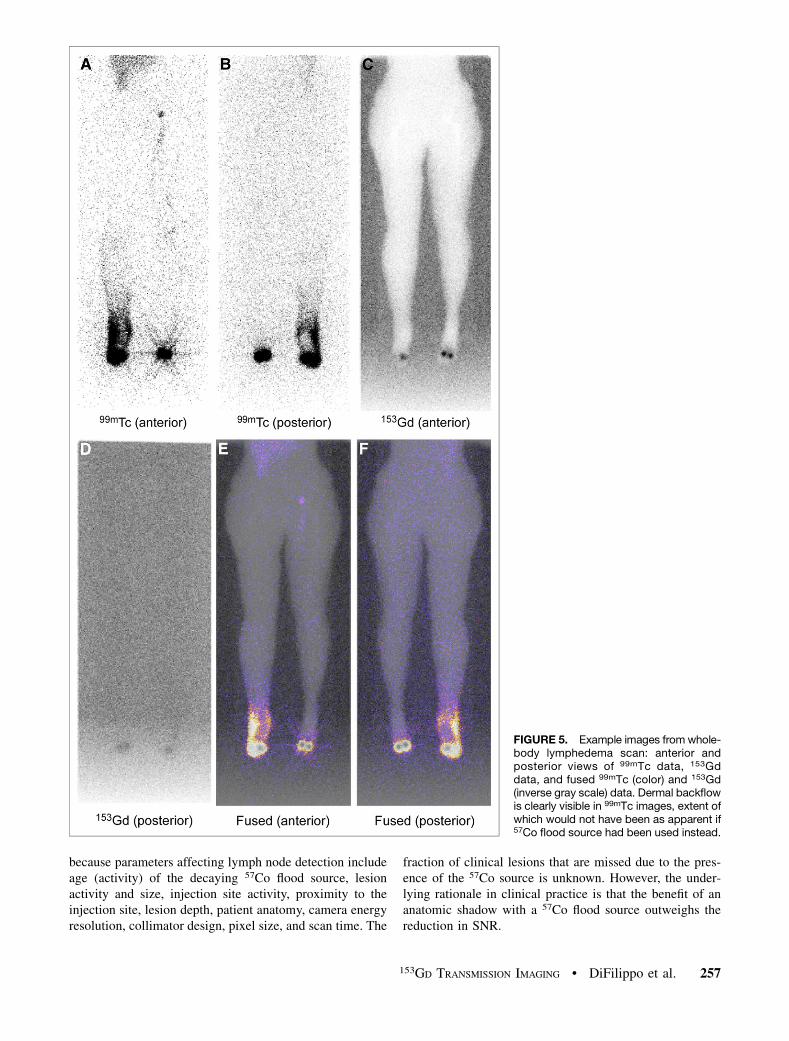
tigraphy, melanoma lymphoscintigraphy, and images areshown in Figures 5, 6, and 7. Lymphedema images wereacquired as a whole-body scan with a 256 · 1,024 matrixand 25 cm/min table speed. Breast and melanoma lympho-scintigraphy images were acquired as 256 · 256 staticviews with 1.23 zoom at 300 s per view.Region-of-interest measurements of the simulated phantom
lesions were compared with lesions in the clinical studies. Inthe phantom images of Figure 3C, the background-subtracted
region-of-interest measurements of the 3 peripheral pointsources were 760, 430, and 250 counts. In the clinical images,the background-subtracted lesion counts were 110 countsfor the inguinal node in Figure 5 (anterior view), 220counts for the axillary node in Figure 6 (oblique view),and 360–560 counts for the 4 inguinal nodes in Figure 7(anterior view).
DISCUSSION
An anatomic shadow in lymphoscintigraphy scans isuseful to interpreting physicians. Lesions appear as isolatedfoci, often near the intense injection site, with virtually noanatomic information included in the 99mTc emission dataitself. The ability to detect and localize lymph nodes inlymphoscintigraphy images may help guide surgery andaffect treatment outcome.
Thus, a concern is whether the presence of the floodsource has a significant negative impact on lymph nodedetection. With a 57Co flood source, 1 approach is to ac-quire sequential scans with and without the source, produc-ing a separate image unaffected by scatter and backgroundcounts from the 57Co source for better lymph node detec-tion. However, it is often not practical to acquire 2 sets ofstatic views or whole-body views, which would add signif-icant scan time, and most laboratories simply acquire 1 setof views with the 57Co source. It has been demonstrated inphantom studies that there is an optimal 57Co flood sourceactivity and scan time for lymph node detection (4).The relationship is more complicated in clinical practice,
FIGURE 4. Graphs of SNR versus activity for 3 peripheralpoint sources in various phantom and flood configurations:anterior view, point sources below phantom (A); posteriorview, point sources above phantom (B); anterior view, pointsources above phantom (C); and posterior view, point sourcesbelow phantom (D).
FIGURE 3. Anterior-view scintigraphy emission images (140-keVwindow) are shown in gray scale of water phantom with 99mTcpoint sources located below phantom: with no flood source (A),with 57Co flood source (B), and with 153Gd flood source (C). In A,circular regions of interest (diameter, 33 mm) for measuring pointsource counts (labeled PS) and adjacent background counts(labeled BK) are shown. For 153Gd flood source, correspondingtransmission image (100-keV window) (D) is shown in inversegray scale. Point source activities were 3,770 kBq in center,800 kBq at top, 380 kBq at lower-right, and 220 kBq at lower-left. All emission images are shown with equivalent intensityscaling relative to central point source.
256 JOURNAL OF NUCLEAR MEDICINE TECHNOLOGY • Vol. 43 • No. 4 • December 2015
because parameters affecting lymph node detection includeage (activity) of the decaying 57Co flood source, lesionactivity and size, injection site activity, proximity to theinjection site, lesion depth, patient anatomy, camera energyresolution, collimator design, pixel size, and scan time. The
fraction of clinical lesions that are missed due to the pres-ence of the 57Co source is unknown. However, the under-lying rationale in clinical practice is that the benefit of ananatomic shadow with a 57Co flood source outweighs thereduction in SNR.
FIGURE 5. Example images from whole-body lymphedema scan: anterior andposterior views of 99mTc data, 153Gddata, and fused 99mTc (color) and 153Gd(inverse gray scale) data. Dermal backflowis clearly visible in 99mTc images, extent ofwhich would not have been as apparent if57Co flood source had been used instead.
153GD TRANSMISSION IMAGING • DiFilippo et al. 257

Replacing the 57Co flood source with a 153Gd floodsource for lymphoscintigraphy scans is attractive for severalreasons. The 153Gd transmission photons in the 100-keVwindow do not interfere directly with the primary emissionphotons in the 140-keV window. Thus, there are fewerbackground counts with 153Gd, compared with 57Co, whichimproves SNR. With the 153Gd source, the acquisition ofemission and transmission counts in separate energy win-dows introduces flexibility in how images are displayed.The emission data can be displayed independently of thetransmission data with a gray scale range that highlights thecontrast of faint lesions that are difficult to detect. Fusedemission and transmission images provide both functionaland anatomic information in color and gray scale, respec-tively, which may aid interpretation and may provide betterinformation to the referring physician or surgeon. The153Gd flood source also allows posterior views to be ac-quired simultaneously, by inverting the transmission imageof the anterior view to create the posterior shadow. Inwhole-body lymphedema scans, for example, a posteriorview may improve detection of popliteal nodes.Another advantage of the 153Gd flood source is that
higher-quality anatomic shadows are generated. Given equalactivity and scan time, an anatomic shadow from a 153Gdflood source and 100-keV window has higher count densitythan a corresponding anatomic shadow from a 57Co floodsource and a 140-keV window, because only a small fractionof the primary 57Co photons (122 keV) are acquired in the140-keV window. Thus, a lower-activity 153Gd source canproduce anatomic shadows of acceptable quality, though
further study is needed to determine the lowest acceptableactivity for clinical use. The costs of a 153Gd flood sourceand a 57Co flood source are comparable, and the half-lives of153Gd (240 d) and 57Co (272 d) are similar. Because thetransmission data are acquired on-peak instead of off-peak,a 153Gd flood source can be replaced less often than a 57Coflood source for the purpose of anatomic shadowing. If
FIGURE 6. Example fused 99mTc (color)and 153Gd (inverse gray scale) imagesfrom breast lymphoscintigraphy scan.99mTc color window has been enhancedin oblique and lateral views to reveallymph node with low tracer activity,which was not visible in anterior viewand which would have had lower SNR ifusing 57Co flood source and single energywindow.
FIGURE 7. Example fused 99mTc (color) and 153Gd (inversegray scale) images from melanoma lymphoscintigraphy scanof injection site and of multiple views of inguinal lymph nodes.
258 JOURNAL OF NUCLEAR MEDICINE TECHNOLOGY • Vol. 43 • No. 4 • December 2015

a 153Gd flood source can replace a 57Co flood source forcamera quality assurance and calibrations, then there is ad-ditional economic justification for replacing the 57Co floodsource entirely. However, equivalent performance between153Gd and 57Co in quality assurance tasks must be validatedfor each specific camera model and supported by the manu-facturer before replacing 57Co with 153Gd.A minor issue with the 153Gd flood source is the contri-
bution to background counts from simultaneous detectionof an approximately 100-keV g-photon and an approxi-mately 40-keV europium x-ray photon, which can producea detected count in the 140-keV emission window. Theadded background counts from this mechanism are rela-tively small in the anterior view but may be noticeable inthe posterior view. A thin layer of a suitable metal can beintroduced to absorb preferentially the lower-energy x-raysand thereby reduce background counts. This effect wasstudied in the phantom experiments by adding brass filtersto the 153Gd source. A more ideal material would have beentin foil of approximately 0.15-mm thickness, but becauseinexpensive tin foil was not readily available, brass sheetmetal of 0.5-mm thickness was used instead.The phantom studies confirmed expectations of lesion
detection versus the various configurations. SNR, whichdepends on the lesion counts and background counts, isinfluenced by many factors that are highly variable inclinical studies. Although the phantom studies encom-passed a range of point source activities with or withoutattenuation and with or without flood sources, they did notrepresent the entire range of parameters encountered inclinical lymphoscintigraphy practice. Nonetheless, the phan-tom studies were valuable in comparing flood source con-figurations over a representative range.As expected, imaging with no flood source produced the
best SNR in all cases. This situation has minimal back-ground counts, arising only from collimator penetration andscatter from 99mTc sources. The presence of a 57Co or153Gd flood source only can reduce SNR, by contributingmore background counts, by attenuating 99mTc photons (inthe posterior view), or by contributing to detector deadtime. SNR was reduced significantly in all cases with the57Co flood source. The degree of SNR reduction in theanterior view depended on signal strength (lesion activityand attenuation). In the posterior view where the 57Co pho-tons were unattenuated, the SNR was greatly reduced. Theclinical implication is that some faint lesions in the anteriorview may not be detected and that the posterior view mostlikely is not worth acquiring at all. The frequency of clin-ical lymph nodes missed when using a 57Co flood source isan interesting topic for future research.Replacing the 57Co flood source with the 153Gd flood
source consistently improved SNR. Introducing brass sheetmetal to filter x-ray photons further improved SNR in mostcases. SNR in anterior views with the 153Gd flood sourceand brass filters was similar to the case with no flood sourcepresent. This observation supports that the 99mTc emission
data experience minimal interference from the 153Gd sourceand that the interference arises from simultaneous detectionof g-rays and x-rays. In the posterior views, however, SNRwith the 153Gd flood source was substantially lower than thecorresponding anterior views with the phantom rotated180�. The reason for this difference is attenuation of99mTc counts by the 153Gd flood source. The presence ofthe brass filters further attenuated the 99mTc signal,though it also noticeably reduced background counts.Depending on whether the point sources were above or be-low the phantom, the presence of the brass filters eitherimproved or reduced SNR in the posterior view. Withpoint sources above the phantom and attenuated, back-ground counts due to x-rays were a significant componentin the posterior emission image, and SNR was improved bythe brass filters. With point sources below the phantom andunattenuated, background counts due to x-rays were negli-gible, and the main effect of the brass filters was to attenuatesignal, reducing posterior SNR.
The phantom studies provided confirmation of the supe-riority of the 153Gd flood source compared with the 57Coflood source for lymphoscintigraphy, and thus the 153Gdflood source was adopted for use in clinical practice. Clin-ical lymphoscintigraphy images with 153Gd shadow were ofhigh quality, as can be seen from the examples in Figures5–7. Figure 5 illustrates the utility of applying the 153Gdflood source to a patient with lower-extremity lymphedema.Determination of the extent of dermal backflow is assistedby the availability of both anterior and posterior imagesand by the availability of emission-only 99mTc images,which avoid background counts arising from a 57Co floodsource. The potential benefit of an enhanced SNR witha 153Gd flood source for anatomic localization is illus-trated in the breast lymphoscintigraphy case in Figure 6,in which faint tracer uptake is identified in a sentinellymph node in the anterior superior left axilla on obliqueand lateral views. Finally, the images in Figure 7 clearlydefine the lymphatic pathway leading to the inguinallymph nodes in a patient with cutaneous melanoma. Thesentinel inguinal lymph nodes are clearly localized, andfaint secondary iliac lymph nodes are visualized posteriorto the inguinal nodes on the lateral view.
Further clinical studies will be needed to systematicallyevaluate the impact of the 153Gd flood source in lympho-scintigraphy on clinical decision making. Initial reactionsto the use of the 153Gd flood source have been positive. In-terpreting nuclear medicine physicians generally prefer theflexibility of emission-only and fused emission–transmissionimage display over their prior experience with a singleimage with 57Co shadow. The brightness and contrast ofthe emission-only images are adjusted more easily withoutthe border and background counts from the 57Co source.Color screenshots of the fused emission–transmission images,in addition to gray-scale screenshots of the emission-onlyimages, are routinely saved and made available to refer-ring physicians and surgeons, facilitating the bedside
153GD TRANSMISSION IMAGING • DiFilippo et al. 259

visualization of sentinel lymph nodes and pathways oflymphatic drainage.
CONCLUSION
A 153Gd flood source offers several advantages overa 57Co flood source for production of anatomic shadowsin lymphoscintigraphy studies. The acquisition of emissionand transmission data in separate energy windows yieldsboth pure-emission and fused images and provides greaterflexibility in image display. Phantom studies simulatinglesion detection demonstrate superior SNRs using a 153Gdflood source, which is attributed mainly to reduced back-ground count levels compared with using a 57Co floodsource. The 153Gd flood source is as practical to use ina clinical environment as a 57Co flood source, having sim-ilar cost and radionuclide half-life. Lymphoscintigraphywith the 153Gd flood source offers the potential for im-proved lymph node detection and improved satisfactionamong referring physicians.
DISCLOSURE
No potential conflict of interest relevant to this articlewas reported.
REFERENCES
1. Giammarile F, Alazraki N, Aarsvold JN, et al. The EANM and SNMMI practice
guideline for lymphoscintigraphy and sentinel node localization in breast cancer.
Eur J Nucl Med Mol Imaging. 2013;40:1932–1947.
2. Law M, Ma WH, Leung R, et al. Evaluation of patient effective dose from
sentinel lymph node lymphoscintigraphy in breast cancer: a phantom study
with SPECT/CT and ICRP-103 recommendations. Eur J Radiol. 2012;81:
e717–e720.
3. Krynyckyi BR, Sata S, Zolty I, Kim CK, Knesaurek K. Reducing exposure from57Co sources during breast lymphoscintigraphy by optimizing energy windows
and other suggested enhancements of acquisition and the display of images.
J Nucl Med Technol. 2004;32:198–205.
4. Mar MV, Dickinson RL, Erwin WD, Wendt RE III. Optimal 57Co flood source
activity and acquisition time for lymphoscintigraphy localization images. J Nucl
Med Technol. 2008;36:82–87.
5. Wilczek B, Sandelin K, Eriksson S, Larsson SA, Jacobsson H. Sentinel node
scintigraphy in breast cancer using a dual tracer technique. Nucl Med Commun.
2004;25:135–138.
6. Krynyckyi BR, Miner M, Ragonese JM, Firestone M, Kim CK, Machac J. Tech-
nical aspects of performing lymphoscintigraphy: optimization of methods used
to obtain images. Clin Nucl Med. 2000;25:978–985.
7. Vallejo Mar M, Gee-Johnson S, Kim EE, Podoloff DA. Whole-body lympho-
scintigraphy using transmission scans. J Nucl Med Technol. 2002;30:12–17.
8. Bailey DL, Hutton BF, Walker PJ. Improved SPECT using simultaneous emis-
sion and transmission tomography. J Nucl Med. 1987;28:844–851.
9. Zaidi H, Hasegawa B. Determination of the attenuation map in emission tomog-
raphy. J Nucl Med. 2003;44:291–315.
10. Clarke E, Notghi A, Harding K. Improved body-outline imaging technique for
localization of sentinel lymph nodes in breast surgery. J Nucl Med. 2002;43:
1181–1183.
260 JOURNAL OF NUCLEAR MEDICINE TECHNOLOGY • Vol. 43 • No. 4 • December 2015


![$ SDUWLUH GD ¼ SS DU ± FRQ SDUWHQ]D GD 7RULQR · 7uhql gd shu 1dsrol h 7udqvihu gd shu o +rwho $ sduwluh gd ¼ ss du ± frq sduwhq]d gd 7rulqr 7uhqr 7rulqr 1dsrol h ulwruqr 7udvihulphqwr](https://img.pdfslide.net/doc/110x75/602b6d423576982f89178c7f/-sduwluh-gd-ss-du-frq-sduwhqd-gd-7rulqr-7uhql-gd-shu-1dsrol-h-7udqvihu-gd.jpg)