Embed Size (px)
Citation preview

Linköping University Postprint
Transplantation of cultured human keratinocytes in single cell suspension: a comparative in vitro study of different
application techniques
Camilla Fredriksson, Gunnar Kratz and Fredrik Huss
N.B.: When citing this work, cite the original article. Original publication: Camilla Fredriksson, Gunnar Kratz and Fredrik Huss, Transplantation of cultured human keratinocytes in single cell suspension: a comparative in vitro study of different application techniques, 2008, Burns, (34), 2, 212-219. http://dx.doi.org/10.1016/j.burns.2007.03.008. Copyright: Elsevier B.V., http://www.elsevier.com/ Postprint available free at: Linköping University E-Press: http://urn.kb.se/resolve?urn=urn:nbn:se:liu:diva-11214

Transplantation of cultured human keratinocytes in single cell suspension: a comparative in vitro study of different application techniques
Camilla Fredriksson Med Biol1, Gunnar Kratz MD PhD, Professor1,2, Fredrik Huss MD
PhD1,2
1) Institution of Biomedicine and Surgery, Department of Experimental plastic
surgery, Faculty of Health Science, Linköpings Universitet, Linköping, Sweden
2) Department of Plastic-, Hand-, and Burn Surgery, University Hospital of
Linköping, Linköping, Sweden
Corresponding author:
Camilla Fredriksson, Medical Biologist
Institution of Biomedicine and Surgery, Department of Experimental plastic surgery, Faculty of Health
Science, Linköpings Universitet
S-581 82 Linköping
Sweden
E-mail: [email protected]
Phone: +46 13 22 73 37
Fax: +46 13 12 74 65
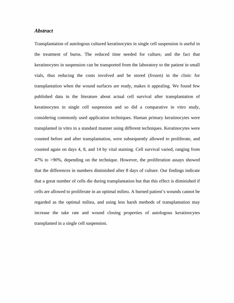
Abstract
Transplantation of autologous cultured keratinocytes in single cell suspension is useful in
the treatment of burns. The reduced time needed for culture, and the fact that
keratinocytes in suspension can be transported from the laboratory to the patient in small
vials, thus reducing the costs involved and be stored (frozen) in the clinic for
transplantation when the wound surfaces are ready, makes it appealing. We found few
published data in the literature about actual cell survival after transplantation of
keratinocytes in single cell suspension and so did a comparative in vitro study,
considering commonly used application techniques. Human primary keratinocytes were
transplanted in vitro in a standard manner using different techniques. Keratinocytes were
counted before and after transplantation, were subsequently allowed to proliferate, and
counted again on days 4, 8, and 14 by vital staining. Cell survival varied, ranging from
47% to >90%, depending on the technique. However, the proliferation assays showed
that the differences in numbers diminished after 8 days of culture. Our findings indicate
that a great number of cells die during transplantation but that this effect is diminished if
cells are allowed to proliferate in an optimal milieu. A burned patient’s wounds cannot be
regarded as the optimal milieu, and using less harsh methods of transplantation may
increase the take rate and wound closing properties of autologous keratinocytes
transplanted in a single cell suspension.

Introduction
Cultured keratinocytes have been used for about 20 years in the treatment of burns and
other cutanous wounds. In 1975, Rheinwald and Green [1] described a reliable method of
culturing human epidermal cells in stratified and coherent layers, and so cultured
epidermal sheet autografts became available to complement autologous split thickness
skin grafts in treating major burns or other large wounds (Figure 1A) [2]. However,
producing confluent grafts of keratinocytes puts heavy demands on laboratory skills,
comprises manual labour, and is expensive, which limits the use of the autografts in many
ways. The sheets are only 8-10 cells thick, which make them fragile and difficult to
handle, and means that they have to be placed on a supportive backing material to be
possible to transfer from laboratory to patient (Figure 1B). When the autografts have been
detached from the culture vessel, they must be transplanted the same day, which requires
the wound surfaces to be ready for grafting at the same time as the autografts are ready to
be transplanted. Because the transplanted epidermal sheets are quite unstable and prone
to blistering, care must also be taken, not only during production and transplantation, but
also during dressing of the applied grafts, mobilisation of the patient, and changing of
dressings [3]. The production of stratified grafts inevitably involves some degree of
maturation and differentiation, of the keratinocytes, which reduces their proliferative
capacity, and this in turn may affect the take-rate and wound-healing capacity [4].
Extrinsic factors including preparation of the wound, nutritional state, and dressings used
influence their success.

Figure 1A Cultured epithelial autografts transplanted to wound surface with a polyamide-mesh backing material. Picture taken five days after transplantation. Figure 1B Cultured epithelial autograft has been enzymatically released from culture vessel’s bottom. Polyamide-mesh backing material (Surfasoft®) is being attached by folding the autograft’s edges over the backing material and securing them with surgical micro-clips. Figure 1C Transport and storage vial for cultured cells, e.g. cultured autologous keratinocytes. Depicted vial contains approximately 20 x106 cultured cells ready for transplantation. Figure 1D Autologous cultured keratinocytes from the vial in picture 1C has been aspirated into the thrombin fraction of the Tisseel Duo Quick™ tissue glue syringe and are being spray-painted on the wound surface using the Duploject™ Spray set.
The attention to, and understanding of, these shortcomings have led to a progressive
development of techniques of skin culture and an increased use of suspensions of single
cells of keratinocytes being transplanted instead of sheet grafts. Fraulin et al [5], in 1998,
described a novel technique in which they used an aerosol device to spray epithelial cells
on wounds in pigs. They noted that re-epithelialisation was quicker than in unsprayed
controls. Navarro et al [6] developed this technique further by combining it with meshed
split thickness skin grafts. They reported faster healing and a better quality of cells when
they were sprayed. Further advantages of suspension transplantation are the reduced time
needed for culture, and avoidance of the manual labour of releasing cell-sheets from
culture flasks and attaching the cell-sheets to backing materials. By culturing and
transplanting the cells in a suspension rather than as a sheet, the use of enzymes like
Dispase® can be avoided. Keratinocytes in suspension can then be transported from
laboratory to patient in a handful of small vials (Figure 1C) and be stored (frozen) at the
clinic to be transplanted when the wound surfaces are ready [7]. The single cell
suspension of keratinocytes can then be transplanted to the patient with whatever method
is available such as being spray-painted on the wound surfaces with or without fibrin-
glue (Figure 1D) [8, 9]. Today, transplantation of keratinocytes in a single cell suspension
overgrafted with meshed allogeneic donor skin is a common approach in the treatment of
burns [9, 10]. Techniques used in clinics today include spraying cells, with or without the
additional use of tissue or fibrin glue, painting the cell suspension with a brush, or
dripping the cell suspension on to the wound bed using a syringe. At our burn unit we
have used the Tissomat applicator together with Tisseel Duo Quick™ tissue glue and the
Duploject™ Spray set (all from Baxter Medical AB, Kista, Sweden) to transplant cultured
keratinocytes. To distribute the cells satisfactorily, pressures as high as 200 kPa must be
used. This has long been thought to damage the cells, both by the passing of the spray
nozzle and by the high velocity impact on to the wound bed. Harkin et al [11] recently
examined the viability of keratinocytes delivered by aerosol, using the Tissomat

applicator and found that the viability after transplantation (93.7% at 70 kPa and 90% at
138 kPa) was similar to the viability of the cells just recently detached from the culture
dish (94%). When they adjusted the Tissomat applicator to deliver 207 kPa, they showed
that fewer cells survived (73.3%), but not significantly so.
If a brush is used to distribute the cells to the wound (the method of choice in some
clinics) it probably causes shear forces that damage the cells, and may leave fibres from
the brush in the wound. Drips from the cell suspension, when using a syringe, may well
cause an uneven distribution of the cells, if the suspension runs off the surface, pools in
cavities, and leaves some areas uncovered. The fact that surprisingly few research
workers have studied the viability and survival of transplanted cells, encouraged us to
design a comparative in vitro study that took into consideration the methods used in
clinics today.
The aim of our study was to compare and evaluate different commonly used techniques
of transplantation of single cell suspension, from the point of view of cell survival,
attachment, and proliferation of cells.
Material and methods
Cell culture
Normal human keratinocytes were isolated and expanded in vitro. Briefly, biopsy
specimens of skin from surgical waste were transferred to the laboratory in gauze soaked
in physiological saline. Keratinocytes were isolated within 24 hours and the tissue was
kept in +8°C until use. The skin was rinsed twice in phosphate buffered saline (PBS) with
antibiotics and mycotics (50 IU/ml penicillin, 50 μg/ml streptomycin). Subcutaneous fat

was removed with scissors and the remaining tissue (dermis and epidermis) was cut into
roughly 1 cm2 pieces and incubated in Dispase® 15 ml (16.7 mg/ml, 1.04 U/ml) for 18
hours at +8°C. After incubation the epidermis was lifted off the dermis with pliers and
transferred to 0.02% EDTA 2 ml and 0.25% trypsin 2 ml. The tissue was incubated in
+37°C at 95% humidity and 5% carbon dioxide for roughly 10-15 minutes, during which
time it was repeatedly removed from the incubator and triturated with a Pasteur pipette to
dissociate the cells. After incubation and trituration, the action of trypsin was inhibited by
transferring the cell suspension to washing medium (10 ml of Dulbecco’s modified
Eagle’s medium (DMEM) supplemented with 10% fetal calf serum and penicillin-
streptomycin and centrifuged at 400 g for 5 minutes. The supernatant was removed and
the cell pellet re-suspended in culture medium 15 ml and seeded into a 75 cm2 culture
flask (BD Falcon, Stockholm, Sweden). Culture medium used was keratinocyte-serum-
free media (Ker-SFM) supplemented with 200 μl/100 ml bovine pituitary extract, 3.3
μl/100 ml endothelial growth factor and penicillin-streptomycin. Medium was changed
every second day throughout the study. When sub confluence had been achieved (for
illustrations of different states of confluence, see Figure 3A-B) the primary culture was
split 1:3 by rinsing the culture twice with 2 ml EDTA 0.02%, and subsequently adding
0.02%/EDTA 2 ml and 0.25% trypsin 2 ml and incubating for 10-15 minutes. The effect
of the trypsin was then inhibited. The supernatant was removed and the cell pellet re-
suspended in culture medium 12 ml and divided into three culture flasks 75 cm2. An
additional 13 ml of culture medium was then added. Cells of the second passage were
used in the study and the cultures were incubated in +37°C, 95% humidity, and 5%

carbon dioxide. All media and supplements were bought from Invitrogen AB, (Lidingö,
Sweden) and EDTA and trypsin from Gibco®/Invitrogen AB, (Lidingö, Sweden).
Preparation of single cell suspensions
The cells were detached from the culture flasks and the effect of trypsin was inhibited as
described. The supernatant was removed and the cell pellet re-suspended in culture
medium 10 ml. Cells were counted in a haemocytometer by triplicate samples, cell
suspension 10 μl mixed with trypan blue 10 μl (Sigma-Aldrich Sweden AB, Stockholm,
Sweden) and incubated in room temperature for 5 minutes.
Application techniques
Well-dispersed cell suspension was aspirated into 1 ml syringes (BD Plastipak™ Becton
Dickinson, Madrid, Spain) and applied by dripping or spraying the cells into individual
100 mm ∅ Petri dishes from a distance of 10 cm (Table 1, Figure 2A-F).
1 Drop (Figure 2A)
The cell suspension was dripped on to the surface while the syringe was moved over the
area of the Petri dish, aiming for an even distribution of the cells and using as much
pressure as needed to obtain a steady flow or drip of suspension.
2 Paint brush (Figure 2B)
An artist’s paintbrush (Dekorima Symfony size 16, Dekorima AS Sandefjord, Norway)
was used. The cell suspension was dripped on to the Petri dish (as for the first group).
The cells were then smeared with the brush over the surface with 10 strokes in different
directions to paint the surface with the cell suspension.

Table 1: Application techniques used in this study to transplant cultured keratinocytes.
1) Drop Dripped with a standard 1 ml syringe.
2) Paint brush Dripped with a standard 1 ml syringe and dispersed on the
culture surface with a paintbrush.
3) Spray nozzle Sprayed through a spray nozzle attached to a 1 ml syringe.
4) Harvest® Sprayed with the Harvest SK/S Spray Applicator Kit®.
5) High pressure Sprayed with the Tissomat and DuplojectTM spray kit with 200 kPa air pressure.
6) Low pressure Sprayed with the Tissomat and DuplojectTM spray kit with 50 kPa air pressure.
7) Duploject™ nozzle Sprayed with the DuplojectTM spray nozzle attached to a 1 ml syringe.
Figure 2A-F: The different transplantation techniques used in this study for transplanting cultured keratinocytes in suspension: (A) – Drop, (B) - Paint brush, (C) – Spray Nozzle
attached to a syringe, (D) – Harvest SK/S Spray Applicator Kit®, (E) – High pressure and Low pressure spraying using the Duploject™ spray kit. (Insert: Tissomat application device adjusted to deliver 200 kPa or 50 kPa respectively), (F) – DuplojectTM nozzle.

3 Spray nozzle (Figure 2C)
The spray nozzle (Male valve, ST300 T3K016K20, LINDAL Group, Bad Oldesloe,
Hamburg), an ordinary spray nozzle for distributing substances such as hairspray, was
attached to the syringe and the suspension sprayed over the surface for an even
distribution of cells. The force applied was just enough to create a fine mist.
4 Harvest® (Figure 2D) The Harvest SK/S Spray Applicator Kit®, referred to as Harvest® (Harvest Technologies
GmbH, München, Germany), is designed to deliver two liquid components
simultaneously, such as cell suspension and tissue glue. The Harvest® nozzle was
attached to the syringe and to prevent back-flush from the second canal this was blocked.
The suspension was then sprayed evenly over the surface.
5 High pressure (Figure 2E)
The Tissomat applicator is designed to deliver and regulate pressures up to 1000 kPa and
is often used with a Duploject™ spray set to transplant cells together with Tisseel Duo
Quick™ tissue glue. The Duploject™ spray set consists of a special Duploject™ spray
nozzle with a two-channel spray system that allows two fluids to be delivered
simultaneously under the same pressure and flow. It comes with a tube to fit onto the
Tissomat applicator.
The syringe was attached to the Duploject™ spray set according to manufacturer’s
directions and subsequently connected to the Tissomat applicator, which was adjusted to

deliver a pressure of 200 kPa. To prevent back flush from the second canal, this was
blocked and the suspension was then sprayed over the surface.
6 Low pressure (Figure 2E)
The Tissomat application device was adjusted to deliver a pressure of 50 kPa and the
suspension was then sprayed over the surface as above.
7 Duploject™ nozzle (Figure 2F)
The syringe was connected to the Duploject™ spray nozzle. To prevent back flush from
the second canal it was blocked, and the suspension was sprayed evenly over the surface
without any additional air pressure from the Tissomat application device.
Figure 3A-B: Phase contrast microphotographs of a non-confluent keratinocyte culture (A) and a confluent keratinocyte culture (B).
Assessment of keratinocyte viability
After application of the cell suspension, culture medium 7 ml was added instantly to each
individual Petri dish, giving a total of 8 ml in each dish. The number of living and dead
cells was counted in a haemocytometer, by triplicate samples of cell suspension 10 μl,
each mixed with trypan blue 10 μl.
Attachment of cells and proliferation assay
Three samples of 2 ml from the 8 ml cell suspension from each event were seeded into 3
wells of a 6-well cell culture plate (Falcon, BD Plastipak™ Becton Dickinson,
Stockholm, Sweden), and then incubated at +37°C, 95% humidity, and 5% carbon
dioxide until it was time to sample them.
Sampling
At 4, 8, and 14 days after transplantation, cells from each group were counted in
triplicates by haemocytometer to assess cell proliferation and the living:dead ratio. The
cells were detached from the wells with EDTA and Trypsin, and incubated with trypan
blue before they were counted.
Figure 4 summarises the study set up.
Statistics
Each experiment was performed three times using cultures established from separate
tissue donors on each occasion. We analysed the data from day 0, day 4, day 8, and day
14 by using two-way variance analysis with the cultures from the different tissue donors
as explaining variables. We used a statistical model without interaction, since the data did
not confirm significant interactions between the different methods. The triplicate samples
from each donor-culture and time point were regarded as random repetitions without

mutual order within the combination tissue donor-method. The significance of difference
was assessed using Tukey’s simultaneous pair wise analysis of variance test, with a 95%
confidence interval. Results are presented as mean if not otherwise stated.
Figure 4: Figure illustrates the workflow of the transplantation process. Well-dispersed cell suspension was aspirated into 1 ml syringes and applied by dripping or spraying the
cells into individual 100 mm ∅ Petri dishes from a distance of 10 cm. After transplantation the cells from each group was counted, added 7 ml of culture media and
subsequently seeded into 3 wells of a 6-well plate for further culturing. At day 4, 8 and 14 one well from each group was harvested and counted to determine proliferation.
Results
Viability and proliferative capacity of transplanted keratinocytes
After harvest of the cultures and before transplantation, the trypan blue exclusion method
was used to assess the viability of the cells in the stock suspension. We found that 94% of

the cells remained viable after treatment with trypsin and EDTA and re-suspension in
Ker-SFM. After the cells had been transplanted there was a variable cell death, see
below. Apart from the High pressure group, all techniques showed significantly better
survival at all time points, except for the Paintbrush group and the Harvest® group, which
showed significantly fewer survivors at day 14 (Table 2).
Table 2: Cell viability in percentage (% of original cell number) at the different time points after transplantation for each technique used. Technique
Day
0 4 8 14 Drop
87.8 44.0 46.3 55.4
Paint brush
59.4 34.1 35.4 39.7
Spray nozzle
93.2 45.7 44.8 48.4
Harvest®
72.8 35.6 40.2 46.0
High pressure
(200 kPa) 47.3 22.5 26.1 39.8
Low pressure
(50 kPa) 76.6 35.3 35.1 49.8
Duploject™ nozzle
84.1 43.8 42.1 53.9
Day 0
The highest cell survival at day 0 was seen in the Spray nozzle group, which was
significantly higher than the High pressure group (p<0.001), the Paintbrush group
(p<0.001), the Harvest® group (p<0.001), and the Low pressure group (p<0.001). It also
had larger numbers, but not significantly so, compared with the Duploject™ and Drop
groups (Figure 5).

Spray nozzle Drop Duploject Low pressure Harvest Paintbrush High pressure
0
10
20
30
40
50
60
70
80
90
100
1
Su
rviv
ing
cell
s aft
er
tran
spla
nta
tio
n d
ay 0
(%
)
Figure 5: Percentage (%) viable cells after transplantation with seven different techniques. The highest cellular survival rate after transplantation was found after using technique Spray nozzle. This technique showed significantly more viable cells than the High pressure, Paintbrush, Harvest®, and Low pressure groups and showed tendencies
towards higher numbers than DuplojectTM and Drop, but not significantly so. (***, p<0.001 compared with Spray nozzle).
Day 4
On day 4 the proliferation assays showed the best viability in the group Spray nozzle,
which had significantly higher survival than the High pressure (p<0.001), Paintbrush
(p<0.01), Low pressure (p<0.01), and Harvest® (p<0.01) groups and a slightly higher
number (but not significantly so) than the Duploject™ and Drop groups (Figure 6).
Day 8
The cell count for proliferation assays on day 8 showed a shift in the highest number of
viable cells from the Spray nozzle group to the Drop group, which showed significantly
higher surviving numbers than the High pressure (p<0.001), Low pressure (p<0.001), or

Paintbrush (p<0.001) groups, and higher survival numbers, but not significantly so, than
the Harvest®, Duploject™ and Spray nozzle groups.
Spray nozzle Drop Duploject Harvest Low pressure Paintbrush High pressure
0
10
20
30
40
50
60
70
80
90
100
1
Via
ble
cell
s d
ay 4
(%
)
Figure 6: Percentage (% of initial cell number) viable cells after 4 days of culturing. The proliferation assays showed highest viability with the Spray nozzle group, showing significantly higher numbers compared with the High pressure, Paint brush, Low
pressure, and Harvest® groups, and a tendency towards higher numbers compared with the DuplojectTM and Drop groups, but not significantly so. (**, p<0.01, ***, p<0.001
compared with Spray nozzle)
The groups Drop, Spray nozzle, and Duploject™ have similar number of viable cells with
p values close to 1, so not significantly different from each other (Figure 7).
Day 14
By day 14 the numbers had levelled out to some extent, with the Drop group having the
most viable cells, slightly more than the Duploject™, Low pressure, Spray nozzle, and
Harvest® groups. All had significantly more surviving cells compared with the High

pressure and Paintbrush groups (p<0.001 in each case) but they did not differ
significantly from them (Figure 8).
Drop Spray nozzle Duploject Harvest Paintbrush Low pressure High pressure
0
10
20
30
40
50
60
70
80
90
100
1
Via
ble
cell
s d
ay 8
(%
)
Figure 7: Percentage (% of initial cell number) viable cells after 8 days of culturing. The cell count for proliferation assays on day 8 showed a shift in highest number of viable
cells from Spray nozzle to Drop. If looking at clinically interesting techniques, the Drop, Spray nozzle, and DuplojectTM groups show equal numbers of cells with a p-value close to 1.0, compared with High pressure, Low pressure, and Harvest® groups, which show significantly lower numbers than the Drop group. (***, p<0.001 compared with Drop)

Drop Duploject Low pressure Spray nozzle Harvest Paintbrush High pressure
0
10
20
30
40
50
60
70
80
90
100
1
Via
ble
cell
s d
ay 1
4 (
%)
Figure 8: Percentage (% of initial cell number) viable cells after 14 days of culturing. At day 14 the numbers had levelled out to some extent, the Drop group showing the highest
numbers of viable cells, close to the DuplojectTM, Low pressure, Spray nozzle, and Harvest® groups. All of them are significantly higher in numbers compared with the High
pressure and Paintbrush groups, but have no significant difference in relation to each other. (***, p<0.001 compared with the Drop group).
Discussion
Wound closure using autologous cultured keratinocytes in suspension will certainly
continue to be a valuable tool in the future treatment of burns. We have shown that
normal human primary keratinocytes, transplanted by different application techniques
have a great variability in the viability and proliferative capacity of the cells, ranging
from 47% (High pressure technique) to 93% (Spray nozzle technique), depending on the
method of transplantation. One of the hypotheses of this study was that the use of a
pressure device, such as the Tissomat applicator, damages the cells when delivering
keratinocytes to a wound bed, and so less viable cells reach the wound to act in the

wound closure. Duncan et al [12] studied the effect of aerosol transfer of keratinocytes
compared with delivery by keratinocytes on a fibrin membrane to a de-epidermalised
dermis. They found that even though cells in suspension experience different types of
stresses (hydrostatic, shear, and elongation) when being sprayed, a total of 20% more
cells were present on the de-epidermalised dermis than in the fibrin membrane. Harkin et
al [11] used the Tissomat applicator and found that the viability after transplantation,
using pressures ranging from 70-138 kPa, was similar to that of cells just recently
detached from the culture dish. When they adjusted the Tissomat applicator to deliver
207 kPa however, the percentage (about 70%) of viable cells was lower. This is closer to,
but still much higher than what we found in this study. The contradictory results we
present are in the region of 50% viable cells after transplantation delivered at the pressure
of 200 kPa, as opposed to around 70% found by Harkin et al, who also presented a
considerably higher overall survival of cells. The reasons for this are not obvious, but the
fact that they used a sealant for co-transplantation might have increased their yield of
viable cells. We think that a combination of cells-carrier, -vehicle or, -soluble matrix used
when transplanting cells in suspension both lessens the physical impact and damage to
the cells and provides better starting conditions for the cells by providing a three
dimensional scaffold, which leads to a quicker and better proliferation of cells or
regeneration of tissue at hand. This theory is supported by a recent study [13] that was
conducted in our laboratory. In this present study however, so that we could evaluate the
actual survival of the cells using different techniques, we excluded sealants and carriers.
In continuum, even though Harkin et al showed unaffected cell viability after
transplantation with low pressures, they noted a significant reduction in the metabolic

activity of the cells. This corresponds quite well to our results, and might be the answer
as to why the keratinocytes, after transplantation, had a diminished capacity to proliferate
compared with what is usually seen when passing keratinocytes in culture. The results of
the Drop, Duploject™ nozzle, and the Spray nozzle techniques, showed that at least half
of the cells adhered and started to proliferate. However, when we studied the surface of
the culture dishes after transplantation with the Drop, and Duploject™ nozzle we saw
large areas in complete loss of cells and suspension (Figure 2B and 2C). In consequence,
the question arises: can these methods fully cover a large sized wound without leaving
major areas uncovered? As burns or other large wounds often are substantially irregular,
and far from smooth, one can imagine that cells pool in cavities or are washed off the
wound and end up in the bed, instead of covering the wound area. We find these methods
more suitable for smaller burns and ulcers. The Spray nozzle technique, in comparison,
covered the whole surface of the culture dish, using the same volume of suspension. The
Paintbrush technique showed surprisingly high cell survival of around 60% after
transplantation. However, as predicted, a number of brush fibres contaminated the surface
(data not shown). The plating efficacy was good, only roughly 40% of the cells failed to
adhere, but the proliferation capacity seemed poor and the cultures never recovered to
reach the cell yield of the other methods even after 14 days of culture. This could be the
result of different stresses, presented by Duncan et al [12], as it is likely that the cells
sustained at least elongation stress by being smeared across the surface and that this in
turn would lead to reduced cellular metabolism and proliferative capacity, which was also
described by Harkin et al [11]. The Harvest® technique gave a cell survival of about 70%
after transplantation, and a proliferation profile that matched the other low pressure

techniques. The system is small and easy to operate, compared with the Tissomat
applicator that requires additional tubes, medical air and a good hand-foot co-ordination.
However, a new and improved pressure regulator (EasySpray®, Baxter Medical AB,
Kista, Sweden) is about to replace the older Tissomat pressure device. The High pressure
technique, frequently used in various clinics, showed only 25% cell adherence and the
cells did not seem to fully recover in proliferative capacity, compared with other regimes.
When we counted the cells sprayed with the High pressure technique in a
haemocytometer, we also noted subjectively a higher number of fragmented cells and a
number of the cells, even though they had not turned blue from trypan blue, looked
morphologically disturbed and gave the impression of unhealthy cells (data not shown).
The fact that the Duploject™ nozzle resulted in almost the same cell survival as the Drop
technique made us think, even more, that pressure alone can cause substantial cell
damage. When evaluating the Low pressure Tissomat method, we noticed that the
pressure, 50 kPa was just enough to cause the suspension to drip on to the culture dish,
making it comparable to the Drop technique. Wood et al [14] presented in 2003 a study in
which they evaluated the clinical potential of keratinocytes in suspension. The device
used to transplant the keratinocytes in that study was an ordinary spray nozzle attached to
a standard syringe, similar to the one we used in this study. The spray nozzle used in our
study had in a previous pilot study (data not shown), in which we compared several
nozzles on the market, proved suitable for this application. We wanted the channel in the
spray cap to be wide enough to let the cell suspension pass through the nozzle but narrow
enough to create a fine mist when distributing the suspension with only minor pressure
being applied. It also had to fit a standard syringe and permit being sterilised through an

autoclave without losing its properties. The spray nozzle chosen provided a good
distribution of suspension over the surface, and showed similar cell viability to the Drop
technique when compared.
The fact that single cell suspensions can be transported to the clinic frozen in small vials,
stored and then thawed in amounts suitable for the occasion just before transplantation
makes it an appealing option to cultured epithelial sheets and other measures. Together
with straightforward equipments like a spray nozzle attached to a syringe, these features
would undoubtedly facilitate the future use of single cell suspension in transplantations of
burns and other cutanous wounds.
This in vitro study is not to be compared to in vivo or situ conditions, where the wound
bed is a hostile environment with different factors that affect survival and adherence of
cells.
However, it does provide valuable information about different measures to be used when
transplanting autologous keratinocytes in a single cell suspension, and we hope that, with
further studies, advances in this field will lead to the development of an equipment that is
fairly cheap and easy to operate and allow cells to be sent to the clinic, loaded in a spray
device and ready to use. We think that investing time and effort in developing the
methods further will decrease both time and costs of treatment, and provide better healing
and less pain and discomfort for patients.
Conflict of interest statement
There are no conflicts of interest concerning this study.

Acknowledgements We thank lab technicians Anita Lönn and Kristina Briheim for expert advice in cell-culture, and medical student Jonas Ockell for helping out in the pilot studies. This study was supported by grants from County of Östergötland, Sweden (dnr 2006-6-19).

References
[1] Rheinwald JG, Green H. Serial cultivation of strains of human epidermal
keratinocytes: the formation of keratinizing colonies from single cells. Cell 1975;6:331-
43.
[2] Gallico GG III, O’Connor NE, Compton CC, Kehinde O, Green H. Permanent
coverage of large burn wounds with autologous cultured human epithelium. N Engl J
Med. 1984 Aug 16;311(7):448-51.
[3] Elliot M, Vandervord J. Initial experience with cultured epithelial autografts in
massively burnt patients. ANZ J Surg. 2002;72:893-95.
[4] Horch RE, Debus M, Wagner G, Stark GB. Cultured human keratinocytes on type I
collagen membranes to reconstitute the epidermis. Tissue Eng. 2000;6:53-67.
[5] Fraulin FO, Bahoric A, Harrop AR, Hirruki T, Clarke HM. Autotransplantion of
epithelial cells in the pig via an aerosol vehicle. J Burn Care Rehabil 1998;19:337-45.
[6] Navarro FA, Stoner ML, Parks CS, Huertas JC, Lee HB, Wood FM et al. Sprayed
keratinocyte suspensions accelerate epidermal coverage in a porcine microwound model.
J Burn Care rehabil 2000;21:513-18.
[7] Gustafson CJ, Kratz G. Cultured autologous keratinocytes on a cell-free dermis in the
treatment of full-thickness wounds. Burns 1999;25:331-5.
[8] Horch RE, Bannasch H, Stark GB. Transplantation of cultured autologous
keratinocytes in fibrin sealant biomatrix to resurface chronic wounds. Transplant Proc
2001;33:642-4.
[9] Currie LJ, Martin R, Sharpe JR, James SE. A comparison of keratinocyte cell sprays
with and without fibrin glue. Burns 2003;29:677-85.

[10] Stark GB, Kaiser HW. Cologne Burn Centre experience with glycerol-preserved
allogenic skin: Part II: Combination with autologous cultured keratinocytes. Burn
1994;20 (suppl 1):S34-8.
[11] Harkin DG, Dawson R, Upton Z. Optimized delivery of skin keratinocytes by
aerosolization and suspension in fibrin tissue adhesive. Wound Repair Regen
2006;14:354-63.
[12] Duncan CO, Shelton RM, Navsaria H, Balderson DS, Papini RP, Barralet JE. In
vitro transfer of keratinocytes: comparison of transfer from fibrin membrane and delivery
by aerosol spray. J Biomed Mater Res B Appl Biomater 2005;73:221-8.
[13] Huss FRM, Junker JPE, Johnson H, Kratz G. Macroporous gelatine spheres as
culture substrate, transplantation vehicle, and biodegradable scaffold for guided
regeneration of soft tissues. In vivo study in nude mice. J Plast Reconstr Aesthet Surg
2006. [accepted October 16 2005, in press ]
[14] Wood F. Clinical potential of autologous epithelial suspension. Wounds.
2003;15:16-22.