Embed Size (px)
Citation preview

Transport
The need for, and functioning of a transport system in multicellular
plants.
The need for, and functioning of, a transport system in mammals.
Structure and functioning of the mammalian heart.
Learning Objective
Explain the need for transport system in multicellular plants and
animals in terms of size and surface area to volume ratios;
All cells need to take in substances from their environment, and get rid of unwanted
substances.
In single- celled organism, this can happen quickly enough by diffusion alone. This is
because:
No point in the cell is very far from the surface, so it does not take long for
gases to diffuse from the cell surface membrane to the centre of the cell, or
vice versa;
The surface area to volume ratio of the cell is relatively large – that is, it has a
large amount of surface area compared to its total volume.
In a large organism, diffusion is no longer sufficient. This is because:
The centre of the organism may be a long way from the surface, so it would
take too long for substances to diffuse all that way;
The surface area to volume ratio is much smaller – that is, it has a small
amount of surface area compared to its total volume.
Large organisms solve these difficulties in two ways:
They have transport systems that carry substances by mass flow from one
part of the body to another, rather than relying solely on diffusion.
They increase the surface area of parts of the body involved in exchange with
the environment, for example by having thin, flat leaves or by having a highly
folded gas exchange surface.
Define the term transpiration and explain that it is an inevitable
consequence of gas exchange in plants
Transpiration - the loss of water vapour from a plant to its environment, by diffusion
down a water potential gradient; most transpiration takes place through the stomata in the
leaves.

The cells in the mesophyll (‘middle leaf’) layers are not tightly packed, and have many
spaces around them filled with air. The walls of the mesophyll cells are wet and some of this
water evaporates into the air spaces so that the air inside the leaf is usually saturated with
water vapour.
The air in the internal spaces of the leaf has direct contact with the air outside the leaf,
through small pores called stomata. If there is a water potential gradient between the air
inside the leaf and the air outside, then water vapour will diffuse out of the leaf down this
gradient.
In order to photosynthesis, the stomata MUST be open so that CO2 can diffuse into the leaf.
Plants cannot therefore avoid losing water vapour by transpiration.
Describe how to investigate experimentally the factors that affect
transpiration rate.
The amount of water vapour lost by transpiration from the leaves of a plant can be very
great. Even in relatively cool and moist conditions of a temperate country, a leaf may lose
the volume of water contained in its leaves every 20 minutes. Thus water must move into
the leaves equally to replace this lost water.
It is not east to measure the rate at which water vapour is leaving a plant’s leaves. This
makes it very difficult to investigate directly how different factors such as humidity,
temperature, wind speed, or light intensity affect the rate of transpiration. However, it is

relatively easy to measure the rate at which a plant stem takes up water. As a very high
proportion of the water takes up by a stem is lost in transpiration, and as the rate at which
transpiration is happening directly affects the rate of water uptake, this measurement can
give a very good approximation of thee rate of transpiration.
The apparatus used to measure the rate at which water is taken up by a plant is called a
photometer.
Everything must be completely water-tight and alright, so that no leakage of water occurs,
and so that no leakage of water occurs, and so that no air bubbles break the continuous
water column. To achieve this, it helps if you can insert the plant stem into the apparatus
with everything submerged in water. It also helps to cut the end of the stem underwater
and with a slanting cut before placing it in the photometer, as air bubbles are less likely to
get trapped against it. Petroleum jelly applied around the joints helps to keep the apparatus
airtight. Photometer can be simpler than this one. You can manage without the reservoir
(although this does mean it takes more time and effort to refill the potometer), and the
tubing can be straight rather than bent, in other words, you can manage with a straight
piece of glass tubing!
As water evaporates from plant’s leaves, it is drawn into the xylem vessels that are exposed
at the cut end of the stem. Water is therefore drawn along the capillary tubing. If you
expose the plant to different conditions, then you can compare the rates of water uptake.

The rates of water uptake will have a close relationship to the rates of transpiration under
different conditions.
Describe the distribution of xylem and phloem tissue in roots, stems
and leaves of dicotyledonous plants.
Leaves of dicotyledonous
plants.
Xylem and Phloem tissue in
roots
Xylem and Phloem tissue in stem

Describe the structure of xylem vessel elements, sieve tube elements
and companion cells and be able to recognise these using the light
microscope;
Relate the structure of xylem vessel elements, sieve tube elements
and companion cells to their functions;

Explain the movement of water between plant cells, and between
them and their environment, in terms of water potential (no
calculations involving water potential will be set);
Describe the pathways and explain the mechanisms by which water is
transported from soil to xylem and from roots to leaves
Water moves from the soil to the ait through a plant down a water potential gradient. The
water potential in the soil is generally higher than in the air. The water potential in the
leaves is kept lower than the water potential in the soil because of the loss of water vapour
by transpiration. Transpiration maintains the water potential gradient.
How water moves from soil to air
Water enters root hair cells by osmosis, moving down a water potential gradient
from the water in the spaces between soils particles, through the cell surface
membrane and into the cytoplasm and vacuole of the root hair cell.
The water then moves from the root hair cell to neighbouring cell by osmosis down a
water potential gradient. This is called the SYMPLAST pathway.
Water also seeps into the cell wall of the root hair cell. This does not involve
osmosis, as no partially permeable membrane is crossed. The water then seeps into
and along the cell walls of neighbouring cells. This is called the APOPLAST pathway.
In most plant roots, the apoplast pathway carries more water than the symplast
pathway.

When the water nears the centre of the root, it encounters a cylinder of cells called
the ENDODERMIS. Each cell has a ring of impermeable SUBERIN around it, forming
the CASPARIAN STRIP. This prevents water continuing to seep through cell walls. It
therefore travels through these cells by the symplast pathway.
The water moves into the xylem vessels from the endodermis.
Water moves up the xylem vessels by mass flow- that, is, in a similar way to water
flowing in a river. The water molecules are held together by hydrogen bonds
between them, keeping the water column unbroken. There is a relatively low
hydrostatic pressure at the top of the column, produced by the loss of water by
transpiration. This lowering of hydrostatic pressure causes a pressure gradient from
the base to the top of the xylem vessel.
In a leaf, water moves out of the xylem vessels through pits, and then across the leaf
by the apoplast and symplast pathways.
Water evaporated from the wet cell walls into the leaf spaces, and then diffuses out
through the stomata.

Outline the roles of nitrate ions and of magnesium ions in plants
Ions Some roles in plant Effect of it deficiency Nitrate, NO3
- Nitrate is used to help make amino acids; organic bases, e.g. adenine; proteins; nucleotides, e.g. ATP; coenzymes, e.g. NAD, NADP; chlorophyll. Nitrate is the mineral ion
Poor growth; old leaves turn yellow (chlorosis) and die properly

needed in greatest amount by plants
Magnesium, Mg2+ Chlorophyll molecules have a magnesium atom at the centre of their structure. Some enzymes have magnesium ions at their active sites, e.g. ATPases.
Yellowing of leaves (chlorosis)
Describe how the leaves of xerophytic plants are adapted to reduce
water loss by transpiration.
A xerophyric is a plant that is adapted to live in an environment where water is in short
supply. The adaptations may include:
Leaves with a small surface area to volume ratio. This reduces the amount of surface
area from which water vapour can diffuse.
Leaves with a thick, waxy cuticle. This reduces the quantity of water that can diffuse
through the surface of the leaf into the air.
Methods of trapping moist air near the stomata, for example rolling lead with
stomata inside, having stomata pits in the leaf surface, having hair around the
stomata. This produces a layer of high water potential around the stomata, reducing
the water potential gradient and therefore reducing the rate of diffusion of water
vapour from inside the leaf to outside.
Explain translocation as an energy-requiring process transporting assimilates,
especially sucrose, between the leaves (sources) and other parts of the plant
(sinks);
Translocation: the transport of assimilates such as sucrose through a plant, in phloem
tissue; translocation requires the input of metabolic energy; the term is more generally to
include transport in the xylem.
Assimilates: substances such as sucrose that have been made within plant.
How translocation works:
Mass flow moves organic solutes about 1 m h-1 on average, about 10 000 times faster than
diffusion would. To create the pressure differences needed for mass flow in phloem, the

plant has to use energy. Phloem transport is therefore an active process, in contrast to the
passive transport in xylem.
The pressure difference is produced by active loading of sucrose into the sieve elements at
the place from which sucrose is to be transported. Any area of a plant in which sucrose is
loaded into the phloem is called a sucrose. This is usually a photosynthesising leaf. Any area
where sucrose is taken out of the phloem is called a sink.
Loading a high concentration of sucrose into a sieve element greatly decreases the water
potential in the sap inside it. Therefore, water enters the sieve element, moving down a
water potential gradient by osmosis. This causes a correspondingly high build up in
pressure. The pressure is referred to as hydrostatic pressure, turgor pressure or pressure
potential. A pressure difference is therefore created between the source and the sink. This
pressure difference causes a mass flow of water and dissolved solutes through the sieve
tubes, from the high pressure area to the low pressure area. At the sink, sucrose mat be
removed and used, causing the water to follow by osmosis, and this maintaining the
pressure gradient.
Sinks can be anywhere in the plant, both above and below the photosynthesising leaves.
Thus, sap flows both upwards and downwards in phloem. Within any vascular bundles,
phloem sap may be flowing upwards in some sieve tubes and downwards in other, but it can
only flow one way in any particular sieve tube at any one time.
Explain the translocation of sucrose using the mass flow hypothesis;
Loading sucrose into phloem
In leaf mesophyll cells, photosynthesis in chloroplasts produces triose sugars, some of which
are converted into sucrose.
The sucrose, in solution, then moves from the mesophyll cell, across the leaf to the phloem
tissue. It may move by the symplast pathway, moving from cell to cell via plasmodesmata.
Alternatively, it may move by the apoplast pathway travelling along cell walls. At the
moment little is known about how the sucrose crosses the cell surface membrane of the
mesophyll cell to enter the apoplast pathway.

It is known that the companion cells and sieve elements work in tandem. Sucrose is loaded
into a companion cell or directly into sieve elements by active transport.
Hydrogen (H+) ions are pumped out of the companion cell into its cell wall, using ATP as an
energy source. This creates a large excess of hydrogen ions in the apoplast outside the
companion cell. The hydrogen ions can move back into the cell down their concentration
gradient, through a protein which acts as a carrier for both hydrogen ions and sucrose at the
same time. The sucrose molecules are carried through the co-transporter molecule into the
companion cell, against the concentration gradient for sucrose. The sucrose molecules can
then move from the companion cell into the sieve tube, through the plasmodesmata which
connect them.
Unloading sucrose from phloem

Unloading occurs into any tissue which requires sucrose. It is probable that sucrose moves
out of the phloem into these tissues using both symplast and apoplast routes, as with
loading. Phloem unloading requires energy, and similar methods to those used for loading
are probably used. Once in the tissue, the sucrose is converted into something else by
enzymes, so decreasing its concentration gradient. One such enzyme is invertase, which
hydrolysis sucrose to glucose and fructose.
Describe the structures of arteries, veins and capillaries and be able
to recognise these vessels using the light microscope;
Explain the relationship between the structure and function of
arteries, veins and capillaries;
Arteries carry blood away from the heart. The blood that flows through them is pulsing and
at a high pressure. They therefore have thick, elastic walls which can expand and recoil as
the blood pulses through. The artery wall also contains variable amounts of smooth muscle.
arterioles.Arteries branch onto smaller vessels called These also contain smooth muscle in
their walls, which can contract and make the lumen (space inside) smaller. This helps to
control the flow of blood to different parts of the body.
Capillaries are tiny vessels with just enough space for red blood cells to squeeze through.
Their walls are only one cell thick, and there are often gaps in the wall through which
plasma (the liquid component of blood) can leak out. Capillaries deliver nutrients, hormones
and other requirements to body cells, and take away their waste products. Their small size
and thin walls minimise diffusion distance, enabling exchange to take place rapidly between
the blood and the blood and the body cells.
Veins carry low-pressure blood back to the heart. Their walls do not need to be as tough or
as those of arteries as the blood is not at high pressure and is not pulsing. The lumen is
larger than in arteries, reducing friction which would otherwise slow down blood
movement. They contain valves, to ensure that the blood does not flow back the wrong
way. Blood is kept moving through many veins, for example those in the legs, by the
squeezing effect produced by contraction of the body muscles close to them, which are
used when walking.
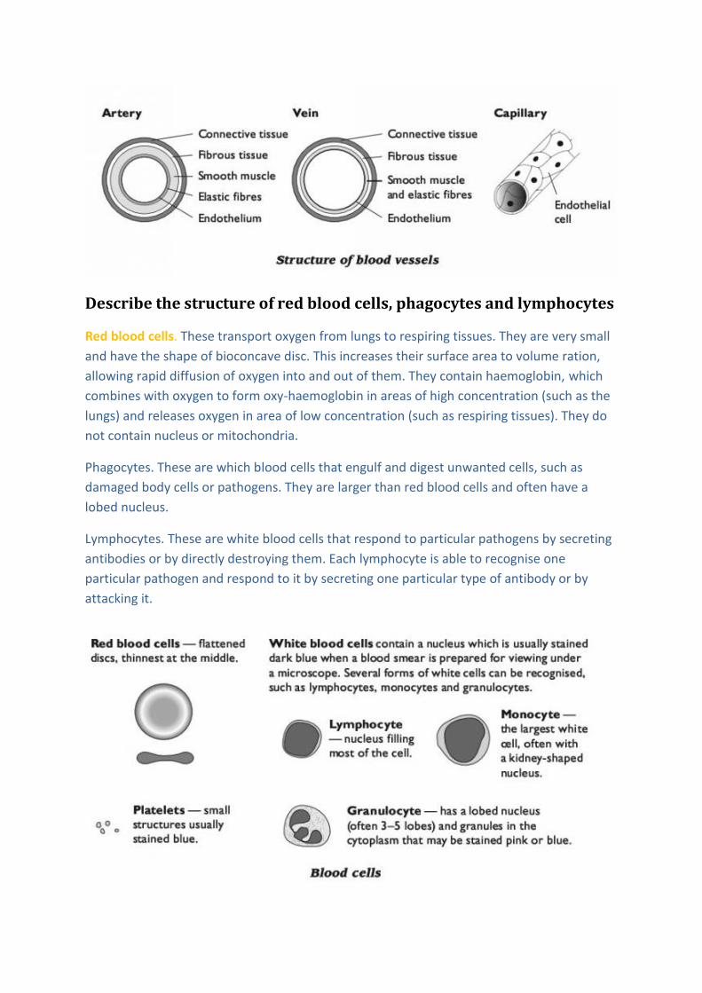
Describe the structure of red blood cells, phagocytes and lymphocytes
Red blood cells. These transport oxygen from lungs to respiring tissues. They are very small
and have the shape of bioconcave disc. This increases their surface area to volume ration,
allowing rapid diffusion of oxygen into and out of them. They contain haemoglobin, which
combines with oxygen to form oxy-haemoglobin in areas of high concentration (such as the
lungs) and releases oxygen in area of low concentration (such as respiring tissues). They do
not contain nucleus or mitochondria.
Phagocytes. These are which blood cells that engulf and digest unwanted cells, such as
damaged body cells or pathogens. They are larger than red blood cells and often have a
lobed nucleus.
Lymphocytes. These are white blood cells that respond to particular pathogens by secreting
antibodies or by directly destroying them. Each lymphocyte is able to recognise one
particular pathogen and respond to it by secreting one particular type of antibody or by
attacking it.

State and explain the differences between blood, tissue fluid and
lymph
Tissue fluid and lymph
Capillaries have tiny gaps between the cells in their walls. Near the arteriole end of a
capillary, there is relatively high pressure inside the capillary, and plasma leaks out through
these gaps to fill the spaces between the body cells. This leaked plasma is called tissue fluid.
Tissue fluid is therefore very similar to blood plasma. However, very large molecules
such as albumin ( a protein carried in solution in blood plasma) and other plasma
protein cannot get through the pores and so remain in the blood plasma.
The tissue fluid baths the body cells. Substance such as oxygen, glucose or urea can
move between the blood plasma and the cells by diffusing through the tissue fluid.
Some tissue fluid moves back into the capillaries, becoming part of the blood plasma once
more. This happens especially at the venule end of the capillary, where blood pressure is
lower, producing a pressure gradient down which the tissue fluid can flow. However, some
of the tissue fluid collects into blind-ending vessels called lymphatic vessels. It is then called
lymph.
Lymphatic vessels have valves that allow fluid to flow into them and along them but not
back out again. They carry the lymph towards the subclavian veins (near the collarbone)
where it is returned to the blood. The lymph passes through lymphatic glands where white
blood cells accumulate. Lymph therefore tends to carry higher densities of white blood cells
than are found in blood plasma or tissue fluid.
Describe the role of haemoglobin in carrying oxygen and carbon
dioxide;
Haemoglobin (Hb) is a protein with quaternary structure. A haemoglobin molecule is made
up of four polypeptide chains, each of which has a haem group at its centre. Each haem
group contains an Fe2+ ion which is able to combine reversibly with oxygen, forming
oxyhaemoglobin. Each iron ion can combine with 2 oxygen atoms, so one Hb molecule can
combine with 8 oxygen atom.
Hb + 4O2 HbO8
Describe and explain the significance of the dissociation curves of
adult oxyhaemoglobin at different carbon dioxide levels (the Bohr
effect);
Oxygen concentration can be measured as partial pressure, in kilopascals (kPa).
Haemoglobin combines with more oxygen at high partial pressures than it does at low

partial pressures. At high partial pressures of oxygen, all the haemoglobin will be combined
with oxygen and we say that it is 100% saturated with oxygen. A graph showing the
relationship between the partial pressure of oxygen and percentage saturation of
haemoglobin with oxygen is known as a dissociation curve.
In
the
lungs, the partial pressure of oxygen may be around 12kPa. You can see from the graph that
the Hb will be about 98% saturated.
In a respiring, the partial pressure of oxygen may be around 2kPa. The Hb will be about 23%
saturated.
Therefore, when Hb from the lungs arrive at a respiring muscle it gives up more than 70% of
the oxygen it is carrying.
The Bohr effect
The presence of CO2 increases acidity that is the concentration of H+ ions. When this
happens, the haemoglobin combines with H+ ions and releases oxygen. Therefore, in areas
of high CO2 concentration, haemoglobin is less saturated with oxygen than it would be if
there was no CO2 present. This is called Bohr effect. It is useful in enabling haemoglobin to
unload more of its oxygen in tissues where respiration (which produces CO2) is taking place.

Describe and explain the significance of the increase in the red blood
cell count of humans at high altitude;
Adaptation to high altitude:
At high altitude, the air is less dense and the partial pressure of oxygen is lower than at sea
level. Haemoglobin is therefore less saturated with oxygen in the lungs and delivers less
oxygen to body tissues.
After some time at high altitude, the number of red blood cells in the blood increases. This
means that there are more haemoglobin molecules in a given volume of blood. Therefore,
even though each Hb molecule carries less oxygen on average than at sea level, the fact that
there are more of them helps to supply the same amount of oxygen to respiring tissues.
Athletes may make use of this by training at high altitude before an important competition.
When they return to low altitude, their extra red blood cells can supply oxygen to their
muscles at a greater rate than in an athlete who has not been to high altitude, giving them a
competitive advantage.
Describe the external and internal structure of the mammalian heart;
Explain the differences in the thickness of the walls of the different
chambers in terms of their functions
The muscle of which the heart is made of is called cardiac muscle. It is made of
interconnecting cells, whose cell surface membranes are very tightly joined together. This
close contact between the muscle cells allows wave of electrical excitation to pass easily
between them.

The large, arching blood vessel is the largest artery, the aorta, with branches leading
upwards towards the head, and the main flow doubling back towards to the rest of the
body. The other blood vessel leaving the heart is the pulmonary artery. This, too, branches
very quickly after leaving the heart, in to two arteries taking blood to the right and left
lungs. Running vertically on the right- hand side of the heart are the two large veins, the
venae cavae, one bringing blood downwards from the heart and the other bringing it
upwards from the rest of the body. The pulmonary veins bring blood back to the heart from
the left and right lungs. On the surface of the heart, the coronary arteries can be seen.
These branch from the aorta, and deliver oxygenated blood to the walls of the heart itself.
If the heart is cut open vertically it can be seen to contain 4 chambers. The two chambers on
the left of the heart are completely separated from those on the right by a wall of muscle
called the septum.
Blood cannot pass through this septum; the only way for blood to get from one side of the
heart to the other is for it to leave the heart, circulate around either the lungs or the rest of
the body, and then return to the heart.

The upper chamber on each side of the heart is called an atrium or sometimes auricle. The
two atria receive blood from the pulmonary veins flows into the left atrium. Blood from the
venae cavae flows into the right atrium, white blood from the pulmonary veins flows into
the left atrium.
The lower chambers are ventricles. Blood flows into the ventricles from the atria, and is
then squeezed out into the arteries. Blood from the left ventricle flows into the aorta, while
blood from the right ventricle flows into the pulmonary arteries.
The atria and ventricles have valves between them, which are known as the atrioventricular
valves. The one on the left is mitral or bicuspid valve.
Describe the mammalian circulatory system as a closed double
circulation;
Cardiovascular system is made up of a pump, the heart and a system of interconnecting
tubes, the blood vessels. The blood is always remains within the vessels and so the system is
known as CLOSED blood system.
Systemic circulation. Blood is pumped out of the left ventricle into the aorta, and travels
from there to all parts of the body except the lungs. It retunes to the right side of the heart
in the vena cava. This is called the systemic circulation.
Pulmonary circulation. The blood is then pumped out of the right ventricle into the
pulmonary arteries, which carry it to the lungs. The final part of the journey is along the
pulmonary veins, which return it to the left side of the heart. This is called the pulmonary
circulation.
The combination of pulmonary circulation and systematic circulation makes double
circulation system.
Describe the cardiac cycle;

When muscle contrasts, it gets shorter. Contraction of the cardiac muscle in the walls of the
heart therefore causes the walls to squeeze inwards on the blood inside the heart. Both
sides of the heart contract and relax together. The complete squeeze inwards on the blood
inside the heart. Both sides of the heart contract and relax together. The complete
sequence of one heart beat is called the cardiac cycle.

Explain how heart action is initiated and controlled (reference should
be made to the sinoatrial node, the atrioventricular node and the
Purkyne tissue);
Cardiac muscle is myogenic – that is, it contracts and relaxes automatically, without the
need of stimulation by nerves. The rhythmic, coordinated contraction of the cardiac muscle
in different parts of the heart is coordinated through electrical impulses passing through the
cardiac muscle tissue.
In the walls of the right atrium, there is a patch of muscle tissue called the
SINOATRIAL NODE (SAN). This has an intrinsic rate of contraction a little higher than
that of the rest of the heart muscle.
As the cells in the SAN contract, they generate action potential (electrical impulses)
which sweep along the muscle in the wall of the right and left atria. This causes the
muscle to contract. This is atrial systole.
When the action potentials reach the atrioventricular node (AVN) in the septum,
they are delayed briefly. They then sweep down the septum between the ventricles,
along fibres of PURKYNE TISSUE, and then up through the ventricle walls. This causes
the ventricle to contract slightly after the atria. The left and right ventricles contract
together from the bottom up. This is ventricle systole.
There is then a short delay before the next wave of action potential is generated in
the SAN. During this time, the heart muscles relax. This is diastole.


![prediabetes - Diabetes Pro - American Diabetes Association · PDF fileDefine prediabetes ... [Epub ahead of print] ... Family history of type 2 diabetes in first- or second-degree](https://img.pdfslide.net/doc/110x75/5aa1bdb67f8b9a84398c1ca6/prediabetes-diabetes-pro-american-diabetes-association-prediabetes-epub.jpg)