Embed Size (px)
Citation preview
REVIEW ARTICLE/BRIEF REVIEW
Transtracheal ultrasound for verification of endotracheal tubeplacement: a systematic review and meta-analysis
Verification du positionnement du tube endotracheal parechographie transtracheale: revue systematique de la litterature etmeta-analyse
Saurabh Kumar Das, MD • Nang Sujali Choupoo, MD •
Rudrashish Haldar, MD • Amitabh Lahkar, MD
Received: 15 May 2014 / Accepted: 9 December 2014 / Published online: 24 December 2014
� Canadian Anesthesiologists’ Society 2014
Abstract
Purpose Early confirmation of endotracheal tube
placement is of paramount importance to prevent hypoxia
and its catastrophic consequences. Despite certain
limitations, capnography is considered the gold standard
to evaluate the proper placement of an endotracheal tube.
Ultrasound is a novel tool with some definitive advantages
over capnography. It enables a real-time view and can be
performed quickly; furthermore, it is independent of
pulmonary blood flow and does not require lung
ventilation. In this review, we aimed to evaluate the
diagnostic accuracy of transtracheal ultrasound in
detecting endotracheal intubation.
Source We completed an extensive search of
MEDLINE�, EMBASETM, The Cochrane Library,
KoreaMed, LILACS, OpenGrey, and the World Health
Organization International Clinical Trials Registry from
their inception to September 4, 2014. The studies that met
the inclusion criteria were pooled and a meta-analysis was
conducted.
Principal findings Eleven studies and 969 intubations
were included in the final analysis. Eight studies and 713
intubations were performed in emergency situations and
the others were carried out in elective situations.
Transtracheal ultrasonography’s pooled sensitivity and
specificity with 95% confidence intervals (CIs) were 0.98
(95% CI 0.97 to 0.99) and 0.98 (95% CI 0.95 to 0.99),
respectively. In emergency scenarios, transtracheal
ultrasonography showed an aggregate sensitivity and
specificity of 0.98 (95% CI 0.97 to 0.99) and 0.94 (95%
CI 0.86 to 0.98), respectively.
Conclusion Transtracheal ultrasound is a useful tool to
confirm endotracheal intubation with an acceptable degree
of sensitivity and specificity. It can be used in emergency
situations as a preliminary test before final confirmation by
capnography.
Resume
Objectif La confirmation rapide du positionnement du
tube endotracheal est d’une importance capitale pour
prevenir l’hypoxie et ses consequences catastrophiques. En
depit de certaines limites, on considere que la capnographie
constitue la reference universelle permettant d’evaluer un
bon positionnement du tube endotracheal. L’echographie est
un nouvel outil qui offre des avantages certains par rapport a
Author contributions Saurabh Kumar Das and Nang SujaliChoupoo are responsible for the integrity of this work from inceptionto manuscript preparation. They contributed to the study design,study selection, quality assessment, records review, data synthesis,data analysis, and manuscript composition. Rudrashish Haldar andAmitabh Lahkar contributed to the review process by searching theliterature and reviewing the search records and manuscripts. Allauthors read the final manuscript.
Electronic supplementary material The online version of thisarticle (doi:10.1007/s12630-014-0301-z) contains supplementarymaterial, which is available to authorized users.
S. K. Das, MD (&) � N. S. Choupoo, MD
Department of Anesthesia and Critical Care, Nazareth Hospital,
Shillong 793003, Meghalaya, India
e-mail: [email protected]
R. Haldar, MD
Department of Anaesthesia, Sanjay Gandhi Post Graduate
Institute of Medical Sciences, Lucknow, India
A. Lahkar, MD
Department of Anesthesia, Milton Keynes Hospital NHS
Foundation Trust, Eaglestone, Milton Keynes, UK
123
Can J Anesth/J Can Anesth (2015) 62:413–423
DOI 10.1007/s12630-014-0301-z
la capnographie. Elle permet une vue en temps reel et peut
etre realisee rapidement; en outre, elle est independante du
debit sanguin pulmonaire et ne necessite pas de ventilation
pulmonaire. Au cours de cette etude, nous avons cherche a
evaluer la precision diagnostique de l’echographie
transtracheale pour la detection de l’intubation
endotracheale.
Source Nous avons mene une recherche extensive dans
les bases de donnees MEDLINE�, EMBASETM, The
Cochrane Library, KoreaMed, LILACS, OpenGrey, ainsi
que dans le Registre des essais cliniques internationaux de
l’Organisation mondiale de la sante depuis leur creation
jusqu’au 4 septembre 2014. Les etudes repondent aux
criteres d’inclusion ont ete regroupees pour permettre la
realisation d’une meta-analyse.
Constatations principales Onze etudes et 969 intubations
ont ete incluses dans l’analyse finale. Huit etudes et
713 intubations ont ete pratiquees dans des situations
d’urgence et les autres ont ete realisees dans des situations
programmees. Apres regroupement, la sensibilite et la
specificite de l’echographie transtracheale, avec
intervalles de confiance (IC) a 95 %, ont ete
respectivement de 0,98 (IC a 95 %: 0,97 a 0,99) et 0,98
(IC a 95 %: 0,95 a 0,99). Dans les situations d’urgence, la
sensibilite et la specificite groupees de l’echographie
transtracheale ont ete, respectivement, de 0,98 (IC a 95 %:
0,97 a 0,99) et 0,94 (IC a 95 %: 0,86 a 0,98).
Conclusion L’echographie transtracheale est un outil utile
pour confirmer l’intubation endotracheale avec des niveaux
de sensibilite et de specificite acceptables. Elle peut etre
utilisee dans les situations d’urgence comme test preliminaire
avant une confirmation definitive par la capnographie.
Introduction
Confirmation of the proper placement of an endotracheal
tube (ETT) is a crucial step in airway management since
unrecognized esophageal intubation leads to catastrophic
consequences. Numerous methods are used to verify ETT
placement, including visual confirmation of the ETT
passing through the vocal cords during laryngoscopy,
expansion of the chest wall during ventilation, visualization
of the tracheal rings and carina using a flexible
bronchoscope, auscultation, capnometry, capnography,
and chest x-ray. These techniques vary in their degree of
accuracy.1,2 The 2010 - Advanced Cardiac Life Support
guidelines recommend the use of quantitative waveform
capnography to confirm ETT placement and to check the
effectiveness of chest compression.3,4 Although,
capnography is considered the gold standard to confirm
tracheal intubation, it has some major limitations. First,
detection of carbon dioxide by capnography depends on
adequate pulmonary blood flow; thus, its accuracy may be
compromised in patients with massive pulmonary
embolism and those suffering from cardiac arrest
(especially where cardiopulmonary resuscitation has not
yet started or the patient is in a state of cardiac arrest for a
prolonged period).5 Second, respiration must be maintained
for several breaths to accurately confirm the ETT
placement. A trauma victim is usually considered to have
a full stomach unless it is proven otherwise, and
inadvertently ventilating the stomach of such a patient
with a misplaced ETT can cause gastric distension,
vomiting, and aspiration.6-8 Third, capnography may
provide false negative results in cases of airway
obstruction or due to the use of epinephrine.9,10 Thus, in
the face of various limitations of the techniques currently
used to verify ETT placement, there is need for a
consistently reliable and safe technique that can be used
in real time without ventilating the lungs.
Ultrasound, once the domain of the radiologist, has now
found its place in pre-hospital applications (e.g., emergency
responders), emergency wards, intensive care units, and
operation theatres. Portable ultrasound is easy to carry, non-
invasive, relatively economical, easily reproducible, and
widely available, and it has a good safety record.11 Various
studies have shown that ultrasound is a novel tool to confirm
proper ETT placement.12-19 Confirmation with ultrasound is
a potential alternative when detection of CO2 by
capnography is compromised, where capnography is not
available, or as an adjunct to capnography.
In recent years, an increasing number of original
research publications have evaluated the accuracy of
ultrasound for confirming ETT intubation and have
reported high sensitivity and specificity using this
technique.6,7,12-19 Two previous attempts were made to
address this important clinical issue, one in 2011 in the
form of a systematic review and one in 2012 in the form of
a systematic review with a meta-analysis.20,21 Both reviews
were presented during conferences, but the full texts were
not published in any scientific journal. While one study did
not report aggregate sensitivity or specificity, the other
included only 323 intubations. After 2012, several high-
quality studies were conducted, which encouraged us to
undertake this systematic review and meta-analysis to test
the accuracy of transtracheal ultrasound in detecting ETT
intubation. Unlike the previous reviews, which included
both transthoracic and transtracheal ultrasound, the focus
of our novel review is on transtracheal ultrasound alone.
Data search
On September 4, 2014, we performed a search of the
published research evaluating the diagnostic accuracy of
414 S. K. Das et al.
123
ultrasound. We searched for published literature in the
English language in MEDLINE� via PubMed,
EMBASETM via Ovid, The Cochrane Library, and Trip
database. For literature published in other languages, we
searched KoreaMed and LILACS, and we searched
OpenGrey (www.opengrey.eu/) and the World Health
Organization Clinical Trials Registry (who.int/ictrp) for
unpublished literature and ongoing studies. Details of our
search strategy are shown in the Appendix (available as
Electronic Supplementary Material). New links displayed
alongside the abstracts were followed and retrieved, and
the bibliographies in the retrieved articles were searched
independently and checked for additional studies. The
authors of the articles were contacted where there was any
confusion regarding the reported data.
Selection of the studies
Studies using transtracheal ultrasound to verify the ETT
position following an emergency or elective intubation were
included in this meta-analysis. The ultrasound examination
was done by placing a linear or curvilinear probe on the
cricothyroid membrane or just above the suprasternal notch.
Probe placement was either transverse or horizontal. The
ultrasound examination was performed simultaneously with
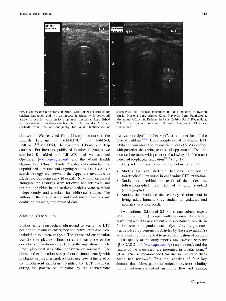
intubation or just afterward. A transverse view at the level of
the cricothyroid membrane identified the ETT placement
during the process of intubation by the characteristic
‘‘snowstorm sign’’, ‘‘bullet sign’’, or a flutter behind the
thyroid cartilage.22,23 Upon completion of intubation, ETT
intubation was identified by one air-mucosa (A-M) interface
with posterior shadowing (comet-tail appearance). Two air-
mucosa interfaces with posterior shadowing (double-track)
indicated esophageal intubation13,14 (Fig. 1).
Study selection was based on the following criteria:
• Studies that evaluated the diagnostic accuracy of
transtracheal ultrasound in confirming ETT intubation.
• Studies that verified the result of the index test
(ultrasonography) with that of a gold standard
(capnography).
• Studies that evaluated the accuracy of ultrasound in
living adult humans (i.e., studies on cadavers and
neonates were excluded).
Two authors (S.D. and S.C.) and one subject expert
(D.P., not an author) independently reviewed the articles,
performed a quality assessment, and ascertained the criteria
for inclusion in the pooled data analysis. Any disagreement
was resolved by consensus. Articles by the same author(s)
were carefully investigated to avoid duplication of studies.
The quality of the study reports was assessed with the
QUADAS-2 tool (www.quadas.org) (supplement), and the
results of the assessment are presented in tabular form.24
QUADAS-2 is recommended for use in Cochrane diag-
nostic test reviews.25 This tool consists of four key
domains that address patient selection, index test (including
timing), reference standard (including, flow and timing),
Fig. 1 Shows one air-mucosa interface with comet-tail artifact for
tracheal intubation and two air-mucosa interfaces with comet-tail
artifact or double-track sign for esophageal intubation. Republished
with permission from American Institute of Ultrasound in Medicine
(AIUM) from Use of sonography for rapid identification of
esophageal and tracheal intubation in adult patients. Bunyamin
Muslu, Huseyin Sert, Ahmet Kaya, Ruveyda Irem Demircioglu,
Muhammet Gozdemir, Brrhanettin Usta, Kadriye Serife Boynuklam,
2011;7 permission conveyed through Copyright Clearance
Center, Inc.
Transtracheal ultrasound 415
123
and flow of patients through the study. The tool should be
used in four phases: 1. summarizing the review question, 2.
developing review-specific guidance, 3. reviewing the
published flow diagram of the primary study or construct a
flow diagram if none is reported, and 4. assessing risk of
bias and concern regarding applicability.24
Data synthesis
We arranged the data in 2 9 2 tables expressing true
positive, false positive, false negative, and true negative. In
the primary studies where data were not expressed in
contingency tables, we reconstructed 2 9 2 tables from the
number of ETT and esophageal intubations.
Data analysis
We constructed the forest plots with freeware Meta-DiSc,
version 1.4 software (http://www.hrc.es/investigacion/
metadisc-en.htm; Ramon y Cajal Hospital; Madrid,
Spain).26 Pooled sensitivity and specificity and their cor-
responding 95% confidence intervals (CIs) were obtained.
To test for heterogeneity among various studies, eyeball
tests and the (Cochran Q) were performed. A P value of\0.10 indicated significant heterogeneity. Meta-DiSc com-
putes the inconsistency index (I2), which has been
proposed as a measure to quantify the amount of hetero-
geneity. Inconsistency (I2) describes the percentage of total
variation across studies that are due to heterogeneity rather
than due to chance. The I2 can be readily calculated from
basic results obtained from a typical meta-analysis as
I2 = 100%(Q - df)/Q, where Q is Cochran’s heterogene-
ity and df is the degree of freedom. Cochran’s Q is
computed by summing the squared deviations of each
study’s estimate from the overall meta-analytic estimate.
Sensitivity analysis was performed to ascertain the
robustness of the result of the review after excluding the
studies where intubations were done electively in a con-
trolled environment and studies that had more than one
area of high or unclear risk of bias. Another sensitivity
analysis was done after excluding studies that did not use
one A-M interface with comet-tail or reverberation artifact
or similar features for tracheal intubation and those that did
not use two A-M interfaces with comet-tail or reverbera-
tion artifact or similar features for esophageal intubation.
The method of analysis was decided prospectively
before commencement of the data search.
Results
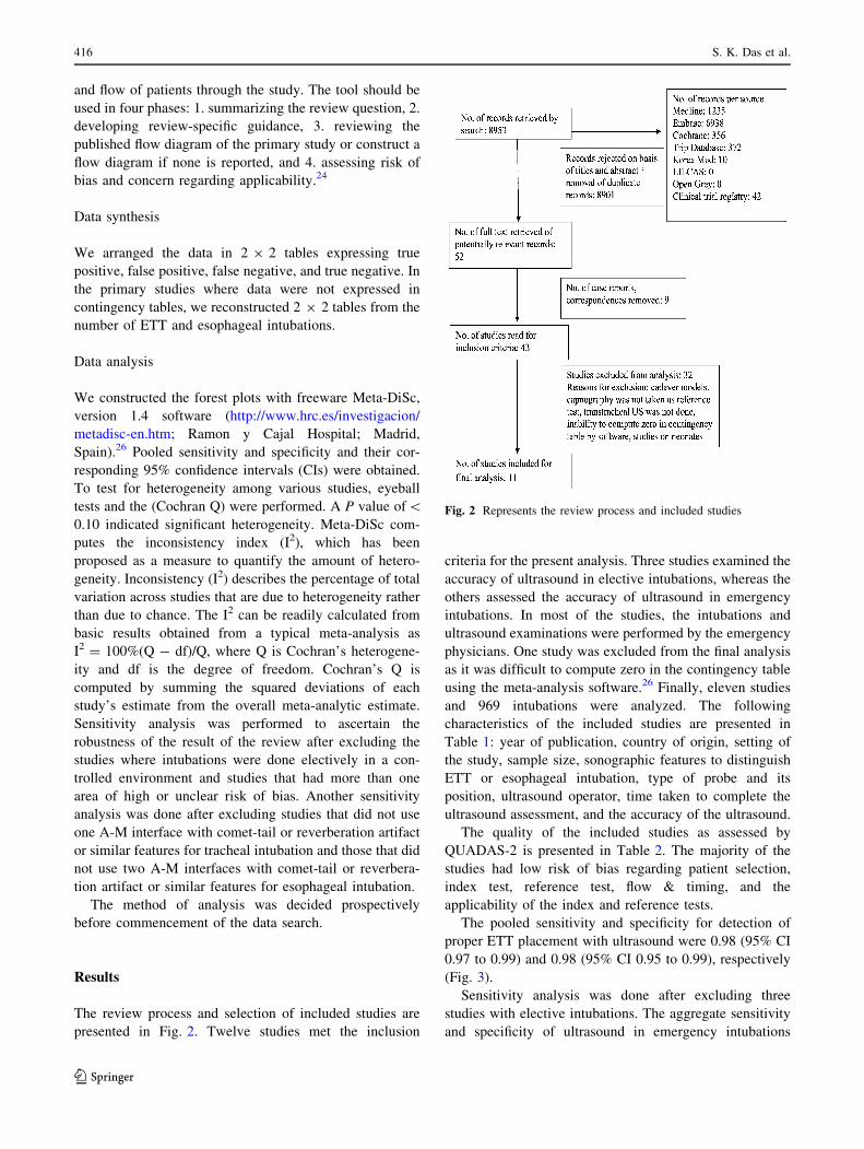
The review process and selection of included studies are
presented in Fig. 2. Twelve studies met the inclusion
criteria for the present analysis. Three studies examined the
accuracy of ultrasound in elective intubations, whereas the
others assessed the accuracy of ultrasound in emergency
intubations. In most of the studies, the intubations and
ultrasound examinations were performed by the emergency
physicians. One study was excluded from the final analysis
as it was difficult to compute zero in the contingency table
using the meta-analysis software.26 Finally, eleven studies
and 969 intubations were analyzed. The following
characteristics of the included studies are presented in
Table 1: year of publication, country of origin, setting of
the study, sample size, sonographic features to distinguish
ETT or esophageal intubation, type of probe and its
position, ultrasound operator, time taken to complete the
ultrasound assessment, and the accuracy of the ultrasound.
The quality of the included studies as assessed by
QUADAS-2 is presented in Table 2. The majority of the
studies had low risk of bias regarding patient selection,
index test, reference test, flow & timing, and the
applicability of the index and reference tests.
The pooled sensitivity and specificity for detection of
proper ETT placement with ultrasound were 0.98 (95% CI
0.97 to 0.99) and 0.98 (95% CI 0.95 to 0.99), respectively
(Fig. 3).
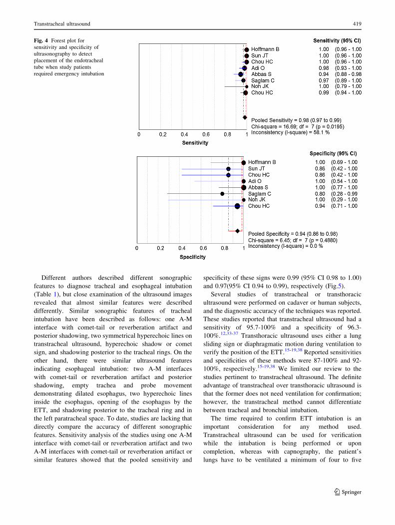
Sensitivity analysis was done after excluding three
studies with elective intubations. The aggregate sensitivity
and specificity of ultrasound in emergency intubations
Fig. 2 Represents the review process and included studies
416 S. K. Das et al.
123
were 0.98 (95% CI 0.97 to 0.99) and 0.94 (95% CI 0.86 to
0.98), respectively (Fig. 4).
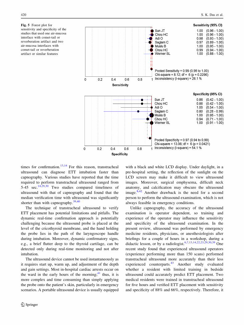
The pooled sensitivity and specificity of the studies
using one A-M interface with comet-tail artifact and two
A-M interfaces with comet-tail artifact or similar features
were 0.99 (95% CI 0.98 to 1.00) and 0.97 (95% CI 0.94 to
0.99), respectively (Fig. 5).
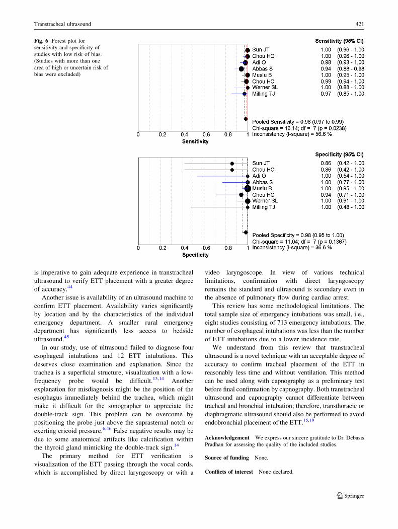
Pooled sensitivity and specificity of studies with a low
risk of bias were 0.98 (95% CI 0.97 to 0.99) and 0.98 (95%
CI 0.95 to 1.00), respectively (Fig. 6).
The Cochran X2 and the inconsistency index (I2) in the
forest plots indicated mild to moderate variation across the
studies.
Discussion
The present study showed that ultrasound is a novel tool to
confirm ETT intubation with an overall pooled sensitivity
and specificity of 98% (95% CI 97 to 99) and 98% (95% CI
95 to 99), respectively. The aggregate sensitivity and
specificity of ultrasound in emergency intubations were
98% (95% CI 97 to 99) and 94% (95% CI 86 to 98),
respectively (Figs. 3, 4).
One meta-analysis of five studies and 323 intubations
reported that the pooled sensitivity and specificity of
ultrasound were 91% (95% CI 74 to 97) and 97% (95% CI
89 to 99), respectively.20 This difference in accuracy
compared with the present study may be attributed to fewer
studies and the smaller sample size of the previous study.
Another study reported that capnography had 100%
sensitivity and specificity in both cardiac arrest and non-
arrest patients.1 Based on 2,192 intubations, a meta-analysis
of capnography trials resulted in an aggregate sensitivity of
93% (95% CI 92 to 94) and an aggregate specificity of 97%
(95% CI 93 to 99) for confirmation of emergency ETT
placement.32 The present review showed comparable
sensitivity and specificity of ultrasound, although the
sample size of the present study was small (2,192 vs 969).
Transtracheal ultrasound is a relatively simple technique
and easy to learn.7 A transverse view just above the
suprasternal notch could be used to visualize both the
trachea and esophagus, but a sagittal view is limited to a
long-axis view of either the trachea or one of the
paratracheal spaces.6 Moreover, the esophagus travels
more laterally than the trachea as it moves inferiorly
from the level of the cricoid cartilage towards the
suprasternal notch.6 The typical ultrasound image of
intubation (air-mucosa artifact) is due to the sound
impedance shift at the interface between the water-filled
mucosa and air. This standard pattern is easy to detect,
although the tube itself is not identified.8
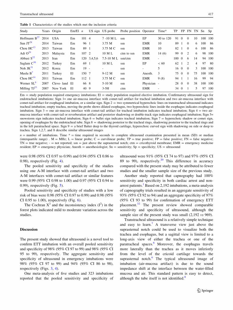
Table 1 Characteristics of the studies which met the inclusion criteria
Study Years Origin Em/El n US sign US probe Probe position Operator Time* TP FP FN TN Sn Sp
Hoffmann B27 2014 USA Em 101 4 7 -10 M L ssn EP 30 to 120 91 0 0 10 100 100
Sun JT28 2014 Taiwan Em 96 1 3.75 M ssn EMR 10 89 1 0 6 100 86
Chou HC13 2013 Taiwan Em 89 1 3.75 M C ssn EMR 10 82 1 0 6 100 86
Adi O29 2013 Malaysia Em 107 2 10 M L ctm to ssn EMR 14 (6) 99 0 2 6 98 100
Abbasi S22 2013 Iran Em 120 3,4,5,6 7.5-10 M L ssn/ctm EMR - 100 0 6 14 94 100
Saglam C30 2012 Turkey Em 69 1 10 M L ssn EP \ 60 62 1 2 4 97 80
Noh JK31 2012 Korea Em 19 - - ssn - 5 16 0 0 3 100 100
Muslu B7 2011 Turkey El 150 7 9-12 M ssn Anesth. 3 75 0 0 75 100 100
Chou HC14 2011 Taiwan Em 112 1 3.75 M C ssn EMR 9 (8) 94 1 1 16 99 94
Werner SL6 2007 Cleve- land El 66 8 5-10 M ssn Physician - 28 0 0 38 100 100
Milling TJ23 2007 New York El 40 9 3-5H ctm EMR - 34 0 1 5 97 100
Em = study population required emergency intubations; El = study population required elective intubation. Confirmatory ultrasound sign for
endotracheal intubations: Sign 1= one air-mucosa interface with comet-tail artifact for tracheal intubation and two air-mucosa interface with
comet-tail artifact for esophageal intubation, or a similar sign. Sign 2 = two symmetrical hyperechoic lines on transtracheal ultrasound indicates
tracheal intubation; empty trachea, moving the probe shows dilated esophagus, two hyperechoic lines inside the esophagus indicates esophageal
intubation. Sign 3 = one air-mucosa interface with comet-tail artifact for tracheal intubation indicates tracheal intubation; Sign 4 = two air-
mucosa interface with comet-tail or reverberation artifact and posterior shadowing or double-track sign indicates esophageal intubation; Sign 5=
snowstorm sign indicates tracheal intubation; Sign 6 = bullet sign indicates tracheal intubation; Sign 7 = hyperechoic shadow or comet sign,
opening of esophagus by the endotracheal tube. Sign 8 = shadowing posterior to the tracheal rings, shadowing posterior to the tracheal rings and
in the left paratracheal space. Sign 9 = a brief flutter deep to the thyroid cartilage, hyperechoic curved sign with shadowing on side or deep to
trachea. Sign 1,2,7, and 8 describe similar ultrasound images
n = number of intubations. Time * = time required in seconds to complete ultrasound examination presented in mean (SD) or median
(interquartile range) . M = MHz; L = linear probe; C = curvilinear probe; TP = true positive; FP = false positive; FN = false negative;
TN = true negative; - = not reported; ssn = just above the suprasternal notch; ctm = cricothyroid membrane; EMR = emergency medicine
resident; EP = emergency physician; Anesth = anesthesiologist; Sn = sensitivity; Sp = specificity; US = ultrasound
Transtracheal ultrasound 417
123
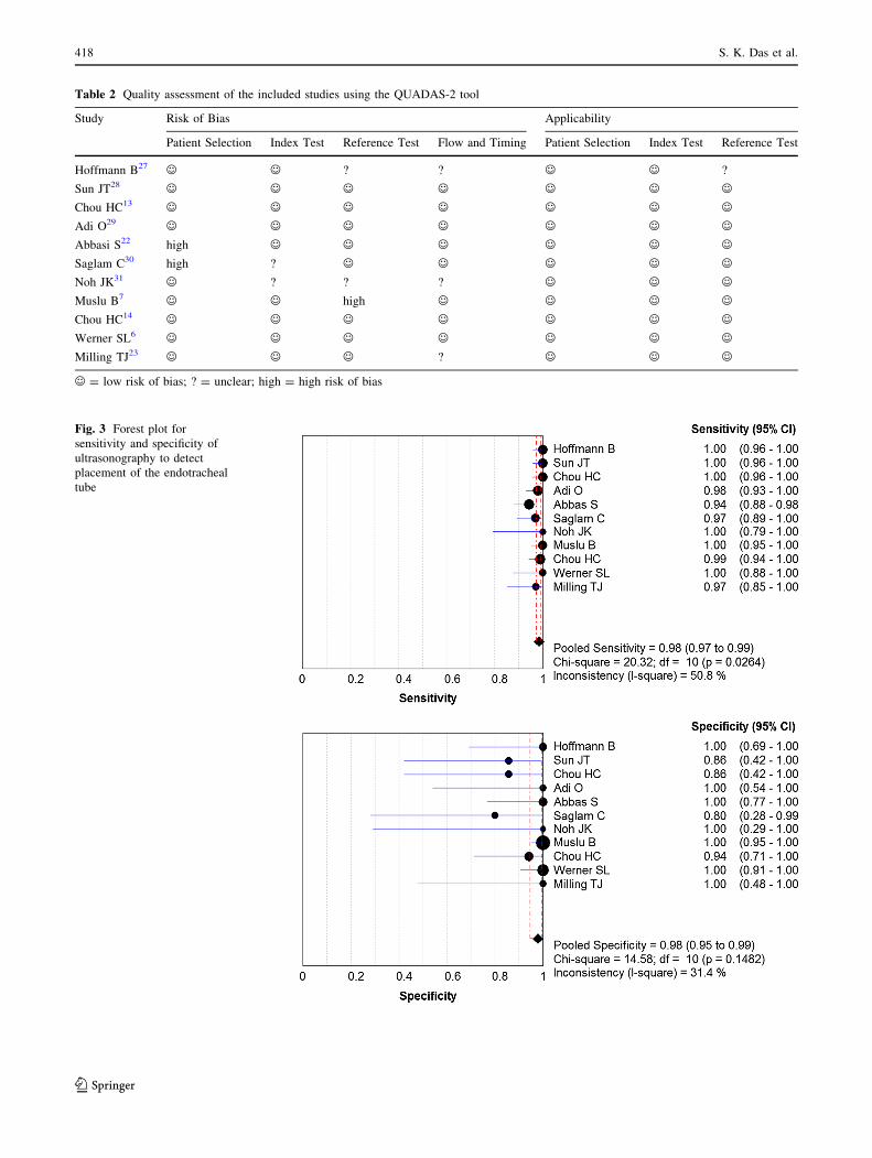
Fig. 3 Forest plot for
sensitivity and specificity of
ultrasonography to detect
placement of the endotracheal
tube
Table 2 Quality assessment of the included studies using the QUADAS-2 tool
Study Risk of Bias Applicability
Patient Selection Index Test Reference Test Flow and Timing Patient Selection Index Test Reference Test
Hoffmann B27 , , ? ? , , ?
Sun JT28 , , , , , , ,Chou HC13 , , , , , , ,Adi O29 , , , , , , ,Abbasi S22 high , , , , , ,Saglam C30 high ? , , , , ,Noh JK31 , ? ? ? , , ,Muslu B7 , , high , , , ,Chou HC14 , , , , , , ,Werner SL6 , , , , , , ,Milling TJ23 , , , ? , , ,
, = low risk of bias; ? = unclear; high = high risk of bias
418 S. K. Das et al.
123
Different authors described different sonographic
features to diagnose tracheal and esophageal intubation
(Table 1), but close examination of the ultrasound images
revealed that almost similar features were described
differently. Similar sonographic features of tracheal
intubation have been described as follows: one A-M
interface with comet-tail or reverberation artifact and
posterior shadowing, two symmetrical hyperechoic lines on
transtracheal ultrasound, hyperechoic shadow or comet
sign, and shadowing posterior to the tracheal rings. On the
other hand, there were similar ultrasound features
indicating esophageal intubation: two A-M interfaces
with comet-tail or reverberation artifact and posterior
shadowing, empty trachea and probe movement
demonstrating dilated esophagus, two hyperechoic lines
inside the esophagus, opening of the esophagus by the
ETT, and shadowing posterior to the tracheal ring and in
the left paratracheal space. To date, studies are lacking that
directly compare the accuracy of different sonographic
features. Sensitivity analysis of the studies using one A-M
interface with comet-tail or reverberation artifact and two
A-M interfaces with comet-tail or reverberation artifact or
similar features showed that the pooled sensitivity and
specificity of these signs were 0.99 (95% CI 0.98 to 1.00)
and 0.97(95% CI 0.94 to 0.99), respectively (Fig.5).
Several studies of transtracheal or transthoracic
ultrasound were performed on cadaver or human subjects,
and the diagnostic accuracy of the techniques was reported.
These studies reported that transtracheal ultrasound had a
sensitivity of 95.7-100% and a specificity of 96.3-
100%.12,33-37 Transthoracic ultrasound uses either a lung
sliding sign or diaphragmatic motion during ventilation to
verify the position of the ETT.15-19,38 Reported sensitivities
and specificities of these methods were 87-100% and 92-
100%, respectively.15-19,38 We limited our review to the
studies pertinent to transtracheal ultrasound. The definite
advantage of transtracheal over transthoracic ultrasound is
that the former does not need ventilation for confirmation;
however, the transtracheal method cannot differentiate
between tracheal and bronchial intubation.
The time required to confirm ETT intubation is an
important consideration for any method used.
Transtracheal ultrasound can be used for verification
while the intubation is being performed or upon
completion, whereas with capnography, the patient’s
lungs have to be ventilated a minimum of four to five
Fig. 4 Forest plot for
sensitivity and specificity of
ultrasonography to detect
placement of the endotracheal
tube when study patients
required emergency intubation
Transtracheal ultrasound 419
123
times for confirmation.13,14 For this reason, transtracheal
ultrasound can diagnose ETT intubation faster than
capnography. Various studies have reported that the time
required to perform transtracheal ultrasound ranged from
5-45 sec.14,29,30 Two studies compared timeliness of
ultrasound with that of capnography and found that the
median verification time with ultrasound was significantly
shorter than with capnography.39,40
The technique of transtracheal ultrasound to verify
ETT placement has potential limitations and pitfalls. The
dynamic real-time confirmation approach is potentially
challenging because the ultrasound probe is placed at the
level of the cricothyroid membrane, and the hand holding
the probe lies in the path of the laryngoscope handle
during intubation. Moreover, dynamic confirmatory signs,
e.g., a brief flutter deep to the thyroid cartilage, can be
detected only during real-time monitoring and not after
intubation.
The ultrasound device cannot be used instantaneously as
it requires start up, warm up, and adjustment of the depth
and gain settings. Most in-hospital cardiac arrests occur on
the ward in the early hours of the morning;41 thus, it is
more complex and time consuming than simply applying
the probe onto the patient’s skin, particularly in emergency
scenarios. A portable ultrasound device is usually equipped
with a black and white LCD display. Under daylight, in a
pre-hospital setting, the reflection of the sunlight on the
LCD screen may make it difficult to view ultrasound
images. Moreover, surgical emphysema, difficult neck
anatomy, and calcification may obscure the ultrasound
image.8,42 Another drawback is the need for a second
person to perform the ultrasound examination, which is not
always feasible in emergency conditions.
Unlike capnography, the accuracy of the ultrasound
examination is operator dependent, so training and
experience of the operator may influence the sensitivity
and specificity of the ultrasound examination. In the
present review, ultrasound was performed by emergency
medicine residents, physicians, or anesthesiologists after
briefings for a couple of hours in a workshop, during a
didactic lesson, or by a radiologist.6,7,13,14,22,23,29,30,39 One
recent study found that experienced ultrasound operators
(experience performing more than 150 scans) performed
transtracheal ultrasound more accurately than their less
experienced counterparts.43 Another study evaluated
whether a resident with limited training in bedside
ultrasound could accurately predict ETT placement. Two
medical residents were trained in transtracheal ultrasound
for five hours and verified ETT placement with sensitivity
and specificity of 88% and 66%, respectively. Therefore, it
Fig. 5 Forest plot for
sensitivity and specificity of the
studies that used one air-mucosa
interface with comet-tail or
reverberation artifact and two
air-mucosa interfaces with
comet-tail or reverberation
artifact or similar features
420 S. K. Das et al.
123
is imperative to gain adequate experience in transtracheal
ultrasound to verify ETT placement with a greater degree
of accuracy.44
Another issue is availability of an ultrasound machine to
confirm ETT placement. Availability varies significantly
by location and by the characteristics of the individual
emergency department. A smaller rural emergency
department has significantly less access to bedside
ultrasound.45
In our study, use of ultrasound failed to diagnose four
esophageal intubations and 12 ETT intubations. This
deserves close examination and explanation. Since the
trachea is a superficial structure, visualization with a low-
frequency probe would be difficult.13,14 Another
explanation for misdiagnosis might be the position of the
esophagus immediately behind the trachea, which might
make it difficult for the sonographer to appreciate the
double-track sign. This problem can be overcome by
positioning the probe just above the suprasternal notch or
exerting cricoid pressure.6,46 False negative results may be
due to some anatomical artifacts like calcification within
the thyroid gland mimicking the double-track sign.14
The primary method for ETT verification is
visualization of the ETT passing through the vocal cords,
which is accomplished by direct laryngoscopy or with a
video laryngoscope. In view of various technical
limitations, confirmation with direct laryngoscopy
remains the standard and ultrasound is secondary even in
the absence of pulmonary flow during cardiac arrest.
This review has some methodological limitations. The
total sample size of emergency intubations was small, i.e.,
eight studies consisting of 713 emergency intubations. The
number of esophageal intubations was less than the number
of ETT intubations due to a lower incidence rate.
We understand from this review that transtracheal
ultrasound is a novel technique with an acceptable degree of
accuracy to confirm tracheal placement of the ETT in
reasonably less time and without ventilation. This method
can be used along with capnography as a preliminary test
before final confirmation by capnography. Both transtracheal
ultrasound and capnography cannot differentiate between
tracheal and bronchial intubation; therefore, transthoracic or
diaphragmatic ultrasound should also be performed to avoid
endobronchial placement of the ETT.15,19
Acknowledgement We express our sincere gratitude to Dr. Debasis
Pradhan for assessing the quality of the included studies.
Source of funding None.
Conflicts of interest None declared.
Fig. 6 Forest plot for
sensitivity and specificity of
studies with low risk of bias.
(Studies with more than one
area of high or uncertain risk of
bias were excluded)
Transtracheal ultrasound 421
123
References
1. Grmec S. Comparison of three different methods to confirm
tracheal tube placement in emergency intubation. Intensive Care
Med 2002; 28: 701-4.
2. Salem MR. Verification of endotracheal tube position.
Anesthesiol Clin North America 2001; 19: 813-39.
3. Neumar RW, Otto CW, Link MS, et al. 2010 American Heart
Association Guidelines for Cardiopulmonary Resuscitation and
Emergency Cardiovascular Care. Part 8: Adult advanced
cardiovascular life support. Circulation 2010; 122: S729-67.
4. Kodali BS. Capnography outside the operating rooms.
Anesthesiology 2013; 118: 192-201.
5. Cook TM, Nolan JP. Use of capnography to confirm correct
tracheal intubation during cardiac arrest. Anaesthesia 2011; 66:
1183-4.
6. Werner SL, Smith CE, Goldstein JR, Jones RA, Cydulka RK. Pilot
study to evaluate the accuracy of ultrasounography in confirming
endotracheal tube placement. Ann Emerg Med 2007; 49: 75-80.
7. Muslu B, Sert H, Kaya A, et al. Use of sonography for rapid
identification of esophageal and tracheal intubation in adult
patients. J Ultrasound Med 2011; 30: 671-6.
8. Zechner PM, Breitkreutz R. Ultrasound instead of capnometry for
confirming tracheal tube placement in an emergency?
Resuscitation 2011; 82: 1259-61.
9. Takeda T, Tanigawa K, Tanaka H, Hayashi Y, Goto E, Tanaka K.
The assessment of three methods to verify tracheal tube placement
in the emergency setting. Resuscitation 2003; 56: 153-7.
10. Levine RI, Wayne MA, Miller CC. End-tidal carbon dioxide and
outcome of out-of-hospital cardiac arrest. N Engl J Med 1997;
337: 301-6.
11. Sustic A. Role of ultrasound in the airway management of
critically ill patients. Crit Care Med 2007; 35(5 Suppl): S173-7.
12. Galicinao J, Bush AJ, Godambe SA. Use of bedside
ultrasonography for endotracheal tube placement in pediatric
patients: a feasibility study. Pediatrics 2007; 120: 1297-303.
13. Chou HC, Chong KM, Sim SS, et al. Real-time tracheal
ultrasonography for confirmation of endotracheal tube
placement during cardiopulmonary resuscitation. Resuscitation
2013; 84: 1708-12.
14. Chou HC, Tseng WP, Wang CH, et al. Tracheal rapid ultrasound
exam (T.R.U.E) for confirming endotracheal tube placement
during emergency intubation. Resuscitation 2011; 82: 1279-84.
15. Hsieh KS, Lee CL, Lin CC, Huang TC, Weng KP, Lu WH.
Secondary confirmation of endotracheal tube position by
ultrasound image. Crit Care Med 2004; 32(9 Suppl): S374-7.
16. Hosseini JS, Talebian MT, Ghafari MH, Eslami V. Secondary
confirmation of endotracheal tube position by diaphragm motion
in right subcostal ultrasound view. Int J Crit Illn Inj Sci 2013; 3:
113-7.
17. Lyon M, Walton P, Bhalla V, Shiver SA. Ultrasound detection of
sliding lung sign by prehospital critical care providers. Am J
Emerg Med 2012; 30: 485-8.
18. Weaver B, Lyon M, Blaivas M. Confirmation of endotracheal tube
placement after intubation using the ultrasound sliding lung sign.
Acad Emerg Med 2006; 13: 239-44.
19. Sim SS, Lien WC, Chou HC, et al. Ultrasonographic lung sliding
sign in confirming proper endotracheal intubation during
emergency intubation. Resuscitation 2012; 83: 307-12.
20. Adhikari S, Farrell I, Stolz U. 46 How accurate is
ultrasonography in confirming endotracheal tube placement? A
meta-analysis. Ann Emerg Med 2012; 60: S18.
21. Herman D, Rudd B. APoo3 Where is the endotracheal tube? A
literature review of rapid ultrasound confirmation. Resuscitation
2011; 82(Suppl 1): S10.
22. Abbasi S, Farsi D, Zare MA, Hajimohammadi M, Rezai M,
Hafezimoghadam P. Direct ultrasound methods: a confirmatory
technique for proper endotracheal intubation in the emergency
department. Eur J Emerg Med 2014; DOI: 10.1097/MEJ.
0000000000000108.
23. Milling TJ, Jones M, Khan T, et al. Transtracheal 2-d ultrasound
for identification of esophageal intubation. J Emerg Med 2007;
32: 409-14.
24. Whiting PF, Rutjes AW, Westwood ME, et al. QUADAS-2: a
revised tool for the quality assessment of diagnostic accuracy
studies. Ann Intern Med 2011; 155: 529-36.
25. Deeks JJ, Wisniewski S, Davenport C. Chapter 4: Guide to the
contents of a Cochrane Diagnostic Test Accuracy Protocol. In:
Deeks JJ, Bossuyt PM, Gatsonic C (Eds). Cochrane Handbook for
Systematic Reviews of Diagnostic Test Accuracy Version 1.0.0.
The Cochrane collaboration, 2013. Available from URL: http://
srdta.cochrane.org/ (accessed November 2014).
26. Zamora J, Abraira V, Muriel A, Khan K, Coomarasamy A. Meta-
DiSc: a software for meta-analysis of test accuracy data. BMC
Med Res Methodol 2006; 6: 31.
27. Hoffmann B, Gullett JP, Hill HF, et al. Bedside ultrasound of the
neck confirms endotracheal tube in emergency intubations.
Ultraschall Med 2014; 35: 451-8.
28. Sun JT, Chou HC, Sim SS, et al. Ultrasonography for proper
endotracheal tube placement confirmation in out-of-hospital
cardiac arrest patients: two-center experience. J Med
Ultrasound 2014; 22: 83-7.
29. Adi O, Chuan TW, Rishya M. A feasibility study on bedside upper
airway ultrasonography compared to waveform capnography for
verifying endotracheal tube location after intubation. Crit
Ultrasound J 2013; 5: 7.
30. Saglam C, Unluer EE, Karagoz A. Confirmation of endotracheal
tube position during resuscitation by bedside ultrasonography.
Am J Emerg Med 2013; 31: 248-50.
31. Noh JK, Cho YS. The comparison of airway ultrasonography and
continuous waveform capnography to confirm endotracheal tube
placement in cardiac arrest patients: prospective observational
study. Crit Ultrasound J 2012; 4(Suppl 1): A6.
32. Li J. Capnography alone is imperfect for endotracheal tube
placement confirmation during emergency Intubation. J Emerg
Med 2001; 20: 223-9.
33. Goksu E, Sayrac V, Oktay C, Kartal M, Akcimen M. How stylet
use can effect confirmation of endotracheal tube position using
ultrasound. Am J Emerg Med 2010; 28: 32-6.
34. Ma G, Davis DP, Schmitt J, Vilke GM, Chan TC, Hayden SR. The
sensitivity and specificity of transcricothyroid ultrasonography to
confirm endotracheal tube placement in a cadaver model. J Emerg
Med 2007; 32: 405-7.
35. Uya A, Spear D, Patel K, Okada P, Sheeran P, McCreight A. Can
novice sonographers accurately locate an endotracheal tube with
a saline—filled cuff in a cadaver model? A pilot study. Acad
Emerg Med 2012; 19: 361-4.
36. Slovis TL, Poland RL. Endotracheal tubes in neoanates:
sonographic positioning. Radiology 1986; 160: 262-3.
37. Park SC, Ryu JH, Yeom SR, Jeong JW, Cho SJ. Confirmation of
endotracheal intubation by combined ultrasonographic methods
in the emergency department. Emerg Med Australas 2009; 21:
293-7.
38. Kerrey BT, Geis GL, Quinn AM, Hornung RW, Ruddy RM. A
prospective comparison of diaphragmatic ultrasound and chest
radiography to determine endotracheal tube position in a pediatric
emergency department. Pediatrics 2009; 123: e1039-44.
39. Pfeiffer P, Rudolph SS, Borglum J, Isbye DL. Temporal
comparison of ultrasound vs. auscultation and capnography in
verification of endotracheal tube placement. Acta Anaesthesiol
Scand 2011; 55: 1190-5.
422 S. K. Das et al.
123

40. Pfeiffer P, Bache S, Isbye DL, Rudolph SS, Rovsing L, Borglum J.
Verification of endotracheal intubation in obese patients –
temporal comparison of ultrasound vs. auscultation and
capnography. Acta Anaesthesiol Scand 2012; 56: 571-6.
41. Buff DD, Fleisher JM, Roca JA, Jaffri M, Wyrwinski PM.
Circadian distribution of in-hospital cardiopulmonary arrests on
the general medical ward. Arch Intern Med 1992; 152: 1282-8.
42. Xue FS, Liao X, Wang Q. Confirmation of endotracheal tube
placement during emergency intubation. Resuscitation 2012; 83:
e67.
43. Stuntz R, Kochert E, Kehrl T, Schrading W. The effect of
sonologist experience on the ability to determine endotracheal
tube location using trastracheal ultrasound. Am J Emerg Med
2014; 32: 267-9.
44. Sharma N, Cardenas-Garcia J, Tibb A. Limited training in
bedside ultrasound can accurately predict endotracheal tube
placement. Chest 2013; 144: 547A (abstract).
45. Talley BE, Ginde AA, Raja AS, Sullivan AF, Espinola JA,
Camargo CA Jr. Variable access to immediate bedside ultrasound
in the emergency department. West J Emerg Med 2011; 12: 96-9.
46. Smith KJ, Dobranowski J, Yip G, Dauphin A, Choi PT. Cricoid
pressure displaces the esophagus: an observational study using
magnetic resonance imaging. Anesthesiology 2003; 99: 60-4.
Transtracheal ultrasound 423
123