Embed Size (px)
Citation preview

This work is subject to an International Creative Commons Attribution 4.0 license.
Treatment of pancreatic necrosis – the Multimodal Glasgow Algorithm
C. Ross Carter, Euan J Dickson, Colin J McKay
West of Scotland Pancreatico-Biliary Unit, Glasgow Royal Infirmary, 84 Castle Street,
Glasgow, United Kingdom, G4 0SF
e-mail: [email protected]
Version 1.0, August 7, 2015 [DOI: 10.3998/panc.2015.30]
1. Introduction
The 2013 APA/IAP consensus document (44)
outlined the principles of early targeted organ
support, nutritional (enteral) optimisation,
avoidance of antibiotic prophylaxis / ERCP (in
the absence of jaundice), and delayed
minimally invasive intervention embedded
within a “step-up” framework where possible.
An in-depth discussion of the evidence
supporting these principles is beyond the
scope of this chapter and will be dealt with
elsewhere. This chapter will focus on the
indications and rationale for intervention, and
the options available within a multi-modal
management algorithm.
2. Revised Atlanta Classification
of Acute Pancreatitis
The original Atlanta classification (7) of acute
pancreatitis characterised clinical behavior as
mild or severe acute pancreatitis and
intervention for necrosis was often focused on
early removal of sterile or infected necrosis
usually by open necrosectomy. This simplistic
dichotomization proved inadequate in clinical
practice until the revised Atlanta Criteria (4)
recognized the importance of early systemic
organ dysfunction and multiple organ failure in
determining disease severity and outcome. The
management of local complications is heavily
influenced by the degree of systemic
disturbance, and this is reflected in an
additional category of “moderately severe”
pancreatitis. In addition to disease severity,
mortality is strongly associated with age,
comorbidity and the presence of infection,
which has been recognized in an addendum
adding a category of “critical” recognizing those
patients with sepsis and organ failure are
associated with the highest mortality (27).
Table 1: Grades of severity for acute pancreatitis (4)
(based on the clinical parameters of the presence or absence of organ failure and / or complications) Mild acute pancreatitis
▸ No organ failure
▸ No local or systemic complications
Moderately severe acute pancreatitis
▸ Organ failure that resolves within 48 h (transient organ failure) and/or
▸ Local or systemic complications without persistent organ failure
Severe acute pancreatitis
▸ Persistent organ failure (>48 h)
–Single organ failure –Multiple organ failure

2
Furthermore, this classification further
categorises local complications on the basis of
time from presentation (< or > 4 weeks) and on
the presence of necrosis, leading to definitions
aimed at permitting comparison of case series
(Table 2). The “early” phase is characterized
by the initial host response to the pancreatitis,
the severity being determined by the
magnitude or organ disturbance /failure, and a
“late” phase typified by the persistence of organ
dysfunction and the management of local or
systemic complications. The vast majority of
acute fluid collections without necrosis will
resolve within 4 weeks and a persistent fluid
collection with minimal or no necrotic
component (“pseudocyst”) is very rare.
Collections may be sterile or infected. The
majority of clinically significant peri-pancreatic
complications are therefore related to either
acute necrotic collections (<4 weeks) or walled-
off pancreatic necrosis (>4 weeks). This
temporal separation is somewhat arbitrary, as
the clinical management and surgical approach
is determined by multifactorial individual patient
factors. However, this does serve to provide a
timeline beyond which, if appropriate,
intervention should be delayed. (Figure 1).
Table 2: Local complications in acute pancreatitis (2012 Revised Atlanta Classification)
Time scale Necrosis absent Necrosis present
< 4 weeks Acute peripancreatic fluid collection
(peripancreatic fluid associated with interstitial
oedematous pancreatitis with no associated
peripancreatic necrosis)
Acute necrotic collection (a collection containing
variable amounts of both fluid and necrosis; the necrosis
can involve the pancreatic parenchyma or the
extrapancreatic tissues)
> 4 weeks Pancreatic pseudocyst (an encapsulated
collection of fluid with a well-defined
inflammatory wall usually outside the pancreas
with minimal or no necrosis)
Walled-off necrosis (a mature, encapsulated collection
of pancreatic or extrapancreatic necrosis that has
developed a well-defined inflammatory wall)
Infection Each collection type may be sterile or infected
Figure 1. Contrast enhanced CT in a 69 yr old woman with severe acute gallstone pancreatitis at (a) 5 days
showing an acute necrotic collection (ANC) and (b) Walled off Necrosis (WON) at 7 weeks subsequently
managed by laparoscopic cystgastrotomy and cholecystectomy (c) fluid level in an acute necrotic collection
suggestive of spontaneous fistulation (clinically well) and (d) loculated gas within an Infected Acute necrotic
collection suggestive of bacterial contamination (clinical sepsis).

3
3. Indications for Intervention for
Pancreatic Necrosis – the Bi-
Phasic Model
Two distinct phases of mortality are seen in
acute pancreatitis: Early death (arbitrarily
defined as within two weeks of onset) is usually
a consequence of progressive multiple organ
failure (23). Late mortality is usually a
consequence of local pancreatic complications
related to pancreatic or peri-pancreatic
necrosis. Whereas intervention during the early
phase of illness is usually counterproductive,
timely and appropriate intervention for specific
local complications (1) can be life-saving.
Although the incidence of acute pancreatitis
has been increasing, the overall case mortality
has been falling for several decades. Mortality
in the sub-group with severe acute pancreatitis
is also falling, attributed to improvements in
intensive care management, minimally-invasive
approaches to management, advances in
vascular intervention, nutritional support and
the development of specialist centres. The
IAP/APA consensus document provides a
broad framework on which to structure
management of what are invariably complex
and individual management algorithms. The
main impact of these improvements has been
to support patients better and for longer
through the early phase of illness, allowing
interventions for local complications to be
carried out later and by less invasive methods.
Surgical intervention for necrosis in the first 2
weeks carries a high risk of morbidity and
mortality and is therefore to be avoided (24), in
the absence of specific complications such as
bleeding or mesenteric ischaemia. Whilst
intervention may eventually be required for a
persistent walled off necrotic collection,
intervention for an acute necrotic collection
before it has matured sufficiently to become
encapsulated is usually only indicated in the
presence of secondary infection as evidenced
by a secondary clinical and biochemical
deterioration, coupled with CT evidence of
infection such as small pockets of gas (9). Gas
within a collection is not in itself an indication
for intervention as spontaneous enteric
discharge of a collection may be associated
with clinical improvement, in which situation
there is often a gas/fluid level, and therefore
any imaging result needs to be interpreted in
the overall clinical context.
Once a decision is made that intervention is
required, these poorly demarcated pancreatic
(and peri-pancreatic) collections can be
managed by a variety of approaches. Freeny
and his colleagues (15) in the 1990’s, showed
that aggressive percutaneous sepsis control
would promote recovery in the absence of
formal necrosectomy, although a number
required subsequent surgical intervention. A
number of minimally invasive approaches have
since been described, including percutaneous
necrosectomy (MIRP) (10), Video-Assisted
Retroperitoneal Debridement(VARD) (21),
endoscopic cystgastrostomy (42), and
laparoscopic cystgastrostomy (17).
Laparoscopic direct necrosectomy was
described in the 1990’s but has failed to gain
popularity due to technical difficulty (16), and
so far there are only 2 recent retrospective
studies describing laparoscopic necrosectomy
alone with a total of 29 highly selected patients
and no follow-up was available for either study
(26, 45).
There is evidence that minimal access
techniques may pose less of a challenge to the
patient’s systemic inflammatory response and
in our own experience, patients have reduced
requirements for the post-operative intensive
care management (12). The choice of
approach in worldwide clinical practice is often
influenced by local resource limitations and
familiarity with a particular technique, but most
now have foundation within a “step-up
framework”.
4
4. Management Techniques for
Sepsis Associated with Acute
Necrotic Collections
Initial “Step-Up” Drainage
Whereas a number of differing minimally
invasive techniques had been described in
cohort series showing benefit over historical
controls, the PANTER trial (40) from the Dutch
Pancreatitis Study Group, provided good
quality randomised data regarding the
management of infected pancreatic necrosis.
Patients requiring surgical intervention for
pancreatic necrosis were randomised to either
primary open necrosectomy or a ‘step-up’
approach based on percutaneous drainage as
the initial intervention, with progression to
retroperitoneal debridement (VARD) with
lavage if no improvement was observed. The
composite endpoint of death or major
complication demonstrated a significant benefit
with the “step-up” approach. Indeed 35% were
successfully managed with percutaneous
drainage alone and did not require any
subsequent debridement. There is now a
consensus advocating a principle of early
organ support, nutritional optimisation, followed
ideally by delayed and selective minimally
invasive intervention if required.
The choice between initial percutaneous or
endoscopic drainage is based on the position
of the collection relative to the stomach, colon,
liver, spleen and kidney. Furthermore, the
ability to perform EUS guided puncture within
an ITU setting, without the need for patient
transfer to the radiology department for CT
guided drainage, may influence the
management decision where a patient is in
extremis, and unstable to transfer. In general,
our practice has been to approach lateral
collections and those extending behind the
colon from the left or right flank by a
percutaneous approach, preferring endoscopic
drainage for medial retrogastric collections
where a percutaneous route may be
compromised by overlying bowel, spleen or
liver. Improved delivery devices (35) to enable
rapid deployment of self-expanding metal
stents SEMS may represent a significant
advance by allowing adequate and rapid initial
drainage, whilst minimizing the risk of
haemorrhage due to lateral compression of the
drain tract by the SEMS. The route of
percutaneous drainage should ideally take into
account the probability of subsequent “step-up”
escalation, siting the drain as lateral and
inferior as possible, avoiding the costal margin,
but the initial priority must be sepsis control. If
the route of initial drainage is suboptimal,
alternative secondary access can be obtained,
sometimes resulting in a combination of
percutaneous and endoscopic techniques.
The choice of one approach over another is
determined by the clinical condition of the
patient, local experience and expertise,
anatomical position / content of the collection,
and the time from presentation / maturation of
the wall of the collection. There is an
acceptance that due to the complexity of
presentation, no single technique will be
suitable for all patients, and the aim should be
to provide a multimodal multi-disciplinary
approach. Our current management algorithm
has emerged from a process of continuous
evolution based on increased experience of the
“step-up” concepts, the approach in the last
decade being for solid predominant or infected
necrotic collections to be managed
percutaneously by MIRP or VARD, and for late,
well-organized and predominantly fluid
collections to be managed by endoscopic or
laparoscopic transgastric drainage, but these
concepts are now being assessed in
randomized trials (2, 39).
Secondary “Step-Up” Management
Following Primary Drainage (Figure 2)
Enhanced Catheter Drainage (+/- Lavage)
The “step-up” concept is based on the
5
stabilisation of patients in organ failure and
sepsis, as a bridge to surgery or as definitive
treatment in a proportion of patients. Some
authors have promoted secondary “upsizing” or
insertion of multiple drains if immediate sepsis
resolution is delayed, rather than proceeding to
one of the necrosectomy techniques described
below. Freeny et al first described a series of 34
patients with infected acute necrotizing
pancreatitis primarily treated with image guided
percutaneous drain (PCD) as an alternative to
primary surgical necrosectomy (15) focusing on
the placement of multiple large-bore catheters
and vigorous irrigation, and was successful in
avoiding the need for surgical necrosectomy in
47% of the patients. Lee and his colleagues
routinely undertook stepwise dilation to 20FG
along with twice weekly lavage (22), with
resolution in 83% but two prospective studies
have suggested a more realistic primary success
rate of PCD of 33% to 35% (20, 40). Early PCD
placement before 3 weeks is associated with a
prolonged course and more frequent drain
exchanges (30, 31), underscoring the importance
of maturation of walled off necrosis before
intervention. Persistent external fistulas occur in
up to one third of patients.
The Dutch Pancreatitis Study Group have
compared the success of further upsizing of PCD
vs VARD as the initial enhanced step-up
procedure if immediate resolution does not occur
and have shown more that 50% of patients will
settle without formal necrosectomy in the
dilatation alone group. Drawbacks include limited
ability to remove necrotic debris, prolonged
hospitalisation and the need for multiple
procedures. The use of grasping forceps (3) to
extract the debris after sequential tract dilatation
has been described in a small series as has the
use of assist devices such as stone retrieval
baskets (11), but these techniques are seldom
performed in clinical practice. A dedicated team of
surgeons/ radiologists willing to perform
meticulous catheter care, with frequent upsizing of
drainage catheters and frequent imaging to
localize the loculated undrained areas is critical
for successful percutaneous management of
necrotizing pancreatitis (15).
Percutaneous Necrosectomy / VARD
Both MIRP and VARD retroperitoneal
techniques are modifications of the open lateral
approach initially described in the 1980’s by
Fagniez (13) which utilised a loin / subcostal
and retrocolic approach to allow debridement
of pancreatic and peripancreatic necrosis. This
open approach was associated with major
morbidity (enteric fistula 45%, haemorrhage
40%, and colonic necrosis 15%), and failed to
gain popularity.
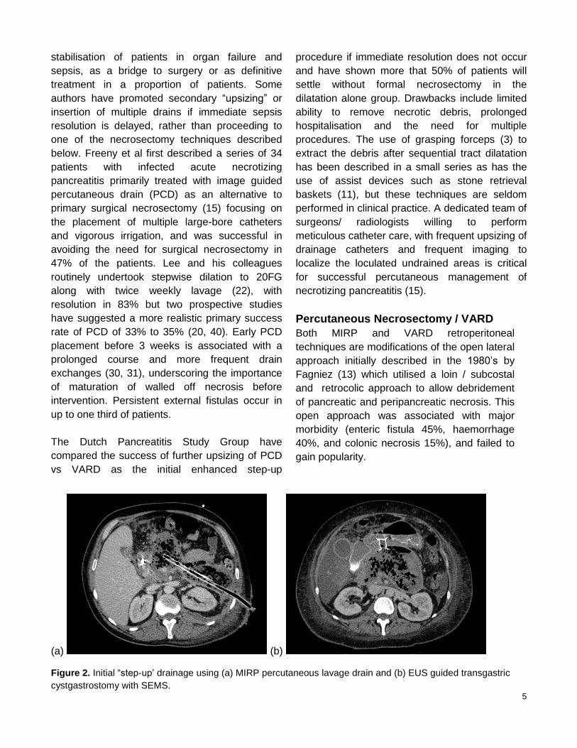
(a) (b)
Figure 2. Initial “step-up’ drainage using (a) MIRP percutaneous lavage drain and (b) EUS guided transgastric
cystgastrostomy with SEMS.

6
For both minimally invasive techniques, a left-
sided small diameter percutaneous drain is
ideally placed into the acute necrotic collection
between the spleen, kidney and colon. Right-
sided, or trans-peritoneal drainage are also
possible. In those who fail to respond
adequately to simple drainage this access drain
is then used as a guide to gain enhanced
drainage of the collection.
Minimally Invasive Pancreatic Necrosectomy
For percutaneous necrosectomy, the catheter
is exchanged for a radiological guidewire then
a low compliance balloon dilator is inserted into
the collection and dilated to 34 FG. Access to
the cavity is then maintained by an Amplatz
sheath through which is passed an operating
nephroscope, which allows debridement under
direct vision. The nephroscope has an
operating channel that permits standard (5 mm)
laparoscopic graspers as well as an
irrigation/suction channel. The directed, high
flow lavage promotes rapid evacuation of pus
and liquefied necrotic material, revealing black
or grey devascularised pancreatic tissue and
peri-pancreatic fat which, if loose, is extracted
in a piecemeal fashion until, after several
procedures, a cavity lined by viable tissue or
granulating pancreas is created. At the end of
each procedure an 8FG catheter sutured to a
24 FG drain is passed into the cavity to allow
continuous post-operative lavage of warmed
fluid initially at 250 ml an hour. Subsequent
conversion of the lavage system to simple
drainage may be all that is required prior to
recovery or the procedure may be repeated
until sepsis control is achieved and interval CT
confirms resolution.
Video-Assisted Retroperitoneal Debridement
(VARD)
A Video-Assisted Retroperitoneal Debridement
(VARD) procedure is performed with the patient
placed in a supine position with the left side
30–40° elevated. A subcostal incision of 5 cm
is placed in the left flank at the mid-axillary line,
close to the exit point of the percutaneous
drain. Using the in situ percutaneous drain as a
guide, the retroperitoneal collection is entered.
The cavity is cleared of purulent material using
a standard suction device. Visible necrosis is
carefully removed with the use of long grasping
forceps, and deeper access under direct vision
is facilitated using a 0° laparoscope, and
further debridement performed with
laparoscopic forceps. As with a percutaneous
necrosectomy, complete necrosectomy is not
the aim of this procedure and only loosely
adherent pieces of necrosis are removed,
minimizing the risk of haemorrhage. Two large
bore single lumen drains are positioned in the
cavity and the fascia closed to facilitate a
closed continuous postoperative lavage
system.
Endoscopic Necrosectomy
Endoscopic cystgastrostomy was initially
reported for the management of a mature
pancreatic abscess with minimal necrosis (5),
but the technique has evolved in the last 10
years to become an established Natural Orifice
(NOTES) procedure, with endoscopic
transmural exploration and debridement of the
retroperitoneum. Single-step drainage under
EUS guidance may be carried out by either a
trans-gastric or less commonly a trans-
duodenal route and is preferred to "blind"
drainage as EUS allows for identification of the
collection where there is no obvious bulge seen
within the stomach and helps identify a safe
route for puncture, free from intervening
vessels (18, 41). The presence of significant
walled off necrosis (WON) is no longer
considered a contraindication, but concerns do
remain regarding the adequacy of endoscopic
drainage, particularly in solid predominant or
larger collections. The principles are similar to
those discussed above, with initial simple
drainage of a collection under pressure,
followed by subsequent “step up” tract
dilatation and potential necrosectomy.

7
The procedure involves puncture of the
collection with either a 19G needle or
cystotome, with dilatation of the track followed
by placement of two or more plastic pigtail
stents. Increasingly, metallic stents may be
used which facilitate subsequent endoscopic
access to the cyst cavity for debridement of
necrosis. Where there is evidence of infection
or systemic sepsis it is our practice to use a
naso-cystic catheter, which can be used for
continuous lavage of the cavity. Factors
associated with a failure of resolution are large
size and retro-colic extension of the collections
and in these cases, other approaches or
combinations of approaches should be
considered (31, 37). Other options include the
multiple gateway technique (41), where two or
three transmural stents are placed under EUS
guidance, one of which is used for nasocystic
cavity lavage and the others to facilitate
drainage of necrotic debris.
Where there is extensive necrosis, delayed
endoscopic necrosectomy may be required
(34). It is our practice to defer this for a week
following the initial drainage procedure to allow
the fluid component to drain and any
associated sepsis to improve. A recent
systematic review (38) of 14 studies including
455 patients found an overall success rate of
81% and mortality of 6%, but these studies are
in highly selected patients and all but one was
retrospective. One small randomized trial (2)
has compared endoscopic with surgical
drainage and found a reduction in significant
complications with the endoscopic approach.
Endoscopic necrosectomy is however a
challenging procedure and not without risk.
Major complications including fatal air
embolism, bleeding and perforation occurred in
26% of patients in the multi-centre GEPARD
(33) study. The use of CO2 insufflation is
therefore now recommended. A persistent
problem is the lack of availability of suitable
endoscopic devices to facilitate necrosectomy
and although endoscopic access to the cyst
cavity is now facilitated by metallic stents,
piecemeal necrosectomy using standard
graspers, baskets and snares is a time
consuming and painstaking process (39). One
possible modification is the use of intra-cavity
hydrogen peroxide to facilitate necrosectomy
although further experience is required before
this can be recommended for routine practice
(36).
Despite these limitations, initial experience has
been promising (33), and an early randomised
pilot study exploring the outcome of
endoscopic transmural drainage vs. minimally
invasive intervention (VARD) (the PENGUIN
trial (2)) suggested at least equivalence, if not
benefit, from endoscopic drainage. This study
has been criticised due to very small numbers
and an excessive mortality (40%), compared to
historical results, within the VARD arm. The
results of the on-going TENSION trial (39) are
awaited with interest.
Open Surgical Necrosectomy
Open necrosectomy is still employed but
increasingly has been replaced by the
procedures described above. Three general
variations of open necrosectomy are currently
practiced, and remain widespread whilst
experience of minimally invasive approaches
increases. These can also be used within a
step up framework with preoperative
percutaneous drainage, allowing control of
sepsis prior to intervention. Although the
procedures are broadly similar in terms of the
necrosectomy, they differ in terms of how they
prevent recurrence of an infected collection
within the debridement cavity: 1) open
necrosectomy with open or closed packing, 2)
open necrosectomy with continuous closed
postoperative lavage, and 3) programmed
open necrosectomy.
In all approaches, the abdomen is entered
though a midline or preferably a bilateral

8
subcostal incision, as this minimizes
contamination of the lower abdomen and
allows bilateral paracolic access. The pancreas
is exposed by dividing the gastrocolic omentum
or gastrohepatic omentum to access the
pancreas through the lesser sac. Open
transgastric debridement has recently been
proposed to minimise post-operative peritoneal
contamination (32).
Open Necrosectomy with Open Packing
Bradley described this technique in 1987 (8),
sepsis control being achieved by leaving the
abdomen open following debridement, packing
the cavity as a laparostomy (8). Planned re-
intervention with sequential pack changes
allows resolution with healing by secondary
intention. Drains may be placed in addition to
the packing. Open packing techniques have
been reported to have higher incidences of
fistulae, bleeding, and incisional hernias as well
as a slightly higher mortality rate (19).
Open Necrosectomy with Closed Packing
Following necrosectomy to achieve sepsis
control (28), primary closure of the abdomen
over gauze-stuffed Penrose drains is
performed with the intention to fill the cavity
and provide some compression (14). Additional
silicone drains (Jackson-Pratt) may be placed
in the pancreatic bed and lesser sac for fluid
drainage. The drains are removed sequentially,
starting 5 to 7 days postoperatively, allowing a
gradual involution of the cavity.
Open Necrosectomy with Continuous Closed
Postoperative Lavage
After debridement, where possible a closed
peripancreatic compartment is reconstituted by
suturing the gastrocolic and duodenocolic
ligaments over large bore drains allowing flank
to flank continuous lavage (6). Postoperative
continuous lavage is instituted at 1 to 10 L per
day and continued until the effluent is clear and
the patient shows improvement in clinical and
laboratory parameters (43). No evidence is
available to suggest the best irrigation fluid, the
optimal number or caliber of drains, or the
duration of irrigation.
Programmed Open Necrosectomy
In response to the bleeding and fistulation that
can arise following aggressive necrosectomy,
this approach attempts to initially perform a
more conservative debridement, with the
intention of performing repeat procedures every
48 hours until debridement is no longer
required. This mimics the “minimal hit” concept
associated with the step up approaches. The
pancreatic bed is drained or packed, and the
abdomen is closed by suturing mesh or a
zipper to the fascial edges of the wound (29).
The addition of intra-abdominal vacuum
dressings may encourage granulation of the
pancreatic bed, and it has been suggested they
may reduce the number of operations and
mortality, but there is little data to support this
and they have been associated with enteric
fistulation (25).
5. Management for late WON
Indications for intervention for WON are: (1)
Infection, (2) Nutritional failure, (3) Persistent
abdominal pain. The decision on when to
intervene and the choice of intervention are made
within a multidisciplinary environment with
consideration of all available options.
Spontaneous resolution of even large acute
walled off necrotic collections are not infrequent
and often continued non-intervention is the best
approach, particularly where continued maturation
of a collection may be anticipated and where the
clinical picture is improving. In any individual
case, the choice of intervention may be guided by
factors including the clinical picture, the position of
the collection in relation to the stomach and
duodenum and available expertise.
Laparoscopic Cystgastrostomy
For many years, the conventional approach to
the management of late WON was open
pancreatic cystgastrostomy, with
9
necrosectomy. This procedure can now be
safely and effectively carried out using a
laparoscopic approach and this represents the
main alternative to endoscopic
cystgastrostomy. Our current technique for
laparoscopic cystgastrostomy is as follows: An
open sub-umbilical cut down is employed.
Further 12 mm and 5mm ports are inserted on
the patient’s left and right side with the specific
port site placement being determined by the
anatomical position of the retro-gastric
collection. Adhesions are divided to expose the
anterior gastric wall. An anterior gastrotomy (5-
10 cm long) is then performed using the
harmonic scalpel (Ethicon Endo-Surgery, Inc,
Cincinnati, Ohio, USA). The superior leaf of the
opened stomach is lifted toward the anterior
abdominal wall to maximise access and
delineate the area of adherence between the
cyst and the posterior aspect of the stomach.
This is achieved by passing a straight needle
2/0 suture through the abdominal wall, the
anterior stomach wall and back out of the
abdomen. A key advance has been the use of
a “Step” dilatation port system (Covidien plc.
Dublin, Ireland) to achieve initial cyst puncture,
allow tract dilatation and maintain access until
insertion of the initial staple device. Following
aspiration of the collection contents to relative
dryness, the port is withdrawn leaving the
suction instrument within the collection to
maintain access, and a stapled
cystgastrostomy is performed using 4-5 firings
of the angulating Universal Endo GIA stapler
(Covidien plc, Dublin Ireland). Necrotic debris
within the cavity is removed and placed in the
fundus of the stomach. Once adequate
debridement and haemostasis have been
assured, the anterior gastrotomy is closed
using a running 3/0 monofilament suture
(BiosynTM , Covidien plc, Dublin Ireland), with
the integrity of the closure then tested by
insufflating the stomach through an oro-gastric
tube, while the anastomosis is held under
lavage fluid. Post-operative fluid and diet is
allowed as tolerated. In this complex cohort of
patients, suitability for hospital discharge is
often multi-factorial, but may be within 36 h of
surgery when dietary intake is adequate.
Where gallstones are present, a simultaneous
laparoscopic cholecystectomy is performed.
Our initial results have been presented
elsewhere and we are currently undertaking a
randomized trial of EUS-guided endoscopic vs
laparoscopic cystgastrostomy for WON (17).
Endoscopic Ultrasound Guided
Cystgastrostomy / Necrosectomy
The technique of EUS guided drainage is as
described above, the principle difference being
the indication of failure to thrive rather than
sepsis control. Many reports in the literature
describe EUS-guided drainage of
"pseudocysts" but is now recognised that true
pancreatic pseudocysts are rare following
acute pancreatitis as some degree of necrosis
is usually present where collections persist.
The revised Atlanta criteria defines these
collections as WON, but there is still a
spectrum of clinical presentations. WON may
have varying degrees of fluid content and
infection may be present, with or without
systemic disturbance or organ failure. EUS-
guided drainage of these collections is now an
established technique in specialist units and
several different modifications to the technique
have been described. The frequent
requirement for repeated endoscopic
procedures, particularly in the presence of
significant necrosis, have led to a former
preference to select fluid predominant WON
collections for this approach, but this
assumption is being currently challenged in a
randomized trial in our unit.
Management of Complications
Early Procedure Related Complications: SIRS /
Bacteraemia Requiring Critical Care Support
For patients with established organ failure,
drainage has an unpredictable effect on
patients and the clinical picture may improve or

10
may worsen, at least temporarily. Evidence
now supports a "step up" approach in the
presence of organ failure and so initial
management in these patients should be either
percutaneous or endoscopic drainage, with
more definitive intervention deferred until organ
failure stabilises or improves.
Following any intervention, however minimal, it
is not unusual for patients to show signs of
significant SIRS or post procedure
bacteraemia, and this may necessitate critical
care admission for organ support. Our
experience has been that minimally invasive
approaches are less likely to cause the
development of new organ failure, and this has
been born out in randomized trials (40). More
significant deterioration is common following
open necrosectomy and this is therefore no
longer the preferred approach.
Acute or Delayed Haemorrhage
Peri-procedural haemorrhage following initial
drainage may be due to bleeding from
submucosal or perigastric vessels during
endoscopic or percutaneous drainage, and is
usually self-limiting. Bleeding from the cavity
itself is more likely during necrosectomy,
particularly if carried out too early or too
aggressively. Venous bleeding is more
common in this situation and may occur during
the procedure or in the post-operative period.
It will usually resolve with correction of any
coagulopathy but tamponade may be required,
either by simply clamping the percutaneous
drain, insertion of a modified Sengstaken-
Blakemore tube (having amputated the gastric
balloon), or gauze packing if there is sufficient
cutaneous access following a VARD
procedure.
Secondary haemorrhage is occasionally
sudden and massive, but there is usually a
prelude with a self-terminating “herald bleed”,
presenting clinically with haemorrhage into a
retroperitoneal drain or by a gastrointestinal
bleed following transluminal drainage.
Secondary haemorrhage is usually of arterial
origin and is often a consequence of persistent
local sepsis. This is now the major cause of
death in patients with infected pancreatic
collections and rapid intervention may be life-
saving. Initial controlled volume support of the
circulation and a simultaneous emergency CT
angiogram is followed by angiography and
embolization if appropriate. Upper
gastrointestinal endoscopy in this setting is
usually non-diagnostic and should therefore not
delay radiological assessment which allows
definitive management. The increased intra-
cavity pressure associated with haemorrhage
into an infected cavity, may result in escalating
organ dysfunction through bacteraemia and
sepsis. Timely consideration of further
intervention to improve surgical drainage is
important once bleeding has been arrested.
Enteric Fistulation
Spontaneous discharge of a pancreatic
collection into the gastrointestinal tract is
common and may occur in the presence or
absence of infection. This should be suspected
when a collection contains gas, particularly
where a gas/fluid level is present, in a patient
who is not systemically unwell. Indeed,
discharge of a collection into the stomach or
duodenum can be associated with an
improvement in a patient's condition. In our
experience, foregut fistulation will usually
resolve without the need for intervention (other
than adequate drainage of a collection by
percutaneous or endoscopic means) but
fistulation into the colon is often associated with
clinical deterioration and persistent sepsis.
Some form of defunctioning procedure is
usually required and in occasional cases,
formal colonic resection with exteriorisation
may be required.

11
6. Late complications
Pancreatic Fistulation
Persistent pancreatic fistula is a common
sequel of percutaneous necrosectomy or
VARDS. Disruption of the pancreatic duct is
common in the presence of extensive necrosis,
and although resolution is the norm, persistent
fistulae can be a challenging management
problem. If a pancreatic fistula persists once
resolution of sepsis and any significant
collection has been confirmed by CT,
pancreatic duct stent insertion at ERCP is the
management of choice. Failure of resolution
thereafter is often associated with more
extensive parenchymal loss, or a disconnected
pancreatic tail with loss of continuity of the
main pancreatic duct. Prolonged catheter
drainage will lead to maturation of the fistula
tract and planned interval drain removal may
result in spontaneous resolution or
development of a late pseudocyst, which can
often be resolved by transmural endoscopic
cystgastrostomy. The avoidance of pancreatic
fistula is one of the main advantages of
endoscopic (or laparoscopic) drainage of
pancreatic collections.
Disconnected Pancreatic Tail
Following extensive necrosis or complete
necrosis of a section of the neck or body of the
pancreas, complete separation of the main
pancreatic duct in the pancreatic tail may occur
leading to a persistent fistula and
“disconnected duct syndrome”. This may lead
to persistence of a pancreatic fistula or a late
"pseudocyst" following initial successful
management of a pancreatic collection. Ductal
occlusion at the pancreatic neck precludes
trans-papillary access but if this has not
occurred, intra-cystic trans-papillary stenting or
a stent bridging the defect into the tail, may
result in resolution. If trans-papillary access is
not possible, the preferred option is transmural
EUS guided drainage with placement of long-
term pigtail stents although in some patients,
distal pancreatectomy may be required. This
however is a challenging procedure,
particularly, as is commonly the case, where
there have been previous interventions.
7. Conclusion
Clinical complexity and diversity precludes
algorithm driven management in severe acute
pancreatitis. Three phases of management exist
(1) organ support (2) sepsis control and (3) failure
to thrive; based on an understanding of the
evolution of necrosis/collections and the dynamic
nature of the physiological response in acute
pancreatitis the rationale and interventional
approach chosen will differ depending on the
specific issues that need to be addressed.
Maintaining nutritional competence throughout is
essential. Individual patient management within a
step-up framework remains key, utilizing a
multimodal approach focused on delayed
minimally invasive intervention where possible.
8. References
1. Aldridge MC, Francis ND, Glazer G and Dudley HA. Colonic complications of severe acute pancreatitis. Br J Surg 76(4): 362-367, 1989. PMID: 2655821.
2. Bakker OJ, van Santvoort HC, van Brunschot S, Geskus RB, Besselink MG, Bollen TL, et al. Endoscopic transgastric vs surgical necrosectomy for infected necrotizing pancreatitis: a randomized trial. JAMA 307(10): 1053-1061, 2012. PMID: 22416101.
3. Bala M, Almogy G, Klimov A, Rivkind AI and Verstandig A. Percutaneous "stepped" drainage technique for infected pancreatic necrosis. Surg Laparosc Endosc Percutan Tech 19(4): e113-118, 2009. PMID: 19692859.
4. Banks PA, Bollen TL, Dervenis C, Gooszen HG, Johnson CD, Sarr MG, et al. Classification of acute pancreatitis--2012: revision of the Atlanta classification and definitions by international consensus. Gut 62(1): 102-111, 2013. PMID: 23100216.
12
5. Baron TH, Thaggard WG, Morgan DE and Stanley RJ. Endoscopic therapy for organized pancreatic necrosis. Gastroenterology 111(3): 755-764, 1996. PMID: 8780582.
6. Beger HG. Operative management of necrotizing pancreatitis--necrosectomy and continuous closed postoperative lavage of the lesser sac. Hepatogastroenterology 38(2): 129-133, 1991. PMID: 1855769.
7. Bradley EL, 3rd. A clinically based classification system for acute pancreatitis. Summary of the International Symposium on Acute Pancreatitis, Atlanta, Ga, September 11 through 13, 1992. Arch Surg 128(5): 586-590, 1993. PMID: 8489394.
8. Bradley EL, 3rd. Management of infected pancreatic necrosis by open drainage. Ann Surg 206(4): 542-550, 1987. PMID: 3662663.
9. Buchler M, Uhl W and Beger HG. Acute pancreatitis: when and how to operate. Dig Dis 10(6): 354-362, 1992. PMID: 1473288.
10. Carter CR, McKay CJ and Imrie CW. Percutaneous necrosectomy and sinus tract endoscopy in the management of infected pancreatic necrosis: an initial experience. Ann Surg 232(2): 175-180, 2000. PMID: 10903593.
11. Echenique AM, Sleeman D, Yrizarry J, Scagnelli T, Guerra JJ, Jr., Casillas VJ, et al. Percutaneous catheter-directed debridement of infected pancreatic necrosis: results in 20 patients. J Vasc Interv Radiol 9(4): 565-571, 1998. PMID: 9684824.
12. Elgammal S MC, Imrie CW, Carter CR. Does surgical approach affect outcome in patients with infected pancreatic necrosis requiring necrosectomy. Br J Surg 90: 93, 2003.
13. Fagniez PL, Rotman N and Kracht M. Direct retroperitoneal approach to necrosis in severe acute pancreatitis. Br J Surg 76(3): 264-267, 1989. PMID: 2720323.
14. Fernandez-del Castillo C, Rattner DW, Makary MA, Mostafavi A, McGrath D and Warshaw AL. Debridement and closed packing for the treatment of necrotizing pancreatitis. Ann Surg 228(5): 676-684, 1998. PMID: 9833806.
15. Freeny PC, Hauptmann E, Althaus SJ, Traverso LW and Sinanan M. Percutaneous CT-guided catheter drainage of infected acute necrotizing pancreatitis: techniques and results. Am J Roentgenol 170(4): 969-975, 1998. PMID: 9530046.
16. Gagner M. Laparoscopic Treatment of Acute Necrotizing Pancreatitis. Semin Laparosc Surg 3(1): 21-28, 1996. PMID: 10401099.
17. Gibson SC, Robertson BF, Dickson EJ, McKay CJ and Carter CR. 'Step-port' laparoscopic cystgastrostomy for the management of organized solid predominant post-acute fluid collections after severe acute pancreatitis. HPB (Oxford) 16(2): 170-176, 2014. PMID: 23551864.
18. Giovannini M, Bernardini D and Seitz JF. Cystogastrotomy entirely performed under endosonography guidance for pancreatic pseudocyst: results in six patients. Gastrointest Endosc 48(2): 200-203, 1998. PMID: 9717789.
19. Heinrich S, Schafer M, Rousson V and Clavien PA. Evidence-based treatment of acute pancreatitis: a look at established paradigms. Ann Surg 243(2): 154-168, 2006. PMID: 16432347.
20. Hollemans RA, Bollen TL, van Brunschot S, Bakker OJ, Ahmed Ali U, van Goor H, et al. Predicting Success of Catheter Drainage in Infected Necrotizing Pancreatitis. Ann Surg, 2015. PMID: 25775071.
21. Horvath KD, Kao LS, Wherry KL, Pellegrini CA and Sinanan MN. A technique for laparoscopic-assisted percutaneous drainage of infected pancreatic necrosis and pancreatic abscess. Surg Endosc 15(10): 1221-1225, 2001. PMID: 11727105.
22. Lee JK, Kwak KK, Park JK, Yoon WJ, Lee SH, Ryu JK, et al. The efficacy of nonsurgical treatment of infected pancreatic necrosis. Pancreas 34(4): 399-404, 2007. PMID: 17446837.
23. McKay CJ, Evans S, Sinclair M, Carter CR and Imrie CW. High early mortality rate from acute pancreatitis in Scotland, 1984-1995. Br J Surg 86(10): 1302-1305, 1999. PMID: 10540138.
24. Mier J, Leon EL, Castillo A, Robledo F and Blanco R. Early versus late necrosectomy in severe necrotizing pancreatitis. Am J Surg 173(2): 71-75, 1997. PMID: 9074366.
25. Olejnik J, Vokurka J and Vician M. Acute necrotizing pancreatitis: intra-abdominal vacuum sealing after necrosectomy. Hepatogastroenterology 55(82-83): 315-318, 2008. PMID: 18613356.
26. Parekh D. Laparoscopic-assisted pancreatic necrosectomy: A new surgical option for treatment of severe necrotizing pancreatitis. Arch Surg 141(9): 895-902; discussion 902-893, 2006. PMID: 16983033.
27. Petrov MS, Shanbhag S, Chakraborty M, Phillips AR and Windsor JA. Organ failure and infection of pancreatic necrosis as determinants of mortality in patients with acute pancreatitis. Gastroenterology 139(3): 813-820, 2010. PMID: 20540942.
28. Pezzilli R, Uomo G, Gabbrielli A, Zerbi A, Frulloni L, De Rai P, et al. A prospective multicentre survey on the treatment of acute pancreatitis in Italy. Dig Liver Dis 39(9): 838-846, 2007. PMID: 17602904.
13
29. Radenkovic DV, Bajec DD, Tsiotos GG, Karamarkovic AR, Milic NM, Stefanovic BD, et al. Planned staged reoperative necrosectomy using an abdominal zipper in the treatment of necrotizing pancreatitis. Surg Today 35(10): 833-840, 2005. PMID: 16175464.
30. Ramesh H, Prakash K, Lekha V, Jacob G and Venugopal A. Are some cases of infected pancreatic necrosis treatable without intervention? Dig Surg 20(4): 296-299; discussion 300, 2003. PMID: 12789025.
31. Ross A, Gluck M, Irani S, Hauptmann E, Fotoohi M, Siegal J, et al. Combined endoscopic and percutaneous drainage of organized pancreatic necrosis. Gastrointest Endosc 71(1): 79-84, 2010. PMID: 19863956.
32. Sasnur P, Nidoni R, Baloorkar R, Sindgikar V and Shankar B. Extended Open Transgastric Necrosectomy (EOTN) as a Safer Procedure for Necrotizing Pancreatitis. J Clin Diagn Res 8(7): NR01-02, 2014. PMID: 25177603.
33. Seifert H, Biermer M, Schmitt W, Jurgensen C, Will U, Gerlach R, et al. Transluminal endoscopic necrosectomy after acute pancreatitis: a multicentre study with long-term follow-up (the GEPARD Study). Gut 58(9): 1260-1266, 2009. PMID: 19282306.
34. Seifert H, Wehrmann T, Schmitt T, Zeuzem S and Caspary WF. Retroperitoneal endoscopic debridement for infected peripancreatic necrosis. Lancet 356(9230): 653-655, 2000. PMID: 10968442.
35. Shah RJ, Shah JN, Waxman I, Kowalski TE, Sanchez-Yague A, Nieto J, et al. Safety and efficacy of endoscopic ultrasound-guided drainage of pancreatic fluid collections with lumen-apposing covered self-expanding metal stents. Clin Gastroenterol Hepatol 13(4): 747-752, 2015. PMID: 25290534.
36. Siddiqui AA, Easler J, Strongin A, Slivka A, Kowalski TE, Muddana V, et al. Hydrogen peroxide-assisted endoscopic necrosectomy for walled-off pancreatic necrosis: a dual center pilot experience. Dig Dis Sci 59(3): 687-690, 2014. PMID: 24282052.
37. Takahashi N, Papachristou GI, Schmit GD, Chahal P, LeRoy AJ, Sarr MG, et al. CT findings of walled-off pancreatic necrosis (WOPN): differentiation from pseudocyst and prediction of outcome after endoscopic therapy. Eur Radiol 18(11): 2522-2529, 2008. PMID: 18563416.
38. van Brunschot S, Fockens P, Bakker OJ, Besselink MG, Voermans RP, Poley JW, et al. Endoscopic transluminal necrosectomy in necrotising pancreatitis: a systematic review. Surg Endosc 28(5): 1425-1438, 2014. PMID: 24399524.
39. van Brunschot S, van Grinsven J, Voermans RP, Bakker OJ, Besselink MG, Boermeester MA, et al. Transluminal endoscopic step-up approach versus minimally invasive surgical step-up approach in patients with infected necrotising pancreatitis (TENSION trial): design and rationale of a randomised controlled multicenter trial [ISRCTN09186711]. BMC Gastroenterol 13: 161, 2013. PMID: 24274589.
40. van Santvoort HC, Besselink MG, Bakker OJ, Hofker HS, Boermeester MA, Dejong CH, et al. A step-up approach or open necrosectomy for necrotizing pancreatitis. N Engl J Med 362(16): 1491-1502, 2010. PMID: 20410514.
41. Varadarajulu S, Phadnis MA, Christein JD and Wilcox CM. Multiple transluminal gateway technique for EUS-guided drainage of symptomatic walled-off pancreatic necrosis. Gastrointest Endosc 74(1): 74-80, 2011. PMID: 21612778.
42. Wiersema MJ, Baron TH and Chari ST. Endosonography-guided pseudocyst drainage with a new large-channel linear scanning echoendoscope. Gastrointest Endosc 53(7): 811-813, 2001. PMID: 11375600.
43. Wig JD, Mettu SR, Jindal R, Gupta R and Yadav TD. Closed lesser sac lavage in the management of pancreatic necrosis. J Gastroenterol Hepatol 19(9): 1010-1015, 2004. PMID: 15304118.
44. Working Group IAPAPAAPG. IAP/APA evidence-based guidelines for the management of acute pancreatitis. Pancreatology 13(4 Suppl 2): e1-15, 2013. PMID: 24054878.
45. Zhu JF, Fan XH and Zhang XH. Laparoscopic treatment of severe acute pancreatitis. Surg Endosc 15(2): 146-148, 2001. PMID: 11285957.
![OPEN ACCESS viruses - Semantic Scholar · , viruses [1], the viral etiology of these previously recognized diseases such as infectious pancreatic necrosis, Oregon sockeye disease,](https://img.pdfslide.net/doc/110x75/5e855f018b3d144fe76983d0/open-access-viruses-semantic-scholar-viruses-1-the-viral-etiology-of-these.jpg)