Embed Size (px)
Citation preview

Journal of Chromatography, 510 (1990) lOlLI Elsevier Science Publishers B.V., Amsterdam
CHROM. 22 407
Tresyl-activated support for high-performance affinity chromatography
KOJI NAKAMURA, TSUTOMU HASHIMOTO and YOSHIO KATO*
Central Research Laboratory, TOSOH Corporation, Tonda, Shinnanyo, Yamaguchi 746 (Japan)
and
KIYOHITO SHIMURA and KEN-ICHI KASAI
Faculty of Pharmaceutical Sciences. Teikyo University. Sagamiko, Kanagawa 199-01 (Japan)
ABSTRACT
A new activated support TSKgel Tresyl-SPW was evaluated for the coupling of antibodies, which was found to occur easily under mild conditions with high yields. Optimum coupling conditions were a 2-h reaction at 25°C in 1 M phosphate buffer (pH 7.5) when 2-3 mg antibody/ml support is to be coupled and a 67-h reaction when ca. 10 mg antibody/ml support is to be coupled. When antibodies were coupled under these conditions, antibody coupling yields >80% and antigen binding effi- ciencies of 7&80% were achieved, probably owing to a selective attachment of the F, region of the antibodies. Antigens (human serum proteins) could be separated rapidly without denaturation on antibody-coupled Tresyl-SPW.
INTRODUCTION
The cyanogen bromide activation method reported by Axen et al.’ in 1967 was one of the most important developments in affinity chromatography. Since then, affinity chromatography has progressed considerably and has become widely accept- ed for the purification of biological substances such as proteins. However, the cyano- gen bromide activation method has some shortcomings, e.g., cyanogen bromide is extremely toxic, the activation reaction is complicated and the linkage between the support matrix and the ligand is not very stable. Therefore, other activation methods have also been investigated. The tresyl(2,2,2-trifluoroethanesulphonyl) chloride acti- vation method developed by Nilsson and Mosbach’ in 1981 has attracted attention recently because the activation reaction is simple and many ligands can be coupled easily under mild conditions via amino, thiol, phenol or imidazole groups.
Some tresyl-activated supports based on agarose or silica became commercially available a few years ago, but there are problems with the mechanical and chemical stability of the base material. However, new tresyl-activated supports based on syn- thetic hydrophilic resins have become available recently as TSKgel Tresyl-Toyopearl
0021-9673/90/$03.50 0 1990 Elsevier Science Publishers B.V.

102 K. NAKAMURA et nl.
650M and Tresyl-5PW (TOSOH, Tokyo, Japan). They are mechanically and chem- ically stable and therefore seem useful as supports for high-performance affinity chro- matography.
We have been evaluating these tresyl-activated supports and have already re- ported results on the study of the coupling conditions for some proteins334. As report- ed there, the optimum coupling conditions were dependent on the proteins. Accord- ingly, we have investigated further the coupling conditions for antibodies, because they constitute one of the most important ligands in affinity chromatography. We have also examined rapid separations of antigens (human serum proteins) by immu- noaffinity chromatography on supports coupled with anti-human serum protein anti- bodies. The results are reported in this paper.
EXPERIMENTAL
Antibodies were coupled to Tresyl-SPW, which was prepared by introducing tresyl groups at a level of ca. 20 pmol/ml support into TSKgel G5000PW of particle diameter 10 pm and pore diameter cu. 1000 A. Potassium phosphate solution (1 M) was employed as a coupling buffer throughout all the experiments because it was very effective in the coupling of other proteins4. The effects of pH of the coupling buffer, reaction time, temperature and amount of ligand antibody on the antibody coupling yield, antigen binding capacity and antigen binding efficiency were investigated.
The antibody coupling reaction and subsequent evaluation of antibody-cou- pled supports were performed as follows. A certain amount of antibody was dissolved in 2.2 ml of coupling buffer and 0,.25 g dried Tresyl-5PW powder, which gives a volume of 1 .O ml in the swollen state, was added to 2 ml of the antibody solution. The remaining 0.2 ml of the antibody solution was used to measure the UV absorption of the solution at 280 nm, which was necessary for determining the antibody coupling yield. After the mixture had been allowed to stand with gentle shaking at a constant temperature for a certain period of time, 18 ml of distilled water was added and the diluted mixture was filtered through a sintered-glass funnel. This dilution was neces- sary to prevent adsorption of antibody on the surface of the support without covalent bonding. The UV absorption of the filtrate was measured at 280 nm. After washing the gel in the funnel three times with 10 ml of 0.1 M Tris-HCl buffer (pH 8.5), the remaining tresyl groups were blocked by suspending the support in 5 ml of 0.1 M Tris-HCl buffer (pH 8.5) for 1 h at 25°C. Then the support was packed into a stain- less-steel column (10 mm x 6 mm I.D.).
The column was installed in a high-performance liquid chromatography (HPLC) system and equilibrated with 0.1 M phosphate buffer (pH 7.4) at a flow-rate of 0.5 ml/min at 25°C then 5 mg of antigen dissolved in 0.5 ml of 0.1 A4 phosphate buffer (pH 7.4) was applied to the column. After unbound excess antigen had been completely washed from the column, bound antigen was eluted with 0.1 A4 citric acid of pH 1.6, adjusted with hydrochloric acid. A 5-ml volume of column effluent con- taining bound antigen was collected and the UV absorption of the fraction was measured at 280 nm. The antibody coupling yield was calculated from the UV ab- sorption of the antibody solutions before and after the coupling reaction. The amount of coupled antibody was calculated from the coupling yield and concentra- tion of antibody solution before the coupling reaction. The antigen binding capacity

TRESYL-ACTIVATED SUPPORT FOR HPAC 103
was calcualted from the UV absorption of the antigen fraction by assuming Ai&, = 5.8, 11.4, 14.7 and 13.3 for albumin, transferrin, immunoglobulin (Ig) G and IgM, respectively. The antigen binding efficiency, which is defined as the percentage of coupled antibody which is active and can bind antigen, was calculated from the amount of coupled antibody and antigen binding capacity considering the fact that one antibody molecule can bind two antigen molecules. We employed values of 67 000 and 150 000 as molecular weights of albumin and antibody, respectively.
Separation of antigens by immunoaffinity chromatography was carried out on a 10 mm x 6 mm I.D. or 20 mm x 10 mm I.D. column with a step gradient from 0.1 M phosphate buffer (pH 7.4) to 0.1 M citric acid-hydrochloric acid (pH 1.6) at a flow-rate of 1 or 2 ml/min at 25°C by using an HPLC system consisting of a Model
CCPM double-plunger pump, a Model UV-8000 variable-wavelength UV detector operated at 280 nm, and a Model CP-8000 data processor (TOSOH). The fractions collected were tested for purity by sodium dodecyl sulphate-polyacrylamide gel elec- trophoresis (SDS-PAGE) in slabs of 4-20% polyacrylamide gradient gel (Tefco, To- kyo, Japan). The recovery of activity was also examined in the purification of plasmi- nogen. Plasminogen activity was measured with a Testzyme PLG kit (Daiich Pure Chemicals, Tokyo, Japan).
All antibodies employed were IgG fractions of rabbit polyclonal antibodies purified by ion-exchange chromatography and gel filtration (Dakopatts. Glostrup, Denmark), except one which was affinity-purified goat polyclonal anti-human albu- min antibody (Biosys, Compiegne, France). Human albumin, transferrin and plasma were purchased from Sigma (St. Louis, MO, U.S.A.), human TgG and serum from Miles Labs. (Elkhart, IN, U.S.A.), human IgM from Protogen (Laufelfingen, Swit- zerland) and human serum standards for single radial immunodiffusion analysis from Hoechst Japan (Tokyo, Japan).
RESULTS AND DISCUSSION
Fig. 1 shows the effect of the pH of the coupling buffer. The antibody coupling yield was almost 100% at pH 37.0, but it decreased to about 50% at pH 6.5. The
I I
” ” 0
I I
6 7 a
PH of Coupling Buffer
Fig. 1. Effect of pH of coupling butfer on (0) antibody coupling yield. (a) antigen binding capacity and (0) antigen binding efficiency. Affinity-purified anti-human albumin antibody (2.5 mg) was reacted with 0.25 g of Tresyl-5PW in 2 ml of 1 M potassium phosphate buffer of pH 6.5-8.5 at 4°C for 16 h.

104 K. NAKAMURA et c/l.
antigen binding efficiency was approximately constant at ca. 70% at pH d 8.0, but it was about 45% at pH 8.5. The antigen binding capacity was maximum at pH cu. 7.5. Therefore, the optimum pH of coupling buffer can be said to be ca. 7.5. Because the reactivity of tresyl group with amino, thiol, phenol and imidazole groups is higher at higher pH, multi-point attachment of proteins to Tresyl-5PW must occur more easily at higher pH. Multi-point attachment sometimes causes changes in the structure of proteins and results in lower binding efficiencies ~ . 5 ’ Accordingly, the multi-point attachment is supposed to be responsible for the lower antigen binding efficiency at pH 8.5. In this study, the amount of antibody was fairly small, 2.5 mg/ml support. A similar study was also performed with a larger amount of antibody, cu. 10 mg/ml support, and the results are shown in Fig. 2.
b / 8 Y
DH of Coupling Buffer
Fig. 2. Etfect of pH of coupling buffer on (0) antibody couphng yield and (a) antigen bmding capacity. IgG fraction of anti-human albumin antibody (10.7 mg) was reacted with 0.25 g of Tresyl-5PW in 2 ml of I A4 potassium phosphate buffer of pH 6.5-8.5 at 25°C for 16 h.
The antibody coupling yield was more than 90% at pH 3 7.5, but it decreased considerably when the pH decreased below 7.0. The antigen binding capacity was maximum at pH 7.5. At higher pH, the antigen binding capacity decreased slightly although the antibody coupling yield was slightly higher. This slight decrease in anti- gen binding capacity at pH 8.0 and 8.5 may be due to a change in antibody structure due to multi-point attachment. Steric hindrance to antigen binding is also assumed to be responsible because the amount of coupled antibody is almost half the total anti- body coupling capacity of Tresyl-5PW (15-20 mg/ml support). Accordingly, a pH of ca. 7.5 is also optimum in the coupling of large amounts of antibody.
Fig. 3 shows the effect of the amount of antibody reacted. The antibody cou- pling yield was 100% for amounts of antibody less than 2.5 mg and decreased to 94% with 5 mg. The antigen binding efficiency was very high ( > 80%) when the amount of antibody was small (< 1 mg) and it gradually decreased with increasing amount of antibody. Steric hindrance to the binding of antigen to antibody is assumed to be responsible for the decrease in antigen binding efficiency with increase in the amount

TRESYL-ACTIVATED SUPPORT FOR HPAC 105
Amount of Antibody (mg)
Fig. 3. Effect of amount of reacted antibody on (0) antibody coupling yield, (a) antigen bindmg capacity and (0) antigen binding efficiency. Affinity-purified anti-human albumin antibody (0.5-5 mg) was reacted
with 0.25 g of Tresyl-SPW in 2 ml of 1 M potassium phosphate buffer (pH 7.5) at 4°C for 16 h.
of antibody. On the other hand, the antigen binding capacity increased continuously with increase in the amount of antibody up to 5 mg. Consequently, when it is desir- able to couple antibody as effectively as possible, the amount of antibody should be less than 2-3 mg/ml support, whereas more antibody should be reacted when a high antigen binding capacity is required.
Fig. 4. shows the effect of reaction time on the coupling of antibody of 2.5 mg/ml support at 25°C. The coupling reaction was fast. The antibody coupling yield was nearly 90% after 1 h and almost 100% after 2 h. The antigen binding capacity and efficiency also reached a maximum after 2 h but decreased afterwards. Therefore, reaction times of ca. 2 h are best in the coupling of small amounts of antibody at 25°C.
I.
I I I I I
L:
I I I I I
0 5 10 15 20 25
Reaction Time (h ) Fig. 4. Elfect of reaction time on (0) antibody coupling yield, (a) antigen binding capacity and (0) antigen binding efficiency. Affinity-purified anti-human albumin antibody (2.5 mg) was reacted with 0.25 g of Tresyl-5PW in 2 ml of 1 M potassium phosphate buffer (pH 7.5) at 25°C.
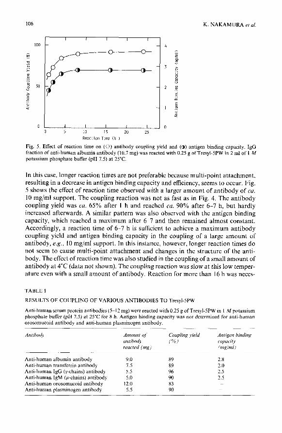
106 K. NAKAMURA et al.
I I I I I
100 - - 4
i50_-_-~
B ._
z -I
0 I I I I I 0
0 5 10 15 20 25
Reaction Time (h 1
Fig. 5. Effect of reaction time on (0) antibody coupling yield and (a) antigen binding capacity. IgG fraction of anti-human albumin antibody (10.7 mg) was reacted with 0.25 g of Tresyl-5PW in 2 ml of 1 M potassium phosphate buffer (pH 7.5) at 25°C.
In this case, longer reaction times are not preferable because multi-point attachment, resulting in a decrease in antigen binding capacity and efficiency, seems to occur. Fig. 5 shows the effect of reaction time observed with a larger amount of antibody of ca. 10 mg/ml support. The coupling reaction was not as fast as in Fig. 4. The antibody coupling yield was ea. 65% after 1 h and reached cu. 90% after 6-7 h, but hardly increased afterwards. A similar pattern was also observed with the antigen binding capacity, which reached a maximum after 6-7 and then remained almost constant. Accordingly, a reaction time of 6-7 h is sufficient to achieve a maximum antibody coupling yield and antigen binding capacity in the coupling of a large amount of antibody, e.g., 10 mg/ml support. In this instance, however, longer reaction times do not seem to cause multi-point attachment and changes in the structure of the anti- body. The effect of reaction time was also studied in the coupling of a small amount of antibody at 4°C (data not shown). The coupling reaction was slow at this low temper- ature even with a small amount of antibody. Reaction for more than 16 h was neces-
TABLE I
RESULTS OF COUPLING OF VARIOUS ANTIBODIES TO Tresyl-5PW
Anti-human serum protein antibodies (5-12 mg) were reacted with 0.25 g of Tresyl-5PW in I M potassium phosphate buffer (pH 7.5) at 25°C for 8 h. Antigen binding capacity was not determined for anti-human orosomucoid antibody and anti-human plasminogen antibody.
Antibody
Anti-human albumin antibody Anti-human transferrin antibody Anti-human IgG (y-chains) antibody Anti-human IgM (p-chains) antibody Anti-human orosomucoid antibody Anti-human plasminogen antibody
Amount qf
antihod) reacted (mg)
9.0 1.5 5.5 5.0
12.0 5.5
Coupling yield
ix)
89 89 96 90 83 90
Antigen binding
capacity
(wlml)
2.8 2.0 2.5 2.5
_ _

TRESYL-ACTIVATED SUPPORT FOR HPAC 107
sary to achieve an antibody coupling yield of more than 90%. However, multi-point attachment seemed to occur even at 4°C although very slowly. Accordingly, there is no advantage in coupling at low temperature.
Table I shows the results ofcoupling ofvarious antibodies under conditions which seemed optimum from the data given above. The coupling yield was more than 80% for all the antibodies examined. The antigen binding capacity was 2-3 mg/ml support. A similar binding capacity was obtained even for a large molecule such as IgM. This is probably due to the large pore size of Tresyl-SPW.
Rapid separations of human serum proteins by using antibody-coupled Tre- syl-SPW were tried. The elution conditions were examined first by using Tresyl-SPW coupled with anti-human albumin antibody. The results are shown in Fig. 6. Almost no albumin was eluted at pH 3.0. Although albumin was eluted at pH 2.0, the recov- ery was still low (55%). However, albumin was recovered quantitatively at pH 1.6. We then employed 0.1 A4 citric acid (pH 1.6) for the elution of bound antigens from antibody-coupled Tresyl-SPW. However, because these conditions seemed severe, the stability of antibody-coupled Tresyl-5 PW was examined. The same separation as in Fig. 6C was repeated 100 times and the albumin binding capacities before and after the separations were compared.
C
B
A
h, I I, A--Y- -I- 0 2 4 6 0 2 4 6 0 2 4 6
Elution Time (min)
Fig. 6. Effect of eluent pH on the elution of antigen from antibody-coupled Tresyl-SPW. Human serum albumin was applied to a column (20 mm x 10 mm I.D.) of Tresyl-SPW coupled with anti-human albumin
antibody in 0. I M phosphate buffer (pH 7.4) at a flow-rate of 2 ml/min, and 2 min after the sample injection the eluent was changed stepwise to 0. I M citric acid the pH of which was adjusted to (A) 3.0, (B) 2.0 or(C) I .6 with NaOH or HCI.
The albumin binding capacity decreased by lo%, which does not seem much in comparison with reported values observed under other conditions’,“.
Examples of the purification of human serum and plasma proteins are shown in Figs. 7711. Very pure transferrin and albumin were obtained in less than 5 min, as indicated in Figs. 7 and 8. Only a single band corresponding to transferrin and albu- min is seen in the SDS-PAGE pattern of the bound fraction. Several bands are seen in the SDS-PAGE patterns of bound fractions in the purification of IgG and TgM. Therefore, the IgG and IgM obtained are not very pure, although two main bands must be heavy and light chains of IgG and TgM. However, these results indicate that

108 K. NAKAMURA et al.
94 000
67 000 43 000
I I I I I I
0 1 2 3 4 5 6 7
Elution Time (min) Fig. 7. Purification of transferrin by high-performance affinity chromatography. Human serum (20 ~1) was applied to a column (10 mm x 6 mm I.D.) of Tresyl-SPW coupled with anti-human transferrin antibody at a flow-rate of 1 ml/min at 25°C. The pH of eluent was changed stepwise from 7.4 to 1.6 3 min after the
sample injection. Bound and unbound peaks were collected and subjected to SDS-PAGE. The results of SDS-PAGE are included; left, center and right lanes represent patterns of unbound fraction, bound frac- tion and molecular weight markers, respectively.
J
I I I I I I I
0 1 2 3 4 5 6 7
Elution Time (min)
94 000
67 000
43 000
Fig. 8. Purification of albumin by high-performance affinity chromatography. Human serum (50 PI) was applied to a column (20 mm x 10 mm I.D.) of Tresyl-5PW coupled with anti-human albumin antibody at a flow-rate of 2 ml/min at 25°C. The elution of bound albumin, collection of peaks and purity test were performed as for transferrin in Fig. 7.

TRESYL-ACTIVATED SUPPORT FOR HPAC 109
antibody-coupled Tresyl-5PW can be applied to large molecules. Purified plasmino- gen also contained small amounts of impurities. A few faint bands are seen in addi- tion to the main band corresponding to plasminogen in the SDS-PAGE pattern of the bound fraction. The recovery of plasminogen activity was 90%. Although 0.1 M citric acid of pH 1.6 was used for the elution of bound antigen proteins here, the separation was very rapid and antigen proteins were exposed to the harsh conditions for only about 1 min. Therefore, the possibility of denaturation of antigen proteins during elution seems low. This is one of advantages of high-speed affinity chromato-
graphy. Rapid analyses of human serum proteins were also examined. Fig. 12 shows a
separation of IgG in human serum. The separation was repeated 300 times, the sam- ple was injected at intervals of 4 min and the IgG content was calculated from the height of bound peak. The separation was very reproducible. IgG contents observed in the first and the 300th analyses were the same within experimental error (the relative standard deviation was 3%). A good linear correlation was observed between
94 000
67 000 43 000
30 000
20 000
14 000
I I I I I I I
0 1 2 3 4 5 6 7
Elution Time (min) Fig. 9. Purification of IgG by high-performance affinity chromatography. Experimental procedure as for transferrin in Fig. 7, except that 20 ~1 of human serum were applied to a column of Tresyl-5PW coupled with anti-human IgG antibody.

110 K. NAKAMURA et al.
the height of the bound peak and the amount of IgG injected. The linearity extended up to 160 pg of IgG. Commercial human serum standards with different IgG contents for use in single radial immunodiffusion analysis were analysed. The IgG contents determined by high-performance affinity chromatography are plotted against IgG contents determined by single radial immunodiffusion in Fig. 13. A good linear corre- lation was obtained between the TgG contents determined by the two methods. Con- sequently, high-performance affinity chromatography can be a good alternative to existing methods for analysing some serum proteins because it is rapid, reproducible, easy to automate, etc.
94 000 67 000
43 000
I I I I I I I I I
0 1 2345678
Elution Time (min) Fig. IO. Purification of IgM by high-performance affinity chromatography. Experimental procedure as for transferrin in Fig. 7, except that 100 ~1 of human serum were applied to a column of Tresyl-SPW coupled with anti-human IgM antibody and the pH of eluent was changed 4 min after the sample injection.

TRESYL-ACTIVATED SUPPORT FOR HPAC 111
94 000 67 000 43 000
30 000 20 000 14 000
I I I I I I I I I
0 1 2 3 4 5 6 7 8
Elution Time (min)
Fig. 1 I. Purification of plasminogen by high-performance affinity chromatography. Experimental proce- dure as for transferrin in Fig. 7, except that 50 ~1 of human plasma were applied to a column of Tresyl-5PW coupled with anti-human plasminogen antibody and the pH of the eluent was changed 4 min after the sample injection.
In conclusion, TSKgel Tresyl-5PW is a suitable activated support for high- performance immunoaffinity chromatography. Antibodies can be coupled easily in high yield under mild conditions. Antibody coupling yields of >80% and antigen- binding efficiencies of 7040% are achieved when antibodies are coupled under suit-
: I ki- 0 2 4
Elution Time (min)
Fig. 12. Analysis of IgG in human serum by high-performance atlinity chromatography. A I-PI volume of human serum diluted IO-fold with 0. I M phosphate buffer (pH 7.4) was applied to a column (10 mm x 6 mm I.D.) of Tresyl-SPW coupled with anti-human IgG antibody at a flow-rate of 1 ml/min at 25°C. Bound IgG was eluted by pulse injection of 2 ml of 0. I M citric acid (pH I .6) I min after the sample injection.

112
I 0 10 20 30 40
IgG Content by HP/X @@/~I)
K. NAKAMURA et al.
Fig. 13. Relationship between IgG contents in serum detcrmincd by single radial immunodilfusion (SRID) and by high-performance affinity chromatography (HPAC). Three human serum standards (1 ~1) of differ- ent 1gG contents were analysed in the same way as in Fig. 12.
able conditions. Directions for the proper selection of antibody coupling conditions are as follows. Phosphate buffer (1 M) is very effective as a coupling buffer. The optimum pH is cu. 7.5. A temperature of ca. 25°C is better than lower temperatures such as 4°C in order to save time without any disadvantages. The reaction time must be properly selected according to the amount of antibody. When a small amount of antibody, e.g., 2.5 mg/ml support, is to be coupled. ca. 2 h is the optimum time, but when large amounts of antibody, e.g., 10 mg/ml support, are to be coupled, 6-7 h are optimum. Antigen can be separated rapidly without denaturation on antibody-cou- pled Tresyl-5PW. Accordingly, antibody-coupled Tresyl-5PW is useful for both puri- fication and analysis of antigen.
Phosphate buffer (1 A4) was particularly effective for the coupling of antibody. Antibody molecules are assumed to be forced to come near the surface of the Tresyl- 5PW matrix owing to the salting-out effect of 1 M phosphate buffer and the coupling reaction between the antibody and the tresyl group occurs more easily. This situation is the same as for other proteins. However, more conveniently in the case of antibody, the F, region rather than the Fat, region is expected to approach the surface of the Tresyl-5PW matrix because the former region is more hydrophobic than the latter. Therefore, there is a possibility of selective attachment of the F, region of antibody without special conditions when 1 M phosphate butfer is used as a coupling buffer. We guess that the high antigen binding efficiency achieved in 1 M phosphate buffer is due to the selective attachment of the F, region of antibody. Other buffers having a similar salting-out effect to 1 M phosphate buffer may also be effective for the cou- pling of antibody.
REFERENCES
1 R. Axen, J. Porath and S. Ernback, Nature (London), 215 (1967) 1302. 2 K. Nilsson and K. Mosbach, Biochem. Biophys. Res. Commun., 102 (1981) 449. 3 K. Nakamura, K. Toyoda, Y. Kato, K. Shimura and K. Kasai, J. Chromatogr., 478 (1989) 159. 4 K. Nakamura, T. Hashimoto, Y. Kato, K. Shimura and K. Kasai, in preparation. 5 G. S. David, T. H. Chinor and R. A. Reisfeld. FEES Left., 43 (1974) 264.

fRESYL-ACTIVATED SUPPORT FOR HPAC 113
6 J. Lasch, M. lwig and R. Koelsch, Eur. J. Biochem., 60 (1975) 163. 7 R. R. Walters, Anal. Chem., 57 (1985) 1099. 8 N. E. Pfeiffer, D. E. Wylie and S. M. Schuster, J. Immunol. Methods, 97 (1987) 1. 9 T. M. Phillips, N. S. More, W. D. Queen and A. M. Thompson, J. Chromatogr., 327 (1985) 205.
10 Dj. Josit, W. Hofmann, R. Habermann, J.-D. Schulzk and W. Reutter, J. Clin. Chem. Clin. Biochem. 26 (1988) 559.


![Recent developments in protein ligand affinity mass spectrometry · frontal affinity chromatography (FAC) [1], size-exclusion chromatography (SEC) [2], (pulsed) ultrafiltration [3],](https://img.pdfslide.net/doc/110x75/604c1f4e3a10f26659366e36/recent-developments-in-protein-ligand-affinity-mass-spectrometry-frontal-affinity.jpg)


![3 l] Affinity Chromatography](https://img.pdfslide.net/doc/110x75/6234c6b4f34ba75ca16e0e55/3-l-affinity-chromatography.jpg)