Embed Size (px)
Citation preview
H O S T E D B Y Available online at www.sciencedirect.com
Biosurface and Biotribology 2 (2016) 173–192
http://dx.doi.org/2405-4518/& 20(http://creativeco
nCorrespondeE-mail addrePeer review u
www.elsevier.com/locate/bsbt
Tribology of medical devices
Z.M. Jina,b,c,n, J. Zhenga, W. Lia, Z.R. Zhoua
aTribology Research Institute, Key Laboratory of Advanced Technologies of Materials, Ministry of Education of China, Southwest Jiaotong University, Chengdu610031, PR China
bSchool of Mechanical Engineering, Xi’an Jiaotong University, Chengdu 610031, PR ChinacSchool of Mechanical Engineering, University of Leeds, Leeds LS2 9JT, UK
Received 23 November 2016; received in revised form 8 December 2016; accepted 8 December 2016
Abstract
Importance of tribology in a number of medical devices and surgical instruments is reviewed, including artificial joints, artificial teeth, dentalimplants and orthodontic appliances, cardiovascular devices, contact lenses, artificial limbs and surgical instruments. The current focus and futuredevelopments of these medical devices are highlighted from a tribological point of view, together with the underlying mechanisms.& 2016 Southwest Jiaotong University. Production and hosting by Elsevier B.V. This is an open access article under the CC BY-NC-ND license(http://creativecommons.org/licenses/by-nc-nd/4.0/).
Keywords: Tribology; Medical devices; Artificial joints; Dental implants; Surgical instruments
Contents
1. Introduction . . . . . . . . . . . . . . . . . . . . . . . . . . . . . . . . . . . . . . . . . . . . . . . . . . . . . . . . . . . . . . . . . . . . . . . . . . . . . . . . . 1742. Literature review. . . . . . . . . . . . . . . . . . . . . . . . . . . . . . . . . . . . . . . . . . . . . . . . . . . . . . . . . . . . . . . . . . . . . . . . . . . . . . 174
2.1. Artificial joints. . . . . . . . . . . . . . . . . . . . . . . . . . . . . . . . . . . . . . . . . . . . . . . . . . . . . . . . . . . . . . . . . . . . . . . . . . . 175
2.1.1. Articular surfaces . . . . . . . . . . . . . . . . . . . . . . . . . . . . . . . . . . . . . . . . . . . . . . . . . . . . . . . . . . . . . . . . . . . 1752.1.2. Modular junctions . . . . . . . . . . . . . . . . . . . . . . . . . . . . . . . . . . . . . . . . . . . . . . . . . . . . . . . . . . . . . . . . . . 1762.1.3. Fixation . . . . . . . . . . . . . . . . . . . . . . . . . . . . . . . . . . . . . . . . . . . . . . . . . . . . . . . . . . . . . . . . . . . . . . . . . 1782.1.4. Challenges . . . . . . . . . . . . . . . . . . . . . . . . . . . . . . . . . . . . . . . . . . . . . . . . . . . . . . . . . . . . . . . . . . . . . . . 1782.2. Fracture fixation. . . . . . . . . . . . . . . . . . . . . . . . . . . . . . . . . . . . . . . . . . . . . . . . . . . . . . . . . . . . . . . . . . . . . . . . . . 1782.3. Dental artificial tooth . . . . . . . . . . . . . . . . . . . . . . . . . . . . . . . . . . . . . . . . . . . . . . . . . . . . . . . . . . . . . . . . . . . . . . 179
2.3.1. Tribology related to dental restorations . . . . . . . . . . . . . . . . . . . . . . . . . . . . . . . . . . . . . . . . . . . . . . . . . . . . 1792.3.2. Tribology related to dental implants and orthodontic appliance . . . . . . . . . . . . . . . . . . . . . . . . . . . . . . . . . . . . 1812.3.3. Challenges . . . . . . . . . . . . . . . . . . . . . . . . . . . . . . . . . . . . . . . . . . . . . . . . . . . . . . . . . . . . . . . . . . . . . . . 182
2.4. Surgical instruments . . . . . . . . . . . . . . . . . . . . . . . . . . . . . . . . . . . . . . . . . . . . . . . . . . . . . . . . . . . . . . . . . . . . . . . 182
2.4.1. Friction at minimally invasive grasper-tissue interface . . . . . . . . . . . . . . . . . . . . . . . . . . . . . . . . . . . . . . . . . . 1822.4.2. Friction at endoscopy and esophagus or colon interface . . . . . . . . . . . . . . . . . . . . . . . . . . . . . . . . . . . . . . . . . 1832.5. Artificial limbs stumps/sockets . . . . . . . . . . . . . . . . . . . . . . . . . . . . . . . . . . . . . . . . . . . . . . . . . . . . . . . . . . . . . . . . 1842.6. Ocular contact lenses . . . . . . . . . . . . . . . . . . . . . . . . . . . . . . . . . . . . . . . . . . . . . . . . . . . . . . . . . . . . . . . . . . . . . . 1852.7. Cardiovascular devices . . . . . . . . . . . . . . . . . . . . . . . . . . . . . . . . . . . . . . . . . . . . . . . . . . . . . . . . . . . . . . . . . . . . . 185
10.1016/j.bsbt.2016.12.00116 Southwest Jiaotong University. Production and hosting by Elsevier B.V. This is an open access article under the CC BY-NC-ND licensemmons.org/licenses/by-nc-nd/4.0/).
nce to: School of Mechanical Engineering, Southwest Jiaotong University, PR China.ss: [email protected] (Z.M. Jin).nder responsibility of Southwest Jiaotong University.
Z.M. Jin et al. / Biosurface and Biotribology 2 (2016) 173–192174
3. Summary. . . . . . . . . . . . . . . . . . . . . . . . . . . . . . . . . . . . . . . . . . . . . . . . . . . . . . . . . . . . . . . . . . . . . . . . . . . . . . . . . . . 186Acknowledgments. . . . . . . . . . . . . . . . . . . . . . . . . . . . . . . . . . . . . . . . . . . . . . . . . . . . . . . . . . . . . . . . . . . . . . . . . . . . . . . . 186References . . . . . . . . . . . . . . . . . . . . . . . . . . . . . . . . . . . . . . . . . . . . . . . . . . . . . . . . . . . . . . . . . . . . . . . . . . . . . . . . . . . . . 186
0
500
1000
1500
2000
2500
3000
1800 1850 1900 1950 2000 2050
0
200
400
600
800
1000
1920 1940 1960 1980 2000 2020
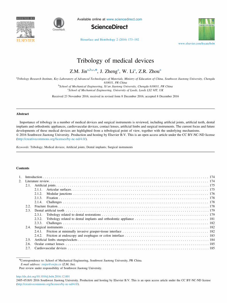
Fig. 1. (a) Number of records searched in Pubmed on 6th October 2016against year published using keywords "Medical device or (Joint ANDimplant) or (Dental AND Implant) or Contact lens or Medical instrument orContact lens OR Cardiovascular devices OR Fracture fixation or (ArtificialAND limb)) AND (tribology OR friction OR wear OR lubrication)".(b) Number of records searched in Pubmed on 6th October 2016 against yearpublished using keywords "Medical device AND (tribology OR friction ORwear OR lubrication)".
1. Introduction
Medical devices are widely used in daily life, ranging fromsimple bandages to complex imaging equipment. Medicaldevices are defined in different ways from various organiza-tions, including the Food and Drug Administration (FDA), theEuropean Union Directive (2007/47/EC) and ISO (13485).Examples of medical devices include instruments, apparatuses,appliances, materials, etc., intended to be used in humanbeings for the purpose of diagnosis, prevention, monitoring,treatment, or alleviation of disease, or compensation for aninjury or handicap, investigation, replacement, or modificationof the anatomy or of a physiological process etc.
Medical devices are heavily regulated because of theirintended uses in human beings. Generally medical devicesare classified into different categories depending upon thedegree of potential risks and regulated accordingly. Anincreasing concern has been raised recently, following on theclinical withdraw of a number of medical devices [1]. Theissue to balance the safety and effectiveness of a medicaldevice is once again called into question. Strict and compre-hensive pre-clinical testing has become even more important inthe evaluation of new innovative medical devices.
Many medical devices are involved with relative moving parts,either in contact to the native tissues or within the biomaterials,and often under loading. Important issues, such as friction andwear of the moving parts, not only affect the functions of thesedevices but also the potential adverse effects on the natural tissues.Biotribology deals with the application of tribological principles,such as friction, wear and lubrication between relatively motionssurfaces, to medical and biological systems. Biotribology plays animportant role in a number of medical devices.
The purpose of this review is focused on the tribology ofmedical devices. Specific aims include the following:
� Review important medical devices that have receivedextensively tribological investigations.
� Identify the corresponding gaps in research and the newdirections.
� Understand the underlying tribological mechanisms that arecommon among different medical devices.
It is beyond the scope of the present review to include allpossible medical devices in which tribology plays an importantrole. Instead implanted medical devices are mainly consideredand only musculoskeletal, dental, and cardiovascular systems arefocused. Other important medical devices for ocular and skinsystems as well as medical instruments are also included. Thispaper is organized with an overall introduction, followed by theliterature review of medical devices in each system, and finally asummary. In each section of the literature review on a biological
system, a general introduction to the use of the medical deviceand the potential clinical problems are firstly outlined and thenthe important tribological issues are discussed.
2. Literature review
A search was performed in Pubmed on 8th October 2016,using the following keywords “(Medical device or (Joint ANDimplant) or (Dental and Implant) or Contact lens or Medicalinstrument or Contact lens OR Cardiovascular devices ORFracture fixation or (Artificial AND limb)) AND (tribologyor friction or wear or lubrication)” and a total of 41,090 recordswere found. Fig. 1a shows the increasing trend of theserecords, particularly after the 1980s.A narrowed down search using the keywords “Medical
device AND (tribology OR friction OR wear OR lubrication)”returned a total record of 15,966, as shown in Fig. 1b.
Z.M. Jin et al. / Biosurface and Biotribology 2 (2016) 173–192 175
The records in each specific search of the sub-areas areshown in Table 1;
It is clear that joint implants have received by far the mostattention, followed by dental implants and cardiovascularimplants, fracture fixation devices and artificial limbs. Contactlenses and medical instruments are widely used, however thetribological research is relatively limited. There are clearlimitations of the above search because of the selection ofthe keywords. Nevertheless, the above search does give anindication of the important medical devices that have receivedimportant considerations of tribology.
2.1. Artificial joints
Artificial joints are one of the most successful medicaldevices used in human beings. There are 206 bones and over300 joints in the body. Of the joints in the body that allow arelatively large motion are the hip, the knee, the shoulder etc.Smaller joint implants such as the ankle, the elbow, the wristand the finger are also increasingly introduced into clinicalpractices. Joint implants also include the spinal disc (total disc)replacement and the temporomandibular joint (TMJ) prosthe-sis. These joints provide a range of complex three-dimensional
Table 1Number of records searched in Pubmed on 8th October 2016 for different areaswith different keywords.
Keywords Records
Joint And implant AND (tribology OR friction OR wear ORlubrication)
1468
Dental AND implant AND (tribology OR friction OR wear ORlubrication)
429
Cardiovascular devices AND (tribology OR friction OR wear ORlubrication)
387
Fracture fixation AND (tribology OR friction OR wear ORlubrication)
295
Artificial AND limb AND (tribology OR friction OR wear ORlubrication)
157
Contact lens AND (tribology OR friction OR lubrication) 114Medical instrument AND (tribology OR friction OR wear ORlubrication)
50
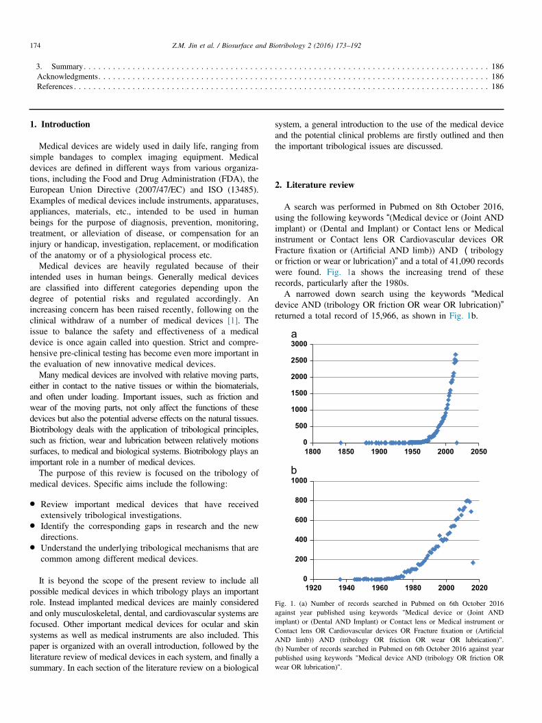
Fig. 2. A typical hip implant and a typical knee implant, showing the joint comp(b) knee: metallic femoral head, plastic tibial insert and metallic tray). a) Hip impl
motion and yet at the same time undertake a significant amountof loading. It is estimated that currently there are well over onemillion artificial joints implanted yearly into patients world-wide. Fig. 2 shows a typical hip implant and a typical kneeimplant.Tribological issues at the articulating surfaces as well as at
the connection between modular components and the fixationto bone are important considerations. Friction, wear andlubrication play important roles in the successful function ofartificial joints and the potential clinical problems.
2.1.1. Articular surfacesVarious biomaterial combinations are used for the articulat-
ing surfaces of artificial joints. These can be broadly dividedinto soft-on-hard and hard-on-hard combinations. Soft-on-hardcombinations mainly include ultra-high molecular weightpolyethylene (UHMWPE) against cobalt chromium alloys oralumina/zirconia toughened alumina composite ceramics(ZTA). Titanium alloys are sometimes preferred, particularlyfor total disc replacements in the spine, due to its lower elasticmodules and improved imaging quality, but surface treatmentsto improve its wear resistance are necessary. Hard-on-hardbearing surface combinations for a hip implant include metal-on-metal, ceramic-on-ceramic and ceramic-on-metal [2]. Themajor issue currently associated with the bearing surfaces ofartificial joints is wear and subsequent wear debris which cancause adverse tissue reactions and the loosening of theprosthetic components. Therefore improving the wear resis-tance of the bearing surfaces has been one of the main driversin the development of artificial joints [3].The major source of wear debris in the soft-on-hard
combination is from the UHMWPE bearing surface. Thereforeimprovements of the UHMWPE bearing surface are essential.Recent developments in this area include highly cross-linkedUHMWPEs, and further addition of vitamin-E and other anti-oxidants [4]. Compared with the conventional UHMWPE,these new polyethylene bearing materials have been shown toreduce wear considerably and to improve clinical outcome.Furthermore, the role of the hard counterface is also veryimportant since its scratch can lead to a marked increase in
onents (a) hip: metallic femoral head, plastic cup and metallic backing shell;ant. b) Knee implant.
Z.M. Jin et al. / Biosurface and Biotribology 2 (2016) 173–192176
UHMWPE wear [2]. Surface coatings of the metallic counter-face or the use of harder materials such as ceramics areintroduced to maintain a low wear level. However, the strengthand the potential long term durability associated with thesecoatings remain problematic [5]. Highly cross-linkedUHMWPEs are now mainly used in the majority of artificialhip joints, while their use in artificial knee joints is alsoreceiving attention [6].
The current hard-on-hard combinations for artificial jointsare mainly for the hip, including metal-on-metal and ceramic-on-ceramic. The wear in a metal-on-metal articulation can below, but can be increased drastically under adverse operatingconditions when the lubrication breaks down [7]. This haslargely led to the high revision rate and clinical withdraw of anumber of hip implants with a metal-on-metal articulation andthe use of these types of devices is greatly reduced [8].Nevertheless, well designed and accurate position may stillallow metal-on-metal articulations, particularly of the resurfa-cing type, to be used in selected patients [9]. Only ceramic-on-ceramic bearing combinations are now mainly used in routineclinics, including alumina and ZTA. In particular, the intro-duction of ZTA has improved both wear resistance andtoughness [10]. As a result, the combination of ZTA-on-ZTAhas reduced the wear of the bearing surfaces considerably,particularly under adverse conditions when the edge of theacetabular cup comes into contact with the femoral head [11]and in younger patients [12]. One of the potential complica-tions with ceramic-on-ceramic articulations is squeaking, and anumber of factors have been suggested and yet the squeakingmechanism is still unclear [13]. Squeaking has been reported torange from 0.5% to 10%, but occasionally up to 25%. Patient,implant and surgical factors can all contribute to the squeaking.This is further complicated by lack of a clear definition. At thepresent time, the squeaking in ceramic-on-ceramic implantscannot be eliminated completely.
New bearing surface combinations are being increasinglyintroduced to reduce wear and improve the longevity of jointimplants even further. Polyether-ether-ketone (PEEK) has beenextensively investigated as a potential material to replaceUHMWPE, in particular against a ceramic counterface forthe hip [14], the knee [15] and the spinal disc [16]. In addition,PEEK-on-PEEK combinations have been investigated forsmaller joints such as the spinal disc [17]. More recently,UHMWPE-on-PEEK bearing combination has been suggestedas a candidate for knee implants [18–20]. All these newmaterials and combinations are currently investigated inlaboratories and extensive pre-clinical testing is still requiredbefore potential clinical applications. Apart from the improve-ments of the wear resistance of the bearing materials, otherfactors such as implant designs, patients and surgeons are alsoimportant considerations. The key parameters in the implantdesign include the radius (size) of the bearing surfaces as wellas the conformity between the two articulating surfaces. It isoften necessary to balance the contradictory design require-ments between biomechanical and tribological functions. Forexample, an increase in the femoral head radius in the hipimplant improves the biomechanical functions such as the
range of motion and stability, and yet the wear of the bearingsurfaces is also increased as a result of the increased slidingdistance [21]. In the knee implant, an increase in theconformity between the articulating surfaces reduces thecontact stress, but may limit the range of motions and alsopotentially increase wear [22]. Furthermore, patient activitiescan affect wear greatly, for example, stair climbing may doublewear in a knee implant, compared with level walking [23].Surgical techniques can affect how the components of artificialjoints are positioned and aligned, and consequently the loadtransmission and wear.Wear testing of artificial joints is carried out extensively in
laboratories using simulators with various degrees of complex-ity before the implants are considered for approval for clinicalapplications. A number of standards for wear testing ofartificial joints have been introduced from both ISO (14242;14243; 18192) and ASTM (F2025). Currently, the major focusis to improve the laboratory based testing in order to be moreclosely representative of clinical settings. These includeintroduction of more adverse conditions to reflect a wide-spectrum use in patients and by surgeons [24,25].
2.1.2. Modular junctionsModular connections are introduced in artificial joints in
order to facilitate their use in patients and by surgeons. Forexample in the hip joint, modular head-neck combinations andmodular neck stems allow for restoration of anatomy andoptimization of joint biomechanical functions. Different bio-materials are often involved in the modular connection as wellas in direct contact with bone,including cobalt chromiumalloy/titanium, ceramics/titanium etc. Therefore, the potentialproblem of corrosion has long been recognized [26]. However,fretting corrosion has only received significant attentionrecently, following on the extensive clinical problems andrecalls reported with a number of hip implants with metal-on-metal articulations initially and then subsequently with mod-ular conjunctions [27]. The problem of fretting corrosion at themodular connection has been found particularly to be asso-ciated with the synergistic mechanical and electrochemicaleffect [28]. A number of terms have been introduced todescribe the related clinical problems, including pseudotumor,adverse local tissue reaction (ALTR), acute lymphocyticvasculitis associated lesions (ALVAL), adverse reaction tometallic debris (ARMD), taperosis, trunnionosis etc.,mainlyas a result of metallic wear debris and released metal ions.Furthermore, the clinical problems associated with frettingcorrosion in different artificial joints are common. Initially thefocus was on the articulation of metal-on-metal bearingsurfaces, particularly in resurfacing prostheses and largediameter total hip implants. Subsequently, a number ofmodular femoral stems in the hip implant were identified asproblems and consequently recalled [29,30]. Similar problemshave also been found at the femoral head and stem junction[31], and at the bearing linear and backing shell connection[32]. Fretting corrosion at the taper interface was found to beparticularly severe in the articulation of metal-on-metal bearingsurfaces with a large diameter [33]. Now it is generally
Z.M. Jin et al. / Biosurface and Biotribology 2 (2016) 173–192 177
accepted that fretting corrosion also occurs widely in thefemoral taper connection in total hip implants associated withUHMWPE-on-metal and UHMWPE-on-ceramics bearing sur-faces [34]. Similar problems have been identified in modularcomponents in the knee implant [35] and in the shoulderimplants [36]. Currently a lot of efforts are devoted to theunderstanding of the tribo-corrosion mechanism at the modularjunction and the identification of implant factors that aremainly responsible through clinical studies, laboratory testingand computational modelling.
Extensive clinical studies have been conducted to correlatethe factors that are related to fretting corrosion, throughmonitoring joint fluid and blood metal ions [37,38] andanalysing retrieval components [39]. The most important factorin fretting corrosion has been found to be associated with thematerial combination of the modular connection. Cobaltchromium alloy against titanium alloy or stainless steel hasbeen shown to produce more extensive fretting corrosion atboth the head-neck junction and the modular stem connectionthan against itself [39]. The use of a ceramic femoral head inconjunction with a titanium alloy appears to be the bestcombination in terms of reducing fretting corrosion, but cannoteliminate the problem completely [40]. In addition, theseauthors have shown that a low modulus titanium alloy(titanium–molybdenum–zirconium–iron alloy, TMZF) led toincreased fretting corrosion damage in the ceramic heads butno differences in the cobalt chromium alloy heads. A PEEKstem has been shown to produce less fretting corrosion [41].Surface coatings may have the potential of reducing frettingcorrosion, however this has not been demonstrated in a clinicalstudy for an oxidized zirconium head [42]. Furthermore, thecoating strength and long term durability are yet to beestablished.
The design and manufacturing parameters at the modularjunction can affect the relative micro-motion and therefore thefretting corrosion. Narrower and shorter stem designs withdifferent offsets are introduced to restore the joint centre, toincrease the range of motion, and decrease the risk ofimpingement and dislocation. However, a stem design directlyaffects its flexural rigidity and the head offset is directly relatedto the frictional torque, all potentially influencing the micro-motion at the taper. The effect of the taper designs fromdifferent manufacturers on fretting corrosion was investigatedby Tan et al. [42] for a given polyethylene-on-cobalt chro-mium alloy articulation with a 28 mm diameter bearing andsignificant differences were found. However, there are manyparameters associated with a taper design, which can allpotentially affect fretting corrosion. Taper geometry (cross-sectional dimensions and lengths) from different retrievedimplants was measured and used to calculate the flexuralrigidity and a wide range of values were found [43]. Threetaper designs with different angle, distal diameter and contactlength were examined and compared for a metal-on-metalarticulation [44]. A further study revealed the effect of thetaper length, and fretting corrosion was increased with longerhead lengths [45]. However, this has not been demonstrated inanother study [46]. The effects of the increased medio-lateral
offsets and longer neck moment arms have been shown to leadto increased taper damage at the modular interfaces for metal-on-metal articulations [32]. The effect of the taper angle hasbeen shown to be inversely correlated with stem fretting, butnot with head fretting and head-neck corrosion [47]. Differentsurface topographies and textures are introduced to increasethe fixation at the modular interface [48]. One study in [49]showed an increased fretting corrosion in the rougher tapersunder normal loading and an even worse performance underhigh loading. The surface topography was also shown to berelated to the damage scores on retrieved head-neck modularjunctions and furthermore to affect different materials combi-nations of cobalt chromium/titanium and titanium/titaniumdifferently [50]. Currently it is still not clear what the bestsurface topography should be for a modular connection. Inaddition to the design parameters, manufacturing parameters ofthe taper interface can also influence fretting corrosionsignificantly. Langton et al. showed that any deviations fromthe design specifications resulting from the manufacturingprocess can significantly increase the problem of frettingcorrosion [51].The bearing surfaces of the articulation between the femoral
head and the acetabular cup can also affect the frettingcorrosion at the modular taper interface. The resultant fric-tional torque at the bearing surfaces plays an important role inthis process. Metal-on-metal bearing surfaces, particularly witha large diameter and under adverse conditions, can produce ahigh friction torque, leading to severe fretting-corrosionproblems at the taper interface. Nevertheless, different bearingsurfaces currently used for total hip implants have all beenshown to produce fretting corrosion at the taper junction. Headdiameter affects the friction, particularly for metal-on-metalarticulations, however for UHMWPE-on-metal articulations,no effects were found [52].The effect of the length of implantation in patients on
fretting corrosion is not clear, with contradictory findings[38,44]. In addition, patient weight was found to be a predictorof fretting corrosion damage at the taper-neck junction in aretrieval series [53]. Furthermore, in a study of retrievedmodular femoral components, female patients were identifiedas high risk factors for failure [54]. However, such a findingmay be confounded by the smaller size of the implants.While the above findings have been found mainly from
clinical studies, laboratory experimental measurements havealso been conducted to understand the underlying mechanismof fretting corrosion from various perspectives. Some of thesestudies have combined a number of factors such as frettingcorrosion under a cyclic load and a corrosion environment[55]. While Panagiotidou et al. [49] have examined the effectof friction torque (bending moment) and corrosion. Thesurface contact area in a taper was measured and found todepend on different assembly forces [56]. Micro-motion andpull-out force were measured under different conditions tosimulate different surgical techniques and conditions [57–61].The effect of impaction force was considered to representsurgical factors [55,62]. A fretting corrosion testing was set upto investigate the effects of fretting amplitude and pH levels
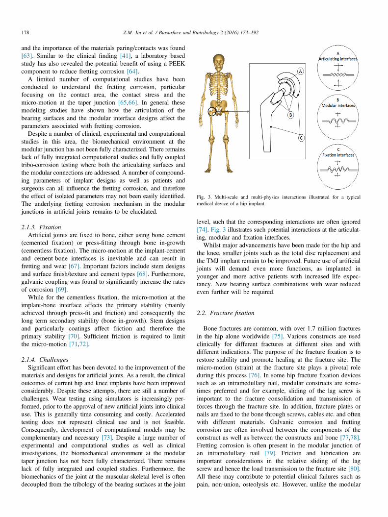
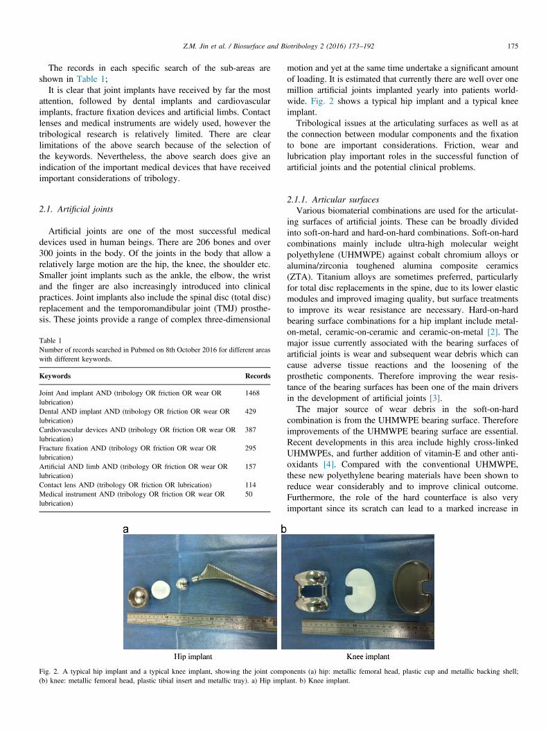
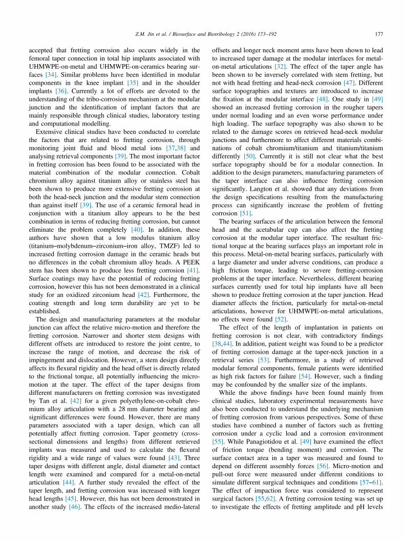
Fig. 3. Multi-scale and multi-physics interactions illustrated for a typicalmedical device of a hip implant.
Z.M. Jin et al. / Biosurface and Biotribology 2 (2016) 173–192178
and the importance of the materials paring/contacts was found[63]. Similar to the clinical finding [41], a laboratory basedstudy has also revealed the potential benefit of using a PEEKcomponent to reduce fretting corrosion [64].
A limited number of computational studies have beenconducted to understand the fretting corrosion, particularfocusing on the contact area, the contact stress and themicro-motion at the taper junction [65,66]. In general thesemodeling studies have shown how the articulation of thebearing surfaces and the modular interface designs affect theparameters associated with fretting corrosion.
Despite a number of clinical, experimental and computationalstudies in this area, the biomechanical environment at themodular junction has not been fully characterized. There remainslack of fully integrated computational studies and fully coupledtribo-corrosion testing where both the articulating surfaces andthe modular connections are addressed. A number of compound-ing parameters of implant designs as well as patients andsurgeons can all influence the fretting corrosion, and thereforethe effect of isolated parameters may not been easily identified.The underlying fretting corrosion mechanism in the modularjunctions in artificial joints remains to be elucidated.
2.1.3. FixationArtificial joints are fixed to bone, either using bone cement
(cemented fixation) or press-fitting through bone in-growth(cementless fixation). The micro-motion at the implant-cementand cement-bone interfaces is inevitable and can result infretting and wear [67]. Important factors include stem designsand surface finish/texture and cement types [68]. Furthermore,galvanic coupling was found to significantly increase the ratesof corrosion [69].
While for the cementless fixation, the micro-motion at theimplant-bone interface affects the primary stability (mainlyachieved through press-fit and friction) and consequently thelong term secondary stability (bone in-growth). Stem designsand particularly coatings affect friction and therefore theprimary stability [70]. Sufficient friction is required to limitthe micro-motion [71,72].
2.1.4. ChallengesSignificant effort has been devoted to the improvement of the
materials and designs for artificial joints. As a result, the clinicaloutcomes of current hip and knee implants have been improvedconsiderably. Despite these attempts, there are still a number ofchallenges. Wear testing using simulators is increasingly per-formed, prior to the approval of new artificial joints into clinicaluse. This is generally time consuming and costly. Acceleratedtesting does not represent clinical use and is not feasible.Consequently, development of computational models may becomplementary and necessary [73]. Despite a large number ofexperimental and computational studies as well as clinicalinvestigations, the biomechanical environment at the modulartaper junction has not been fully characterized. There remainslack of fully integrated and coupled studies. Furthermore, thebiomechanics of the joint at the muscular-skeletal level is oftendecoupled from the tribology of the bearing surfaces at the joint
level, such that the corresponding interactions are often ignored[74]. Fig. 3 illustrates such potential interactions at the articulat-ing, modular and fixation interfaces.Whilst major advancements have been made for the hip and
the knee, smaller joints such as the total disc replacement andthe TMJ implant remain to be improved. Future use of artificialjoints will demand even more functions, as implanted inyounger and more active patients with increased life expec-tancy. New bearing surface combinations with wear reducedeven further will be required.
2.2. Fracture fixation
Bone fractures are common, with over 1.7 million fracturesin the hip alone worldwide [75]. Various constructs are usedclinically for different fractures at different sites and withdifferent indications. The purpose of the fracture fixation is torestore stability and promote healing at the fracture site. Themicro-motion (strain) at the fracture site plays a pivotal roleduring this process [76]. In some hip fracture fixation devicessuch as an intramedullary nail, modular constructs are some-times preferred and for example, sliding of the lag screw isimportant to the fracture consolidation and transmission offorces through the fracture site. In addition, fracture plates ornails are fixed to the bone through screws, cables etc. and oftenwith different materials. Galvanic corrosion and frettingcorrosion are often involved between the components of theconstruct as well as between the constructs and bone [77,78].Fretting corrosion is often present in the modular junction ofan intramedullary nail [79]. Friction and lubrication areimportant considerations in the relative sliding of the lagscrew and hence the load transmission to the fracture site [80].All these may contribute to potential clinical failures such aspain, non-union, osteolysis etc. However, unlike the modular
Z.M. Jin et al. / Biosurface and Biotribology 2 (2016) 173–192 179
junction in the hip implant, the effect of using dissimilarmaterials for the fracture constructs does not appear to besignificant [81].
Tribological problems are clearly present in fracture con-structs and fixations. However, the scope of the investigationin this area is much limited, compared with artificial joints.The main reasons for this may be the relatively short period ofin vivo implantation and different biomechanical environmentsof fracture constructs.
2.3. Dental artificial tooth
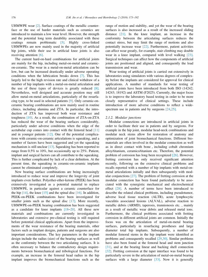
Human teeth are not only the important masticatory organ butalso closely associated with both the pronunciation and the facialesthetics of human being. Due to ageing, various pathologicfactors and traumas, tooth lesion such as caries, partial or overalltooth tissue loss will occur unavoidably. Generally the lesion ofhuman teeth is restored and treated with dental restorations and/or implants in dental clinic, which are called artificial dentaltooth. Dental restorations include dental restorative materialsused to restore the function, integrity and morphology ofmissing tooth structure and the replacement of missing toothstructure that is supported by dental implants. Due to oralphysiological functions, dental restorations and implants inevi-tably suffer friction and wear in the mouth every day. Nowa-days, metals and alloys, ceramics and composites materials aremost widely used for dental restorations and implants [82].Fig. 4 illustrates dental restorations and implants commonlyused in clinic. Normally different dental materials encounterdifferent tribological problems in their clinical uses, as shown inTable 2 [83]. Excessive wear could result in the failure of dentalrestorations and implants. Thus, much work has been done toinvestigate the tribological behavior of artificial dental teeth andthen to improve their anti-wear properties.
2.3.1. Tribology related to dental restorationsDental restorations are classified as either direct or indirect.
Direct restorations are made directly inside the mouth of thepatient, while indirect restorations are made outside of thepatient's mouth and then placed inside. Restorations includefilling, (composite filling and amalgam filling), crown (com-posite crown, metal-ceramic crown and full ceramic crown),
Fig. 4. Dental restorations and imp
veneer, inlay, onlay (mainly made from ceramic), and bridge(mainly made from stainless steel). Most tribological studiesrelated to dental restorations focused on dental composite,ceramic and amalgam.
2.3.1.1. Dental composite. Due to good aesthetics, theability to bond to tooth structures and the need for an amalgamalternative, resin-based dental composites have been usedincreasingly widely in the field of restorative dentistry recently[84]. The most widely used dental composites are compositeresin fillings (also called white fillings), which contain fillerparticles (borosilicate glass, colloidal silica, etc.) in a polymermatrix (Bis-GMA, TEGDMA, etc.) generally. Composite resinfillings are commonly utilized to restore cavities, replace themissing tooth tissue that has been worn away by grinding, andresemble the appearance of the natural tooth [83,85,86]. Amain problem of dental composites in clinic is their weakwear-resistance [83]. Therefore, many new technologies andmethods have been developed to optimize the composites inorder to improve their wear resistance [87].The wear resistance of dental composite is closely associated
with its material characteristic [88]. The material factors of resin-based dental composites are normally related to particle size,shape and hardness, the filler content, the inter-particle spacing,the filler distribution, the degree of conversion, the interfacialbond strength between filler and matrix, the nature of the matrix,and the surface hardness [89].The size of inorganic fillers has been found to be enormously
essential for the wear resistance of the dental composites. Micro-filled materials had a better wear resistance than traditionalmacro-filled materials [90]. Micro-filled and hybrid materialspossessed similar wear resistance [90]. Although a few earlierstudies revealed that composites containing smaller sphericalparticles showed better wear resistance [91], nano-filled materi-als seemed to experience more or equal wear to micro-filledmaterials [92]. It was indicated that there was a critical value offiller particle size (1.2–1.5 μm), under which the strait-linerelation was different from that above the value [89].Aside from particle size, the content of filler particles also
could affect the wear resistance of dental composite signifi-cantly. As the filler volume increased, wear was reducedregardless of the filler treatment [88]. The wear resistance of
lants commonly used in clinic.
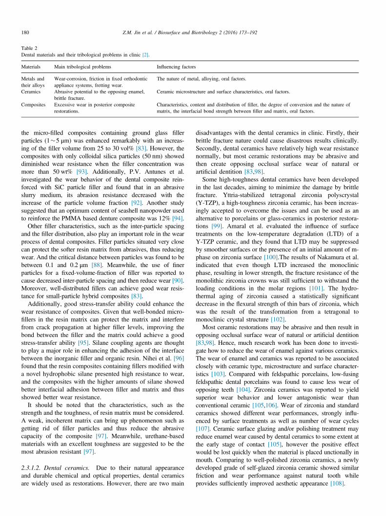
Table 2Dental materials and their tribological problems in clinic [2].
Materials Main tribological problems Influencing factors
Metals andtheir alloys
Wear-corrosion, friction in fixed orthodonticappliance systems, fretting wear.
The nature of metal, alloying, oral factors.
Ceramics Abrasive potential to the opposing enamel,brittle fracture.
Ceramic microstructure and surface characteristics, oral factors.
Composites Excessive wear in posterior compositerestorations.
Characteristics, content and distribution of filler, the degree of conversion and the nature ofmatrix, the interfacial bond strength between filler and matrix, oral factors.
Z.M. Jin et al. / Biosurface and Biotribology 2 (2016) 173–192180
the micro-filled composites containing ground glass fillerparticles (1�5 μm) was enhanced remarkably with an increas-ing of the filler volume from 25 to 30 vol% [83]. However, thecomposites with only colloidal silica particles (50 nm) showeddiminished wear resistance when the filler concentration wasmore than 50 wt% [93]. Additionally, P.V. Antunes et al.investigated the wear behavior of the dental composite rein-forced with SiC particle filler and found that in an abrasiveslurry medium, its abrasion resistance decreased with theincrease of the particle volume fraction [92]. Another studysuggested that an optimum content of seashell nanopowder usedto reinforce the PMMA based denture composite was 12% [94].
Other filler characteristics, such as the inter-particle spacingand the filler distribution, also play an important role in the wearprocess of dental composites. Filler particles situated very closecan protect the softer resin matrix from abrasives, thus reducingwear. And the critical distance between particles was found to bebetween 0.1 and 0.2 μm [88]. Meanwhile, the use of finerparticles for a fixed-volume-fraction of filler was reported tocause decreased inter-particle spacing and then reduce wear [90].Moreover, well-distributed fillers can achieve good wear resis-tance for small-particle hybrid composites [83].
Additionally, good stress-transfer ability could enhance thewear resistance of composites. Given that well-bonded micro-fillers in the resin matrix can protect the matrix and interferefrom crack propagation at higher filler levels, improving thebond between the filler and the matrix could achieve a goodstress-transfer ability [95]. Silane coupling agents are thoughtto play a major role in enhancing the adhesion of the interfacebetween the inorganic filler and organic resin. Nihei et al. [96]found that the resin composites containing fillers modified witha novel hydrophobic silane presented high resistance to wear,and the composites with the higher amounts of silane showedbetter interfacial adhesion between filler and matrix and thusshowed better wear resistance.
It should be noted that the characteristics, such as thestrength and the toughness, of resin matrix must be considered.A weak, incoherent matrix can bring up phenomenon such asgetting rid of filler particles and thus reduce the abrasivecapacity of the composite [97]. Meanwhile, urethane-basedmaterials with an excellent toughness are suggested to be themost abrasion resistant [97].
2.3.1.2. Dental ceramics. Due to their natural appearanceand durable chemical and optical properties, dental ceramicsare widely used as restorations. However, there are two main
disadvantages with the dental ceramics in clinic. Firstly, theirbrittle fracture nature could cause disastrous results clinically.Secondly, dental ceramics have relatively high wear resistancenormally, but most ceramic restorations may be abrasive andthen create opposing occlusal surface wear of natural orartificial dentition [83,98].Some high-toughness dental ceramics have been developed
in the last decades, aiming to minimize the damage by brittlefracture. Yttria-stabilized tetragonal zirconia polyscrystal(Y-TZP), a high-toughness zirconia ceramic, has been increas-ingly accepted to overcome the issues and can be used as analternative to porcelains or glass-ceramics in posterior restora-tions [99]. Amaral et al. evaluated the influence of surfacetreatments on the low-temperature degradation (LTD) of aY-TZP ceramic, and they found that LTD may be suppressedby smoother surfaces or the presence of an initial amount of m-phase on zirconia surface [100].The results of Nakamura et al.indicated that even though LTD increased the monoclinicphase, resulting in lower strength, the fracture resistance of themonolithic zirconia crowns was still sufficient to withstand theloading conditions in the molar regions [101]. The hydro-thermal aging of zirconia caused a statistically significantdecrease in the flexural strength of thin bars of zirconia, whichwas the result of the transformation from a tetragonal tomonoclinic crystal structure [102].Most ceramic restorations may be abrasive and then result in
opposing occlusal surface wear of natural or artificial dentition[83,98]. Hence, much research work has been done to investi-gate how to reduce the wear of enamel against various ceramics.The wear of enamel and ceramics was reported to be associatedclosely with ceramic type, microstructure and surface character-istics [103]. Compared with feldspathic porcelains, low-fusingfeldspathic dental porcelains was found to cause less wear ofopposing teeth [104]. Zirconia ceramics was reported to yieldsuperior wear behavior and lower antagonistic wear thanconventional ceramic [105,106]. Wear of zirconia and standardceramics showed different wear performances, strongly influ-enced by surface treatments as well as number of wear cycles[107]. Ceramic surface glazing and/or polishing treatment mayreduce enamel wear caused by dental ceramics to some extent atthe early stage of contact [105], however the positive effectwould be lost quickly when the material is placed unctionally inmouth. Comparing to well-polished zirconia ceramics, a newlydeveloped grade of self-glazed zirconia ceramic showed similarfriction and wear performance against natural tooth whileprovides sufficiently improved aesthetic appearance [108].
Z.M. Jin et al. / Biosurface and Biotribology 2 (2016) 173–192 181
The chemical attack in the mouth may result in the surfacedegradation of dental ceramics [103], and then accelerate itswear process. The highest degradation of Y-TZP dentalceramics occurred in acidic environment [109]. Generallyceramic surface is prone to degradation by acidulated fluoride,that can increase wear rates. The results of Guilherme TeixeiraTheodoro et al. suggested that ceramic type and fluoride gelaffected the wear and roughness of worn surface, but the typeof failure was only affected by ceramic type [109].
2.3.1.3. Dental amalgam. Considering the color being verydifferent from that of dental tissue, metals and alloys aremainly applied to orthodontic appliances and dental implantsnowadays, and only amalgam filling is used as dental restora-tion. Amalgam, commonly called amalgam alloy, is composedfrom a mixture of mercury and powdered alloy made mostly ofsilver, tin, copper and so on [110], and has been successfullyused as one of the most popular direct restorative materials bydental profession for more than 200 years.
The major attraction of amalgam alloy is the proven longevitydue to its high wear resistance from the metallic character inclinical service and ease of clinical use [111]. Thus, manyresearchers use the amalgam as a comparator to evaluate otherdental materials in earlier studies. Hu et al. [112] compared therelative wear resistance of a selection of current dental compo-sites and amalgam to assess their relative potential clinical wearresistance under variable masticatory loads, and finally theydivided the tested composites into two degrees of wearresistance including better and worse than the amalgam.Additionally, it was found that an increase in the hardness valueof amalgam usually led to improved abrasion resistance [111].
However, there are still some disadvantages about theamalgam, such as its aesthetics and the high toxicity ofmercury, the weak corrosion-resistance, the low fracturetoughness and tensile strength, the brittleness, and so on[111]. It has been widely accepted that ions, especiallymercury and some other heavy metal, of the constituentelements are released into the body during the corrosion ofdental amalgam in the long run [113,114]. Given that thecorrosive characteristics of dental alloys are of both funda-mental and applied interest, because corrosion not only affectsthe functionality of dental constructions but may also causepathological phenomena, related research work mainly focusedon the corrosion of amalgam in the last decade [115]. Muchhas been done on the corrosion of dental silver amalgam fromdifferent point of views, such as the release of mercury andother metals, the electrochemistry under sliding wear condi-tion, the metallography, the discoloration and other aspects.From the aspect of electrochemistry, a recent research provedthat the existence of an electrically insulating layer, which isprobably composed of non-metallic corrosion products, bio-films, and dental calculus, could reduce galvanic corrosionrates to small or negligible values [116]. While from the aspectof metallography, considering both the corrosion and thestrength, the results of Chung et al. [117] indicated that thecorrosion resistance of high-copper single-composition amal-gam, whose mechanical properties could be significantly
improved by a certain amount of steel fibers [118], could beimproved by Ag–Cu nanoparticle-doping.
2.3.2. Tribology related to dental implants and orthodonticappliance2.3.2.1. Dental implants. Pure titanium (CP-Ti) and itsalloys have been widely applied to dental implants due toexcellent biocompatibility, corrosion resistance and lightweight. However, CP-Ti is inferior to conventional dentalalloys in tribological characteristics [119,120], and its wearresistance can be improved by alloying [121].Nowadays, although CP-Ti and its alloys are the most
commonly used materials for dental implants, the release oftoxic elements (e.g. Al and V) due to tribocorrosion in themouth and the so-called stress-shielding effect are still aconcern. Recently, β and near-β titanium alloys with reducedelastic modulus and biocompatible alloying elements havebeen developed to overcome these issues [122]. Golvanostudied the tribocorrosion behavior of the near-β Ti13Nb13Zralloy in oral environment, and their results revealed a negativeinfluence of the increase of fluoride concentration and theacidified artificial saliva on the material degradation [123]. Itwas suggested that both the cast and sintered Ti6Al4V alloysexhibited same tribocorrosion mechanisms, and there existed acritical fluoride concentration above which corrosion andtribocorrosion rates of Ti6Al4V alloys increased [124]. Copperin titanium-copper biomedical alloy was proven to increase theamount of eutectoid in the grain boundary, favouring theformation of Ti2Cu intermetallics and increasing the hardnessof the alloys, and thus total material loss due to the wear andcorrosion decreased with the increase in the Ti2Cu interme-tallics [125]. In order to improve the tribocorrosion resistanceof Ti alloy implant surface, Oliveira et al. focused on theincorporation of magnesium, together with calcium andphosphorous, in the structure of titanium oxide films producedby micro-arc oxidation, and they found that the addition ofmagnesium would support the formation of rutile which couldimprove the tribocorrosion properties of the surfaces [126].The results of Mathew et al. indicated that the lipopolysac-charide in saliva could negatively affect the corrosion/wearbehavior of titanium, which may contribute to the failure ofdental implants [127].It should be noted that fretting wear may result in the failure
of dental implants [128]. Yu et al. investigated the tangentialfretting behavior of titanium alloy (TC4) against humancortical thighbone to understand the fretting behavior of thefixation interface of dental implants [129].Their results indi-cated that during the long service process of dental implants,the repeated action of occlusal load would result in a variationof the initial contact condition of the bone-implant interfacewith the accumulation of surface damage, and thus looseningoccurs to dental implants. That is the non-medicinal reasonwhy the failure rate of dental implants' fixation interfaceincreases over time after osseointegration.
2.3.2.2. Orthodontic appliance. It has been accepted thatincreased friction between mucosa tissue and the surface of
Z.M. Jin et al. / Biosurface and Biotribology 2 (2016) 173–192182
metallic brackets can cause pain and discomfort of oral mucosa[130,131]. The friction behavior of orthodontic metallicbracket-wire combinations is associated with such factors asarchwire and bracket materials, their size and shape, width andslot dimensions, surface composition, roughness and cleanli-ness, bracket-to-wire positioning in a 3-dimensional space, theligature force and the type of ligation, interbracket distances,and lubrication.
2.3.3. ChallengesSignificant effort has been devoted to improving the
tribological properties of dentalmaterials. As a result, theclinical outcomes of current artificial dental teeth have beenimproved considerably. Nonetheless, there are still a number ofchallenges. Ideally, the tribological properties of artificialdental teeth should be similar to those of human tooth enamel.To date, these properties may only befound in dental ceramicmaterials and particular metal alloys [82], and the wear ofmany dental resincomposites is still considerablein vivo in thelong run. Moreover, most in vitro studies have only focused onproviding comparative ranking of various dental materials, butnot aimed at revealing their wear mechanism.Given that anunderstanding of the fundamental underlying wear mechan-isms involved will lead to a better understanding of in vivofailure patterns, the lack of these aspects may be one of themain obstacles hindering the development of dental materials.
2.4. Surgical instruments
Surgical instruments can be generally divided into sixclasses by function. These classes are: cutting instruments,grasping or holding instruments, haemostatic forceps, retrac-tors, clamps and distractors, accessories and implants. In recentyears, Minimally Invasive Surgery (MIS) and gastrointestinalendoscopy are generally popular surgical operations, which isaccompanied by some tribological problems occurred at theinterface between minimally invasive grasper or endoscopyand tissues.
2.4.1. Friction at minimally invasive grasper-tissue interfaceMinimally Invasive Surgery (MIS) is almost self-evident
that minimally invasive procedures have clear clinical benefitsto patients when compared to “open” procedures. By virtue ofthe minimal invasion, performing any procedure less inva-sively results in less soft tissue disruption, with the effects ofreduced pain, faster healing and better recovery [132]. As abyproduct of minimally invasive techniques, patients requireshorter hospital stays and return faster to normal activity [133].However, these minimally invasive procedures have alsoincorporated the disadvantages of limited dexterity, lack of3D visualization, poor ergonomic design and lack of hapticfeedback, which reduce the accuracy of force feedback to thesurgeon from the tool-tissue interaction [134–136]. As thesurgeon is no longer in direct contact with the patient orsurgical tools and must use only their visual sense toapproximate the tool-tissue interaction forces, the surgeon'sperception of the tool-tissue interaction forces may be higher
or lower than the actual force at the tool tip. Higher forceusually induces tissue trauma, while lower force can causegrasper and tissue slipping when dragging tissue, reducingoperation efficiency [137,138]. The function of laparoscopicgraspers is to realize clamping, gripping and dragging organ ortissue. There exists friction behavior at the laparoscopicgrasper-tissue interface, which would usually result in tissuedamage. Excessive pressure during organ and tissue retractionwith laparoscopic graspers is one of the causes of intraopera-tive injury in laparoscopic interventions [139,140]. It isreported that grasper-related traumaduring laparoscopic proce-dures has a 2–4% risk of injury to the bile duct, bowel,vascular structures, significantly higher than in open abdom-inal surgery [140–143]. An observational study by Tang et al.[144] found that 66% of human errors identified duringlaparoscopic cholecystectomy were related to graspers, 13%of which, in turn, were related to excessive force exertion.On the investigation of the pressure distribution of grasper-
tissue interaction, Payandeh et al. [145] has shown that theaverage magnitude of the grasping force in a typical palpationtask is approximately 12.5 N. Similar research studies havefoundthe maximum grasping force was 16 N [145,146]. Cart-mill et al. [147] found that the pinch force required to preventtissue slipping out of the grasper, while hanging from thetissue a 250 g load at a direction perpendicular to the plane ofthe end effector, generated localized peak tissue stresses ashigh as 800 kPa, which was beyond the safety threshold of200 kPa estimated by De et al. for cell apoptosis in abdominalorgans [148,149]. Some researchers designed a laparoscopicgrasper equipped with strain gages or sensors and thenconducted in vivo and in situ experiments with different tissuesto measure forces during grasping [135–137,150].On the investigation refered to the friction between laparo-
scopic grasper and soft tissues interfaces, Frank et al. [151]preliminarily tested the friction behavior between the clampand the small intestine in laparoscopic operation, the resultshowed that the friction coefficient was 0.6�0.9 and 7 N ofsealing force was recommended to prevent leakage. Li et al.[152] studied the friction behavior at minimally invasivegrasper-liver tissue interface under different clamping forcesand dragging speeds. The results revealed that the injurydegree of the liver gradually increased with increasing clamp-ing force. The maximum static friction force increased withincreasing clamping force and dragging speed, and draggingdisplacement before sliding increased with increasing clamp-ing force and decreasing dragging speed, which indicated thatlow clamping force and high dragging speed may be morelikely to cause slipping at the jaw-liver interface. Oldfield et al.[153] examined tool-tissue interactions, strain energy releaserate and deformation by using blade insertions into a gelatinsoft tissue phantom experiments and accompanying finiteelement simulations.In most of the studies above, the compressive stress was
measured and computed by finite element analysis, however,the tractive force or friction force during dragging tissue wasstill rarely studied. The combination of critical pressure valuesand friction force which neither induced tissue injury nor cause
Z.M. Jin et al. / Biosurface and Biotribology 2 (2016) 173–192 183
tissue slipping out of the grasper are not clear. Little currentdata is available to suggest stress magnitudes that are safe fortissue manipulation, which is an important safe threshold fordoctors during grasping task in MIS.
Moreover, modern laparoscopic graspers usually have atooth structure at the end effector to improve the efficiency ofclamping. However, this structure may cause non-uniformpressure distribution, and the obvious damage appeared in thetissue site contacted along the jaw edge, which suggested thatthe tissue damage is not only associated with excessiveclamping force, also related to the structure of the grasper[152]. Thus the pressure distribution from graspers withdifferent structures is important for analysis of tissue damagemechanisms, which should be done as future work by usingnumerical simulation.
2.4.2. Friction at endoscopy and esophagus or colon interfaceFor gastrointestinal diseases, conveying gastrointestinal
endoscope through the digestive tract, such as, to the lesionlocation, and conducting operation such as checking, ablation,removing or stripping the diseased tissue, are the most basicclinical treatments in digestive system (Fig. 5). For example,endoscopic submucosa dissection [154] and endoscopic muco-sal resection [155,156] are the proper treatment of earlygastrointestinal tumors. However, either diagnosis or treatmentof gastrointestinal tract is a kind of invasive operation throughthe narrow single port of digestive tract. During gastrointest-inal endoscopy, endoscopy is pushed into human digestivetract with the aid of outside force, which may cause a series ofcomplications such as throat abrasion, bleeding, mucosaltearing and perforation of digestive tract due to repeatedlyinserting, rotating, pushing and retrieving operation. Never-theless, these are serious friction damage problems that fewstudies have focused on.
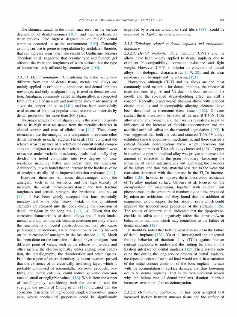
On the investigation of friction behavior of the digestivetract tissue surface, Accoto et al. [157] first acknowledged andproposed that it was very difficult for minimally invasivedevices to pass through the collapsed and bended digestivetract in the initial stages of NOTES (Natural orifices translum-inal endoscopic surgery). They measured the variation of
Fig. 5. Schematic diagram during gastrointestinal endoscopy, (a) Gastrointestinal entract and (c) Endoscopic diagnosis and treatment through lower gastrointestinal tra
friction coefficient by using the constant weight blocks slidingon the surface of digestive tract at different speeds and foundthat it increased proportionally with speed. Since then, thestudy of friction behavior on the digestive tract surface wasmainly concentrated on the small intestine. Kim et al. [158]tested the friction coefficient of intestinal surface in closed andopen cavity and put forward a five-element viscoelastic modelto establish the friction model of small intestine. Li et al. [159]investigated the friction trauma mechanism of small intestinecaused by the pulling operation of endoscopy under differentnormal force and friction time in the process of surgery, andfound that the total friction energy dissipation on the smallintestine increased with the increasing normal force andfriction time, which induced the damage degree of the smallintestine aggravation. For the friction between capsule endo-scopy and digestive tract surface, some studies examined theinfluence of capsule shape, dimension, weight, contact areaand speed parameters on the friction coefficient of smallintestine, and found that the friction coefficient changedbetween 0.08 and 0.2, which increased with the increasingspeed, and decreased with the increasing normal load. Theinfluence of capsule weight and contact area on the frictioncoefficient was trivial [160–164].Although a few studies referred to the friction between
endoscopy and digestive tract tissue surface, the viscoelasticcharacteristics of the internal organs and the frictional resistanceof the endoscope inside the body are still not clear. These aredefinitely necessary since the power consumption and positioncontrol of the endoscope are largely affected by these character-istics [165]. Moreover, it is hard to perform experimentalinvestigations inside the human body because of the cost andsafety, whenever the data are required for the endoscope design.In addition, complex digestive tract environment has been one ofthe greatest obstacles of the development of the endoscopy andcapsule robot. Different parts of the digestive tract have differentdiameters, wall thickness, lengths of villi and so on. Theinfluence of the biodiversity of the digestive tract on thefrictional resistance can be revealed by a lot of experiments,which can provide the basic data for safety operation anddamage control during gastrointestinal endoscopy.
doscope, (b) Endoscopic diagnosis and treatment through upper gastrointestinalct.
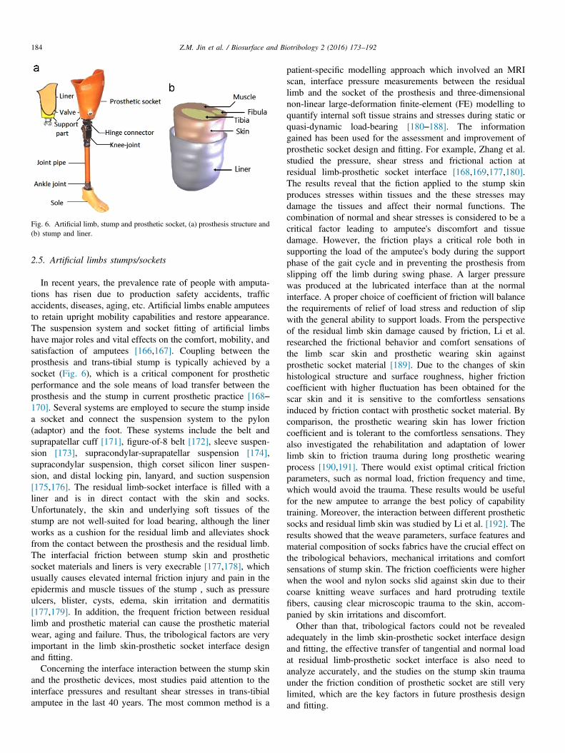
Fig. 6. Artificial limb, stump and prosthetic socket, (a) prosthesis structure and(b) stump and liner.
Z.M. Jin et al. / Biosurface and Biotribology 2 (2016) 173–192184
2.5. Artificial limbs stumps/sockets
In recent years, the prevalence rate of people with amputa-tions has risen due to production safety accidents, trafficaccidents, diseases, aging, etc. Artificial limbs enable amputeesto retain upright mobility capabilities and restore appearance.The suspension system and socket fitting of artificial limbshave major roles and vital effects on the comfort, mobility, andsatisfaction of amputees [166,167]. Coupling between theprosthesis and trans-tibial stump is typically achieved by asocket (Fig. 6), which is a critical component for prostheticperformance and the sole means of load transfer between theprosthesis and the stump in current prosthetic practice [168–170]. Several systems are employed to secure the stump insidea socket and connect the suspension system to the pylon(adaptor) and the foot. These systems include the belt andsuprapatellar cuff [171], figure-of-8 belt [172], sleeve suspen-sion [173], supracondylar-suprapatellar suspension [174],supracondylar suspension, thigh corset silicon liner suspen-sion, and distal locking pin, lanyard, and suction suspension[175,176]. The residual limb-socket interface is filled with aliner and is in direct contact with the skin and socks.Unfortunately, the skin and underlying soft tissues of thestump are not well-suited for load bearing, although the linerworks as a cushion for the residual limb and alleviates shockfrom the contact between the prosthesis and the residual limb.The interfacial friction between stump skin and prostheticsocket materials and liners is very execrable [177,178], whichusually causes elevated internal friction injury and pain in theepidermis and muscle tissues of the stump,such as pressureulcers, blister, cysts, edema, skin irritation and dermatitis[177,179]. In addition, the frequent friction between residuallimb and prosthetic material can cause the prosthetic materialwear, aging and failure. Thus, the tribological factors are veryimportant in the limb skin-prosthetic socket interface designand fitting.
Concerning the interface interaction between the stump skinand the prosthetic devices, most studies paid attention to theinterface pressures and resultant shear stresses in trans-tibialamputee in the last 40 years. The most common method is a
patient-specific modelling approach which involved an MRIscan, interface pressure measurements between the residuallimb and the socket of the prosthesis and three-dimensionalnon-linear large-deformation finite-element (FE) modelling toquantify internal soft tissue strains and stresses during static orquasi-dynamic load-bearing [180–188]. The informationgained has been used for the assessment and improvement ofprosthetic socket design and fitting. For example, Zhang et al.studied the pressure, shear stress and frictional action atresidual limb-prosthetic socket interface [168,169,177,180].The results reveal that the fiction applied to the stump skinproduces stresses within tissues and the these stresses maydamage the tissues and affect their normal functions. Thecombination of normal and shear stresses is considered to be acritical factor leading to amputee's discomfort and tissuedamage. However, the friction plays a critical role both insupporting the load of the amputee's body during the supportphase of the gait cycle and in preventing the prosthesis fromslipping off the limb during swing phase. A larger pressurewas produced at the lubricated interface than at the normalinterface. A proper choice of coefficient of friction will balancethe requirements of relief of load stress and reduction of slipwith the general ability to support loads. From the perspectiveof the residual limb skin damage caused by friction, Li et al.researched the frictional behavior and comfort sensations ofthe limb scar skin and prosthetic wearing skin againstprosthetic socket material [189]. Due to the changes of skinhistological structure and surface roughness, higher frictioncoefficient with higher fluctuation has been obtained for thescar skin and it is sensitive to the comfortless sensationsinduced by friction contact with prosthetic socket material. Bycomparison, the prosthetic wearing skin has lower frictioncoefficient and is tolerant to the comfortless sensations. Theyalso investigated the rehabilitation and adaptation of lowerlimb skin to friction trauma during long prosthetic wearingprocess [190,191]. There would exist optimal critical frictionparameters, such as normal load, friction frequency and time,which would avoid the trauma. These results would be usefulfor the new amputee to arrange the best policy of capabilitytraining. Moreover, the interaction between different prostheticsocks and residual limb skin was studied by Li et al. [192]. Theresults showed that the weave parameters, surface features andmaterial composition of socks fabrics have the crucial effect onthe tribological behaviors, mechanical irritations and comfortsensations of stump skin. The friction coefficients were higherwhen the wool and nylon socks slid against skin due to theircoarse knitting weave surfaces and hard protruding textilefibers, causing clear microscopic trauma to the skin, accom-panied by skin irritations and discomfort.Other than that, tribological factors could not be revealed
adequately in the limb skin-prosthetic socket interface designand fitting, the effective transfer of tangential and normal loadat residual limb-prosthetic socket interface is also need toanalyze accurately, and the studies on the stump skin traumaunder the friction condition of prosthetic socket are still verylimited, which are the key factors in future prosthesis designand fitting.
Z.M. Jin et al. / Biosurface and Biotribology 2 (2016) 173–192 185
Moreover, the improper prosthetic materials often cause skindamage from the tribological point of view. Therefore, it isnecessary for prosthetic limb manufacturers to improve prosthe-tic socket design and fitting, and choose better biocompatiblematerials with the skin such as silica gel, etc, which can improvethe contact comfort and avoid the skin damage.
2.6. Ocular contact lenses
Contact lenses are used widely to correct vision. Currentlyover 140 million contact lenses are used worldwide [193].Despite the improvements of new lens materials and caresystems, clinical problems such as dryness and discomfort arestill widely reported and more severe complications such ascontact lens-induced lid-wiper epitheliopathy and lid-parallelconjunctival folds are still present [194]. The insertion of acontact lens forms two interfaces with the natural eye, one withthe lid wiper (pre-lens) and the other with the ocular surface(post-lens). Both relative motion and loading are involved atthese two interfaces and therefore tribology plays an importantrole in the blinking of the eye as well as the successful functionof a contact lens.
Lubrication between the contact lens and the eye is criticallyimportant [194]. Fluid film lubrication in the presence of thetear film ensures minimum friction, smooth motion andnegligible damage in the eye during blinking. However, atthe beginning, end, and return points of the blinking cyclewhere there is relatively small motion between the lid wiperand a contact lens, boundary lubrication may be dominant, anda complementary surface-brush boundary lubrication mechan-ism comes into operation. At the back of the contact lens incontact with the ocular surface, the speed is relatively low andthe brush lubrication mechanism is also dominant. Therefore asynergistic lubrication mechanism is expected for a contactlens to function normally in the eye. Important considerationsinclude the lens materials, the wetting agents etc. as well as thetear film, and particularly their interactions [195]. The structureof the tear film and composition (proteins, lipids, and mucin)are critically important in the tribological and clinical functionsof a contact lens [196,197].
New soft contact lens materials with high water contentsurfaces or incorporated wetting agents such as poly(vinylpyr-rolidone) (PVP) or poly(vinyl alcohol) (PVA) have beendeveloped to reduce friction between the contact lens surfaceand the lid-wiper [194]. The coefficients of friction in thesenew materials have been shown to be much lower, than thoseof the first-generation soft, silicone-hydrogel contact lenses[215]. The tribology of the pre-lens interface in these lowfriction soft materials can be expected to be similar to thenormal eye [194]. Under this ideal condition, the effects of thecontact lens on the tribology of the eye may not be important.However, a high friction contact lens or lack of the tear film indye eye patients may induce high shear stress and maynegatively impact the brush lubrication, potentially leading towear. Wetting agents such as water-soluble surface-brushes are
introduced, particularly for dry eye patients [198]. It has beenfurther revealed that adding a wetting agent is important tomaintain low friction even when the contact lens was aged andworn [199]. In addition, the elastic modulus of the soft contactlens materials may also plays an important role [200]. Thetribology of the post-lens interface may influence the overallfunction of a contact lens, particularly the brush lubricationmechanism, because of a much reduced speed [201]. Thesurface topography of a contact lens is another important factorin the tribological function [202].The successful clinical function of a contact lens depends
critically on the tribology of the two interfaces formed with theeye. Both fluid film lubrication and brush-type boundarylubrication mechanisms are important to maintain low frictionand minimum shear stress. The material composition andstructure of a contact lens are important considerations.
2.7. Cardiovascular devices
Cardiovascular disease includes conditions that affect thestructures or function of the heart or blood vessels, such ascoronary artery disease, heart failure, heart valve disease,vascular disease (blood vessel disease) etc. It is the leadingcause of death globally among noncommunicable diseases.Sometimes mechanical interventions using medical devices arenecessary for end-stage diseases. These include blood vesselprosthesis, heart valve, pacemaker, stent, catheter, heart assistdevices etc. These devices interact with blood in relativemotion and must be designed to avoid blood damage incontact, while at the same time, allowing sufficient washouts.Blood damage can lead to thrombosis, coagulopathy etc. [203].Furthermore, mechanical cardiovascular assist devices oftenexhibit relative motions between components. Tribologicalprinciples of cardiovascular implants are therefore importantconsiderations in the design of these medical devices.Mechanical circulatory support medical devices include left
ventricular assist devices and total artificial hearts [204,205].One of the key elements in the design of these devices is thebearing of the rotating components [206]. It is important tomaintain an adequate lubricant (blood) film to avoid blooddamage and provide sufficient washout. This is usuallyachieved through the optimization of the bearing geometry[207,208].Mechanical heart valves are still used extensively to treat
aortic valve diseases [209]. Erosion and wear of heart valvecomponents is often present [210]. New coatings are con-stantly developed to minimize the formation of thrombosiswhile at the same time to improve the wear resistance of theleaflets [211].Friction is important when a vascular stent is inserted [212]
and at the contact between the stent and the blood vessel [213].The passage of a lead in tissues and in the cardiovascularsystem, such as a pacemaker or a defibrillator, may triggerwear, particularly when a combination of two or morematerials is used [214].
Z.M. Jin et al. / Biosurface and Biotribology 2 (2016) 173–192186
3. Summary
Medical devices are extensively used in current clinicalpractices to treat various diseases. There are a number oflimitations of current medical devices, some of which areclosely associated with tribological problems. The futurerequirements for medical devices are even more challenging,as a result of more active and younger patients and increasedlife expectance of patients. It is also important to considermedical devices in the context of patients and surgeons. It isbeing increasingly recognized that the design of a medicaldevice must be considered in conjunction with patients andsurgeons. Although patient specific designs have beenadvocated, only the patient anatomy is mainly addressed.More important considerations in the development of patientspecific implants should include the functions, particularlythe biomechanics of loading and function. Improvements ofthe medical devices should help develop more naturalphysiology, while at the same increase durability and long-evity. Compromises may have to be thought. Furthermore,while innovations in medical devices are important, increasedregulations are also required to balance the potential risks andthe safety [1].
Biotribology considerations are important for a number ofmedical devices involving relative motions currently in clinicaluse. While a fluid film lubrication mechanism is preferred, asaccompanied with minimum friction and negligible wear, suchan ideal lubrication regime is often difficult to achieve insidethe body. Furthermore, intermittent motion and adverse con-ditions must also be addressed, as the break of the fluid filmlubrication may significantly increase friction and wear.Effective boundary lubrication mechanisms and intrinsicallywear resistance properties of the bearing surfaces are alsorequired. While this is feasible in a contact lens, difficulties inartificial joints are still present.
Modularity is an important design consideration of medicaldevices to balance patient specificity and cost. Differentmaterials are often used in a modular connection for differentpurposes. Galvanic corrosion and more importantly frettingcorrosion is common at these interfaces of a medical deviceimplanted in the body. It is important to choose appropriatebiomaterials as well as to optimise designs. The patient andsurgical factors are also important considerations.
Medical devices are working in the human body as asystem. It is increasingly important to address the multi-scaleand multi-physics problems encountered in a medical device.As illustrated with a typical medical device of a hip implant(Section 2.1.2), coupling the biomechanics of the joint at theskeletal level and the tribology of the bearing surfaces at thejoint level is essential to address the effects of the patients andthe surgeons on the performance. The interactions between thebiomechanics of the jaw and the tribology of the tooth can alsobe expected to be true and should be investigated together.Furthermore, it is important to investigate the interactionsbetween the tribology at the bearing surfaces and the frettingcorrosion at the modular junctions. Such interactions are alsoexpected to be present in other medical devices.
Realistic in vitro simulation of the operating environment isbecoming more important as part of pre-clinical evaluation ofthe safety and effectiveness of a medical device. Extensivedevelopment has been made in the area of artificial joints.However, for other medical devices such as dental implant andcontact lens, full in vitro simulation of the working environ-ment is still lacking. Furthermore, more robust testing undereven more severe conditions is required in order to capture theworse-case scenario and to ensure the safety of medicaldevices.New materials and medical devices are constantly being
developed to improve the treatment of existing and new clinicalproblems. There is also a paradigm shift for medical treatmentsto be more conservative and more minimally invasive, such asmore natural healthy tissues are kept. This has been the case indental care, however, in the joint, cartilage repair and regenera-tion has only received significant attention recently. As a result,the natural tissue may become an integral part of a medicaldevice or the scaffolds may become natural tissues ultimately.Design and testing of such devices is challenging, since not onlythe mechanical environment but also the biological environmentneed to be simulated. Furthermore, new technologies, such as3D printing, are being pushed into the field of medical devicesand allow patient-specific implants and custom-made devices tobe made. Design and manufacturing as well as testing andevaluation of these innovative medical devices may requirespecific considerations.
Acknowledgments
We appreciate the financial support of the National NaturalScience Foundation of China (Nos. 51535010, 51290291,51222511, 51175440, 51323007, 51675447 and 51675449).
References
[1] J.J. Howard, Balancing innovation and medical device regulation: thecase of modern metal-on-metal hip replacements, Med. Devices (Auckl.)9 (2016) 267–275, http://dx.doi.org/10.2147/MDER.S113067 eCollection2016. (Review).
[2] J. Fisher, Z. Jin, J. Tipper, M. Stone, E. Ingham, Tribology of alternativebearings, Clin. Orthop. Relat. Res. 453 (2006) 25–34.
[3] T.W. Bauer, P.A. Campbell, G. Hallerberg, Biological Working Group, Howhave new bearing surfaces altered the local biological reactions to byproductsof wear and modularity? Clin. Orthop. Relat. Res. 472 (12) (2014)3687–3698, http://dx.doi.org/10.1007/s11999-014-3817-1 Review.
[4] P. Massin, S. Achour, Wear products of total hip arthroplasty: the case ofpolyethylene, Morphologie (2016) http://dx.doi.org/10.1016/j.morpho.2016.06.001.
[5] Ł. Łapaj, J. Wendland, J. Markuszewski, A. Mróz, T. Wiśniewski,Retrieval analysis of titanium nitride (TiN) coated prosthetic femoralheads articulating with polyethylene, J. Mech. Behav. Biomed. Mater. 55(2015) 127–139, http://dx.doi.org/10.1016/j.jmbbm.2015.10.012.
[6] N.T. Dion, C. Bragdon, O. Muratoglu, A.A. Freiberg, Durability ofhighly cross-linked polyethylene in total hip and total knee arthroplasty,Orthop. Clin. N. Am. 46 (3) (2015) 321–327, http://dx.doi.org/10.1016/j.ocl.2015.02.001.
[7] S. Williams, I. Leslie, G. Isaac, Z. Jin, E. Ingham, J. Fisher, Tribologyand wear of metal-on-metal hip prostheses: influence of cup angle andhead position, J. Bone Joint Surg. Am. 90 (Suppl 3) (2008)111–117, http://dx.doi.org/10.2106/JBJS.H.00485.
Z.M. Jin et al. / Biosurface and Biotribology 2 (2016) 173–192 187
[8] Y.K. Lee, B.H. Yoon, Y.S. Choi, W.L.3 Jo, Y.C. Ha, K.H. Koo, Metal onmetal or ceramic on ceramic for cementless total hip arthroplasty: a meta-analysis, J. Arthroplasty 31 (11) (2016) 2637–2645.e1, http://dx.doi.org/10.1016/j.arth.2016.04.014.
[9] H.C. Amstutz, M.J. Le Duff, Hip resurfacing: history, current status, andfuture, Hip Int. 25 (4) (2015) 330–338, http://dx.doi.org/10.5301/hipint.5000268.
[10] G.C. Lee, R.H. Kim, Incidence of modern alumina ceramic and aluminamatrix composite femoral head failures in nearly 6 million hip implants,J. Arthroplasty (2016) http://dx.doi.org/10.1016/j.arth.2016.08.011.
[11] M. Al-Hajjar, S. Carbone, L.M. Jennings, S. Begand, T. Oberbach,D. Delfosse, J. Fisher, Wear of composite ceramics in mixed-materialcombinations in total hip replacement under adverse edge loadingconditions, J. Biomed. Mater. Res. B Appl. Biomater. (2016) http://dx.doi.org/10.1002/jbm.b.33671.
[12] U. Sentuerk, P. von Roth, C. Perka, Ceramic on ceramic arthroplasty ofthe hip: new materials confirm appropriate use in young patients, Bone Jt.J. 98-B (1 Suppl A) (2016) 14–17, http://dx.doi.org/10.1302/0301-620X.98B1.36347.
[13] G.L. Wu, W. Zhu, Y. Zhao, Q. Ma, X.S. Weng, Hip Squeaking afterceramic-on-ceramic total hip arthroplasty, Chin. Med. J. (Engl.) 129 (15)(2016) 1861–1866, http://dx.doi.org/10.4103/0366-6999.186654.
[14] C.L. Brockett, G. John, S. Williams, Z. Jin, G.H. Isaac, J. Fisher, Wear ofceramic-on-carbon fiber-reinforced poly-ether ether ketone hip replace-ments, J. Biomed. Mater. Res. B Appl. Biomater. 100 (6) (2012)1459–1465, http://dx.doi.org/10.1002/jbm.b.32664.
[15] T.M. Grupp, S. Utzschneider, C. Schröder, J. Schwiesau, B. Fritz, A. Maas,W. Blömer, V. Jansson, Biotribology of alternative bearing materials forunicompartmental knee arthroplasty, Acta Biomater. 6 (9) (2010)3601–3610, http://dx.doi.org/10.1016/j.actbio.2010.04.003 Erratum in:Acta Biomater. 2012 Apr;8(4):1659.
[16] R. Siskey, L. Ciccarelli, M.K. Lui, S.M. Kurtz, Are PEEK-on-Ceramicbearings an option for total disc arthroplasty? An in vitro tribology study,Clin. Orthop. Relat. Res. 474 (11) (2016) 2428–2440.
[17] T.M. Grupp, H.J. Meisel, J.A. Cotton, J. Schwiesau, B. Fritz, W. Blömer,V. Jansson, Alternative bearing materials for intervertebral disc arthro-plasty, Biomaterials 31 (3) (2010) 523–531, http://dx.doi.org/10.1016/j.biomaterials.2009.09.064.
[18] R.H. East, A. Briscoe, A. Unsworth, Wear of PEEK-OPTIMAs andPEEK-OPTIMAs-Wear performance articulating against highly cross-linked polyethylene, Proc. Inst. Mech. Eng. H 229 (3) (2015)187–193, http://dx.doi.org/10.1177/0954411915576353.
[19] D. Baykal, R.S. Siskey, R.J. Underwood, A. Briscoe, S.M. Kurtz, Thebiotribology of PEEK-on-HXLPE bearings is comparable to traditionalbearings on a multidirectional Pin-on-disk tester, Clin. Orthop. Relat. Res.474 (11) (2016) 2384–2393.
[20] R.M. Cowie, A. Briscoe, J. Fisher, L.M. Jennings, PEEK-OPTIMA™ asan alternative to cobalt chrome in the femoral component of total kneereplacement: a preliminary study, Proc. Inst. Mech. Eng. H (2016) Sep16. pii: 0954411916667410.
[21] J. Girard, Femoral head diameter considerations for primary total hiparthroplasty, Orthop. Traumatol. Surg. Res. 101 (1 Suppl) (2015)S25–S29, http://dx.doi.org/10.1016/j.otsr.2014.07.026 Review.
[22] A. Abdelgaied, C.L. Brockett, F. Liu, L.M. Jennings, Z. Jin, J. Fisher, Theeffect of insert conformity and material on total knee replacement wear,Proc. Inst. Mech. Eng. H 228 (1) (2014) 98–106, http://dx.doi.org/10.1177/0954411913513251.
[23] S. Abdel-Jaber, C. Belvedere, A. Leardini, S. Affatato, Wear simulation oftotal knee prostheses using load and kinematics waveforms from stairclimbing, J. Biomech. 48 (14) (2015) 3830–3836, http://dx.doi.org/10.1016/j.jbiomech.2015.09.007.
[24] L.M. Jennings, M. Al-Hajjar, C.L. Brockett, S. Williams, J.L. Tipper,E. Ingham, J. Fisher, Enhancing the safety and reliability of jointreplacement implants, Orthop. Trauma 26 (4) (2012) 246–252.
[25] C. Zietz, C. Fabry, J. Reinders, R. Dammer, J.P. Kretzer, R. Bader,R. Sonntag, Wear testing of total hip replacements under severe conditions,Expert Rev. Med. Devices 12 (4) (2015) 393–410, http://dx.doi.org/10.1586/17434440.2015.1050378 Review.
[26] J.P. Collier, M.B. Mayor, R.E. Jensen, V.A. Surprenant, H.P. Surprenant,J.L. McNamar, L. Belec, Mechanisms of failure of modular prostheses,Clin. Orthop. Relat. Res. 285 (1992) 129–139.
[27] I. De Martino, J.B. Assini, M.E. Elpers, T.M. Wright, G.H. Westrich,Corrosion and fretting of a modular hip system: a retrieval analysis of 60rejuvenate stems, J. Arthroplasty 30 (8) (2015)1470–1475, http://dx.doi.org/10.1016/j.arth.2015.03.010.
[28] J.L. Gilbert, C.A. Buckley, J.J. Jacobs, In vivo corrosion of modular hipprosthesis components in mixed and similar metal combinations. Theeffect of crevice, stress, motion, and alloy coupling, J. Biomed. Mater.Res. 27 (12) (1993) 1533.
[29] R.1 Pivec, R.M. Meneghini, W.J. Hozack, G.H. Westrich, M.A. Mont,Modular taper junction corrosion and failure: how to approach a recalledtotal hip arthroplasty implant, J. Arthroplasty 29 (1) (2014)1–6, http://dx.doi.org/10.1016/j.arth.2013.08.026.
[30] H. Krishnan, S.P. Krishnan, G. Blunn, J.A. Skinner, A.J. Hart, Modularneck femoral stems, Bone Jt. J. 95-B (8) (2013)1011–1021, http://dx.doi.org/10.1302/0301-620X.95B8.31525.
[31] S. Hussenbocus, D. Kosuge, L.B. Solomon, D.W. Howie, R.H. Oskouei,Head-neck taper corrosion in hip arthroplasty, Biomed. Res. Int. 2015(2015) 758123, http://dx.doi.org/10.1155/2015/758123 Review.
[32] G.B. Higgs, J.A. Hanzlik, D.W. MacDonald, et al., Is increasedmodularity associated with increased fretting and corrosion damage inmetal-on-metal total hip arthroplasty devices? A retrieval study, J.Arthroplasty 28 (Suppl) (2013) 2–6.
[33] Z.H. Goldstein, K. Estrera, B.R. Levine, Taper failure after large-diameterMetal-on-Metal total hip arthroplasty, Orthopedics 39 (5) (2016)e984–e987, http://dx.doi.org/10.3928/01477447-20160526-07.
[34] K. Osman, A.P. Panagiotidou, M. Khan, G. Blunn, F.S. Haddad,Corrosion at the head-neck interface of current designs of modularfemoral components: essential questions and answers relating to corrosionin modular head-neck junctions, Bone Jt. J. 98-B (5) (2016)579–584, http://dx.doi.org/10.1302/0301-620X.98B5.35592.
[35] C.M. Arnholt, D.W. MacDonald, A.L. Malkani, G.R. Klein, C.M. Rimnac, S.M. Kurtz, Implant Research Center Writing Committee,S.B.1 Kocagoz, J.L.6 Gilbert, Corrosion damage and wear mechanisms inlong-term retrieved CoCr femoral components for total knee arthroplasty,J. Arthroplasty (2016) http://dx.doi.org/10.1016/j.arth.2016.05.006.
[36] J.A. Eckert, U. Mueller, S. Jaeger, B. Panzram, J.P. Kretzer, Fretting andcorrosion in modular shoulder arthroplasty: a retrieval analysis, Biomed.Res. Int. 2016 (2016) 1695906, http://dx.doi.org/10.1155/2016/1695906.
[37] B.J. Vundelinckx, L.A. Verhelst, J. De Schepper, Taper corrosion inmodular hip prostheses: analysis of serum metal ions in 19 patients, J.Arthroplasty 28 (7) (2013) 1218–1223, http://dx.doi.org/10.1016/j.arth.2013.01.018.
[38] R.P. Sidaginamale, T.J. Joyce, J.G. Bowsher, J.K. Lord, P.J. Avery, S. Natu,A.V.F. Nargol, D.J. Langton, The clinical implications of metal debris releasefrom the taper junctions and bearing surfaces of metal-on-metal hiparthroplasty: joint fluid and blood metal ion concentrations, Bone Jt. J. 98-B(7) (2016) 925–933, http://dx.doi.org/10.1302/0301-620X.98B7.37029.
[39] J.R. Goldberg, J.L. Gilbert, J.J. Jacobs, T.W. Bauer, W. Paprosky,S. Leurgans, A multicenter retrieval study of the taper interfaces ofmodular hip prostheses, Clin. Orthop. Relat. Res. 401 (2002) 149–161.
[40] S.M. Kurtz, S.B. Kocagöz, J.A. Hanzlik, et al., Do ceramic femoral headsreduce taper fretting corrosion in hip arthroplasty? A retrieval study, Clin.Orthop. Relat. Res. 471 (2013) 3270–3282.
[41] I. Nakahara, M. Takao, S. Bandoh, N. Sugano, Fixation strength of taperconnection at head-neck junction in retrieved carbon fiber-reinforced PEEKhip stems, J. Artif. Organs 17 (4) (2014) 358–363, http://dx.doi.org/10.1007/s10047-014-0789-6.
[42] S.C. Tan, M.G. Teeter, C. Del Balso, J.L. Howard, B.A. Lanting, Effect oftaper design on trunnionosis in metal on polyethylene total hip arthroplasty,J. Arthroplasty 30 (7) (2015) 1269–1272, http://dx.doi.org/10.1016/j.arth.2015.02.031.
[43] D.A. Porter, R.M. Urban, J.J. Jacobs, J.L. Gilbert, J.A. Rodriguez, H.J. Cooper, Modern trunnions are more flexible: a mechanical analysis ofTHA taper designs, Clin. Orthop. Relat. Res. 472 (12) (2014)3963–3970, http://dx.doi.org/10.1007/s11999-014-3965-3.
Z.M. Jin et al. / Biosurface and Biotribology 2 (2016) 173–192188
[44] N.A. Nassif, D.H. Nawabi, K. Stoner, M. Elpers, T. Wright, D.E. Padgett,Taper design affects failure of large-head metal-on-metal total hipreplacements, Clin. Orthop. Relat. Res. 472 (2) (2014)564–571, http://dx.doi.org/10.1007/s11999-013-3115-3.
[45] C. Del Balso, M.G. Teeter, S.C. Tan, B.A. Lanting, J.L. Howard, Taperosis:does head length affect fretting and corrosion in total hip arthroplasty?, BoneJt. J. 97-B (7) (2015) 911–916, http://dx.doi.org/10.1302/0301-620X.97B7.35149.
[46] G.B. Higgs, D.W. MacDonald, J.L. Gilbert, C.M. Rimnac, S.M. Kurtz,Implant Research Center Writing Committee, Does taper size have an effecton taper damage in retrieved Metal-on-Polyethylene total hip devices? J.Arthroplasty 31 (9 Suppl) (2016) 277–281, http://dx.doi.org/10.1016/j.arth.2016.06.053.
[47] Y.Y. Kao, C.N. Koch, T.M. Wright, D.E. Padgett, Flexural rigidity, taperangle, and contact length affect fretting of the femoral stem trunnion intotal hip arthroplasty, J. Arthroplasty 31 (9 Suppl) (2016)254–258, http://dx.doi.org/10.1016/j.arth.2016.02.079.
[48] S. Munir, W.L. Walter, W.R. Walsh, Variations in the trunnion surfacetopography between different commercially available hip replacementstems, J. Orthop. Res. 33 (2015) 98–105.
[49] A. Panagiotidou, J. Meswania, J. Hua, et al., Enhanced wear and corrosion inmodular tapers in total hip replacement is associated with the contact area andsurface topography, J. Orthop. Res. 31 (2013) 2032–2039.
[50] R. Pourzal, D.J. Hall, N.Q. Ha, R.M. Urban, B.R. Levine, J.J. Jacobs, H.J. Lundberg, Does surface topography play a role in taper damage inhead-neck modular junctions? Clin. Orthop. Relat. Res. 474 (10) (2016)2232–2242, http://dx.doi.org/10.1007/s11999-016-4933-x.
[51] D.J. Langton, R.P. Sidaginamale, P. Avery, S. Waller, G. Tank, J. Lord,T. Joyce, N. Cooke, R. Logishetty, A.V. Nargol, Retrospective cohortstudy of the performance of the pinnacle metal on metal (MoM) total hipreplacement: a single-centre investigation in combination with thefindings of a national retrieval centre, BMJ Open 6 (4) (2016)e007847, http://dx.doi.org/10.1136/bmjopen-2015-007847.
[52] G.K. Triantafyllopoulos, M.E. Elpers, J.C. Burket, C.I. Esposito, D.E. Padgett, T.M. Wright, Otto Aufranc Award: large heads do notincrease damage at the head-neck taper of metal-on-polyethylene total hiparthroplasties, Clin. Orthop. Relat. Res. 474 (2) (2016)330–338, http://dx.doi.org/10.1007/s11999-015-4468-6.
[53] G.B. Higgs, D.W. MacDonald, J.L. Gilbert, C.M. Rimnac, S.M. Kurtz,Implant Research Center Writing Committee, Does taper size have an effecton taper damage in retrieved Metal-on-Polyethylene total hip devices?, J.Arthroplasty 31 (9 Suppl) (2016) 277–281, http://dx.doi.org/10.1016/j.arth.2016.06.053.
[54] E. Ghanem, D.M. Ward, C.E. Robbins, S. Nandi, J.V. Bono, C.T. Talmo,Corrosion and adverse local tissue reaction in one type of modular neck stem,J. Arthroplasty 30 (10) (2015) 1787–1793, http://dx.doi.org/10.1016/j.arth.2015.04.039.
[55] M.L. Mroczkowski, J.S. Hertzler, S.M. Humphrey, T. Johnson, C.R. Blanchard, Effect of impact assembly on the fretting corrosion ofmodular hip tapers, J. Orthop. Res. 24 (2) (2006) 271–279.
[56] F. Witt, J. Gührs, M.M. Morlock, N.E. Bishop, Quantification of thecontact area at the Head-Stem taper interface of modular hip prostheses,PLoS One 10 (8) (2015) e0135517, http://dx.doi.org/10.1371/journal.pone.0135517.
[57] S.Y. Jauch, G. Huber, E. Hoenig, M. Baxmann, T.M. Grupp, M.M. Morlock,Influence of material coupling and assembly condition on the magnitude ofmicromotion at the stem-neck interface of a modular hip endoprosthesis, J.Biomech. 44 (9) (2011) 1747–1751, http://dx.doi.org/10.1016/j.jbiomech.2011.04.007.
[58] A. Rehmer, N.E. Bishop, M.M. Morlock, Influence of assembly procedureand material combination on the strength of the taper connection at the head-neck junction of modular hip endoprostheses, Clin. Biomech. (Bristol, Avon)27 (1) (2012) 77–83, http://dx.doi.org/10.1016/j.clinbiomech.2011.08.002.
[59] M. Baxmann, S.Y. Jauch, C. Schilling, W. Blömer, T.M. Grupp, M.M. Morlock, The influence of contact conditions and micromotions on thefretting behavior of modular titanium alloy taper connections, Med. Eng.Phys. 35 (5) (2013) 676–683, http://dx.doi.org/10.1016/j.medengphy.2012.07.013.
[60] M. Baxmann, S.Y. Jauch, C. Schilling, W. Blömer, T.M. Grupp, M.M. Morlock, The influence of contact conditions and micromotions on thefretting behavior of modular titanium alloy taper connections, Med. Eng.Phys. 35 (5) (2013) 676–683, http://dx.doi.org/10.1016/j.medengphy.2012.07.013.
[61] H. Haschke, S.Y. Jauch-Matt, K. Sellenschloh, G. Huber, M.M. Morlock,Assembly force and taper angle difference influence the relative motion at thestem-neck interface of bi-modular hip prostheses, Proc. Inst. Mech. Eng. H230 (7) (2016) 690–699, http://dx.doi.org/10.1177/0954411916648717Epub 2016 May 10.
[62] L. Scholl, G. Schmidig, A. Faizan, K. TenHuisen, J. Nevelos, Evaluation ofsurgical impaction technique and how it affects locking strength of the head-stem taper junction, Proc. Inst. Mech. Eng. H 230 (7) (2016)661–667, http://dx.doi.org/10.1177/0954411916644477 Epub 2016 Apr 22.
[63] D. Royhman, M. Patel, M.J. Runa, M.A. Wimmer, J.J. Jacobs, N.J. Hallab,M.T. Mathew, Fretting-corrosion behavior in hip implant modular junctions:the influence of friction energy and pH variation, J. Mech. Behav. Biomed.Mater. 62 (2016) 570–587, http://dx.doi.org/10.1016/j.jmbbm.2016.05.024.
[64] E.S. Ouellette, J.L. Gilbert, Properties and corrosion performance of self-reinforced composite PEEK for proposed use as a modular taper gasket,Clin. Orthop. Relat. Res. 474 (11) (2016) 2414–2427.
[65] F.E. Donaldson, J.C. Coburn, K.L. Siegel, Total hip arthroplasty head-neckcontact mechanics: a stochastic investigation of key parameters, J. Biomech.47 (7) (2014) 1634–1641, http://dx.doi.org/10.1016/j.jbiomech.2014.02.035.
[66] C.J. Lavernia, D.A. Iacobelli, J.M. Villa, K. Jones, J.L. Gonzalez, W.K. Jones, Trunnion-Head stresses in THA: are big heads trouble?, J.Arthroplasty 30 (6) (2015) 1085–1088, http://dx.doi.org/10.1016/j.arth.2015.01.021.
[67] H.Y. Zhang, L.A. Blunt, X.Q. Jiang, L.T. Fleming, S.M. Barrans, Theinfluence of bone cement type on production of fretting wear on the femoralstem surface: a preliminary study, Clin. Biomech. (Bristol, Avon) 27 (7)(2012) 666–672, http://dx.doi.org/10.1016/j.clinbiomech.2012.02.008 Epub 2012 Mar 28.
[68] B. Beksac, N.A. Taveras, A.G. Valle, E.A. Salvati, Surface finish mechanicsexplain different clinical survivorship of cemented femoral stems for total hiparthroplasty, J. Long Term Eff. Med. Implants 16 (6) (2006)407–422, http://dx.doi.org/10.1016/j.clinbiomech.2012.02.008 Epub 2012Mar 28.
[69] M. Bryant, R. Farrar, R. Freeman, K. Brummitt, J. Nolan, A. Neville,Galvanically enhanced fretting-crevice corrosion of cemented femoralstems, J. Mech. Behav. Biomed. Mater. 40 (2014)275–286, http://dx.doi.org/10.1016/j.jmbbm.2014.08.021.
[70] N.B. Damm, M.M. Morlock, N.E. Bishop, Friction coefficient andeffective interference at the implant-bone interface, J. Biomech. 48 (12)(2015) 3517–3521, http://dx.doi.org/10.1016/j.jbiomech.2015.07.012.
[71] L. Pulido, S.R. Rachala, M.E. Cabanela, Cementless acetabular revision:past, present, and future. Revision total hip arthroplasty: the acetabularside using cementless implants, Int. Orthop. 35 (2) (2011)289–298, http://dx.doi.org/10.1007/s00264-010-1198-y.
[72] N. Harrison, P.E. McHugh, W. Curtin, P. Mc Donnell, Micromotion andfriction evaluation of a novel surface architecture for improved primaryfixation of cementless orthopaedic implants, J. Mech. Behav. Biomed.Mater. 21 (2013) 37–46, http://dx.doi.org/10.1016/j.jmbbm.2013.01.017.
[73] JingZhang, ZhenxianChen, LingWang, DichenLi and ZhongminJin,Tribology International, in press.
[74] Z. Chen, X. Zhang, M.M. Ardestani, L. Wang, Y. Liu, Q. Lian, J. He,D. Li, Z. Jin, Prediction of in vivo joint mechanics of an artificial kneeimplant using rigid multi-body dynamics with elastic contacts, Proc. Inst.Mech. Eng. H 228 (6) (2014) 564–575.
[75] S.A. Olson, G. Schemitsch, M. Morwood, E. Schemitsch, T.A. Russell, L.L. Latta, Hot topics in biomechanics: hip fracture fixation, J. Orthop. Trauma29 (Suppl 12) (2015) S1–S5, http://dx.doi.org/10.1097/BOT.0000000000000471 Review.
[76] D.S. Elliott, K.J. Newman, D.P. Forward, D.M. Hahn, B. Ollivere,K. Kojima, R. Handley, N.D. Rossiter, J.J. Wixted, R.M. Smith, C.G. Moran, A unified theory of bone healing and nonunion: BHN theory,Bone Jt. J. 98-B (7) (2016) 884–891, http://dx.doi.org/10.1302/0301-620X.98B7.36061.
Z.M. Jin et al. / Biosurface and Biotribology 2 (2016) 173–192 189
[77] S.A. Brown, J.P. Simpson, Crevice and fretting corrosion of stainless-steel plates and screws, J. Biomed. Mater. Res. 15 (6) (1981) 867–878.
[78] U. Vieweg, D. van Roost, H.K. Wolf, C.A. Schyma, J. Schramm,Corrosion on an internal spinal fixator system, Spine (Phila, Pa 1976).,24, , 1999, p. 946–951.
[79] D.M. Jones, J.L. Marsh, J.V. Nepola, J.J. Jacobs, A.K. Skipor, R.M. Urban, J.L. Gilbert, J.A. Buckwalter, Focal osteolysis at the junctionsof a modular stainless-steel femoral intramedullary nail, J. Bone Jt. Surg.Am. 83-A (4) (2001) 537–548.
[80] F.J. Kummer, R. Schwarzkopf, R.C. Takemoto, K.A. Egol, Sliding oftwo lag screw designs in a highly comminuted fracture model, Bull. NYUHosp. Jt. Dis. 69 (4) (2011) 289–291.
[81] P.J. Høl, A. Mølster, N.R. Gjerdet, Should the galvanic combination oftitanium and stainless steel surgical implants be avoided? Injury 39 (2)(2008) 161–169.
[82] Z.R. Zhou, Z.M. Jin, Biotribology: recent progresses and future perspec-tives[J], Biosurf. Biotribol. 1 (1) (2015) 3–24.
[83] Z.R. Zhou, J. Zheng, Tribology of dental materials: a review[J], J. Phys.D Appl. Phys. 41 (11) (2008) 1441–1446.
[84] J.A. Arsecularatne, N.R. Chung, M. Hoffman, An in vitro, study of thewear behaviour of dental composites[J], Biosurf. Biotribol. (2016).
[85] P. Padipatvuthikul, F.D. Jarad, L. Mair, Determination of surface andsubsurface fatigue damage in dental composites[J], Wear 268 (11) (2010)1483–1489.
[86] C.D.R. Perez, M.R. Gonzalez, Restoration of noncarious cervical lesions:when, why, and how[J], Int. J. Dent. 2012 (2012).
[87] S.D. Heintze, V. Rousson, Clinical effectiveness of direct class IIrestorations – a meta-analysis[J], J. Adhes. Dent. 14 (5) (2012) 407–431.
[88] P. Lambrechts, K. Goovaerts, D. Bharadwaj, et al., Degradation of toothstructure and restorative materials: a review[J], Wear 261 (9) (2006)980–986.
[89] B.A.M. Venhoven, A.J.D. Gee, A. Werner, et al., Influence of fillerparameters on the mechanical coherence of dental restorative resincomposites[J], Biomaterials 17 (7) (1996) 735–740.
[90] S. Hahnel, S. Schultz, C. Trempler, et al., Two-body wear of dental restorativematerials ☆[J], J. Mech. Behav. Biomed. Mater. 4 (4) (2011) 237–244.
[91] L. Wang, P.H. D'Alpino, L.G. Lopes, et al., Mechanical properties ofdental restorative materials: relative contribution of laboratory tests[J], J.Appl. Oral Sci. 11 (3) (2003) 162–167.
[92] P.V. Antunes, A. Ramalho, E.V.P. Carrilho, Mechanical and wearbehaviours of nano and microfilled polymeric composite: effect of fillerfraction and size[J], Mater. Des. 61 (2014) 50–60.
[93] N.C. Lawson, J.O. Burgess, Wear of nanofilled dental composites atvarying filler concentrations[J], J. Biomed. Mater. Res. Part B Appl.Biomater. 103 (2) (2014) 424–429.
[94] R. Karthick, P. Sirisha, M.R. Sankar, Mechanical and tribologicalproperties of PMMA-sea shell based biocomposite for dental application☆[J], Procedia Mater. Sci. 6 (5) (2014) 1989–2000.
[95] J.D. Scholtanus, M.C.D.N.J.M. Huysmans, Clinical failure of class-IIrestorations of a highly viscous glass-ionomer material over a 6-yearperiod: a retrospective study[J], J. Dent. 35 (2) (2007) 156–162.
[96] T. Nihei, D.T. Teranaka, S. Kurata, et al., Three-body-wear resistance ofthe experimental composites containing filler treated with hydrophobicsilane coupling agents[J], Dent. Mater. Off. Publ. Acad. Dent. Mater. 24(6) (2008) 760–764.
[97] N. Lawson, Composite dental materials: wear[J], Ref. Modul. Mater. Sci.Mater. Eng. (2016) Current as of 12 April 2016.
[98] J.R. Kelly, Ceramics in restorative and prosthetic dentistry1[J], Annu.Rev. Mater. Sci. 27 (7) (1997) 443–468.
[99] H.C. Wang, M.M. Stack, W.L. Li, et al., On the construction of wearmaps for Y-TZP dental ceramics in aqueous environments: pH, exposuretime and impact angle effects[J], Tribol. Int. 43 (12) (2010) 2258–2267.
[100] M. Amaral, L.F. Valandro, M.A. Bottino, et al., Low-temperaturedegradation of a Y-TZP ceramic after surface treatments[J], J. Biomed.Mater. Res. Part B Appl. Biomater. 101 (8) (2013) 1387–1392.
[101] K. Nakamura, A. Harada, T. Kanno, et al., The influence of low-temperature degradation and cyclic loading on the fracture resistance of
monolithic zirconia molar crowns[J], J. Mech. Behav. Biomed. Mater.47 (3) (2015) 49–56.
[102] B.D. Flinn, A.J. Raigrodski, A. Singh, et al., Effect of hydrothermaldegradation on three types of zirconias for dental application[J], J.Prosthet. Dent. 112 (6) (2014) 1377–1384.
[103] W.S. Oh, R. Delong, K.J. Anusavice, Factors affecting enamel andceramic wear: a literature review[J], J. Prosthet. Dent. 87 (4) (2002)451–459.
[104] P. Derand, P. Vereby, Wear of low-fusing dental porcelains[J], J.Prosthet. Dent. 81 (4) (1999) 460–463.
[105] V. Preis, M. Behr, G. Handel, et al., Wear performance of dentalceramics after grinding and polishing treatments[J], J. Mech. Behav.Biomed. Mater. 10 (6) (2012) 13–22.
[106] M.J. Kim, S.H. Oh, J.H. Kim, et al., Wear evaluation of the humanenamel opposing different Y-TZP dental ceramics and other porcelains[J], J. Dent. 40 (11) (2012) 979–988.
[107] V. Preis, K. Grumser, S. Schneider-Feyrer, et al., Cycle-dependent invitro wear performance of dental ceramics after clinical surfacetreatments[J], J. Mech. Behav. Biomed. Mater. 53 (2015) 49–58.
[108] Y. Liu, Y. Wang, D. Wang, et al., Self-glazed zirconia reducing thewear to tooth enamel[J], J. Eur. Ceram. Soc. 36 (12) (2016) 2889–2894.
[109] G.T. Theodoro, L. Fiorin, I.C.M. Moris, et al., Wear resistance andcompression strength of ceramics tested in fluoride environments[J], J.Mech. Behav. Biomed. Mater. 65 (2017).
[110] A. Shenoy, Is it the end of the road for dental amalgam? A criticalreview.[J], J. Conserv. Dent. 11 (3) (2008) 99–107.
[111] P.V. Antunes, A. Ramalho, Study of abrasive resistance of compositesfor dental restoration by ball-cratering[J], Wear 255 (7) (2003) 990–998.
[112] X. Hu, P.M. Marquis, A.C. Shortall, et al., Two-body in vitro wearstudy of some current dental composites and amalgams[J], J. Prosthet.Dent. 82 (2) (1999) 214–220.
[113] J.C. Wataha, R.G. Craig, C.T. Hanks, The release of elements of dentalcasting alloys into cell-culture medium[J], J. Dent. Res. 70 (6) (1991)1014–1018.
[114] J.C. Wataha, S.K. Nelson, P.E. Lockwood, Elemental release fromdental casting alloys into biological media with and without protein[J],Dent. Mater. Off. Publ. Acad. Dent. Mater. 17 (5) (2001) 409–414.
[115] S.P. Kedici, A.A. Aksüt, M.A. Kílíçarslan, et al., Corrosion behaviour ofdental metals and alloys in different media[J], J. Oral Rehabil. 25 (10)(1998) 800–808.
[116] E.J. Sutow, J.C. Taylor, W.A. Maillet, et al., Existence of an electricallyinsulating layer in amalgam-containing galvanic couples[J], Dent.Mater. 24 (7) (2008) 874–879.
[117] K.H. Chung, L.Y. Hsiao, Y.S. Lin, et al., Morphology and electro-chemical behavior of Ag–Cu nanoparticle-doped amalgams[J], ActaBiomater. 4 (3) (2008) 717–724.
[118] C.T. Cochran, J.R.V. Hoose, P.B. Mcgill, et al., Improving the strengthof amalgams by including steel fibers[J], Mater. Sci. Eng. A 545 (545)(2012) 44–50.
[119] D. Steinberg, A. Klinger, D. Kohavi, et al., Adsorption of humansalivary proteins to titanium powder. I. Adsorption of human salivaryalbumin[J], Biomaterials 16 (17) (1995) 1339–1343.
[120] M.A. Khan, R.L. Williams, D.F. Williams, In-vitro corrosion and wearof titanium alloys in the biological environment[J], Biomaterials 17 (22)(1996) 2117–2126.
[121] A.C. Faria, R.C. Rodrigues, A.P. Claro, et al., Wear resistance ofexperimental titanium alloys for dental applications[J], J. Mech. Behav.Biomed. Mater. 4 (8) (2011) 1873–1879.
[122] M. Niinomi, M. Nakai, Titanium-based biomaterials for preventingstress shielding between implant devices and bone[J], Int. J. Biomater.2011 (2011) (2011) 836587.
[123] I. Golvano, I. Garcia, A. Conde, et al., Influence of fluoride content andpH on corrosion and tribocorrosion behaviour of Ti13Nb13Zr alloy inoral environment[J], J. Mech. Behav. Biomed. Mater. 49 (2015)186–196.
[124] M.P. Licausi, M.A. Igual, B.V. Amigó, Influence of the fabricationprocess and fluoride content on the tribocorrosion behaviour of Ti6Al4V
Z.M. Jin et al. / Biosurface and Biotribology 2 (2016) 173–192190
biomedical alloy in artificial saliva[J], J. Mech. Behav. Biomed. Mater.20 (4) (2013) 137–148.
[125] V.G. Pina, V. Amigó, A.I. Muñoz, Microstructural, electrochemical andtribo-electrochemical characterisation of titanium-copper biomedicalalloys[J], Corros. Sci. 109 (2016) 115–125.
[126] F.G. Oliveira, A.R. Ribeiro, G. Perez, et al., Understanding growthmechanisms and tribocorrosion behaviour of porous TiO 2, anodic filmscontaining calcium, phosphorous and magnesium[J], Appl. Surf. Sci.341 (2015) 1–12.
[127] M.T. Mathew, V.A. Barão, J.C. Yuan, et al., What is the role oflipopolysaccharide on the tribocorrosive behavior of titanium?[J], J.Mech. Behav. Biomed. Mater. 8 (2) (2012) 71–85.
[128] R.B. Waterhouse, Fretting Fatigue, Elsevier Applied Science, London, 1981.[129] H.Y. Yu, S.S. Gao, Z.B. Cai, et al., Dual-motion fretting behavior of
mandibular cortical bone against pure titanium[J], Tribol. Int. 42 (9)(2009) 1365–1372.
[130] G. Willems, K. Clocheret, J.P. Celis, et al., Frictional behavior of stainlesssteel bracket-wire combinations subjected to small oscillating displacements[J], Am. J. Orthod. Dentofac. Orthop. 120 (4) (2001) 371–377.
[131] G.T. Kluemper, D.G. Hiser, M.K. Rayens, et al., Efficacy of a waxcontaining benzocaine in the relief of oral mucosal pain caused byorthodontic appliances[J], Am. J. Orthod. Dentofac. Orthop. 122 (4)(2002) 359–365.
[132] T.J. Huang, R.W. Hsu, S.H. Chen, Y.Y. Lee, Minimal access surgery inmanaging anterior lumbar disorders, Clin. Orthop. Relat. Res. no. 387(2001) 140–147.
[133] A. Toniato, P. Bernante, G.P. Rosse, A. Piotto, M.R. Pelizzo, Laparo-scopic versus open adrenalectomy: outcome in 35 consecutive patients,Int. J. Surg. Investig. 1 (6) (2000) 503–507.
[134] G. Tholey, J.P. Desai, A compact and modular laparoscopic grasperwith tridirectional force measurement capability, J. Med. Devices 2(2008) 031001–031008.
[135] I.B. Wanninayakea, K. Althoefera, L.D. Seneviratnea, Novel air-floattactile array for stiffness characterization in soft tissue palpation,Procedia Eng. 41 (2012) 281–288.
[136] J. Bos, E.W.L.J. Doornebosch, J.G. Engbers, O. Nyhuis, D. Dodou,Methods for reducing peak pressure in laparoscopic grasping, Proc.IMechE Part H: J. Eng. Med. 0 (2013) 1–9.
[137] S. De, J. Rosen, A. Dagan, B. Hannaford, P. Swanson, M. Sinanan,Assessment of tissue damage due to mechanical stresses, Int. J. Robot.Res. 26 (2007) 1159–1171.
[138] D.D. Marucci, A.J. Shakeshaft, J.A. Cartmill, M.R. Cox, S.G. Adams,C.J. Martin, Grasper trauma during laparoscopic cholecysectomy, Aust.N. Z. J. Surg. 70 (2000) 578–581.
[139] T. Kimura, H. Goto, Y. Takeuchi, et al., Intraabdominal contaminationafter gallbladder perforation during laparoscopic cholecystectomy andits complications, Surg. Endosc. 10 (1996) 888–891.
[140] J.H. Peters, G.D. Gibbons, J.T. Innes, et al., Complications of laparo-scopic cholecystectomy, Surgery 110 (1991) 769–778.
[141] T. Sammour, A. Kahokehr, S. Srinivasa, et al., Laparoscopic colorectalsurgery is associated with a higher intraoperative complication rate thanopen surgery, Ann. Surg. 253 (2011) 35–43.
[142] D.R. Fletcher, M.S.T. Hobbs, P. Tan, et al., Complications ofcholecystectomy: risks of the laparoscopic approach and protectiveeffects of operative cholangiography. A population-based study, Ann.Surg. 229 (1999) 449–457.
[143] D.J. Deziel, K.W. Millikan, S.G. Econumou, et al., Complications oflaparoscopic cholecystectomy: a national survey of 4,292 hospitals andan analysis of 77,604 cases, Am. J. Surg. 165 (1993) 9–14.
[144] B. Tang, G.B. Hanna, P. Joice, et al., Identification and categorization oftechnical errors by Observational Clinical Human Reliability Assess-ment (OCHRA) during laparoscopic cholecystectomy, Arch. Surg. 139(2004) 1215–1220.
[145] S. Payandeh, T. Li, Towards new designs of haptic devices forminimally invasive surgery. International congress series, Comput.Assist. Radiol. Surg. Lond. UK 1256 (2003) 775–781.
[146] J. Rosen, B. Hannaford, M.P. MacFarlane, M.N. Sinanan, Forcecontrolled and teleoperated endoscopic grasper for minimally invasive
surgery-experimental performance evaluation, IEEE Trans. Biomed.Eng. 46 (1999) 1212–1221.
[147] J.A. Cartmill, A.J. Shakeshaft, W.R. Walsh, et al., High pressures aregenerated at the tip of laparoscopic graspers, Aust. N. Z. J. Surg. 69(1999) 127–130.
[148] S. De, The grasper-tissue interface in minimally invasive surgery: stressand acute indicators of injury (Ph.D. thesis), University of Washington,Seattle, WA, 2008.
[149] J. Rosen, J.D. Brown, S. De, et al., Biomechanical properties ofabdominal organs in vivo and postmortem under compression loads,J. Biomech. Eng. 130 (2008) 021020.
[150] J.D. Brown, J. Rosen, Y.S. Kim, L. Chang, M.N. Sinanan,B. Hannaford, In-vivo and in-situ compressive properties of porcineabdominal soft tissues, Med. Meets Virtual Real. 94 (2003) 26–32.
[151] T. Frank, G.J. Willetts, Clamping the small intestine during surgery:predicted and meased sealing forces, Proc. Inst. Mech. Eng. Part H: J.Eng. Med. 209 (1995) 111–115.
[152] W. Li, Z.G. Jia, J. Wang, L. Shi, Z. Zhou, Friction behavior atminimally invasive grasper/liver tissue interface, Tribol. Int. 81 (2015)190–198.
[153] M.J. Oldfield, D. Dini, T. Jaiswal, F. Rodriguez y Baena, Thesignificance of rate dependency in blade insertions into a gelatin softtissue phantom, Tribol. Int. 63 (2013) 226–234.
[154] T. Gotoda, H. Yamamoto, R.M. Soetikno, Endoscopic submucosaldissection of early gastric cancer, J. Gastroenterol. 41 (2006) 929–942.
[155] H. Ono, H. Kondo, T. Gotoda, Endoscopic mucosal resection fortreatment of early gastric cancer, Gut 48 (2001) 225–229.
[156] D.G. Adler, T.H. Baron, R.E. Davila, ASGE guideline: the role ofERCP in diseases of the biliary tract and the pancreas, Gastrointest.Endosc. 62 (2005) 1–8.
[157] D. Accoto, C. Stefanini, L. Phee, Measurements of the frictionalproperties of the gastrointestinal tract, World Tribol. Congr. 3 (2001) 7.
[158] J.S. Kim, I.H. Sung, Y.T. Kim, Analytical model development for theprediction of the frictional resistance of a capsule endoscope inside anintestine, Proc. Inst. Mech. Eng. Part H: J. Eng. Med. 221 (2007)837–845.
[159] W. Li, L. Shi, H. Deng, Investigation on friction trauma of smallintestine in vivo under reciprocal sliding conditions, Tribol. Lett. 55(2014) 261–270.
[160] J.S. Kim, I.H. Sung, Y.T. Kim, Experimental investigation of frictionaland viscoelastic properties of intestine for micro-endoscope application,Tribol. Lett. 22 (2006) 143–149.
[161] X.Wang, M.Q.H.Meng, Y.Chan, Physiological factors of the smallintestine in design of active capsule endoscopy, in: Proceeding of the27th Annual International Conference of Engineering in Medicine andBiology Society, IEEE–EMBS 2005, (2006) pp. 2942–2945.
[162] X.Wang, M.Q.H.Meng, Study of frictional properties of the smallintestine for design of active capsule endoscope, in: Proceeding of theFirst IEEE/RAS-EMBS International Conference on BiomedicalRobotics and Biomechatronics, BioRob 2006, (2006) pp. 124–129.
[163] X. Wang, M.Q.H. Meng, An experimental study of resistant propertiesof the small intestine for an active capsule endoscope, Proc. Inst. Mech.Eng. Part H: J. Eng. Med. 224 (2010) 107–118.
[164] S.H. Woo, T.W. Kim, J.H. Cho, Stopping mechanism for capsuleendoscope using electrical stimulus, Med. Biol. Eng. Comput. 48 (2010)97–102.
[165] B. Kim, S. Lee, J.H. Park, J.O. Park, Design surface and fabrication of alocomotive mechanism for capsule-type endoscopes using shape mem-ory alloys (SMAs), IEEE–ASME Trans. Mechatron. 10 (2005) 77–86.
[166] G.K. Klute, B.C. Glaister, J.S. Berge, Prosthetic liners for lower limbamputees: a review of the literature, Prosthet. Orthot. Int. 34 (2010)146–153.
[167] G. Pirouzi, N.A.A. Osman1, A.A. Oshkour, S. Ali, et al., Developmentof an air pneumatic suspension system for transtibial prostheses, Sensors14 (2014) 16754–16765.
[168] M. Zhang, A.F.T. Mak, In vivo friction properties of human skin,Prosthet. Orthot. Int. 23 (2) (1999) 135–141.
Z.M. Jin et al. / Biosurface and Biotribology 2 (2016) 173–192 191
[169] M. Zhang, A.R. Turner-Smith, A. Tanner, V.C. Roberts, Clinicalinvestigation of the pressure and shear stress on the transtibial stumpwith a prosthesis, Med. Eng. Phys. 20 (1998) 188–198.
[170] K. Isozaki, M. Hosoda, T. Masuda, S. Morita, Cad/cam evaluation of thefit of trans-tibial sockets for trans-tibial amputation stumps, J. Med.Dent. Sci. 53 (2006) 51–56.
[171] C. Radcliff, J. Foort, V. Inman, The Patella-Tendon Bearing Below-KneeProsthesis, University of California, Berkeley, CA, USA, 1961, p. 119–130.
[172] J. Girling, G. Cummings, Artificial-limb fabrication without the use ofcommercially available components, Prosthet. Orthot. Int. 4 (1972)21–25.
[173] N. Chino, J.R. Pearson, J.L. Cockrell, H.A. Mikishko, G.H. Koepke,Negative pressures during swing phase in below-knee prostheses withrubber sleeve suspension, Arch. Phys. Med. Rehabil. 56 (1975) 22–26.
[174] J. Breakey, Criteria for use of supracondylar and supracondylarsuprapatellar suspension for below-knee prostheses, Prosthet. Orthot.Int. 27 (1973) 14–18.
[175] H. Charlesh., C.P.O. Pritham, Suspension of the below-knee prosthesis:an overview, Prosthet. Orthot. Int. 33 (1979) 1–19.
[176] K.L. Coleman, D.A. Boone, L.S. Laing, D.E. Mathews, D.G. Smith,Quantification of prosthetic outcomes: elastomeric gel liner with lockingpin suspension versus polyethylene foam liner with neoprene sleevesuspension, J. Rehabil. Res. Dev. 41 (2004) 591–602.
[177] M. Zhang, A.F.T. Mak, In vivo friction properties of human Skin,Prosthet. Orthot. Int. 23 (1999) 135–141.
[178] C.C. Lyon, J. Kulkarni, E. Zimerson, E. Van Ross, M.H. Beck, Skindisorders in amputees, J. Am. Acad. Dermatol. 42 (2000) 501–507.
[179] M.F. Allende, S.W. Levy, G.H. Barnes, Epidermoid cysts in amputees,Acta Derm.Venereol. 43 (1963) 56–67.
[180] M. Zhang, A.R. Turner-Smith, A. Tanner, V.C. Roberts, Frictionalaction at residual limb/prostheticsocketinterface, Med. Eng. Phys. 18(1996) 207–214.
[181] S. Portnoy, J. van Haare, R.P.J. Geers, et al., Real-time subject-specificanalyses of dynamic internal tissue loads in the residual limb oftranstibial amputees, Med. Eng. Phys. 32 (4) (2010) 312–323.
[182] S. Portnoy, Z. Yizhar, N. Shabshin, Y. Itzchak, Internal mechanicalconditions in the soft tissues of a residual limb of a trans-tibial amputee,J. Biomech. 41 (2008) 1897–1909.
[183] G. Simpson, C. Fisher, D.K. Wright, Modeling the interactions betweena prosthetic socket, polyurethane liners and the residual limb intranstibial amputees using non-linear finite element analysis, Biomed.Sci. Inst. 37 (2000) 343–347.
[184] A.F.T. Mak, M. Zhang, D.A. Boone, State-of-the-artresearchinlower-limb prosthetic biomechanics-socketinterface:areview, J.Rehabil.Res.Dev. 38 (2001) 161–174.
[185] J.E. Sanders, S.G. Zachariah, A.B. Baker, J.M. Greve, C. Clinton,Effects of changes in cadence, prosthetic componentry, and time oninterface pressures and shear stresses of three trans-tibial amputees, Clin.Biomech. 15 (2000) 684–694.
[186] X.H. Jia, M. Zhang, R.C. Wang, D.W. Jin, Dynamic investigation ofinterface stress on below-knee residual limb in a prosthetic socket[J],Tsinghua Sci. Technol. 06 (2004) 680–683.
[187] D.P. Reynolds, M. Lord, Interface load analysis for computer-aided designof below-knee prosthetic sockets[J], Med. Biol. Eng. Comput. 304 (1992).
[188] M.B. Silver-Thorn, D.S. Childress, Parametric analysis using the finiteelement method to investigate prosthetic interface stresses for persons withtrans-tibial amputation[J], J. Rehabil. Res. Dev. 33 (1996) 227–238.
[189] W. Li, M. Kong, X.D. Liu, Z.R. Zhou, Tribological behavior of scarskin and prosthetic skin in vivo, Tribol. Int. 41 (7) (2008) 640–647.
[190] W. Li, Q. Pang, M. Lu, Y. Liu, Z.R. Zhou, Rehabilitation and adaptationof lower limb skin to friction trauma during friction contact, Wear 332-333 (2015) 725–733.
[191] W. Li, S.X. Qu, Y.J. Zheng, Q. Pang, J. Zheng, Z.R. Zhou, Skin self-adaptation to friction trauma under reciprocal sliding conditions, Tribol.Int. 44 (2011) 1782–1789.
[192] W. Li, X.D. Liu, Z.B. Cai, J. Zheng, Z.R. Zhou, Effect of prostheticsocks on the frictional properties of residual limb skin, Wear 271 (11–12) (2011) 2804–2811.
[193] A. Muntz, L.N. Subbaraman, L. Sorbara, L. Jones, Tear exchange andcontact lenses: a review, Optometry 8 (1) (2015) 2–11, http://dx.doi.org/10.1016/j.optom.2014.12.001 Review.
[194] H. Pult, S.G. Tosatti, N.D. Spencer, J.M. Asfour, M. Ebenhoch, P.J. Murphy, Spontaneous Blinking from a tribological viewpoint, Ocul.Surf. 13 (3) (2015) 236–249, http://dx.doi.org/10.1016/j.jtos.2014.12.004 Review.
[195] A. Mann, B. Tighe, Contact lens interactions with the tear film, Exp.Eye Res. 117 (2013) 88–98, http://dx.doi.org/10.1016/j.exer.2013.07.013 Review.
[196] A. Panaser, B.J. Tighe, Function of lipids � their fate in contact lenswear: an interpretive review, Contact Lens Anterior Eye 35 (3) (2012)100–111, http://dx.doi.org/10.1016/j.clae.2012.01.003 Review.
[197] D. Silva, A.C. Fernandes, T.G. Nunes, R. Colaço, A.P. Serro, The effect ofalbumin and cholesterol on the biotribological behavior of hydrogels forcontact lenses, Acta Biomater. 26 (2015) 184–194, http://dx.doi.org/10.1016/j.actbio.2015.08.011.
[198] A.C. Dunn, J.M. Uruena, Y. Huo, et al., Lubricity of surface hydrogenlayers, Tribol. Lett. 49 (2013) 371–378.
[199] O. Sterner, R. Aeschlimann, S. Zürcher, K. Osborn Lorenz,J. Kakkassery, N.D. Spencer, S.G. Tosatti, Friction measurements oncontact lenses in a physiologically relevant environment: effect oftesting conditions on friction, Invest. Ophthalmol. Vis. Sci. 57 (13)(2016) 5383–5392, http://dx.doi.org/10.1167/iovs.16-19713.
[200] J.S. Wolffsohn, O.A. Hunt, A. Chowdhury, Objective clinical perfor-mance of 'comfort-enhanced' daily disposable soft contact lenses,Contact Lens Anterior Eye 33 (2010) 88–92.
[201] M. Fukui, M. Yamada, Y. Akune, C. Shigeyasu, K. Tsubota, Fluorophoto-metric analysis of the ocular surface glycocalyx in soft contact lens wearers,Curr. Eye Res. 41 (1) (2016) 9–14, http://dx.doi.org/10.3109/02713683.2014.999948.
[202] S. Stach, Ş. Tălu, S. Trabattoni, S. Tavazzi, A. Głuchaczka, P. Siek,J. Zając, S. Giovanzana, Morphological properties of siloxane-hydrogelcontact lens surfaces, Curr. Eye Res. (2016) 1–8.
[203] D.L. Jennings, P.A. Weeks, Thrombosis in continuous-flow leftventricular assist devices: pathophysiology, prevention, and pharmaco-logic management, Pharmacotherapy 35 (1) (2015)79–98, http://dx.doi.org/10.1002/phar.1501.
[204] A. Prinzing, U. Herold, A. Berkefeld, M. Krane, R. Lange, B. Voss, Leftventricular assist devices-current state and perspectives, J. Thorac. Dis. 8(8) (2016) E660–E666, http://dx.doi.org/10.21037/jtd.2016.07.13.
[205] M.K. Halushka, R.N. Mitchell, R.F. Padera, Heart failure therapies: newstrategies for old treatments and new treatments for old strategies,Cardiovasc. Pathol. 25 (6) (2016) 503–511, http://dx.doi.org/10.1016/j.carpath.2016.08.008.
[206] N. Moazami, K.J. Hoercher, K. Fukamachi, M. Kobayashi, N.G. Smedira, A. Massiello, D.J. Horvath, Mechanical circulatory supportfor heart failure: past, present and a look at the future, Expert Rev. Med.Devices 10 (1) (2013) 55–71, http://dx.doi.org/10.1586/erd.12.69.
[207] R. Kosaka, T. Yada, M. Nishida, O. Maruyama, T. Yamane, Geometricoptimization of a step bearing for a hydrodynamically levitatedcentrifugal blood pump for the reduction of hemolysis, Artif. Organs37 (9) (2013) 778–785, http://dx.doi.org/10.1111/aor.12114.
[208] Q. Han, J. Zou, X. Ruan, X. Fu, H. Yang, A novel design of spiral groovebearing in a hydrodynamically levitated centrifugal rotary blood pump,Artif. Organs 36 (8) (2012) 739–746, http://dx.doi.org/10.1111/j.1525-1594.2012.01467.x.
[209] D.F. Zhao, M. Seco, J.J. Wu, J.B. Edelman, M.K. Wilson, M.P. Vallely, M.J. Byrom, P.G. Bannon, Mechanical versus bioprosthetic aortic valvereplacement in middle-aged adults: a systematic review and meta-analysis,Ann. Thorac. Surg. 102 (1) (2016) 315–327, http://dx.doi.org/10.1016/j.athoracsur.2015.10.092.
[210] S. Aoyagi, S. Fukunaga, K. Arinaga, Disc wear and entrapment in aStarr-Edwards mitral caged-disc valve, J. Heart Valve Dis. 20 (4) (2011)474–476.
[211] M.J. Jackson, G.M. Robinson, N. Ali, Y. Kousar, S. Mei, J. Gracio,H. Taylor, W. Ahmed, Surface engineering of artificial heart valve disks
Z.M. Jin et al. / Biosurface and Biotribology 2 (2016) 173–192192
using nanostructured thin films deposited by chemical vapour deposition
and sol-gel methods, J. Med. Eng. Technol. 30 (5) (2006) 323–329.[212] A. Lelasi, A. Anzuini, Guide-catheter extension system facilitated multiple
bioresorbable vascular scaffolds (ABSORBs) delivery in a very long and
resistant coronary artery lesion), Cardiovasc. Revasc. Med. 15 (2) (2014)
117–120, http://dx.doi.org/10.1016/j.carrev.2013.08.004.[213] A. Prasad, N. Xiao, X.Y. Gong, C.K. Zarins, C.A. Figueroa, A
computational framework for investigating the positional stability of
aortic endografts, Biomech. Model Mechanobiol. 12 (5) (2013)
869–887, http://dx.doi.org/10.1007/s10237-012-0450-3.
[214] B. Małecka, A. Ząbek, A. Ciaś, J. Stępiński, A. Kutarski, J. Rońda,J. Lelakowski, J. Małecki, Endocardial silicone lead wear: description oftribological phenomena on the basis of microscopic examination ofremoved leads. Preliminary report, Kardiol. Pol. 72 (10) (2014)960–968.
[215] L. Jones, N.A. Brennan, J. Gonzalez-Meijome, J. Lally, C. Maldonado-Codina, T.A. Schmidt, L. Subbaraman, G. Young, J.J. Nichols, mem-bers of the TIWoCLD, The TFOS international workshop on contactlens discomfort: report of the contact lens materials, design, and caresubcommittee, Investig. Ophthalmol. Vis. Sci. 54 (2013)TFOS37–TFOS70.