Embed Size (px)
Citation preview

Trigeminal System 2010 1
Dental Neuranatomy Tuesday March 16th, 2010
David A. Morton, Ph.D.
THE TRIGEMINAL SYSTEM Somatic Sensation of the Face and Head
Objectives for Trigeminal: 1. Outline the two pathways for facial sensation from the head. 2. Contrast facial sensation from the head and somatic sensation from the body. In what
ways are they similar? Different? Try drawing this on the Haines atlas diagram at the end of the lecture.
3. Diagram the corneal reflex: the afferent and efferent limbs as well as nuclei involved in the brainstem.
4. If a person does not blink, how would you determine if the problem is in the sensory (afferent) limb, motor (efferent) limb, or brainstem interconnections for the corneal reflex?
5. Explain how a single, small medullary vascular lesion could abolish pain and temperature from the face on the right side and pain and temperature from the body on the left side. What vessel is most likely occluded?
Introduction – The trigeminal system for the face and oral cavity is organized in a manner similar to the spinal cord. It has the equivalent of both the DCML pathway and the ALS pathway. The TWO trigeminal pathways will converge in the thalamus. The most confusing thing is that one of them descends before crossing and the other crosses immediately. I. Peripheral Receptors and Sensation Structures served by trigeminal system.
1. Cornea 2. Mucocutaneous tissues around mouth and nostrils. 3. Oral and nasal mucosae 4. Paranasal sinuses 5. Tongue (anterior two thirds) 6. Teeth and gums 7. Dura of anterior and middle cranial fossae 8. Skin of face to the vertex except angle of jaw 9. Parts of external ear
II. Primary Sensory Neurons A. Trigeminal ganglion (= Gasserian or semilunar ganglion). 1. Unipolar neurons similar to dorsal root ganglion cells
2. Three nerve roots give rise to: a. Ophthalmic nerve, (CN V-1) b. Maxillary nerve, (CN V-2)
c. Mandibular nerve, (CN V-3) 3. Peripheral distribution of the three branches. Back of head and the angle of
the jaw are not supplied by the trigeminal (Areas around ear supplied by CNs VII, IX, and X also use this pathway but we will not discuss or test on this.)
Trigeminal System 2010 2
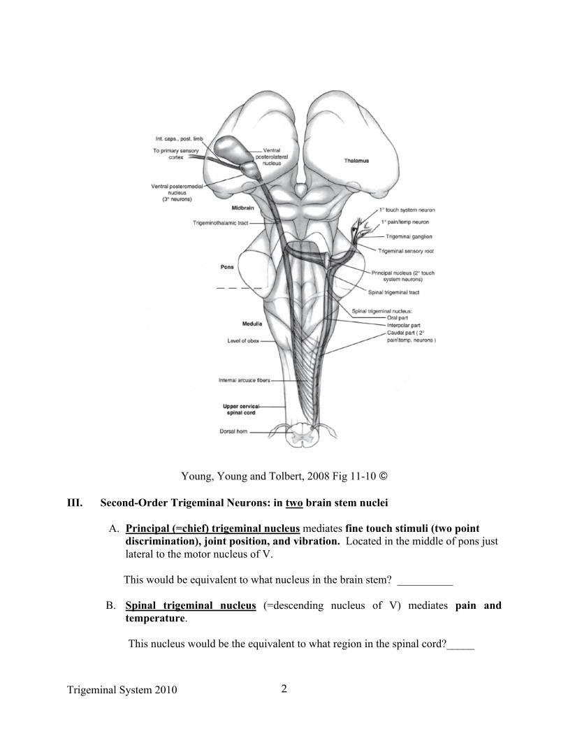
Young, Young and Tolbert, 2008 Fig 11-10 ©
III. Second-Order Trigeminal Neurons: in two brain stem nuclei
A. Principal (=chief) trigeminal nucleus mediates fine touch stimuli (two point discrimination), joint position, and vibration. Located in the middle of pons just lateral to the motor nucleus of V.
This would be equivalent to what nucleus in the brain stem? __________
B. Spinal trigeminal nucleus (=descending nucleus of V) mediates pain and
temperature.
This nucleus would be the equivalent to what region in the spinal cord?_____

Trigeminal System 2010 3
1. Spinal (descending) trigeminal tract contains primary afferents that will synapse in spinal (or descending) nucleus of V. The tract is continuous with the dorsolateral fasciculus (=Lissauer’s tract) in the spinal cord, again emphasizing the similarities with the ALS system.
2. The Spinal (descending) nucleus extends caudally as far as C 2-3 and is
continuous with the dorsal horn. This means it is several cm long and can be involved in lesions of caudal pons and medulla.
C. Ascending Trigeminal Pathways. Axons of second order neurons form the
trigeminothalamic pathway to VPM (ventroposteromedial) thalamus.
The trigeminothalamic pathway is located near the medial lemniscus. Axons from the spinal trigeminal nucleus are also referred to as the (ventral) trigeminothalamic tract and fibers coming from the principal trigeminal nucleus join it in mid pons. There is a dorsal trigeminothalamic tract we will ignore. We will use the term trigeminothalamic. These are not tracts you can identify, but vascular lesions can involve them.
a. Axons from neurons in the chief sensory nucleus carry sensations such as
______________________?
b. Axons from neurons in the spinal trigeminal nucleus carry sensations such as _______________________?
IV. Third Order Neurons in Thalamus Project to Postcentral Gyrus (Primary sensory
cortex).
VPM projects somatotopically to the face areas of the cortex via the internal capsule. The face is represented in primary, secondary, and association cortex (superior parietal cortex) just as the projections from VPL.
V. Primary Somatosensory Cortex, Postcentral Gyrus. There are four different body maps to
help extract texture, form, and motion that come from the head and body. A. INPUT - via internal capsule from VPM and VPL for both ALS, DCML and
trigeminothalamic systems. B.INJURY to postcentral gyrus - Deficits in position sense and ability to discriminate
sizes, texture, shape. Pain and temperature are altered but not abolished.

Trigeminal System 2010 4
VI. Association Somatosensory Cortex: Posterior Superior Parietal Areas 5 and 7 of Brodmann.
A. Relays information to sensory association cortex, posterior superior parietal cortex areas 5, 7 for stereognosis, the ability to recognize objects by "handling”, perception of your body image.
B Relays information to Motor and Premotor parts of cortex to guide intentional movement.
C. Connects homotopic areas via corpus callosum. D. Lesions of Association Area 5 And 7
Inability to perform simple acts requiring information on bodily orientation. "Cortical Neglect" - inability to perceive objects or parts of body in "body space". With lesions of the Non-dominant, usually right, hemisphere
VII. Output Of Postcentral Gyrus: To Association Somatosensory Cortex: Posterior
Superior Parietal Areas 5 AND 7

Trigeminal System 2010 5
Self-assessment: When you think you have mastered the pathways, select 2 colors in both a dark and light shade. Use the dark color for the body and the lighter color for the face pathways. e.g. Light blue-Trigeminothalamic (ventral) Dark blue – ALS Pink – trigeminothalamic (dorsal) from chief Red – DCML