Embed Size (px)
Citation preview

Tryptase 4, a New Member of the Chromosome 17 Family of MouseSerine Proteases*
Received for publication, November 16, 2000, and in revised form, March 1, 2001Published, JBC Papers in Press, March 20, 2001, DOI 10.1074/jbc.M010422200
Guang W. Wong‡§, Lixin Li¶, Mallur S. Madhusudhani, Steven A. Krilis¶, Michael F. Gurish‡,Marc E. Rothenberg**, Andrej Salii‡‡, and Richard L. Stevens‡§§
From the ‡Department of Medicine, Brigham and Women’s Hospital, and Department of Medicine, Harvard MedicalSchool, Boston, Massachusetts 02115, ¶Department of Medicine, University of New South Wales, and Department ofImmunology, Allergy, and Infectious Disease, St. George Hospital, Kogarah, New South Wales 2217, Australia, iRockefellerUniversity, New York, New York 10021, and **Department of Pediatrics, Division of Pulmonary Medicine, Allergy, andClinical Immunology, Children’s Hospital Medical Center, University of Cincinnati Medical Center,Cincinnati, Ohio 45229
Genomic blot analysis raised the possibility that un-characterized tryptase genes reside on chromosome 17at the complex containing the three genes that encodemouse mast cell protease (mMCP) 6, mMCP-7, and trans-membrane tryptase (mTMT). Probing of GenBank’s ex-pressed sequence tag data base with these threetryptase cDNAs resulted in the identification of an ex-pressed sequence tag that encodes a portion of a novelmouse serine protease (now designated mouse tryptase4 (mT4) because it is the fourth member of this family).5*- and 3*-rapid amplification of cDNA ends approacheswere carried out to deduce the nucleotide sequence ofthe full-length mT4 transcript. This information wasthen used to clone its ;5.0-kilobase pair gene. Chromo-some mapping analysis of its gene, sequence analysis ofits transcript, and comparative protein structure mod-eling of its translated product revealed that mT4 is anew member of the chromosome 17 family of mousetryptases. mT4 is 40–44% identical to mMCP-6, mMCP-7,and mTMT, and this new serine protease has all of thestructural features of a functional tryptase. Moreover,mT4 is enzymatically active when expressed in insectcells. Due to its 17-mer hydrophobic domain at its Cterminus, mT4 is a membrane-anchored tryptase moreanalogous to mTMT than the other members of its fam-ily. As assessed by RNA blot, reverse transcriptase-po-lymerase chain reaction, and/or in situ hybridizationanalysis, mT4 is expressed in interleukin-5-dependentmouse eosinophils, as well as in ovaries and testes. Theobservation that recombinant mT4 is preferentially re-tained in the endoplasmic reticulum of transientlytransfected COS-7 cells suggests a convertase-like rolefor this integral membrane serine protease.
A complex of genes resides on human chromosome 16p13.3that encodes the homologous tryptases a1, a2, bI, bII, bIII,transmembrane tryptase (TMT)1/tryptase g, and eosinophilserine protease-1 (Esp-1)2 (1–7). The corresponding complex ofgenes resides on the syntenic region of mouse chromosome 17at the interface between bands 17A3.3 and 17B1 (7–11). Al-though our previous genomic blot analysis suggested the pres-ence of additional mouse tryptase genes in the family (7), theonly genes and/or transcripts cloned so far at the mouse com-plex are those that encode mouse mast cell protease (mMCP) 6,mMCP-7, and mouse TMT (mTMT).
The amino acid sequences of mMCP-6 and mMCP-7 are;75% identical, and the mast cells (MCs) in the skin andskeletal muscle of BALB/c mice express both tryptases (12, 13).Nevertheless, in vivo and in vitro studies have established thatthese two homologous serine proteases are metabolized quitedifferently during allergic reactions (12). Moreover, they arefunctionally distinct. Because fibrinogen is a physiologic sub-strate of mMCP-7 (14), this tryptase appears to help preventthe deposition of fibrin/platelet clots in inflammatory sites sothat the various granulocytes and lymphocytes in the blood arenot physically prevented from entering an inflamed mousetissue site. Recent data have suggested that mMCP-7 alsoregulates eosinophil extravasation in tissues.3 MCs are essen-tial for combating bacterial infections in the peritoneal cavity(15–17). Because peritoneal MCs express mMCP-6 (11, 18),because the complement factors C3a and C5a induce peritonealMCs to degranulate (19), and because recombinant mMCP-6 isa potent and selective inducer of neutrophil extravasation intothe peritoneal cavity (20), mMCP-6 appears to play an impor-tant and protective role in bacterial infections. The function ofmTMT is not yet known, but its C-terminal hydrophobic do-main (7) probably causes its prolonged retention at the plasmamembrane when mTMT1 MCs are immunologically activated.
* This work was supported in part by National Institutes of HealthGrants AI-23483, HL-36110, and HL-63284 and by a grant from theMizutani Foundation for Glycoscience. The costs of publication of thisarticle were defrayed in part by the payment of page charges. Thisarticle must therefore be hereby marked “advertisement” in accordancewith 18 U.S.C. Section 1734 solely to indicate this fact.
The nucleotide sequence(s) reported in this paper has been submittedto the GenBankTM/EBI Data Bank with accession number(s) AF176209and AF226710.
§ Pharmacia Allergy Research Foundation Fellow.‡‡ Alfred P. Sloan Research Fellow.§§ To whom correspondence and reprint requests should be ad-
dressed: Brigham and Women’s Hospital, Dept. of Medicine, SmithBldg., Rm. 616B, 1 Jimmy Fund Way, Boston, MA 02115. Tel.: 617-525-1231; Fax: 617-525-1310; E-mail: [email protected].
1 The abbreviations used are: TMT, transmembrane tryptase; ER,endoplasmic reticulum; hEsp-1, human eosinophil serine protease-1;FISH, fluorescent in situ hybridization; MC, mast cell; mMCP, mouseMC protease; mT4, mouse tryptase 4; PBS, phosphate-buffered saline;RACE, rapid amplification of cDNA ends; mTesp, mouse testicularserine protease; UTR, untranslated region; EST, expressed sequencetag; kb, kilobase pairs; bp, base pairs; PCR, polymerase chain reaction;RT-PCR, reverse transcriptase-PCR; IL, interleukin; PAGE, polyacryl-amide gel electrophoresis; PNGase F, peptide N-glycosidase F.
2 Genomic sequencing of the 35.7-kb cosmid clone 352F10 by D. O.Ricke and co-workers (GenBankTM direct submission, accession numberAC005361) resulted in the initial identification and chromosomal loca-tion of the 4.6-kb human gene in 1998 that others (4, 5, 44) designatedas the hEsp-1/testisin gene in 1999.
3 R. L. Stevens, unpublished observations.
THE JOURNAL OF BIOLOGICAL CHEMISTRY Vol. 276, No. 23, Issue of June 8, pp. 20648–20658, 2001© 2001 by The American Society for Biochemistry and Molecular Biology, Inc. Printed in U.S.A.
This paper is available on line at http://www.jbc.org20648
Because our previous genomic blot analyses suggested thepresence of undiscovered tryptase-like genes in the mouse ge-nome and because mouse chromosome 17A3.3 to 17B1 has notbeen sequenced in its entirety, we used a gene-hunt approachin the present study to identify a new member of this importantfamily of serine proteases. We now describe the isolation, char-acterization, and expression of the fourth member of the mousetryptase family of serine proteases.
EXPERIMENTAL PROCEDURES
Cloning of the mT4 cDNA and Gene—The nucleotide sequences of themTMT, mMCP-6, and mMCP-7 transcripts were used as templates tosearch for novel, but related, mouse tryptase-like ESTs in the Gen-BankTM data base. Sequence analysis of the mouse testis-derived cloneAI326140, obtained from the “Integrated Molecular Analysis of GeneExpression” consortium, revealed that its insert corresponded to aportion of what appeared to be a novel serine protease. Based on thededuced nucleotide sequence of this clone, 59- and 39-rapid amplificationof cDNA ends (RACE) approaches were carried out on a pool of testiscDNAs (CLONTECH) to deduce the nucleotide and amino acid se-quences of the full-length transcript. The 39-RACE reaction was carriedout with 59-CACCTACAATAACTTCATCCAGC-39 and the anchor oli-gonucleotide 59-CCATCCTAATACGACTCACTATAGGGC-39. The re-sulting DNA products were purified on a 1% agarose gel, subcloned intopCR2.1 (Invitrogen, Carlsbad, CA), and the inserts in two of the arbi-trarily selected clones were sequenced. The 59-RACE was carried outwith the RLM-RACE kit (Ambion, Austen, TX). The mT4-specificprimer 59-GACTGGATCCAGTTGTAGTGATGACTG-39 and the outerprimer 59-GCTGATGGCGATGAATGAACACTG-39 were used in thefirst PCR. One microliter of the generated product was then used as atemplate in the second PCR step with the mT4-specific primer 59-CCCAGCAGTCAGTTCGGTTCTC-39 and the inner primer 59-CGCG-GATCCGACACTCGTTTGCTGGCTTTGATGAAA-39. The resultingproducts were purified and subcloned, and the inserts in six of thearbitrarily selected clones were sequenced.
Because all mouse and human tryptase genes so far cloned in thisfamily are ,6 kb in size, a long range nested PCR approach was used toisolate and characterize the mT4 gene from BALB/c mice. The oligonu-cleotides 59-GCAAGACGTTGGTGCCACTGCTG-39 and 59-GTGACG-TACACGTGTGGGCTCAGGCAG-39 were used in the initial 30-cyclePCR, whereas the oligonucleotides 59-ATGGCCTTACAGTCAACCTAT-TTGCAG-39 and 59-GCCTGAGCAGCCCATTGCGGATC-39 were usedin the subsequent 28-cycle reaction. Recombinant Thermus thermophi-lus DNA polymerase (PerkinElmer Life Sciences) was used in bothPCRs.
Genomic Blot Analysis and Chromosomal Location of the mT4Gene—Some of the human tryptases that have been cloned during thelast decade are ;98% identical (3). Thus, to determine whether or notthe mouse genome contains other genes that closely resemble the mT4gene, replicate 25-mg samples of BALB/c mouse genomic DNA weredigested separately at 37 °C for ;15 h with EcoRI, DraI, BamHI, AvrII,EcoRV, BglII, or HindIII (New England Biolabs, Beverly, MA). Thedigests were fractionated on a 1% agarose gel, and the separated frag-ments were blotted onto a MagnaGraph nylon membrane (Micron Sep-arations Inc., Westborough, MA). The resulting DNA blot was incu-bated for 2 h at 65 °C in QuikHyb solution (Stratagene, La Jolla, CA)containing a 32P-labeled 536-bp probe corresponding to the 59 end of themT4 cDNA. The blot was washed twice at room temperature for 20 mineach in 23 SSC containing 0.1% SDS and then twice at 65 °C for 20 mineach in 0.23 SSC containing 0.1% SDS before being exposed toBIOMAX film.
A fluorescent in situ hybridization (FISH) technique was used byHuman Genome Systems (St. Louis, MO) to determine the chromo-somal location of the mT4 gene. Preliminary studies revealed that themT4 gene resided in the company’s BAC mouse genomic clone F1062.Slides containing normal metaphase chromosomes derived from mouseembryonic fibroblasts were therefore incubated with the digoxigenindUTP-labeled clone F1062 in the presence of 50% formamide, 10%dextran sulfate, 23 SSC, and sheared mouse DNA. After this hybrid-ization step, the slides were incubated with fluoresceinated anti-digoxi-genin antibody and counterstained with 4,6-diamidino-2-phenylindole.
Expression of mT4 at the mRNA Level—To determine which tissuesin the mouse contain abundant levels of mT4 mRNA, a blot (CLON-TECH) containing ;2 mg of poly(A)1 RNA from mouse heart, brain,spleen, lung, liver, skeletal muscle, kidney, and testis was probed underconditions of high stringency with a radiolabeled 536-bp probe that
corresponded to the 59 end of the mT4 transcript. After the probe wasrandom primed with [a-32P]dCTP using the Rediprime kit (AmershamPharmacia Biotech), it was hybridized to the RNA blot at 65 °C for 2 hin Quickhyb solution. The blot was washed twice at room temperaturefor 20 min each in 23 SSC containing 0.1% SDS and then twice at 65 °Cfor 20 min each in 0.23 SSC containing 0.1% SDS before being exposedto BIOMAX film for 1–2 days. After the bound mT4 probe was removed,the blot was reprobed with a b-actin cDNA (CLONTECH) to evaluatethe amount of RNA loaded in each lane. In a similar manner, total RNAfrom normal mouse bone marrow cells, IL-5 transgenic mouse bonemarrow cells, and eosinophils purified from IL-5 transgenic mice (21)were evaluated for the presence of the mT4 transcript. Day-7, -11, -15,and -17 mouse embryos also were evaluated for their mT4 mRNA levels.
BALB/c mice exhibit a substantial, but transient, T cell-dependentincrease in the number of eosinophils and MCs in their jejunum 7 and14 days, respectively, after the animals are infected with Trichinellaspiralis (22). To determine if the eosinophils or MCs that develop in thejejunum also contain mT4 mRNA, total RNA was isolated from thesmall intestine, spleen, and liver of day-7 helminth-infected mice (n 52) and day-14 helminth-infected mice (n 5 2). RT-PCR was then used todetermine whether or not any of the tissue specimens contained mT4mRNA. In this assay, the RT step was carried out at 55 °C for 30 min.Forty five cycles of PCR were performed with the primers 59-CAACAG-CATGTGTAACCATATG-39 and 59-GCCTGAGCAGCCCATTGCG-GATC-39; each cycle consisted of a 5-s denaturing step at 94 °C, a 5-sannealing step at 60 °C, and a 15-s extension step at 72 °C. ThisRT-PCR approach also was used to evaluate mT4 expression in theovaries and testis before and after sexual maturation. In these reac-tions, 32 cycles were carried out rather than 45.
Location of mT4 mRNA-expressing Cells in the Testis—For the non-radioactive in situ hybridization approach, the mT4-specific oligonu-cleotide 59-CTATTTGGTAACGGTTGGAATAGGCCTGTAGGTTCCAG-AGAGATGGCCTGG-39 was labeled with digoxigenin-alkaline phos-phatase with a commercially available 39 end labeling kit (Roche Mo-lecular Biochemicals). BALB/c mouse testis was fixed in 4% paraform-aldehyde in 0.1 M phosphate-buffered saline (PBS) at 4 °C. The prepa-ration was washed twice with PBS containing 2% dimethyl sulfoxideand then dehydrated and embedded in JB4 glycomethacrylate accord-ing to the manufacturer’s instructions (Polysciences Inc., Warrington,PA). Sections were cut on a Reichert-Jung Supracut microtome (Leica,Deerfield, IL) at 5-mm thickness and picked up on glass slides. Theslides were incubated sequentially for 15 min at 37 °C in 0.025% trypsincontaining 2 mM calcium chloride and then for 30 min in Target Re-trieval solution (Dako, Carpinteria, CA) at 95 °C. After the slides werewashed three times in distilled water, they were incubated in prehy-bridization solution containing 50% formamide, 43 SSC, 13 Denhardt’ssolution, sonicated salmon sperm DNA, and 10% dextran sulfate at42 °C for 30 min. The prehybridization solution was removed; 50 ml ofhybridization buffer containing the digoxigenin-labeled oligonucleotidewas added, and each specimen was incubated overnight at 42 °C. Thenext morning, each slide was washed twice with 23 SSC, once with 13SSC at room temperature, and then incubated with anti-digoxigenin-biotin complex for 30 min at room temperature. The slides were washedtwice with Tris-buffered saline, incubated with streptavidin complexconjugated to horseradish peroxidase, and washed twice again withTris-buffered saline. Color development was performed according to themanufacturer’s instructions (Dako). In this analysis, cells that con-tained abundant levels of mT4 mRNA stain brown.
A radioactive in situ hybridization approach was used to confirm theobtained data with the above digoxigenin-alkaline phosphatase-labeledprobe. For this second type of in situ hybridization analysis, nucleotides1–536 of the mT4 cDNA (Fig. 1) were subcloned into pCR2.1 in both thesense and antisense directions. The resulting two plasmid DNA sam-ples were linearized with SpeI (New England Biolabs) and transcribedwith T7 RNA polymerase (Promega, Madison, WI) in the presence of[a-33P]dUTP (PerkinElmer Life Sciences) to generate antisense andsense radiolabeled RNA probes. Tissue sections were placed on slides,deparaffinized, fixed in 4% paraformaldehyde in PBS, and treated withproteinase K. After washing in 0.53 SSC, the sections were coveredwith 50 ml of hybridization solution (50% deionized formamide, 0.3 M
NaCl, 5 mM EDTA, 13 Denhardt’s solution, 10% dextran sulfate, 10 mM
dithiothreitol, and 20 mM Tris-HCl, pH 8.0) and incubated for 2 h at55 °C. 33P-Labeled antisense or sense RNA probes (3 million cpm/slide)were added to the hybridization solution, and the sections were incu-bated for an additional 12–18 h at 55 °C. The resulting sections werewashed for 20 min in 23 SSC, 10 mM b-mercaptoethanol, and 1 mM
EDTA and then were exposed to RNase A (10 mg/ml) for 30 min at roomtemperature. To minimize the possibility of nonspecific binding of the
Mouse Tryptase 4 20649

radiolabeled probe, each section was incubated for an additional 2 h at60 °C in 0.13 SSC, b-mercaptoethanol, and 1 mM EDTA. The resultingsections were dehydrated, dipped in photographic emulsion NTB3
(Kodak), and stored at 4 °C. After 7 days of exposure, the radiolabeledsections were developed, and cells in these sections were counterstainedwith hematoxylin and eosin. In this radioactive analysis, black silvergranules appear over those cells that contain abundant levels of mT4mRNA.
Expression of mT4 Protein in COS-7 Cells and Insect Cells andProtein Modeling of Its Translated Mature Product—To addresswhether or not the mT4 transcript encodes an enzymatically active,membrane-anchored protease, the entire coding region of mT4 cDNAwas placed in the expression vector pcDNA3.1/V5-His-TOPO (Invitro-gen). This expression vector was chosen because the resulting productwill contain the 14-mer V5 peptide (Gly-Lys-Pro-Ile-Pro-Asn-Pro-Leu-Leu-Gly-Leu-Asp-Ser-Thr) at its C terminus, thereby allowing its de-tection with anti-V5 antibody. Vector lacking an insert was used as anegative control in the transfection experiments. African green mon-key, SV40-transformed kidney COS-7 cells (line CRL-1651, ATCC, Ma-nassas, VA) were cultured in Dulbecco’s modified Eagle’s medium con-taining 10% fetal bovine serum. Transient transfections wereperformed with SuperFect (Qiagen, Valencia, CA) according to themanufacturer’s instructions. Cells were plated at a density of 2 3 105
cells/well in 6-well plates 24 h prior to transfection. After the cells weretransfected for 2–3 h, they were trypsinized, and portions of the result-ing cells were placed in 24-well plates containing 11-mm coverslips forimmunofluorescence microscopy. The remainder of the cells were placedin 12-well plates for SDS-PAGE/immunoblot analysis. Conditioned me-dia and cells were collected 24 and 48 h post-transfection.
For immunofluorescence microscopy, transfected COS-7 cells grownon coverslips were washed once with PBS, fixed in 4% paraformalde-hyde for 10 min, and permeabilized in methanol for 10 min. The treatedcells were washed three times with PBS. They were then exposed to10% donkey serum in PBS for 1 h at room temperature to preventnonspecific binding of the relevant mouse and rabbit antibodies. Afterthis step, the cells were stained with mouse anti-V5 antibody (Invitro-gen) in the absence or presence of a mixture of rabbit anti-calnexin andanti-calreticulin antibodies (StressGen, Victoria, British Columbia,Canada). Calnexin and calreticulin reside in the endoplasmic reticulum(ER). Thus, antibodies directed against these two proteins were used toconfirm the ER location of recombinant mT4 protein in the transfec-tants. Cy®2-conjugated donkey anti-mouse antibody (Jackson Immu-noResearch, West Grove, PA) was used to detect the mT4-V5 fusionprotein, whereas Cy®3-conjugated donkey anti-rabbit antibody (Jack-son ImmunoResearch) was used to detect calnexin and calreticulin.Stained cells were viewed with a Nikon Eclipse 800 microscope. Imageswere digitally captured using a CCD-SPOT RT camera and compiledusing Adobe Photoshop software.
For SDS-PAGE/immunoblot analysis, samples of the transfectedCOS-7 cells and their conditioned media were boiled in SDS samplebuffer containing b-mercaptoethanol. After electrophoresis, the re-solved proteins were blotted onto polyvinylidene difluoride membranes(Bio-Rad). Each protein blot was exposed to Tris-buffered saline (15 ml)containing 5% non-fat milk, 0.1% Tween 20, 0.5% goat serum, and 3 mgof mouse anti-V5 antibody (Invitrogen) for 2 h at room temperature.After each blot was washed 3 times with Tris-buffered saline containing0.1% Tween 20, it was exposed to Tris-buffered saline (15 ml) contain-ing 5% non-fat milk, 0.1% Tween 20, 0.5% goat serum, and a 1:1000dilution of a stock solution of horseradish peroxidase-conjugated goatanti-mouse IgG (Bio-Rad) for 1 h at room temperature. The immuno-reactive proteins were then visualized using a chemiluminescence kit(Genotech, St. Louis, MO) and BioMax MR film (Kodak). In order toconfirm that mT4 is a membrane-anchored protein, ;4 3 106 mT4-expressing COS-7 cells were suspended in 750 ml of buffer (4 mM
HEPES, pH 7.0, containing 50 mM sucrose, 0.4 mM EDTA, and 0.2 mM
dithiothreitol) and lysed by subjecting the cell suspension to multiplefreeze-thaw cycles using liquid nitrogen and a 45 °C water bath. Aftercentrifugation for 1 h at .100,000 3 g, aliquots of the obtained super-natant representing the cytosolic fraction and aliquots of the pelletedmicrosomal fraction were boiled in Laemmli buffer in the presence ofb-mercaptoethanol and then subjected to SDS-PAGE/immunoblot anal-ysis as described above.
Other members of the tryptase family contain N-linked glycans (23,24). Analysis of the predicted primary amino acid sequence of mT4revealed four potential N-linked glycosylation sites. Thus, to determineif mT4 contains N-linked glycans, a sample of the COS-7 cell-derivedrecombinant material was incubated with PNGase F (New EnglandBiolabs) using the manufacturer’s suggested reaction conditions. The
resulting digest was then subjected to SDS-PAGE/immunoblotanalysis.
As noted under “Results,” mT4 can be generated in COS-7 cells usingthe above expression strategy. Unfortunately, because mT4 remainstightly associated with the ER membrane of the cell, contaminating ERproteins cannot be removed easily to evaluate the enzymatic activity ofthe generated recombinant protein. We previously generated pseudozy-mogen forms of mMCP-6 and mMCP-7 in High Five insect cells that ineach instance contain an enterokinase-susceptibility sequence betweenthe natural propeptide and catalytic domain (14, 20). Because eachpseudozymogen also contains the FLAG peptide Asp-Tyr-Lys-Asp-Asp-Asp-Asp-Lys at its C terminus, it can be purified using an anti-FLAGantibody immunoaffinity column. Thus, to generate a bioengineeredform of recombinant mT4 that can be purified to near homogeneity forfunctional studies, an expression construct was created in which theC-terminal membrane-spanning domain of mT4 (i.e. residues 249–270)was replaced by the 8-residue FLAG peptide. Insect cells might beunable to activate constitutively the generated product. To guardagainst that possibility, an enterokinase-susceptibility sequence alsowas created in the construct by inserting the 5-residue peptide Asp-Asp-Asp-Asp-Lys in between the natural propeptide of mT4 and thefirst Ile residue in its catalytic domain. In this way, the purifiedpseudozymogen could be activated by enterokinase if needed. High Fiveinsects cells, cultured in serum-free media, were induced to express thebioengineered form of mT4 as described previously (14, 20, 25, 26) forother recombinant mouse and human tryptases. Insect cell lysis bufferfrom PharMingen was used to liberate the cell-associated recombinantmT4 from pelleted cells. After a 30-min incubation on ice, the resultinglysates were centrifuged at ;20,000 3 g for 30 min at 4 °C. Thesupernatant was removed and passed through a 0.45-mm celluloseacetate filter. The cleared supernatant was then loaded at 4 °C onto an;1-ml column containing anti-FLAG M1 antibody (Sigma). After thecolumn was washed with 250 ml of Tris-buffered saline, pH 7.0, 0.1 M
glycine, pH 3.5, buffer was used to elute bound mT4. Ten 1-ml fractionswere collected into tubes containing 20 ml of 1 M Tris-HCl, pH 8.0.Samples of resulting fractions were analyzed by SDS-PAGE for thepresence of Coomassie Blue1 proteins and for immunoreactive mT4.
Casein is a protease-sensitive protein. Thus, resorufin-labeled casein(Roche Molecular Biochemicals) was used to determine whether or notinsect cell-derived mT4 is enzymatically active. In this assay, a 50-mlsample of the eluate from the above immunoaffinity column was addedto 50 ml of 5 mM calcium chloride and 10 mM Tris-HCl, pH 5.0, contain-ing or lacking 0.4 unit of enterokinase (New England Biolabs); thesample was then incubated at 37 °C for 2 h. To evaluate the enzymaticactivity of the resulting recombinant material, 100 ml of 0.1 M Tris-HCl,pH 7.8, containing 10 mM calcium chloride and 200 mg of resorufin-labeled casein was added. After an 18-h incubation at 37 °C, eachreaction was stopped by the addition of 480 ml of 5% trichloroacetic acid,followed by a 10-min centrifugation at .8,000 3 g. A 400-ml sample ofthe resulting supernatant was mixed with 600 ml of 0.5 M Tris-HCl, pH8.8, and the absorbance was measured at 574 nm. Because low molec-ular weight resorufin-labeled peptides released from its substrate arenot precipitated by the trichloroacetic acid step, the concentration ofresorufin-labeled peptides in the supernatant is directly proportional tothe general proteolytic activity present in the sample. Trypsin (Sigma)was used as a positive control in this enzymatic assay.
A three-dimensional model of the extracellular domain of maturemT4 (i.e. residues 1–256) was built by MODELLER (27, 28) as de-scribed for other MC proteases (7, 12, 29, 30). The primary template forcomparative protein structure modeling was the crystal structure ofhuman tryptase bII (31). Within this region, the amino acid sequencesof the two tryptases are 39% identical.
RESULTS
Cloning and Sequence Analysis of mT4 cDNA—When themTMT, mMCP-6, and mMCP-7 cDNAs were used as templatesto screen GenBank’s EST data base, an EST (accession numberAI326140) was identified that was somewhat homologous to allthree target tryptases. The relevant EST was obtained from theIntegrated Molecular Analysis of Gene Expression consortium,and nucleotide sequence analysis of the entire insert in theclone revealed that it encoded a portion of a serine protease(designated mT4 because it is the fourth member of this familyof serine proteases) not previously described. 59- and 39-RACEapproaches (Fig. 1A) were therefore carried out to deduce thenucleotide sequence of the missing portion of the transcript
Mouse Tryptase 420650

(Fig. 1B). The full-length mT4 cDNA consists of ;1095 nucle-otides, and the 59- and 39-untranslated regions (UTRs) of thetranscript consist of 23 and .97 nucleotides, respectively. The
putative translation-initiation codon conforms to the sequencepresent in most eukaryotic transcripts (32).
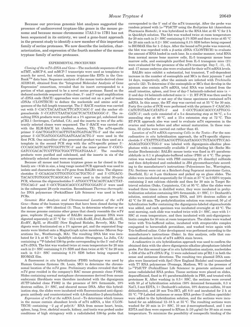
Analysis of mT4 at the Protein Level—The deduced aminoacid sequence of its cDNA suggested that mT4 could be initiallytranslated as an ;36-kDa zymogen consisting of 324 aminoacids (Fig. 1B). Hydropathy plot analysis (Fig. 1C) disclosedthat mT4 possesses an unusual hydrophobic domain in its Cterminus at residues 251–267. Phylogenetic analysis (Fig. 2A)of all known mouse proteins revealed that mT4 is most closelyrelated to mTMT. However, the degree of identity is only 44%.Every tryptase in this family has an N-terminal sequence ofIle-Val-Gly-Gly when its propeptide is removed (Fig. 2B). Be-cause the zymogen form of mT4 also possesses this sequencenear its initially translated N terminus, it is predicted that the54-residue prepropeptide is cleaved at the Arg-Ile site indicatedin Fig. 1B. In its mature form, the protein portion of thecatalytic domain of mT4 is predicted to have a molecular massof ;30 kDa. However, because the mature protein containsfour potential N-linked glycosylation sites at Asn116, Asn123,Asn156, and Asn229, post-translationally modified mT4 is ex-pected to be somewhat larger in size in vivo.
When transfected into COS-7 cells, the level of immunoreac-tive mT4 in the conditioned media of the transfectants wasbelow detection by SDS-PAGE/immunoblot analysis (Fig. 3A).Thus, very little, if any, recombinant mT4 was constitutivelyreleased from the transfectants. The fact that mT4 was pref-erentially recovered in the microsomal fraction of the cell ly-sates (Fig. 3B) confirmed that mT4 is a membrane-anchoredprotein. Immunohistochemical analysis of the transfectantsrevealed that mT4 was preferentially retained in the calexin/calreticulum-enriched ER (Fig. 3, D–F). The fact that immuno-reactive mT4 was not released after trypsin treatment (datanot shown) confirmed that very little, if any, mT4 targets to theplasma membrane in transfected COS-7 cells. As assessed bySDS-PAGE analysis, an immunoreactive protein of ;40 kDawas identified in COS-7 cell transfectants that shifted to ;35kDa after PNGase F treatment (Fig. 3C). Based on the magni-tude of this change in its molecular weight, mT4 contains morethan one N-linked glycan.
By using resorufin-labeled casein as a substrate, the level ofproteolytic activity in the lysates of the mT4-expressing COS-7cells was significantly greater than that in the lysates of controlnon-transfectants (data not shown). However, the amounts ofactive enzyme were low, and recombinant mT4 remainedtightly associated with the membrane of the ER (Fig. 3). Thus,a bioengineered form of mT4 possessing the FLAG peptide atits C terminus was expressed in High Five insect cells (Fig. 4).In contrast to what occurs when similar FLAG derivatives ofmMCP-6 (20) and mMCP-7 (14) are expressed in insect cells,the FLAG derivative of mT4 was not constitutively secretedfrom the insect cells. Nevertheless, the recombinant proteincould be purified from the lysates of its expressing cells usingthe immunoaffinity column (Fig. 4A). Even though the amountof protein in fractions 5 and 6 of the eluate of the column wasbelow detection by Coomassie Blue staining of a duplicate gel(data not shown), the recombinant mT4 in these fractions ex-hibited substantial proteolytic activity in vitro (Fig. 4B). Thefact that the enzymatic activity of the recombinant material didnot increase dramatically following enterokinase treatment in-dicated that much of the purified mT4 was constitutively ac-tive. At present, we cannot ascertain whether activation of themT4 pseudozymogen occurred inside the insect cells or duringits purification. Nevertheless, because more trypsin was usedon a weight basis in the depicted experiment, the broad enzy-matic activity of recombinant mT4 is at least as good as that oftrypsin in regard to its ability to degrade casein.
FIG. 1. Cloning of the mT4 cDNA. A, a three-step approach wasused to isolate the full-length mT4 transcript. A computer search ofGenBank’s EST data base resulted in the identification of an EST thatencodes a portion of a novel mouse serine protease. Based on its nucle-otide sequence, 59- and 39-RACE approaches were then carried out on apool of mouse testis-derived cDNAs to deduce the remaining portion ofthe expressed transcript. The diagram in A highlights the 59-UTR,prepropeptide, the catalytic domain of the mature enzyme with itscatalytic triad (circled H, D, and S amino acid residues), C-terminalhydrophobic segment, 39-UTR, and poly(A) tail. B, the nucleotide andamino acid sequences of the full-length mT4 transcript were deduced.The four potential N-linked glycosylation sites in mT4 are circled in Band the C-terminal hydrophobic extension is boxed. Components of thecatalytic triad (f), translation-initiation site (*), translation-termina-tion site (2), leader peptide (single bracket), and propeptide (doublebracket) are indicated. Nucleotide numbering begins at the 59-UTR ofthe isolated transcript; amino acid numbering (within brackets at left)begins with the putative mature protein. C, a Kyte-Doolittle hydropa-thy plot of the translated product was generated. The individual resi-dues in the coding region of mT4 are indicated on the x axis; the extentof their hydrophobicity and hydrophilicity are on the y axis.
Mouse Tryptase 4 20651
The overall fold of mature mT4 is predicted to be similar tothat of most serine proteases (Fig. 5A). For example, like allother functional serine proteases, mT4 possesses the conservedtriad (i.e. His41, Asp93, and Ser194) in its putative catalytic site.Like mTMT, mT4 lacks a number of the Pro and Tyr residues(31) that are needed for human tryptase bII, mMCP-6, andmMCP-7 to form tetramers. However, mT4 possesses the func-tionally important surface Trp-rich domain found in all othertryptases in this family. The presence of Asp188, Gly215, and
Gly225 in mT4 also strongly suggests that it is a tryptase.Nevertheless, the seven loops that form its substrate-bindingcleft are unique (Figs. 2B and 5B). For example, residue sub-stitutions in loops 3 and A in mT4 are predicted to result inshape differences relative to human tryptase bII (Fig. 5B).Although mature mT4 has an overall net 25 charge at neutralpH, it is predicted to have two positively charged surface re-gions at diametrically opposite ends of the folded protein (Fig.5A). Arg33, Arg34, and Arg243 reside in one region, whereas
FIG. 2. Comparison of the amino acid sequences of mT4 with the most closely related known mouse serine proteases. A, dendrogramcomparing mT4 with the closely related serine proteases in the mouse was generated by the GCG program “Distances” using the unweighted pairgroup with arithmetic mean (UPGMA) algorithm. (The mTesp-3 gene, transcript, and translated product have not been described. Thus, therelationship of mT4 to this putative testis-specific serine protease cannot be evaluated at this time.) B, the amino acid sequences of hEsp-1, mTMT,mMCP-6, mMCP-7, mAcrosin, mTesp-1, mTesp-2, mTesp-4, mHepsin, mKallikrein, mTrypsin, hLeydin, and hProstasin were extracted from SwissProtein Database and aligned with the PILEUP program of the Eugene “GCG” software package. Identical amino acids are shaded. Numbering(left) begins at the first residue in the mature portion of each protease. The C-terminal hydrophobic segment of mT4 is bracketed. The sevenputative loops (designated A–D and 1–3) that form the substrate-binding pocket of each of these proteases (also see Fig. 5B) are underlined. Thedepicted amino acid sequence of mAcrosin is truncated at the C terminus.
Mouse Tryptase 420652

His162, Lys165, Lys166, Arg170, and Arg223 reside in the otherregion.
Nucleotide Sequence and Chromosomal Location of the mT4Gene—Although two DNA fragments were detected when a blotcontaining AvrII-, HindIII, or BglII-digested mouse genomicDNA was probed with the mT4 cDNA (Fig. 6A), subsequentnucleotide sequence analysis revealed that these findings weredue to the presence of internal enzyme restriction sites withinthe gene. Thus, a single gene encodes mT4. The fact that noadditional DNA fragment was observed when the genomic blotwas probed at moderate stringency (data not shown) suggeststhat no closely related gene is present in the mouse genome.
The initial FISH analysis revealed that the mT4 gene resideson the proximal region of a small-sized chromosome believed tobe mouse chromosome 17. Based on that data, a second exper-iment was conducted in which a probe specific for the telomericregion of chromosome 17 was co-hybridized with the mT4-containing genomic clone. This experiment resulted in the spe-cific labeling of the telomere and the proximal portion of mousechromosome 17. Measurements of 10 specifically labeled chro-mosomes 17 demonstrated that the mT4 gene is located at aposition that is 23% of the distance from the heterochromatic-euchromatic boundary to the telomere of the chromosome. Thisarea corresponds to the interface between bands 17A3.3 and 17B1 (Fig. 6B) where the mMCP-6, mMCP-7, and mTMT genesreside. When 80 metaphase cells were analyzed, 77 exhibitedspecific labeling.
With a long range PCR approach, the entire mT4 gene wasisolated and sequenced. The mT4 gene is ;5.0 kb in size and
contains 6 exons (Fig. 7A). All exon/intron splice sites con-formed to the GT/AG rule for other eukaryotic genes (33).Exons 1–6 consist of 126, 27, 166, 284, 155, and 237 bp, respec-tively, whereas introns 1–5 consist of 101, 291, 599, 2717, and269 bp, respectively. The exon/intron organization of the mT4gene differs somewhat from that of the other three mouse genesin this family (Fig. 7B). For example, although the mT4 tran-script is similar in size to that of the other three mouse MCtryptase transcripts, its gene is larger due primarily to intron4. The mT4 gene also has six exons, whereas the mTMT andmMCP-7 genes have five exons.
Expression of mT4 in Immune and Non-immune Cells—Asassessed by RNA blot analysis, the level of mT4 mRNA wasbelow detection in normal mouse bone marrow (Fig. 8A). Trans-genic mice that have been induced to express abnormally highlevels of IL-5 exhibit a constitutive eosinophilia (21). Althoughbarely detectable amounts of mT4 mRNA were found in thebone marrow of IL-5 transgenic mice, larger amounts of thistranscript were present in the IL-5-dependent eosinophils pu-rified from the transgenic animals (Fig. 8A). In confirmation ofthese data, mT4 mRNA also could be detected in the jejunum ofa T. spiralis-infected BALB/c mouse (Fig. 8B) precisely whenthe number of eosinophils are maximal in this tissue (34).
mT4 mRNA could not be detected by RNA blot analysis innormal heart, brain, spleen, lung, liver, skeletal muscle, andkidney (Fig. 8C), as well as ear, tongue, stomach, and intestine(data not shown). Because these tissues contain substantialnumbers of MCs, mT4 is the only member of its family that isnot preferentially expressed in MCs. The level of mT4 mRNAalso was below detection in day-7–17 mouse embryos (Fig. 8C).However, using an RT-PCR approach, mT4 mRNA was de-
FIG. 3. SDS-PAGE/immunoblot and immunohistochemicalanalysis of mT4-expressing COS-7 cells. A, COS-7 cells were trans-fected with expression vector alone (left lanes) or expression vectorcontaining an insert that encodes a bioengineered form of mT4 possess-ing the immunogenic V5 peptide at its C terminus (right lanes). Fortyeight hours later, samples of the resulting conditioned media/superna-tants (S) and lysates of the cell pellets (P) were analyzed for thepresence of recombinant protein with anti-V5 antibody. B, lysates ofmT4-expressing COS-7 cells were fractionated to determine whether ornot mT4 is a membrane-anchored protein. Shown is the immunoblotanalysis of the membrane- (MF) and cytosol (CF)-enriched fractions. C,a sample of the cell lysates of mT4-expressing COS-7 cells was incu-bated 1 h in the absence (2) or presence (1) of PNGase F prior toSDS-PAGE/immunoblot analysis to determine whether or not mT4contains N-linked glycans. Molecular mass markers are shown on theleft. The arrow and the open arrowhead on the right point to glycosy-lated and nonglycosylated forms of mT4, respectively. The deglycosy-lated product is ;35 kDa because it contains the additional V5 and His6peptides at its C terminus. D–F, mT4-expressing cells were stained witha mouse monoclonal antibody directed against the V5 peptide (D), amixture of rabbit antibodies directed against calnexin and calreticulin(E), or antibodies directed against all three epitopes (F). Yellow color inF indicates co-localization of mT4 with calnexin and calreticulin. Basedon this double staining approach, a substantial portion of the expressedmT4 is anchored in the ER membrane of the cell.
FIG. 4. Evaluation of the enzymatic activity of insect cell-de-rived recombinant mT4. A, recombinant mT4-FLAG was purifiedfrom the lysates of High Five insect cells using an immunoaffinitychromatography approach. The soluble proteins in lysates of mT4-FLAG-expressing cells were applied to an anti-FLAG antibody column.After the column was washed extensively, the pH of the buffer waschanged to elute the bound recombinant protein. B, fractions 5 and 6 ofthe immunoaffinity column were pooled and evaluated for their enzy-matic activity before (2) and after (1) enterokinase (EK) treatment.Trypsin and activating buffer containing enterokinase alone were usedas positive and negative controls, respectively, in this casein-suscepti-bility assay. As assessed by Coomassie Blue staining of a duplicate gel,the amount of protein in fractions 5 and 6 was below detection. Thus,the amount of trypsin used in the depicted experiment exceeds that ofrecombinant mT4.
Mouse Tryptase 4 20653

tected in the testes and ovaries of adult mice (Fig. 8D). Asassessed by two different in situ hybridization methods, mT4 istransiently expressed relatively late in spermatogenesis duringthe cap phase of acrosome formation (Fig. 9).
DISCUSSION
While at least six distinct tryptase genes reside at a complexon human chromosome 16 (1–3, 6, 7), only three correspondinggenes have been identified so far on the syntenic region ofmouse chromosome 17 (7–11). We now describe a new mousegene in this family that is related to the genes that encodemTMT, mMCP-6, and mMCP-7.
The mT4 cDNA (Fig. 1) and gene (Fig. 7) encode a 324-residue polypeptide having a 54-residue prepropeptide and a17-residue, C-terminal hydrophobic domain. When transientlyexpressed in COS-7 cells, mT4 remains cell-associated (Fig. 3).Thus, as predicted based on analysis of its cDNA and gene,translated mT4 is a membrane-anchored serine protease. Likethe transmembrane protease angiotensin-converting enzyme(35), the C-terminal hydrophobic domain of mT4 is flanked byAsp and Arg residues. Because angiotensin-converting enzyme(35), prostasin (36), and acrosin (37) can be released from cells
FIG. 5. Three-dimensional model of the catalytic portion ofmature mT4 based on the crystal structure of human tryptasebII. A, a three-dimensional model of the catalytic domain of maturemT4 was created. Shown is the overall structure of residues 1–256 ofmT4. Because human tryptase bII lacks a C-terminal hydrophobicdomain, the short membrane-spanning domain in mT4 was not mod-eled. The active site residues (His41, Asp93, and Ser194) are representedas green sticks. The free Cys residue 113 is shown in orange. The sidechains of the residues (His162, Lys165, Lys166, Arg170, Arg220, Arg223,Arg33, Arg34, and Arg243) that consist of the two positively chargedsurface regions are shown as blue sticks. The C-a atoms of the conservedresidues (Trp12, Trp14, Trp128, Trp206, Trp214, and Trp236) that form thehydrophobic domain opposite the substrate-binding cleft are shown asred spheres. Two of the conserved residues (Trp35 and Trp174) in thedomain are hidden in this view. The figure was created with the pro-grams Molscript (50) and Raster3D (51). The general orientation of themT4 model is similar to that of the mMCP-6 (12), mMCP-7 (30), andmTMT (7) models in our previous publications. B, the putative sub-strate-binding cleft of mT4 was analyzed at a higher resolution usingthe modeling approach. The 7 loops that form the substrate-bindingcleft of mT4 are marked A–D and 1–3 and are superimposed on thecorresponding loops of human tryptase bII. The loops in mT4 andhuman tryptase bII are shown in red and blue, respectively; the activesite residues are shown as green sticks.
FIG. 6. Genomic blot analysis and chromosomal location of themT4 gene. A, a blot containing mouse genomic DNA digested withEcoRI, DraI, BamHI, AvrII, EcoRV, BglII, or HindIII was probed underconditions of high stringency with a radiolabeled 536-bp fragment de-rived from the 59 end of the mT4 cDNA. DNA fragments of knownmolecular weight (HindIII-digested l DNA) are indicated on the left ofthe blot. As noted in Fig. 7A, the mT4 gene contains internal sites thatare susceptible to AvrII, HindIII, and BglII. B, the chromosome locationof the mT4 gene was determined by FISH analysis. The fluorescent-labeled, mT4-containing BAC clone hybridized specifically to a small-sized chromosome (arrow, left panel) that was subsequently shown to bechromosome 17. The location (arrow) of the mT4 gene on this chromo-some is more clearly indicated in the right panel.
Mouse Tryptase 420654

by a proteolytic processing mechanism, the possibility has notbeen ruled out that under certain situations mT4 undergoes asimilar post-translational processing event in vivo to cause itsrelease from cells.
The zymogen form of mT4 contains 10 Cys residues. Basedon the crystal structure of human tryptase bII (31), Cys26,Cys42, Cys127, Cys160, Cys179, Cys190, Cys200, and Cys218 arepredicted to form 4 disulfide bonds in the catalytic portion ofthe mature, properly folded protease (Fig. 5A). One of theadditional Cys resides at residue 29 in the propeptide; the
other resides at residue 113. Although Cys113 is not present inmMCP-6 or mMCP-7 (Fig. 2B), a corresponding Cys is presentin the two-chained proteases factor XI (38), plasma kallikrein(39), and acrosin (40). Because this Cys forms a disulfide bondwith a Cys residue in the propeptide of each zymogen, maturemT4 could be a two-chain serine protease consisting of a 33-residue, non-catalytic N-terminal chain covalently linked to thelarger sized catalytic C-terminal chain.
Although its physiologic substrate(s) was not deduced in thisinitial study, mT4 is enzymatically active when expressed in
FIG. 7. Structure of the mT4 gene. A, the nucleotides that consist of the six exons and five introns of the mT4 gene were deduced and are shownin upper and lowercase letters, respectively. The exons are boxed, and the deduced amino acid sequence of the translated product is indicated, aswell as the components of the catalytic triad (f). The putative polyadenylation signal site in exon 6 is underlined. The BglII and AvrII restrictionsites in introns 3 are italicized. B, the exon/intron organization of the mT4 gene was compared with that of the mTMT, mMCP-6, and mMCP-7genes. Boxes (f) indicate exons. The size of each gene is indicated on the right. H, D, and S refer to the catalytic triad amino acids in each tryptase.
Mouse Tryptase 4 20655

insect cells (Fig. 4). mT4 has several features that indicate thatit probably exhibits tryptic-like activity in vivo. For example,all tryptic serine proteases possess an Asp six residues N-terminal of the catalytic Ser residue. This Asp is needed forinteraction of the serine protease with the P1 Lys or Argresidue in the susceptible substrate (41). Not only does mT4have the conserved Asp at residue 188 (Figs. 1, 2A, 5, and 6),but also this residue is predicted to reside at the base of thesubstrate-binding cleft. As in other tryptic proteases, Gly215
and Gly225 are present in mT4. Based on the crystal structuresof rat trypsin (42) and human tryptase bII (31), Gly225 isconserved because it contacts Asp188 at the base of the sub-strate-binding cleft. Gly215 resides in loop 2 (Fig. 5), and thissurface loop helps define the substrate specificity of the serineprotease (42). Although human tryptase aI is an exception (25),tryptases have a Gly at the corresponding site presumablybecause this small-sized amino acid residue facilitates entry ofthe bulky P1 residue of the substrate into the pocket of theenzyme.
When properly folded, a hydrophobic domain consisting ofeight Trp residues forms on the surface of every MC tryptaseopposite that of the substrate-binding cleft (31, 43). Expression/site-directed mutagenesis studies revealed that this domain is offunctional importance in the maturation of mMCP-7 (26). Theobservation that mT4 contains these conserved residues (Trp12,Trp14, Trp35, Trp128, Trp132, Trp206, Trp214, and Trp236) (Fig. 2B)and that they reside on the appropriate surface region (Fig. 5A) isfurther evidence that mT4 is a functional tryptase in vivo.
FIG. 8. Distribution of the mT4 transcript in tissues and cells.A, a blot containing total RNA from normal mouse bone marrow (lane2), bone marrow from IL-5 transgenic mice (lane 1), and eosinophilspurified from IL-5 transgenic mice (lane 3) was probed under conditionsof high stringency with the mT4 cDNA (upper panel). The amount of28 S ribosomal RNA in each lane is shown in the lower panel. Much lesstotal RNA was intentionally loaded in lane 3 in order to show that theeosinophils are the major cell type in the bone marrow of the IL-5transgenic mice that expresses mT4. B, RT-PCR was used to evaluatethe levels of mT4 mRNA in the liver, spleen, and intestine of a day-7-infected mouse and a day-14-infected mouse. The arrow (left) points tothe ;280-bp product that was generated only from the eosinophil-enriched intestine of the day-7-infected animal. The positive (testisRNA) and negative (no RNA) controls used in this RT-PCR are shown.Control RT-PCRs also were carried out with b-actin-specific primers toconfirm that each sample contained non-degraded mRNA (data notshown). C, a blot containing poly(A)1 RNA from various mouse tissueswas probed under conditions of high stringency with the mT4 cDNA(upper panel). The blot was then reprobed with the b-actin cDNA (lowerpanel) to demonstrate that comparable amounts of RNA are present ineach lane. Molecular mass markers are indicated on the left. D, RT-PCRanalysis was carried out on RNA samples isolated from the testis andovaries of 3- and 8-week-old mice. RNA blot analysis (data not shown)confirmed the presence of mT4 in the testes and ovaries of 8-week-old mice.
FIG. 9. Location of mT4-expressing cells in the testis by in situhybridization. To identify the cell type(s) in the testis that containsabundant levels of mT4 mRNA, a JB4 glycomethacrylate-embeddedmouse testis was sectioned and probed with an mT4-specific, digoxige-nin-labeled oligonucleotide (A and C) using an in situ hybridizationapproach. For a negative control (B and D), the reaction was carried outon a replicate slide in the absence of the labeled oligonucleotide. Shownare different magnifications of the resulting tissue sections. Based onthis analysis, the mT4 transcript (arrows, A and C) is transientlyexpressed relatively late in spermatogenesis. This conclusion was con-firmed using a radioactive in situ hybridization approach with anti-sense (E) and sense (F) mT4 RNA probes.
Mouse Tryptase 420656

Genomic blot analysis (Fig. 6A) revealed that there is onlyone mT4-like gene in the mouse genome, and FISH analysis(Fig. 6B) revealed that this new gene resides on mouse chro-mosome 17 quite close to the mMCP-6, mMCP-7, and mTMTgenes. Two transcripts that differ slightly in their size wereseen in the testis (Fig. 8C). Although the possibility has notbeen ruled out that the mT4 transcript can undergo alternativespicing in the testis, this seems unlikely because analysis of itsgene (Fig. 7A) predicts that a functional enzyme would not begenerated if any one of the 6 exons is deleted. Because sequenceanalysis of the eight RACE products failed to reveal a differ-entially spliced transcript, a more likely explanation for theRNA blot data is that different transcription-initiation sites areused to generate transcripts with variable sized 59-untrans-lated regions.
The deduced amino acid sequence of mT4 is ,45% identicalto that of mMCP-6, mMCP-7, and mTMT (Fig. 2). AlthoughmT4 is most homologous to human Esp-1 (hEsp-1), the se-quence identity is still only 68%, and mT4 and hEsp-1 differ ina number of ways. At the protein level, hEsp-1 lacks the 3-res-idue cytoplasmic tail found in mT4 (Fig. 1B). Cytoplasmic tailsoften regulate intracellular routing of membrane proteins.hEsp-1 is a plasma membrane-anchored protease (4, 44). Thefact that recombinant mT4 is unable to reach the plasma mem-brane in transfected COS-7 cells implies a regulatory role forthe three cytoplasmic residues in the ER retention of mT4. Themembrane-spanning domains of mT4 and hEsp-1 also differsubstantially in their length and primary amino acid se-quences, as do their prepropeptides. More important, theamino acid sequences that consist of 6 of the 7 loops that formthe substrate-binding clefts of mT4 and hEsp-1 are very differ-ent (Figs. 2B and 5B). Thus, the preferred substrate specifici-ties of these two tryptases are most certainly distinct in vivo.Although the mT4 (Figs. 8C and 9) and hEsp-1 (44) transcriptsare present in abundance in the testis, their precise location inthis tissue also differs. hEsp-1 has been reported to be ex-pressed exclusively by primary spermatocytes before their firstmeiotic division (44). In contrast, as assessed by two differentin situ hybridization methods, mT4 is transiently expressed insecondary spermatocytes (Fig. 9). Although RNA blot (Fig. 8A)and RT-PCR (Fig. 8B) data indicate that IL-5-dependent mouseeosinophils express mT4, the level of the mT4 transcript inthose mouse eosinophils also appears to be considerably lessthan the level of hEsp-1 mRNA in human eosinophils (4) andhuman K562 leukemia cells.4 Several tissues (e.g. lung, pan-creas, spleen, and bone marrow) that lack mT4 mRNA in mice(Fig. 8, B and C) contain hEsp-1 mRNA in humans (4, 44).Moreover, mT4 is expressed in both the testis and ovaries (Fig.8D). Finally, at the genome level, intron 4 is notably larger inthe mT4 gene (2717 versus 1985 bp), whereas introns 2 and 3are notably larger in the hEsp-1 gene (291 versus 344 bp and599 versus 710 bp, respectively) (Fig. 7B). The regions of themouse and human chromosomes where the tryptase complexesof genes reside have not yet been sequenced in their entirety.Nevertheless, based on all of the above differences, it is un-likely that mT4 is the mouse ortholog of hEsp-1/testisin. If it isthe mouse ortholog, substantial divergence occurred in thisgene, its translated product, its expression pattern, and pre-sumably its substrate preference during the last 40–100 mil-lion years of evolution.
Penetration of the egg zona pellucida by sperm is essentialfor fertilization. Because various trypsin inhibitors can blocksperm penetration of the zona pellucida (45), one or moretryptic proteases appears to play an essential role in fertiliza-
tion. Acrosin is a major tryptic protease in the testis. However,no fertilization defects were observed in mice when the acrosingene was disrupted (46). Thus, an unidentified tryptase mustplay a more critical role in this biologic process. Althoughmouse testicular serine protease (mTesp)-1 (47), mTesp-2 (47),and mTesp-4 (48) were recently cloned from mouse testis andfound to reside in the acrosomal compartment of sperm, noth-ing is known about their in vivo functions and substrate spec-ificities. The observation that each of these proteases possessan Arg residue in its propeptide adjacent to the N-terminal Ilein the mature enzyme suggests that an undefined tryptic-likeenzyme is required for the proteolytic processing of theirpropeptides. Membrane-anchored, tryptic-like convertasessuch as human furin/PACE and yeast Kex2 play importantroles in the post-translational processing of a diverse array ofbiologically active proteins (49). Because mT4 is a membrane-anchored serine protease that resides in the ER of transfectedCOS-7 cells and because this serine protease is predicted topossess tryptic-like enzymatic activity in vivo, this proteaseprobably plays an important convertase-like role in the matu-ration of certain families of proteins.
Acknowledgment—We thank Carmen Tam (In Situ Core Facility,Dana Farber Cancer Institute, Boston, MA) for technical assistance inthe radioactive in situ hybridization analysis of mT4-expressing cells inthe testis.
REFERENCES
1. Miller, J. S., Westin, E. H., and Schwartz, L. B. (1989) J. Clin. Invest. 84,1188–1195
2. Miller, J. S., Moxley, G., and Schwartz, L. B. (1990) J. Clin. Invest. 86,864–870
3. Vanderslice, P., Ballinger, S. M., Tam, E. K., Goldstein, S. M., Craik, C. S., andCaughey, G. H. (1990) Proc. Natl. Acad. Sci. U. S. A. 87, 3811–3815
4. Inoue, M., Kanbe, N., Kurosawa, M., and Kido, H. (1998) Biochem. Biophys.Res. Commun. 252, 307–312
5. Inoue, M., Isobe, M., Itoyama, T., and Kido, H. (1999) Biochem. Biophys. Res.Commun. 266, 564–568
6. Pallaoro, M., Fejzo, M. S., Shayesteh, L., Blount, J. L., and Caughey, G. H.(1999) J. Biol. Chem. 274, 3355–3362
7. Wong, G. W., Tang, Y., Feyfant, E., Sali, A., Li, L., Li, Y., Huang, C., Friend,D. S., Krilis, S. A., and Stevens, R. L. (1999) J. Biol. Chem. 274,30784–30793
8. Gurish, M. F., Nadeau, J. H., Johnson, K. R., McNeil, H. P., Grattan, K. M.,Austen, K. F., and Stevens, R. L. (1993) J. Biol. Chem. 268, 11372–11379
9. Gurish, M. F., Johnson, K. R., Webster, M. J., Stevens, R. L., and Nadeau, J. H.(1994) Mamm. Genome 5, 656–657
10. McNeil, H. P., Reynolds, D. S., Schiller, V., Ghildyal, N., Gurley, D. S., Austen,K. F., and Stevens, R. L. (1992) Proc. Natl. Acad. Sci. U. S. A. 89,11174–11178
11. Reynolds, D. S., Gurley, D. S., Austen, K. F., and Serafin, W. E. (1991) J. Biol.Chem. 266, 3847–3853
12. Ghildyal, N., Friend, D. S., Stevens, R. L., Austen, K. F., Huang, C., Penrose,J. F., Sali, A., and Gurish, M. F. (1996) J. Exp. Med. 184, 1061–1073
13. Stevens, R. L., Friend, D. S., McNeil, H. P., Schiller, V., Ghildyal, N., andAusten, K. F. (1994) Proc. Natl. Acad. Sci. U. S. A. 91, 128–132
14. Huang, C., Wong, G. W., Ghildyal, N., Gurish, M. F., Sali, A., Matsumoto, R.,Qiu, W. T., and Stevens, R. L. (1997) J. Biol. Chem. 272, 31885–31893
15. Echtenacher, B., Mannel, D. N., and Hultner, L. (1996) Nature 381, 75–7716. Malaviya, R., Ikeda, T., Ross, E., and Abraham, S. N. (1996) Nature 381, 77–8017. Prodeus, A. P., Zhou, X., Maurer, M., Galli, S. J., and Carroll, M. C. (1997)
Nature 390, 172–17518. Reynolds, D. S., Stevens, R. L., Lane, W. S., Carr, M. H., Austen, K. F., and
Serafin, W. E. (1990) Proc. Natl. Acad. Sci. U. S. A. 87, 3230–323419. Johnson, A. R., Hugli, T. E., and Muller-Eberhard, H. J. (1975) Immunology
28, 1067–108020. Huang, C., Friend, D. S., Qiu, W. T., Wong, G. W., Morales, G., Hunt, J., and
Stevens, R. L. (1998) J. Immunol. 160, 1910–191921. Dent, L. A., Strath, M., Mellor, A. L., and Sanderson, C. J. (1990) J. Exp. Med.
172, 1425–143122. Friend, D. S., Ghildyal, N., Austen, K. F., Gurish, M. F., Matsumoto, R., and
Stevens, R. L. (1996) J. Cell Biol. 135, 279–29023. Benyon, R. C., Enciso, J. A., and Befus, A. D. (1993) J. Immunol. 151,
2699–270624. Ghildyal, N., Friend, D. S., Freelund, R., Austen, K. F., McNeil, H. P., Schiller,
V., and Stevens, R. L. (1994) J. Immunol. 153, 2624–263025. Huang, C., Li, L., Krilis, S. A., Chanasyk, K., Tang, Y., Li, Z., Hunt, J. E., and
Stevens, R. L. (1999) J. Biol. Chem. 274, 19670–1967626. Huang, C., Morales, G., Vagi, A., Chanasyk, K., Ferrazzi, M., Burklow, C., Qiu,
W. T., Feyfant, E., Sali, A., and Stevens, R. L. (2000) J. Biol. Chem. 275,351–358
27. Sali, A., and Blundell, T. L. (1993) J. Mol. Biol. 234, 779–81528. Sali, A., and Overington, J. P. (1994) Protein Sci. 3, 1582–15964 G. W. Wong and R. L. Stevens, unpublished data.
Mouse Tryptase 4 20657
29. Sali, A., Matsumoto, R., McNeil, H. P., Karplus, M., and Stevens, R. L. (1993)J. Biol. Chem. 268, 9023–9034
30. Matsumoto, R., Sali, A., Ghildyal, N., Karplus, M., and Stevens, R. L. (1995)J. Biol. Chem. 270, 19524–19531
31. Pereira, P. J., Bergner, A., Macedo-Ribeiro, S., Huber, R., Matschiner, G.,Fritz, H., Sommerhoff, C. P., and Bode, W. (1998) Nature 392, 306–311
32. Kozak, M. (1989) J. Cell Biol. 108, 229–24133. Breathnach, R., and Chambon, P. (1981) Annu. Rev. Biochem. 50, 349–38334. Friend, D. S., Gurish, M. F., Austen, K. F., Hunt, J., and Stevens, R. L. (2000)
J. Immunol. 165, 344–35235. Wei, L., Alhenc-Gelas, F., Soubrier, F., Michaud, A., Corvol, P., and Clauser, E.
(1991) J. Biol. Chem. 266, 5540–554636. Yu, J. X., Chao, L., and Chao, J. (1995) J. Biol. Chem. 270, 13483–1348937. Baba, T., Watanabe, K., Kashiwabara, S., and Arai, Y. (1989) FEBS Lett. 244,
296–30038. McMullen, B. A., Fujikawa, K., and Davie, E. W. (1991) Biochemistry 30,
2056–206039. McMullen, B. A., Fujikawa, K., and Davie, E. W. (1991) Biochemistry 30,
2050–205640. Topfer-Petersen, E., Calvete, J., Schafer, W., and Henschen, A. (1990) FEBS
Lett. 275, 139–142
41. Ruhlmann, A., Kukla, D., Schwager, P., Bartels, K., and Huber, R. (1973) J.Mol. Biol. 77, 417–436
42. Perona, J. J., Tsu, C. A., Craik, C. S., and Fletterick, R. J. (1993) J. Mol. Biol.230, 919–933
43. Johnson, D. A., and Barton, G. J. (1992) Protein Sci. 1, 370–37744. Hooper, J. D., Nicol, D. L., Dickinson, J. L., Eyre, H. J., Scarman, A. L.,
Normyle, J. F., Stuttgen, M. A., Douglas, M. L., Loveland, K. A.,Sutherland, G. R., and Antalis, T. M. (1999) Cancer Res. 59, 3199–3205
45. Saling, P. M. (1981) Proc. Natl. Acad. Sci. U. S. A. 78, 6231–623546. Baba, T., Azuma, S., Kashiwabara, S., and Toyoda, Y. (1994) J. Biol. Chem.
269, 31845–3184947. Kohno, N., Yamagata, K., Yamada, S., Kashiwabara, S., Sakai, Y., and Baba,
T. (1998) Biochem. Biophys. Res. Commun. 245, 658–66548. Ohmura, K., Kohno, N., Kobayashi, Y., Yamagata, K., Sato, S., Kashiwabara,
S., and Baba, T. (1999) J. Biol. Chem. 274, 29426–2943249. Steiner, D. F., Smeekens, S. P., Ohagi, S., and Chan, S. J. (1992) J. Biol. Chem.
267, 23435–2343850. Kraulis, P. J. (1991) J. Appl. Crystallogr. 24, 946–95051. Merritt, E. A., and Murphy, M. E. P. (1994) Acta Crystallogr. Sect. D Biol.
Crystallogr. 50, 869–873
Mouse Tryptase 420658