Embed Size (px)
Citation preview
Tryptophan Scanning Reveals Dense Packing of ConnexinTransmembrane Domains in Gap Junction ChannelsComposed of Connexin32*
Received for publication, March 11, 2015, and in revised form, April 29, 2015 Published, JBC Papers in Press, May 12, 2015, DOI 10.1074/jbc.M115.650747
Matthew J. Brennan‡1, Jennifer Karcz‡, Nicholas R. Vaughn‡, Yvonne Woolwine-Cunningham§1, Adam D. DePriest¶,Yerko Escalona�**, Tomas Perez-Acle�**2, and I. Martha Skerrett‡3
From the ‡Biology Department, State University of New York Buffalo State, Buffalo, New York 14222, the §Clinical and TranslationalResearch Center, State University of New York at Buffalo, Buffalo, New York 14214, the ¶Department of Cancer Genetics, RoswellPark Cancer Institute, Buffalo, New York 14263, the �Computational Biology Lab, Fundación Ciencia and Vida, 7780344 Santiago,Chile, and the **Centro Interdisciplinario de Neurociencias de Valparaíso, Universidad de Valparaíso, 2360102 Valparaíso, Chile
Background: Transmembrane domain interactions in gap junction channels are poorly understood.Results: Tryptophan substitution experiments involving all four TM domains of Cx32 revealed tight packing.Conclusion: After modeling, tight packing was found to occur in the midregion. Pore-facing residues were highly sensitive tosubstitution, whereas lipid-facing residues were variably tolerant.Significance: Connexin-based channels are more densely packed than their innexin-based counterparts.
Tryptophan was substituted for residues in all four transmem-brane domains of connexin32. Function was assayed using dual celltwo-electrode voltage clamp after expression in Xenopus oocytes.Tryptophan substitution was poorly tolerated in all domains, withthe greatest impact in TM1 and TM4. For instance, in TM1, 15substitutions were made, six abolished coupling and five otherssignificantly reduced function. Only TM2 and TM3 included a dis-tinct helical face that lacked sensitivity to tryptophan substitution.Results were visualized on a comparative model of Cx32 hemichan-nel. In this model, a region midway through the membrane appearshighly sensitive to tryptophan substitution and includes residuesArg-32, Ile-33, Met-34, and Val-35. In the modeled channel, pore-facing regions of TM1 and TM2 were highly sensitive to trypto-phan substitution, whereas the lipid-facing regions of TM3 andTM4 were variably tolerant. Residues facing a putative intracellularwater pocket (the IC pocket) were also highly sensitive to trypto-phan substitution. Although future studies will be required to sep-arate trafficking-defective mutants from those that alter channelfunction, a subset of interactions important for voltage gating wasidentified. Interactions important for voltage gating occurredmainly in the mid-region of the channel and focused on TM1. Todetermine whether results could be extrapolated to other connex-ins, TM1 of Cx43 was scanned revealing similar but not identicalsensitivity to TM1 of Cx32.
Gap junctions mediate direct intercellular communicationbetween animal cells. A typical gap junction includes hundredsor thousands of gap junction channels localized to a region ofcell contact. The channels permit the passage of ions, nutrients,and cellular metabolites up to about 1 kDa in size (1). The con-nexin protein family constitutes gap junction channels in mam-malian tissues, and 21 different connexin proteins have beenidentified in humans (2). Connexins are named according totheir molecular mass; for example, connexin32 (Cx32) has anestimated molecular mass of 32 kDa. Connexins are also classi-fied in groups based on sequence and evolutionary origins (3).For example, Cx32 is a �-connexin, and it shows highersequence identity with �-connexins (e.g. Cx26, Cx30, and Cx31)than �-connexins (e.g. Cx40, Cx43, and Cx50).
Connexins are expressed in specific and overlapping pat-terns, and Cx32 is expressed in liver, Schwann cells, and oligo-dendrocytes (2). Mutations in the human Cx32 gene (GJB1) areassociated with a peripheral neuropathy known as Charcot-Marie-Tooth disease type X (CMTX)4 (4) with over 400 muta-tions identified in patients (5). In Schwann cells, Cx32 appearsto form a critical pathway for the flow of cellular metabolitesbetween layers of the myelin sheath (6), and mutations rangingfrom complete loss of function to fairly conservative missensemutations induce similar severity of disease (7). CMTX muta-tions have been characterized in a variety of experimental sys-tems and have been shown to alter trafficking, voltage gating,and permeability (5). Because neuropathy is usually the onlyclinical symptom associated with CMTX mutations, it is sus-pected that other connexins compensate for the loss of Cx32 intissue such as liver (5).
Connexins have four transmembrane domains (Fig. 1A,TM1–TM4) and cytoplasmic N and C termini (8) as indicatedin Fig. 1. An intercellular gap junction channel is formed when
* This work was supported in part by an American Heart Association NationalAffiliate Scientist Development grant and the State University of New YorkBuffalo State Incentive Funding Program. The authors declare that theyhave no conflicts of interest with the contents of this article.
1 Supported by the Buffalo State Undergraduate Research Fellowshipprogram.
2 Supported in part by FONDECYT 1130652, ACT1107, PFB16 Fundación Cien-cia para la Vida, and Centro Interdisciplinario de Neurociencias de Val-paraíso Millennium Institute that is funded by ICM-Chilean Ministry ofEconomy Grant P09-022-F.
3 To whom correspondence should be addressed: Biology Dept., SUNY at Buf-falo, 1300 Elmwood Ave, Buffalo, NY 14222. Tel.: 716-878-5203; Fax: 716-878-4208; E-mail: [email protected].
4 The abbreviations used are: CMTX, Charcot-Marie-Tooth disease type X; TM,transmembrane; MD, molecular dynamics; �S, microsiemens; h, human; r,rat.
THE JOURNAL OF BIOLOGICAL CHEMISTRY VOL. 290, NO. 28, pp. 17074 –17084, July 10, 2015© 2015 by The American Society for Biochemistry and Molecular Biology, Inc. Published in the U.S.A.
17074 JOURNAL OF BIOLOGICAL CHEMISTRY VOLUME 290 • NUMBER 28 • JULY 10, 2015
by guest on Novem
ber 15, 2016http://w
ww
.jbc.org/D
ownloaded from
six connexins oligomerize around a central pore, creating aconnexon, also referred to as a hemichannel, which docks witha connexon in an adjacent cell (9). To date, only one atomicstructure of a gap junction channel has been published (10).The structure of Cx26 obtained by x-ray crystallography repre-sents a complex intercellular channel formed by head to headdocking of two connexons. Each connexon is composed of sixconnexins arranged symmetrically around a central pore. Assuch, each pore is surrounded and stabilized by 24 membrane-spanning transmembrane (TM) domains. The TM domainsmaintain consistent �-helical secondary structure throughoutthe membrane (10).
In the Cx26 structure, TM1 lies adjacent to the pore, inter-acting at the cytoplasmic end with an infolded N terminus.TM2 is kinked in the middle due to the presence of a conservedproline, and TM2 and TM3 extend beyond the membrane bor-der at the cytoplasmic face. TM2 contributes to the pore at thecytoplasmic end, whereas TM3 and TM4 lie distal to the pore atthe lipid-protein interface. Within the membrane-spanningregion, the pore of the channel ranges from 15 Å in diameter atthe extracellular end to 40 Å in diameter at the cytoplasmic end(10). Important elements of the pore include a hinged plugwithin the cytoplasmic vestibule of the channel, induced by aninfolded N-terminal helix and a short 310 helix at the extracel-lular end, which is formed by EL1 (10). These important porefeatures are supported by three-dimensional projection struc-tures (11, 12) and molecular dynamic refinements (13, 14).
Overall, the crystal structure (10) and MD simulated channel(13, 14) provide similar images of TM domain arrangement.However, TM domain interactions are dynamic, and eachmethod provides only a snapshot of interactions in a channelconformation biased toward stability. In vivo, TM domaininteractions occur soon after the co-translational process is ini-tiated in the endoplasmic reticulum and play important roles inoligimerization and trafficking (15–17). In addition, TM
domain interactions are important for dynamic processes, suchas gating and signaling (18 –20).
Tryptophan scanning analysis is a functional assay of TMdomain interactions (21), based on the premise that the largebulky side chain of tryptophan will be poorly tolerated ifinserted at a site of close interaction in a folded protein. It hasbeen primarily applied to membrane proteins. For instance, thetechnique was first applied to the MotB protein, a bacterialintegral membrane protein associated with flagellar motion(21), and it has since been applied to a range of membraneproteins, including the �-subunit of the nicotinic acetylcholinereceptor (nAChR (22)), the mammalian hyperpolarization-ac-tivated cyclic nucleotide-gated channel (HCN (23)), and aninnexin-based gap junction channel (20). Most commonly,tryptophan-scanning analysis leads to identification of sensitivesites positioned along a common face of a membrane-spanninghelix (20 –24), but in tightly packed regions of a membraneprotein, broad sensitivity to tryptophan substitution isexpected (21, 24).
Although reduced or abolished function is the most commonreporter that tryptophan has been inserted at a site of interac-tion, a range of other consequences has also been described,including altered agonist sensitivity in the GABAA receptor(24), slow activation of HERG channels (25), and alteredresponse to transjunctional voltage of innexin-based gap junc-tions (20). This suggests that tryptophan scanning can be usedas a reporter of TM domain interactions that are critical forparticular protein functions, as well as an overall reporter ofprotein structure.
In this study, tryptophan scanning mutagenesis was appliedto all four transmembrane domains (TM1–TM4) of the gapjunction protein Cx32 to broadly identify sites of transmem-brane domain interaction that are essential for channel func-tion. Scanning all four domains allowed the results to be inter-preted from the broadest possible perspective. Sensitive sites
FIGURE 1. Topology and sequence of Cx32 shown together with one subunit of the modeled gap junction channel. A, membrane topology of Cx32showing four transmembrane helices, cytoplasmic N and C termini, and two extracellular loops. Transmembrane helices are colored as follows: TM1 (blue), TM2(green), TM3 (yellow), and TM4 (red). B, alignment of rCx26 and hCx32 obtained using HHpred, which was used to create the model of rCx32. C, one subunit ofthe modeled Cx32 channel viewed from the center of the pore. The model was created using MODELLER.
Tryptophan Scan of Gap Junction Protein Cx32
JULY 10, 2015 • VOLUME 290 • NUMBER 28 JOURNAL OF BIOLOGICAL CHEMISTRY 17075
by guest on Novem
ber 15, 2016http://w
ww
.jbc.org/D
ownloaded from
were mapped onto a molecular model of Cx32 channel, createdby comparative modeling using as a template the Cx26 atomicmodel. The broad map of TM domain interactions will serve asthe basis for studies aimed at identifying interactions essentialfor protein trafficking and localization, docking, and gating.
Materials and Methods
Site-directed Mutagenesis—The genes encoding rat con-nexin32 (rCx32) and rat connexin Cx43 (rCx43) were clonedinto the EcoRI site of PGem7zf (�) and were a generous giftfrom Dr. Bruce Nicholson (University of Texas, Health ScienceCenter at San Antonio). The Stratagene QuikChange� orQuikChange Lightning� mutagenesis method (Agilent Tech-nologies-Stratagene Products, Santa Clara, CA) was used tocreate tryptophan substitution mutants. Primers were designedusing the QuikChange� Primer Design Program (Agilent Tech-nologies-Stratagene Products, Santa Clara, CA) and custom-synthesized by Integrated DNA Technologies (Coralville, IA) in25-nmol quantities with standard desalting. Mutations wereconfirmed by sequencing through the coding region (RoswellPark Cancer Institute DNA Sequencing Facility, Buffalo, NY).
In Vitro Transcription—Prior to in vitro transcription, plas-mid DNA was linearized with XbaI, which cut downstream ofthe Cx32 coding region or KpnI which cut downstream of theCx43 coding region. Linearized DNA was cleaned and concen-trated according to the GENECLEAN protocol (MP Biomedi-cals, Santa Ana, CA), and RNA was prepared using either anSP6 (Cx32) or T7 (Cx32) mMessage mMachine RNA kit(Applied Biosystems/Ambion, Austin, TX). RNA was purifiedwith lithium chloride and quantified using gel electrophoresisand ethidium bromide staining by comparison with an RNA250 control (Applied Biosystems/Ambion, Austin, TX). Cx32RNA was diluted to �50 ng/�l prior to injection of 41 nl peroocyte (total RNA � 2 ng/oocyte) and Cx43 RNA was diluted to�12 ng/�l prior to injection of 41 nl per oocyte (total RNA �0.5ng/oocyte).
Oocyte Expression—The technique of recording intercellularcurrents from paired Xenopus oocytes was carried out asdescribed previously (26). Oocytes were removed from ovulat-ing Xenopus laevis females. They were cleaned and digested inOocyte Ringers 2 (OR2: 82.5 mM NaCl, 2 mM KCl, 1 mM MgCl2,5 mM HEPES, pH 7.4) and maintained in modified Barth’s (MB)solution (88 mM NaCl, 1 mM KCl, 0.41 mM CaCl2, 0.82 mM
MgSO4, 1 mM MgCl2, 0.33 mM Ca(NO3)2, 20 mM HEPES, pH7.4) for injection, pairing, and recording. Oocytes clumps werecut into small sections and incubated in OR2 supplementedwith collagenase (type 1A, Sigma) for 10 –30 min, and the fol-licular layer was subsequently removed using fine forceps. Thefollowing day oocytes were pre-injected with 0.05 ng of mor-pholino antisense oligonucleotide directed against XenopusCx38 (Gene Tools LLC, Philomath, OR). Approximately 24 hafter pre-injection, cRNA was injected, and oocytes were incu-bated at 18 °C for 12–24 h, stripped of their vitelline mem-branes, and paired overnight in agar wells. Agar wells were pre-pared with 1% agar dissolved in OR2, whereas the bathingmedia consisted of MB1. All mutants were tested in heterotypicpairings (e.g. mutant/Cx32 or mutant/Cx43) to avoid additiveeffects of mutations on function.
Functional Analysis—To assess junctional conductance,paired oocytes were clamped at �20 mV using two Geneclampamplifiers (Molecular Devices, Sunnyvale, CA). One cell wasthen pulsed to �80 and �120 mV eliciting a 2-s transjunctionalcurrent (Ij) in the partnered oocyte. Currents were measured attheir maximal level, which occurred within the first 100 ms ofthe voltage pulse for most wild type channels and mostmutants. For a few mutants, current activated rather than inac-tivated in response to transjunctional voltage (Vj). For these“reverse-gating” mutants, the current was measured at the endof a 2-s voltage pulse. Functional mutants were further assessedwith a set of longer voltage steps applied in 10-mV incrementsto maximum Vj values of �100 mV.
We calculated transjunctional conductance (Gj) for each pair(mutant/Cx32). Between three and 20 pairs were tested for eachmutant. Results were normalized to the conductance of oocytepairs expressing wild type connexins on the same day. ForCx32-associated mutants, pairs were only included for analysiswhen positive control pairs displayed high junctional conduc-tance (Gj �25 �S for Cx32/Cx32) and negative controls wereuncoupled (Gj �0.5 �S for Cx32/oligonucleotide). For Cx43-associated mutants, experiments were considered when themean conductance of wild type controls was greater than 10 �S,and the mean conductance of negative controls (Cx43/oligonu-cleotide) was less than 2 �S.
Data Management —Pairings between wild type connexinsserved as a positive control and provided the baseline conduc-tance for each batch of cells. For the tryptophan scan of Cx32,the primary focus of this study, very high conductance positivecontrols were obligatory as they allowed clear identification ofnonfunctional mutants and aided with discrimination betweennonfunctional mutants and those with reduced function. A typ-ical Cx32/Cx32-positive control resulted in a mean Gj �50 �S(n � 8). Working within this range of saturating conductancealso served to negate the effect of small variations in RNA quan-tification or injection on overall coupling levels (20).
To determine whether tryptophan substitution significantlyaltered function, the conductance induced by each mutant wasaveraged. This average was then compared with the averageconductance induced by wild type Cx32 on the same day. ForCx32, statistical significance was determined at a level of p �0.05 after a Student’s t test was applied to the raw data. Also forCx43, a Student’s t test was used to compare each mutant towild type Cx43 pairs on the same day. Conductance was con-sidered to be abolished when the mean Gj was zero, andreduced when the mean Gj was significantly smaller than wildtype (p � 0.05).
Model of rCx32 Hemichannel—The program MODELLER9.13 (27) was used to develop a comparative molecular model ofthe rCx32 hemichannel using the atomic model structure ofhCx26 as a template (10) (Protein Data Bank code 2zw3). Toimprove the alignment, HHpred (28) was employed to matchthe secondary structure in the hCx26 model with the secondarystructure predicted for rCx32 (Fig. 1). Symmetry restraintsobtained from the hCx26 crystal structure were then applied tothe Cx32 hemichannel molecular model, identifying the struc-ture with the lowest objective function (molpdf). In doing so, weselected as representative structure the model with the highest
Tryptophan Scan of Gap Junction Protein Cx32
17076 JOURNAL OF BIOLOGICAL CHEMISTRY VOLUME 290 • NUMBER 28 • JULY 10, 2015
by guest on Novem
ber 15, 2016http://w
ww
.jbc.org/D
ownloaded from
fitness to structural restraints obtained from hCx26. Furtherstereochemical quality check of the model was assessed by eval-uating the selected model by both PROCHECK (29) andWHAT_CHECK (30).
Results
Effects of Tryptophan Substitution on Junctional Con-ductance—Tryptophan substitutions were created at about 15sites in each TM domain with targeted regions determined byhydropathy analysis (TMHMM 2.0; Ref. 31). All substitutionsfell within the membrane-spanning region defined during earlybiochemical experiments of connexins (8) and also within themembrane-spanning regions of the Cx26 atomic model (10).Tryptophan substitutions were avoided at predicted membraneboundaries as large aromatic side chains can influence posi-tioning of helices within the membrane (32). In the Cx26 crystalstructure, helices TM2 and TM3 extend beyond the membraneat the cytosolic face (10), and these cytoplasmic regions werenot analyzed in this study.
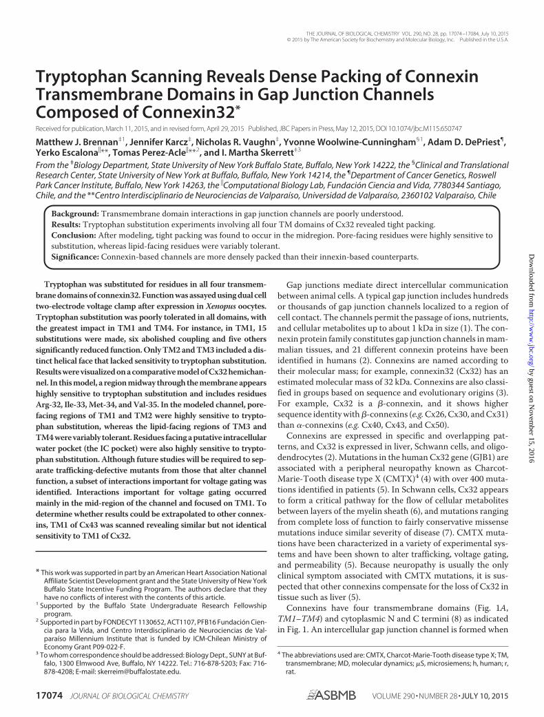
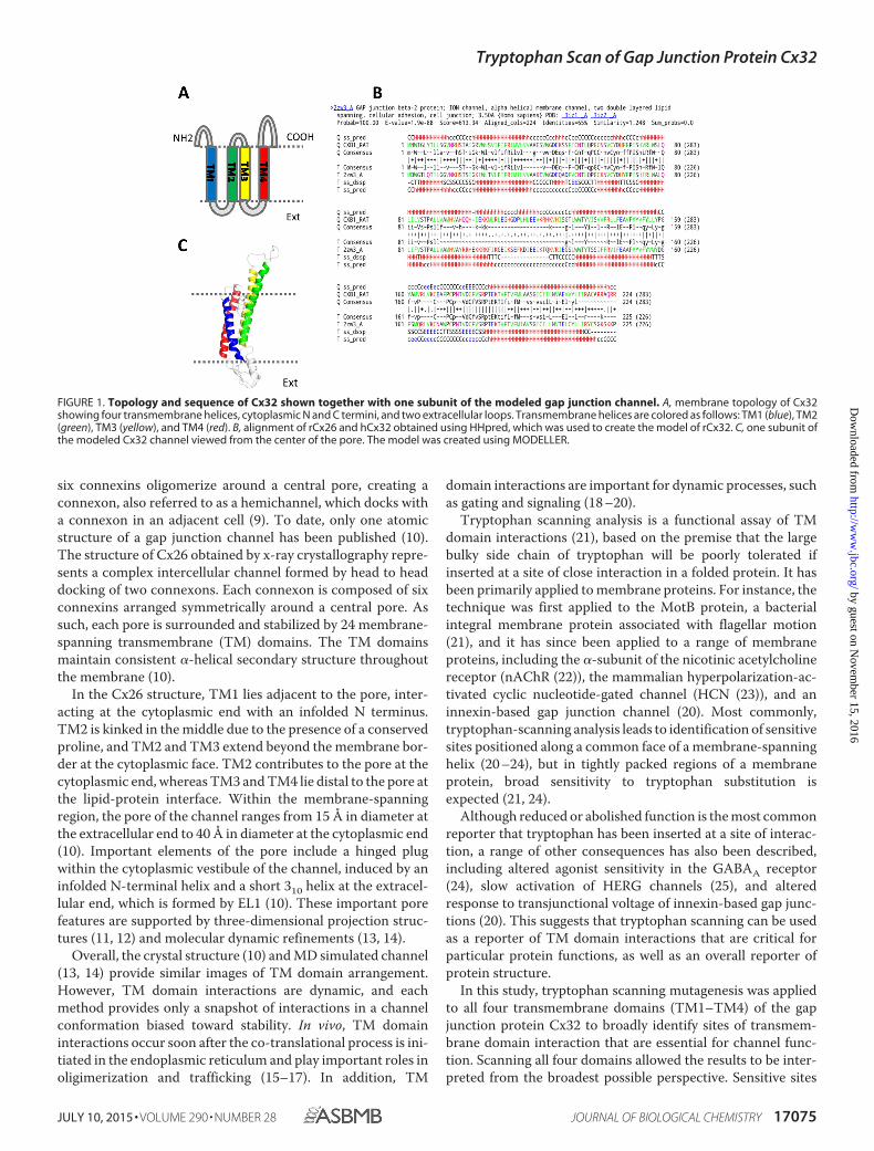
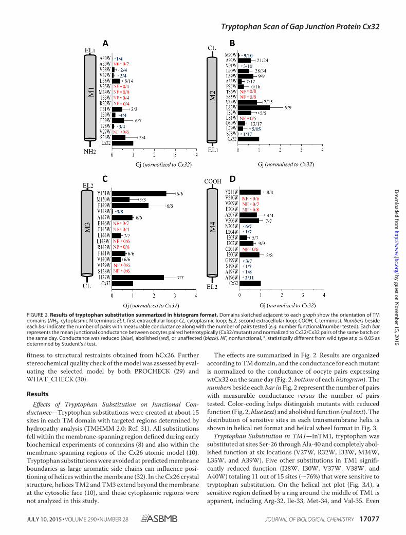
The effects are summarized in Fig. 2. Results are organizedaccording to TM domain, and the conductance for each mutantis normalized to the conductance of oocyte pairs expressingwtCx32 on the same day (Fig. 2, bottom of each histogram). Thenumbers beside each bar in Fig. 2 represent the number of pairswith measurable conductance versus the number of pairstested. Color-coding helps distinguish mutants with reducedfunction (Fig. 2, blue text) and abolished function (red text). Thedistribution of sensitive sites in each transmembrane helix isshown in helical net format and helical wheel format in Fig. 3.
Tryptophan Substitution in TM1—InTM1, tryptophan wassubstituted at sites Ser-26 through Ala-40 and completely abol-ished function at six locations (V27W, R32W, I33W, M34W,L35W, and A39W). Five other substitutions in TM1 signifi-cantly reduced function (I28W, I30W, V37W, V38W, andA40W) totaling 11 out of 15 sites (�76%) that were sensitive totryptophan substitution. On the helical net plot (Fig. 3A), asensitive region defined by a ring around the middle of TM1 isapparent, including Arg-32, Ile-33, Met-34, and Val-35. Even
FIGURE 2. Results of tryptophan substitution summarized in histogram format. Domains sketched adjacent to each graph show the orientation of TMdomains (NH2, cytoplasmic N terminus; EL1, first extracellular loop; CL, cytoplasmic loop; EL2, second extracellular loop; COOH, C terminus). Numbers besideeach bar indicate the number of pairs with measurable conductance along with the number of pairs tested (e.g. number functional/number tested). Each barrepresents the mean junctional conductance between oocytes paired heterotypically (Cx32/mutant) and normalized to Cx32/Cx32 pairs of the same batch onthe same day. Conductance was reduced (blue), abolished (red), or unaffected (black). NF, nonfunctional, *, statistically different from wild type at p � 0.05 asdetermined by Student’s t test.
Tryptophan Scan of Gap Junction Protein Cx32
JULY 10, 2015 • VOLUME 290 • NUMBER 28 JOURNAL OF BIOLOGICAL CHEMISTRY 17077
by guest on Novem
ber 15, 2016http://w
ww
.jbc.org/D
ownloaded from
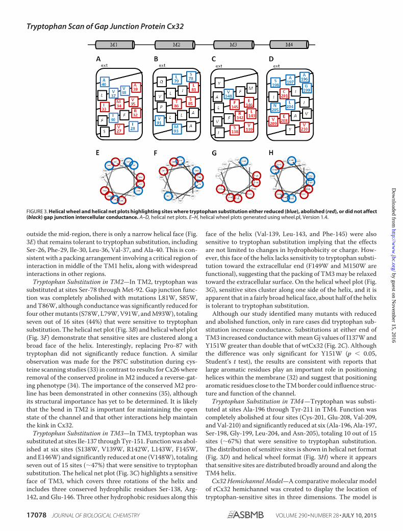
outside the mid-region, there is only a narrow helical face (Fig.3E) that remains tolerant to tryptophan substitution, includingSer-26, Phe-29, Ile-30, Leu-36, Val-37, and Ala-40. This is con-sistent with a packing arrangement involving a critical region ofinteraction in middle of the TM1 helix, along with widespreadinteractions in other regions.
Tryptophan Substitution in TM2—In TM2, tryptophan wassubstituted at sites Ser-78 through Met-92. Gap junction func-tion was completely abolished with mutations L81W, S85W,and T86W, although conductance was significantly reduced forfour other mutants (S78W, L79W, V91W, and M93W), totalingseven out of 16 sites (44%) that were sensitive to tryptophansubstitution. The helical net plot (Fig. 3B) and helical wheel plot(Fig. 3F) demonstrate that sensitive sites are clustered along abroad face of the helix. Interestingly, replacing Pro-87 withtryptophan did not significantly reduce function. A similarobservation was made for the P87C substitution during cys-teine scanning studies (33) in contrast to results for Cx26 whereremoval of the conserved proline in M2 induced a reverse-gat-ing phenotype (34). The importance of the conserved M2 pro-line has been demonstrated in other connexins (35), althoughits structural importance has yet to be determined. It is likelythat the bend in TM2 is important for maintaining the openstate of the channel and that other interactions help maintainthe kink in Cx32.
Tryptophan Substitution in TM3—In TM3, tryptophan wassubstituted at sites Ile-137 through Tyr-151. Function was abol-ished at six sites (S138W, V139W, R142W, L143W, F145W,and E146W) and significantly reduced at one (V148W), totalingseven out of 15 sites (�47%) that were sensitive to tryptophansubstitution. The helical net plot (Fig. 3C) highlights a sensitiveface of TM3, which covers three rotations of the helix andincludes three conserved hydrophilic residues Ser-138, Arg-142, and Glu-146. Three other hydrophobic residues along this
face of the helix (Val-139, Leu-143, and Phe-145) were alsosensitive to tryptophan substitution implying that the effectsare not limited to changes in hydrophobicity or charge. How-ever, this face of the helix lacks sensitivity to tryptophan substi-tution toward the extracellular end (F149W and M150W arefunctional), suggesting that the packing of TM3 may be relaxedtoward the extracellular surface. On the helical wheel plot (Fig.3G), sensitive sites cluster along one side of the helix, and it isapparent that in a fairly broad helical face, about half of the helixis tolerant to tryptophan substitution.
Although our study identified many mutants with reducedand abolished function, only in rare cases did tryptophan sub-stitution increase conductance. Substitutions at either end ofTM3 increased conductance with mean Gj values of I137W andY151W greater than double that of wtCx32 (Fig. 2C). Althoughthe difference was only significant for Y151W (p � 0.05,Student’s t test), the results are consistent with reports thatlarge aromatic residues play an important role in positioninghelices within the membrane (32) and suggest that positioningaromatic residues close to the TM border could influence struc-ture and function of the channel.
Tryptophan Substitution in TM4 —Tryptophan was substi-tuted at sites Ala-196 through Tyr-211 in TM4. Function wascompletely abolished at four sites (Cys-201, Glu-208, Val-209,and Val-210) and significantly reduced at six (Ala-196, Ala-197,Ser-198, Gly-199, Leu-204, and Asn-205), totaling 10 out of 15sites (�67%) that were sensitive to tryptophan substitution.The distribution of sensitive sites is shown in helical net format(Fig. 3D) and helical wheel format (Fig. 3H) where it appearsthat sensitive sites are distributed broadly around and along theTM4 helix.
Cx32 Hemichannel Model—A comparative molecular modelof rCx32 hemichannel was created to display the location oftryptophan-sensitive sites in three dimensions. The model is
FIGURE 3. Helical wheel and helical net plots highlighting sites where tryptophan substitution either reduced (blue), abolished (red), or did not affect(black) gap junction intercellular conductance. A–D, helical net plots. E–H, helical wheel plots generated using wheel.pl, Version 1.4.
Tryptophan Scan of Gap Junction Protein Cx32
17078 JOURNAL OF BIOLOGICAL CHEMISTRY VOLUME 290 • NUMBER 28 • JULY 10, 2015
by guest on Novem
ber 15, 2016http://w
ww
.jbc.org/D
ownloaded from
based on the crystal structure of human Cx26 (10), which, likeCx32, is a �-type connexin. rCx32 and hCx26 share about 65%sequence identity (Fig. 1B); therefore, the architecture of gapjunction channels composed on rCx32 and hCx26 is expectedto be similar.
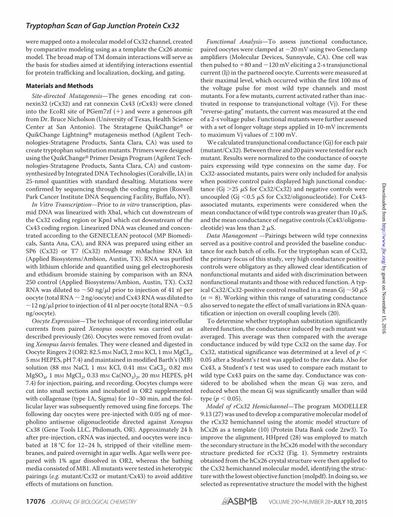
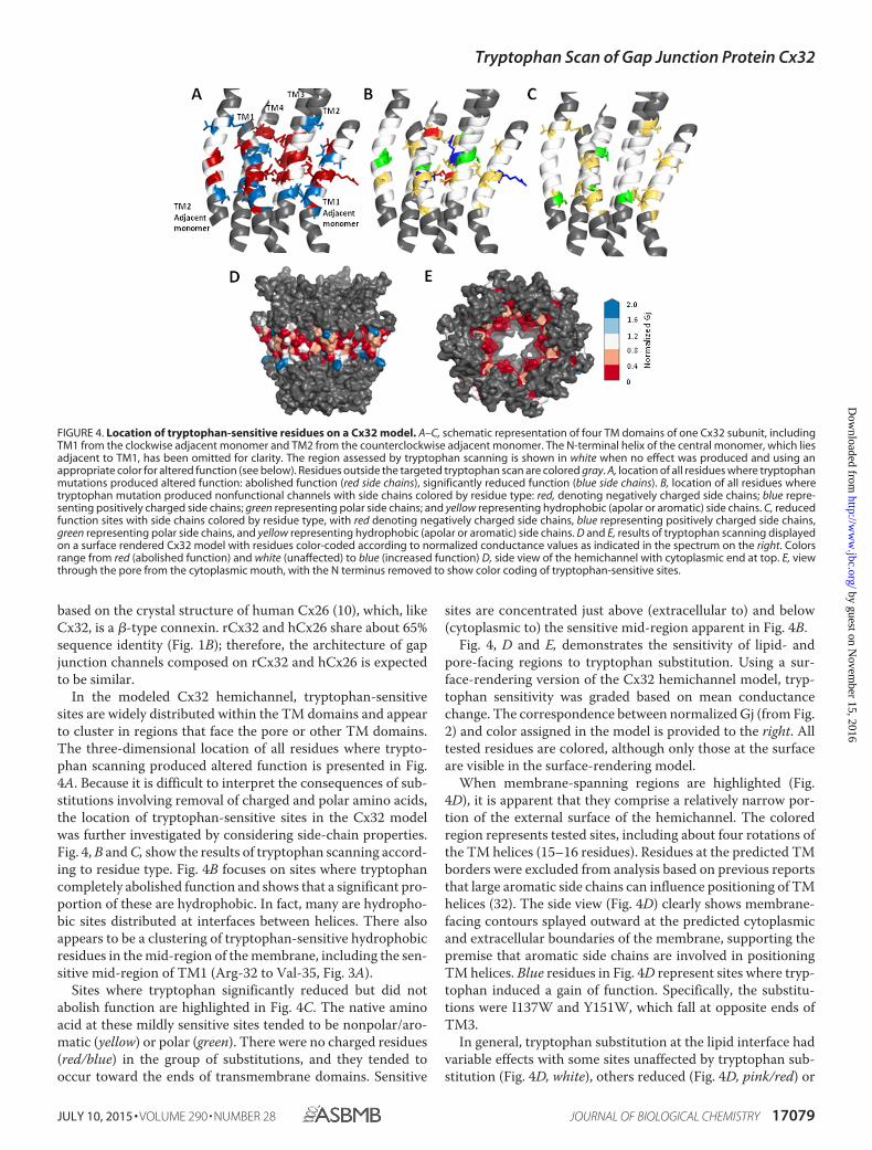
In the modeled Cx32 hemichannel, tryptophan-sensitivesites are widely distributed within the TM domains and appearto cluster in regions that face the pore or other TM domains.The three-dimensional location of all residues where trypto-phan scanning produced altered function is presented in Fig.4A. Because it is difficult to interpret the consequences of sub-stitutions involving removal of charged and polar amino acids,the location of tryptophan-sensitive sites in the Cx32 modelwas further investigated by considering side-chain properties.Fig. 4, B and C, show the results of tryptophan scanning accord-ing to residue type. Fig. 4B focuses on sites where tryptophancompletely abolished function and shows that a significant pro-portion of these are hydrophobic. In fact, many are hydropho-bic sites distributed at interfaces between helices. There alsoappears to be a clustering of tryptophan-sensitive hydrophobicresidues in the mid-region of the membrane, including the sen-sitive mid-region of TM1 (Arg-32 to Val-35, Fig. 3A).
Sites where tryptophan significantly reduced but did notabolish function are highlighted in Fig. 4C. The native aminoacid at these mildly sensitive sites tended to be nonpolar/aro-matic (yellow) or polar (green). There were no charged residues(red/blue) in the group of substitutions, and they tended tooccur toward the ends of transmembrane domains. Sensitive
sites are concentrated just above (extracellular to) and below(cytoplasmic to) the sensitive mid-region apparent in Fig. 4B.
Fig. 4, D and E, demonstrates the sensitivity of lipid- andpore-facing regions to tryptophan substitution. Using a sur-face-rendering version of the Cx32 hemichannel model, tryp-tophan sensitivity was graded based on mean conductancechange. The correspondence between normalized Gj (from Fig.2) and color assigned in the model is provided to the right. Alltested residues are colored, although only those at the surfaceare visible in the surface-rendering model.
When membrane-spanning regions are highlighted (Fig.4D), it is apparent that they comprise a relatively narrow por-tion of the external surface of the hemichannel. The coloredregion represents tested sites, including about four rotations ofthe TM helices (15–16 residues). Residues at the predicted TMborders were excluded from analysis based on previous reportsthat large aromatic side chains can influence positioning of TMhelices (32). The side view (Fig. 4D) clearly shows membrane-facing contours splayed outward at the predicted cytoplasmicand extracellular boundaries of the membrane, supporting thepremise that aromatic side chains are involved in positioningTM helices. Blue residues in Fig. 4D represent sites where tryp-tophan induced a gain of function. Specifically, the substitu-tions were I137W and Y151W, which fall at opposite ends ofTM3.
In general, tryptophan substitution at the lipid interface hadvariable effects with some sites unaffected by tryptophan sub-stitution (Fig. 4D, white), others reduced (Fig. 4D, pink/red) or
FIGURE 4. Location of tryptophan-sensitive residues on a Cx32 model. A–C, schematic representation of four TM domains of one Cx32 subunit, includingTM1 from the clockwise adjacent monomer and TM2 from the counterclockwise adjacent monomer. The N-terminal helix of the central monomer, which liesadjacent to TM1, has been omitted for clarity. The region assessed by tryptophan scanning is shown in white when no effect was produced and using anappropriate color for altered function (see below). Residues outside the targeted tryptophan scan are colored gray. A, location of all residues where tryptophanmutations produced altered function: abolished function (red side chains), significantly reduced function (blue side chains). B, location of all residues wheretryptophan mutation produced nonfunctional channels with side chains colored by residue type: red, denoting negatively charged side chains; blue repre-senting positively charged side chains; green representing polar side chains; and yellow representing hydrophobic (apolar or aromatic) side chains. C, reducedfunction sites with side chains colored by residue type, with red denoting negatively charged side chains, blue representing positively charged side chains,green representing polar side chains, and yellow representing hydrophobic (apolar or aromatic) side chains. D and E, results of tryptophan scanning displayedon a surface rendered Cx32 model with residues color-coded according to normalized conductance values as indicated in the spectrum on the right. Colorsrange from red (abolished function) and white (unaffected) to blue (increased function) D, side view of the hemichannel with cytoplasmic end at top. E, viewthrough the pore from the cytoplasmic mouth, with the N terminus removed to show color coding of tryptophan-sensitive sites.
Tryptophan Scan of Gap Junction Protein Cx32
JULY 10, 2015 • VOLUME 290 • NUMBER 28 JOURNAL OF BIOLOGICAL CHEMISTRY 17079
by guest on Novem
ber 15, 2016http://w
ww
.jbc.org/D
ownloaded from
increased (Fig. 4D, blue). A small number of lipid-facing sitesshowed high sensitivity to tryptophan substitution ((Fig. 4D,red). These residues could mediate interactions with lipids,adjacent proteins, or be involved in TM domain interactions inchannel conformations that differ from the conformation of themodel. The highly sensitive ((Fig. 4D, red) residues facing thelipid bilayer occur within a narrow strip along the TM4 helix(Ala-196, Ile-203, and Val-210) suggesting that a slight helicalrotation could alter their position relative to other TMdomains.
Fig. 4E highlights the sensitivity of pore-facing residues totryptophan substitution. The pore is viewed from the cytoplas-mic mouth, with the N terminus omitted for clarity. Only resi-dues considered to be within the membrane-spanning regionare colored, representing their sensitivity to tryptophan substi-tution. As one might predict, pore-facing residues appear to behighly sensitive with most of the pore contours appearing red(Fig. 4E). In the Cx32 model, the pore lining is contributed byTM1 (Ile-30, Ile-33, Met-34, and Val-37) and TM2 (Leu-81,Ile-82, Ser-85, Thr-86, Leu-89, and Met-93).
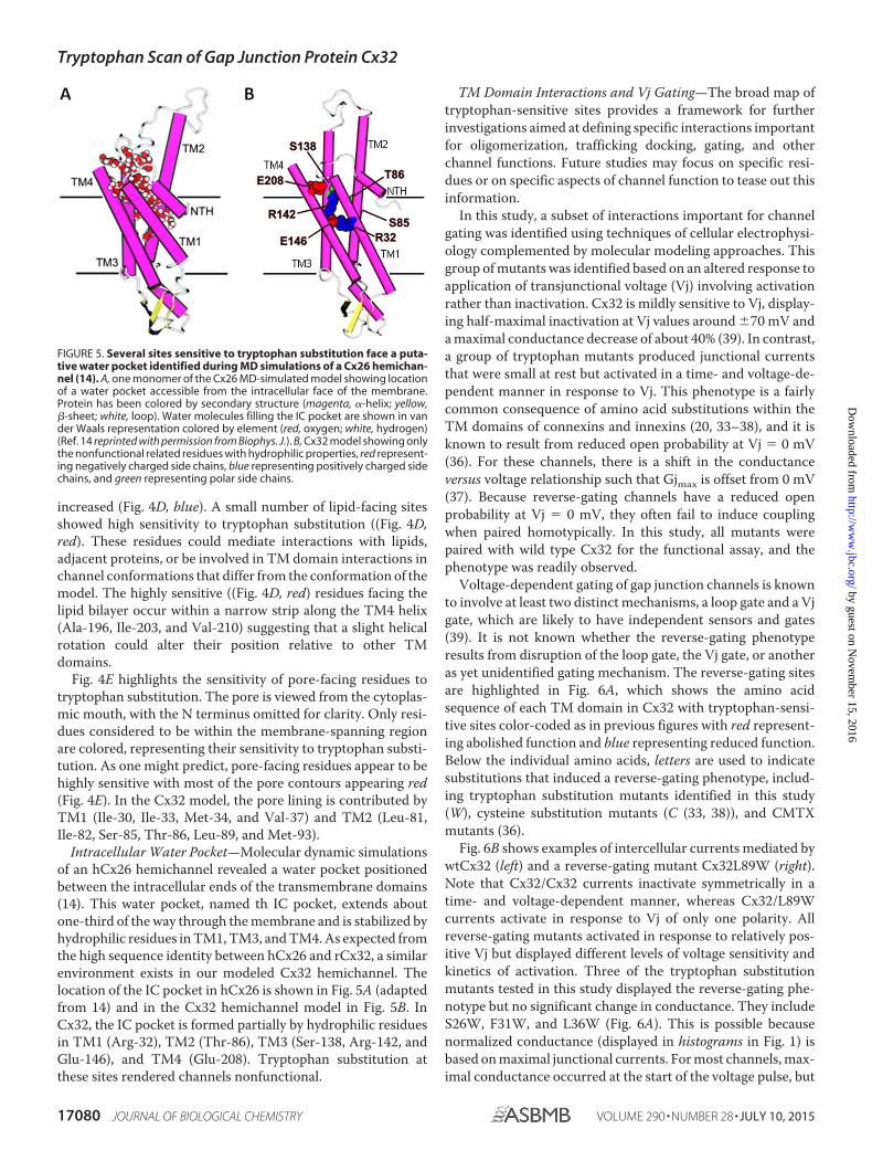
Intracellular Water Pocket—Molecular dynamic simulationsof an hCx26 hemichannel revealed a water pocket positionedbetween the intracellular ends of the transmembrane domains(14). This water pocket, named th IC pocket, extends aboutone-third of the way through the membrane and is stabilized byhydrophilic residues in TM1, TM3, and TM4. As expected fromthe high sequence identity between hCx26 and rCx32, a similarenvironment exists in our modeled Cx32 hemichannel. Thelocation of the IC pocket in hCx26 is shown in Fig. 5A (adaptedfrom 14) and in the Cx32 hemichannel model in Fig. 5B. InCx32, the IC pocket is formed partially by hydrophilic residuesin TM1 (Arg-32), TM2 (Thr-86), TM3 (Ser-138, Arg-142, andGlu-146), and TM4 (Glu-208). Tryptophan substitution atthese sites rendered channels nonfunctional.
TM Domain Interactions and Vj Gating—The broad map oftryptophan-sensitive sites provides a framework for furtherinvestigations aimed at defining specific interactions importantfor oligomerization, trafficking docking, gating, and otherchannel functions. Future studies may focus on specific resi-dues or on specific aspects of channel function to tease out thisinformation.
In this study, a subset of interactions important for channelgating was identified using techniques of cellular electrophysi-ology complemented by molecular modeling approaches. Thisgroup of mutants was identified based on an altered response toapplication of transjunctional voltage (Vj) involving activationrather than inactivation. Cx32 is mildly sensitive to Vj, display-ing half-maximal inactivation at Vj values around �70 mV anda maximal conductance decrease of about 40% (39). In contrast,a group of tryptophan mutants produced junctional currentsthat were small at rest but activated in a time- and voltage-de-pendent manner in response to Vj. This phenotype is a fairlycommon consequence of amino acid substitutions within theTM domains of connexins and innexins (20, 33–38), and it isknown to result from reduced open probability at Vj � 0 mV(36). For these channels, there is a shift in the conductanceversus voltage relationship such that Gjmax is offset from 0 mV(37). Because reverse-gating channels have a reduced openprobability at Vj � 0 mV, they often fail to induce couplingwhen paired homotypically. In this study, all mutants werepaired with wild type Cx32 for the functional assay, and thephenotype was readily observed.
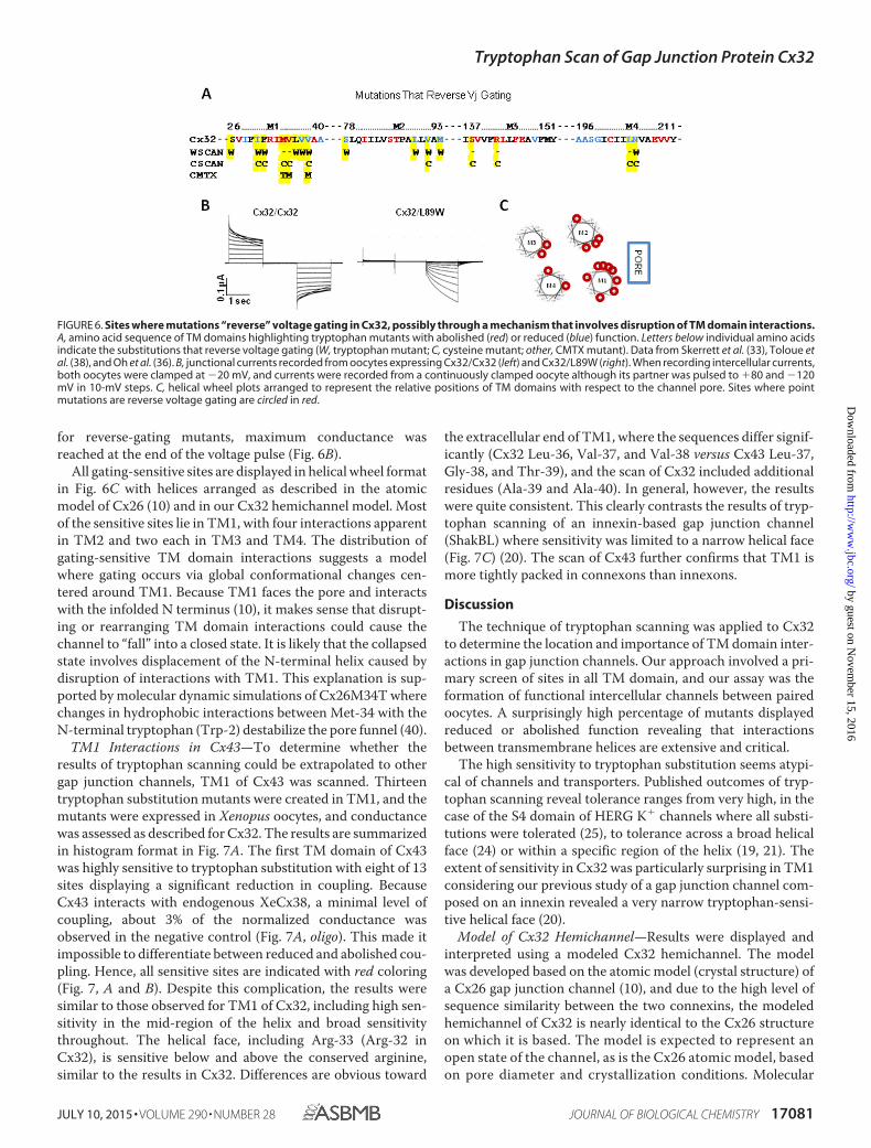
Voltage-dependent gating of gap junction channels is knownto involve at least two distinct mechanisms, a loop gate and a Vjgate, which are likely to have independent sensors and gates(39). It is not known whether the reverse-gating phenotyperesults from disruption of the loop gate, the Vj gate, or anotheras yet unidentified gating mechanism. The reverse-gating sitesare highlighted in Fig. 6A, which shows the amino acidsequence of each TM domain in Cx32 with tryptophan-sensi-tive sites color-coded as in previous figures with red represent-ing abolished function and blue representing reduced function.Below the individual amino acids, letters are used to indicatesubstitutions that induced a reverse-gating phenotype, includ-ing tryptophan substitution mutants identified in this study(W), cysteine substitution mutants (C (33, 38)), and CMTXmutants (36).
Fig. 6B shows examples of intercellular currents mediated bywtCx32 (left) and a reverse-gating mutant Cx32L89W (right).Note that Cx32/Cx32 currents inactivate symmetrically in atime- and voltage-dependent manner, whereas Cx32/L89Wcurrents activate in response to Vj of only one polarity. Allreverse-gating mutants activated in response to relatively pos-itive Vj but displayed different levels of voltage sensitivity andkinetics of activation. Three of the tryptophan substitutionmutants tested in this study displayed the reverse-gating phe-notype but no significant change in conductance. They includeS26W, F31W, and L36W (Fig. 6A). This is possible becausenormalized conductance (displayed in histograms in Fig. 1) isbased on maximal junctional currents. For most channels, max-imal conductance occurred at the start of the voltage pulse, but
FIGURE 5. Several sites sensitive to tryptophan substitution face a puta-tive water pocket identified during MD simulations of a Cx26 hemichan-nel (14). A, one monomer of the Cx26 MD-simulated model showing locationof a water pocket accessible from the intracellular face of the membrane.Protein has been colored by secondary structure (magenta, �-helix; yellow,�-sheet; white, loop). Water molecules filling the IC pocket are shown in vander Waals representation colored by element (red, oxygen; white, hydrogen)(Ref. 14 reprinted with permission from Biophys. J.). B, Cx32 model showing onlythe nonfunctional related residues with hydrophilic properties, red represent-ing negatively charged side chains, blue representing positively charged sidechains, and green representing polar side chains.
Tryptophan Scan of Gap Junction Protein Cx32
17080 JOURNAL OF BIOLOGICAL CHEMISTRY VOLUME 290 • NUMBER 28 • JULY 10, 2015
by guest on Novem
ber 15, 2016http://w
ww
.jbc.org/D
ownloaded from
for reverse-gating mutants, maximum conductance wasreached at the end of the voltage pulse (Fig. 6B).
All gating-sensitive sites are displayed in helical wheel formatin Fig. 6C with helices arranged as described in the atomicmodel of Cx26 (10) and in our Cx32 hemichannel model. Mostof the sensitive sites lie in TM1, with four interactions apparentin TM2 and two each in TM3 and TM4. The distribution ofgating-sensitive TM domain interactions suggests a modelwhere gating occurs via global conformational changes cen-tered around TM1. Because TM1 faces the pore and interactswith the infolded N terminus (10), it makes sense that disrupt-ing or rearranging TM domain interactions could cause thechannel to “fall” into a closed state. It is likely that the collapsedstate involves displacement of the N-terminal helix caused bydisruption of interactions with TM1. This explanation is sup-ported by molecular dynamic simulations of Cx26M34T wherechanges in hydrophobic interactions between Met-34 with theN-terminal tryptophan (Trp-2) destabilize the pore funnel (40).
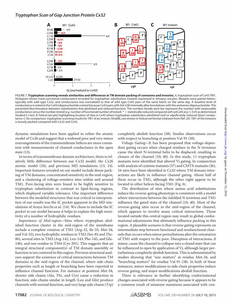
TM1 Interactions in Cx43—To determine whether theresults of tryptophan scanning could be extrapolated to othergap junction channels, TM1 of Cx43 was scanned. Thirteentryptophan substitution mutants were created in TM1, and themutants were expressed in Xenopus oocytes, and conductancewas assessed as described for Cx32. The results are summarizedin histogram format in Fig. 7A. The first TM domain of Cx43was highly sensitive to tryptophan substitution with eight of 13sites displaying a significant reduction in coupling. BecauseCx43 interacts with endogenous XeCx38, a minimal level ofcoupling, about 3% of the normalized conductance wasobserved in the negative control (Fig. 7A, oligo). This made itimpossible to differentiate between reduced and abolished cou-pling. Hence, all sensitive sites are indicated with red coloring(Fig. 7, A and B). Despite this complication, the results weresimilar to those observed for TM1 of Cx32, including high sen-sitivity in the mid-region of the helix and broad sensitivitythroughout. The helical face, including Arg-33 (Arg-32 inCx32), is sensitive below and above the conserved arginine,similar to the results in Cx32. Differences are obvious toward
the extracellular end of TM1, where the sequences differ signif-icantly (Cx32 Leu-36, Val-37, and Val-38 versus Cx43 Leu-37,Gly-38, and Thr-39), and the scan of Cx32 included additionalresidues (Ala-39 and Ala-40). In general, however, the resultswere quite consistent. This clearly contrasts the results of tryp-tophan scanning of an innexin-based gap junction channel(ShakBL) where sensitivity was limited to a narrow helical face(Fig. 7C) (20). The scan of Cx43 further confirms that TM1 ismore tightly packed in connexons than innexons.
Discussion
The technique of tryptophan scanning was applied to Cx32to determine the location and importance of TM domain inter-actions in gap junction channels. Our approach involved a pri-mary screen of sites in all TM domain, and our assay was theformation of functional intercellular channels between pairedoocytes. A surprisingly high percentage of mutants displayedreduced or abolished function revealing that interactionsbetween transmembrane helices are extensive and critical.
The high sensitivity to tryptophan substitution seems atypi-cal of channels and transporters. Published outcomes of tryp-tophan scanning reveal tolerance ranges from very high, in thecase of the S4 domain of HERG K� channels where all substi-tutions were tolerated (25), to tolerance across a broad helicalface (24) or within a specific region of the helix (19, 21). Theextent of sensitivity in Cx32 was particularly surprising in TM1considering our previous study of a gap junction channel com-posed on an innexin revealed a very narrow tryptophan-sensi-tive helical face (20).
Model of Cx32 Hemichannel—Results were displayed andinterpreted using a modeled Cx32 hemichannel. The modelwas developed based on the atomic model (crystal structure) ofa Cx26 gap junction channel (10), and due to the high level ofsequence similarity between the two connexins, the modeledhemichannel of Cx32 is nearly identical to the Cx26 structureon which it is based. The model is expected to represent anopen state of the channel, as is the Cx26 atomic model, basedon pore diameter and crystallization conditions. Molecular
FIGURE 6. Sites where mutations “reverse” voltage gating in Cx32, possibly through a mechanism that involves disruption of TM domain interactions.A, amino acid sequence of TM domains highlighting tryptophan mutants with abolished (red) or reduced (blue) function. Letters below individual amino acidsindicate the substitutions that reverse voltage gating (W, tryptophan mutant; C, cysteine mutant; other, CMTX mutant). Data from Skerrett et al. (33), Toloue etal. (38), and Oh et al. (36). B, junctional currents recorded from oocytes expressing Cx32/Cx32 (left) and Cx32/L89W (right). When recording intercellular currents,both oocytes were clamped at �20 mV, and currents were recorded from a continuously clamped oocyte although its partner was pulsed to �80 and �120mV in 10-mV steps. C, helical wheel plots arranged to represent the relative positions of TM domains with respect to the channel pore. Sites where pointmutations are reverse voltage gating are circled in red.
Tryptophan Scan of Gap Junction Protein Cx32
JULY 10, 2015 • VOLUME 290 • NUMBER 28 JOURNAL OF BIOLOGICAL CHEMISTRY 17081
by guest on Novem
ber 15, 2016http://w
ww
.jbc.org/D
ownloaded from
dynamic simulations have been applied to refine the atomicmodel of Cx26 and suggest that a widened pore and very minorrearrangements of the transmembrane helices are more consis-tent with measurements of channel conductance in the openstate (13).
In terms of transmembrane domain architecture, there is rel-atively little difference between our Cx32 model, the Cx26atomic model (10), and previous MD simulations (13, 14).Important features revealed on our model include dense pack-ing of TM domains, concentrated sensitivity in the mid-region,and a clustering of voltage-sensitive sites within and aroundTM1. Pore-facing sites were found to be highly sensitive totryptophan substitution in contrast to lipid-facing regions,which displayed variable tolerance. One important differencebetween the modeled structures that was critical to interpreta-tion of our results was the IC pocket apparent in the MD sim-ulations of Araya-Secchi et al. (14). We chose to include the ICpocket in our model because it helps to explain the high sensi-tivity of a number of hydrophilic residues.
Importance of Mid-region—Sites where tryptophan abol-ished function cluster in the mid-region of the membraneinclude a complete rotation of TM1 (Arg-32, Ile-33, Met-34,and Val-35), two hydrophilic residues in TM2 (Ser-85 and Thr-86), several sites in TM3 (Arg-142, Leu-143, Phe-145, and Glu-146), and one residue in TM4 (Cys-201). This suggests that anintegral structural component(s) of TM domain assembly orfunction is/are centered in this region. Previous structural anal-yses support the existence of critical interactions between TMdomains in the mid-region of the channel, where side-chainproperties such as length or branching have been reported toinfluence channel function. For instance at position Met-34,shorter side chains (Ala, Thr, and Cys) cause a reduction infunction; side-chains similar in length (Leu and Gln) producechannels with normal function, and very large side chains (Trp)
completely abolish function (38). Similar observations occurwith respect to branching at position Val-91 (38).
Voltage Gating—It has been proposed that voltage-depen-dent gating occurs when charged residues in the N terminuscause the short N-terminal helix to be displaced, resulting inclosure of the channel (10, 40). In this study, 11 tryptophanmutants were identified that altered Vj gating. In conjunctionwith analysis of cysteine mutants (37) and CMTX mutants (36),16 sites have been identified in Cx32 where TM domain inter-actions are likely to influence channel gating. About half ofthese occur in TM1, although the other eight appear to belocated in other helices facing TM1 (Fig. 6).
The distribution of sites where amino acid substitutionsinduce the reverse-gating phenotype is consistent with a modelwhere interactions between the infolded N terminus and TM1influence the gated state of the channel (10, 40). Most of thereverse-gating sites occur in the mid-region of the channel,which appears to involve many critical interactions. Thoselocated outside this central region may result in global confor-mational adjustments that ultimately influence packing in thisregion. A plausible scenario is that reverse gating represents anintermediate step between functional and nonfunctional chan-nels that occurs when minor perturbations alter the orientationof TM1 with respect to the pore. Disruption of interactions, ifminor, cause the channel to collapse into a closed state that canbe influenced to open by application of Vj, although larger per-turbations completely abolish function. This is substantiated bystudies showing that “size matters” at residue Met-34, and“branching matters” for residue Val-91 (38). In both of theseinstances, minor modifications to side chain properties inducereverse gating, and major modifications abolish function.
There is relevance in further identifying conformationalchanges associated with reverse gating because it appears to bea common result of missense mutations associated with con-
FIGURE 7. Tryptophan scanning reveals similarities and differences in TM domain packing of connexins and innexins. A, tryptophan scan of Cx43 TM1.Histogram shows mean junctional conductance recorded for tryptophan substitution mutants expressed in Xenopus oocytes. Mutants were paired hetero-typically with wild type Cx32, and conductance was normalized to that of wild type Cx43 pairs of the same batch on the same day. A baseline level ofconductance is noted in the Cx43/oligonucleotide control because Cx43 pairs with XeCx38 minimally after knockdown with the antisense oligonucleotide. Thisprevented discrimination between substitutions that abolished and reduced function. The numbers beside each bar represent the number with measurableconductance versus the number tested (e.g. number of functional/number of tested). * � statistically reduced compared with wtCx43 at p � 0.05 as determinedStudent’s t test. B, helical net plot highlighting location of sites in Cx43 where tryptophan substitution abolished (red) or significantly reduced (blue) conduc-tance. C, for comparison, tryptophan scanning results for TM1 of an innexin (ShakBL) are shown in helical net format (redrawn from Ref. 20). TM1 of the innexinsis loosely packed compared with Cx32 and Cx43.
Tryptophan Scan of Gap Junction Protein Cx32
17082 JOURNAL OF BIOLOGICAL CHEMISTRY VOLUME 290 • NUMBER 28 • JULY 10, 2015
by guest on Novem
ber 15, 2016http://w
ww
.jbc.org/D
ownloaded from
nexin diseases and therefore a pathological state of the channel.For instance, the reverse-gating mutations in Cx26 M34T causedeafness (41), and the mutations M34T, V35M, and V38M inCx32 cause CMTX (36). Voltage gating in gap junction chan-nels is complex and involves gates that act in response to Vmand Vj (39). The reverse-gating phenotype may involve a shift inVj sensitivity (36, 37), a modified response of the Vm gate totransjunctional voltage, or a conformation unrelated to eitherof the previously characterized voltage-gating mechanisms.One observation argues against the involvement of typical Vj-gating mechanisms. Cx26 and Cx32 have Vj gates of oppositepolarity (38, 39) yet reverse-gating mutants of both connexinsopen in response to relatively positive Vj.
ResiduesFacingthePore—OurCx32hemichannelmodel,con-sistent with currently accepted models of connexin-based gapjunction structure, places TM1 and TM2 adjacent to the pore(10, 13, 14). Tryptophan substitutions were poorly tolerated atTM domain sites associated with pore-lining positions (Fig.4E), which might be expected for a couple of reasons. First, atthe cytoplasmic end of the channel, inserting a large bulky sidechain is likely to disrupt positioning of the N terminus. Second,high sensitivity to tryptophan insertion is expected at the extra-cellular end of the pore where it narrows.
Previous cysteine accessibility studies presented TM3 as themain pore-lining domain due to extensive accessibility alongthe helix; however, many residues in TM1 were accessible tosulfhydryl reagents. These correlate well with pore-lining resi-dues in the atomic model (10) and the Cx32 hemichannelmodel developed for this study. Five cysteine mutants in TM1(I30C, F31C, I33C, M34C, and V35C) were accessible to sulfhy-dryl reagent (33). Three of these are sites where tryptophansubstitutions rendered the channels nonfunctional (I33W,M34W, and V35W) and one exhibited reduced function(I30W). Only Phe-31 was functional as a tryptophan mutant(F31W), which might be expected given the nature of the sub-stitution. Interpretation of the results of cysteine scanning wascomplicated by the fact that four of the cysteine mutants (I30C,F31C, M34C, and V35C) displayed the reverse-gating pheno-type, and one was accessible in nonperfused cells (I33C) wheresulfhydryl reagents should not have been able to access thepore. However, the results of cysteine accessibility are largelyconsistent with the position of these TM1 residues in our Cx32hemichannel model.
Residues Facing the Lipid Bilayer—It was generally expectedthat substitutions would be well tolerated at the protein-lipidinterface where the bulky hydrophobic side chain of tryptophanwould be easily accommodated by the flexible environment ofthe fatty acid tails (21). Surprisingly, tryptophan substitutionson the external surface of the channel had a wide range ofeffects when viewed on the Cx32 hemichannel model (Fig. 4D,membrane-spanning regions colored). The colored region rep-resents about four rotations of a TM helix, corresponding toabout 15 residues that were subjected to tryptophan scanning.The membrane-spanning domains appear as a relatively nar-row region on the external surface of the hemichannel, and thechannel appears to splay outward at the membrane borders.Several substitutions at the TM domain borders caused anincrease in conductance (blue residues in Fig. 3A). Several sub-
stitutions at the TM domain borders caused an increase in con-ductance (Fig. 4D, blue). Other residues at the protein-lipidinterface range from highly sensitive (Fig. 4D, red) to slightlysensitive (Fig. 4D, pink) and tolerant (Fig. 4D, white). High sen-sitivity was not expected at the lipid interface. The presence ofTrp-sensitive sites in this region may suggest that the lipid-facing region of the channel plays important functions thathave not yet been identified or that sensitive residues are sites oftransmembrane domain interaction in a channel conformationnot apparent in the modeled channel. Future studies should beaimed at clarifying the role of TM3 residues.
Comparing Connexin- and Innexin-based Channels—One ofthe earliest and most significant applications of tryptophanscanning involved a comparison of related proteins. Sharp et al.(21) identified structural differences between flagellar motilityproteins MotA and MotB based on differing sensitivities totryptophan. More recently De Feo et al. (43) have identifiedstructural similarities between distantly related copper trans-porters using tryptophan scanning. DePriest et al. (20) previ-ously demonstrated that TM1 of an innexin was quite tolerantto tryptophan substitution. The results of tryptophan scanningin Cx32 suggest that connexin-based channels are more denselypacked than their innexin-based counterparts. To determinewhether other connexins are also densely packed, TM1 of Cx43was scanned with results comparable with those obtained forCx32. Both Cx32 and Cx43 displayed a highly sensitive regionin the middle of TM1, where tryptophan substitution was verypoorly tolerated over at least an entire rotation of the helix. Thisis very different from the innexin, which showed limited sensi-tivity along one face of the TM1 helix (20).
Connexins and innexins reportedly evolved convergently(44) but are surprisingly similar in their topology and func-tion (1, 12). Innexin channels generally have a larger outerdiameter (12, 42), consistent with looser packing of TMdomains. Advances in the biology of innexin channels arelikely to provide exciting information about their similaritiesand differences.
Acknowledgments—We thank the students in Advanced Cell Biologyat SUNY Buffalo State for technical assistance.
References1. Goodenough, D. A., and Paul, D. L. (2009) Gap junctions. Cold Spring
Harb. Perspect. Biol. 1, a0025762. Willecke, K., Eiberger, J., Degen, J., Eckardt, D., Romualdi, A., Güldenagel,
M., Deutsch, U., and Söhl, G. (2002) Structural and functional diversity ofconnexin genes in the mouse and human genome. Biol. Chem. 383,725–737
3. Kumar, N. M., and Gilula, N. B. (1996) The gap junction communicationchannel. Cell 84, 381–388
4. Bergoffen, J., Scherer, S. S., Wang, S., Scott, M. O., Bone, L. J., Paul, D. L.,Chen, K., Lensch, M. W., Chance, P. F., and Fischbeck, K. H. (1993) Con-nexin mutations in X-linked Charcot-Marie-Tooth disease. Science 262,2039 –2042
5. Kleopa, K. A., Abrams, C. K., and Scherer, S. S. (2012) How do mutationsin GJB1 cause X-linked Charcot-Marie-Tooth disease? Brain Res. 1487,198 –205
6. Balice-Gordon, R. J., Bone, L. J., and Scherer, S. S. (1998) Functional gapjunctions in the Schwann cell myelin sheath. J. Cell Biol. 142, 1095–1104
7. Shy, M. E., Siskind, C., Swan, E. R., Krajewski, K. M., Doherty, T., Fuerst,
Tryptophan Scan of Gap Junction Protein Cx32
JULY 10, 2015 • VOLUME 290 • NUMBER 28 JOURNAL OF BIOLOGICAL CHEMISTRY 17083
by guest on Novem
ber 15, 2016http://w
ww
.jbc.org/D
ownloaded from
D. R., Ainsworth, P. J., Lewis, R. A., Scherer, S. S., and Hahn, A. F. (2007)CMT1X phenotypes represent loss of GJB1 gene function. Neurology 68,849 – 855
8. Milks, L. C., Kumar, N. M., Houghten, R., Unwin, N., and Gilula, N. B.(1988) Topology of the 32-kd liver gap junction protein determined bysite-directed antibody localizations. EMBO J. 7, 2967–2975
9. Bruzzone, R., White, T. W., and Paul, D. L. (1996) Connections with con-nexins: the molecular basis of direct intercellular signaling. Eur.J. Biochem. 238, 1–27
10. Maeda, S., Nakagawa, S., Suga, M., Yamashita, E., Oshima, A., Fujiyoshi,Y., and Tsukihara, T. (2009) Structure of the connexin 26 gap junctionchannel at 3.5 Å resolution. Nature 458, 597– 602
11. Oshima, A., Tani, K., Hiroaki, Y., Fujiyoshi, Y., and Sosinsky, G. E. (2007)Three-dimensional structure of a human connexin26 gap junction chan-nel reveals a plug in the vestibule. Proc. Natl. Acad. Sci. U.S.A. 104,10034 –10039
12. Oshima, A., Matsuzawa, T., Nishikawa, K., and Fujiyoshi, Y. (2013) Olig-omeric structure and functional characterization of Caenorhabditis el-egans Innexin-6 gap junction protein. J. Biol. Chem. 288, 10513–10521
13. Kwon, T., Harris, A. L., Rossi, A., and Bargiello, T. A. (2011) Moleculardynamics simulations of the Cx26 hemichannel: evaluation of structuralmodels with Brownian dynamics. J. Gen Physiol. 138, 475– 493
14. Araya-Secchi, R., Perez-Acle, T., Kang, S. G., Huynh, T., Bernardin, A.,Escalona, Y., Garate, J. A., Martínez, A. D., García, I. E., Sáez, J. C., andZhou, R. (2014) Characterization of a novel water pocket inside the humanCx26 hemichannel structure. Biophys. J. 107, 599 – 612
15. Lee, S. P., Xie, Z., Varghese, G., Nguyen, T., O’Dowd, B. F., and George,S. R. (2000) Oligomerization of dopamine and serotonin receptors. Neu-ropsychopharmacology 23, S32–S40
16. Laird, D. W. (2006) Life cycle of connexins in health and disease. Biochem.J. 394, 527–543
17. Smith, T. D., Mohankumar, A., Minogue, P. J., Beyer, E. C., Berthoud,V. M., and Koval, M. (2012) Cytoplasmic amino acids within the mem-brane interface region influence connexin oligomerization. J. Membr. Biol.245, 221–230
18. Matthews, E. E., Zoonens, M., and Engelman, D. M. (2006) Dynamic helixinteractions in transmembrane signaling. Cell 127, 447– 450
19. Otero-Cruz, J. D., Báez-Pagán, C. A., Caraballo-González, I. M., and La-salde-Dominicci, J. A. (2007) Tryptophan-scanning mutagenesis in the�M3 transmembrane domain of the muscle-type acetylcholinereceptor–a spring model revealed. J. Biol. Chem. 282, 9162–9171
20. Depriest, A., Phelan, P., and Martha Skerrett, I. (2011) Tryptophan-scan-ning mutagenesis of the first transmembrane domain of the innexin Shak-ing-B(Lethal). Biophys. J. 101, 2408 –2416
21. Sharp, L. L., Zhou, J., and Blair, D. F. (1995) Tryptophan-scanning mu-tagenesis of MotB, an integral membrane protein essential for flagellarrotation in Escherichia coli. Biochemistry 34, 9166 –9171
22. Guzmán, G. R., Santiago, J., Ricardo, A., Martí-Arbona, R., Rojas, L. V.,and Lasalde-Dominicci, J. A. (2003) Tryptophan scanning mutagenesisin the �M3 transmembrane domain of the Torpedo californica acetyl-choline receptor: functional and structural implications. Biochemistry 42,12243–12250
23. Ishii, T. M., Nakashima, N., and Ohmori, H. (2007) Tryptophan-scanningmutagenesis in the S1 domain of mammalian HCN channel reveals resi-dues critical for voltage-gated activation. J. Physiol. 579, 291–301
24. Ueno, S., Lin, A., Nikolaeva, N., Trudell, J. R., Mihic, S. J., Harris, R. A., andHarrison, N. L. (2000) Tryptophan scanning mutagenesis in TM2 of theGABAA receptor � subunit: effects on channel gating and regulation byethanol. Br. J. Pharmacol. 131, 296 –302
25. Subbiah, R. N., Kondo, M., Campbell, T. J., and Vandenberg, J. I. (2005)Tryptophan scanning mutagenesis of the HERG K� channel: the S4 do-
main is loosely packed and likely to be lipid-exposed. J. Physiol. 569,367–379
26. Skerrett, I. M., Merritt, M., Zhou, L., Zhu, H., Cao, F.-L., Smith, J. F., andNicholson, B. J. (2001) Applying the Xenopus oocyte expression system tothe analysis of gap junction proteins. Methods Mol. Biol. 154, 225–249
27. Sali, A., and Blundell, T. L. (1993) Comparative protein modelling by sat-isfaction of spatial restraints. J. Mol. Biol. 234, 779 – 815
28. Söding, J., Biegert, A., and Lupas, A. N. (2005) The HHpred interactiveserver for protein homology detection and structure prediction. NucleicAcids Res. 33, W244 –W248
29. Laskowski, R. A., MacArthur, M. W., Moss, D. S., and Thornton, J. M.(1993) PROCHECK–a program to check the stereochemical quality ofprotein structures. J. App. Crystallogr. 26, 283–291
30. Hooft, R. W., Vriend, G., Sander, C., and Abola, E. E. (1996) Errors inprotein structures. Nature 381, 272–272
31. Krogh, A., Larsson, B., von Heijne, G., and Sonnhammer E. L. (2001)Predicting transmembrane protein topology with a hidden Markov mod-el: application to complete genomes. J. Mol. Biol. 305, 567–580
32. Mall, S., Broadbridge, R., Sharma, R. P., Lee, A. G., and East, J. M. (2000)Effects of aromatic residues at the end of transmembrane �-helices onhelix interactions with lipid bilayers. Biochemistry 39, 2071–2078
33. Skerrett, I. M., Aronowitz, J., Shin, J. H., Cymes, G., Kasperek, E., Cao,F.-L., and Nicholson, B. J. (2002) Identification of amino acid residueslining the pore of a gap junction channel. J. Cell Biol. 159, 349 –360
34. Suchyna, T. M., Xu, L. X., Gao, F., Fourtner, C. R., and Nicholson, B. J.(1993) Identification of a proline residue as a transduction element in-volved in voltage gating of gap junctions. Nature 365, 847– 849
35. Arora, A., Minogue, P. J., Liu, X., Reddy, M. A., Ainsworth, J. R., Bhattacha-rya, S. S., Webster, A. R., Hunt, D. M., Ebihara, L., Moore, A. T., Beyer,E. C., and Berthoud, V. M. (2006) A novel GJA8 mutation is associatedwith autosomal dominant lamellar pulverulent cataract: further evidencefor gap junction dysfunction in human cataract. J. Med. Genet. 43, e2
36. Oh, S., Ri, Y., Bennett, M. V., Trexler, E. B., Verselis, V. K., and Bargiello,T. A. (1997) Changes in permeability caused by connexin 32 mutationsunderlie X-linked Charcot-Marie-Tooth disease. Neuron 19, 927–938
37. Skerrett, I. M., Smith, J. F., and Nicholson, B. J. (1999) Mechanistic differ-ences between chemical and electrical gating of gap junctions. Curr. Top.Membr. 49, 249 –269
38. Toloue, M., Woolwine, Y., Karcz, J., Kasperek, E., Nicholson, B. J., andSkerrett, I. M. (2008) Site-directed mutagenesis reveals putative regions ofprotein interaction within the transmembrane domains of connexins. CellCommun. Adhes. 15, 95–105
39. Harris, A. L. (2001) Emerging issues of connexin channels: biophysics fillsthe gap. Q. Rev. Biophys. 34, 325– 472
40. Zonta, F., Buratto, D., Cassini, C., Bortolozzi, M., and Mammano, F. (2014)Molecular dynamics simulations highlight structural and functional alter-ations in deafness-related M34T mutation of connexin 26. Front. Physiol.5, 85
41. Skerrett, I. M., Di, W. L., Kasperek, E. M., Kelsell, D. P., and Nicholson, B. J.(2004) Aberrant gating, but a normal expression pattern, underlies therecessive phenotype of the deafness mutant Connexin26M34T. FASEB J.18, 860 – 862
42. Peracchia, C. (1973) Low resistance junctions in crayfish. II. Two arrays ofglobules in junctional membranes. J. Cell Biol. 57, 66 –76
43. De Feo, C. J., Mootien, S., and Unger, V. (2010) Tryptophan scanninganalysis of the membrane domain of CTR-copper transporters. J. Membr.Biol. 234, 113–123
44. Alexopoulos, H., Böttger, A., Fischer, S., Levin, A., Wolf, A., Fujisawa, T.,Hayakawa, S., Gojobori, T., Davies, J. A., David, C. N., and Bacon, J. P.(2004) Evolution of gap junctions: the missing link? Curr. Biol. 14,R879 –R880
Tryptophan Scan of Gap Junction Protein Cx32
17084 JOURNAL OF BIOLOGICAL CHEMISTRY VOLUME 290 • NUMBER 28 • JULY 10, 2015
by guest on Novem
ber 15, 2016http://w
ww
.jbc.org/D
ownloaded from
Martha SkerrettWoolwine-Cunningham, Adam D. DePriest, Yerko Escalona, Tomas Perez-Acle and I.
Matthew J. Brennan, Jennifer Karcz, Nicholas R. Vaughn, YvonneDomains in Gap Junction Channels Composed of Connexin32
Tryptophan Scanning Reveals Dense Packing of Connexin Transmembrane
doi: 10.1074/jbc.M115.650747 originally published online May 12, 20152015, 290:17074-17084.J. Biol. Chem.
10.1074/jbc.M115.650747Access the most updated version of this article at doi:
Alerts:
When a correction for this article is posted•
When this article is cited•
to choose from all of JBC's e-mail alertsClick here
http://www.jbc.org/content/290/28/17074.full.html#ref-list-1
This article cites 44 references, 13 of which can be accessed free at
by guest on Novem
ber 15, 2016http://w
ww
.jbc.org/D
ownloaded from