Embed Size (px)
Citation preview
HAL Id: tel-03340318https://tel.archives-ouvertes.fr/tel-03340318
Submitted on 10 Sep 2021
HAL is a multi-disciplinary open accessarchive for the deposit and dissemination of sci-entific research documents, whether they are pub-lished or not. The documents may come fromteaching and research institutions in France orabroad, or from public or private research centers.
L’archive ouverte pluridisciplinaire HAL, estdestinée au dépôt et à la diffusion de documentsscientifiques de niveau recherche, publiés ou non,émanant des établissements d’enseignement et derecherche français ou étrangers, des laboratoirespublics ou privés.
Étude du potentiel des biomarqueurs sérologiques pourévaluer le risque de transmission de la dengue dans le
nord-est de la ThaïlandeBénédicte Fustec
To cite this version:Bénédicte Fustec. Étude du potentiel des biomarqueurs sérologiques pour évaluer le risque de trans-mission de la dengue dans le nord-est de la Thaïlande. Sciences agricoles. Université Montpellier,2020. Français. �NNT : 2020MONTT079�. �tel-03340318�

THÈSE POUR OBTENIR LE GRADE DE DOCTEUR
DE L’UNIVERSITÉ DE MONTPELLIER
En Biologie-Sante
École doctorale CBS2
Unité de recherche "MIVEGEC: Maladies Infectieuses et Vecteurs: Ecologie, Génétique, Evolution et Contrôle" UMR IRD 224-CNRS 5290-Université Montpellier
Présentée par Benedicte Fustec
Le 21 Décembre 2020
Sous la direction de Vincent CORBEL et Hans J. OVERGAARD
Étude du potentiel des biomarqueurs sérologiques pour évaluer le risque de transmission de la dengue dans le nord-est de la Thaïlande.
Devant le jury composé de
Mme. Nicole L. Achee, Professeur, University of Notre-Dame
Mr. Lionel Almeras, CR, Université d’Aix-Marseille
Mr. Jean-Pierre Dedet, Professeur Emérite Université de Montpellier
Mme. Audrey Lenhart, CR, United States Centers for Diseases Control and Prevention
Mr. Richard Paul, CR, Global Health, Pasteur Institute
Mr. Vincent Corbel, Directeur de recherche, HDR, IRD.
Mr. Hans J. Overgaard, CR, University of Life Sciences Norway
Rapporteur
Rapporteur
Président du jury
Examinateur
Examinateur
Directeur de thèse
Co-encadrant de thèse

2
3
THÈSE POUR OBTENIR LE GRADE DE DOCTEUR
DE L’UNIVERSITÉ DE MONTPELLIER
En Biologie-Sante
École doctorale CBS2
Unité de recherche "MIVEGEC: Maladies Infectieuses et Vecteurs: Ecologie, Génétique, Evolution et Contrôle" UMR IRD 224-CNRS 5290-Université Montpellier
Exploring the potential of serological biomarkers to assess the
risk of dengue transmission in north-Eastern Thailand.
Defended by Benedicte Fustec
21st of December 2020
Under the supervision of Vincent CORBEL and Hans J. OVERGAARD
Before the jury composed of
Mme. Nicole L. Achee, Professeur, University of Notre-Dame
Mr. Lionel Almeras, CR, Université d’Aix-Marseille
Mr. Jean-Pierre Dedet, Professeur Emérite Université de Montpellier
Mme. Audrey Lenhart, CR, United States Centers for Diseases Control and Prevention
Mr. Richard Paul, CR, Global Health, Pasteur Institute
Mr. Vincent Corbel, Directeur de recherche, HDR, IRD.
Mr. Hans J. Overgaard, CR, University of Life Sciences Norway
Rapporteur
Rapporteur
President of the jury
Examinateur
Examinateur
Directeur de thèse
Co-encadrant de thèse

4
Contents
LIST OF FIGURES ..................................................................................................................9
LIST OF TABLES .................................................................................................................. 11
LIST OF ABBREVIATIONS ................................................................................................. 12
LIST OF PUBLICATIONS .................................................................................................... 16
LIST OF SCIENTIFIC COMMUNICATIONS .................................................................... 17
ACKNOWLEDGEMENTS .................................................................................................... 18
PREAMBLE ............................................................................................................................ 21
FIRST PART: GENERALITIES ........................................................................................... 24
1. DENGUE DISEASE ................................................................................................................ 24
1.2. Epidemiology.................................................................................................................. 24
1.3. Viruses ............................................................................................................................ 26
1.4. Dengue distribution ....................................................................................................... 28
1.4.1. Dengue in South East Asia ...................................................................................................... 29
1.4.2. Dengue in Western Pacific region ........................................................................................... 30
1.4.3. Dengue in Americas ................................................................................................................ 30
1.4.4. Dengue in Africa ..................................................................................................................... 31
1.4.5. Dengue in the Mediterranean region ...................................................................................... 31
1.5. Symptoms ....................................................................................................................... 32
1.6. Diagnostic ....................................................................................................................... 33
1.6.1. Epidemiological diagnostic ..................................................................................................... 34

5
1.6.2. Laboratory diagnostic ............................................................................................................. 34
1.6.2.1. Direct diagnosis .................................................................................................................... 34
1.6.2.2. Indirect diagnosis ................................................................................................................. 36
2. THE DENGUE VECTORS ....................................................................................................... 39
2.1. Life cycle ........................................................................................................................ 40
2.2. Aedes vectors .................................................................................................................. 41
2.2.1. Aedes aegypti........................................................................................................................... 42
2.2.2. Aedes albopictus ...................................................................................................................... 43
3. GLOBAL STRATEGIES FOR DENGUE PREVENTION AND CONTROL ........................................ 45
3.1. Vaccine ........................................................................................................................... 45
3.2. Treatment & prophylaxis .............................................................................................. 46
3.3. Dengue vector control .................................................................................................... 47
3.3.1. Integrated Aedes management strategies ............................................................................... 48
3.3.1.1. Social mobilisation and community engagement ................................................................ 49
3.3.1.2. Larval control ....................................................................................................................... 51
3.3.1.3. Adult control ........................................................................................................................ 52
3.3.2. Alternative strategies for vector control ................................................................................. 56
3.3.2.1. Wolbachia-based strategies .................................................................................................. 56
3.3.2.2. Genetically modified mosquitoes ......................................................................................... 57
3.3.2.3. Other tools currently under evaluation ............................................................................... 57
3.3.3. Insecticide resistance ............................................................................................................... 58
3.3.3.1. Global distribution ............................................................................................................... 58
3.3.3.2. Mechanisms .......................................................................................................................... 60
3.3.3.3. Impact on vector control ...................................................................................................... 61
6
4. METHODS AND INDICATORS FOR ASSESSING AND PREDICTING THE RISK OF DENGUE
TRANSMISSION .......................................................................................................................... 61
4.1. Definition: concept of transmission ............................................................................... 61
4.2. Epidemiological surveillance and it’s limitations ......................................................... 64
4.3. Mathematical tools and their limitations ...................................................................... 65
4.4. Entomology surveillance and it’s limitations ................................................................ 66
4.4.1. Immature indices and their limitations .................................................................................. 70
4.4.2. Adult indices and their limitations ......................................................................................... 71
4.5. Serological tools to estimate dengue transmission risk................................................. 71
4.5.1. Concept.................................................................................................................................... 71
4.5.2. Application of salivary biomarkers to Aedes transmitted diseases ....................................... 73
SECOND PART: CONTEXT OF THE THESIS .................................................................. 76
1. CHALLENGES IN PREDICTING DENGUE TRANSMISSION AND OUTBREAKS IN THAILAND ...... 76
2. THE DENGUE INDEX PROJECT ....................................................................................... 78
3. OBJECTIVES OF THE THESIS ............................................................................................... 79
3.1. Objective #1: Assessing the spatial and temporal dynamic of dengue in North-eastern
Thailand .................................................................................................................................. 80
3.2. Objective #2: Addressing the complex relationship between Aedes vectors, dengue
transmission and socio-economic factors ............................................................................... 80
3.3. Objective #3: Assessing fine-scale variations in human exposure to Aedes mosquito
bites using salivary biomarker during a vector control intervention. ................................... 81
3.4. Objective #4: Evaluating the impact of vector control intervention on the selection of
insecticide resistance in dengue vectors .................................................................................. 81
4. STUDY DESIGN .................................................................................................................... 82
4.1. Study area ...................................................................................................................... 82

7
4.2. Case control study .......................................................................................................... 82
4.3. Cluster-randomized controlled trial ............................................................................ 87
THIRD PART: RESULTS OF THE THESIS ....................................................................... 91
CHAPTER 1. ASSESSING THE SPATIAL AND TEMPORAL PATTERNS OF DENGUE INCIDENCE IN
NORTH-EASTERN THAILAND .................................................................................................... 91
Summary of the results: .......................................................................................................... 92
CHAPTER 2: ADDRESSING THE COMPLEX RELATIONSHIPS BETWEEN AEDES VECTORS, SOCIO-
ECONOMICS AND DENGUE TRANSMISSION IN NORTH-EASTERN THAILAND. .............................. 95
Summary of the results: .......................................................................................................... 95
CHAPTER 3: ASSESSING FINE SCALE VARIATIONS IN HUMAN EXPOSURE TO AEDES MOSQUITO
BITES USING AEDES SALIVARY BIOMARKER DURING A RANDOMIZED VECTOR CONTROL
INTERVENTION TRIAL. .............................................................................................................. 99
Summary of results: .............................................................................................................. 100
CHAPTER 4: ASSESSING THE IMPACT OF VECTOR CONTROL INTERVENTION ON THE SELECTION
OF INSECTICIDE RESISTANCE GENES IN DENGUE VECTORS ...................................................... 103
Summary of results: .............................................................................................................. 103
FOURTH PART: DISCUSSION AND PERSPECTIVES OF THE THESIS .................... 107
Challenges in assessing dengue transmission risk using conventional entomology indices 110
Potential of serological biomarkers for assessing Aedes-human relationships ................... 112
Potential of serological biomarkers for assessing dengue transmission risk....................... 113
Prospect of serological biomarkers for assessing vector control interventions................... 114
Barriers to dengue vector control in Thailand ..................................................................... 115
CONCLUSION ..................................................................................................................... 116

8
REFERENCES ...................................................................................................................... 117
RESUME DE LA THESE ..................................................................................................... 148

9
List of figures
Figure 1: Overlapping of global distribution of major mosquito borne diseases (malaria,
dengue, chikungunya, Zika, yellow fever, Japanese encephalitis, lymphatic filariasis) ............... 23
Figure 2: Spread of dengue in the world since 1943 from Messina et al. ........................ 25
Figure 3: Dengue virus genome from Guzman et al. ...................................................... 27
Figure 4: Dengue virus envelope structure from Rey. ..................................................... 28
Figure 5: Global distributions of dengue serotypes in 1970 and 2004 from Mackenzie et
al. .............................................................................................................................................. 29
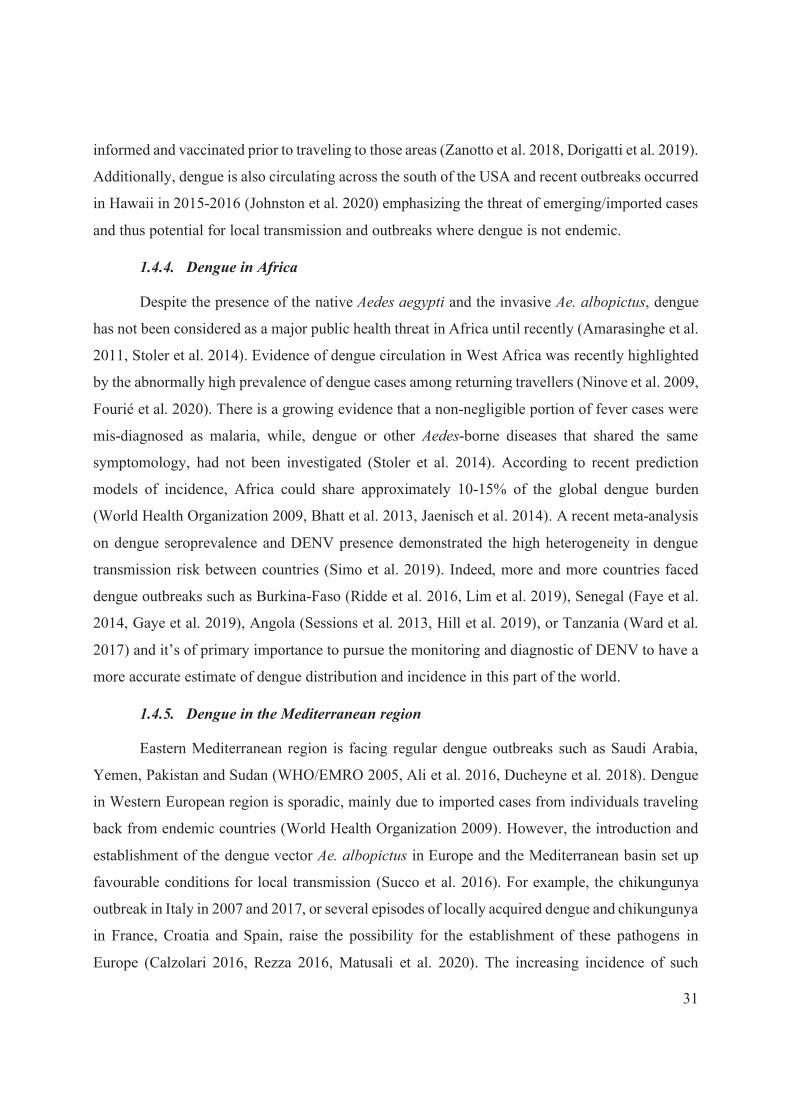
Figure 6: Dengue cases classification and level of severity, from WHO. ........................ 33
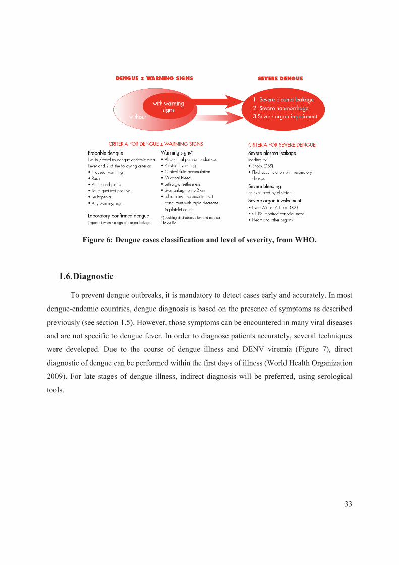
Figure 7: Dengue illness course from WHO. .................................................................. 34
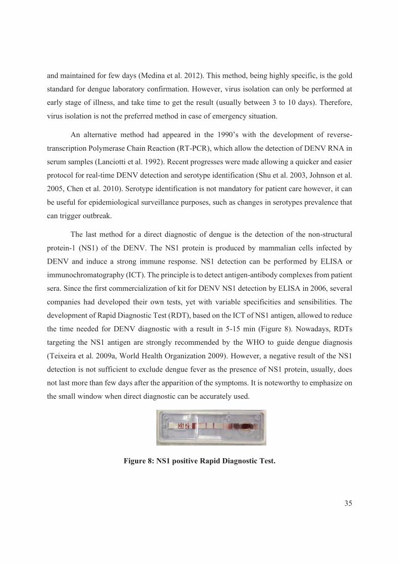
Figure 8: NS1 positive Rapid Diagnostic Test. ............................................................... 35
Figure 9: Time-line of immunoglobulins in primary and secondary dengue infection from
WHO. ....................................................................................................................................... 36
Figure 10: Principle of hemagglutination assay for dengue diagnosis. ............................ 37
Figure 11: Principle of MAC-ELISA experiment. .......................................................... 38
Figure 12: IgG positive and IgM negative RDT ............................................................. 39
Figure 13: Aedes simplified life cycle from Biogents©. ................................................. 40
Figure 14: Aedes aegypti (left) and Aedes albopictus (right). ......................................... 42
Figure 15: Map showing the predicted distribution of Aedes aegypti from Kraemer et al.
................................................................................................................................................. 43
Figure 16: Map showing the predicted distribution of Aedes albopictus from Kraemer et
al. .............................................................................................................................................. 44
Figure 17: Global Vector Control Response Framework. ............................................... 48
Figure 18: Conceptual framework of the IAM system from Roiz et al. ........................... 49

10
Figure 19: Insecticide resistance against pyrethroid and organophosphate worldwide from
Moyes et al................................................................................................................................ 59
Figure 20: Dengue transmission cycles from Ahammad et al. ........................................ 62
Figure 21: Human-vector relationships during arthropod-borne diseases from Sagna et al.
................................................................................................................................................. 72
Figure 22: Amino-acid sequence of Nterm-34 kDa peptide. ........................................... 73
Figure 23: Ab response to Nterm-34 kDa salivary peptide according to the season in
children from Benin from Elanga et al. ...................................................................................... 74
Figure 24: Number of monthly dengue cases reported in Thailand 2005-2015. .............. 76
Figure 25: Number of dengue reported cases in Khon Kaen, Roi Et, Kalasin and Maha
Sarakham provinces, in northeastern Thailand between 2005-2019. .......................................... 78
Figure 26: Isan typical landscape. .................................................................................. 82
Figure 27: Map and characteristics of study sites of the case-control study in northeastern
Thailand.. .................................................................................................................................. 84
Figure 28: Flow diagram of case-control study design. .................................................. 85
Figure 29: Map of the study sites of the RCT. ................................................................ 88
Figure 30: Flow chart of the RCT study design .............................................................. 89
Figure 31: Mean monthly dengue incidence per 100,000 persons, from Phanitchat et al..
................................................................................................................................................. 93
Figure 32: Mean dengue prevalence by sub-district, from Phanitchat et al...................... 94
Figure 33: Immune response to Aedes salivary peptide Nterm-34 (∆OD) in dengue case
and control patients. .................................................................................................................. 97
Figure 34: Seasonal variation of human IgG and AIc ................................................... 100
Figure 35: Copy number variant in CCEAE3A gene in Ae. aegypti populations before and
after PPF treatment. ................................................................................................................. 106

11
List of tables
Table 1: Dengue vaccines under development from Yauch and Shresta ........................ 46
Table 2: Strengths and limitations of larval control strategies from Roiz et al. ............... 50
Table 3: Adult control strategies for Aedes-borne disease from Roiz et al. ..................... 53
Table 4: Strengths and weakness of entomological surveillance tools from Roiz et al. ... 67
Table 5: Variable definition for the case control study. .................................................. 86
Table 6: Evolution of pyriproxyfen resistance in Aedes populations in different clusters of
Khon Kaen following implementation of vector control intervention. ...................................... 104
12
List of abbreviations
Ab: Antibody
ABTS: 2,2’-Azino-Bis (3-ethylbenzThiazoline 6-Sulfonic acid) di-ammonium
AchE: Acetylcholine esterase.
AGO: Autocidal Gravid Oviposition trap
AI: Adult Aedes Index
AI_in: Adult Aedes Index Indoor
AIc: Adult Index at the cluster level
AIC: Akaike Information Criterion
ATSB: Attractive Toxic Sugar Baited
BI: Breteau Index
CCE: Carboxylesterase
CHIKV: chikungunya virus
CI: Container Index
CDC: Center for Disease Control
COMBI: Communication for Behavioral Impact
CNV: Copy Number Variation
CRISP-Cas9: Clustered Regularly Interspaced Short Palindromic Repeats associated protein 9.
CYP: Cytochrome P450
DALY: Disability-Adjusted Life Years
DDT: DichloroDiphenyl-Trichloroethane
DENV: Dengue virus
DF: Dengue Fever
DHF: Dengue Haemorrhagic fever

13
DNA: Desoxyribonucleic acid
DSS: Dengue Shock Syndrome
ELISA: Enzyme-Linked ImmunoSorbent Assay
ENSO: El Niño Southern Oscillation
GAT: Gravid Autocidal Trap
GMO: Genetically Modified Organism
GST: Glutathione-S-transferase
HI: House Index
HIA: Hemagglutination Inhibition Assay
IAM: Integrated Aedes Management
ICT: Immunochromatography
IgG: Immunoglobulin G
IgM: Immunoglobulin M
IGR: Insect Growth Regulator
IRS: Indoor Residual Spraying
IRM: Insecticide Resistance Management
ISS: Indoor Space Spray
IVM: Integrated Vector Management
JEV: Japanese Encephalitis Virus
KAP: Knowledge Attitude and Practice
Kdr: Knock down rate
KK: Khon Kaen
MAC-ELISA: immunoglobulin M Antibody Capture Enzyme-Linked ImmunoSorbent Assay
MEI: Mosquito Exposure Index
14
MET: Mosquito Electrocuting Trap
MoPH: Ministry of Public Health
NS: Non-structural
PCR: Polymerase Chain Reaction
PHI: Pupae per House Index
POC: Point of care
PPF: Pyriproxyfen
PPI: Pupae per Person Index
PNRT: Plaque Neutralization Reduction Test
PYR: Pyrethroid
ODPC: Office of Disease Prevention and Control
OP: Organophosphate
ORS: Outdoor Residual Spraying
RCT: Randomized Control Trial
RDT: Rapid Diagnostic Test
RE: Roi Et
RIDL: Release Insect with Dominant Lethality
RNA: Ribonucleic acid
RT-PCR: Retro Transcription Polymerase Chain Reaction
RR50: Resistance Ratio 50
SEA : South East Asia
SES: Socio-Economic Status
SIT: Sterile Insect Technic
SRRT: Surveillance Rapid Response Team

15
ULV: Ultra Low Volume
VBDU: Vector Borne Disease Unit
VGCR: Vector Global Control Response
VGSC: Voltage Gated Sodium Channel
WIN: Worldwide Insecticide resistance Network
WHO: World Health Organisation
WHO-TDR: World Health Organisation and special group for tropical diseases research.
WHO-VCAG: World Health Organisation
YFV: Yellow Fever Virus
ZIKV: Zika Virus

16
List of publications
1. Fustec B, Phanitchat T, Hoq M. I., Aromseree S., Pientong C., Thaewnongiew K., Ekalaksananan T, Bangs MJ., Corbel V, Alexander N., Overgaard H. J. (2020). Complex relationships between Aedes vectors, socio-economics and dengue transmission— lessons learned from a case-control study in northeastern Thailand. PLoS Negl Trop Dis. 1;14(10). doi: 10.1371/journal.pntd.0008703.
2. Fustec B, Phanitchat T, Aromseree S, Pientong C, Thaewnongview K, Ekalaksananan T, Cerqueira D, Poinsignon A, Elguero E, Bangs M.J., Alexander N, Overgaard H.J, Corbel V. Assessing human exposure to Aedes mosquitoes by the use of Aedes salivary biomarker in a context of a vector control intervention: a randomized control trial in northeastern Thailand. (submitted to Plos Negl Trop Dis. in October 2020)
3. Phanitchat T, Zhao B, Haque U, Pientong C, Ekalaksananan T, Aromseree S, Thaewnongiew K, Fustec B, Bangs MJ, Alexander N, Overgaard HJ. (2019). Spatial and temporal patterns of dengue incidence in northeastern Thailand 2006-2016. BMC Infect Dis, 19(1), 743. doi: 10.1186/s12879-019-4379-3.
4. Overgaard HJ, Pientong C, Thaewnongiew K, Bangs MJ, Ekalaksananan T, Aromseree S, Phanitchat T, Phanthanawiboon S, Fustec B, Corbel V, Cerqueira D, Alexander N. (2018). Assessing dengue transmission risk and a vector control intervention using entomological and immunological indices in Thailand: study protocol for a cluster-randomized controlled trial. Trials, 19(1), 122. doi: 10.1186/s13063-018-2490-1. Corrected in Trials, in December 2018 19(1), 703. doi: 10.1186/s13063-018-3110-9
17
List of scientific communications
- B. Fustec, S. Aromseree, C. Pientong, K. Thaewnongiew, M. J. Bangs, N. Alexander, H. J. Overgaard, V. Corbel. Immunological and Entomological Indices to Evaluate the Risk of Dengue Transmission in Northeastern Thailand. Joint International Tropical Medicine
Meeting (JITMM), 6-8 December 2017, Amari Watergate, Bangkok, Thailand. (POSTER)
- B. Fustec, H. J. Overgaard, T. Phanitchat, S. Aromseree, C. Pientong, K. Thaewnongiew, V. Corbel. Characterization of insecticide resistance in Aedes aegypti populations from Khon Kaen district, northeastern Thailand. 2nd Win International Conference 2018: Grand
Copthorne Waterfront Hotel, 1-3 October 2018, Singapore. (POSTER)
- B. Fustec, H. J. Overgaard, T. Phanitchat, S. Aromseree, C. Pientong, T. Ekalaksananan, K. Thaewnongiew, N. Alexander, M. J. Bangs, V. Corbel. Aedes abundance and dengue fever transmission, a difficult relationship: lessons from an ongoing case control study in northeastern Thailand. The 6th International Forum for Surveillance and Control of
Mosquitoes and Vector-borne Diseases (IFSCMVD) in Conjunction with 4th Meeting of the
ASVEMC and 12th Conference of Medical and Veterinary Entomology of the Entomological
Society of China, May 26-30, 2019, Xiamen, China. (ORAL COMMUNICATION)
- B. Fustec, T. Phanitchat, S. Aromseree, C. Pientong, T. Ekalaksananan, K., Thaewnongiew, N. Alexander, M. J. Bangs, H.J. Overgaard, V. Corbel. Seasonal variations in immunological and entomological indices to estimate dengue vector infestation, a longitudinal study in Northeastern Thailand. XXVI International Congress of Entomology, in Helsinki, Finland
(ICE 2020), postpone to July 2021. (ORAL COMMUNICATION)
- T. Phanitchat, C. Pientong, T. Ekalaksananan, S. Phanthanawiboon, S. Aromseree, B. Fustec, K. Thaewnongiew, M. J. Bangs, N. Alexander, H. J. Overgaard. Spatio-temporal analysis of dengue during 2006-2016 in Khon Kaen Province, Thailand. 10th Conference on Global
Health and vaccination research (GLOBVAC), 14-15 March 2017, Trondheim, Norway. (POSTER)
- H. J. Overgaard, C. Pientong, T. Ekalaksananan, S. Aromseree, T. Phanitchat, B. Fustec, K. Thaewnongiew, M. J. Bangs, V. Corbel, N. Alexander. Can Entomological and Immunological Indices Distinguish between Dengue Positive and Negative Households? A Prospective, Hospital-Based, Case-Control Study in Northeastern Thailand. 10th Conference on Global
Health and vaccination research (GLOBVAC), 14-15 March 2017, Trondheim, Norway. (POSTER)
18
Acknowledgements
This work would not have been possible without the support and help of many people,
whose valuable contributions have enabled me to carry out this study, and to whom I would like
to express my deepest and most sincere thanks. First of all, I wish to warmly thank Dr. VINCENT
CORBEL, as my thesis director and Dr. HANS J. OVERGAARD as my co-director, without
whom this study could not have taken place. I would like to express my gratitude to them for their
patience, their trust in my work and their kind guidance. You gave me the opportunity to follow
“my dreams” and I will be forever grateful for that.
Vincent, you believed in me, even when I didn’t and you gave me the strength to pursue
my career. For more than six years now, we’ve been working together and you trusted me and
guided me all that long. I will be eternally thankful for your support, help, guidance and friendship.
More than only my thesis director, you’ve been a friend to me and I hope this is only the beginning
of the story. I would never been able to complete this work without your encouragement and your
kind supervision, so really, thank you!
To Hans, I would like to express my deepest acknowledgement for your guidance, support
and help during these four years in Khon Kaen, Thailand. Thank you for letting me pursue my
doctoral study in the framework of the DENGUE-INDEX project sponsored by the Research
Council of Norway. I also thank you for helping me to settle in our loved Siwalee village, and
introduce me and Alex in the crazy “tchin-tchin” group of soi 15!! We spent amazing nights with
you guys and we enjoyed discovering unseen part of Thailand.
I extend my acknowledgements to Prof. CHAMSAI PIENTONG and Dr. TIPEYA
ELAKASAN from the Khon Kaen University, Thailand, for their warmly welcome. You made
everything for me to make me feel like home in KKU. You gave me precious advices for the
serological studies and enlarged my knowledge especially in the field of virology. Thank you for
your rigor, kindness and for allowing me to participate and contribute to the weekly journal club.
Prof. Chamsai, you are the driving force of your team, keep it up!
I also want to deeply thank Dr. DOMINIQUE CEIRQUERA (formerly KU/MORU), Dr.
ANNE POINSIGNON (IRD) and Dr. ANDRE SAGNA (IRD) for their valuable contribution and
knowledge sharing in the fields of immunology, parasitology and entomology. A special “big up”

19
for Dominique for visiting me in Khon Kaen (despite delayed flights!), and helping me setting-up
and calibrate ELISA protocols for serological studies, and more importantly, for your longstanding
friendship since your stayed in Bangkok.
This work wouldn’t have been possible without the support of the DENGUE INDEX
project members, whom I want to particularly acknowledge. THIRPUETHAI (Kook) thank you
for coordinating the field entomology work package activities of the programme. You are an
amazing manager and a valuable colleague. SIRINART (Mon) and PANWAD (Dream) thank you
for your support in collecting human blood samples and for the conduct of serology assays.
MICHEAL J. BANGS (Mike) thank you for your support and your great insights in entomology
and paper’s writing. I always appreciate your visits in Khon Kaen for the DENGUE INDEX
meetings. NEAL ALEXANDER thank you so much for your help in statistics and model analysis.
I look forward to collaborating with you in the future.
A special thanks to the members of the office of disease prevention and control 7 (ODPC7),
especially P’ BOONSONG and P’ KIEW for your continual support in mosquito collection and
identification. I am grateful to P’BOONSONG for producing enough mosquitoes for lab testing
and for maintaining “my” colonies when I was away. P’KIEW your help was so precious during
bioassays, and you provided me with valuable advices when I faced technical problems (especially
with Pyriproxyfen assays...). I hope you’ll appreciate that I let the mosquito cages for you after my
departure from Khon Kaen, because you loved them so much….
The resistance part of this work wouldn’t have been possible without the great support and
help of JEAN-PHILIPPE DAVID (LECA-CNRS) and FREDERIQUE LAPORTE (LECA-
CNRS). Thank you for your help and support during the study on resistance markers. I would
never been able to conduct all this work without your strong commitment especially during the
COVID-19 lockdown.
I extend my sincere thanks to DR. FREDERIC SIMARD (IRD, Unit Director) for giving
me the opportunity to join the internationally esteemed MIVEGEC research during my Ph.D.
The last but not least acknowledgement will go to my husband ALEXANDRE CARTIER,
my family and friends without whom I would not be here today. Alex, you trusted me and followed
me in Thailand and I will be forever grateful. More than everything, you helped me when I was

20
sick or exhausted after lab or field work and you made my life easier every day. To my family,
PIERRE, ANNE, LAURE and GUILLAUME FUSTEC, you supported me when I was down, and
you lifted me up. I dedicate this work to you; you gave the strength to never give-up.
I don’t forget my good friends! Thank you all for your patience and your support during
these four long years. I promise, I will have more time for you soon! The “Bercousteck” family,
only you know how far we’ve been through, you will always can count on me. ISABELLE JALA
thank you for your friendship and for introducing me to the badminton team in KKU. It was a real
relief to meet you there. Beebee, P’Um, N’Q, thank you for the badminton games at KKU, you
gave me so much fun and laugh during our matches and karaoke!! To the HUNTER DOG group,
and particularly P’Ass, and P’Jip, thank you for welcoming us as full members of your big and
great family. Your friendship means a lot to me and I wouldn't have been able to do this job without
your help and hindsight.
To all that I’ve probably forgotten to mention here, but whom know that I deeply thanks,
they will recognize themselves !"#$…
21
Preamble
Over 80% of the world’s population lives in areas at risk of one or more of the seven major
vector-borne diseases. Of these seven diseases, four are transmitted by mosquitoes of the genus
Aedes (Golding et al. 2015) (Figure 1). During the last 10 years, infectious diseases caused by
arthropod-borne viruses (“arboviruses”), including dengue (DENV), chikungunya (CHIKV), Zika
(ZIKV) and yellow fever (YFV) viruses have been emerging throughout the world, driven by the
two key mosquito vectors, Aedes aegypti and Ae. albopictus (Girard et al. 2020). The expansion
of Aedes-borne diseases is attributed to factors that favour the dispersal and proliferation of Aedes
mosquitoes as a result of climate change, global trade and unplanned urbanization, inefficient
implementation of vector control programs, and a lack of community engagement and political
will (Roiz et al. 2018). Efforts to address this increasingly urgent challenge have been recently
boosted by a renewed focus on strengthening vector control, as witnessed at the May 2017 World
Health Assembly, where the Global Vector Control Response (GVCR) received strong support
from the member states (Organization 2017). The GVCR provides countries with high-level,
strategic guidance to reduce the burden and threat of vector-borne diseases - including Aedes-borne
diseases-, through effective, locally optimized and sustainable vector control. Despite this fresh
impetus, many countries are still unprepared to address the challenge of Aedes-borne diseases, lack
adequate guidance and tools to prevent the introduction, establishment and/or spread of both the
mosquito vectors and the viruses (Roiz et al. 2018).
Substantial gaps exist in the surveillance systems for arboviral vectors, most notably in
South East Asia and Latin America facing increasing arbovirus outbreaks (Weetman et al. 2018).
Aedes borne diseases do not exhibit simple dynamic and outbreaks are particularly difficult to
predict (Brady et al 2015). This raises concerns about the application of current outbreak guidelines
and indicators for early warning and identification systems. Clearly, sensitive surveillance tools
do not exist today, and most studies have failed to find good correlations between entomological
indices and episodes of dengue (Bowman et al. 2014), and no entomological thresholds have
proven effective in predicting Aedes-borne virus epidemics (Bowman et al. 2016, Reiner et al.
2016). Unfortunately, recent predictive models based on climatic conditions and urban growth
suggest that both Ae. aegypti and Ae. albopictus are anticipated to continue expanding beyond their
current distributions hence extending the risk of autochthonous transmission in new territories
22
(Kraemer et al. 2019). More cost-effective approaches and practical tools that can reliably measure
real-time dengue transmission dynamics are needed to enable more accurate and useful predictions
of incidence and outbreaks.
This thesis has been conducted in the framework of the DENGUE INDEX project funded
by the Norway Research Council that aimed to develop practical and sensitive entomological and
immunological indicators for dengue transmission that may be used to forecast dengue outbreaks.
This thesis explores the determinants associated with dengue transmission risk in North-eastern
Thailand using different approaches (entomology, immunology, virology) and design
(retrospective study, case-control study and a randomized controlled trial). The first part of the
thesis will present generalities related to dengue disease, the virus and the vectors and will review
the main strategies actually deployed for the surveillance and control of the disease. The second
part will present the context and the specific objectives of the thesis. The key findings will be
resumed in the third part; The first chapter will describe the spatial and temporal dynamic of
dengue incidence in North-eastern Thailand where the thesis has been carried out. The second
chapter will discuss the complex relationships between dengue infection, vector infestation and
human exposure risk to Aedes mosquito bites and will evaluate the accuracy of entomology and
immunology indices to discriminate between dengue case and control (non-case) houses. The third
chapter will investigate the close association between the levels of Aedes infestations and mosquito
exposure risk as measured by the level of antibody response to Aedes salivary antigens to validate
the use of salivary biomarkers as proxy for estimating “human-vector” contact and dengue
transmission risk in the context of vector control intervention based on pyriproxyfen (a new Insect
Growth regulator). The last chapter, which slightly differs from the three previous ones, will
address the impact of the vector control intervention on the selection of insecticide resistance in
order to guide vector control polices for dengue prevention. Altogether, the results presented in
this thesis are expected to provide national authorities with more accurate information and tools
for improving dengue surveillance and for monitoring and evaluation of vector control in Thailand
and abroad. This thesis has led to 4 publications in peer review journals (including 2 as first
authors) and 6 communications (4 poster and 2 lectures) at various symposium and international
conferences.

23
Figure 1: Overlapping of global distribution of major mosquito borne diseases
(malaria, dengue, chikungunya, Zika, yellow fever, Japanese encephalitis, lymphatic
filariasis)
24
First Part: Generalities
1. Dengue disease
Dengue is a viral vector-borne disease founded in tropical and subtropical area, caused by a
Flavivirus, and transmitted by mosquito vectors, mainly Aedes aegypti and to a lesser extent Aedes
albopictus. Dengue infection is characterized by a sudden feverish state, flu-like symptoms are
very commonly observed, thus dengue fever is often called the “tropical-flu”. In some cases,
dengue infection can induce plasma leakage which may result in massive haemorrhage and death.
1.2. Epidemiology
Dengue is an old viral vector-borne disease widespread through the tropical and sub-
tropical regions. While dengue was suspected in Asia, America and Africa in the 1780’s, the first
reports of dengue-like illness may be as older as the Chin dynasty (265 to 420 A.D.). However,
the World War II set-up the perfect conditions for the spread of the dengue and other vector borne
diseases. From local and sporadic outbreaks, countries started to demonstrate increased
transmission and a new disease appeared in South East Asia (SEA), known as the dengue
haemorrhagic fever (Gubler 1998). The first outbreak of dengue haemorrhagic fever was reported
in Philippines in 1953. Within 30 years, dengue spread over the SEA region and was the first cause
of hospitalization among children (World Health Organization 1986). Despite an interruption of
dengue transmission in Americas between 1930 and 1977, granted by the massive use of DDT and
the elimination of the mosquito vectors, Aedes aegypti, it re-invaded Latin America and dengue
soared by 1980’s. Although there are few reports of dengue outbreaks in Africa before the 80’s,
nowadays outbreaks are reported in more and more countries across the continent (Gubler 1998,
Weetman et al. 2018). Most tropical and sub-tropical countries have now reported the circulation
of the four DENV serotypes coupled with epidemic episodes (World Health Organization 2009).
In early 2000’s World Health Organization (WHO) raised the alarm and urged member states to
fight dengue, noticing the global expansion of the disease (Figure 2) (Messina et al. 2014).
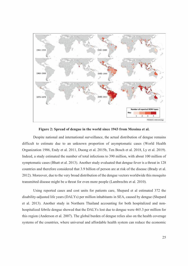
25
Figure 2: Spread of dengue in the world since 1943 from Messina et al.
Despite national and international surveillance, the actual distribution of dengue remains
difficult to estimate due to an unknown proportion of asymptomatic cases (World Health
Organization 1986, Endy et al. 2011, Duong et al. 2015b, Ten Bosch et al. 2018, Ly et al. 2019).
Indeed, a study estimated the number of total infections to 390 million, with about 100 million of
symptomatic cases (Bhatt et al. 2013). Another study evaluated that dengue fever is a threat in 128
countries and therefore considered that 3.9 billion of person are at risk of the disease (Brady et al.
2012). Moreover, due to the very broad distribution of the dengue vectors worldwide this mosquito
transmitted disease might be a threat for even more people (Lambrechts et al. 2010).
Using reported cases and cost units for patients care, Shepard et al estimated 372 the
disability-adjusted life years (DALYs) per million inhabitants in SEA, caused by dengue (Shepard
et al. 2013). Another study in Northern Thailand accounting for both hospitalized and non-
hospitalized febrile dengue showed that the DALYs lost due to dengue were 465.3 per million for
this region (Anderson et al. 2007). The global burden of dengue relies also on the health coverage
systems of the countries, where universal and affordable health system can reduce the economic

26
costs for those afflicted with dengue. For example, the global economic losses due to dengue have
been estimated to be at least US$ 9 billion per year (Bradshaw et al. 2016).
In most dengue endemic countries, cases occurred during all the year, yet the rainy season
is associated with local or wider epidemic episode. Dengue epidemiology is characterized by
seasonal peaks during the rainy season with major outbreaks every three to six years (van Panhuis
et al. 2015, Churakov et al. 2019). Dengue epidemiology is also characterized by pluri-annual
seasonal variations, with intra and inter-epidemic periods. Annual seasonality of dengue can be
related to climatic factors, vector abundance and individual factors. Larger epidemic episodes are
usually associated with changes in serotype distribution in a defined area. Global climatic changes
foresaw more and more people at risk for dengue with an increase of the temperature and changes
in rainfall patterns (Hales et al. 2002, Hii et al. 2012, Phaijoo et al. 2017). In addition, the
phenomenon known as the El Niño Southern Oscillation (ENSO) is suspected to increase dengue
transmission risk and to synchronize dengue outbreaks especially in SEA (Cummings et al. 2004,
Huang et al. 2015, van Panhuis et al. 2015, Vincenti-Gonzalez et al. 2018). In addition, climate
changes may contribute to extend the geographical distribution of both the mosquito vectors and
the viruses. Global warming can contribute to increase dengue transmission risk by enhancing viral
replication and by increasing the density, aggressiveness, survival, and reproduction rates of the
mosquito vectors (Fan et al. 2014, Samuel et al. 2016).
Another key factor explaining the global expansion of dengue and other Aedes-arboviral
diseases, is the increase of travels and exchange. Indeed, Ae. albopictus geographical expansion is
very well correlated to the circulation of goods, tires trade and human movements (Hawley et al.
1987, Paupy et al. 2009). As a result, many countries have faced a resurgence/emergence of dengue
cases due to a growing proportion of infected travellers returning home which can then facilitate
local and autochthonous disease transmission if the vector is present (Wilder-Smith 2012, Jentes
et al. 2016, Succo et al. 2016).
1.3. Viruses
The dengue virus is a positive sense single-stranded ribonucleic acid (RNA) of about 11
kbp, belonging to the genus Flavivirus, Flaviviridae family, to which other pathogens such as the
YFV, ZIKV, and Japanese encephalitis virus (JEV) also belong. The disease is caused by four
27
genotypic distinct serotypes (DENV 1-4), however a fifth serotype was recently reported (Mustafa
et al. 2015). Yet caution needs to be taken regarding this putative new serotype reported only in
Malaysia as it may be a variant of the DENV-4 (Joob et al. 2016). The four characterised DENV
serotypes share approximately 60% to 75% of the genome. The mature viral particle is about 50nm
diameter and contains several copies of three structural proteins, host-derived membrane bilayer
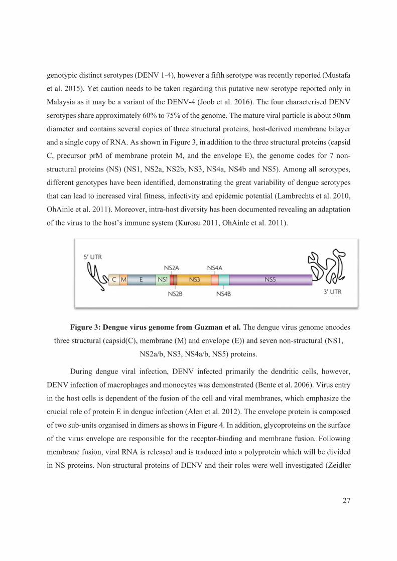
and a single copy of RNA. As shown in Figure 3, in addition to the three structural proteins (capsid
C, precursor prM of membrane protein M, and the envelope E), the genome codes for 7 non-
structural proteins (NS) (NS1, NS2a, NS2b, NS3, NS4a, NS4b and NS5). Among all serotypes,
different genotypes have been identified, demonstrating the great variability of dengue serotypes
that can lead to increased viral fitness, infectivity and epidemic potential (Lambrechts et al. 2010,
OhAinle et al. 2011). Moreover, intra-host diversity has been documented revealing an adaptation
of the virus to the host’s immune system (Kurosu 2011, OhAinle et al. 2011).
Figure 3: Dengue virus genome from Guzman et al. The dengue virus genome encodes
three structural (capsid(C), membrane (M) and envelope (E)) and seven non-structural (NS1,
NS2a/b, NS3, NS4a/b, NS5) proteins.
During dengue viral infection, DENV infected primarily the dendritic cells, however,
DENV infection of macrophages and monocytes was demonstrated (Bente et al. 2006). Virus entry
in the host cells is dependent of the fusion of the cell and viral membranes, which emphasize the
crucial role of protein E in dengue infection (Alen et al. 2012). The envelope protein is composed
of two sub-units organised in dimers as shows in Figure 4. In addition, glycoproteins on the surface
of the virus envelope are responsible for the receptor-binding and membrane fusion. Following
membrane fusion, viral RNA is released and is traduced into a polyprotein which will be divided
in NS proteins. Non-structural proteins of DENV and their roles were well investigated (Zeidler
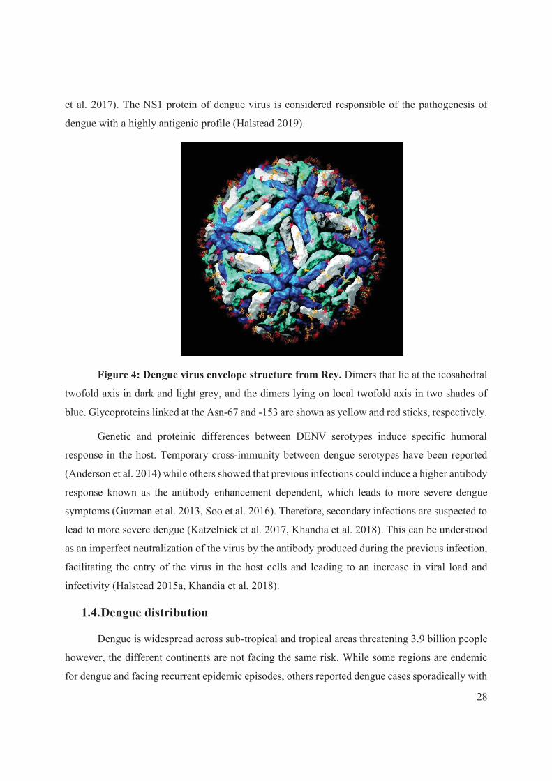
28
et al. 2017). The NS1 protein of dengue virus is considered responsible of the pathogenesis of
dengue with a highly antigenic profile (Halstead 2019).
Figure 4: Dengue virus envelope structure from Rey. Dimers that lie at the icosahedral
twofold axis in dark and light grey, and the dimers lying on local twofold axis in two shades of
blue. Glycoproteins linked at the Asn-67 and -153 are shown as yellow and red sticks, respectively.
Genetic and proteinic differences between DENV serotypes induce specific humoral
response in the host. Temporary cross-immunity between dengue serotypes have been reported
(Anderson et al. 2014) while others showed that previous infections could induce a higher antibody
response known as the antibody enhancement dependent, which leads to more severe dengue
symptoms (Guzman et al. 2013, Soo et al. 2016). Therefore, secondary infections are suspected to
lead to more severe dengue (Katzelnick et al. 2017, Khandia et al. 2018). This can be understood
as an imperfect neutralization of the virus by the antibody produced during the previous infection,
facilitating the entry of the virus in the host cells and leading to an increase in viral load and
infectivity (Halstead 2015a, Khandia et al. 2018).
1.4. Dengue distribution
Dengue is widespread across sub-tropical and tropical areas threatening 3.9 billion people
however, the different continents are not facing the same risk. While some regions are endemic
for dengue and facing recurrent epidemic episodes, others reported dengue cases sporadically with
29
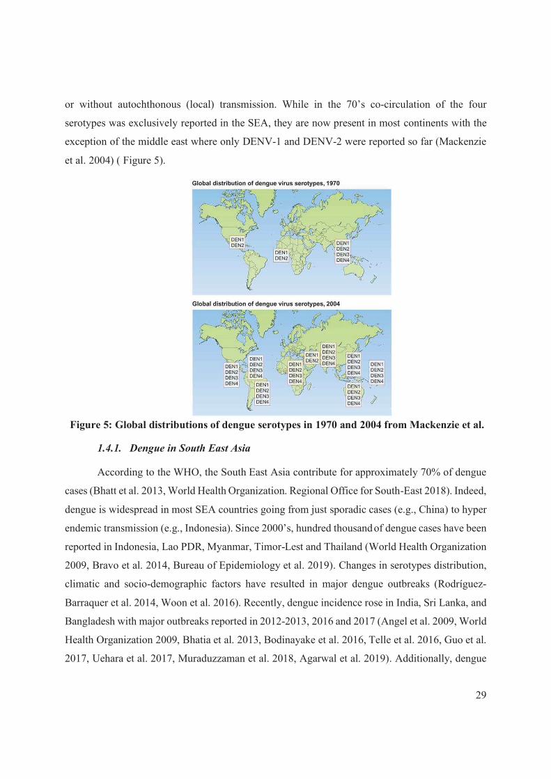
or without autochthonous (local) transmission. While in the 70’s co-circulation of the four
serotypes was exclusively reported in the SEA, they are now present in most continents with the
exception of the middle east where only DENV-1 and DENV-2 were reported so far (Mackenzie
et al. 2004) ( Figure 5).
Figure 5: Global distributions of dengue serotypes in 1970 and 2004 from Mackenzie et al.
1.4.1. Dengue in South East Asia
According to the WHO, the South East Asia contribute for approximately 70% of dengue
cases (Bhatt et al. 2013, World Health Organization. Regional Office for South-East 2018). Indeed,
dengue is widespread in most SEA countries going from just sporadic cases (e.g., China) to hyper
endemic transmission (e.g., Indonesia). Since 2000’s, hundred thousand of dengue cases have been
reported in Indonesia, Lao PDR, Myanmar, Timor-Lest and Thailand (World Health Organization
2009, Bravo et al. 2014, Bureau of Epidemiology et al. 2019). Changes in serotypes distribution,
climatic and socio-demographic factors have resulted in major dengue outbreaks (Rodríguez-
Barraquer et al. 2014, Woon et al. 2016). Recently, dengue incidence rose in India, Sri Lanka, and
Bangladesh with major outbreaks reported in 2012-2013, 2016 and 2017 (Angel et al. 2009, World
Health Organization 2009, Bhatia et al. 2013, Bodinayake et al. 2016, Telle et al. 2016, Guo et al.
2017, Uehara et al. 2017, Muraduzzaman et al. 2018, Agarwal et al. 2019). Additionally, dengue

30
re-emerged in Singapore after 35 years of effective control (Ooi et al. 2006, Bravo et al. 2014).
Dengue is also present in several provinces of China, including Yunnan, Guangdong, and Guangxi
(Zhang et al. 2014). Today, dengue was reported in all countries in the WHO South-East Asian
region except in North Korea hence highlighting the global trend of disease expansion worldwide.
1.4.2. Dengue in Western Pacific region
Cambodia, Vietnam, Malaysia, Philippines are the most affected countries by dengue in
the western pacific region. In addition, the dengue outbreak in 2008 in Cambodia indicated a rapid
change in dengue epidemiology, with more rural transmission than previously observed (Huy et
al. 2010). Moreover, dengue is spreading to the Pacific Islands such as Selangor (Malaysia), Fiji
and Vanuatu, due to the re-introduction of DENV-3 serotype which had been absent for a decade
(Getahun et al. 2019). Between 2008 and 2014, WHO reported a 2-fold increase in the number of
dengue cases in the region, however, with a lower the fatality rate compared to previous years
(Regional Committee for the Western 2016). Finally, dengue is also circulating sporadically in
Australia (Queensland), with both imported and autochthonous cases, due to the presence of the
very effective vector Ae. aegypti (Akter et al. 2019).
1.4.3. Dengue in Americas
For more than 30 years, dengue was absent from the Americas, as a result of the Ae. aegypti
eradication campaign using DDT (1970-1980) (van den Berg et al. 2012, Epelboin et al. 2018).
However, the discontinuation of vector control contributed to the re-invasion of Ae. aegypti in the
early 80’s (Guzman et al. 2003, Kotsakiozi et al. 2017). Following the vector (re) introduction
DENV started to re-circulate in America, invading more and more countries (Teixeira et al.
2009b). In 2013, a major outbreak occurred in Latin America causing more than 2 million cases,
including 38,000 severe dengue cases and 1,280 deaths (Pan America Health Organization 2020).
Brazil was the most afflicted country with about 1.5 million cases reported (Nunes et al. 2019).
Since then, recurrent dengue outbreaks occurred in this country causing about 1.6 and 3.1 million
cases in 2015 and 2019, respectively (Nunes et al. 2019, Pan America Health Organization 2020).
In the same time, Latin America was strongly also affected by other Aedes-borne diseases such as
Zika outbreak causing >5 million of cases, mainly in Brazil. More recently yellow fever outbreaks
were historically reported in Brazil (2000 human cases including 800 death during the 2016-2018)
and the country has taken necessary actions to vaccinate the populations and keep travellers
31
informed and vaccinated prior to traveling to those areas (Zanotto et al. 2018, Dorigatti et al. 2019).
Additionally, dengue is also circulating across the south of the USA and recent outbreaks occurred
in Hawaii in 2015-2016 (Johnston et al. 2020) emphasizing the threat of emerging/imported cases
and thus potential for local transmission and outbreaks where dengue is not endemic.
1.4.4. Dengue in Africa
Despite the presence of the native Aedes aegypti and the invasive Ae. albopictus, dengue
has not been considered as a major public health threat in Africa until recently (Amarasinghe et al.
2011, Stoler et al. 2014). Evidence of dengue circulation in West Africa was recently highlighted
by the abnormally high prevalence of dengue cases among returning travellers (Ninove et al. 2009,
Fourié et al. 2020). There is a growing evidence that a non-negligible portion of fever cases were
mis-diagnosed as malaria, while, dengue or other Aedes-borne diseases that shared the same
symptomology, had not been investigated (Stoler et al. 2014). According to recent prediction
models of incidence, Africa could share approximately 10-15% of the global dengue burden
(World Health Organization 2009, Bhatt et al. 2013, Jaenisch et al. 2014). A recent meta-analysis
on dengue seroprevalence and DENV presence demonstrated the high heterogeneity in dengue
transmission risk between countries (Simo et al. 2019). Indeed, more and more countries faced
dengue outbreaks such as Burkina-Faso (Ridde et al. 2016, Lim et al. 2019), Senegal (Faye et al.
2014, Gaye et al. 2019), Angola (Sessions et al. 2013, Hill et al. 2019), or Tanzania (Ward et al.
2017) and it’s of primary importance to pursue the monitoring and diagnostic of DENV to have a
more accurate estimate of dengue distribution and incidence in this part of the world.
1.4.5. Dengue in the Mediterranean region
Eastern Mediterranean region is facing regular dengue outbreaks such as Saudi Arabia,
Yemen, Pakistan and Sudan (WHO/EMRO 2005, Ali et al. 2016, Ducheyne et al. 2018). Dengue
in Western European region is sporadic, mainly due to imported cases from individuals traveling
back from endemic countries (World Health Organization 2009). However, the introduction and
establishment of the dengue vector Ae. albopictus in Europe and the Mediterranean basin set up
favourable conditions for local transmission (Succo et al. 2016). For example, the chikungunya
outbreak in Italy in 2007 and 2017, or several episodes of locally acquired dengue and chikungunya
in France, Croatia and Spain, raise the possibility for the establishment of these pathogens in
Europe (Calzolari 2016, Rezza 2016, Matusali et al. 2020). The increasing incidence of such

32
episodes demonstrates that Europe is not immune to mosquito-borne diseases, and that the
continent is increasingly exposed to the threat of (re-) emerging pathogens. In addition, the
presence of Ae. aegypti in Madeira Islands (Portugal) following its introduction in 2005, has led
to recurrent epidemics that have affected thousands of people (Schaffner et al. 2014, Wilder-Smith
A. 2014). Predictive models based on climatic conditions and urban growth suggest that Ae.
aegypti is likely to establish in specific isolated regions in Europe such as southern Italy and
Turkey, and may then contribute to the transmission in the future.
1.5. Symptoms
Dengue infection exhibits a broad range of symptoms, from no clinical symptoms or mild-
fever, to severe haemorrhage or even deaths, which make the diagnosis difficult. Overall, the case-
fatality rate of dengue fever is relatively low (»1%) despite a possible increase during outbreaks
due to the public health structures overwhelming.
Prior 2009, dengue fever disease was classified into 3 categories: dengue fever (DF),
dengue haemorrhagic fever (DHF) and dengue shock syndrome (DSS). In 2009, WHO re-
evaluated the dengue classification to dengue with or without warning signs and severe dengue
(Figure 6).
As shown in Figure 6, symptoms of dengue infection range from mild to acute fever, rashes,
nausea, retro-orbital pain, arthralgia, leukopenia and positive tourniquet test. Some warning signs,
which necessitate medical attention include rapid increased in haematocrit combined with a
significant decrease in platelet count, mucosal bleeding, persistent vomiting, abdominal
tenderness, or lethargy. Dengue infection, in some cases can evolve to more severe symptoms and
to severe plasma leakage which might result in organ impairment and/or haemorrhagic syndrome
(Halstead 2015b). Moreover, weakness and fatigue can persist for weeks which may increase
dengue overall burden (Seet et al. 2007, Umakanth 2017).
33
Figure 6: Dengue cases classification and level of severity, from WHO.
1.6. Diagnostic
To prevent dengue outbreaks, it is mandatory to detect cases early and accurately. In most
dengue-endemic countries, dengue diagnosis is based on the presence of symptoms as described
previously (see section 1.5). However, those symptoms can be encountered in many viral diseases
and are not specific to dengue fever. In order to diagnose patients accurately, several techniques
were developed. Due to the course of dengue illness and DENV viremia (Figure 7), direct
diagnostic of dengue can be performed within the first days of illness (World Health Organization
2009). For late stages of dengue illness, indirect diagnosis will be preferred, using serological
tools.
34
Figure 7: Dengue illness course from WHO.
1.6.1. Epidemiological diagnostic
Differential diagnosis, based on the clinical symptoms and laboratory analysis, is the first
step for dengue diagnostic. During dengue illness course, thrombocytopenia, plasma leakage, joint
pain and fever are typical. Any patients presenting at least two dengue symptoms (see section 1.4)
are eligible for laboratory confirmation of dengue infection (World Health Organization 2009)
(Figure 6).
1.6.2. Laboratory diagnostic
1.6.2.1. Direct diagnosis
Direct diagnostic of dengue, which can only be performed at early stage of disease, rely on
virus detection, viral RNA detection or antigen detection. Virus detection is historically based on
virus isolation by cell culture. Briefly, patient sera are incubated on susceptible cell lines, such as
the C6/36 cell line from Ae. albopictus mosquitoes or Vero cells (from green monkey kidney cells),
35
and maintained for few days (Medina et al. 2012). This method, being highly specific, is the gold
standard for dengue laboratory confirmation. However, virus isolation can only be performed at
early stage of illness, and take time to get the result (usually between 3 to 10 days). Therefore,
virus isolation is not the preferred method in case of emergency situation.
An alternative method had appeared in the 1990’s with the development of reverse-
transcription Polymerase Chain Reaction (RT-PCR), which allow the detection of DENV RNA in
serum samples (Lanciotti et al. 1992). Recent progresses were made allowing a quicker and easier
protocol for real-time DENV detection and serotype identification (Shu et al. 2003, Johnson et al.
2005, Chen et al. 2010). Serotype identification is not mandatory for patient care however, it can
be useful for epidemiological surveillance purposes, such as changes in serotypes prevalence that
can trigger outbreak.
The last method for a direct diagnostic of dengue is the detection of the non-structural
protein-1 (NS1) of the DENV. The NS1 protein is produced by mammalian cells infected by
DENV and induce a strong immune response. NS1 detection can be performed by ELISA or
immunochromatography (ICT). The principle is to detect antigen-antibody complexes from patient
sera. Since the first commercialization of kit for DENV NS1 detection by ELISA in 2006, several
companies had developed their own tests, yet with variable specificities and sensibilities. The
development of Rapid Diagnostic Test (RDT), based on the ICT of NS1 antigen, allowed to reduce
the time needed for DENV diagnostic with a result in 5-15 min (Figure 8). Nowadays, RDTs
targeting the NS1 antigen are strongly recommended by the WHO to guide dengue diagnosis
(Teixeira et al. 2009a, World Health Organization 2009). However, a negative result of the NS1
detection is not sufficient to exclude dengue fever as the presence of NS1 protein, usually, does
not last more than few days after the apparition of the symptoms. It is noteworthy to emphasize on
the small window when direct diagnostic can be accurately used.
Figure 8: NS1 positive Rapid Diagnostic Test.

36
1.6.2.2. Indirect diagnosis
As a consequence of the broad symptoms and the difficulty to diagnose directly dengue
infection, indirect diagnostic tools are commonly used in practice. One of the indirect diagnosis of
dengue is based on the detection of specific antibody response against DENV (Salje et al. 2018).
According to the course of dengue illness (Figure 7) and the time-line of antibody production
(Figure 9), humoral response can be separated into two phases: the mid-early response with the
production of IgM against dengue virus within few days after viral infection, and the later stage
with the production of IgG, which confers the durable immunity against a given serotype.
Therefore, the detection of IgM or IgG against DENV from blood or serum samples, and the
comparison with the previous levels of immune response can provide information on
seroconversion. Additionally, the IgM/IgG detection can provide information on the number of
infection (i.e. primary or secondary infection). Indeed, the concomitantly presence of both type of
immunoglobulin indicates secondary infections which may lead to more severe symptoms (Figure
9). There are several assays to detect immunoglobulins related to dengue infection (De Paula et al.
2004).
Figure 9: Time-line of immunoglobulins in primary and secondary dengue infection from WHO.
1.6.2.2.1. Plaque reduction neutralisation test
The plaque reduction neutralisation test (PRNT) has been developed to measure changes
in the titters of neutralizing immunoglobulin against dengue virus (Peeling et al. 2010). The
37
principle of PRNT is to allow virus-antibody interactions and to measure the efficiency of
antibodies to neutralize the virus (plaques). Briefly, virus-susceptible cells are cultured in a semi-
solid media to avoid dispersal of virus progeny. Patients sera are incubated at various dilution prior
mixing with constant amount of virus in order to maximize observation of plaques (local infection)
which can be detected in various ways such as direct coloration of the cells (e.g., using neutral red
or crystal violet) (World Health Organization 2007a, Timiryasova et al. 2013), or staining by using
DENV-reactive antibodies (Roehrig et al. 2008). This assay is the gold standard to assess the level
of neutralizing antibodies against the different DENV serotypes, however, PRNT is labour-
intensive (e.g., approximately 5 to 7 days are required for plaques formation), requires BSL-2
laboratory facilities, and qualified staff for cell cultures and virus manipulation and cannot be
performed in early dengue illness (World Health Organization 2009).
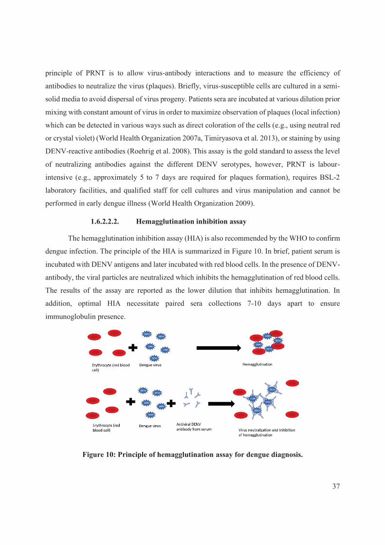
1.6.2.2.2. Hemagglutination inhibition assay
The hemagglutination inhibition assay (HIA) is also recommended by the WHO to confirm
dengue infection. The principle of the HIA is summarized in Figure 10. In brief, patient serum is
incubated with DENV antigens and later incubated with red blood cells. In the presence of DENV-
antibody, the viral particles are neutralized which inhibits the hemagglutination of red blood cells.
The results of the assay are reported as the lower dilution that inhibits hemagglutination. In
addition, optimal HIA necessitate paired sera collections 7-10 days apart to ensure
immunoglobulin presence.
Figure 10: Principle of hemagglutination assay for dengue diagnosis.
38
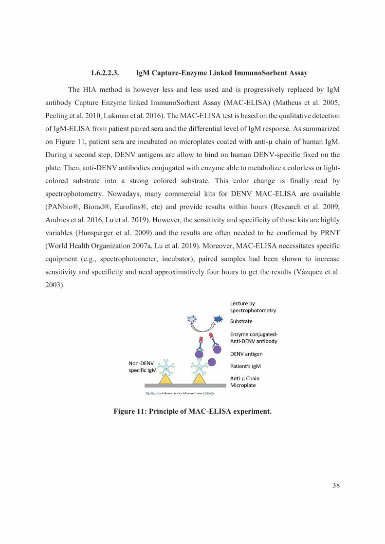
1.6.2.2.3. IgM Capture-Enzyme Linked ImmunoSorbent Assay
The HIA method is however less and less used and is progressively replaced by IgM
antibody Capture Enzyme linked ImmunoSorbent Assay (MAC-ELISA) (Matheus et al. 2005,
Peeling et al. 2010, Lukman et al. 2016). The MAC-ELISA test is based on the qualitative detection
of IgM-ELISA from patient paired sera and the differential level of IgM response. As summarized
on Figure 11, patient sera are incubated on microplates coated with anti-µ chain of human IgM.
During a second step, DENV antigens are allow to bind on human DENV-specific fixed on the
plate. Then, anti-DENV antibodies conjugated with enzyme able to metabolize a colorless or light-
colored substrate into a strong colored substrate. This color change is finally read by
spectrophotometry. Nowadays, many commercial kits for DENV MAC-ELISA are available
(PANbio®, Biorad®, Eurofins®, etc) and provide results within hours (Research et al. 2009,
Andries et al. 2016, Lu et al. 2019). However, the sensitivity and specificity of those kits are highly
variables (Hunsperger et al. 2009) and the results are often needed to be confirmed by PRNT
(World Health Organization 2007a, Lu et al. 2019). Moreover, MAC-ELISA necessitates specific
equipment (e.g., spectrophotometer, incubator), paired samples had been shown to increase
sensitivity and specificity and need approximatively four hours to get the results (Vázquez et al.
2003).
Figure 11: Principle of MAC-ELISA experiment.

39
1.6.2.2.4. Rapid Diagnostic Test
Finally, Rapid Diagnostic Test (RDT) targeting IgM and/or IgG were developed for dengue
diagnostic. The principle of RDT is the migration of sample on a membrane and the detection of
the target by immunochromatography test (ICT). Briefly, blood or sera sample are deposed into
the cassette, then a specific buffer is added to allow migration of sample for few minutes
(depending on the manufacturer’s instruction). The results of the RDT are given in the form band
indicting the presence of the target antigen and a control band indicating the validity of the test
(Figure 12). The detection of IgM and IgG using RDT test allow the health officers to obtain results
within few minutes (usually 5-30 min). However, those tests had been criticized regarding their
lack of sensitivity and specificity (Hunsperger et al. 2009, Jang et al. 2019). Indeed, studies had
risen the potential high rate of false negatives from RDT IgM/IgG, due either to the time course
of dengue illness, or to the high antibody titers needed to trigger a visible band. Moreover, others
had pointed the cross-reactivity of RDT IgM/IgG, with other Aedes -borne transmitted viruses,
especially with CHIKV or ZIKV (Blacksell et al. 2011).
Figure 12: IgG positive and IgM negative RDT
2. The dengue vectors
Aedes aegypti (Linnaeus) (Diptera: Culicidae) and Ae. albopictus (Skuse) (Diptera:
Culicidae) are mosquito species that can transmit several viruses to humans that cause diseases,
such as dengue, Zika, chikungunya, and yellow fever. Over the last few decades, those diseases
have spread rapidly partly due to the global expansion of the vectors. The distribution of Aedes
mosquitoes is the widest ever recorded in history (Kraemer et al. 2015, Kraemer et al. 2019), and
further research are need to better understand the causes and consequences of this rapid
geographical expansion in order to propose more effective, durable and locally-adapted tools for
vector control. In the following sections, I will describe current knowledge on Aedes vector
40
biology and ecology and provide new insight into the spatial distribution of Aedes aegypti and Ae.
albopictus worldwide.
2.1. Life cycle
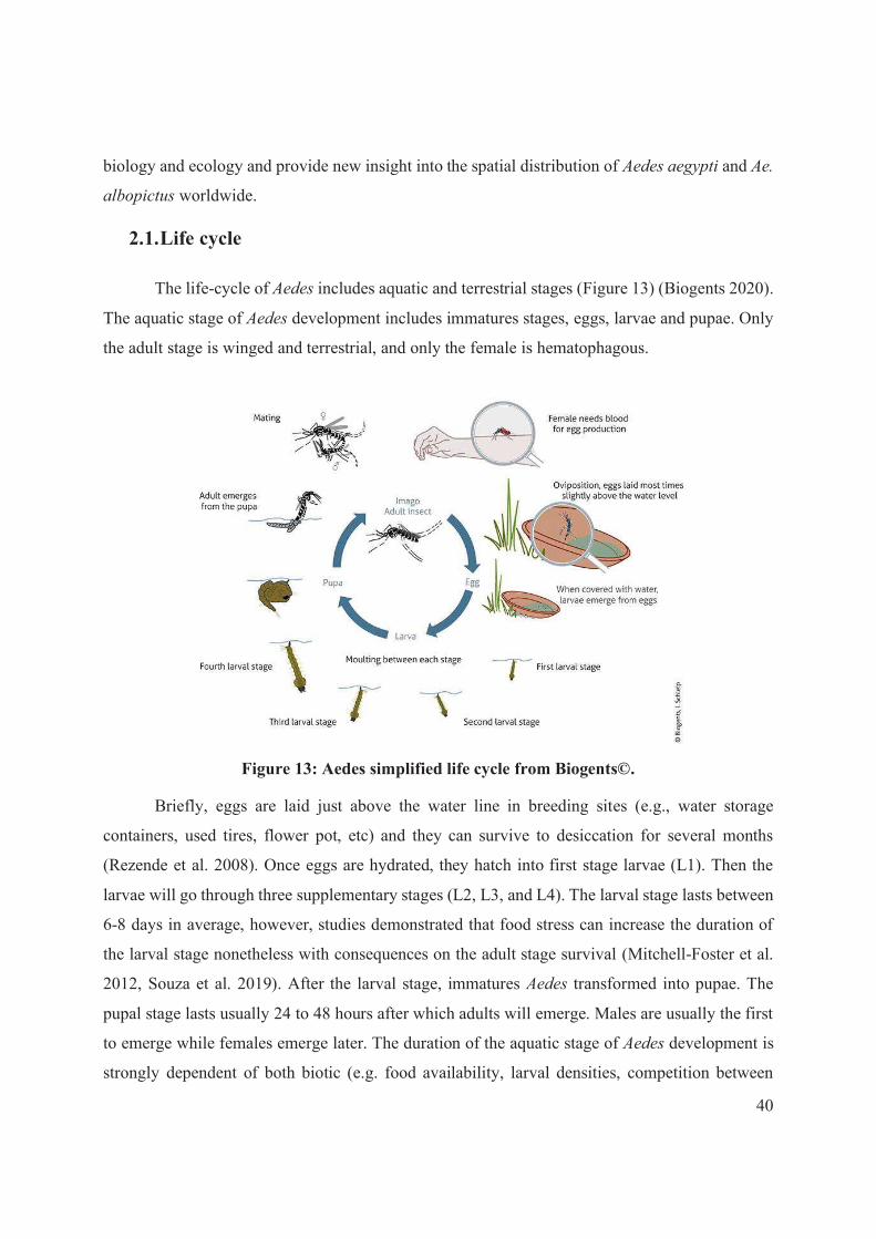
The life-cycle of Aedes includes aquatic and terrestrial stages (Figure 13) (Biogents 2020).
The aquatic stage of Aedes development includes immatures stages, eggs, larvae and pupae. Only
the adult stage is winged and terrestrial, and only the female is hematophagous.
Figure 13: Aedes simplified life cycle from Biogents©.
Briefly, eggs are laid just above the water line in breeding sites (e.g., water storage
containers, used tires, flower pot, etc) and they can survive to desiccation for several months
(Rezende et al. 2008). Once eggs are hydrated, they hatch into first stage larvae (L1). Then the
larvae will go through three supplementary stages (L2, L3, and L4). The larval stage lasts between
6-8 days in average, however, studies demonstrated that food stress can increase the duration of
the larval stage nonetheless with consequences on the adult stage survival (Mitchell-Foster et al.
2012, Souza et al. 2019). After the larval stage, immatures Aedes transformed into pupae. The
pupal stage lasts usually 24 to 48 hours after which adults will emerge. Males are usually the first
to emerge while females emerge later. The duration of the aquatic stage of Aedes development is
strongly dependent of both biotic (e.g. food availability, larval densities, competition between

41
species, predation) and abiotic factors such as the rainfall, the relative humidity and the
temperature. Indeed, increased in mean temperature was related to a reduce development time
(Scott et al. 2000b, Tun-Lin et al. 2000, Couret et al. 2014).
After emergence, adult mosquitoes will rest in shade places for 24 to 48 hours, in order to
dry their cuticle, spread their wings and wait for their reproductive system to be functional. The
male reproductive system needs in average 24 to 48 hours to be functional while it takes
approximately 30-60 hours for the female reproductive system. Then, adult mosquitoes (males and
females) will take their first sugar-meal, from flower nectar, which will be the only food source
for male mosquitoes. Only the female needs to take a blood meal, rich in protein, for the egg
maturation (Day et al. 1994, Styer et al. 2007). The mating occurs during flight and females usually
mate only once shortly after emergence however, polyandry (i.e., mating with several males) was
demonstrated in semi-field experiments (Helinski et al. 2012). Then the gonotrophic cycle starts
with the host-seeking behaviour which is strongly related to anthropogenic environment, and ends
with the oviposition. After blood meal, Aedes usually rests in shaded areas to complete eggs
maturation. Depending on the amount of blood ingested, females Aedes will seek another host
and/or will rest to digest the blood and mature the eggs. In average, Aedes produces around 100
eggs per clutch and about 4 to 5 batches in their life (Chadee et al. 2002, CDC 2020). Both Ae.
aegypti and Ae. albopictus have a small flight range, then it follows that adults often stay close to
their emerging site depending on the availability of breeding sites and (human) host to provide
blood meal.
2.2. Aedes vectors
Although there are several “potential” Aedes dengue vectors, the field isolation of viruses
and epidemiological evidence clearly show that Ae. aegypti (Figure 14) is responsible for the
majority of dengue transmission (Gubler et al. 1997). The intrinsic ability of an arthropod to carry
pathogens, ensure their multiplication and or development and to transmit the pathogens to a
vertebrate host is defined as the vector competence. The vectorial capacity, which is the level of
efficacy of the vector to transmit a pathogen, is highly dependent on abiotic factors such as the
temperature, but also intrinsic characteristics of vector, virus and hosts (Liu-Helmersson et al.
2014). Aedes aegypti is the main vector of dengue due to its wide distribution, high vector
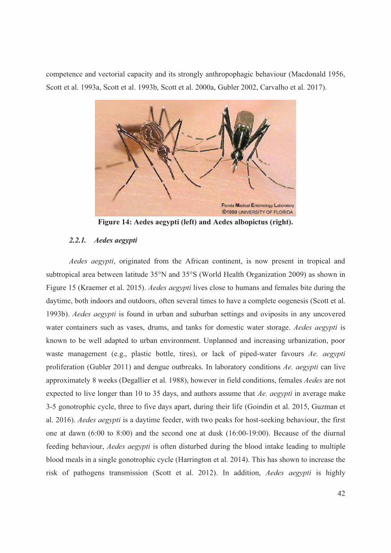
42
competence and vectorial capacity and its strongly anthropophagic behaviour (Macdonald 1956,
Scott et al. 1993a, Scott et al. 1993b, Scott et al. 2000a, Gubler 2002, Carvalho et al. 2017).
Figure 14: Aedes aegypti (left) and Aedes albopictus (right).
2.2.1. Aedes aegypti
Aedes aegypti, originated from the African continent, is now present in tropical and
subtropical area between latitude 35°N and 35°S (World Health Organization 2009) as shown in
Figure 15 (Kraemer et al. 2015). Aedes aegypti lives close to humans and females bite during the
daytime, both indoors and outdoors, often several times to have a complete oogenesis (Scott et al.
1993b). Aedes aegypti is found in urban and suburban settings and oviposits in any uncovered
water containers such as vases, drums, and tanks for domestic water storage. Aedes aegypti is
known to be well adapted to urban environment. Unplanned and increasing urbanization, poor
waste management (e.g., plastic bottle, tires), or lack of piped-water favours Ae. aegypti
proliferation (Gubler 2011) and dengue outbreaks. In laboratory conditions Ae. aegypti can live
approximately 8 weeks (Degallier et al. 1988), however in field conditions, females Aedes are not
expected to live longer than 10 to 35 days, and authors assume that Ae. aegypti in average make
3-5 gonotrophic cycle, three to five days apart, during their life (Goindin et al. 2015, Guzman et
al. 2016). Aedes aegypti is a daytime feeder, with two peaks for host-seeking behaviour, the first
one at dawn (6:00 to 8:00) and the second one at dusk (16:00-19:00). Because of the diurnal
feeding behaviour, Aedes aegypti is often disturbed during the blood intake leading to multiple
blood meals in a single gonotrophic cycle (Harrington et al. 2014). This has shown to increase the
risk of pathogens transmission (Scott et al. 2012). In addition, Aedes aegypti is highly
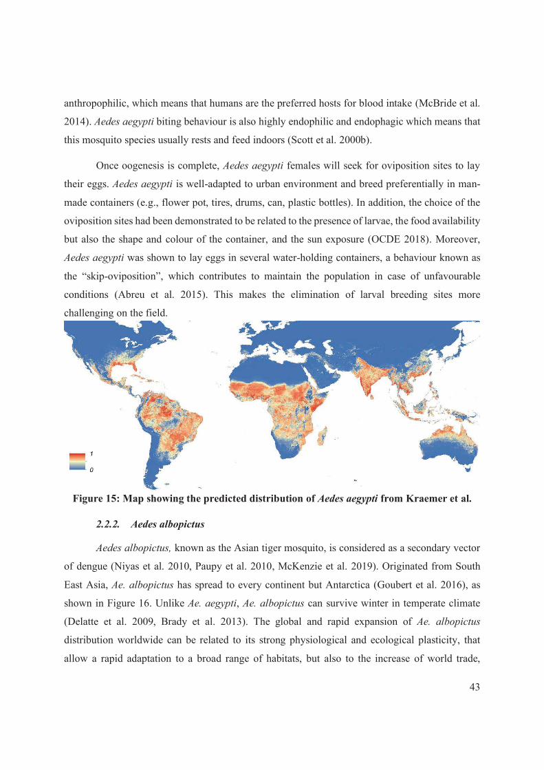
43
anthropophilic, which means that humans are the preferred hosts for blood intake (McBride et al.
2014). Aedes aegypti biting behaviour is also highly endophilic and endophagic which means that
this mosquito species usually rests and feed indoors (Scott et al. 2000b).
Once oogenesis is complete, Aedes aegypti females will seek for oviposition sites to lay
their eggs. Aedes aegypti is well-adapted to urban environment and breed preferentially in man-
made containers (e.g., flower pot, tires, drums, can, plastic bottles). In addition, the choice of the
oviposition sites had been demonstrated to be related to the presence of larvae, the food availability
but also the shape and colour of the container, and the sun exposure (OCDE 2018). Moreover,
Aedes aegypti was shown to lay eggs in several water-holding containers, a behaviour known as
the “skip-oviposition”, which contributes to maintain the population in case of unfavourable
conditions (Abreu et al. 2015). This makes the elimination of larval breeding sites more
challenging on the field.
Figure 15: Map showing the predicted distribution of Aedes aegypti from Kraemer et al.
2.2.2. Aedes albopictus
Aedes albopictus, known as the Asian tiger mosquito, is considered as a secondary vector
of dengue (Niyas et al. 2010, Paupy et al. 2010, McKenzie et al. 2019). Originated from South
East Asia, Ae. albopictus has spread to every continent but Antarctica (Goubert et al. 2016), as
shown in Figure 16. Unlike Ae. aegypti, Ae. albopictus can survive winter in temperate climate
(Delatte et al. 2009, Brady et al. 2013). The global and rapid expansion of Ae. albopictus
distribution worldwide can be related to its strong physiological and ecological plasticity, that
allow a rapid adaptation to a broad range of habitats, but also to the increase of world trade,

44
especially related to the transport of used tires (Hawley et al. 1987). While Ae. albopictus was
originally found in rural areas, the species recently adapted to suburban and urban areas (Paupy et
al. 2009, Aida et al. 2011). Aedes. albopictus is an aggressive, opportunistic daytime feeder, that
can bite humans and animals for blood meals (Kek et al. 2014). However, many studies
demonstrated a preference for human hosts hence highlighting a strong anthropophilic behaviour
of this species (Delatte et al. 2008, Paupy et al. 2009, Benelli et al. 2020). Aedes albopictus bites
several times for complete oogenesis. In contrast to Ae. aegypti, Ae. albopictus is more often
observed outdoors, either for host-seeking (exophagic behaviour) or for resting (exophilic
behaviour) (Delatte et al. 2010). Aedes albopictus dwells in mostly in rural and suburban areas,
lay eggs in water-filled artificial containers as used tires, flower pots, cans, but also in natural
containers such as tree holes, or axil of orchid leaves, challenging breeding sites elimination
(Paupy et al. 2010, World Health Organization 2011a).
Some studies showed that Ae. albopictus was as competent as Ae. aegypti for DENV-1 and
DENV-3 but less competent for DENV-2 and DENV-4 (Christofferson 2015, Whitehorn et al.
2015). Moreover, Ae. albopictus can play a role in dengue transmission where Ae. aegypti is absent
(e.g., La Reunion Island) (Delatte et al. 2008). Additionally, Ae. albopictus is a very competent
vector for CHIKV and is considered as the primary vector of the disease (Paupy et al. 2009). Aedes
albopictus is also competent for ZIKV (McKenzie et al. 2019). The global spread of Ae. albopictus
worldwide and the increase of international travels foresaw an increasing part of the world’s
population at risk for Aedes-vector-borne diseases.
Figure 16: Map showing the predicted distribution of Aedes albopictus from Kraemer et al.

45
3. Global strategies for dengue prevention and control
The control of mosquito-borne diseases generally relies on four pillars: vaccination,
chemo-prophylaxis, chemo-therapy and vector control. For dengue however, vaccination is limited
(see section 3.1), and there is no preventive nor curative treatment (see section 3.2). Consequently,
preventing or reducing dengue and other arboviral diseases caused by currently recognised or
novel Aedes-borne viruses on a global scale continues to depend largely on controlling mosquito
vector populations or interrupting human–vector contact (see section 3.3). A brief description of
each strategy will be presented in the following sections.
3.1. Vaccine
The example of the YFV vaccine for disease control has shown the efficacy of this strategy,
when deployed accurately, for Aedes-borne diseases. However, dengue vaccine development is far
more complicated due to the complex and indeterminate immunoprotective and/or
immunopathogenic response between the four serotypes (McArthur et al. 2013). Several vaccines
for dengue are under clinical development (Yauch et al. 2014) (Table 1) but the Dengvaxia®, a
tetravalent vaccine developed by Sanofi, has been licensed recently in more than ten dengue
endemic countries including Brazil, the Philippines, Costa Rica, and Mexico. It has shown
promising results in phase III of clinical trials (Capeding et al. 2014), but its efficacy was lower
for serotype 1 (50.2%) and 2 (39.6%) than for serotype 3 (74.9%) and 4 (76.6%) (World Health
Organization 2016a). After consultations, the manufacturer (Sanofi-Pasteur) and the WHO-
Strategic Advisory Group of Experts on Immunization (SAGE) had warned on increased
prevalence of severe cases in vaccinated children who never experienced dengue (World Health
Organization 2016c, World Health Organization 2018). Therefore, this vaccine is recommended
only for dengue endemic regions with high transmission and where seroprevalence is >50% to
limit the risk to develop severe dengue in vaccinated-seronegative individuals (Flasche et al. 2019).
Consequently, Sanofi does not recommend the use of Dengvaxia® in children <9 years old and in
individuals who have not been previously infected with dengue. Despite that, the government of
Philippines has recently suspended the distribution of dengue vaccine due to suspicious deaths
(Flasche et al. 2019). The mitigate results obtained with the Dengvaxia®, might had reduced the
enthusiasm for further vaccines development. However, vaccination should be considered as part

46
of global strategy including vector control and chemo-therapy and chemo-prophylaxis as
developed in the following sections.
Table 1: Dengue vaccines under development from (Yauch and Shresta 2014)
Vaccine name Vaccine type Developer/ manufacturer
Progress
Dengvaxia (CYD-TDV)
Live attenuated vaccine
Sanofi Pasteur Licensed
TAK-003 (DENVax)
Live attenuated vaccine + chimeric DENV2/DENV4
Mahidol university, Inviragen and Takeda
Phase III trial
TetraVax-DV TV003
Live attenuated vaccine
NIAID and Butantan Phase III trial
LATV Tetravalent, Live attenuated vaccine + chimeric
NIAID and Butantan In vivo (Phase I-III)
DPIV Inactivated virus tetravalent
GSK, Walter Reed Army Institute of Research, Fiocruz
In vivo (Phase I trial)
V180 Subunit vaccine Merck In vivo (Phase I trial)
TVDV DNA vaccine US AMRDC, WRAIR, NMRC Vical inc
Phase I trial
TLAV Prime/PIV boost
Heterologous prime/boost and reverse order Phase I trial
3.2. Treatment & prophylaxis
Currently, there is no curative treatment for arboviral diseases. Patient care mainly aimed
at reducing the symptoms by prescribing antipyretics (i.e., paracetamol), anti-nausea and pain
killers. It is noteworthy to remind that nonsteroidal anti-inflammatory drugs (e.g., ibuprofen),
aspirin and/or other salicylates should not be prescribed in case of suspected dengue fever because
of their blood thinning effect leading to an increased risk of developing haemorrhage. In addition,
patients presenting dengue cases with warning signs or severe dengue usually need fluid therapy
(oral or intravenous) and/or blood or plasma transfusion to prevent severe organ hypoperfusion
(World Health Organization 2009). Nevertheless, some drugs, already licensed for other diseases,
are explored in dengue treatment, yet they failed in addressing strong clinical endpoints (Low et
al. 2017). The absence of specific treatment against dengue emphasized the need for more effective
strategies of prevention and control to prevent dengue outbreaks.

47
3.3. Dengue vector control
Considering the challenges mentioned above, dengue control and prevention depends
essentially on controlling the vector mosquito and reducing human-vector contact (Guzman and
Kouri 2003). Historically, vector control was initiated following the discovery of the implication
of mosquitoes in pathogens transmission. In the United States, three strategies were successively
used to control mosquito populations. The first strategy was based on mechanical control and was
used between 1920-1940. The discovery of DDT opened a new path in vector control, and the
chemical control strategy was used during the 1940-1970 period (Patterson 2016). The strong
organization of the DDT campaigns, enabled the eradication of several diseases including malaria
in Western Europe, dengue and Yellow fever in America. However, the acknowledgement of the
harmful and carcinogenic effects of DDT led to its ban in the USA for vector control measure in
the 1970’s. Since then, USA and other countries have adopted integrated vector control strategies
to maintain efficient and sustainable vector control intervention using the available tools. The
GVCR recently endorsed by member states emphasizes on 4 pillars of action with 2 crucial
elements: the reinforcement of vector surveillance and control capacities and capabilities; and the
increase of fundamental and applied research and innovation in order to optimize vector control
(Figure 17). The GVCR also underlines the critical factors necessary to achieve these objectives,
such as country leadership, resource mobilization and coordination across sectors and diseases.
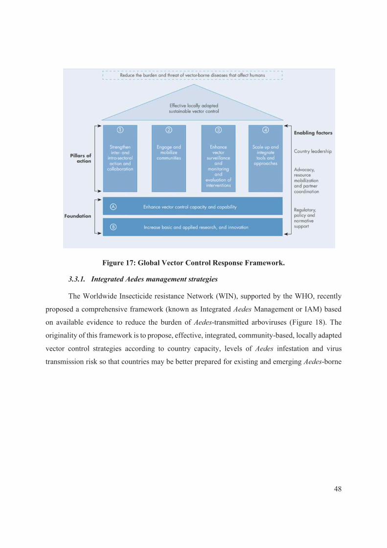
48
Figure 17: Global Vector Control Response Framework.
3.3.1. Integrated Aedes management strategies
The Worldwide Insecticide resistance Network (WIN), supported by the WHO, recently
proposed a comprehensive framework (known as Integrated Aedes Management or IAM) based
on available evidence to reduce the burden of Aedes-transmitted arboviruses (Figure 18). The
originality of this framework is to propose, effective, integrated, community-based, locally adapted
vector control strategies according to country capacity, levels of Aedes infestation and virus
transmission risk so that countries may be better prepared for existing and emerging Aedes-borne

49
disease threats. A brief review of the vector control tools proposed in IAM framework, with their
strength and weakness, will be presented in the following sections and in the Table 2 and Table 3.
Figure 18: Conceptual framework of the IAM system from Roiz et al.
3.3.1.1. Social mobilisation and community engagement
According to WHO, achieving sustainable vector control without community involvement
might not be feasible (World Health Organization 2011a, Roiz et al. 2018). Community control is
based on educational programs, social mobilization, and government policies. At the state or
regional level, that imply educational programs and health communications (Roiz et al. 2018).
Increasing knowledge on dengue prevention is mandatory to achieve this goal. Indeed, most the
studies conducted worldwide on the knowledge, attitudes and practices (KAP) regarding dengue
infection risk, showed that communities are quite aware of dengue symptoms yet, the relations
between vector control, human behaviours and dengue risk remain poor (Brusich et al. 2015,
Alyousefi et al. 2016, Kumaran et al. 2018). Inspiring behavioural changes through education or
Communication for Behavioural Impact (COMBI) activities (Andersson et al. 2017), regarding
self-implication in vector control and dengue prevention, is crucial to ensure sustainable vector
control (Kumaran et al. 2018, Roiz et al. 2018).

Table 2: Strengths and limitations of larval control strategies from Roiz et al.
Stage/ scenario
Methodology Type of intervention/
product
Strength of evidence* Constraints/advantages Specifications References
Larval control
for routine
Environmental management
Source reduction and educational outreach visits (door-to-door)
Epidemiological evidence (level 1) of community- based campaigns. Entomological evidence (level 3a and 3b).
Labour intensive. Larval development habitats need to be accurately identified. Must be done diligently and conscientiously and with access to a high number of dwellings
Requires a high level of education and community participation. Difficult to sustain over time. Need to characterize larval development habitats, including urban cryptic habitats. Essential to reduce mosquito larval development habitats in the long-term in private and public domains
(Heintze et al. 2007, Erlanger et al. 2008, Bartlett-Healy et al. 2011, Andersson et al. 2015, Bouzid et al. 2016, Bowman et al. 2016, Alvarado-Castro et al. 2017 )
Larviciding Organophosphates (Temephos, Chlorpyrifos, Pirimephos methyl, Fenthion)
Entomological evidence for Temephos (level 3b).
Affordable Not acceptable for treating drinking water containers and sources (except Temephos) Temephos resistance in several areas Regulatory constraints (e.g., OPs are not notified in the EU for mosquito control)
Cholinesterase inhibitors Different formulations (EC, GR) and application methods (manual or with hand sprayers)
(George et al. 2015, Alvarado-Castro et al. 2017)
Insect growth regulators (pyriproxyfen, diflubenzuron, novaluron)
Epidemiological evidence for pyriproxyfen as part of community base (level 2b). Entomological evidence (level 3b).
More expensive Late acting effect (pupae) on juvenoids Acceptable for treating drinking water sources and containers Constraints for the treatment of cryptic breeding sites
Disruption of endocrine system for juvenoids (pyriproxyfen) and chitin synthesis inhibitor for ecdysoids (novaluron and diflubenzuron). Different formulations (WG, GR, DT) and application methods (manual or with hand sprayers)
(Bowman et al. 2016, Maoz et al. 2017)
Bti Entomological evidence (level 3a and 3b) for Bti.
No resistance Selective and safe Acceptable for treating drinking water sources and containers Low residual action in polluted habitats
Bacterial toxins targeting midgut epithelium cells Different formulations (WG, GR) and application methods (manual or with hand sprayers and fogging).
(Boyce et al. 2013, Faraji et al. 2016)
Biological control
Fish (Gambusia, etc.)
Limited entomological evidence (level 3b) for fish.
Well accepted in several countries, needs a delivery mechanism and maintenance. Adequate for treating large and/or permanent mosquito habitats, not generally accepted for drinking water storage containers.
Predators of mosquito larvae (kill all stages). Controversial, harmful impacts of nonnative species, such as Gambusia.
(Kay et al. 2005, Han et al. 2015, Lazaro et al. 2015, Benelli et al. 2016, Faraji and Unlu 2016, Alvarado-Castro et al. 2017, Azevedo-Santos et al. 2017)
Copepods (Mesocyclops)
Limited epidemiological (level 2b) and entomological evidence (level 3b) for copepods depending on settings.
Predators of mosquito larvae (kill young instar larvae).

3.3.1.2. Larval control
Targeting the immature stages, has been the standard method to prevent adult Aedes
emergence thus, to reduce adult density. The methods used for larval control had been described
with regards to epidemiological outcomes by Roiz et al. as shown in Table 2. Yet, larval
management should be considered as a part of a global strategy to improve its effectiveness and
sustainability. Moreover, larval control must be carried routinely to achieve effective dengue
vector control.
3.3.1.2.1. Environmental management
Environmental management is a key pillar of Aedes control and is recommended as part of
IAM for all transmission settings and/or Aedes invasion stages (Roiz et al. 2018). It can be
implemented at the community level for example to improve water supplies and waste
management (Table 2). A pilot study in Merida, Mexico, demonstrated that involving communities
by prompting plastic recycling and targeting the more at-risk population reduced Aedes density
(Barrera-Perez et al. 2015). At the individual level, source reduction by covering the water storage
containers, and removing water from flower pots, are efficient to reduce vector density. However,
elimination of all potential breeding sites is challenging and time consuming as some are cryptic,
especially for Ae. albopictus.
3.3.1.2.2. Chemical control
The use of chemicals remains the method of choice for dengue vector control. Larvicides
are usually added into the water containers to prevent immature stage development (World Health
Organization 2009). Yet, larvicides should be used with parsimony in order to extend life span of
chemical insecticides and avoid resistance selection (Roiz et al. 2018). Only few larvicides are
recommended by the WHO for the treatment of drinking water containers (i.e., temephos,
metoprophene, pyriproxyfen and Bacillus thuringiensis israelensis –Bti) (World Health
Organization 2009). Yet temephos, remains the most used insecticides globally for larval control
due to its low cost, with the exception of Europe because OPs are not anymore registered for
mosquito control (George et al. 2015). However, the rise of temephos resistance reinforce the
needs for new insecticides for maintaining effective control of wild mosquito populations.
Pyriproxyfen (PPF) is an insect growth regulator (IGR), active against the pupal stage by
preventing transformation into adult stage can also decrease in adult fertility and fecundity (World

52
Health Organization 2001). Pyriproxyfen presents also the advantage of being “disseminated” by
adults in order to contaminate breeding sites and kill the larvae (Caputo et al. 2012, Chandel et al.
2016). Resistance against this compound has not yet been reported in Aedes mosquitoes (Del Rio-
Galvan et al. 2016), nevertheless, a recent study has shown that PPF can be metabolized by mono-
oxygenase P450 (Yunta et al. 2016) Other larvicides are recommended by the WHO such as
pirimiphos-methyl, diflubenzuron, novaluron, and spinosad but are not widely deployed for larval
control.
3.3.1.2.3. Biological control
Biological control is an environmentally sound and effective means of reducing or
mitigating insect pests through the use of natural enemies (Eilenberg et al. 2001). Natural enemies
of insects also known as biological control agents include predators, parasites, and pathogens
(Benelli et al. 2016). Since the 19th century, the use of beneficial organisms had been recognized
for the control of mosquitoes (Lamborn 1890). For instance, the use fish such as Gambusia affinis
or other related species were successfully introduced into many countries to control mosquito
larvae since the early 1900s (Legner 1995). Introduction of fish and copepods (i.e., crustaceans)
in water filled containers showed to reduce the number of larvae per container in Vietnam (Tran
et al. 2015) and Thailand (Chansang et al. 2004, Kittayapong et al. 2012). However, their use
presents some limitations and epidemiological evidences had been lacking so far (Lazaro et al.
2015).
The Bacillus thuringiensis israelensis or Bti, is an entomopathogenic bacterium that induce
the death of larvae when it is ingested, through a wide range of toxins targeting gut lining. Its
larviciding activity was demonstrated in various insect species, including Ae. aegypti and no
resistance had been demonstrated until now. However, despite entomological evidence of
reduction of Aedes populations (Kittayapong et al. 2012), no robust epidemiological evidence had
been reported (Roiz et al. 2018).
3.3.1.3. Adult control
The aim of adult control is to reduce the density of vectors and to minimize human-vector
contact, to interrupt or reduce the risk of virus transmission. The current methods for Aedes vector
control with their limitations were recently described by Roiz et al. and are summarized in Table
3.
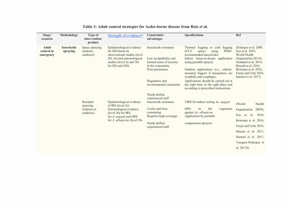
Table 3: Adult control strategies for Aedes-borne disease from Roiz et al.
Stage/ scenario
Methodology Type of intervention/
product
Strength of evidence*
Constraints/ advantages
Specifications Ref
Adult control in emergency
Insecticide spraying
Space spraying (indoors, outdoors)
Epidemiological evidence for ISS based on observational studies (level 2b). Several entomological studies (level 3a and 3b) for ISS and OSS.
Insecticide resistance Thermal fogging or cold fogging (ULV spray) using WHO-recommended insecticides
(Erlanger et al. 2008, Esu et al. 2010, World Health Organization 2011b, Stoddard et al. 2014, Bouzid et al. 2016, Bowman et al. 2016, Faraji and Unlu 2016, Samuel et al. 2017)
Low acceptability and limited sense of security in the community
Indoor house-to-house application using portable sprayer.
Poor persistence Outdoor applications (i.e., vehicle-mounted fogger) if mosquitoes are exophilic and exophagic.
Regulatory and environmental constraints
Applications should be carried out at the right time, in the right place and according to prescribed instructions.
Needs skilled, experienced staff
Residual spraying (indoors or outdoors)
Epidemiological evidence of IRS (level 2a). Entomological evidence (level 3b) for IRS for A. aegypti and ORS for A. albopictus (level 3b).
Insecticide resistance TIRS for indoor resting Ae. aegypti (World Health
Organization 2007b,
Esu et al. 2010,
Bowman et al. 2016,
Faraji and Unlu 2016,
Muzari et al. 2017,
Samuel et al. 2017,
Vazquez-Prokopec et
al. 2017b)
Costly and time-consuming
ORS on the vegetation against Ae. albopictus
Requires high coverage Application by portable
Needs skilled, experienced staff
compression sprayers

Table 3: Adult control strategies for Aedes-borne disease from Roiz et al. (continued)
Stage/ scenario
Methodology Type of intervention/ product
Strength of evidence* Constraints/ advantages Specifications Ref
Adult control for routine and emergency
Mass trapping
Gravid traps (AGO or GAT)
Epidemiological evidence based on observational studies (level 2b). Entomological evidence (level 3b) for A. aegypti.
Low cost Need for a coverage of greater than 80%
(Lorenzi et al. 2016, Barrera et al. 2017, Johnson et al. 2017) Possible to combine with
community participation Use large autocidal gravid traps, as AGO or GAT, to maximise visual and olfactory attraction using grass or hay infusion
Sustainable, able to be reused for several seasons
Individual-based action (requires high degree of compliance)
DEET, the longest-lasting; IR3535 or picaridin, medium-long lasting protection; plant-derived oils (eucalyptus, citronella, or geranium), short-term (frequency of applications according to national legislation and/or manufacturer’s
recommendations)
No residual activity
Insecticide-treated materials (clothes, curtains, house screens, water container covers, etc.)
Epidemiological evidence for house screening (level 2b). Entomological evidence for ITCs, house screening, and water container covers (level 3a and 3b). No evidence for bed nets.
Individual- and community-based action
Clothes, curtains, and bed nets treated with WHO-recommended insecticides.
(Wilson et al. 2014, Bowman et al. 2016, Kittayapong et al. 2017) Residual activity with
long-lasting technology Most evidence supports house screening for preventing dengue transmission
Insecticide resistance
Low protection against UV Degradation of insecticide

3.3.1.3.1. Chemical control
Chemicals remains the most widely used method for targeting Aedes adult mosquitoes.
Adult control is usually implemented using two families of insecticides, i.e., the organophosphates
(OP) and the pyrethroids (PYR). Adult chemical vector control can be implemented using very
limited number of molecules and type of applications as summarized in Table 3 (World Health
Organization 2009, Roiz et al. 2018).
3.3.1.3.1.1. Space sprays
Space sprays using mainly PYR, are widely used in emergencies to rapidly kill the adult
females and curtail the cycle of transmission. However, the evidence to support the deployment of
this intervention is rather weak (Table 3). Indoor space spray (ISS) showed better results in
controlling adult populations and dengue transmission, than outdoor space spray using either
thermal fogging or cold fogging of Ultra Low Volume (ULV) against the highly endophilic Ae.
aegypti. (Esu et al. 2010, Stoddard et al. 2014, Faraji and Unlu 2016).
To improve the sustainability of vector control insecticide residual treatment had been
investigated (Table 3). Indoor residual spraying (IRS) has demonstrated strong evidence in
reducing malaria burden, however, it remains so far, scarcely used for Aedes-borne disease control.
Yet, study in Australia demonstrated the effectiveness of targeted IRS to prevent dengue in areas
where Ae. aegypti is the sole vector (Vazquez-Prokopec et al. 2017b). Similarly, outdoors residual
spraying (ORS) can be performed to control Ae. albopictus, by targeting the vegetation or external
walls of habitations (Faraji and Unlu 2016). Nevertheless, IRS and ORS required qualified staff
and must be repeated regularly in order to keep mosquito populations at low levels (Roiz et al.
2018).
3.3.1.3.1.2. Insecticide treated materials
Another strategy, taking advantage of the endophilic and anthropophagic behaviour of Ae.
aegypti, is the use of insecticide-treated curtains, house screens and clothes to reduce adult
population. However, insecticide-treated curtains were deployed in Mexico and Thailand with
mitigated results (Wilson et al. 2014). While Kroeger et al. and Vanlerberghe et al. reported an
entomological impact on the local vector population, Lenhart et al. found no evidence of
effectiveness of insecticide-treated curtains for Aedes control (Kroeger et al. 2006, Lenhart et al.
2013, Vanlerberghe et al. 2013, Wilson et al. 2014). In addition, the use of insecticide-treated
56
uniforms was not associated with a reduction of dengue incidence in Thailand probably due to a
rapid decline of insecticide efficacy after washing (Kittayapong et al. 2017).
It is worth to note that PYR are the gold standard for adult control but PYR resistance has
been reported in many dengue endemic countries (Corbel et al. 2016, Moyes et al. 2017). The rise
of PYR resistance foresaw a decline of efficiency of those class of adulticides for adult control and
therefore, research on new tools and paradigms for mosquito control has become a priority.
3.3.2. Alternative strategies for vector control
Several tools and methods are under consideration by the World Health Organization
Vector Control Advisory Group (WHO VCAG) and they have been the subjected of a recent
review by (Achee et al. 2019). For instance, genetically modified mosquitoes and Wolbachia-
infected mosquitoes for population suppression or population replacement have shown promising
results in small-medium scale pilot trials (O'Connor et al. 2012, Indriani et al. 2020). The strength
and weakness of the new tools and strategies will be briefly presented in the following sections.
3.3.2.1. Wolbachia-based strategies
Wolbachia is a bacterial endosymbiont originally found in Culex species and is naturally
present in 60% of insect species, including Ae. albopictus, and is known to disturb host’s
reproduction, disrupt pathogen transmission, and prevent eggs from hatching by cytoplasmic
incompatibility (Yen et al. 1971, Bourtzis et al. 2014, Farnesi et al. 2019). Population suppression
strategy using Wolbachia-infected Aedes mosquitoes was shown to be species-specific (O'Connor
et al. 2012, Mains et al. 2016) in California, Thailand, Singapore and Australia (Hoffmann et al.
2014, Tan et al. 2017, Kittayapong et al. 2019, Crawford et al. 2020). The population replacement
strategy using strains of Wolbachia, seems also promising for interrupting DENV, CHIKV, and
even ZIKV transmission, in particular the wMel strain, during field releases in Australia and
Indonesia (Moreira et al. 2009, Aliota et al. 2016, Tan et al. 2017, Gonçalves et al. 2019, Indriani
et al. 2020, Tantowijoyo et al. 2020). Although promising Wolbachia based strategies are
challenging by several factors including a lack of community’s adhesion, ethical and regulatory
constraints and a lack of facilities and capacities for scaling-up the intervention (Lambrechts et al.
2015, Ritchie et al. 2017, Ritchie et al. 2018, Xue et al. 2018, Achee et al. 2019, Maïga et al. 2019,
Crawford et al. 2020).

57
3.3.2.2. Genetically modified mosquitoes
Genetic control is defined as the genetic modification of the vectors by introducing genes,
conferring a fitness-cost, that can interfere with the fertility, or prevent the emergence of adults, or
confer resistance to a given pathogen (Thomas et al. 2000). Originally based on the sterile insect
technique (SIT) which imply the release of insect males sterilized by irradiation (Achee et al. 2019,
Kittayapong et al. 2019), advances in genetics, enable the release of insects of dominant lethality
(RIDL) strategies, based on the insertion of a repressible dominant sex-specific lethal transgene
spreading in the vector population (Alphey et al. 2013). The RIDL Ae. aegypti OX513A strain
(Oxitec company), released in 2009 in Brazil, Panama, and Cayman Islands, has shown to reduce
the wild Ae. aegypti population by >90% (Carvalho et al. 2015, Gorman et al. 2016). More
recently, the development of Clustered Regularly Interspaced Short Palindromic Repeats
associated protein 9 (CRISPR-Cas9) systems had enable designing gene drives that precisely
cleave the DNA to insert a gene of interest that becomes heritable. While promising results has
been shown for malaria vectors, only proof-of-principle was illustrated in Ae. aegypti (Achee et
al. 2019, Li et al. 2020). Although these methods appear promising, they still need a strong
evaluation of their ecological impact and community acceptance for mosquito releases, especially
regarding the introduction of genetic modified organism in the environment (Achee et al. 2019).
Moreover, these strategies are challenged by potential “re-emergence” of the native population
and/or “replacement” by new invasive species (through migration and introduction) that may
enable diseases transmission (Gorman et al. 2016).
3.3.2.3. Other tools currently under evaluation
In addition to the conventionally used methods mentioned above, a certain number of
innovative vector control tools are under development and are currently examined by the WHO
VCAG (Achee et al. 2019, Corbel et al. 2019). First, mosquito traps were modified to improve
mosquito attractiveness and reduce vector populations by either killing the gravid females looking
for oviposition sites or by eliminating the progeny. The gravid Aedes traps (GAT) developed by
Biogents® and the autocidal gravitraps (AGO) developed by the Centers for Disease Control and
prevention (CDC) were demonstrated successful in medium scale field trials to control Aedes
population in Latin America and Australia (Perich et al. 2003, Rapley et al. 2009, Wesson et al.
2012, Barrera et al. 2019, Montenegro et al. 2020). In addition, Attractive Toxic-Sugar Baited

58
(ATSB) were developed to attract both males and females and incite them to feed on “toxic” sugar
meals applied on plants or used in bait station. So far, ATSB efficacy strongly relies on the
attractiveness of the bait and the type of toxin used (e.g., spinosad, neonicotinoids, or fipronil)
(Müller et al. 2005, Achee et al. 2019). In contrast to GMO and traps, ATSB can be challenged by
insecticide resistance and by potential adverse effects of the toxin against non-targeted species and
further investigations are need to assess relevance for vector control. An alternative to chemical
insecticides may come from natural compounds produced by fungi or by using the mosquitoes
themselves to distribute the insecticide (i.e., pyriproxyfen) into cryptic breeding sites, known as
the autodissemination strategy. Another strategy is to reduce the human-vector contact by using
spatial repellent. Noteworthy other methods, not involving insecticides are explored such as the
destruction of Ae. aegypti larvae using acoustic emission (Britch et al. 2016). Yet, these approaches
are quite new and will need further research to ensure their efficiency and safety.
3.3.3. Insecticide resistance
Insecticide resistance is an increasing challenge for Aedes-borne disease prevention
because dengue, Zika and chikungunya control strategies rely heavily on chemical control (see
section 3.3). Moreover, few molecules are registered for vector control and the challenges to
develop new chemicals for public health are extremely high. Thus, increasing insecticide
resistance, enhanced by the massive use of pesticides in agriculture and public health, is now
considered by the WHO as a major threat to dengue control and prevention worldwide (Corbel et
al. 2016).
3.3.3.1. Global distribution
Insecticide resistance in Ae. aegypti and Ae. albopictus against the four main classes of
insecticide (e.g., carbamates, organochlorine, OPs and PYRs) was reported in at least 57 countries,
including South East Asia, the Americas and the Caribbean (Harris et al. 2010, Dusfour et al. 2011,
Marcombe et al. 2012, Kasai et al. 2014, Corbel et al. 2017, Moyes et al. 2017, Marcombe et al.
2019).
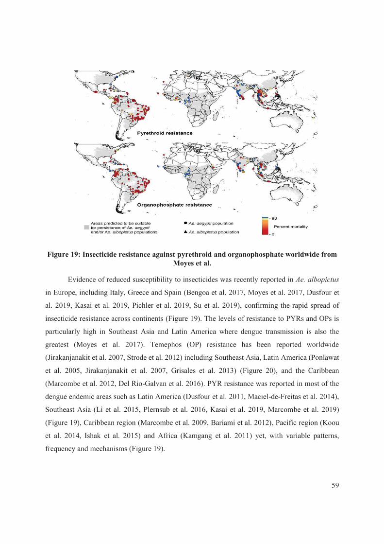
59
Figure 19: Insecticide resistance against pyrethroid and organophosphate worldwide from Moyes et al.
Evidence of reduced susceptibility to insecticides was recently reported in Ae. albopictus
in Europe, including Italy, Greece and Spain (Bengoa et al. 2017, Moyes et al. 2017, Dusfour et
al. 2019, Kasai et al. 2019, Pichler et al. 2019, Su et al. 2019), confirming the rapid spread of
insecticide resistance across continents (Figure 19). The levels of resistance to PYRs and OPs is
particularly high in Southeast Asia and Latin America where dengue transmission is also the
greatest (Moyes et al. 2017). Temephos (OP) resistance has been reported worldwide
(Jirakanjanakit et al. 2007, Strode et al. 2012) including Southeast Asia, Latin America (Ponlawat
et al. 2005, Jirakanjanakit et al. 2007, Grisales et al. 2013) (Figure 20), and the Caribbean
(Marcombe et al. 2012, Del Rio-Galvan et al. 2016). PYR resistance was reported in most of the
dengue endemic areas such as Latin America (Dusfour et al. 2011, Maciel-de-Freitas et al. 2014),
Southeast Asia (Li et al. 2015, Plernsub et al. 2016, Kasai et al. 2019, Marcombe et al. 2019)
(Figure 19), Caribbean region (Marcombe et al. 2009, Bariami et al. 2012), Pacific region (Koou
et al. 2014, Ishak et al. 2015) and Africa (Kamgang et al. 2011) yet, with variable patterns,
frequency and mechanisms (Figure 19).

60
3.3.3.2. Mechanisms
Several studies on Ae. aegypti have highlighted a certain number of non-synonymous
mutations in the Voltage Gated Sodium Channel (VGSC), known as kdr mutations, especially the
S989P, V1016G, and F1534C conferring resistance to PYRs (Brengues et al. 2003, Saavedra-
Rodriguez et al. 2007, Kasai et al. 2011, Yanola et al. 2011, Hirata et al. 2014). Moreover, the
F1534S mutation in Ae. albopictus is associated with PYR resistance in many populations across
the world (Chen et al. 2016, Xu et al. 2016). Recent findings suggested a reduction in AChE
sensitivity to propoxur in Ae. aegypti populations from Trinidad and Tobago (Polson et al. 2011,
Vontas et al. 2012), although no evidence for the presence of mutations in the acetylcholine
esterase (AChE) gene was reported.
Metabolic resistance caused by changes in the patterns of enzyme’s expression resulting in
an enhanced insecticide detoxification system was reported in dengue vectors (Strode et al. 2008,
Strode et al. 2012). Metabolic resistance is mainly due to three families of enzymes: cytochrome
P450 mono-oxygenase (P450s or CYPs for genes), carboxylesterases (CCEs) and glutathione-S-
transferases (GSTs) (Hemingway et al. 2004). While PYRs and OPs have different target and mode
of action, their detoxification pathway can involve similar enzymes or class of enzymes such as
P450s, CCEs, or GSTs (Dusfour et al. 2011, David et al. 2014, Grigoraki et al. 2016). For instance,
the carboxylesterase 3 (CCE3A) was implicated in both resistance to OP and PYR in SEA and
SEA regions. Consequently, some genes overexpressed in mosquito after exposure to OP can lead
to a cross-resistance to PYR and vice versa.
Changes in enzymes expression can be caused by an up-regulation of gene transcription
but also due to gene amplification resulting in multiple copies variants (CNV) of the genes coding
for the enzymes (Bariami et al. 2012, Kasai et al. 2014, Faucon et al. 2015, Goindin et al. 2017,
Cattel et al. 2020b). Duplications were demonstrated in several genes including CYPs, CCEs, and
GSTs genes linked to PYR and OP resistance in Ae. aegypti in Latin America, Caribbean region,
and Asia yet with different genetic profiles (Faucon et al. 2015, Faucon et al. 2017, Goindin et al.
2017, Cattel et al. 2020a, Cattel et al. 2020b).
While many studies focus on Ae. aegypti, there is also increasing evidence for the presence
of insecticide resistance in the tiger mosquito Ae. albopictus (Kasai et al. 2019). Recently Ishak et
al revealed that 10 genes were up-regulated in a pyrethroid-resistant population of Ae. albopictus
61
in Malaysia including five P450s (three CYP6, and two CYP9), two GSTs, one ABC transporter
and two short-chain dehydrogenases were implicated (Ishak et al. 2016).
3.3.3.3. Impact on vector control
A potential consequence of insecticide resistance is the loss of effectiveness of vector control
intervention (Marcombe et al. 2011). Despite increasing concern, the degree to which insecticide
resistance compromises dengue control in the field remains largely unknown (Corbel et al. 2019).
Recent studies performed in Latin America and Caribbean demonstrated the negative impact of
insecticide resistance on vector control activities, targeting either larvae (Montella et al. 2007), or
adults (Marcombe et al. 2011, Dusfour et al. 2015, Vazquez-Prokopec et al. 2017a). Gray et al also
demonstrated that the use of household insecticide spray led to a selection of resistance mechanism
such as kdr-alleles. Lessons learnt from the past suggest that monitoring the susceptibility level
and changes in genotype frequency regularly allow to readjust vector control policies before vector
control failure occur (Corbel et al. 2019). Nevertheless, further investigations are needed to address
clearly the relationships between resistance mechanisms, kdr frequencies, and the impact of
resistance on vector control interventions. This would help authorities to implement timely
insecticide resistance management plan.
4. Methods and indicators for assessing and predicting the risk of dengue
transmission
Dengue transmission remains complex to assess and even predict using actual tools and
indicators. Indeed, dengue transmission risk is highly dependent on the interactions between Aedes
vectors, human hosts, dengue viruses and the environment. This section will review the concept
of dengue transmission, and will describe the actual epidemiological, mathematical, and
entomological methods and indicators used to estimate dengue transmission risk with their
limitations. Finally, the potential of new immunological tools for estimating “human-vector”
contact will be discussed in relation to dengue transmission studies.
4.1. Definition: concept of transmission
Dengue virus is transmitted to human through an infected Aedes mosquito bites and
mosquitoes can become infected while biting a human infected by dengue virus (Figure 20).

62
Complex interaction between the mosquito, the host and the vector are driven by key factors such
as the intrinsic and extrinsic incubation periods. The intrinsic incubation period is the period when
the pathogen (in our case, the dengue virus) is detectable in the (human) host’s blood. Chan et al.
reviewed 35 studies, published between 1903 and 2011, with relevant data on intrinsic incubation
period (Chan et al. 2012) and they concluded that the most probable intrinsic incubation period is
comprised between 3-10 days. However, virus can replicate in humans host only if the hosts never
experienced this dengue serotype (see section 1.3 and 1.5). Thus, individuals can be infected
multiple times by different serotypes. If the human host is already immune against dengue, the
virus will not be able to replicate and the transmission will stop. Therefore, herd immunity,
serotype distribution and circulation are decisive in estimating (and predicting) dengue
transmission.
In addition, DENV transmission from human to human, by sexual transmission was
recently reported in South Korea (Lee et al. 2019, Wilder-Smith 2019). Indeed, a woman
contracted DENV after her partner previously got infected by dengue overseas. Nevertheless, such
cases remain anecdotal, the Aedes mosquitoes being the principal vectors of the disease.
Figure 20: Dengue transmission cycles from Ahammad et al.
Once the virus is disseminated into the Aedes organism, it will remain until the mosquito
dies. The extrinsic incubation period is defined as the period of time required, after ingestion of
the virus during the blood-feeding process, for the vector to be able to transmit the pathogen
(Fontaine et al. 2016). Aedes extrinsic incubation period for dengue virus, ranges from 8 to 12 days
63
(World Health Organization 2009), depending on temperature, mosquito populations, and virus
serotypes (Whitehorn et al. 2015, Christofferson et al. 2016, Gloria-Soria et al. 2017). For instance,
temperature about 29°C were shown to reduce the incubation period, producing more Aedes able
to transmit the virus. Additionally, temperature above 32°C may reduce Aedes lifespan despite a
quicker extrinsic incubation period (Christofferson and Mores 2016, Liu et al. 2017). On the other
hand, temperature below 18°C strongly slow down viral “migration” thus preventing
dissemination to the salivary glands. In addition, Gloria-Soria et al, demonstrated that the success
of viral dissemination has variable influence depending on Ae. aegypti populations (Gloria-Soria
et al. 2017).
In addition to the ability of transmitting dengue virus during their whole life, Aedes female
can transmit the virus to their offspring (known as vertical transmission) (Castro et al. 2004,
Arunachalam et al. 2008). Firstly demonstrated in laboratory conditions, vertical transmission of
dengue virus in Ae aegypti and Ae. albopictus have been reported worldwide, particularly in Latin
America and southeast Asia (Thenmozhi et al. 2007, Le Goff et al. 2011, Ferreira-de-Lima et al.
2018). Although experimental studies demonstrated that vertical transmission cannot be sustained
after the fifth generation (Rohani et al. 2008, Sanchez-Vargas et al. 2018), vertical transmission of
the viruses is a possible explanation for the persistence of dengue serotypes between epidemics
(Ferreira-de-Lima and Lima-Camara 2018, Ferreira-de-Lima et al. 2020).
Nonetheless, dengue transmission is highly focal, and characterized by the occurrence of
“hotspots”, i.e., area of higher risk or higher probability of disease incidence (Martinez-Vega et
al. 2015). Aedes mosquitoes are known to have small flight range (<100 meters), and usually rest
near their sites of emergence. Aedes aegypti is often breed in people’s yard, thus somehow it can
be considered as a “pet”-mosquito. The limited Aedes flight range explains the highly focal pattern
of dengue transmission (Stoddard et al. 2013). Human movements, however, by moving between
areas with relative dengue transmission risks, can introduce dengue virus into new areas/territories,
whether by being dengue infected or by transporting infected vectors (e.g., eggs or adults).
Therefore, the spread of dengue at large scale, such as countries, is mainly driven by human
movement (Stoddard et al. 2013, Smith et al. 2014). Nonetheless, the presence of competent
vectors is required to maintain the transmission.

64
Dengue and other Aedes-borne diseases transmission risk can be assessed through the use
of multiple epidemiological, entomological, mathematical, and/or immunological surveillance
tools and indicators each one having their strengths and weaknesses. This will be further discussed
in the following sections.
4.2. Epidemiological surveillance and it’s limitations
Epidemiological surveillance is the cornerstone of the assessment of dengue transmission risk.
Epidemiological surveillance aims to monitor dengue incidence trends in order to detect outbreaks
in a timely manner and trigger interventions from public health stakeholders (World Health
Organization 2009). Indicators used for epidemiological surveillance are based on dengue cases
detection and serotype distributions.
Dengue case surveillance is based on case notification yet, only symptomatic cases from
hospitals are accounted. Moreover, the difficult diagnosis of dengue based on symptoms lead to a
non-negligible proportion of misdiagnosed patients (see section 1.5 and 1.6) which impoverish
transmission risk assessment (Bhatt et al. 2013). Moreover, Duong et al. demonstrated that
asymptomatic cases can transmit dengue virus to mosquitoes, and thus actively participate to
dengue transmission (Duong et al. 2015a). Consequently, dengue transmission assessment is
strongly challenged by asymptomatic and mild-symptomatic cases that would not seek medical
attention or being misdiagnosed (see section 1.5). Moreover, the temporal disconnection between
acquiring an infection to time of presenting illness and testing (i.e., identification of a case) may
greatly affect attempts to link transmission with actual epidemiological conditions many days
prior.
In addition, the possible multi-infections and immunities make outbreaks even more difficult
to predict. Many the of biggest outbreaks were related to the sudden increase of prevalence of a
serotype that was not circulating (i.e., introduction of a new serotype) or was circulating a lower
prevalence during the past years (Mammen et al. 2008, Mondini et al. 2009, Getahun et al. 2019,
Phanitchat et al. 2019). Better knowledge on serotype distribution and herd immunity dynamics
would help to better predict and prevent dengue outbreaks by re-enforcing dengue vector
prevention and control in a timely manner (Bhatia et al. 2013, Bhatt et al. 2013, Reich et al. 2016).
Moreover, outbreak definition is subjective and many countries use their own definition (Brady et
al. 2015). In most of dengue endemic countries, an outbreak is declared when the number of
65
hospitalized cases per week (or month) rises above the average of the number of cases in the
previous years plus two standard deviation (World Health Organization 2009, Brady et al. 2015).
Unfortunately, when the number of cases exceeds the threshold, it is often too late to implement
effective vector control interventions and curtail the transmission. Additionally, most of dengue
surveillance systems in endemic areas rely on hospital reports, thus missing mild symptomatic
cases. Therefore, epidemiological surveillance must be combined to mathematical and
entomological surveillance tools to better address transmission risk and prevent dengue outbreaks.
4.3. Mathematical tools and their limitations
Considering the complex relationships between, the vector, the human host, the virus and the
environment, mathematical and computational tools were developed to predict transmission risk
and prevent outbreak. Indeed, computational technologies enable complex calculation and take
into account large datasets with many types of parameters such as case incidence, Aedes densities,
but also climatic and demographic data or human movements. Predictive models of dengue
incidence historically demonstrated variable accuracies in terms of dengue incidence and outbreak
predictions (Ramadona et al. 2016, Olliaro et al. 2018, Johansson et al. 2019). The first model of
dengue transmission risk (DENSiM) used mainly vectors characteristics and climatic factors
(Focks et al. 1993b, Focks et al. 1995). Since then, numerous studies tried to predict dengue
incidence using a combination of entomological, epidemiological and climatic factors (Morin et
al. 2013, Lee et al. 2017, Johansson et al. 2019, Stolerman et al. 2019). Yet, models of dengue
transmission using retrospective epidemiological and climatic data could demonstrate clear
evidence of seasonal pattern of dengue transmission in South East Asia (van Panhuis et al. 2015).
Recently, the WHO and the special group for tropical diseases research (WHO-TDR) developed a
new adaptable model for dengue surveillance and outbreak response (World Health et al. 2017).
Using retrospective country datasets, they could define and detect dengue outbreaks using
probable/ hospitalised cases as the outbreak variable (defined in section 4.2), and then successfully
predict these outbreaks using early changes in various entomological, meteorological and
epidemiological “alarm” variables (World Health et al. 2017, Hussain-Alkhateeb et al. 2018).
Although predictive models for dengue have been shown to be successful, particularly using
climate and incidence data, the inconsistency of entomological data, highlighted the need for more
reliable response for estimating dengue transmission risks (Bowman et al. 2016).

66
To conclude, despite the large number of dengue transmission models developed, they failed
to be universally applicable. Indeed, the dengue transmission models developed for the Latin
America are difficultly transposable to SEA and vice and versa (Johansson et al. 2016, Reich et al.
2016). In particular, integrating social (e.g., rent value or education level), demographic (e.g.,
population density or distance to urban habitats), and landscape (e.g., vegetation cover or type of
urbanization) data in mathematical models could be helpful to better asses transmission risk and
predict further spread and seasonal dynamics and could be less expensive than field studies. In
addition, better understanding of the correlations between Aedes vectors and dengue transmission
risk might improve mathematical models in assessing dengue transmission risk and thus would
result in better predictions of dengue outbreaks.
4.4. Entomology surveillance and it’s limitations
Entomological surveillance is essential to provide information on the local vector population,
especially the density and diversity of vector species, their vectorial capacity and their host seeking
preferences (World Health Organization 2011b). Several entomological indices are used to assess
the risk of transmission targeting different stages of the mosquito development, each presenting
their strength and weakness (Table 4 for details).

Table 4: Strengths and weakness of entomological surveillance tools from Roiz et al.
Trap methodology
Index name Index description
Formula Unit Target Strengths/weaknesses References
Ovitraps Ovitrap index (OI)
Average proportion of positive ovitraps
Positive ovitraps / No. of ovitraps examined in a given area per month/week/ fortnight
% Eggs
Sensitive and economical method for detecting Aedes introduction and/or presence in large area (surveillance). Information not reliable for measuring Aedes density.
(European Centre for Disease Prevention and Control (ECDC) 2012, Flacio et al. 2015)
Trap positivity index (TPI)
Proportion of positive traps
(Total no. of traps infested with eggs / Total traps) x 100
% Eggs
Egg density index (EDI)
Ratio of no. of eggs/traps
Total no. of eggs / Total no. of traps
No. of eggs per trap
Eggs Information not reliable for measuring Aedes density
Larval indices House index (HI), (also called premise index)
Proportion of houses positive for immature Aedes
(No. of houses infested / Total households) x 100
% Pupae, larvae
Not reliable for measuring Aedes population level No information on the number of positive containers Does not take productivity into account Poor indication of adult production
(Focks 2004, European Centre for Disease Prevention and Control (ECDC) 2012, Bowman et al. 2016)
Container index (CI)
Proportion of containers positive for immature Aedes
(No. of containers infested / Total containers inspected) x 100
% Pupae, larvae
Relevant for focussing larval control efforts and for orienting educational messages Can provide data on larval development habitat characteristics Does not take productivity into account Poor indication of adult production
Breteau index (BI)
No. of Aedes-
positive containers per 100 houses
(No. of containers infested / Total houses inspected) x 100
No. per 100 houses
Pupae, larvae
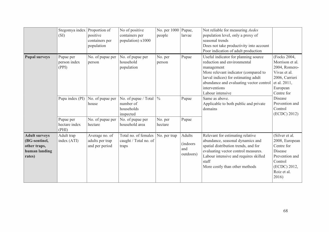
68
Stegomya index (SI)
Proportion of positive containers per population
No of positive containers per population) x1000
No. per 1000 people
Pupae, larvae
Not reliable for measuring Aedes population level, only a proxy of seasonal trends Does not take productivity into account Poor indication of adult production
Pupal surveys Pupae per person index (PPI)
No. of pupae per person
No. of pupae per household population
No. per person
Pupae Useful indicator for planning source reduction and environmental management More relevant indicator (compared to larval indices) for estimating adult abundance and evaluating vector control interventions Labour intensive
(Focks 2004, Morrison et al. 2004, Romero-Vivas et al. 2006, Carrieri et al. 2011, European Centre for Disease Prevention and Control (ECDC) 2012)
Pupa index (PI) No. of pupae per house
No. of pupae / Total number of households inspected
% Pupae Same as above. Applicable to both public and private domains
Pupae per hectare index (PHI)
No. of pupae per hectare
No. of pupae per household area
No. per hectare
Pupae
Adult surveys (BG-sentinel, other traps, human landing rates)
Adult trap index (ATI)
Average no. of adults per trap and per period
Total no. of females caught / Total no. of traps
No. per trap Adults
(indoors and outdoors)
Relevant for estimating relative abundance, seasonal dynamics and spatial distribution trends, and for evaluating vector control measures. Labour intensive and requires skilled staff More costly than other methods
(Silver et al. 2008, European Centre for Disease Prevention and Control (ECDC) 2012, Roiz et al. 2016)

69
Sticky trap surveys
Sticky trap index (STI)
No. of adults caught by the sticky trap per unit of time
(No of traps positive for Aedes sp./ Total no of inspected traps) x 100
% Adults Poor proxy of adult abundance. Useful for collecting gravid females. Easy to use, affordable, and can be deployed at large scale. Can be used to screen virus infections in mosquitoes
(Ritchie et al. 2003, Facchinelli et al. 2007 , Gama et al. 2007)
Adult surveys for viral detection
Vector infection index (VII)
Proportion of Infected females
(No. of virus-infected females / Total no. of females inspected)*100
% Adults Relevant for identifying the role of local species in virus transmission and/or for characterising viral strain. Costly, labour intensive, requires skilled staff and strong diagnostic capacity (RT-PCR).
(European Centre for Disease Prevention and Control (ECDC) 2012)
Adult surveys using human hosts
Human-baited double net (HDN) or mosquito electrocuting traps (MET)
Mean number of mosquito females per person per unit of time
No. of females collected per person per unit of time
No. mosquitoes collected per person per unit of time
Adults Alternative techniques to the human landing catch (HLC) method for collecting anthropophagic mosquito species, and for measuring mosquito biting densities and mosquito behaviour. Labour-intensive, costly, and may pose logistic difficulties (e.g., access to power sources) Human-baited double traps are a good alternative to collecting outdoor mosquitoes. Mosquito electrocuting traps are a good exposure-free alternative to HLC. HDN and MET are well accepted and pose fewer ethical problems than HLC.
(Tangena et al. 2015, Govella et al. 2016)
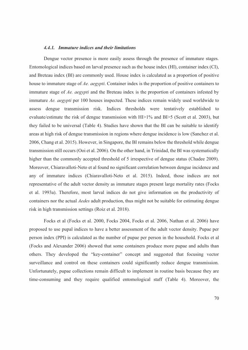
70
4.4.1. Immature indices and their limitations
Dengue vector presence is more easily assess through the presence of immature stages.
Entomological indices based on larval presence such as the house index (HI), container index (CI),
and Breteau index (BI) are commonly used. House index is calculated as a proportion of positive
house to immature stage of Ae. aegypti. Container index is the proportion of positive containers to
immature stage of Ae. aegypti and the Breteau index is the proportion of containers infested by
immature Ae. aegypti per 100 houses inspected. These indices remain widely used worldwide to
assess dengue transmission risk. Indices thresholds were tentatively established to
evaluate/estimate the risk of dengue transmission with HI>1% and BI>5 (Scott et al. 2003), but
they failed to be universal (Table 4). Studies have shown that the BI can be suitable to identify
areas at high risk of dengue transmission in regions where dengue incidence is low (Sanchez et al.
2006, Chang et al. 2015). However, in Singapore, the BI remains below the threshold while dengue
transmission still occurs (Ooi et al. 2006). On the other hand, in Trinidad, the BI was systematically
higher than the commonly accepted threshold of 5 irrespective of dengue status (Chadee 2009).
Moreover, Chiaravalloti-Neto et al found no significant correlation between dengue incidence and
any of immature indices (Chiaravalloti-Neto et al. 2015). Indeed, those indices are not
representative of the adult vector density as immature stages present large mortality rates (Focks
et al. 1993a). Therefore, most larval indices do not give information on the productivity of
containers nor the actual Aedes adult production, thus might not be suitable for estimating dengue
risk in high transmission settings (Roiz et al. 2018).
Focks et al (Focks et al. 2000, Focks 2004, Focks et al. 2006, Nathan et al. 2006) have
proposed to use pupal indices to have a better assessment of the adult vector density. Pupae per
person index (PPI) is calculated as the number of pupae per person in the household. Focks et al
(Focks and Alexander 2006) showed that some containers produce more pupae and adults than
others. They developed the “key-container” concept and suggested that focusing vector
surveillance and control on these containers could significantly reduce dengue transmission.
Unfortunately, pupae collections remain difficult to implement in routine basis because they are
time-consuming and they require qualified entomological staff (Table 4). Moreover, the

71
correlation between pupal indices and dengue cases is heterogeneous across studies (Romero-
Vivas et al. 2005, Favaro et al. 2013) and this would merit further investigations.
4.4.2. Adult indices and their limitations
Adult mosquito collections have been used to estimate the risk of dengue transmission (Lau
et al. 2017, Parra et al. 2018), but they have also their weakness (Table 4). Several methods have
been developed to sample adult Aedes such as sticky traps, gravitraps (see section 3.3.2.3), or
mechanical batterie-driven aspirators (Vazquez-Prokopec et al. 2009). Recently, a new method
inspired by the human landing catches was developed, the mosquito electrocuting trap (MET) and
demonstrated similar results to the BG-sentinel traps (Ortega-López et al. 2020). The authors
suggested a better assessment of host-seeking preferences and human biting rates using MET than
BG-Sentinel traps as the latter method is less species-specific and is highly dependent on the types
of lures (e.g., CO2, hay infusion) used to attract mosquitoes (Bazin et al. 2018). Moreover, all adult
collection methods are used as a proxy to estimate Aedes “density” but they cannot predict the real
exposure time between the human host and the vector (Barnard et al. 2014). This information is
yet crucial to identify population subsets at higher exposure risk to dengue vector bites and then
virus transmission.
4.5. Serological tools to estimate dengue transmission risk
To counter the actual difficulties in estimating dengue transmission risk (see above), new
serological tools relying on the human antibody response against arthropod salivary proteins were
developed (Doucoure et al. 2015). These tools known as “salivary biomarkers” offer the
opportunity to provide more direct and accurate estimation of the human exposure to vector bites,
at both community and individual levels, and may then be used as proxy to estimate local disease
transmission risk (Sagna et al. 2018). This section will describe the concept of serological
biomarkers and how they can be used to predict Aedes-borne disease transmission risk.
4.5.1. Concept
Vector-borne diseases have in common to be caused by pathogens that are transmitted to
vertebrate hosts (human or animal) through infective bite of the arthropod vector (Doucoure and
Drame 2015, Sagna et al. 2018). During the blood feeding process arthropods inject saliva in
order to facilitate the blood intake (Figure 21). Saliva of hematophagous insects is composed of

72
several proteins which modulate the immune response of the host and inhibits coagulation, in
order to get a full blood meal. Interestingly, among this cocktail of molecules, some were shown
to induce a specific immune response. Several studies have shown that the human antibody (Ab)
response to arthropods bites such as ticks (Lane et al. 1999), Phlebotomus (Rohousova et al.
2005), and mosquitoes (Remoue et al. 2006, Poinsignon et al. 2008) can be used as relevant
markers for assessing human exposure to insect bites and to estimate pathogens transmission risk
(Ya-Umphan et al. 2017, Ya-Umphan et al. 2018). Patented biomarkers1 have been developed to
measure the levels of human exposure to Anopheles mosquito bites (Remoue et al. 2006,
Poinsignon et al. 2008). Additionally, they have been successful to assess the effectiveness of
vector control measures for malaria prevention (Drame et al. 2010a, Drame et al. 2013). More
recently, salivary biomarker showed to be accurate enough to identify “hotspots” of vector
abundance and malaria transmission (Poinsignon et al. 2009, Marie et al. 2015, Ya-Umphan et
al. 2017, Ya-Umphan et al. 2018), and then represent promising tools for epidemiology studies.
Figure 21: Human-vector relationships during arthropod-borne diseases from Sagna et al. During the bite, the vector (Aedes) injects its saliva in the human skin. Once in the skin, salivary proteins take the control of (1) the human hemostatic system by inhibiting the platelet activation and clotting mechanism, and (2) the inflammatory system. (3) The salivary proteins modulate the human immune response and promote the production of anti-salivary antibodies. (4) If ever the (Aedes) vector carries a pathogen, the salivary proteins contribute to its transmission into the
human.
1 Internationally patented by Remoue et al. 2011 (patent no. 2009.630 37 33 09).

73
4.5.2. Application of salivary biomarkers to Aedes transmitted diseases
Several studies have shown that the measurement of IgG response against salivary gland
extracts from different Aedes species, such as Ae. aegypti, Ae. polynesiensis, Ae. caspius were
reliable indicators of human-Aedes exposure in South-America (Doucoure et al. 2012, Londono-
Renteria et al. 2013), Pacific Islands (Mathieu-Daude et al. 2018), Africa (Doucoure et al. 2014)
and even Europe (Fontaine et al. 2011). However, the use salivary gland extracts presents some
limitations because proteins amount varies between individuals and batches, and may lack of
specificity (Sagna et al. 2018). Indeed, salivary extracts of Anopheles, Culex, and Aedes share
similar proteins that can induce cross-reactivity. To solve these problems, a salivary peptide
(named Nterm-34 kDa) was identified and designed as a specific biomarker of dengue vector
mosquito bites (Wasinpiyamongkol et al. 2010, Elanga Ndille et al. 2012, Elanga Ndille et al.
2014) (Figure 22). Sequence alignment with the BLAST program on VectorBase showed no
similarity with other mosquito species, indicating the Nterm-34 salivary peptide was specific to
the Aedes genus (Elanga Ndille et al. 2012).
Figure 22: Amino-acid sequence of Nterm-34 kDa peptide.
Ideally, a reliable and accurate biomarker should be able to discriminate between un-
exposed and exposed individuals, and should reflect the intensity of exposure (Sagna et al. 2018).
This has been demonstrated by Elanga et al in Benin and Laos where individuals leaving in areas
where both Ae. aegypti and Ae. albopictus are present had significantly higher levels of Ab
response to the Nterm-34 kDa than unexposed individuals (e.g., living in the North of France).
Moreover, the authors showed that the level of IgG response was positively correlated with the

74
density of Ae. aegypti (Elanga Ndille et al. 2012, Elanga Ndille et al. 2014) and varied according
to the season (Boonklong et al. 2016) (Figure 23). More recently, the Nterm-34 kDa salivary
peptide was used to assess the spatial distribution of Aedes aegypti in several urban districts of
Senegal (Sagna et al. 2019). The authors demonstrated that the levels of Ab response against the
Aedes salivary peptide varied according to the quality of sanitation services, i.e., with a lower Ab
response in individuals living in districts with better sanitation compared to the one’s leaving in
poor sanitary areas. Finally, Yobo et al in Cote d’Ivoire showed that agricultural practices could
also increase the risk of Aedes mosquito bites by providing suitable breeding habitats for Aedes
mosquitoes all year round (Yobo et al. 2018).
Figure 23: Ab response to Nterm-34 kDa salivary peptide according to the season in children from Benin from Elanga et al.
Overall, the results demonstrated the potential of the Nterm34 salivary peptide to assess
variations in Aedes exposure risk in various entomological/social/demographical settings.
Unfortunately, most of studies estimated the level of Aedes infestation using indirect proxy such
as rainfall, immature indices, or urbanization levels which have some limitations to accurately
address human-vector contact (see section 4.4.1 and 4.4.2 for details). The variations in vector
abundance and Ab response to the Nterm-34kDa salivary peptide over time were rarely measured
in those studies due to a lack of longitudinal follow-up of both entomology and immunological
endpoints (Elanga Ndille et al. 2012, Elanga Ndille et al. 2014, Sagna et al. 2019). Moreover,
human-vector exposure and Ab response to salivary peptide can be strongly influenced by
individual characteristics and behaviour, such as age, sex, professional habits and/or presence of

75
vector control interventions as demonstrated previously with malaria vectors. Therefore, more
evidence on the capability of the Nterm-34 kDa salivary peptide to assess small-scale variations
in Aedes abundance and dengue transmission risk is needed to fully validate the Nterm-34 kDa
peptide as a relevant serological biomarker for dengue epidemiology study.
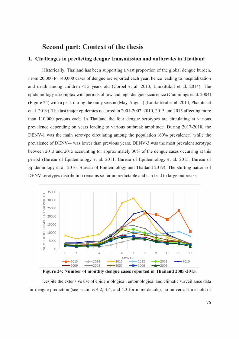
76
Second part: Context of the thesis
1. Challenges in predicting dengue transmission and outbreaks in Thailand
Historically, Thailand has been supporting a vast proportion of the global dengue burden.
From 20,000 to 140,000 cases of dengue are reported each year, hence leading to hospitalization
and death among children <15 years old (Corbel et al. 2013, Limkittikul et al. 2014). The
epidemiology is complex with periods of low and high dengue occurrence (Cummings et al. 2004)
(Figure 24) with a peak during the rainy season (May-August) (Limkittikul et al. 2014, Phanitchat
et al. 2019). The last major epidemics occurred in 2001-2002, 2010, 2013 and 2015 affecting more
than 110,000 persons each. In Thailand the four dengue serotypes are circulating at various
prevalence depending on years leading to various outbreak amplitude. During 2017-2018, the
DENV-1 was the main serotype circulating among the population (60% prevalence) while the
prevalence of DENV-4 was lower than previous years. DENV-3 was the most prevalent serotype
between 2013 and 2015 accounting for approximately 30% of the dengue cases occurring at this
period (Bureau of Epidemiology et al. 2011, Bureau of Epidemiology et al. 2015, Bureau of
Epidemiology et al. 2016, Bureau of Epidemiology and Thailand 2019). The shifting pattern of
DENV serotypes distribution remains so far unpredictable and can lead to large outbreaks.
Figure 24: Number of monthly dengue cases reported in Thailand 2005-2015.
Despite the extensive use of epidemiological, entomological and climatic surveillance data
for dengue prediction (see sections 4.2, 4.4, and 4.3 for more details), no universal threshold of
0
5000
10000
15000
20000
25000
30000
35000
1 2 3 4 5 6 7 8 9 1 0 1 1 1 2
NU
MB
ER
OF D
EN
GU
E C
ASE
S R
EP
OR
TE
D
MONTH2015 2014 2013 2012 2011 2010
2009 2008 2007 2006 2005

77
transmission risk could be yet established. More importantly, outbreaks continue to occur and the
overall dengue incidence is increasing, despite efforts of national program to control the disease.
Thailand has established entomological thresholds to assess higher risk of dengue transmission as
CI<1%, BI<50 and, HI<10% (see section 4.4.1), however, there are no evidence of general
adoption of these thresholds to predict any outbreaks. In addition, the decentralization of vector
control has led to differences in Vector Borne Disease Unit (VBDU) leaderships and capacities
resulting in varying efficacy in measuring entomology indices and dengue vector risk
(Bhumiratana et al. 2014).
In addition, each region of Thailand has distinct patterns of dengue transmission. Using
retrospective climatic and epidemiological data over the past 10 years Lauer et al. demonstrated
that predictive models performed differently according to the provinces especially regarding the
climatic data (Lauer et al. 2018). Moreover, they highlighted that interactions between climate
variables and dengue incidence varies over time and space, thus model should take into account
vector populations dynamic and their interactions with climatic factors to improve the prediction
accuracy (Chumpu et al. 2019). Yet, several gaps exist in estimating dengue transmission risk and
there is an urgent need to develop more cost-effective and practical tools that can reliably measure
dengue transmission risk and prevent outbreaks.
The current thesis took place in the context of the DENGUE-INDEX Project that was
developed to contribute to the development of early warning systems for dengue epidemics in
Thailand. The project was supported by the Research Council of Norway and was conducted in
North-eastern region of Thailand where dengue incidence is moderate (
Figure 25) but where several unpredictable outbreaks occurred in the last 5 years (see
section 1 for details). More information about the objectives and study design of the project are
briefly resume in the next section.
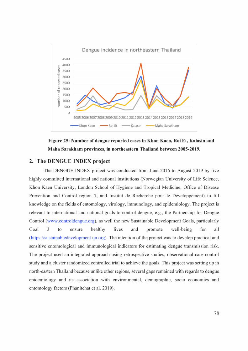
78
Figure 25: Number of dengue reported cases in Khon Kaen, Roi Et, Kalasin and
Maha Sarakham provinces, in northeastern Thailand between 2005-2019.
2. The DENGUE INDEX project
The DENGUE INDEX project was conducted from June 2016 to August 2019 by five
highly committed international and national institutions (Norwegian University of Life Science,
Khon Kaen University, London School of Hygiene and Tropical Medicine, Office of Disease
Prevention and Control region 7, and Institut de Recherche pour le Developpement) to fill
knowledge on the fields of entomology, virology, immunology, and epidemiology. The project is
relevant to international and national goals to control dengue, e.g., the Partnership for Dengue
Control (www.controldengue.org), as well the new Sustainable Development Goals, particularly
Goal 3 to ensure healthy lives and promote well-being for all
(https://sustainabledevelopment.un.org). The intention of the project was to develop practical and
sensitive entomological and immunological indicators for estimating dengue transmission risk.
The project used an integrated approach using retrospective studies, observational case-control
study and a cluster randomized controlled trial to achieve the goals. This project was setting up in
north-eastern Thailand because unlike other regions, several gaps remained with regards to dengue
epidemiology and its association with environmental, demographic, socio economics and
entomology factors (Phanitchat et al. 2019).
0
500
1000
1500
2000
2500
3000
3500
4000
4500
2005 2006 2007 2008 2009 2010 2011 2012 2013 2014 2015 2016 2017 2018 2019
nu
mb
er
of
rep
ort
ed
ca
ses
Dengue incidence in northeastern Thailand
Khon Kaen Roi Et Kalasin Maha Sarakham
79
The specific outcomes of this project were:
1. To assess the seasonal pattern of dengue transmission in North-eastern Thailand and to
identify local clusters of symptomatic disease based on reported dengue cases (retrospective
study)
2. To assess the accuracy of entomology and immunology indices in discriminating between
dengue positive and negative households and identify potential cofactors as part of a Case
Control study with passive detection (see details in section 4.1).
3. To assess the relationships between entomological and immunological indices and dengue
incidence to develop dengue outbreak predictive models though a cohort longitudinal study
(see details in section 4.3).
4. To assess the accuracy of various entomological, epidemiological and immunology indices in
evaluating vector control intervention based on pyriproxyfen as part of a Cluster Randomized
Control Trial (RCT) with active case detection (see details in section 4.3).
The project included a strong collaboration with the Ministry of Public Health (MoPH) through
the involvement of local authorities. Moreover, dissemination and communication of findings to
local communities, national authorities and regional and international stakeholders were provided
throughout the lifetime of the project at local and stakeholder engagement meetings. The expected
outcomes of the project were to provide national authorities solutions to better forecast epidemics
and plan and execute appropriate and timely interventions. These were not only important for
Thailand, but also for the whole Southeast Asia region and further afield.
3. Objectives of the thesis
This thesis was conducted in the framework of the DENGUE-INDEX project and aimed
specifically to estimate the risk of dengue transmission in North-eastern Thailand using various
entomological and serological indicators and to identify the main determinants associated with
Aedes mosquito exposure using specific salivary biomarker. The thesis had four specific objectives
as follows;
80
3.1. Objective #1: Assessing the spatial and temporal dynamic of dengue in
North-eastern Thailand
This baseline study aimed to assess the seasonal patterns of dengue incidence in Khon Kaen
province and to identify potential factors contributing to dengue dispersion at a fine-spatial
resolution scale. To do so, we carried out a retrospective epidemiological study using monthly
dengue incidence and climatic data at the sub-district level, to better understand dengue-climate
relationships and to identify periods and areas at higher risk of dengue transmission. Data on
dengue cases were retrieved from the national communicable disease surveillance system in
Thailand. The association between monthly disease incidence and climate variations was analysed
at the sub-district level using Bayesian poison regression models while Local Indicators of Spatial
Association (LISA) were used to identify significant “hotspots” (and “coldspots”) for dengue
transmission. This study aimed to get a better picture of the dengue epidemiology in the study area
and to get more reliable prediction models for future projections applied in early warning and
response systems.
3.2. Objective #2: Addressing the complex relationship between Aedes
vectors, dengue transmission and socio-economic factors
The objective was to identify entomological and immunological indices capable of
discriminating between dengue case and control (non-case) houses in North-eastern Thailand. To
do so, we conducted a case-control study (see section 4.1) to assess whether houses with and
without dengue cases exhibited different “profiles” in terms of human exposure risk to Aedes
mosquito bites, as measured by the levels of IgG response to salivary antigens and, Aedes
infestation levels as measured by the presence and abundance of immature and adult stages. We
assumed that people at higher risk of mosquito exposure risk (as measured by entomology and/or
immunology outcomes) may be also at higher risk of dengue transmission. Finally, we assessed
whether socio-economics, individual and household characteristics may represent additional “risk
factors” for acquiring dengue infection.
81
3.3. Objective #3: Assessing fine-scale variations in human exposure to Aedes
mosquito bites using salivary biomarker during a vector control
intervention.
This study was conducted as part of a RCT (see section 4.3) aiming to evaluate the efficacy
of a new vector control intervention for dengue prevention based on Pyriproxyfen (IGR). The
objective was to assess the relationship between the intensity of the Ab response to the Aedes
salivary peptide and the levels of Aedes infestation prior and after the deployment of the
intervention. Risk factors associated with “Aedes exposure risk” including individual and
household characteristics, human behaviour, and environmental factors were also explored.
Finally, we investigated potential relationships between the intensity of Ab response to Aedes
mosquito bites and DENV vector infectivity at the household levels. The idea was to generate
robust evidence to validate the use of the Aedes salivary biomarker as proxy for estimating dengue
transmission risk and evaluate vector control intervention in Thailand.
3.4. Objective #4: Evaluating the impact of vector control intervention on the
selection of insecticide resistance in dengue vectors
This chapter, which slightly differs from the three previous ones, aimed to assess changes
in insecticide resistance traits in local dengue vector populations following the deployment of the
pyriproxyfen-based vector control intervention (see details in section 4.3). The rational beyond
that study is that pyriproxyfen deployed in permanent breeding containers may select for
insecticide resistance hence potentially impacting on the effectiveness of the intervention. To do
so, we conducted mosquito collections to assess the levels and mechanisms of insecticide
resistance through a combination of biological and molecular assays, prior, during and after the
deployment of the intervention. Several candidate resistance markers were followed up for almost
2 years and correlation between resistance phenotype and genotype were addressed. This
information is deemed important to determine possible causes of vector control failure and to guide
vector control policies in Thailand.
The study design, including methods, randomization, endpoints and statistics are summarized in
the following sections.

82
4. Study design
4.1. Study area
This study was conducted in the North-Eastern region of Thailand, mainly in Khon Kaen
and Roi Et provinces but also in Maha Sarakham and Kalasin provinces. This region, known as a
part of the Isan area, is on the Khorat Plateau (up to 187m of elevation) and crossed by the Chi
river. The landscape is slightly hilly, with numerous swamps and the altitude varies between 90
and 180m above sea level (
Figure 26). Isan region is the third is terms of inhabitants yet, the region contributes only
to ten percent of the national gross domestic product. The region is mostly rural with few densely
populated urban city centers. The main sector of Isan economy is the agriculture, in particular
sticky rice, yet since the 1970’s trade and service sectors have increased due to the difficulties of
farming (World Bank Group 2016) and rural-urban migration is common in this region of Thailand
for multiple reasons including job opportunities, standard of living, better education, and health
facilities (Katewongsa 2015). The total population of these four provinces is about 5 million
inhabitants (National Statistical Office 2010).
Figure 26: Isan typical landscape.
4.2. Case control study
The first part of the study included a hospital-based prospective case control study
conducted in the north-eastern region of Thailand (Fustec et al. 2020). The case control study was
initially carried out in two provinces of north-eastern Thailand, Khon Kaen and Roi Et, and was

83
extended to two additional provinces, Kalasin and Maha Sarakham. A total of nineteen community
district and sub-district hospitals in Khon Kaen, Roi Et, Kalasin and Maha Sarakham provinces
were asked to participate (Figure 27). Hospitals were selected based on good clinical practices to
detect dengue cases and willingness to participate in the study. The study collections began in June
2016 and was carried until sample size, calculated using data from Thomas et al. (Thomas et al.
2015), was reached in August 2019.
At the hospital, presumptive dengue cases were diagnosed using SD Duo Bioline RDT
(Figure 28). Consenting dengue positive (cases) and negative patients (controls) were included in
the study. Venous blood was taken for DENV detection and serotyping (Shu et al. 2003). Within
the day of recruitment, entomological teams were mandated to visit each patient household to
collect data on household characteristics, e.g., number of family members, sex, age, travel history,
socioeconomic status, GPS position, etc. Additionally, entomological collections were performed
at the houses of case and control patients, and in the four neighbouring houses. Collections
included immature and adult stage of Aedes, captured using batterie-driven aspirators for 15 min
each, indoors and outdoors. Furthermore, DENV infection was investigated among Aedes females
using RT-qPCR (Lanciotti et al. 1992). Several entomological and immunological indices were
used as described in Table 5. Entomological indices in patients’ houses were distinguished from
those at the neighbourhood level (i.e., patient’s house + four surrounding houses, Table 5). The
Aedes-specific immune response was evaluated in each case and control patient from dry blood
spots by an indirect Enzyme-Linked Immunosorbent Assay (ELISA) using the Nterm-34kDa
salivary peptide (Genepep, St Jean de Vedas, France) and was expressed as a differential optical
density (ΔOD). The specific immune threshold (TR) was calculated by measuring the Ab response
to the Nterm-34 salivary peptide from individuals with no known exposure history to Ae. aegypti.
Thus, the mosquito exposure index (MEI) was defined as the sample-specific immune response to
the salivary peptide (Table 5).

84
Figure 27: Map and characteristics of study sites of the case-control study in northeastern Thailand. A: Location of four provinces and study districts in north-eastern Thailand included in
the case-control study. Map of study sites was built using QGis 3.10 software and shapefiles were obtained from the Humanitarian Data Exchange project (Humanitarian Data Exchange Project 2019) (CC BY 4.0). B: Study area characteristics, population and average number of
dengue cases per year from 2005-2019 (National Statistical Office 2010).

85
Figure 28: Flow diagram of case-control study design.
The data were analysed using R software version 3.5.1 (R Core Team, Vienna, Austria).
The study population was analysed with descriptive statistics, and individuals’ information and
household characteristics were analysed with the dengue case occurrence as categorical variables
using univariable logistic regression. The socio-economic status (SES) of each patient was
calculated as a score based on the household questionnaire (e.g., assets, income) using principal
component analysis (Vyas et al. 2006). Univariable binomial logistic regression was performed
between each entomological and immunological index and dengue case/control status.
Multivariable logistic regression was performed using all variables (i.e., individual characteristics,
house characteristics, SES, entomological and immunological indices) with a statistically
significant association (p<0.1) with case/control status on the univariable analysis. Model selection

86
was based on backward/forward Akaike Information Criterion (AIC) selection with the selected
model was the one with the lowest AIC.
This study was approved from the Khon Kaen University Ethics Committee (KKUEC,
project number HE591099), the London School of Hygiene and Tropical Medicine Ethical
Committee (LSHTM Ethics, project number 10534), and the Norwegian Regional Committees for
Medical and Health Research Ethics (REC, no. 2016/357).
Table 5: Variable definition for the case control study.
Variable Definition / Formula Aedes life stage Individual level
MEI MEI= ∆OD-TR (with TR=0.450) IgG response to Nterm 34 kDa salivary peptide
Adult
Household level (patient house) CI (%) (No. positive container/ total no of wet container) x 100 Larvae & Pupae AI No. of adult female Aedes collected Adult AI_in No. of adult female Aedes collected indoors (only) Adult AI+ Proportion of infected females Aedes in the patient house Adult PHI No. of pupae collected at the patient house Pupae PPI No. of pupae collected per person at the patient house Pupae Neighborhood level
CIn (%) (No. positive container/ total no. of wet container) x 100 Larvae & Pupae HI (%) (No. positive house/ no. households visited) x 100 Larvae & Pupae
BI No. positive container x100/ No. containers inspected / no. house visited
Larvae & Pupae
AIn No. of adult female Aedes collected/ no. households visited Adult
AIn_in No of adult female Aedes collected indoor/ no. households visited
Adult
AIn+ Proportion of infected adult female Aedes/ no. houses visited Adult PHIn No. of pupae collected/ no. houses visited Pupae

87
4.3. Cluster-randomized controlled trial
The second part of the study included a cohort cluster-randomized controlled trial (RCT) that
was conducted in Khon Kaen and Roi Et cities. The complete and described protocol was published
in Trials in February 2018 (see the section Publication4) and corrected in December 2018 (see the
section Publication 4).
Briefly, the study started in September 2017 in KK city and October 2017 in RE city, and was
conducted during 24-months in each city (Figure 29). The effectiveness of the vector control
intervention was measured using immunological and entomological indices (see details in
Overgaard et al 2018) including the abundance of Aedes adult female and vector infectivity and
the intensity of the human Ab response to the Aedes salivary antigen. The effect of the intervention
on the number of dengue cases was also assessed as a secondary outcome measure (paper under
preparation). In addition, the entomological, immunological and climatic indices were used to
develop predictive models of dengue transmission and outbreaks.
The full protocol was published in Trials (2018) 19:122, https://doi.org/10.1186/s13063-018-2490-1, Assessing dengue transmission risk and a vector control intervention using entomological and immunological indices in Thailand: study protocol for a cluster randomized controlled trial. Hans J. Overgaard, Chamsai Pientong, Kesorn Thaewnongiew, Michael J. Bangs, Tipaya Ekalaksananan, Sirinart Aromseree, Thipruethai Phanitchat, Supranee Phanthanawiboon, Benedicte Fustec, Vincent Corbel, Dominique Cerqueira and Neal Alexander. And corrected in Trials (2018), 19(1), 703. https://doi.org/10.1186/s13063-018-3110-9, Correction to: Assessing dengue transmission risk and a vector control intervention using entomological and immunological indices in Thailand: study protocol for a cluster-randomized controlled trial.

88
Figure 29: Map of the study sites of the RCT.
Sample size was calculated based on mosquito data from the case control study and the
dengue incidence during the previous 10 years (2006-2015). A total of 18 clusters (city blocks) in
each city were included with 10 HHs per cluster (n=180 HHs per city). Statistical methods for
cluster-randomized trials were used to calculate this sample size (Hayes et al. 1999). Households
were monitored weekly for presumptive dengue cases (fever cases) during 24 months using RDT
by health volunteers. In addition, dried blood spots on filter paper were taken from each
presumptive dengue case for immunological assays. In addition, entomological collections,
including adult, pupae and larvae Aedes, were conducted in all households every four months
(Figure 30). Concomitantly to the entomological investigation, dried blood spots were taken from
selected inhabitants to assess the human exposure to mosquito bites. Additionally, entomological
collections and dried blood spot collections were done every month in three sentinel HHs per
cluster.

89
Figure 30: Flow chart of the RCT study design
After a 10 months baseline and just before the next rainy season, half of clusters in each
city were randomly selected for a vector control intervention consisting of pyriproxyfen (applied
as a 0.5% granule formulation) distributed every four months in permanent breeding habitats
(targeted dose of 0.01 mg/L as per WHO recommendation). The other clusters remained as control
and did not receive PPF intervention. Intervention and control clusters were followed-up for 14
additional months. Immunological index was calculated at the individual level and entomological
indices were calculated for HH clusters all long the study. Impact of the trial was measured using
logistic regression on adult Aedes index (AI) summarized by cluster using logistic negative
binomial regression on the total number of Aedes collected and the total number of houses
inspected. Additionally, dengue incidence in study households will be analyzed using negative
binomial regression. Others entomological endpoints were analyzed the same way as the AI.
Moreover, the study attempted to predict dengue incidence over time using entomological and
immunological indices, by predicting the risk of a future outbreak, and by estimating associations

90
between dengue incidence and the indices. This trial was registered (ISRCTN, ISRCTN73606171)
and approved by the Khon Kaen University Ethics Committee (KKUEC Record No. 4.4.01:
29/2017, Reference No. HE601221, 1 September 2017), the London School of Hygiene and
Tropical Medicine Ethical Committee, UK (LSHTM Ethics Ref: 14275, 16 August 2017), and the
Regional Committee for Medical and Health Research Ethics, Section B, South East Norway (REK
Ethics ref: 2017/1826b, 03 March 2018).
Simultaneously, complementary mosquito collections were conducted as part of the RCT
to evaluate the susceptibility levels of Aedes mosquitoes to the insecticide used (PPF)
comparatively to the one’s used in routine by the ODPC7 (temephos and deltamethrin at 0.5%).
Briefly, larvae collections were performed in sentinel HHs, three times; at the beginning of the
RCT (baseline), six months after the beginning of the vector control intervention and hence one-
year post intervention to assess change in the levels and/or mechanisms of resistance over time.
All collections included both treated and control clusters. Susceptibility status of Ae. aegypti
populations was investigated using standard WHO susceptibility tests (World Health Organization
2005, World Health Organization 2013, World Health Organization 2016b). Additionally, relevant
DNA markers (kdr mutations) and changes in CNV in genes associated with metabolic resistance
were explored following protocols already established by Saavedra-Rodriguez et al (Saavedra-
Rodriguez et al. 2007), Yanola et al (Yanola et al. 2011) and Cattel et al (Cattel et al. 2020b).

STUDY PROTOCOL Open Access
Assessing dengue transmission risk and avector control intervention usingentomological and immunological indicesin Thailand: study protocol for a cluster-randomized controlled trialHans J. Overgaard1* , Chamsai Pientong2,3, Kesorn Thaewnongiew4, Michael J. Bangs5,6, Tipaya Ekalaksananan2,3,Sirinart Aromseree2,3, Thipruethai Phanitchat2, Supranee Phanthanawiboon2,3, Benedicte Fustec2,7, Vincent Corbel8,Dominique Cerqueira6 and Neal Alexander9
Abstract
Background: Dengue fever is the most common and widespread mosquito-borne arboviral disease in the world.There is a compelling need for cost-effective approaches and practical tools that can reliably measure real-timedengue transmission dynamics that enable more accurate and useful predictions of incidence and outbreaks.Sensitive surveillance tools do not exist today, and only a small handful of new control strategies are available.Vector control remains at the forefront for combating dengue transmission. However, the effectiveness of manycurrent vector control interventions is fraught with inherent weaknesses. No single vector control method iseffective enough to control both vector populations and disease transmission. Evaluations of novel larval and adultcontrol interventions are needed.
Methods/design: A cluster-randomized controlled trial will be carried out between 2017 and 2019 in urbancommunity clusters in Khon Kaen and Roi Et cities, northeastern Thailand. The effectiveness of a pyriproxyfen/spinosad combination treatment of permanent water storage containers will be evaluated on epidemiological andentomological outcomes, including dengue incidence, number of female adult dengue vectors infected or notinfected with dengue virus (DENV), human exposure to Aedes mosquito bites, and several other indices. Theseindices will also be used to develop predictive models for dengue transmission and impending outbreaks.Epidemiological and entomological data will be collected continuously for 2 years, with the interventionimplemented after 1 year.(Continued on next page)
* Correspondence: [email protected] University of Life Sciences, Ås, NorwayFull list of author information is available at the end of the article
© The Author(s). 2018 Open Access This article is distributed under the terms of the Creative Commons Attribution 4.0International License (http://creativecommons.org/licenses/by/4.0/), which permits unrestricted use, distribution, andreproduction in any medium, provided you give appropriate credit to the original author(s) and the source, provide a link tothe Creative Commons license, and indicate if changes were made. The Creative Commons Public Domain Dedication waiver(http://creativecommons.org/publicdomain/zero/1.0/) applies to the data made available in this article, unless otherwise stated.
Overgaard et al. Trials (2018) 19:122
https://doi.org/10.1186/s13063-018-2490-1
(Continued from previous page)
Discussion: The aims of the trial are to simultaneously evaluate the efficacy of an innovative dengue vector controlintervention and developing predictive dengue models. Assessment of human exposure to mosquito bites bydetecting antibodies generated against Aedes saliva proteins in human blood samples has, so far, not been appliedin dengue epidemiological risk assessment and disease surveillance methodologies. Likewise, DENV detection inmosquitoes (adult and immature stages) has not been used in any practical way for routine disease surveillancestrategies. The integration of multiple outcome measures will assist health authorities to better predict outbreaks forplanning and applying focal and timely interventions. The trial outcomes will not only be important for Thailand,but also for the entire Southeast Asian region and further afield.
Trial registration: ISRCTN, ISRCTN73606171. Registered on 23 June 2017.
Keywords: Dengue monitoring, Entomology, Immunology, Dengue index, Risk assessment, Vector control
Background
Dengue fever is the most common and widespread arbo-
viral disease in the world, with an estimated four billion
people in at least 128 countries at risk of infection [1].
The exact global burden of dengue is not known, but
there are estimates of about 390 million infections annu-
ally, of which only a minority (~ 25%) manifest clinically
[2]. Yearly mortality figures of > 20,000 deaths have been
reported [3]. Dengue fever and other arboviral diseases,
such as Zika and chikungunya, are transmitted to
humans primarily by Aedes aegypti and Aedes albopictus
mosquitoes. There is currently no specific treatment for
dengue and only recently has a vaccine been licensed,
but it does not confer full protection for all virus sero-
types [4]. Even with effective therapies and vaccines, vec-
tor control will likely remain important to curtail disease
incidence and outbreaks. Perennial dengue incidence
varies seasonally, and dengue outbreaks occur periodic-
ally in most endemic countries [5]. Infection of one of
the four dengue virus serotypes (DENV1—4) typically
confer lifelong protective homotypic (type-specific)
immunity as well as production of more time-limited
cross-reactive heterotypic neutralizing antibodies [6];
however, antibody-dependent enhancement may result
in a second DENV serotype infection inducing a more
severe clinical course [5].
For improved dengue control, reliable epidemic fore-
casting systems for early detection of temporal anomal-
ies in disease incidence are needed, as well as more
effective control strategies that affect both entomological
and epidemiological endpoints. Sensitive surveillance
tools do not exist today, and only a small handful of new
control strategies are available [7–14]. For example, al-
though temephos — the most commonly used chemical-
based vector control method, used against immature
mosquito stages — may be effective in reducing entomo-
logical indices, there is no evidence showing it reduces
dengue transmission [7].
There is a compelling need to develop cost-effective
approaches and practical tools that can reliably measure
real-time dengue transmission dynamics that enable
more accurate and useful predictions of outbreaks. Cur-
rently, there is no universally accepted definition of what
constitutes an outbreak [15]. This complicates the inter-
pretation of early detection of cases that exceed expected
normal seasonal variations. In many endemic countries,
a dengue outbreak is declared when the number of
reported cases during a specific time period (week or
month) surpasses the historical average of the preceding
5 to 7 years above two standard deviations (SD), known
as the endemic channel [5]. Outbreak definitions vary
depending on how the historical average is calculated,
which may involve, for example, the number of years
used, type of mean (e.g., monthly or moving mean),
whether or not outbreak years are included, how the
critical threshold is calculated (e.g., ±2 SD), and criteria
used to define the outbreak (e.g., time above the thresh-
old before a response is triggered) [15]. Ideally, when an
outbreak alert has been triggered, standard vector con-
trol strategies should be implemented. However, current
early warning systems and detection of outbreaks are
usually neither accurate nor timely enough to initiate
effective control interventions (outbreak response) to
curb increased transmission after it has begun [11].
Various entomological indices are used to measure
dengue vector infestation in and around structures
(homes, buildings, etc.). However, these indices are sel-
dom sensitive enough to precisely estimate dengue
transmission risk or predict impending outbreaks [16,
17]. The Stegomyia indices, i.e., House index (HI, pro-
portion of Aedes positive houses) and Container index
(CI, proportion of Aedes positive containers) were devel-
oped nearly a century ago [18], followed by the Breteau
index (BI, number of Aedes positive containers per 100
houses) [19]. These three measures are currently the
most commonly used indices to assess dengue vector
larval habitat infestations. They are relatively easy to
measure, but are generally not correlated with disease
incidence or outbreak risk [16]. In the 1990s, Focks et al.
explored the use of pupal surveys as a potentially more
Overgaard et al. Trials (2018) 19:122 Page 2 of 19

epidemiologically relevant index, correlating total pupal
densities with resultant adult densities [20]. This led to
further development of entomological thresholds using a
pupal/demographic method, ambient temperature, and
seroprevalence of dengue antibodies in the population
[21]. As a result, container-specific, targeted source re-
duction was proposed by identifying the relative import-
ance of major types of container habitats with high
pupal productivity that contribute significantly to the
transmission threshold [21]. However, it remains unclear
if targeting only containers that are responsible for the
vast majority, say 80–90%, of the pupal production [22–
26] is sufficient to have an epidemiological impact on
transmission. Other aquatic habitats may be important,
such as unusual and cryptic sites, which are typically
overlooked during vector control interventions [27, 28].
Furthermore, in many settings, such as in northeastern
Thailand and southern Laos, as many as eight to ten of
the most productive container types might only produce
< 70% of all pupae [29]. Although there are perceived
benefits to targeting only the most productive con-
tainers, such as reduced time and effort, they may not
compensate for ignoring control of other less obvious
breeding habitats. The difficulty in finding and effect-
ively treating such cryptic sites can be addressed by
using pyriproxyfen, a potent insect growth regulator,
which can be transferred between habitats by female Ae-
des mosquitoes during oviposition, a strategy called
auto-dissemination [30–32].
A recent study from Iquitos, Peru investigated the
relationship between several indicators of Ae. aegypti
abundance and DENV infection in humans using more
than 8000 paired serological samples with corresponding
entomological data [17]. The researchers found that in-
dicators based on cross-sectional entomological surveil-
lance, i.e., data from a single survey observation, are of
little practical use. On the other hand, longitudinal-
based, household-level entomological indicators using
data from up to three yearly visits before a 6-month
seroconversion period showed that the presence of adult
female Ae. aegypti in a household increased the risk of
DENV seroconversion by approximately 29% compared
to households without mosquito vectors. The authors
therefore challenged the assumption that most common
Ae. aegypti indicators provide adequate proxies for
DENV risk and transmission [17].
The general response by national dengue control pro-
grams to indications of increased disease transmission
and possible outbreaks mainly consists of reactive vector
control. Typically, control activities involve application
of temephos (an organophosphate compound) to domes-
tic water storage containers for larval control and/or
peridomestic space spraying with an insecticide, most
commonly a pyrethroid-based formulation, for adult
control. Although, these interventions may reduce vector
populations dramatically, there is no evidence that they
reduce dengue transmission substantially [7, 9]. Other
possible options for vector control are community-based
source reduction campaigns, application of bacteria-based
larvicides, larvivorous fish, or copepods, or combinations
of these approaches [8, 33–35]. Newer paradigms for
Aedes vector control include microbial control of human
pathogens in adult vectors, such as Wolbachia bacteria
that shorten the lifespan of mosquitoes [36] and release of
transgenic Ae. aegypti engineered to carry a dominant
lethal gene that suppresses mosquito populations [37].
These novel approaches are currently not recommended
for full-scale programmatic deployment by the World
Health Organization (WHO) Vector Control Advisory
Group, but rather implemented as carefully planned pilot
interventions under operational conditions [38]. The ef-
fectiveness of many vector control interventions is fraught
with inherent weaknesses, e.g., widespread insecticide re-
sistance, quality of delivery, and other operational issues,
such as availability and cost of insecticide, dedicated and
trained personnel, and appropriate application equipment
[39, 40].
The WHO Global Strategic Framework for integrated
vector management (IVM) was released in 2004 and
recommends a range of interventions, in combination,
to increase impact [41]. This means there is no single
vector control method that is effective enough to control
both vector populations and disease transmission. Com-
binations of larval control interventions, such as mix-
tures of pyriproxyfen (an insect growth regulator) and
spinosad (a biopesticide) have been evaluated. This com-
bination reduced larval and pupal relative densities by
90% for at least 4 months in the French West Indies
[42]. Pyriproxyfen, even in minute doses, can induce
complete inhibition of adult emergence for several weeks
after treatment [43]. Pyriproxyfen used alone and
applied to storm drains in Colombia reduced dengue
cases by 80% [44]. The benefit of using these two com-
pounds in combination is that they have different modes
of action and that pyriproxyfen targets the pupal stage
while spinosad is active against larval stages. Both com-
pounds have very low toxicity for humans and most other
non-target fauna [45, 46]. The WHO draft on global vec-
tor control response for 2017–2030 [47] builds on the
IVM approach but places stronger emphasis on enhancing
human capacity and health education, increasing research
and innovation by strengthening infrastructure, and
increasing intersectoral and interdisciplinary action. The
targets of the global response are to reduce mortality and
incidence due to all vector-borne diseases globally relative
to 2016 by at least 75% and 60%, respectively.
In view of the preceding discussion, this study aims to
assess a specific vector control intervention and to
Overgaard et al. Trials (2018) 19:122 Page 3 of 19

contribute to the development of a practical early warn-
ing system that can more accurately predict changes in
dengue transmission and impending outbreaks. The trial
will determine the efficacy of a pyriproxyfen/spinosad
combination in water storage containers to reduce ento-
mological risk indicators and dengue incidence. The
hypothesis is that the study arm receiving the combin-
ation treatment in household water storage containers
will have a lower density of adult female Ae. aegypti per
house, both indoors and outdoors, compared to the
study arm receiving an alternative intervention involving
normal governmental action. Furthermore, the study
aims to determine one or more entomological indices
and an immunological index that best predicts dengue
incidence for the study area.
Methods/design
Objectives
The specific objectives of this trial are to:
1. Assess the effect of periodically treating water
storage containers with a pyriproxyfen/spinosad
combination on entomological and epidemiological
outcomes
2. Determine the most accurate and precise index or
indices to predict variation in dengue incidence in
time
Trial design
A stratified, cluster-randomized controlled trial is de-
signed to study the effect of a vector control interven-
tion in households located in pre-selected clusters in
two urban areas in northeastern Thailand. Each cluster
is randomized to one of two arms: intervention clusters
receiving treatment of water containers with a pyriprox-
yfen/spinosad combination, and control clusters not
receiving any intervention from this project. Control
areas will rely on normal operational vector control
interventions performed by the local public services.
Randomization of arms is stratified by city (Khon Kaen
and Roi Et, i.e., two levels). Stratification is done because
there are potential differences between the two cities
that may affect the outcomes (e.g., population size, and
regional importance in terms of travel, commerce,
services, health care, and education); therefore, stratifica-
tion may reduce the residual statistical error when one
compares the two arms. Including two cities may also
alleviate problems of low incidence caused by the spatial
and temporal variation in dengue transmission; i.e., one
area functions as a backup if there are few cases in the
other. Including two cities should also increase the
generalizability of the trial.
A cluster design is considered the best option because
the intervention is not performed on the individual level,
but is rather a spatial, area-wide approach involving
treatment of containers in and immediately around each
house and property in a study area (cluster). Within a
household, it is not feasible to randomize some individ-
uals to one intervention and other individuals to an-
other. Furthermore, the entomological outcomes, both
primary (Adult index) and secondary (e.g., Pupal index
per person, and the Breteau index), will be estimated on
a household level. We are using the larger clusters rather
than single houses because (1) there may be mass (area)
effects of the interventions, whereby entomological indi-
ces in each house may depend partly on the abundance
of mosquitoes in neighboring ones, and (2) entire clus-
ters having the same intervention more closely resem-
bles how the intervention would be implemented,
should it be scaled up.
Setting
The trial is carried out in two urban areas, Khon Kaen
(N16.440236, E102.828272) and Roi Et (N16.055637,
E103.652417) cities in northeastern Thailand (Fig. 1).
Khon Kaen is the capital city of the province with the
same name. The province has an area of ~ 10,900 km2,
divided into 26 districts and a population of 1,741,980 in
the 2010 national census [48]. Khon Kaen district is the
largest by area and population, with a population of
around 400,000 over an area of 953.4 km2 (population
density 416 persons/km2). The district is divided into 17
sub-districts with 272 villages. In 2016, there was a
population of 269,247 in six sub-districts that make up
greater Khon Kaen (within the ring road) with a resident
density of 2500/km2 in the central parts. Roi Et is the
capital city of Roi Et province and is divided into 20 dis-
tricts covering a total area of 8300 km2 with a popula-
tion of 1,084,985 in 2010 [48].The largest district is also
called Roi Et, and it has ~ 160,000 inhabitants, covers an
area of 493.6 km2, and has a population density of
approximately 311 inhabitants/km2. There are 15 sub-
districts and 195 villages in Roi Et district. The largest
sub-district is Nai Mueang Roi Et municipality with a
population of approximately 34,000 inhabitants. To
delimit the study area, only villages completely within
each city’s primary access ring road are selected. We use
the English word “village”, although most are urban
divisions. In Khon Kaen, there are 162 villages in six
sub-districts within the ring road. In Roi Et, there are 56
villages in nine sub-districts within the ring road,
although only 39 have clearly demarcated administrative
boundaries.
Between 2006 and 2016, the total number of reported
dengue cases (uncomplicated and severe categories)
reported in Khon Kaen province was 15,195 (mean 1381
cases/year, range 439–3014), providing an incidence rate
of 76.7 cases/100,000 population. In Khon Kaen district,
Overgaard et al. Trials (2018) 19:122 Page 4 of 19
the number of cases during the same period was 7209
(mean 655 cases/year, range 204–1705), with an estimated
incidence of 455.3 cases/100,000. The corresponding num-
bers for Roi Et province for the same period were 20,174
total cases (mean 1834 cases/year, range 402–4141) and an
incidence of 140.2 cases/100,000. In Roi Et district, 3956
cases were reported during the period (mean 360 cases/
year, range 71–914) with an incidence of 329.1 cases/
100,000. All dengue case data were provided by the Office
of Disease Prevention and Control Region 7, Khon Kaen,
Ministry of Public Health.
Outcomes
The primary outcome is the Adult index (AI, the
number of female adult Ae. aegypti and Ae. albopictus
collected per house) (Table 1). The AI is based on com-
bined indoor and outdoor collections using mechanical,
battery-powered aspirators for 30 min (2 × 15 min) by
staff of the local public health departments. The AI for
each species will be recorded separately, although the
general expectation is to find a predominance of Ae.
aegypti in all urbanized clusters.
The secondary outcomes are the dengue incidence rate
(DIR), mosquito exposure index (MEI), infected adult
index (IAI), adult sticky trap index (ASTI), pupae per
person index (PPI) and BI (Table 1). Dengue cases (inci-
dence) is a secondary endpoint, because the sample size
required to detect a difference would be unfeasibly large.
In addition to the intervention-related outcomes, the
study will attempt to predict dengue incidence over time
using entomological and immunological indices. The
main outcomes for this part are identification of mea-
sures (indices) with sufficient accuracy and precision in
terms of predicting dengue outbreaks as defined by the
Ministry of Public Health.
Sample size
The sample size is calculated based on data on adult
female Aedes mosquitoes collected using mechanical
aspirators (15 min outdoors and indoors each) from a
case-control study in nearby districts during 2016 and
2017. The mean capture was 0.78 mosquito per house
(indoors + outdoors). At minimum, 34 clusters are
needed to detect a 90% difference in adult female
mosquitoes per house with 90% statistical power and a
two-sided significance level (α) of 0.05 assuming 10
households are visited three times each after the inter-
vention begins and a between-cluster coefficient of
variation of 0.33. Sample size methods for cluster-
randomized trials were used [49], as implemented in the
‘clustersampsi’ add-on to Stata® statistical software [50].
The existing data were over-dispersed relative to a
Poisson distribution; therefore, to represent a negative
binomial distribution with a dispersion parameter (α, or
1/k) of 2.03 estimated from the same data, the ‘means’
option was used, with the variance in each arm equal to
the mean plus the square of the mean times α [51]. The
Fig. 1 Study locations in northeastern Thailand. The red borders around the cities (right insets) are the respective ring roads.
Overgaard et al. Trials (2018) 19:122 Page 5 of 19

Table 1 Primary and secondary outcome measures and other entomological indices
Outcome No. Indexabbreviation
Indexname
Description Unit Frequency of data collection Details
Primaryoutcome
1 AI Adultindex
Number of female adultAe. aegypti and Ae.albopictus per housecollected both indoorsand outdoors
No./house
Once every 4 months in allhouseholds (HHs) and onceevery month in 3 HHs percluster (the same ones each time)
Adult collections using amechanical battery-poweredaspirator for 30 min per house(indoors and outdoors)
Secondaryoutcomes
2 DIR Dengueincidencerate
Number of confirmeddengue cases/observation days ofhousehold populations
Rate Weekly VHVs detect fever cases.Hospital and project staffcollect blood samples
3 MEI Mosquitoexposureindex
(1) Differential opticaldensity for antibodies toAe. aegypti saliva(2) Proportion abovethe immune thresholdfor this assay
(1)Number(2) %
Once every 4 months in all HHsand once every month in 3 HHsper cluster (the same HH asfor the AI)
Recurring blood spots fromtwo persons per HH takenon filter paper. IgG antibodyresponse (positive or negative)to the Ae. aegypti Nterm-34kDa salivary peptide
4 IAI Infectedadultindex
Number of DENV-infected adult femaleAe. aegypti and Ae. albopictus
No./house
Once every 4 months in all HHsand once every month in 3 HHsper cluster
Based on adult mosquitocollections indoors using amechanical battery-poweredaspirator for 15 min per houseand DENV detection inindividual mosquitoes
5 ASTI Adultsticky trapindex
Total number of Ae.aegypti and Ae.albopictus femalescollected by sticky trapsper month
No./trap/month
7 consecutive days per month Adult mosquitoes collectedby sticky traps baited withhay infusion for 7 daysevery month
6 PPI Pupae perpersonindex
Number of Aedes pupaeper person
No./person
Once every 4 months in all HHsand once every month in 3 HHsper cluster
From immature collections.All pupae collected dividedby the number of householdparticipants
7 BI Breteauindex
Number of Aedespositive containers per100 houses
No./100houses
Once every 4 months in all HHsand once every month in 3 HHsper cluster
From immature collections.Cluster-level result
Stegomyiaindices
8 HI Houseindex
Proportion of housespositive for immature Aedes
% Once every 4 months in all HHsand once every month in 3 HHsper cluster
From immature collections.Cluster-level result
9 CI Containerindex
Proportion of containerspositive for immature Aedes
% Once every 4 months in all HHsand once every month in 3 HHsper cluster
From immature collections.Cluster-level result
Pupalindices
10 IPPI Infectedpupae perpersonindex
Number of DENVinfected Aedes pupaeper person
No./person
Once every 4 months in all HHsand once every month in 3 HHsper cluster
Based on PPI and DENVdetection. All infected pupaecollected divided by thenumber of householdparticipants
11 PHI Pupae perhouseindex
Number of pupaeper house
No./house
Once every 4 months in all HHsand once every month in 3 HHsper cluster
Pupal collections
12 IPHI Infectedpupae perhouseindex
Number of DENV-infected Aedes pupaeper house
No./house
Once every 4 months in all HHsand once every month in 3 HHsper cluster
Pupal collections andDENV detection
Adultindices
13 AII Adultindoorindex
Number of Ae. aegyptiand Ae. albopictusfemales per houseindoors
No./house
Once every 4 months in all HHsand once every month in 3 HHsper cluster
Adult collections indoors usinga mechanical battery-poweredaspirator for 15 min per house
14 IAII Infectedadultindoorindex
Number of DENV-infected Ae. aegypti andAe. albopictus femalesper house indoors
No./house
Once every 4 months in all HHsand once every month in 3 HHsper cluster
Based on AII andDENV detection
Overgaard et al. Trials (2018) 19:122 Page 6 of 19

clusters are split equally between the strata (Khon Kaen
and Roi Et). To provide an equal number of clusters in
each arm, one extra cluster is added per stratum, i.e.,
18 clusters per stratum (city) and 9 clusters per arm in
each stratum.
Eligibility criteria
Eligibility for participation in the trial is determined on
four levels: (1) location or village, (2) cluster of houses
within village, (3) households within cluster, and (4)
individuals within households (household residents)
(Table 2).
Recruitment
The 162 villages in Khon Kaen and 39 villages in Roi Et
located within the respective ring roads are the sampling
frames for each stratum. In each stratum, villages are
randomly sampled based on probability proportional to
population size, i.e., the population of occupied houses
(the target denominator of the primary endpoint). These
Table 1 Primary and secondary outcome measures and other entomological indices (Continued)
Outcome No. Indexabbreviation
Indexname
Description Unit Frequency of data collection Details
15 AOI Adultoutdoorindex
Number of Ae. aegypti andAe. albopictus females perhouse outdoors
No./house
Once every 4 months in all HHsand once every month in 3 HHsper cluster
Adult collections outdoorsusing a mechanical battery-powered aspirator for15 min per house
16 IAOI Infectedadultoutdoorindex
Number of DENV-infectedAe. aegypti and Ae. albopictusfemales per house outdoors
No./house
Once every 4 months in all HHsand once every month in 3 HHsper cluster
Based on AOI andDENV detection
17 IASTI Infectedadultsticky trapindex
Number of DENV-infectedAe. aegypti and Ae. albopictusfemales per sticky trap
No./trap/week
Once every 4 months in all HHsand once every month in 3 HHsper cluster
Based on ASTI andDENV detection
Premiseindex
18 PCI Premiseconditionindex
Degree of shade + conditionof house + condition of yard
Number(min = 3,max 9)
Once every 4 months in all HHs Observation criteria [90]
VHV village health volunteer, IgG immunoglobulin G
Table 2 Eligibility criteria by location, cluster, household, and individual
Level Inclusion criteria Exclusion criteria
Village - Within ring roads of each city (stratum) - Area < 0.125 km2
- Populated residential areas - Number of houses < 100
- Population < 300
- Coverage of residential area 70–80% (scattered housing)
- Non-residential areas, e.g., agricultural fields, airports,industrial areas, commercial areas, (e.g., shopping malls),government offices, lakes, army camps, hospitals, and schools
Cluster - All points of the cluster are at least 100 m fromthe nearest point of the village border
Household - Households that are permanently inhabited - Apartment buildings
- Abandoned houses
- Households that are built or re-populated duringthe study period
- Non-permanent households
Individual - Individuals in households where household head has signedinformed consent for household to participate in project
- A travel history outside the village during the previous 7 days
- Self-reported fever within the last 7 days - Chronic disease, such as HIV/AIDS, or other healthcondition that preclude participation in the study
- Age ≥ 1 year old - Apparent inability to give informed consent,e.g., due to mental disability
There is a distinction between being included in the final evaluation of endpoints and inclusion for receiving interventions. For example, abandoned houses and
non-permanent household structures are not included in evaluation of endpoints, but they may be treated with an intervention if they are located within a radius
of 100 m and as feasibly possible
Overgaard et al. Trials (2018) 19:122 Page 7 of 19

selected villages will be randomly allocated between the
arms (see the section on Assignment of interventions
below).
Villages are normally much larger than the target clus-
ter size (10 houses); therefore, to select a starting point
for the house selection, a 50 × 50 m grid and a 100-m
buffer zone inside of the village perimeter will be super-
imposed over each village map. The buffer zone of
approximately 100 m on the inside of each village border
is applied to reduce potential “contamination” (in-flying
mosquitoes) from neighboring villages. A random grid
cell is selected in each village and 10 houses nearest the
centroid of that cell selected. This procedure is followed
in each village in this manner as far as practically
possible. For example, if a selected household does not
want to participate in the study, a neighboring house will
be selected.
Informational meetings are held at the sub-district and
village administrative levels to provide information about
the project and benefits to the communities. House-
holders are visited and carefully informed about the
study, and informed consent is obtained from the house-
hold head. A complete enumeration of all participants in
the selected clusters will be completed with assistance
from the local administration and village health volun-
teers (VHVs). This enumeration will be done three times
during the study, allowing monitoring of potential
participants’ discontinuation in the trial. Data on discon-
tinuation, whether due to movement outside the study
area or withdrawal of consent, are relevant for the
secondary outcomes of DIR and MEI. Reasons for
potential discontinuation will be monitored and taken
into account in communication strategies to promote
retention. In addition, a minor monetary compensation
for those who provide blood samples for the mosquito
exposure study will promote participant retention to
complete follow-up of individuals.
Interventions
Following approximately 10–12 months of baseline data
collections, household interventions will begin in the
selected intervention clusters (Fig. 2). The intervention
specifically targets mosquito immature stages by applying
a mixture of pyriproxyfen and spinosad to all permanent
household containers, whether indoor or outdoor, found
to contain water up to a 10-m perimeter from the house.
Pyriproxyfen is an insect growth regulator (insect juvenile
hormone analog) that is active against pupal stages, result-
ing in the inhibition of adult development (preventing
emergence). It has low mammal toxicity and is recom-
mended by WHO for vector control [52]. Spinosad is a
natural insecticide produced by the soil bacterium
Saccharopolyspora spinosa. It has a neurotoxic mode of ac-
tion in insects, but with low mammal toxicity, and it is also
recommended by WHO [52]. The doses recommended by
WHO for Aedes immature mosquito control are 0.01 mg/L
active ingredient (a.i.) pyriproxyfen (applied as a 0.5%
granule formulation) and 0.1–0.5 mg/L a.i. spinosad
(also a 0.5% granule formulation) [45, 53]. Both pyri-
proxyfen and spinosad have also been assessed and
approved by WHO for use in drinking water containers
[54, 55]. The reasons for selecting this novel intervention
are that the combination of pyriproxyfen and spinosad has
not yet been tested in a national dengue vector control
program; that it should be effective, easy, and practical to
use for national control authorities; and that its combined
Fig. 2 Time schedule of enrollment, interventions, and pre- and post-allocation data collections (based on SPIRIT 2013 figure [91])
Overgaard et al. Trials (2018) 19:122 Page 8 of 19

use reduces the risk of resistance development. The inter-
ventions will be implemented by project staff from the
Ministry of Public Health, thereby ensuring adherence to
intervention protocols.
The combination larvicide will be applied simultan-
eously to containers every 3 months. A buffer zone of
approximately 100 m will be established around the
selected intervention clusters. All selected households
and other households inside this buffer zone will be
treated. As far as feasibly possible, abandoned house-
holds, non-permanent households, non-occupied prop-
erties, and vacant lots inside the buffer zone will be
treated in the same manner.
The households in the other half — the control arm
clusters — will not receive any specific intervention ini-
tiated by the project. However, for ethical reasons, the
comparator, i.e., the control arm, will receive normal
governmental dengue control activities. Therefore,
during the study period, both intervention and control
clusters may be subjected to governmental action as part
of the existing national dengue control program
response. This may consist of space spraying with pyre-
throids in and around a household where a dengue
(index) case has been reported, including surrounding
houses within a radius of 100 m from an index case.
Additionally, larval control with temephos applied to
household water-holding containers may occur. Larval
control activities depend on the availability of staff,
insecticides, and time. Although space spraying can be
used in clusters of either arm (e.g., if a dengue case is
detected), temephos will not be applied in the interven-
tion clusters to avoid biased results and concerns from
the public about potential negative effects on water qual-
ity. The pyriproxyfen/spinosad combination may be
more effective than temephos, particularly since teme-
phos resistance has been detected in Ae. aegypti in
several sites in Thailand [56].
Assignment of interventions
Sequence generation and implementation
The assignment of intervention (allocation) and control
to clusters will be accomplished by two open public
lottery events, one in each city (Fig. 2). Allocation will
be done several months after and independently from
cluster recruitment (Fig. 2). The lottery events will be
carried out just before the first intervention. Representa-
tives from each respective sub-district and village,
including householders, district village heads, VHVs, and
sub-district hospitals, will be invited to attend. Informa-
tion about dengue and the purpose of the project will be
provided. The reasons for randomization, its procedures,
and the concepts of intervention and control will be
explained. Attendants will also have a chance to ask
questions about dengue, vector control, health-seeking
behaviors, personal experiences of dengue, and specific
details about the project.
Each of the two lotteries will be performed as follows.
Small pieces of paper, of the same color and size and thus
indistinguishable from one another, numbered from 1 to
18, will be folded and placed in small opaque envelopes
and then placed in a bowl. Each number represents a clus-
ter (village). A large screen with the numbered list of vil-
lage names (from 1 to 18) will be shown above the bowl
and visible to all. A person not involved in the study,
and accepted by all participants, will be selected to
make the draw. Two flip boards with large sheets of
paper will be placed on either side of the bowl with
the respective headings “Intervention” and “Control”
(in Thai). The village on the first paper drawn will be
assigned to the intervention arm, the village on the
next paper drawn will be assigned to the control arm,
and so on. Following the draw, the implications of
being in either of the two arms will be discussed and
the roles of participants, health volunteers, and sub-
district hospital staff will be reviewed. By following
this lottery scheme, the interventions are allocated at
the same time as the sequence is generated, obviating
the need for allocation concealment.
Blinding
This study is unblinded for both participants and data
collectors because of the nature of the intervention and
because it is neither practical nor financially feasible to
obtain placebo (blank) granules of pyriproxyfen and
spinosad to serve as a control. However, although know-
ledge of treatment allocation could affect mosquito end-
points (e.g., differential collection efforts), the MEI,
which relies on the antibody response to Ae. aegypti
saliva, should not be affected significantly. In terms of
performance bias (i.e., systematic differences in care),
dengue incidence is an endpoint, and dengue may initi-
ate contact with health care personnel. However, the
subsequent course of the episode does not affect any of
the endpoints. In other words, care from health
personnel will not affect the dengue diagnosis status, so
performance bias should not be a concern.
Data collection
Household questionnaire
Following the consent (see more details later in the paper),
the household head will be asked to complete a question-
naire on the normal number of people living in the house,
their age and sex, and socioeconomic status, including ob-
servations of type and quality of house structure and facil-
ities. The household questionnaire will be repeated
annually. However, parts of the questionnaire relating to
vector control activities will be carried out every 4 months.
Overgaard et al. Trials (2018) 19:122 Page 9 of 19
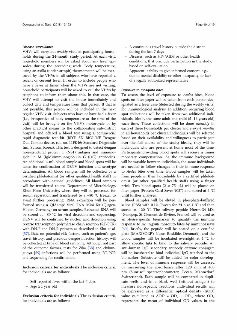
Disease surveillance
VHVs will carry out weekly visits at participating house-
holds during the 24-month study period. At each visit,
household members will be asked about any fever epi-
sodes during the preceding week. Body temperature,
using an axilla (under-armpit) thermometer, will be mea-
sured by the VHVs in all subjects who have reported a
recent or current fever. In order to include people who
have a fever at times when the VHVs are not visiting,
household participants will be asked to call the VHVs by
telephone to inform them about this. In that case, the
VHV will attempt to visit the house immediately and
collect data and temperature from that person. If that is
not possible, this person will be included in the next
regular VHV visit. Subjects who have or have had a fever
(i.e., irrespective of body temperature at the time of the
visit) will be brought on the VHV’s motorcycle or by
other practical means to the collaborating sub-district
hospital and offered a blood test using a commercial
rapid diagnostic test kit (RDT: SD BIOLINE Dengue
Duo Combo device, cat. no. 11FK46; Standard Diagnostic
Inc., Suwon, Korea). This test is designed to detect dengue
non-structural protein 1 (NS1) antigen and immuno-
globulin M (IgM)/immunoglobulin G (IgG) antibodies.
An additional 4-mL blood sample and blood spots will be
taken for confirmation of DENV infection and serotype
determination. All blood samples will be collected by a
certified phlebotomist (or other qualified health staff ) in
accordance with national guidelines. All blood samples
will be transferred to the Department of Microbiology,
Khon Kaen University, where they will be processed for
serum separation and transferred to a –80 °C freezer to
await further processing. RNA extraction will be per-
formed using a QIAamp® Viral RNA Mini Kit (Qiagen,
Hilden, Germany) on serum samples. Extracted RNA will
be stored at –80 °C for viral detection and sequencing.
DENV will be confirmed by nucleic acid detection using
reverse transcription polymerase chain reaction (RT-PCR)
with DN-F and DN-R primers as described in Shu et al.
[57]. Data on potential risk factors, such as patient’s age,
travel history, and previous dengue infection history, will
be collected at time of blood sampling. Although not part
of the outcome factors, tests for Zika [58] and chikun-
gunya [59] infections will be performed using RT-PCR
and sequencing for confirmation.
Inclusion criteria for individuals The inclusion criteria
for individuals are as follows:
– Self-reported fever within the last 7 days
– Age ≥ 1 year old
Exclusion criteria for individuals The exclusion criteria
for individuals are as follows:
– A continuous travel history outside the district
during the last 7 days
– Diseases, such as HIV/AIDS or other health
conditions, that preclude participation in the study,
based on self-evaluation
– Apparent inability to give informed consent, e.g.,
due to mental disability or other incapacity, or lack
of a legally authorized representative
Exposure to mosquito bites
To assess the level of exposure to Aedes bites, blood
spots on filter paper will be taken from each person des-
ignated as a fever case (detected during the weekly visits)
for immunological analysis. In addition, recurring blood
spot collections will be taken from two additional indi-
viduals, ideally the same adult and child (5–14 years old)
each time. These collections will be done monthly in
each of three households per cluster and every 4 months
in all households per cluster. Individuals will be selected
based on their availability and willingness to participate
over the full course of the study; ideally, they will be
individuals who are present at home most of the time.
Participants providing blood spots will receive a minor
monetary compensation. As the immune background
will be variable between individuals, the same individuals
are needed to follow changes in their immune response
to Aedes bites over time. Blood samples will be taken
from people in their households by a certified phlebot-
omist (or other qualified health staff ) using a finger
prick. Two blood spots (2 × 75 μL) will be placed on
filter paper (Protein Card Saver 903™) and stored at 4 °C
until further analyses.
Blood samples will be eluted in phosphate-buffered
saline (PBS) with 0.1% Tween for 24 h at 4 °C and then
stored at –20 °C. The salivary peptide Nterm-34 kDa
(Genepep, St Clement de Rivière, France) will be used as
an Aedes-specific biomarker to quantify the immune
response to Ae. aegypti mosquito bites by immunoassays
[60]. Briefly, the peptide will be coated on a certified
plate (MAXISORP®; Nunc, Roskilde, Denmark), and the
blood samples will be incubated overnight at 4 °C to
allow specific IgG to bind to the salivary peptide. An
anti-human IgG secondary antibody enzyme conjugate
will be incubated to bind individual IgG attached to the
biomarker. Substrate will be added for color develop-
ment. The level of immune response will be assessed
by measuring the absorbance after 120 min at 405
nm (Sunrise™ spectrophotometer, Tecan, Männedorf,
Switzerland). Each sample will be compared in dupli-
cate wells and in a blank well (without antigen) to
measure non-specific reactions. Individual results will
be expressed as a differential optical density (ΔOD)
value calculated as ΔOD = ODx − ODn, where ODx
represents the mean of individual OD values in the
Overgaard et al. Trials (2018) 19:122 Page 10 of 19

two wells containing antigen, and ODn represents the
OD value in the well without antigen. Specific anti-
Nterm-34 kDa IgG response will be assayed in indi-
viduals who have not been exposed to Ae. aegypti
mosquitoes to quantify the non-specific background
antibody level and to calculate the specific immune
threshold (TR) as follows:
TR ¼ mean ΔODunexposed
� �
þ 3SD:
The main outcome for this immune response assay
will be ΔOD, which is a continuous variable. In addition,
a binary outcome will be calculated by considering an
individual to be “exposed” if the ΔOD value is higher
than the TR calculated from unexposed individuals.
Entomological collections
Mosquito collections will be carried out in all participat-
ing households every 4 months (Fig. 2). In addition,
monthly collections will be done in the three sentinel
households per cluster, using the same households as
those used for the blood spot collections for logistical
reasons. The following data will be recorded from each
household: number of total containers (potential breed-
ing sites, wet or dry), number of containers with water,
number of mosquito positive and negative containers
(any species), container type, and location (indoors/out-
doors) using defined criteria. Mosquito larvae will be
collected from all positive containers using a standard
larval dipper to determine species composition (both Ae.
aegypti and Ae. albopictus will be identified and
recorded). Pupae will be collected using the pupal/
demographic survey method [19] and the “five-sweep”
net procedure for very large containers [61].
Adult mosquitoes will be collected for 15 min indoors
(in living rooms, bedrooms, etc.) and 15 min outdoors
(among man-made articles, vegetation, etc.) from each
household using a Prokopack mechanical aspirator [62].
Adult mosquitoes will also be collected using stationary
sticky lure gravid Aedes traps [63] placed in a location
where mosquitoes are abundant (based on householders’
knowledge) at four selected households for 7 consecutive
days every month. Specimens will be taken to a labora-
tory for sorting and identification using a stereomicro-
scope and morphological keys [64, 65]. Larvae
(separated by species) and pupae (separated by species
and sex) will be stored in absolute 99.5% ethanol in la-
beled 1.5-mL Eppendorf® tubes. Blood digestion status
(fed or not fed) of female mosquitoes will be determined
by external examination of abdomens. Adult mosquitoes
(separated by species and sex) will be stored individually
in absolute 99.5% ethanol in 1.5-mL labeled Eppendorf
tubes. All specimens will be transported to Khon Kaen
University and stored at –80 °C until further processing.
Virus detection in mosquitoes
Virus detection will be performed on adults and
pupae of Ae. aegypti and Ae. albopictus. The heads
and abdomens of adult mosquitoes will be stored sep-
arately. Abdomens will be pooled using a pool size of
5–10 individual abdomens depending on abundance.
Virus detection will first be performed on all pools;
then, if positive, serotype detection will be done on
individual mosquitoes (heads). As heads and abdo-
mens cannot be separated in pupae, virus and sero-
type detection will be done on pools of whole bodies
of pupae. The prevalence of infection in the pupal
population, based on the proportion of positive
pools, will be estimated using previously described
methods [66, 67]. The total RNA will be isolated
from mosquito specimens using Favorgen® reagent
(FavorPrepTM Tissue Total RNA Mini Kit) following
manufacturer instructions. The final solution will be
stored at –80 °C. DENV presence will be confirmed
by real-time quantitative RT-PCR (qRT-PCR) con-
ducted in the LightCycler® 480 Real-Time qPCR Sys-
tem using KAPA SYBR® FAST qPCR Master Mix
(2X) Universal [68]. The Master Mix contains an
optimized MgCl2 concentration. Positive samples will
be submitted to a second specific qPCR to determine
the DENV serotype [69, 70].
Climate data
Climate data, including daily temperature, rainfall,
and humidity data, will be collected from permanent
weather stations located in Khon Kaen and Roi Et
(Department of Meteorology of Thailand). Addition-
ally, four rainfall gauges (three manual and one auto-
matic) and eight temperature-humidity data loggers
(iButtons Hygrochron Loggers, DS1923-F5) will be placed
in each city at suitable locations to capture local
variations.
Data management
Each participating household will be given a 6-digit
identification number (indicating province, village, and
household number) and an identification plate (with
project name and ID number) attached in a secure
location to the house. Each household member will
also receive a unique ID number. Data from house-
hold questionnaires at household enrollment, entomo-
logical collections, blood spot sampling at households
and hospitals (venipuncture for dengue positivity con-
firmation), and disease surveillance data by VHVs will
be collected on paper forms. Data will be securely
stored in a password-protected central database. All
hardcopy and electronic data will be placed in locked
spaces or password-protected computers. Data man-
agement procedures will be detailed in specific
Overgaard et al. Trials (2018) 19:122 Page 11 of 19
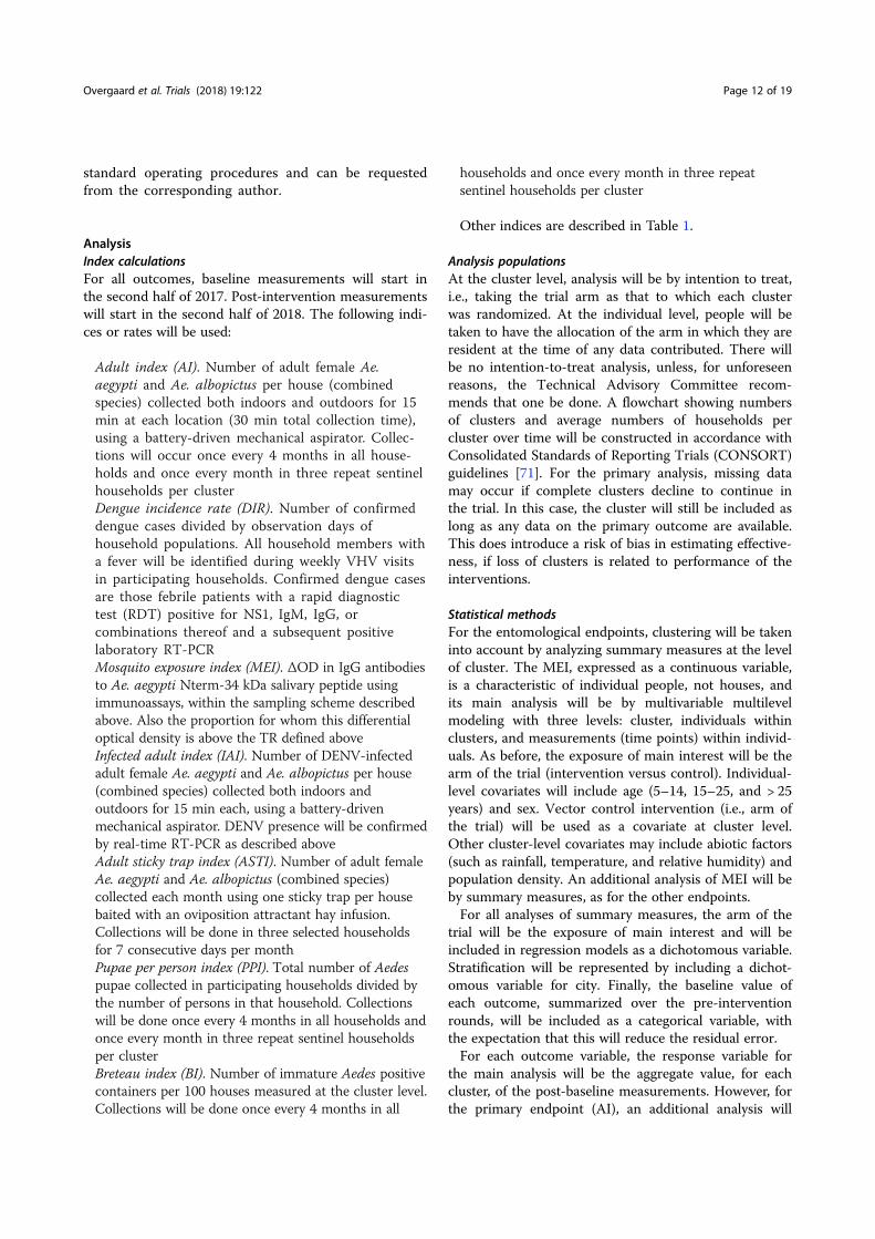
standard operating procedures and can be requested
from the corresponding author.
Analysis
Index calculations
For all outcomes, baseline measurements will start in
the second half of 2017. Post-intervention measurements
will start in the second half of 2018. The following indi-
ces or rates will be used:
Adult index (AI). Number of adult female Ae.
aegypti and Ae. albopictus per house (combined
species) collected both indoors and outdoors for 15
min at each location (30 min total collection time),
using a battery-driven mechanical aspirator. Collec-
tions will occur once every 4 months in all house-
holds and once every month in three repeat sentinel
households per cluster
Dengue incidence rate (DIR). Number of confirmed
dengue cases divided by observation days of
household populations. All household members with
a fever will be identified during weekly VHV visits
in participating households. Confirmed dengue cases
are those febrile patients with a rapid diagnostic
test (RDT) positive for NS1, IgM, IgG, or
combinations thereof and a subsequent positive
laboratory RT-PCR
Mosquito exposure index (MEI). ΔOD in IgG antibodies
to Ae. aegypti Nterm-34 kDa salivary peptide using
immunoassays, within the sampling scheme described
above. Also the proportion for whom this differential
optical density is above the TR defined above
Infected adult index (IAI). Number of DENV-infected
adult female Ae. aegypti and Ae. albopictus per house
(combined species) collected both indoors and
outdoors for 15 min each, using a battery-driven
mechanical aspirator. DENV presence will be confirmed
by real-time RT-PCR as described above
Adult sticky trap index (ASTI). Number of adult female
Ae. aegypti and Ae. albopictus (combined species)
collected each month using one sticky trap per house
baited with an oviposition attractant hay infusion.
Collections will be done in three selected households
for 7 consecutive days per month
Pupae per person index (PPI). Total number of Aedes
pupae collected in participating households divided by
the number of persons in that household. Collections
will be done once every 4 months in all households and
once every month in three repeat sentinel households
per cluster
Breteau index (BI). Number of immature Aedes positive
containers per 100 houses measured at the cluster level.
Collections will be done once every 4 months in all
households and once every month in three repeat
sentinel households per cluster
Other indices are described in Table 1.
Analysis populations
At the cluster level, analysis will be by intention to treat,
i.e., taking the trial arm as that to which each cluster
was randomized. At the individual level, people will be
taken to have the allocation of the arm in which they are
resident at the time of any data contributed. There will
be no intention-to-treat analysis, unless, for unforeseen
reasons, the Technical Advisory Committee recom-
mends that one be done. A flowchart showing numbers
of clusters and average numbers of households per
cluster over time will be constructed in accordance with
Consolidated Standards of Reporting Trials (CONSORT)
guidelines [71]. For the primary analysis, missing data
may occur if complete clusters decline to continue in
the trial. In this case, the cluster will still be included as
long as any data on the primary outcome are available.
This does introduce a risk of bias in estimating effective-
ness, if loss of clusters is related to performance of the
interventions.
Statistical methods
For the entomological endpoints, clustering will be taken
into account by analyzing summary measures at the level
of cluster. The MEI, expressed as a continuous variable,
is a characteristic of individual people, not houses, and
its main analysis will be by multivariable multilevel
modeling with three levels: cluster, individuals within
clusters, and measurements (time points) within individ-
uals. As before, the exposure of main interest will be the
arm of the trial (intervention versus control). Individual-
level covariates will include age (5–14, 15–25, and > 25
years) and sex. Vector control intervention (i.e., arm of
the trial) will be used as a covariate at cluster level.
Other cluster-level covariates may include abiotic factors
(such as rainfall, temperature, and relative humidity) and
population density. An additional analysis of MEI will be
by summary measures, as for the other endpoints.
For all analyses of summary measures, the arm of the
trial will be the exposure of main interest and will be
included in regression models as a dichotomous variable.
Stratification will be represented by including a dichot-
omous variable for city. Finally, the baseline value of
each outcome, summarized over the pre-intervention
rounds, will be included as a categorical variable, with
the expectation that this will reduce the residual error.
For each outcome variable, the response variable for
the main analysis will be the aggregate value, for each
cluster, of the post-baseline measurements. However, for
the primary endpoint (AI), an additional analysis will
Overgaard et al. Trials (2018) 19:122 Page 12 of 19
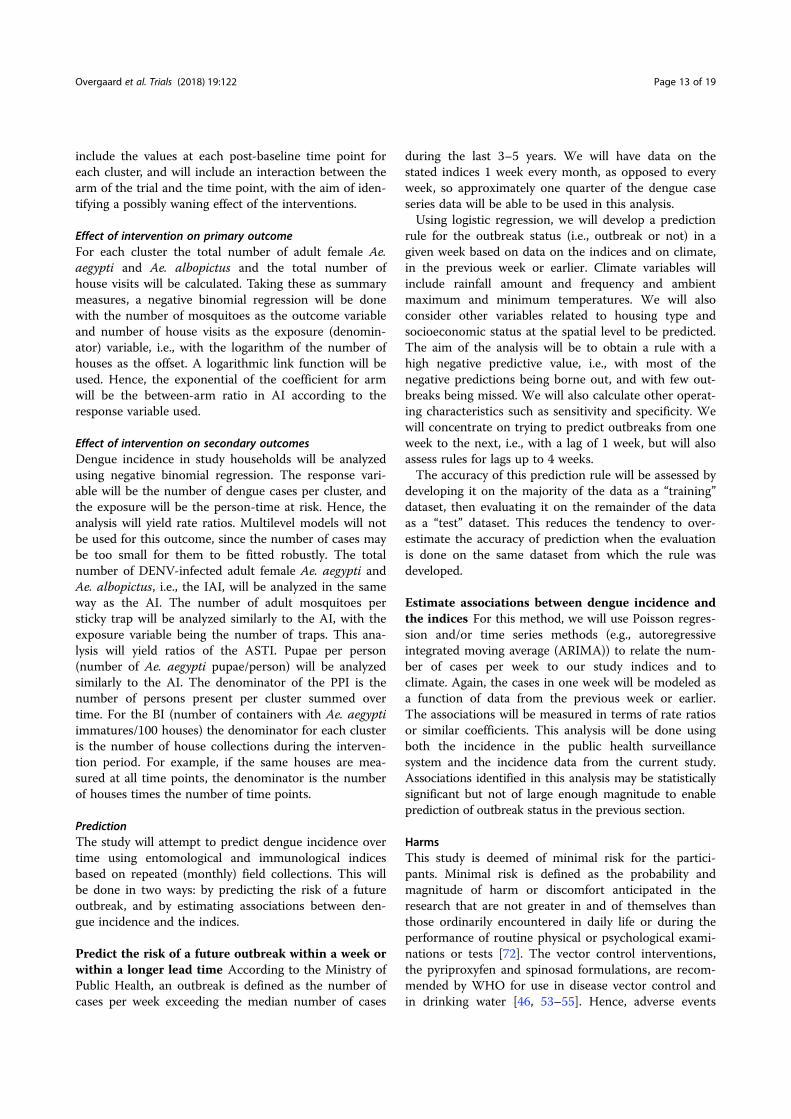
include the values at each post-baseline time point for
each cluster, and will include an interaction between the
arm of the trial and the time point, with the aim of iden-
tifying a possibly waning effect of the interventions.
Effect of intervention on primary outcome
For each cluster the total number of adult female Ae.
aegypti and Ae. albopictus and the total number of
house visits will be calculated. Taking these as summary
measures, a negative binomial regression will be done
with the number of mosquitoes as the outcome variable
and number of house visits as the exposure (denomin-
ator) variable, i.e., with the logarithm of the number of
houses as the offset. A logarithmic link function will be
used. Hence, the exponential of the coefficient for arm
will be the between-arm ratio in AI according to the
response variable used.
Effect of intervention on secondary outcomes
Dengue incidence in study households will be analyzed
using negative binomial regression. The response vari-
able will be the number of dengue cases per cluster, and
the exposure will be the person-time at risk. Hence, the
analysis will yield rate ratios. Multilevel models will not
be used for this outcome, since the number of cases may
be too small for them to be fitted robustly. The total
number of DENV-infected adult female Ae. aegypti and
Ae. albopictus, i.e., the IAI, will be analyzed in the same
way as the AI. The number of adult mosquitoes per
sticky trap will be analyzed similarly to the AI, with the
exposure variable being the number of traps. This ana-
lysis will yield ratios of the ASTI. Pupae per person
(number of Ae. aegypti pupae/person) will be analyzed
similarly to the AI. The denominator of the PPI is the
number of persons present per cluster summed over
time. For the BI (number of containers with Ae. aegypti
immatures/100 houses) the denominator for each cluster
is the number of house collections during the interven-
tion period. For example, if the same houses are mea-
sured at all time points, the denominator is the number
of houses times the number of time points.
Prediction
The study will attempt to predict dengue incidence over
time using entomological and immunological indices
based on repeated (monthly) field collections. This will
be done in two ways: by predicting the risk of a future
outbreak, and by estimating associations between den-
gue incidence and the indices.
Predict the risk of a future outbreak within a week or
within a longer lead time According to the Ministry of
Public Health, an outbreak is defined as the number of
cases per week exceeding the median number of cases
during the last 3–5 years. We will have data on the
stated indices 1 week every month, as opposed to every
week, so approximately one quarter of the dengue case
series data will be able to be used in this analysis.
Using logistic regression, we will develop a prediction
rule for the outbreak status (i.e., outbreak or not) in a
given week based on data on the indices and on climate,
in the previous week or earlier. Climate variables will
include rainfall amount and frequency and ambient
maximum and minimum temperatures. We will also
consider other variables related to housing type and
socioeconomic status at the spatial level to be predicted.
The aim of the analysis will be to obtain a rule with a
high negative predictive value, i.e., with most of the
negative predictions being borne out, and with few out-
breaks being missed. We will also calculate other operat-
ing characteristics such as sensitivity and specificity. We
will concentrate on trying to predict outbreaks from one
week to the next, i.e., with a lag of 1 week, but will also
assess rules for lags up to 4 weeks.
The accuracy of this prediction rule will be assessed by
developing it on the majority of the data as a “training”
dataset, then evaluating it on the remainder of the data
as a “test” dataset. This reduces the tendency to over-
estimate the accuracy of prediction when the evaluation
is done on the same dataset from which the rule was
developed.
Estimate associations between dengue incidence and
the indices For this method, we will use Poisson regres-
sion and/or time series methods (e.g., autoregressive
integrated moving average (ARIMA)) to relate the num-
ber of cases per week to our study indices and to
climate. Again, the cases in one week will be modeled as
a function of data from the previous week or earlier.
The associations will be measured in terms of rate ratios
or similar coefficients. This analysis will be done using
both the incidence in the public health surveillance
system and the incidence data from the current study.
Associations identified in this analysis may be statistically
significant but not of large enough magnitude to enable
prediction of outbreak status in the previous section.
Harms
This study is deemed of minimal risk for the partici-
pants. Minimal risk is defined as the probability and
magnitude of harm or discomfort anticipated in the
research that are not greater in and of themselves than
those ordinarily encountered in daily life or during the
performance of routine physical or psychological exami-
nations or tests [72]. The vector control interventions,
the pyriproxyfen and spinosad formulations, are recom-
mended by WHO for use in disease vector control and
in drinking water [46, 53–55]. Hence, adverse events
Overgaard et al. Trials (2018) 19:122 Page 13 of 19

associated with the products are expected to be few.
However, an adverse event, should one occur, will be
registered by the community-based VHVs through the
weekly visits to all households. The project information
sheet, given to all participants, also contains contact
telephone numbers of the principal investigators and the
Khon Kaen University Ethical Committee should any
questions or reservations arise. Any adverse event during
the trial interventions or trial conduct will be discussed
during weekly meetings of the research team at Khon
Kaen University. Expedited decisions will be made as to
whether any follow-up action is necessary. The opinion
of the Technical Advisory Committee (see the following
section) will be sought should there be adverse events
believed possibly related to the interventions. Based on
these considerations, no criteria have been set for dis-
continuing or modifying the interventions, nor have any
trial stopping guidelines been deemed necessary.
Data monitoring
A Technical Advisory Committee consisting of three
independent researchers assumes the role of a Data
Monitoring Committee (DMC). The duties of the com-
mittee are to stay informed about the progress of the
trial; provide advice to the research team when needed;
assist in solving ethical issues and unforeseen or adverse
events; and determine any potential termination of the
trial. This committee is independent and will not benefit
from the trial or otherwise influence the trial. The terms
of reference of the Technical Advisory Committee can
be accessed from the corresponding author. No interim
analysis is planned.
Auditing
There will be no formal auditing of this trial.
Confidentiality
As described above, the personal information of enrolled
participants will be stored in a safe website ensuring
confidentiality before, during, and after the trial. Analysis
and publication of the results will ensure that no identi-
fiable information is released.
Ancillary and post-trial care
This trial is deemed of minimal risk to study partici-
pants. Therefore, there are no provisions for ancillary or
post-trial care or for compensation to those who suffer
harms from trial participation, beyond the existing Thai
social security system.
Dissemination policy and access to data
Results from this trial will be published in open access,
peer-reviewed journals. The presentation of the final
results of this trial will follow the CONSORT 2010
statement and the extension to cluster-randomized trials
[71] and, if needed, extensions on non-pharmacological
interventions and pragmatic designs [73, 74]. This study
protocol followed the recommendations of items to ad-
dress in a clinical trial protocol (Additional file 1)and
the minimum trial registration information of WHO
(Additional file 2), in addition to what was registered in
the primary ISRCTN registry. Access to the trial dataset
will be made available upon publication of results. Ac-
cess to data will also be archived and made available
through the Norwegian University of Life Sciences and
the Norwegian Centre for Research Data (http://
www.nsd.uib.no/nsd/english/) after the project has offi-
cially ended. Results will be communicated to trial par-
ticipants in easy-to-read local language pamphlets and
through post-project dissemination events. Access to
data collection forms can be requested from the corre-
sponding author.
Discussion
This field trial has a novel combination of aims: to
evaluate simultaneously the efficacy of an innovative
dengue vector control intervention and to develop
methods and indices to anticipate changes in dengue
transmission and predict impending outbreaks. Such
objectives are in harmony with recent published recom-
mendations on global frameworks on vector control and
contingency planning for dengue outbreaks [39, 47] as
well as recommendations from several review papers on
these topics [16, 75]. If successful, results from this study
will provide important information on dengue vector
control and contribute to the further development of
early warning systems and deployment of effective
responses to dengue outbreaks.
Currently, the primary vector control methods used by
the majority of public-funded dengue control programs
are treatment of water storage containers with a larvi-
cide (commonly temephos) and/or peridomestic space
spraying of insecticides. Additionally, source reduction
practices through community-based clean-up campaigns
are common vector control interventions. Although
these standard interventions are recommended by WHO
[5], there is currently no clear evidence that they have
any demonstrable effect on reducing dengue transmis-
sion [7, 9, 33].
A systematic literature review on the effectiveness of
temephos found that as a single community-based inter-
vention it controlled larvae for 2–3 months, depending
on study design, local circumstances, water turnover
rates, and season [7]. However, temephos appears not to
work well in combination with other interventions, pos-
sibly due to an inordinate trust in (or reliance on) its
effectiveness when used alone, poor implementation and
coverage, and low acceptability for its use in drinking
Overgaard et al. Trials (2018) 19:122 Page 14 of 19
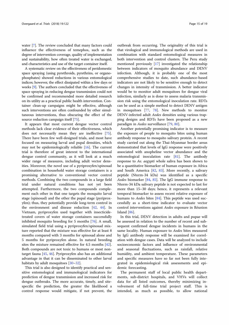
water [7]. The review concluded that many factors could
influence the effectiveness of temephos, such as the
degree of intervention coverage, quality of implementation
and sustainability, how often treated water is exchanged,
and characteristics and use of the target container itself.
A systematic review on the effectiveness of peridomestic
space spraying (using pyrethroids, pyrethrins, or organo-
phosphates) showed reductions in various entomological
indices; however, the effect dissipated within a few days or
weeks [9]. The authors concluded that the effectiveness of
space spraying in reducing dengue transmission could not
be confirmed and recommended more detailed research
on its utility as a practical public health intervention. Con-
tainer clean-up campaigns might be effective, although
such interventions are often confounded by other simul-
taneous interventions, thus obscuring the effect of the
source reduction campaign itself [75].
It appears that most current dengue vector control
methods lack clear evidence of their effectiveness, which
does not necessarily mean they are ineffective [75].
There have been few well-designed trials, and most have
focused on measuring larval and pupal densities, which
may not be epidemiologically reliable [16]. The current
trial is therefore of great interest to the international
dengue control community, as it will look at a much
wider range of measures, including adult vector dens-
ities. Moreover, the novel use of a pyriproxyfen/spinosad
combination in household water storage containers is a
promising alternative to conventional vector control
methods. Combining the two compounds in a large field
trial under natural conditions has not yet been
attempted. Furthermore, the two compounds comple-
ment each other in that one targets the mosquito larval
stage (spinosad) and the other the pupal stage (pyriprox-
yfen); thus, they potentially provide long-term control in
the environment and disease reduction [42, 44]. In
Vietnam, pyriproxyfen used together with insecticide-
treated covers of water storage containers successfully
inhibited mosquito breeding for 5 months [76]. A small,
simulated field trial using a pyriproxyfen/spinosad mix-
ture reported that the mixture was effective for at least 8
months compared with 3 months for spinosad alone and
5 months for pyriproxyfen alone. In natural breeding
sites the mixture remained effective for 4.5 months [42].
Both compounds are not toxic to humans or most non-
target fauna [45, 46]. Pyriproxyfen also has an additional
advantage in that it can be disseminated to other larval
habitats by adult mosquitoes [30–32].
This trial is also designed to identify practical and sen-
sitive entomological and immunological indicators for
prediction of dengue transmission and increased risk for
dengue outbreaks. The more accurate, timely, and site-
specific the prediction, the greater the likelihood a
control response would mitigate, if not prevent, the
outbreak from occurring. The originality of this trial is
that virological and immunological methods are used in
combination with standard entomological measures in
both intervention and control clusters. The Peru study
mentioned previously [17] investigated the relationship
between indicators of mosquito abundance and DENV
infection. Although, it is probably one of the most
comprehensive studies to date, such abundance-based
indicators are not likely to be sensitive enough to detect
changes in intensity of transmission. A better indicator
would be to monitor adult mosquitoes for dengue viral
infection, similarly as is done to assess malaria transmis-
sion risk using the entomological inoculation rate. RDTs
can be used as a simple method to detect DENV antigen
in mosquitoes [77, 78]. New methods to monitor
DENV-infected adult Aedes densities using various trap-
ping designs and RDTs have been proposed as a new
paradigm in Aedes surveillance [79, 80].
Another potentially promising indicator is to measure
the exposure of people to mosquito bites using human
antibody response to mosquito salivary protein. A recent
study carried out along the Thai-Myanmar border areas
demonstrated that levels of IgG response were positively
associated with anopheline vector abundance and the
entomological inoculation rate [81]. The antibody
response to Ae. aegypti whole saliva has been shown to
be a quantitative biomarker of human exposure in Africa
and South America [82, 83]. More recently, a salivary
peptide (Nterm-34 kDa) was identified as a specific
Aedes biomarker [84, 85]. The IgG immune response to
Nterm-34 kDa salivary peptide is not expected to last for
more than 15–30 days; hence, it represents a relevant
temporal biomarker to assess recent relative exposure of
humans to Aedes bites [84]. This peptide was used suc-
cessfully as a short-time indicator to evaluate vector
control interventions against Aedes exposure in Réunion
Island [86].
In this trial, DENV detection in adults and pupae will
be assessed in relation to the number of recent and sub-
sequent confirmed dengue incidents in humans in the
same locality. Human exposure to Aedes bites measured
by IgG antibody response will be examined for correl-
ation with dengue cases. Data will be analyzed to include
socioeconomic factors and influence of environmental
and seasonal fluctuations, such as rainfall, relative
humidity, and ambient temperature. These parameters
and specific measures have so far not been fully inte-
grated in epidemiological risk assessments and epi-
demic forecasting.
The permanent staff of local public health depart-
ments, sub-district hospitals, and VHVs will collect
data for all listed outcomes, thereby minimizing in-
volvement of full-time trial project staff. This is
intended, as much as possible, to allow national
Overgaard et al. Trials (2018) 19:122 Page 15 of 19

authorities to emulate the project procedures in
follow-on surveillance and intervention activities or
adoption of these methods into routine vector control
program activities. The exception to this is molecular-
based assay confirmation of DENV in human blood
and mosquitoes and human antibody response to Ae.
aegypti salivary peptides; this testing will be con-
ducted by project staff.
Study limitations
Several potential limiting factors may affect study out-
comes. High spatial and temporal variation in dengue
transmission dynamics may result in an insufficient
number of incident infections to allow reliable associa-
tions between indices and dengue risk. This is why
collections will be conducted over a 2-year period and in
two urban areas to increase the potential of witnessing
an upsurge in transmission as opposed to an interepi-
demic period. Nevertheless, the sample size required to
detect significant differences in dengue incidence
between the intervention and control arms was deemed
unfeasibly large, thus relegating dengue incidence as a sec-
ondary outcome. Conversely, a dengue outbreak could
likely overwhelm data collection systems used in the trial
and further compel public health authorities to intervene
with standard vector control interventions on a broad
scale, thus potentially interfering with study outcomes. If
outcome measures are substantially suppressed, this
would negatively affect the power of the study.
Lastly, the proportion of asymptomatic (inapparent)
and infectious persons in the study area may affect pre-
diction outcomes, because they will not be captured by
the data collection procedures used in the trial, although
they may contribute to transmission [87]. Although the
ratio of asymptomatic to symptomatic cases can be as
high as 14:1 or higher, the epidemiological role of
asymptomatic infections remains unclear [88].
Trial status
At the time of submission of this manuscript, the trial has
enrolled village clusters, requested household participation,
and started baseline data collections in Khon Kaen (but not
yet in Roi Et). Recruitment of patients has not started.
Additional files
Additional file 1: SPIRIT checklist. (DOCX 53 kb)
Additional file 2: WHO Trial Registration Data Set (Version 1.3). (DOCX 21 kb)
Additional file 3: Consent forms. (DOCX 47 kb)
Abbreviations
Ae.: Aedes; AI: Adult index; ARIMA: Autoregressive integrated moving average;ASTI: Adult sticky trap index; BI: Breteau index; CI: Container index;DENV: Dengue virus; DIR: Dengue incidence rate; DMC: Data MonitoringCommittee; HH: Household; HI: House index; IAI: Infected adult index;
IgG: Immunoglobulin G; IVM: Integrated vector management; kDa: kilodalton;KKUEC: Khon Kaen University Ethics Committee; MEI: Mosquito exposureindex; NSD: Norwegian Centre for Research Data; OD: Optical density;ΔOD: Differential optical density; PBS: Phosphate-buffered saline; PPI: Pupaeper person index; qPCR: Quantitative polymerase chain reaction; RDT: Rapiddiagnostic test; RNA: Ribonucleic acid; RT-PCR: Reverse transcriptionpolymerase chain reaction; SD: Standard deviation; TR: Immune threshold;USD: United States dollar; VHV: Village health volunteer; WHO: WorldHealth Organization
Acknowledgements
Not applicable.
Funding
The Research Council of Norway (RCN, FRIPRO project no. 250443) fundedthis study. The funding body did not have any role in project design or inthe writing of the manuscript. The trial sponsor is the Norwegian Universityof Life Sciences.
Availability of data and materials
The datasets generated and/or analyzed during this study will be madeavailable in the NSD - Norwegian Centre for Research Data repository, http://www.nsd.uib.no/nsd/english/, or from the corresponding author onreasonable request.
Authors’ contributions
HJO conceived, designed, and coordinated the study; wrote the firstmanuscript draft, reviewed and edited the manuscript; and secured fundingfor the project. CP, SA, TP, SP, BF, TE, and MJB developed protocols fordisease surveillance and virological laboratory technical aspects. TP, SP, BF,and MJB developed protocols for entomological collections and technicalaspects related to virus detection in mosquitoes. BF, DC, and VC developedprotocols for exposure to mosquito bites. MJB provided substantialcontributions to the study design and drafting and revision of themanuscript. KT contributed to the setting of the study. NA contributed todefining the objectives, the analysis plan, sample size calculations, andsampling methods. All authors contributed to the study design, and writing,reviewing, and editing of the manuscript, and read and approved the finalversion of the manuscript.
Ethics approval and consent to participate
This trial was approved by the Khon Kaen University Ethics Committee(KKUEC) (Record No. 4.4.01: 29/2017, Reference No. HE601221, 1 September2017) and the London School of Hygiene and Tropical Medicine EthicalCommittee, UK (LSHTM Ethics Ref: 14275, 16 August 2017). Ethical review ofthe Regional Committee for Medical and Health Research Ethics, Section B,South East Norway (REK) is ongoing. Any follow-on protocol amendmentswill be reviewed and approved by these committees. The ISRCTN trialregistry will also be informed in a timely manner of such amendments.All subjects will be engaged in a process of documented informed consent,and assent where appropriate, before participating in any study activities(Additional file 3). These processes will be conducted by staff of the Office ofDisease Prevention and Control in Khon Kaen supervised by Khon KaenUniversity. There will be three consent processes in this trial:1. Request for household participation in a research project. Before starting thebaseline data collections, households will be informed about the trial, andthe household head will sign a consent form requesting his/her householdto participate in the trial. Household agreement to participate means that itsmembers may be subjected to individual request for participation, as statedin the following two points.2. Request for individual participation in a research project. Individualconsent, and assent if appropriate, to participate will be sought from eachhousehold member at the time when they have been identified as having afever during the study observation period. Fever cases will be identified bythe VHVs.3. Request for blood spot collections. Consent (assent) will be sought from thetwo persons per household identified and selected for regular monthlyblood spot collections each time blood spots are requested. These twoindividuals will be provided a small monetary compensation (50 baht = USD1.50) for each blood sample.
Overgaard et al. Trials (2018) 19:122 Page 16 of 19

Each of the three consent processes will take place at the households.Informed consent will be documented by means of a written signed anddated consent form after (1) a full description of the study on theinformation sheet is given to the participant, and (2) the procedures,advantages, disadvantages, and responsibilities and participant’s rights towithdraw at any time have been discussed, clarified, and fully understood bythe participant. All forms and questionnaires are translated from English intothe Thai language and tested for accuracy and interpretation before use.Children will be given a chance to decide for themselves whether they wantto participate or not without pressure or coercion from parents or projectstaff. The assent forms, adapted to the children’s ability to read and write,provide a simple and general overview of what participation in the projectentails. The children will also be informed about the project verbally in easy-to-understand language. Children will sign the forms before their parents orlegal guardians sign. The assent form is for children 7–13 years old. Children14–17 years old will use the same consent form as that for adults, which hasspace for both children and parents/guardians to sign. Although the age ofmajority in Thailand is 20 years, 18 is commonly used as an age of consentfor treatment purposes [89]. All consent and assent forms explain the risksand benefits of the study. For participants who cannot read, the entireinformed consent form will be read and explained by the project staff in thepresence of a community witness. After consenting, these people will markan inked thumb impression on the form, and the witness will be asked tosign it. Consent and assent forms approved and stamped by the KKUEC areused in accordance with the Ethics Committee’s guidelines.
Consent for publication
All authors have consented to publication of this article.
Competing interests
The authors declare that they have no competing interests.
Publisher’s NoteSpringer Nature remains neutral with regard to jurisdictional claims inpublished maps and institutional affiliations.
Author details1Norwegian University of Life Sciences, Ås, Norway. 2Khon Kaen University,Khon Kaen, Thailand. 3HPV & EBV and Carcinogenesis Research Group, KhonKaen University, Khon Kaen, Thailand. 4Office of Disease Prevention andControl, Region 7, Khon Kaen, Thailand. 5PT Freeport Indonesia/InternationalSOS Indonesia, Kuala Kencana, Indonesia. 6Kasetsart University, Bangkok,Thailand. 7Université de Montpellier, Montpellier, France. 8Institut deRecherche pour le Développement (IRD), Maladies Infectieuses et Vecteurs,Ecologie, Génétique, Evolution et Contrôle (MIVEGEC, UM1-CNRS 5290-IRD224), Montpellier, France. 9MRC Tropical Epidemiology Group, London, UK.
Received: 16 September 2017 Accepted: 19 January 2018
References
1. WHO. Dengue and severe dengue. Factsheet, updated April 2017. WorldHealth Organization; 2017. http://www.who.int/mediacentre/factsheets/fs117/en/. Accessed 6 July 2017.
2. Bhatt S, Gething PW, Brady OJ, Messina JP, Farlow AW, Moyes CL, et al. Theglobal distribution and burden of dengue. Nature. 2013;496(7446):504–7.
3. WHO. Global Strategy for dengue prevention and control, 2012–2020.Geneva: World Health Organization; 2012. WHO/HTM/NTD/VEM/2012.5
4. Hadinegoro SR, Arredondo-Garcia JL, Capeding MR, Deseda C,Chotpitayasunondh T, Dietze R, et al. Efficacy and long-term safety of adengue vaccine in regions of endemic disease. N Engl J Med. 2015;373(13):1195–206.
5. WHO. Dengue — Guidelines for diagnosis, treatment, prevention andcontrol. Geneva: World Health Organization; 2009.
6. Sharp TM, Tomashek KM, Read JS, Margolis HS, Waterman SH. A new look atan old disease: recent insights into the global epidemiology of dengue.Curr Epidemiol Rep. 2017;4(1):11–21.
7. George L, Lenhart A, Toledo J, Lazaro A, Han WW, Velayudhan R, et al.Community-effectiveness of temephos for dengue vector control: asystematic literature review. PLoS Negl Trop Dis. 2015;9(9):e0004006.
8. Han WW, Lazaro A, McCall PJ, George L, Runge-Ranzinger S, Toledo J, et al.Efficacy and community effectiveness of larvivorous fish for dengue vectorcontrol. Tropical Med Int Health. 2015;20(9):1239–56.
9. Esu E, Lenhart A, Smith L, Horstick O. Effectiveness of peridomestic spacespraying with insecticide on dengue transmission; systematic review.Tropical Med Int Health. 2010;15(5):619–31.
10. Horstick O, Runge-Ranzinger S, Nathan MB, Kroeger A. Dengue vector-control services: how do they work? A systematic literature review andcountry case studies. Trans R Soc Trop Med Hyg. 2010;104(6):379–86.
11. Runge-Ranzinger S, Horstick O, Marx M, Kroeger A. What does denguedisease surveillance contribute to predicting and detecting outbreaks anddescribing trends? Tropical Med Int Health. 2008;13(8):1022–41.
12. Runge-Ranzinger S, McCall PJ, Kroeger A, Horstick O. Dengue diseasesurveillance: an updated systematic literature review. Tropical Med IntHealth. 2014;19(9):1116–60.
13. Morrison AC, Zielinski-Gutierrez E, Scott TW, Rosenberg R. Definingchallenges and proposing solutions for control of the virus vector Aedesaegypti. PLoS Med. 2008;5(3):e68.
14. Achee NL, Gould F, Perkins TA, Reiner Jr RC, Morrison AC, Ritchie SA, et al. Acritical assessment of vector control for dengue prevention. PLoS Negl TropDis. 2015;9(5):e0003655.
15. Brady OJ, Smith DL, Scott TW, Hay SI. Dengue disease outbreak definitionsare implicitly variable. Epidemics. 2015;11:92–102.
16. Bowman LR, Runge-Ranzinger S, McCall PJ. Assessing the relationshipbetween vector indices and dengue transmission: a systematic review ofthe evidence. PLoS Negl Trop Dis. 2014;8(5):e2848.
17. Cromwell EA, Stoddard ST, Barker CM, Van Rie A, Messer WB, MeshnickSR, et al. The relationship between entomological indicators of Aedesaegypti abundance and dengue virus infection. PLoS Negl Trop Dis.2017;11(3):e0005429.
18. Connor ME, Monroe WM. Stegomyia indices and their value in yellow fevercontrol. Am J Trop Med Hyg. 1923;1:9–19.
19. Focks DA. A review of entomological sampling methods and indicators fordengue vectors. Geneva: Special Programme for Research and Training inTropical Diseases (TDR) WHO; 2003. TDR/IDE/Den/03.1
20. Focks DA, Chadee DD. Pupal survey: an epidemiologically significantsurveillance method for Aedes aegypti: an example using data from Trinidad.Am J Trop Med Hyg. 1997;56(2):159–67.
21. Focks DA, Brenner RJ, Hayes J, Daniels E. Transmission thresholds fordengue in terms of Aedes aegypti pupae per person with discussion oftheir utility in source reduction efforts. Am J Trop Med Hyg. 2000;62(1):11–8.
22. Edillo FE, Roble ND, Otero ND 2nd. The key breeding sites by pupal surveyfor dengue mosquito vectors, Aedes aegypti (Linnaeus) and Aedes albopictus
(Skuse), in Guba, Cebu City, Philippines. Southeast Asian J Trop Med PublicHealth. 2012;43(6):1365–74.
23. Romero-Vivas C, Arango-Padilla P, Falconar A. Pupal-productivity surveys toidentify the key container habitats of Aedes aegypti (L.) in Barranquilla, theprincipal seaport of Colombia. Ann Trop Med Parasitol. 2006;100:S87–95.
24. Midega JT, Nzovu J, Kahindi S, Sang RC, Mbogo C. Application of the pupal/demographic-survey methodology to identify the key container habitats ofAedes aegypti (L.) in Malindi district, Kenya. Ann Trop Med Parasitol. 2006;100(Suppl 1):S61–s72.
25. Lenhart AE, Castillo CE, Oviedo M, Villegas E. Use of the pupal/demographic-survey technique to identify the epidemiologically importanttypes of containers producing Aedes aegypti (L.) in a dengue-endemic areaof Venezuela. Ann Trop Med Parasitol. 2006;100:S53–9.
26. Wai KT, Arunachalam N, Tana S, Espino F, Kittayapong P, Abeyewickreme W,et al. Estimating dengue vector abundance in the wet and dry season:implications for targeted vector control in urban and peri-urban Asia.Pathog Glob Health. 2012;106(8):436–45.
27. Barrera R, Amador M, Diaz A, Smith J, Munoz-Jordan JL, Rosario Y. Unusualproductivity of Aedes aegypti in septic tanks and its implications for denguecontrol. Med Vet Entomol. 2008;22(1):62–9.
28. Chitolina RF, Anjos FA, Lima TS, Castro EA, Costa-Ribeiro MC. Raw sewage asbreeding site to Aedes (Stegomyia) aegypti (Diptera, culicidae). Acta Trop.2016;164:290–6.
29. Dada N, Vannavong N, Seidu R, Lenhart A, Stenstrom TA, ChareonviriyaphapT, et al. Relationship between Aedes aegypti production and occurrence ofEscherichia coli in domestic water storage containers in rural and sub-urbanvillages in Thailand and Laos. Acta Trop. 2013;126(3):177–85.
Overgaard et al. Trials (2018) 19:122 Page 17 of 19

30. Chandel K, Suman DS, Wang Y, Unlu I, Williges E, Williams GM, et al.Targeting a hidden enemy: pyriproxyfen autodissemination strategy for thecontrol of the container mosquito Aedes albopictus in cryptic habitats. PLoSNegl Trop Dis. 2016;10(12):e0005235.
31. Caputo B, Ienco A, Cianci D, Pombi M, Petrarca V, Baseggio A, et al. The“auto-dissemination” approach: a novel concept to fight Aedes albopictus inurban areas. PLoS Negl Trop Dis. 2012;6(8):e1793.
32. Sihuincha M, Zamora-Perea E, Orellana-Rios W, Stancil JD, Lopez-Sifuentes V, Vidal-Ore C, et al. Potential use of pyriproxyfen for controlof Aedes aegypti (Diptera: Culicidae) in Iquitos, Peru. J Med Entomol.2005;42(4):620–30.
33. Heintze C, Velasco Garrido M, Kroeger A. What do community-baseddengue control programmes achieve? A systematic review of publishedevaluations. Trans R Soc Trop Med Hyg. 2007;101(4):317–25.
34. Boyce R, Lenhart A, Kroeger A, Velayudhan R, Roberts B, Horstick O. Bacillusthuringiensis israelensis (Bti) for the control of dengue vectors: systematicliterature review. Tropical Med Int Health. 2013;18(5):564–77.
35. Lazaro A, Han WW, Manrique-Saide P, George L, Velayudhan R, Toledo J,et al. Community effectiveness of copepods for dengue vector control:systematic review. Tropical Med Int Health. 2015;20(6):685–706.
36. McMeniman CJ, Lane RV, Cass BN, Fong AW, Sidhu M, Wang YF, et al. Stableintroduction of a life-shortening Wolbachia infection into the mosquitoAedes aegypti. Science. 2009;323(5910):141–4.
37. Harris AF, McKemey AR, Nimmo D, Curtis Z, Black I, Morgan SA, et al.Successful suppression of a field mosquito population by sustained releaseof engineered male mosquitoes. Nat Biotechnol. 2012;30(9):828–30.
38. WHO. Mosquito (vector) control emergency response and preparednessfor Zika virus. World Health Organization; 2016. http://www.who.int/neglected_diseases/news/mosquito_vector_control_response/en/.Accessed 6 July 2017.
39. Runge-Ranzinger S, Kroeger A, Olliaro P, McCall PJ, Sanchez Tejeda G, LloydLS, et al. Dengue contingency planning: from research to policy andpractice. PLoS Negl Trop Dis. 2016;10(9):e0004916.
40. Ranson H, Burhani J, Lumjuan N, Black WC IV. Insecticide resistance indengue vectors. TropIKAnet. 2010;1:1–12.
41. WHO. Global strategic framework for integrated vector management.Geneva: World Health Organization; 2004. WHO/CDS/CPE/PVC/2004.10
42. Darriet F, Marcombe S, Etienne M, Yebakima A, Agnew P, Yp-Tcha MM, et al.Field evaluation of pyriproxyfen and spinosad mixture for the control ofinsecticide resistant Aedes aegypti in Martinique (French West Indies). ParasitVectors. 2010;3:88.
43. Nayar JK, Ali A, Zaim M. Effectiveness and residual activity comparison ofgranular formulations of insect growth regulators pyriproxyfen and s-methoprene against Florida mosquitoes in laboratory and outdoorconditions. J Am Mosq Control Assoc. 2002;18(3):196–201.
44. Ocampo CB, Mina NJ, Carabali M, Alexander N, Osorio L. Reduction indengue cases observed during mass control of Aedes (Stegomyia) instreet catch basins in an endemic urban area in Colombia. Acta Trop.2014;132:15–22.
45. WHO. Report of the 4th WHOPES Working Group meeting — IR3535,KBR3023, (RS)-methoprene 20% EC, pyriproxyfen 0.5% GR and lambda-cyhalothrin 2.5% CS. Geneva: World Health Organization; 2001. WHO/CDS/WHOPES/2001.2
46. WHO. Report of the 14th WHOPES working group meeting: WHO/HQ,Geneva, 11-15 April 2011: review of Spinosad® EC, Lifenet® LN, MagnetTMLN, Royal Sentry® LN, Yahe® LN. Geneva: World Health Organization; 2011.WHO/HTM/NTD/WHOPES/2011.7
47. WHO. Global vector control response 2017-2030. Geneva: World HealthOrganization; 2017. Background document to inform deliberations duringthe 70th session of the World Health Assembly (version 5.4)
48. NSO. The 2010 Population and Housing Census. National Statistical Office,Ministry of Information and Communication Technology; 2010. http://web.nso.go.th/en/census/poph/cen_poph_10.htm. Accessed 6 July 2017.
49. Hayes RJ, Bennett S. Simple sample size calculation for cluster-randomizedtrials. Int J Epidemiol. 1999;28(2):319–26.
50. Hemming K, Marsch J. A menu-driven facility for sample-size calculations incluster randomized controlled trials. Stata J. 2013;13(1):114–35.
51. Alexander N. Review: analysis of parasite and other skewed counts. TropicalMed Int Health. 2012;17(6):684–93.
52. WHO. WHOPES-recommended compounds and formulations for control ofmosquito larvae. Geneva: WHO Pesticides Evaluation Scheme (WHOPES);
2013. http://www.who.int/whopes/Mosquito_Larvicides_sep_2011.pdf.Accessed 14 June 2012.
53. WHO. Report of the 10th WHOPES Working Group meeting — Review ofspinosad 0.5% GR and 12% SC, Lambda-cyhalothrin 10% CS, K-O Tab 1-2-3®,Interceptor®. Geneva: World Health Organization; 2007. WHO/CDS/NTD/WHOPES/2007.1
54. WHO. Pyriproxyfen in drinking-water: use for vector control in drinking-water sources and containers. Geneva: World Health Organization; 2008.Background document for development of WHO Guidelines for Drinking-water Quality. WHO/HSE/AMR/08.03/9
55. WHO. Spinosad DT in drinking-water: use for vector control in drinking-water sources and containers. Geneva: World Health Organization; 2010.Background document for development of WHO Guidelines for Drinking-water Quality. WHO/HSE/WSH/10.01/12
56. Chareonviriyaphap T, Bangs MJ, Suwonkerd W, Kongmee M, Corbel V,Ngoen-Klan R. Review of insecticide resistance and behavioral avoidance ofvectors of human diseases in Thailand. Parasit Vectors. 2013;6:280.
57. Shu PY, Chang SF, Kuo YC, Yueh YY, Chien LJ, Sue CL, et al. Development ofgroup- and serotype-specific one-step SYBR green I-based real-time reversetranscription-PCR assay for dengue virus. J Clin Microbiol. 2003;41(6):2408–16.
58. Corman VM, Rasche A, Baronti C, Aldabbagh S, Cadar D, Reusken CB, et al.Assay optimization for molecular detection of Zika virus. Bull World HealthOrgan. 2016;94(12):880–92.
59. Niyas KP, Abraham R, Unnikrishnan RN, Mathew T, Nair S, Manakkadan A,et al. Molecular characterization of Chikungunya virus isolates from clinicalsamples and adult Aedes albopictus mosquitoes emerged from larvae fromKerala, South India. Virol J. 2010;7:189.
60. Elanga Ndille E, Doucoure S, Damien G, Mouchet F, Drame PM,Cornelie S, et al. First attempt to validate human IgG antibodyresponse to Nterm-34kDa salivary peptide as biomarker for evaluatingexposure to Aedes aegypti bites. PLoS Negl Trop Dis. 2012;6(11):e1905.
61. Knox TB, Yen NT, Nam VS, Gatton ML, Kay BH, Ryan PA. Critical evaluation ofquantitative sampling methods for Aedes aegypti (Diptera: Culicidae)immatures in water storage containers in Vietnam. J Med Entomol. 2007;44(2):192–204.
62. Vazquez-Prokopec GM, Galvin WA, Kelly R, Kitron U. A new, cost-effective,battery-powered aspirator for adult mosquito collections. J Med Entomol.2009;46(6):1256–9.
63. Heringer L, Johnson BJ, Fikrig K, Oliveira BA, Silva RD, Townsend M, et al.Evaluation of alternative killing agents for Aedes aegypti (Diptera: Culicidae)in the gravid Aedes trap (GAT). J Med Entomol. 2016;53(4):873–9.
64. Bangs MJ, Focks DA. Abridged pupa identification key to the commoncontainer-breeding mosquitoes in urban Southeast Asia. J Am Mosq ControlAssoc. 2006;22(3):565–72.
65. Rueda LM. Pictorial keys for the identification of mosquitoes (Diptera: Culicidae)associated with dengue virus transmission. Zootaxa. 2004;589:1–60.
66. Katholi CR, Toe L, Merriweather A, Unnasch TR. Determining the prevalenceof Onchocerca volvulus infection in vector populations by polymerase chainreaction screening of pools of black flies. J Infect Dis. 1995;172(5):1414–7.
67. Gu W, Lampman R, Novak RJ. Assessment of arbovirus vector infection ratesusing variable size pooling. Med Vet Entomol. 2004;18(2):200–4.
68. Sim S, Aw PP, Wilm A, Teoh G, Hue KD, Nguyen NM, et al. Tracking denguevirus intra-host genetic diversity during human-to-mosquito transmission.PLoS Negl Trop Dis. 2015;9(9):e0004052.
69. Zandi K, Teoh B, Sam S, Wong P, Mustafa M, AbuBakar S. In vitro antiviralactivity of Fisetin, Rutin and Naringenin against dengue virus type-2. J MedPlant Res. 2011;5(23):5534–9.
70. Seah CL, Chow VT, Tan HC, Can YC. Rapid, single-step RT-PCR typing of dengueviruses using five NS3 gene primers. J Virol Methods. 1995;51(2-3):193–200.
71. Campbell MK, Piaggio G, Elbourne DR, Altman DG. Consort 2010 statement:extension to cluster randomised trials. BMJ. 2012;345:e5661.
72. US DHHS. Code of Federal Regulations, 45 CFR 46, Public Welfare,Protection of Human Subjects. Washington, DC: Office for Human ResearchProtections, US Department of Health & Human Services; 2009.
73. Boutron I, Altman DG, Moher D, Schulz KF, Ravaud P. CONSORT statementfor randomized trials of nonpharmacologic treatments: a 2017 update and aCONSORT extension for nonpharmacologic trial abstracts. Ann Intern Med.2017;167(1):40–7.
74. Zwarenstein M, Treweek S, Gagnier JJ, Altman DG, Tunis S, Haynes B, et al.Improving the reporting of pragmatic trials: an extension of the CONSORTstatement. BMJ. 2008;337:a2390.
Overgaard et al. Trials (2018) 19:122 Page 18 of 19
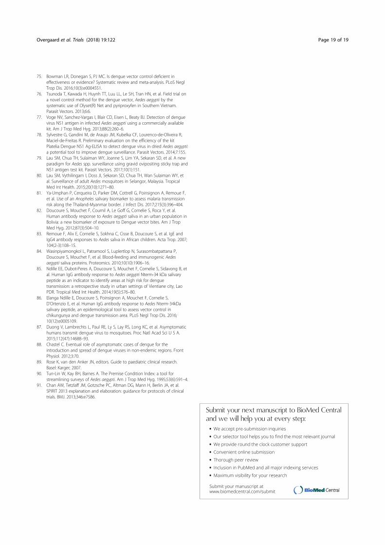
75. Bowman LR, Donegan S, PJ MC. Is dengue vector control deficient ineffectiveness or evidence? Systematic review and meta-analysis. PLoS NeglTrop Dis. 2016;10(3):e0004551.
76. Tsunoda T, Kawada H, Huynh TT, Luu LL, Le SH, Tran HN, et al. Field trial ona novel control method for the dengue vector, Aedes aegypti by thesystematic use of Olyset(R) Net and pyriproxyfen in Southern Vietnam.Parasit Vectors. 2013;6:6.
77. Voge NV, Sanchez-Vargas I, Blair CD, Eisen L, Beaty BJ. Detection of denguevirus NS1 antigen in infected Aedes aegypti using a commercially availablekit. Am J Trop Med Hyg. 2013;88(2):260–6.
78. Sylvestre G, Gandini M, de Araujo JM, Kubelka CF, Lourenco-de-Oliveira R,Maciel-de-Freitas R. Preliminary evaluation on the efficiency of the kitPlatelia Dengue NS1 Ag-ELISA to detect dengue virus in dried Aedes aegypti:a potential tool to improve dengue surveillance. Parasit Vectors. 2014;7:155.
79. Lau SM, Chua TH, Sulaiman WY, Joanne S, Lim YA, Sekaran SD, et al. A newparadigm for Aedes spp. surveillance using gravid ovipositing sticky trap andNS1 antigen test kit. Parasit Vectors. 2017;10(1):151.
80. Lau SM, Vythilingam I, Doss JI, Sekaran SD, Chua TH, Wan Sulaiman WY, etal. Surveillance of adult Aedes mosquitoes in Selangor, Malaysia. TropicalMed Int Health. 2015;20(10):1271–80.
81. Ya-Umphan P, Cerqueira D, Parker DM, Cottrell G, Poinsignon A, Remoue F,et al. Use of an Anopheles salivary biomarker to assess malaria transmissionrisk along the Thailand-Myanmar border. J Infect Dis. 2017;215(3):396–404.
82. Doucoure S, Mouchet F, Cournil A, Le Goff G, Cornelie S, Roca Y, et al.Human antibody response to Aedes aegypti saliva in an urban population inBolivia: a new biomarker of exposure to Dengue vector bites. Am J TropMed Hyg. 2012;87(3):504–10.
83. Remoue F, Alix E, Cornelie S, Sokhna C, Cisse B, Doucoure S, et al. IgE andIgG4 antibody responses to Aedes saliva in African children. Acta Trop. 2007;104(2-3):108–15.
84. Wasinpiyamongkol L, Patramool S, Luplertlop N, Surasombatpattana P,Doucoure S, Mouchet F, et al. Blood-feeding and immunogenic Aedesaegypti saliva proteins. Proteomics. 2010;10(10):1906–16.
85. Ndille EE, Dubot-Peres A, Doucoure S, Mouchet F, Cornelie S, Sidavong B, etal. Human IgG antibody response to Aedes aegypti Nterm-34 kDa salivarypeptide as an indicator to identify areas at high risk for denguetransmission: a retrospective study in urban settings of Vientiane city, LaoPDR. Tropical Med Int Health. 2014;19(5):576–80.
86. Elanga Ndille E, Doucoure S, Poinsignon A, Mouchet F, Cornelie S,D’Ortenzio E, et al. Human IgG antibody response to Aedes Nterm-34kDasalivary peptide, an epidemiological tool to assess vector control inchikungunya and dengue transmission area. PLoS Negl Trop Dis. 2016;10(12):e0005109.
87. Duong V, Lambrechts L, Paul RE, Ly S, Lay RS, Long KC, et al. Asymptomatichumans transmit dengue virus to mosquitoes. Proc Natl Acad Sci U S A.2015;112(47):14688–93.
88. Chastel C. Eventual role of asymptomatic cases of dengue for theintroduction and spread of dengue viruses in non-endemic regions. FrontPhysiol. 2012;3:70.
89. Rose K, van den Anker JN, editors. Guide to paediatric clinical research.Basel: Karger; 2007.
90. Tun-Lin W, Kay BH, Barnes A. The Premise Condition Index: a tool forstreamlining surveys of Aedes aegypti. Am J Trop Med Hyg. 1995;53(6):591–4.
91. Chan AW, Tetzlaff JM, Gotzsche PC, Altman DG, Mann H, Berlin JA, et al.SPIRIT 2013 explanation and elaboration: guidance for protocols of clinicaltrials. BMJ. 2013;346:e7586.
• We accept pre-submission inquiries
• Our selector tool helps you to find the most relevant journal
• We provide round the clock customer support
• Convenient online submission
• Thorough peer review
• Inclusion in PubMed and all major indexing services
• Maximum visibility for your research
Submit your manuscript atwww.biomedcentral.com/submit
Submit your next manuscript to BioMed Central and we will help you at every step:
Overgaard et al. Trials (2018) 19:122 Page 19 of 19

CORRECTION Open Access
Correction to: Assessing denguetransmission risk and a vector controlintervention using entomological andimmunological indices in Thailand: studyprotocol for a cluster-randomizedcontrolled trialHans J. Overgaard1*, Chamsai Pientong2,3, Kesorn Thaewnongiew4, Michael J. Bangs5,6, Tipaya Ekalaksananan2,3,Sirinart Aromseree2,3, Thipruethai Phanitchat2, Supranee Phanthanawiboon2,3, Benedicte Fustec2,7, Vincent Corbel8,Dominique Cerqueira6 and Neal Alexander9
Correction to: Trials (2018) 19:122
https://doi.org/10.1186/s13063-018-2490-1
In the original publication [1], the first of two objectives
was to “Assess the effect of periodically treating water
storage containers with a pyriproxyfen/spinosad combin-
ation on entomological and epidemiological outcomes”.
However, spinosad will not be a part of the intervention.
The approval by the Food and Drug Administration of the
Ministry of Public Health, Thailand to use spinosad in this
research project was delayed and not in place when the
interventions were supposed to begin. Therefore, a decision
was made to exclude spinosad in the trial and only use
pyriproxyfen. The correct version of objective 1 should
now read “Assess the effect of periodically treating water
storage containers with pyriproxyfen on entomological and
epidemiological outcomes”. The second objective remains
the same: “Determine the most accurate and precise index
or indices to predict variation in dengue incidence in time”.
The exclusion spinosad in the trial has implications for
several sections in the original article. The word spinosad
appears 18 times in the main text of document; in the
abstract, background, methods, and discussion. The reader
should be aware of this important change when reading
the article and its implications for the trial and outcomes.
Author details1Norwegian University of Life Sciences, Ås, Norway. 2Khon Kaen University,Khon Kaen, Thailand. 3HPV & EBV and Carcinogenesis Research Group, KhonKaen University, Khon Kaen, Thailand. 4Office of Disease Prevention andControl, Region 7, Khon Kaen, Thailand. 5PT Freeport Indonesia/InternationalSOS Indonesia, Kuala Kencana, Indonesia. 6Kasetsart University, Bangkok,Thailand. 7Université de Montpellier, Montpellier, France. 8Institut deRecherche pour le Développement (IRD), Maladies Infectieuses et Vecteurs,Ecologie, Génétique, Evolution et Contrôle (MIVEGEC, UM1-CNRS 5290-IRD224), Montpellier, France. 9MRC Tropical Epidemiology Group, London, UK.
Received: 6 December 2018 Accepted: 6 December 2018
Reference
1. Overgaard HJ, et al. Assessing dengue transmission risk and a vector controlintervention using entomological and immunological indices in Thailand:study protocol for a cluster-randomized controlled trial. Trials. 2018;19:122.https://doi.org/10.1186/s13063-018-2490-1.
* Correspondence: [email protected] University of Life Sciences, Ås, NorwayFull list of author information is available at the end of the article
© The Author(s). 2018 Open Access This article is distributed under the terms of the Creative Commons Attribution 4.0International License (http://creativecommons.org/licenses/by/4.0/), which permits unrestricted use, distribution, andreproduction in any medium, provided you give appropriate credit to the original author(s) and the source, provide a link tothe Creative Commons license, and indicate if changes were made. The Creative Commons Public Domain Dedication waiver(http://creativecommons.org/publicdomain/zero/1.0/) applies to the data made available in this article, unless otherwise stated.
Overgaard et al. Trials (2018) 19:703
https://doi.org/10.1186/s13063-018-3110-9

91
Third part: Results of the thesis
Chapter 1. Assessing the spatial and temporal patterns of dengue
incidence in North-eastern Thailand
A retrospective epidemiological study using monthly dengue incidence at the sub-district
level and climatic data was conducted by our team in order to better understand the spatial dengue-
climate relationships at fine scale and to identify areas and periods at higher dengue transmission
risk (Phanitchat et al. 2019) (see details in Phanitchat et al. 2019). The study was conducted in
Khon Kaen province comprising 26 districts, 199 sub-districts and 2,139 villages. The province is
primarily rural, with a few large urban centres. Dengue cases from the 1st of January 2006 to 31
December 2016 were retrieved from the MoPH and classified according to WHO dengue
classification prior 2009 (i.e., DF, DHF, DSS). Meteorological data for the same period of time
were downloaded from the data library of the International Research Institute for Climate and
Society. For each sub-district, daily temperatures were aggregated to monthly average with a 0.2
x 0.2 degrees resolution, and daily rainfall was aggregated to monthly average with a 0.05 x 0.05
degrees resolution. Monthly data on dengue cases and climate (rainfall and temperature) from the
study period were combined to visualize seasonal patterns and temporal trends. Bayesian Poisson
model regression were used to assess associations with the number of monthly cases in 199 sub-
districts. Population was used as the denominator in the model. For the main model the covariates
were the population density per km2, gender, mean age, mean rainfall, and minimum and
maximum temperature. Population density was included in the regression model as a “proxy” for
estimating the levels of urbanization. Conditional autoregressive structure was used as a random
effect capturing the spatio-temporal autocorrelation. Local Indicators of Spatial Association
(LISA) were used to identify “hotspots” of dengue (i.e., where incidence is higher than the
The results from this study were published in BMC Infectious Disease, 19(1), 74 https://doi.org/10.1186/s12879-019-4379-3 (2019). Spatial and temporal patterns of dengue incidence in northeastern Thailand 2006-2016. Phanitchat T, Zhao B, Haque U, Pientong C, Ekalaksananan T, Aromseree S, Thaewnongiew K, Fustec B, Bangs MJ, Alexander N, Overgaard HJ.

92
expected number given a random distribution of cases) and “coldspots”, and outliers of dengue
incidence at the sub district level.
Summary of the results:
Over the 11-years period, >15,000 cases were reported, half of them being classified as
severe dengue DHF/DSS. The highest incidence was recorded in 2013 with approximately 80
cases per 100,000 inhabitants. We demonstrated a shift over the last 10 years in case ages, with
the age group 15-29 years old being the most affected by the disease. Our observation was
consistent with a population age shift, potentially influenced by changes in birth and death rates.
Similar trend in dengue infection pattern was observed in other countries in the SEA region
(Limkittikul et al. 2014, Mohd-Zaki et al. 2014, Thomas et al. 2015, Alera et al. 2016).
Additionally, we showed that dengue incidence had a clear seasonal pattern with about
73% of the dengue cases occurring during the rainy season (Figure 31). Our findings showed a
good correlation between dengue incidence and climatic factors, especially temperature and
rainfall. Indeed, the rate ratio for maximum temperature was 1.055, implying 5.5% (95% CI 0.9–
11.5%) increase in cases with an increase of 1 °C per month. The rate ratio for mean rainfall was
1.004, indicating that increasing rainfall by one unit (1 cm) per month would increase dengue
incidence by about 0.4%. Other studies in Thailand and Timor Lest also demonstrated a strong
correlation between dengue incidence and meteorological data (Wangdi et al. 2018). Although the
dynamic of dengue incidence was clearly influenced by rainfall and temperature, our data show
apparent spatial clustering of dengue cases associated with environmental parameters such as
urbanization (Figure 32). Greater vulnerability to dengue infection has been previously observed
in areas situated closer to urban centers (Tipayamongkholgul et al. 2011) and such neighboring
effects have been related to similarities in human behavior, development infrastructure, and
ecological surroundings. Spatial regression analysis suggests that other variables than urbanization
may explain the differences in dengue incidence as half of dengue hotspots were found in rural
areas located in the southwest of the province, hence corroborating the influence of other factors
in dengue transmission. One speculation could be that the lakes and swamps that are common in
this area may provide suitable humidity for mosquitoes to thrive, but this was not studied here.
93
Figure 31: Mean monthly dengue incidence per 100,000 persons, from Phanitchat et al. (a) and monthly average of rainfall (bar) and temperature (line) (b) in Khon Kaen Province,
Thailand, 2006–2016.
To conclude, this baseline study clearly showed the involvement of climatic factors on
dengue transmission in the province. Spatial clustering of dengue cases was partly associated with
urban areas closer to Khon Kaen city and rural areas in the southwest of the province. However,
the current analysis was not able to detect a close proxy factors to quantify a relationship between
urbanization and dengue incidence. This first study highlighted the need for further investigations
on dengue-related risk factors in the study area in order to develop dengue early warning systems
to guide vector control operations.
94
Figure 32: Mean dengue prevalence by sub-district, from Phanitchat et al. (a) and spatial distribution of the posterior means of random effects for dengue (b) in Khon Kaen
Province, Thailand, 2006–2016

RESEARCH ARTICLE Open Access
Spatial and temporal patterns of dengueincidence in northeastern Thailand2006–2016Thipruethai Phanitchat1,2, Bingxin Zhao3, Ubydul Haque4, Chamsai Pientong1,5, Tipaya Ekalaksananan1,5,Sirinart Aromseree1,5, Kesorn Thaewnongiew6, Benedicte Fustec1,7, Michael J. Bangs8, Neal Alexander9 andHans J. Overgaard10*
Abstract
Background: Dengue, a viral disease transmitted by Aedes mosquitoes, is an important public health concernthroughout Thailand. Climate variables are potential predictors of dengue transmission. Associations betweenclimate variables and dengue have usually been performed on large-scale first-level national administrativedivisions, i.e. provinces. Here we analyze data on a finer spatial resolution in one province, which is often morerelevant for effective disease control design. The objective of this study was to investigate the effect of seasonalvariations, monthly climate variability, and to identify local clusters of symptomatic disease at the sub-district levelbased on reported dengue cases.
Methods: Data on dengue cases were retrieved from the national communicable disease surveillance system inThailand. Between 2006 and 2016, 15,167 cases were recorded in 199 sub-districts of Khon Kaen Province,northeastern Thailand. Descriptive analyses included demographic characteristics and temporal patterns of diseaseand climate variables. The association between monthly disease incidence and climate variations was analyzed atthe sub-district level using Bayesian Poisson spatial regression. A hotspot analysis was used to assess the spatialpatterns (clustered/dispersed/random) of dengue incidence.
Results: Dengue was predominant in the 5–14 year-old age group (51.1%). However, over time, dengue incidencein the older age groups (> 15 years) gradually increased and was the most affected group in 2013. Dengueoutbreaks coincide with the rainy season. In the spatial regression model, maximum temperature was associatedwith higher incidence. The hotspot analysis showed clustering of cases around the urbanized area of Khon Kaencity and in rural areas in the southwestern portion of the province.
Conclusions: There was an increase in the number of reported dengue cases in older age groups over the studyperiod. Dengue incidence was highly seasonal and positively associated with maximum ambient temperature.However, climatic variables did not explain all the spatial variation of dengue in the province. Further analyses areneeded to clarify the detailed effects of urbanization and other potential environmental risk factors. These resultsprovide useful information for ongoing prediction modeling and developing of dengue early warning systems toguide vector control operations.
Keywords: Dengue, Climate, Seasonal, Temperature, Rainfall, Thailand
© The Author(s). 2019 Open Access This article is distributed under the terms of the Creative Commons Attribution 4.0International License (http://creativecommons.org/licenses/by/4.0/), which permits unrestricted use, distribution, andreproduction in any medium, provided you give appropriate credit to the original author(s) and the source, provide a link tothe Creative Commons license, and indicate if changes were made. The Creative Commons Public Domain Dedication waiver(http://creativecommons.org/publicdomain/zero/1.0/) applies to the data made available in this article, unless otherwise stated.
* Correspondence: [email protected] of Science and Technology, Norwegian University of Life Sciences,Ås, NorwayFull list of author information is available at the end of the article
Phanitchat et al. BMC Infectious Diseases (2019) 19:743
https://doi.org/10.1186/s12879-019-4379-3
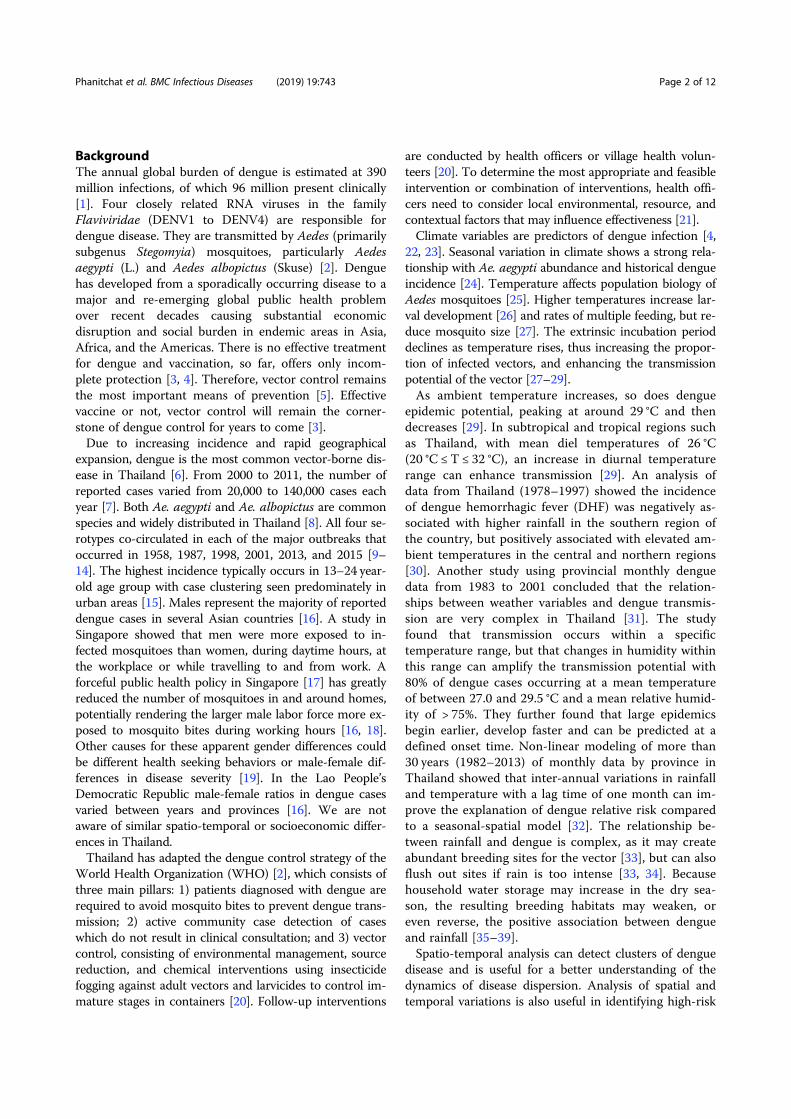
BackgroundThe annual global burden of dengue is estimated at 390
million infections, of which 96 million present clinically
[1]. Four closely related RNA viruses in the family
Flaviviridae (DENV1 to DENV4) are responsible for
dengue disease. They are transmitted by Aedes (primarily
subgenus Stegomyia) mosquitoes, particularly Aedes
aegypti (L.) and Aedes albopictus (Skuse) [2]. Dengue
has developed from a sporadically occurring disease to a
major and re-emerging global public health problem
over recent decades causing substantial economic
disruption and social burden in endemic areas in Asia,
Africa, and the Americas. There is no effective treatment
for dengue and vaccination, so far, offers only incom-
plete protection [3, 4]. Therefore, vector control remains
the most important means of prevention [5]. Effective
vaccine or not, vector control will remain the corner-
stone of dengue control for years to come [3].
Due to increasing incidence and rapid geographical
expansion, dengue is the most common vector-borne dis-
ease in Thailand [6]. From 2000 to 2011, the number of
reported cases varied from 20,000 to 140,000 cases each
year [7]. Both Ae. aegypti and Ae. albopictus are common
species and widely distributed in Thailand [8]. All four se-
rotypes co-circulated in each of the major outbreaks that
occurred in 1958, 1987, 1998, 2001, 2013, and 2015 [9–
14]. The highest incidence typically occurs in 13–24 year-
old age group with case clustering seen predominately in
urban areas [15]. Males represent the majority of reported
dengue cases in several Asian countries [16]. A study in
Singapore showed that men were more exposed to in-
fected mosquitoes than women, during daytime hours, at
the workplace or while travelling to and from work. A
forceful public health policy in Singapore [17] has greatly
reduced the number of mosquitoes in and around homes,
potentially rendering the larger male labor force more ex-
posed to mosquito bites during working hours [16, 18].
Other causes for these apparent gender differences could
be different health seeking behaviors or male-female dif-
ferences in disease severity [19]. In the Lao People’s
Democratic Republic male-female ratios in dengue cases
varied between years and provinces [16]. We are not
aware of similar spatio-temporal or socioeconomic differ-
ences in Thailand.
Thailand has adapted the dengue control strategy of the
World Health Organization (WHO) [2], which consists of
three main pillars: 1) patients diagnosed with dengue are
required to avoid mosquito bites to prevent dengue trans-
mission; 2) active community case detection of cases
which do not result in clinical consultation; and 3) vector
control, consisting of environmental management, source
reduction, and chemical interventions using insecticide
fogging against adult vectors and larvicides to control im-
mature stages in containers [20]. Follow-up interventions
are conducted by health officers or village health volun-
teers [20]. To determine the most appropriate and feasible
intervention or combination of interventions, health offi-
cers need to consider local environmental, resource, and
contextual factors that may influence effectiveness [21].
Climate variables are predictors of dengue infection [4,
22, 23]. Seasonal variation in climate shows a strong rela-
tionship with Ae. aegypti abundance and historical dengue
incidence [24]. Temperature affects population biology of
Aedes mosquitoes [25]. Higher temperatures increase lar-
val development [26] and rates of multiple feeding, but re-
duce mosquito size [27]. The extrinsic incubation period
declines as temperature rises, thus increasing the propor-
tion of infected vectors, and enhancing the transmission
potential of the vector [27–29].
As ambient temperature increases, so does dengue
epidemic potential, peaking at around 29 °C and then
decreases [29]. In subtropical and tropical regions such
as Thailand, with mean diel temperatures of 26 °C
(20 °C ≤ T ≤ 32 °C), an increase in diurnal temperature
range can enhance transmission [29]. An analysis of
data from Thailand (1978–1997) showed the incidence
of dengue hemorrhagic fever (DHF) was negatively as-
sociated with higher rainfall in the southern region of
the country, but positively associated with elevated am-
bient temperatures in the central and northern regions
[30]. Another study using provincial monthly dengue
data from 1983 to 2001 concluded that the relation-
ships between weather variables and dengue transmis-
sion are very complex in Thailand [31]. The study
found that transmission occurs within a specific
temperature range, but that changes in humidity within
this range can amplify the transmission potential with
80% of dengue cases occurring at a mean temperature
of between 27.0 and 29.5 °C and a mean relative humid-
ity of > 75%. They further found that large epidemics
begin earlier, develop faster and can be predicted at a
defined onset time. Non-linear modeling of more than
30 years (1982–2013) of monthly data by province in
Thailand showed that inter-annual variations in rainfall
and temperature with a lag time of one month can im-
prove the explanation of dengue relative risk compared
to a seasonal-spatial model [32]. The relationship be-
tween rainfall and dengue is complex, as it may create
abundant breeding sites for the vector [33], but can also
flush out sites if rain is too intense [33, 34]. Because
household water storage may increase in the dry sea-
son, the resulting breeding habitats may weaken, or
even reverse, the positive association between dengue
and rainfall [35–39].
Spatio-temporal analysis can detect clusters of dengue
disease and is useful for a better understanding of the
dynamics of disease dispersion. Analysis of spatial and
temporal variations is also useful in identifying high-risk
Phanitchat et al. BMC Infectious Diseases (2019) 19:743 Page 2 of 12

locations and times of higher transmission risk, which are
important for disease surveillance and control [15, 40].
The above-mentioned research on climate and
dengue focused on larger spatiotemporal scales, such
as monthly dengue surveillance and climate records at
the provincial level [31, 41, 42]. The current study is
novel because it uses data on the lowest administra-
tive level, the sub-district, in one province to under-
stand fine-scale spatial dengue-climate relationships.
This is useful for developing more reliable prediction
models for future projections applied in early warning
and response systems, thus ultimately improving
timely control interventions.
We analyzed data on reported dengue cases in Khon
Kaen Province, northeastern Thailand collected between
2006 and 2016 to 1) describe demographic characteris-
tics and seasonal variations of dengue cases; 2)
determine the potential impact of climate variability on
dengue incidence; and 3) identify clusters of dengue
cases at the sub-district level.
MethodsStudy area
The study was conducted in Khon Kaen Province, an
area of approximately 10,900 km2 (16°25′12″N to 16°42′
12″N and 102°49′48″E to 102°83′48″E). The province
has 26 districts, 199 sub-districts, and 2139 villages. In
2010, the population was 1,767,601, of which 387,279
people lived in Mueang District that includes the provin-
cial capital Khon Kaen (see Additional file 1). This prov-
ince was selected as the study area because dengue is
endemic with typical seasonal increases and occasional
outbreaks. The province is primarily rural with a few
large urban centers. Mueang District, the most densely
populated area in the province, is a regional center for
education, health, finance and commerce. The northern
and southern parts of the district, along the major
highway linking Bangkok with Lao People’s Democratic
Republic, are rapidly developing. The districts in the
northwestern and southeastern parts of the province are
rural and agricultural. Classification of urban and rural
areas depends on population density. An urban area is
defined as a municipality or town with a population
over 100,000 and a population density above 300 per-
sons per square kilometer [43]. The average minimum
and maximum seasonal temperatures are 16.7 °C
(December–January) and 36.4 °C (April–May). The
monthly minimum and maximum rainfall vary from
0mm (dry season: November–April) to 240 mm (wet
season: May–October).
Data collection
The Office of Disease Prevention and Control, Region 7
Khon Kaen (ODPC7), Department of Disease Control,
Ministry of Public Health, Thailand provided data on
the weekly number of reported dengue cases in Khon
Kaen Province from 1 January 2006 to 31 December
2016. Dengue is a notifiable disease based on the
National Communicable Disease Control Law, i.e., all
government and private hospitals, clinics and other
healthcare facilities must report all cases (confirmed and
suspected) to the local health authority within 24 h of
diagnosis [12]. Cases are recorded by degree of disease
severity into one of three categories (at peak of illness):
1) dengue fever (DF), 2) dengue hemorrhagic fever
(DHF), and 3) dengue shock syndrome (DSS), but the
serotype is not recorded (or typically known except
retrospectively). A patient is diagnosed with suspected
DF when the following criteria are met and signs and
symptoms are present: residence or recent travel to a
dengue endemic area, acute fever accompanied by any
two of the following: headache, myalgia, arthralgia, rash,
positive tourniquet test and leucopenia, with no evidence
of plasma leakage. DHF is recorded in patients with a
temperature ≥ 38 °C, petechiae, ecchymosis, or a positive
tourniquet test, thrombocytopenia (platelets < 100,000
cells/mm3), and evidence of plasma leakage. DSS, the
most severe disease manifestation, is defined as
having the same signs and symptoms as DHF, but
progressing to circulatory failure. The Provincial
Health Offices enter patient data into the standard-
ized Disease Surveillance Report (Report 506) for re-
cording communicable diseases in Thailand. The form
provides the patient’s age, gender, house address,
signs and symptoms, and date of medical consult-
ation. DHF and DSS are based on both clinical symp-
toms and laboratory tests (usually complete blood
count), and sometimes accompanied with a rapid
diagnostic test (RDT); whereas, DF is seldom based
on additional laboratory tests or by RDT.
Meteorological data from 1 January 2006 to 31
December 2016 were downloaded from the data library
of the International Research Institute for Climate and
Society [44], which contains specific climate data from
different sources, such as The National Centers for
Environmental Prediction (NCEP), Climate Forecast Sys-
tem Reanalysis (CFSR) [45], and Climate Hazards Group
InfraRed Precipitation with Station data (CHIRPS) global
rainfall datasets [46]. For each sub-district, daily temper-
atures (°C) were retrieved from NCEP and daily rainfall
(mm) from CHIRPS. These data generated the monthly
means used in the analysis (see Additional files 2 and 3).
The spatial resolution of rainfall is 0.05 × 0.05 degrees
(CHIRPS) and for temperature 0.2 × 0.2 degrees (NCEP
CFSR v2, https://rda.ucar.edu/datasets/ds094.1/). A
centroid was created for each sub-district. Rainfall and
temperature data for each sub-district was determined
based on the grid cell in which the centroid was located.
Phanitchat et al. BMC Infectious Diseases (2019) 19:743 Page 3 of 12

Analysis
Monthly data on dengue cases and climate (rainfall and
temperature) from the study period were combined to
visualize seasonal patterns and temporal trends. Dengue
incidence was calculated using the monthly number of
reported cases and sub-district population size in 2010
reflecting the mid-study denominator [47].
Bayesian Poisson regression models were used to
assess associations with the number of monthly cases in
199 sub-districts. Population was used as the denomin-
ator in the model (i.e. log-population as an offset). The
neighborhood relationship between the sub-districts
were defined using their adjacency matrix; ‘1’ for a pair
of sub-districts sharing a border, otherwise ‘0’. Hence
the following model was used:
Y ij ∼ Poisson μij
� �
log μij
� �
¼ log Pið Þ þ θij
θij ¼ αþ βkxijk þ uij;
where Yij is the observed mean number of cases for the
ith sub-district in jth month (i = 1, …,199; j = 1, …,12), Piis the sub-district population size, α is the intercept, and
βk is the regression coefficient for covariate k. For the
main model, the covariates (xk) were: population density
per square kilometer; gender (proportion of males
among the cases), mean age in years of the cases; mean
rainfall; and minimum and maximum temperature. As a
non-mechanistic way of measuring the seasonality of in-
cidence, a second set of covariates was obtained by re-
placing three meteorological variables by sine and cosine
terms with period 12months. Finally, uij is the random
effect that captures the spatio-temporal autocorrelation
in response data Yij, whose variance depends on the
adjacency matrix.
Conditional autoregressive (CAR) priors [48] structure
were used on uij and for (α, βk), non-informative normal
prior distributions was used. Flat and conjugate priors
were specified for uij using inverse gamma distributions
with shape and scale parameters equal to 0.001. Markov
chain Monte Carlo simulation was used to estimate the
model parameters, sampling 300,000 times, with the first
150,000 as the burn-in, and keeping the results from every
tenth iteration. The “ST.CARar” function of the R statis-
tical software package CARBayesST (www.r-project.org)
was used to fit the model. Convergence was assessed by
trace plots and checked by the convergence Z-score
diagnostic function [49]. The Watanabe-Akaike
Information Criterion (WAIC) was used as a measure
of goodness of fit [50].
Local Indicators of Spatial Association (LISA) were
used to identify significant hotspots, coldspots, and
outliers of dengue incidence at the sub-district level [51].
A hotspot is defined as an area that is surrounded by
other high incidence areas, i.e. incidence is higher than
the expected number given a random distribution of
cases (so called high-high cluster). A coldspot is defined
as an area surrounded by other low incidence areas
(low-low cluster). Hotspot detection can be useful, even
if the global pattern is not clustered. Moreover, case
clusters that occur randomly can also have an influence
on the spread of an infectious disease [52].
Results
General results
Dengue cases numbering 15,167 were reported over the
11-year period by all hospitals and clinics in Khon Kaen
Province. Of these, there were 7461 dengue fever cases
(49.2%) and 7706 severe dengue cases (50.8%), comprising
both DHF and DSS. The demographic characteristics of
patients are summarized in Table 1. Males represented
the majority of patients (8057; 53.1%). Ages ranged from
4months to 92 years old (median 13 years). The highest
number of patients was in the 5–14 year-old age group
(7758; 51.1%), followed by 15–29 years (5026; 33.1%) and
30–44 years (937; 6.2%). The proportion of older age
groups (> 15 years), increased from nearly 20% of all cases
in 2006 to more than 50% in 2016 (Fig. 1). The highest re-
corded disease incidence was in 2013, approximately 80
per 100,000 population (Fig. 2). Incidence was high during
the rainy season (May–September), with July having the
highest incidence (Fig. 3).
Table 1 Demographic characteristics of dengue reported casesin Khon Kaen Province, Thailand, 2006–2016
Characteristics Number of cases Percentage (%)
Gender
Male 8057 53.1
Female 7110 46.9
Age group (years)
< 1 72 0.5
1–<5 857 5.7
5–<15 7758 51.1
15–<30 5026 33.1
30–<45 937 6.2
45–<60 391 2.6
> 60 126 0.8
Diagnosis
Dengue fever (DF) 7461 49.2
Dengue haemorrhagic fever (DHF) 7186 47.4
Dengue shock syndrome (DSS) 520 3.4
Phanitchat et al. BMC Infectious Diseases (2019) 19:743 Page 4 of 12

Association between dengue cases and climatic factors
Mean rainfall and maximum temperature were positively
associated with dengue incidence, and minimum
temperature was negatively associated, in terms of their
point estimates (Table 2). However, among the three 95%
credible intervals (CIs), only the one for maximum
temperature excluded 1 (null effect). The rate ratio for
maximum temperature was 1.055, implying 5.5% (95% CI
0.9–11.5%) increase in cases with an increase of 1 °C per
month. The range of this variable was from 30.7 °C to
44.9 °C. The rate ratio for mean rainfall was 1.004, indicat-
ing that increasing rainfall by one unit (1 cm) per month
would increase dengue incidence by about 0.4%. The
Watanabe-Akaike Information Criterion (WAIC) for this
model was 10,028.75. For the model with two sinusoid
terms replacing the three meteorological variables, the
WAIC was very similar, at 10028.23. This sinusoid terms
had a peak to trough rate ratio of 5.8, and a peak in mid-
July, i.e. a roughly six-fold difference in fitted incidence
from mid-July to mid-January.
The mean dengue incidence was high in the central
northeastern sub-districts, around Khon Kaen city, and
in the southwestern sub-districts of the province (red
and orange in Fig. 4a). The distribution of the posterior
means of the random effects (from the CAR model with
meteorological variables) show some clustering, indicat-
ing that the variables in the model did not account fully
for the spatial variation in the data (Fig. 4b). Posterior
distribution plots are shown in Additional file 4. High
clusters were present around Khon Kaen city and the
southwestern portion of the province and low clusters
were present in the northwestern area (Fig. 5), from the
LISA analysis. When broken down by month, the
incidences show the same clustering patterns, especially
during July–August (Additional file 5).
Discussion
The majority (~ 90%) of patients were below the age of
30 years. The trend during the study period showed that
the proportion of dengue cases younger than 15 years
declined from almost 80% in 2006 to below 50% in 2016.
Dengue fever is generally more common in younger age
groups [53], although there is evidence showing increas-
ing incidence of more severe disease and outcomes
among older age groups [54]. Our observations are also
consistent with a population age shift, potentially influ-
enced by demographic changes, such as the birth and
death rates that show decreasing trends during 2011 and
2015 [55]. Thailand, in general, is undergoing a demo-
graphic transition where the proportion older adults are
gradually increasing with an increase in median age of
the general population. A higher proportion of adults
will also increase the number of immune individuals
(those with previous exposure to dengue virus) in the
population, which might theoretically decrease the risk
of dengue infection in younger people by providing
alternative blood sources for infectious mosquitoes [56].
This age shift has also been observed in other Asian
countries with a higher frequency of dengue cases
among people 15 years of age and older [16]. Increases
in disease incidence in older age groups may be
explained by an increase in secondary infections and
changes in circulating dengue virus serotypes [57], which
have been shown to be important risk factors for severe
clinical presentations [58–62].
Fig. 1 Age distribution of reported dengue cases (DF, DHF and DSS) in Khon Kaen Province, Thailand, 2006–2016
Phanitchat et al. BMC Infectious Diseases (2019) 19:743 Page 5 of 12

There were clear seasonal patterns of dengue inci-
dence in Khon Kaen Province during the study
period. Dengue occurs throughout the rainy season,
with 73% of cases reported between May and Septem-
ber. Although maximum temperature was associated
with higher incidence (Table 2), the model with
meteorological covariates had similar performance (in
terms of the WAIC) to a non-mechanistic model,
which simply fitted a sinusoidal pattern with a period
of 12 months. In our study, a 1 cm increase in
monthly rainfall was associated with a 0.4% increase
in dengue incidence. In Timor Leste, results from
similar modeling analyses showed a far larger effect: a
47% increase in incidence per 1 mm increase in an-
nual rainfall [63]. Different climate patterns between
Timor Leste and Thailand might explain these differ-
ences. Rainfall can affect the availability of mosquito
larval habitats [34]. During rainy and dry periods of
the year, permanent water containers are common in
and around households; some located in toilet or
bathroom spaces providing continuous year round
mosquito production [35–39, 64]. Large water storage
jars and tanks are the most commonly used con-
tainers in Thailand [64]. A study correlating rainfall
A
B
C
D
E
F
Fig. 2 Monthly dengue incidence (a), dengue anomaly (b), rainfall (c), rainfall anomaly (d), temperature (e) and temperature anomaly (f) in KhonKaen Province, Thailand, 2006–2016. DF = dengue fever, DHF = dengue hemorrhagic fever, DSS = dengue shock syndrome
Phanitchat et al. BMC Infectious Diseases (2019) 19:743 Page 6 of 12
and clinical dengue cases in Thailand from 2002 to
2003 also found that the dengue incidence was closely
related with rainfall [65].
Temperature is another primary environmental risk
factor for dengue transmission. Sea surface temperature
(SST) changes, generally related to periodic El Niño
Southern Oscillation effects, and air temperature, having
more direct short-term effects, have both been shown to
influence dengue incidence [63, 66]. Dengue incidence
increased by 19.4% with a 1 °C increase in SST and 2.6%
with a 1 °C increase in weekly maximum temperature in
the Texas-Mexico border region [66]. Another study
found that a 1 °C monthly increase in mean ambient
temperature, dengue incidence increased by 0.7% [63].
In our study, the rate ratio for maximum temperature
was 1.055 per °C, within the range from 30.7 °C to
44.9 °C. Higher temperatures enhance viral replication in
A
B
Fig. 3 Mean monthly dengue incidence per 100,000 persons (a) and monthly average of rainfall (bar) and temperature (line) (b) in Khon KaenProvince, Thailand, 2006–2016
Table 2 Point estimates and 95% credible interval of theBayesian Poisson regression model on number of all monthlydengue cases (DF, DHF and DSS) and covariates in Khon KaenProvince, Thailand, 2006–2016
Parameter Rate ratios
Median 2.5% 97.5%
Mean monthly rainfall (cm) 1.004 0.990 1.017
Maximum temperature (°C) 1.055 1.009 1.115
Minimum temperature (°C) 0.958 0.927 1.024
Age (years) 0.990 0.985 0.994
Gender (proportion femalea) 0.933 0.854 1.020
Density (thousands of people per km2) 0.925 0.827 1.047aHence the rate ratio is for 100% female case composition relative to 100%
male case composition
Phanitchat et al. BMC Infectious Diseases (2019) 19:743 Page 7 of 12

the vector mosquito in a shorter amount of time and
thus increase transmission potential of dengue viruses. A
study of the extrinsic incubation period (EIP) of dengue
serotype 2 in Aedes albopictus found that the virus
remained in the midgut at 18 °C but could disseminate
and invade the salivary glands at temperatures between
23 °C and 32 °C [67], thereby showing higher tempera-
tures produce a shorter EIP and greater transmission po-
tential. The strong and consistent relationships between
climate, particularly rainfall and temperature, and the
number of dengue cases have been used to develop
prediction models to implement more timely dengue
control measures [68, 69]. Relationships between dengue
transmission and climatic variables have been examined
in numerous studies, as shown above, but the question
remains how to use such relationships in predicting
impending outbreaks and applying effective interven-
tions in time to avert them. User-friendly tools, such as
the operational guide on Early Warning and Response
System developed with support from the WHO/TDR
and the European Union [70], are needed and will be
tested in forthcoming work in Khon Kaen Province.
The highest dengue incidence seen in this study oc-
curred in two areas of the province: around Khon Kaen
Mueang District in the northeast, and in Manchakhiri
and Khokphochai districts in the southwest. Mueang
District includes the provincial capital and has the
highest human population density, and in general, more
conducive to dengue transmission. Manchakhiri and
Khokphochai districts have lower population densities,
but are, from our observations, seemingly similar to
other districts in the province, i.e. vector species are
present, larval habitats are plentiful, with a susceptible
human population; therefore there must be other yet un-
explored factors that support high dengue transmission
in these two districts.
Although dengue incidence is influenced by rainfall
and temperature, in our data there is no apparent spatial
clustering of cases associated with the spatial variability
in these environmental parameters. Rather, other factors
such as urbanization are likely causes of the observed
clustering effect [71]. However, population density,
which was included in the regression model as a meas-
ure of urbanization, was not independently associated
with dengue incidence. The residual spatial variation
visible in Fig. 4b suggests that variables beyond those
included in the spatial regression model are needed to
explain differences in incidence between urban and rural
subdistricts. Moreover, hotspots in more rural areas of
southwestern Khon Kaen Province, further corroborate
Fig. 4 Mean dengue prevalence by sub-district (a) and spatial distribution of the posterior means of random effects for dengue (b) in Khon KaenProvince, Thailand, 2006–2016
Phanitchat et al. BMC Infectious Diseases (2019) 19:743 Page 8 of 12
the influence of factors other than urbanization driving
transmission. We do not know of any specific reasons
for why these rural areas should have elevated dengue
prevalence. One speculation could be that the lakes and
swamps that are common in this area may provide suit-
able humidity for mosquitoes to thrive, but this was not
studied here. Large changes in population size over time
will affect outcomes. However, during 2000 and 2015,
the average annual population growth rate in Thailand
was less than 0.5% [72], which might not have affected
the results substantially. Rural-urban migration is com-
mon in Thailand, with people drawn by, for example,
better education, job opportunities, health facilities,
standard of living, and wages [73]. Human movement is
also an important factor in the dynamics of dengue
transmission [74]. Adults are more likely to have greater
mobility than younger age groups; therefore, to under-
stand the circulation of the virus information on recent
travel history and working conditions (location, time of
work, etc.) is required. Elsewhere in Thailand, greater
vulnerability to dengue infection has been observed in
villages situated closer to urban centers [75]. Such neigh-
boring effects are related to similarities in human behav-
ior, development infrastructure, and ecological
surroundings. Moreover, similar lifestyles and social in-
teractions between neighboring areas are evident
Fig. 5 High and low clustering of dengue incidence in Khon Kaen Province, Thailand, 2006–2016
Phanitchat et al. BMC Infectious Diseases (2019) 19:743 Page 9 of 12

between villages that share social and religious centers
such as schools, temples, mosques and community halls
[75]. Hence, the results presented here are generalizable
to most of northern Thailand, Laos, and Cambodia, and
potentially Vietnam and Myanmar as well, under similar
epidemiological settings.
Data collected from national surveillance systems
come with inherent limitations, including underreport-
ing and misreporting of symptomatic cases as well as
the absence of subclinical and asymptomatic infec-
tions [76]. Moreover, dengue cases are seldom labora-
tory confirmed or identified to serotype. Another
limitation of this study is inaccuracy, albeit minor, of
the population denominators within sub-districts, as
these were taken as fixed values from a single census
(2010). Lastly, the possibility of travel-related infections
was not determined in this study, which would provide
potential misclassification bias. Nationally, the import-
ance of travel-related dengue would vary by locality
based on mobility. Obviously, we cannot exclude the
possibility that some dengue infections were acquired
outside the study area, thus potentially affecting the
analysis and conclusions. However, if the general travel
patterns had not changed significantly over the 11-year
observation period, the dengue disease trends reported
in this study would remain valid.
ConclusionWe examined the epidemiology of dengue in Khon Kaen
Province, Thailand between 2006 and 2016. There was
an increase in older age groups reporting symptomatic
dengue. Symptomatic dengue disease in people > 15
years of age is now more common than in children in
this province, an observation that has been seen in other
Asian countries. This study used monthly sub-district
level data to show that rainfall and temperature have sig-
nificant effects on dengue transmission in the province.
Spatial clustering of cases is partly associated with urban
areas closer to Khon Kaen city and rural areas in the
southwest of the province. However, the current analysis
was not able to detect a close proxy factor to quantify a
relationship between urbanization and dengue incidence.
The data set awaits further analysis for temporal pat-
terns of infection for use in disease prediction modeling
and developing dengue early warning systems to guide
vector control operations.
Additional files
Additional file 1: Population density per sub-district, Khon Kaen province,Thailand, 2006 to 2016. Location of province in northeastern Thailand (inset).(PDF 128 kb)
Additional file 2: Average monthly temperature (°C) per sub-district, KhonKaen province, Thailand, January to December 2006–2016. (PDF 51 kb)
Additional file 3: Average monthly rainfall (mm) per sub-district, KhonKaen province, Thailand, January to December 2006–2016. (PDF 49 kb)
Additional file 4: Posterior distribution plots of A) Mean rainfall, B)Minimum temperature, C) Maximum temperature, D) Age, E) Gender,and F) Population density. (PDF 265 kb)
Additional file 5: Average monthly incidence of dengue (DF, DHF,and DSS) per 10,000 persons in Khon Kaen province, Thailand, January toDecember 2006–2016. (PDF 58 kb)
Abbreviations
CAR: Conditional autoregressive; DF: Dengue fever; DHF: Denguehemorrhagic fever; DSS: Dengue shock syndrome; EIP: Extrinsic incubationperiod; ODPC7: Office of Disease Prevention and Control 7; SST: Sea surfacetemperature
Acknowledgements
We thank the Office of Disease Prevention and Control, Region 7, Khon Kaen,Department of Disease Control, Ministry of Public Health, Thailand, for theirkind cooperation and providing the dengue case data used in this study.
Consent for publications
Not applicable.
Authors’ contributions
TP designed the study, performed data analysis, interpretation and draftedthe manuscript. BZ and UH contributed to study design, analysis and writing.CP, TE, SA, BF, MJB and NA guided the research and reviewed themanuscript. KT provided dengue data and guided the research. HJOproposed the research, made substantial contributions to conception, designand coordination of the study, participated in interpretation of data anddrafting the manuscript. All authors read and approved the final manuscript.
Funding
This project is funded by the Research Council of Norway (project no.250443). The funding body had no role in study design, data collection,analysis, interpretation or preparation of the manuscript.
Availability of data and materials
The datasets used during the current study are available from thecorresponding author on reasonable request.
Ethics approval and consent to participate
This study is part of the DENGUE-INDEX Project funded by the ResearchCouncil of Norway (Project no. 250443), which received ethical approval fromKhon Kaen University Ethics Committee for Human Research (HE591099–20/04/2016 and HE601221–01/09/2017), the London School of Hygiene andTropical Medicine Ethical Committee, UK (LSHTM Ethics Ref: 10534–19/04/2016 and LSHTM Ethics Ref: 14275–16/08/2017), and the Regional Committeefor Medical and Health Research Ethics, South East Norway (2016/357–15/04/2016 and 2017/1826–13/03/2018). Permission to use these data was ap-proved by the Ministry of Public Health, Thailand.
Competing interests
The authors declare that they have no competing interests.
Author details1Department of Microbiology, Khon Kaen University, Khon Kaen, Thailand.2Department of Medical Entomology, Faculty of Tropical Medicine, MahidolUniversity, Bangkok, Thailand. 3Department of Biostatistics, University ofNorth Carolina at Chapel Hill, Chapel Hill, USA. 4Department of Biostatisticsand Epidemiology, University of North Texas Health Science Center, FortWorth, TX, USA. 5HPV & EBV and Carcinogenesis Research Group, Khon KaenUniversity, Khon Kaen, Thailand. 6Department of Disease Control, Office ofDisease Prevention and Control, Region 7 Khon Kaen, Ministry of PublicHealth, Khon Kaen, Thailand. 7Université de Montpellier, Montpellier, France.8Public Health & Malaria Control, PT Freeport Indonesia, International SOS,Kuala Kencana, Papua, Indonesia. 9London School of Hygiene and TropicalMedicine, London, UK. 10Faculty of Science and Technology, NorwegianUniversity of Life Sciences, Ås, Norway.
Phanitchat et al. BMC Infectious Diseases (2019) 19:743 Page 10 of 12
Received: 21 July 2018 Accepted: 13 August 2019
References
1. Bhatt S, Gething PW, Brady OJ, Messina JP, Farlow AW, Moyes CL, et al. Theglobal distribution and burden of dengue. Nature. 2013;496(7446):504–7.
2. WHO. Dengue: Guidelines for Diagnosis, Treatment, Prevention and Control.:New Edition. edn. World Health Organization, Geneva; 2009.
3. Christofferson RC, Mores CN. A role for vector control in dengue vaccineprograms. Vaccine. 2015;33(50):7069–74.
4. Descloux E, Mangeas M, Menkes CE, Lengaigne M, Leroy A, Tehei T, et al.Climate-based models for understanding and forecasting dengueepidemics. PLoS Negl Trop Dis. 2012;6(2):e1470.
5. Wen TH, Lin NH, Chao DY, Hwang KP, Kan CC, Lin KC, et al. Spatial-temporalpatterns of dengue in areas at risk of dengue hemorrhagic fever inKaohsiung, Taiwan, 2002. Int J Infect Dis. 2010;14(4):e334–43.
6. WHO. Vector-borne diseases (Dengue). World Health Organization. Availablefrom: http://www.who.int/mediacentre/factsheets/fs387/en/index2.html.
7. Limkittikul K, Brett J, L'Azou M. Epidemiological trends of dengue disease inThailand (2000-2011): a systematic literature review. PLoS Negl Trop Dis.2014;8(11):e3241.
8. Ponlawat A, Scott JG, Harrington LC. Insecticide susceptibility of Aedesaegypti and Aedes albopictus across Thailand. J Med Entomol.2005;42(5):821–5.
9. Halstead SB, Yamarat C. Recent epidemics of hemorrhagic fever in Thailand.Observations related to pathogenesis of a “new” dengue disease.Am J Public Health Nations Health. 1965;55:1386–95.
10. Ministry of Public Health, Thailand. Epidemiology. Annual epidemiologicalsurveillance report. Ministry of Public Health, Nonthaburi, Thailand. 1987.
11. Ministry of Public Health, Thailand. Epidemiology. Annual epidemiologicalsurveillance report. Ministry of Public Health, Nonthaburi, Thailand. 1998.
12. Ministry of Public Health, Thailand.Epidemiology. Annual epidemiologicalsurveillance report. Ministry of Public Health, Nonthaburi, Thailand. 2001.
13. Ministry of Public Health, Thailand. Epidemiology. Annual epidemiologicalsurveillance report. Ministry of Public Health, Nonthaburi, Thailand. 2013.
14. Ministry of Public Health, Thailand. Annual epidemiological surveillancereport. 2015. Ministry of Public Health, Nonthaburi, Thailand. Available from:http://www.boe.moph.go.th/Annual/AESR2015/.
15. Jeefoo P, Tripathi NK, Souris M. Spatio-temporal diffusion pattern andhotspot detection of dengue in Chachoengsao province, Thailand.Int J Environ Res Public Health. 2011;8(1):51–74.
16. Anker M, Arima Y. Male-female differences in the number of reportedincident dengue fever cases in six Asian countries. Western Pac SurveillResponse J. 2011;2(2):17–23.
17. Ooi EE. Changing pattern of dengue transmission in Singapore. DengueBulletin. 2001;25.
18. Ooi EE, Goh KT, Gubler DJ. Dengue prevention and 35 years of vectorcontrol in Singapore. Emerg Infect Dis. 2006;12(6):887–93.
19. Yew YW, Ye T, Ang LW, Ng LC, Yap G, James L, et al. Seroepidemiology ofdengue virus infection among adults in Singapore. Ann Acad Med Singap.2009;38(8):667–75.
20. Ministry of Public Health, Thailand. Reporting of Priority Diseases Guideline.Ministry of Public Health, Nonthaburi, Thailand.Thailand. Available from:http://www.boe.moph.go.th/files/report/20121008_18818829.pdf.
21. Thawillarp S. Evaluation of possible dengue outbreak detectionmethodologies for Thailand, which one should be implemented? JohnsHopkins University; 2017.
22. Gould EA, Higgs S. Impact of climate change and other factors onemerging arbovirus diseases. Trans R Soc Trop Med Hyg.2009;103(2):109–21.
23. Sang S, Gu S, Bi P, Yang W, Yang Z, Xu L, et al. Predicting unprecedenteddengue outbreak using imported cases and climatic factors in Guangzhou,2014. PLoS Negl Trop Dis. 2015;9(5):e0003808.
24. Hopp MJ, Foley JA. Worldwide fluctuations in dengue fever cases related toclimate variability. Clim Res. 2003;25:85–94.
25. Alto BW, Juliano SA. Temperature effects on the dynamics of Aedesalbopictus (Diptera: Culicidae) populations in the laboratory. J Med Entomol.2001;38(4):548–56.
26. Rueda LM, Patel KJ, Axtell RC, Stinner RE. Temperature-dependentdevelopment and survival rates of Culex quinquefasciatus and Aedes aegypti
(Diptera: Culicidae). J Med Entomol. 1990;27(5):892–8.
27. Scott TW, Amerasinghe PH, Morrison AC, Lorenz LH, Clark GG, Strickman D,et al. Longitudinal studies of Aedes aegypti (Diptera: Culicidae) in Thailand andPuerto Rico: blood feeding frequency. J Med Entomol. 2000;37(1):89–101.
28. Rohani A, Wong YC, Zamre I, Lee HL, Zurainee MN. The effect of extrinsicincubation temperature on development of dengue serotype 2 and 4 viruses inAedes aegypti (L.). Southeast Asian J Trop Med Public Health. 2009;40(5):942–50.
29. Liu-Helmersson J, Stenlund H, Wilder-Smith A, Rocklov J. Vectorial capacityof Aedes aegypti: effects of temperature and implications for global dengueepidemic potential. PLoS One. 2014;9(3):e89783.
30. Thammapalo S, Chongsuwiwatwong V, McNeil D, Geater A. The climaticfactors influencing the occurrence of dengue hemorrhagic fever inThailand. Southeast Asian J Trop Med Public Health. 2005;36(1):191–6.
31. Campbell KM, Lin CD, Iamsirithaworn S, Scott TW. The complex relationshipbetween weather and dengue virus transmission in Thailand. Am J TropMed Hyg. 2013;89(6):1066–80.
32. Lowe R, Cazelles B, Paul R, Rodo X. Quantifying the added value of climateinformation in a spatio-temporal dengue model. Stoch Env Res Risk A.2016;30(8):2067–78.
33. Rozilawati H, Zairi J, Adanan CR. Seasonal abundance of Aedes albopictus inselected urban and suburban areas in Penang. Malaysia Trop Biomed.2007;24(1):83–94.
34. Li CF, Lim TW, Han LL, Fang R. Rainfall, abundance of Aedes aegypti anddengue infection in Selangor, Malaysia. Southeast Asian J Trop Med PublicHealth. 1985;16(4):560–8.
35. Lambdin BH, Schmaedick MA, McClintock S, Roberts J, Gurr NE, Marcos K, etal. Dry season production of filariasis and dengue vectors in AmericanSamoa and comparison with wet season production. Am J Trop Med Hyg.2009;81(6):1013–9.
36. Bermudi PMM, Kowalski F, Menzato MM, Ferreira MDC, Passos W, Oku VJA,et al. Aedes aegypti breeding site in an underground rainwater reservoir: awarning. Rev Saude Publica. 2017;51:122.
37. Serpa LL, Costa KV, Voltolini JC, Kakitani I. Seasonal variation of Aedes aegyptiand Aedes albopictus in a city of southeastern Brazil. Rev Saude Publica.2006;40(6):1101–5.
38. Kittayapong P, Strickman D. Distribution of container-inhabiting Aedes larvae(Diptera: Culicidae) at a dengue focus in Thailand. J Med Entomol. 1993;30(3):601–6.
39. Tonn RJ, Sheppard PM, Macdonald WW, Bang YH. Replicate surveys of larvalhabitats of Aedes aegypti in relation to dengue haemorrhagic fever inBangkok. Thailand Bull World Health Organ. 1969;40(6):819–29.
40. Sharma KD, Mahabir RS, Curtin KM, Sutherland JM, Agard JB, Chadee DD.Exploratory space-time analysis of dengue incidence in Trinidad: aretrospective study using travel hubs as dispersal points, 1998-2004. ParasitVectors. 2014;7:341.
41. Cummings DA, Irizarry RA, Huang NE, Endy TP, Nisalak A, Ungchusak K,Burke DS. Travelling waves in the occurrence of dengue haemorrhagic feverin Thailand. Nature. 2004;427(6972):344–7.
42. van Panhuis WG, Choisy M, Xiong X, Chok NS, Akarasewi P, Iamsirithaworn S, etal. Region-wide synchrony and traveling waves of dengue across eightcountries in Southeast Asia. Proc Natl Acad Sci U S A. 2015;112(42):13069–74.
43. Lolekha S, Tanthiphabha W, Sornchai P, Kosuwan P, Sutra S, Warachit B,Chup-Upprakarn S, Hutagalung Y, Weil J, Bock HL. Effect of climatic factorsand population density on varicella zoster virus epidemiology within atropical country. Am J Trop Med Hyg. 2001;64(3–4):131–6.
44. International Research Institute for Climate and Society: Climate DataLibrary. International Research Institute for Climate and Society. 2017.
45. NCEP Climate Forecast System Reanalysis (CFSR) Selected Hourly Time-Series Products, January 1979 To December 2010. Research Data Archive atthe National Center for Atmospheric Research, Computational andInformation Systems Laboratory.
46. Funk C, Peterson P, Landsfeld M, Pedreros D, Verdin J, Shukla S, et al. Theclimate hazards infrared precipitation with stations-a new environmentalrecord for monitoring extremes. Sci Data. 2015;2:150066.
47. National Statistical Office. The 2010 Population and housing census.Bangkok, Thailand 2010.
48. Rushworth A, Lee D, Sarran C. An adaptive spatiotemporal smoothingmodel for estimating trends and step changes in disease risk. J Royal StatSoc Series C-Applied Stat. 2017;66(1):141–57.
49. Geweke J. In: Bernardo JM, Berger JO, Dawid AP, Smith AFM, editors.Evaluating the Accuracy of Sampling-Based Approaches to CalculatingPosterior Moments. In Bayesian Statistics 4. Oxford: Clarendon Press; 1992.
Phanitchat et al. BMC Infectious Diseases (2019) 19:743 Page 11 of 12
50. Burnham KP, Anderson DR. Multimodel inference: understanding AIC andBIC in model selection. Sociol Methods Res. 2004;33(2):261–304.
51. Anselin L. Local indicators of spatial association-LISA. Geogr Anal.1995;27:93–115.
52. Pessanha JE, Caiaffa WT, Almeida MC, Brandao ST, Proietti FA. Diffusionpattern and hotspot detection of dengue in Belo Horizonte, Minas Gerais.Brazil J Trop Med. 2012;2012:760951.
53. Rigau-Perez JG, Vorndam AV, Clark GG. The dengue and denguehemorrhagic fever epidemic in Puerto Rico, 1994-1995. Am J Trop Med Hyg.2001;64(1–2):67–74.
54. Guha-Sapir D, Schimmer B. Dengue fever: new paradigms for a changingepidemiology. Emerg Themes Epidemiol. 2005;2(1):1.
55. National Statistical Office. Population and Housing census (Births andDeaths by Sex: 2011–2015, Khon Kaen province). Khon Kaen, Thailand 2018.
56. Cummings DA, Iamsirithaworn S, Lessler JT, McDermott A, Prasanthong R,Nisalak A, et al. The impact of the demographic transition on dengue inThailand: insights from a statistical analysis and mathematical modeling.PLoS Med. 2009;6(9):e1000139.
57. Kanakaratne N, Wahala WM, Messer WB, Tissera HA, Shahani A, AbeysingheN, et al. Severe dengue epidemics in Sri Lanka, 2003-2006. Emerg Infect Dis.2009;15(2):192–9.
58. Guzman MG, Kouri G. Dengue and dengue hemorrhagic fever in theAmericas: lessons and challenges. J Clin Virol. 2003;27(1):1–13.
59. Deen JL, Harris E, Wills B, Balmaseda A, Hammond SN, Rocha C, et al. TheWHO dengue classification and case definitions: time for a reassessment.Lancet. 2006;368(9530):170–3.
60. Karyanti MR, Uiterwaal CS, Kusriastuti R, Hadinegoro SR, Rovers MM,Heesterbeek H, et al. The changing incidence of dengue haemorrhagicfever in Indonesia: a 45-year registry-based analysis. BMC Infect Dis.2014;14:412.
61. Guzman MG, Alvarez M, Halstead SB. Secondary infection as a risk factor fordengue hemorrhagic fever/dengue shock syndrome: an historicalperspective and role of antibody-dependent enhancement of infection.Arch Virol. 2013;158(7):1445–59.
62. Mohd-Zaki AH, Brett J, Ismail E, L'Azou M. Epidemiology of dengue diseasein Malaysia (2000-2012): a systematic literature review. PLoS Negl Trop Dis.2014;8(11):e3159.
63. Wangdi K, Clements ACA, Du T, Nery SV. Spatial and temporal patterns ofdengue infections in Timor-Leste, 2005-2013. Parasit Vectors. 2018;11(1):9.
64. Vannavong N, Seidu R, Stenstrom TA, Dada N, Overgaard HJ. Effects ofsocio-demographic characteristics and household water management onAedes aegypti production in suburban and rural villages in Laos andThailand. Parasit Vectors. 2017;10(1):170.
65. Wiwanitkit V. An observation on correlation between rainfall and theprevalence of clinical cases of dengue in Thailand. J Vector Borne Dis.2006;43(2):73–6.
66. Brunkard JM, Cifuentes E, Rothenberg SJ. Assessing the roles oftemperature, precipitation, and ENSO in dengue re-emergence on theTexas-Mexico border region. Salud Publica Mex. 2008;50(3):227–34.
67. Liu Z, Zhang Z, Lai Z, Zhou T, Jia Z, Gu J, et al. Temperature increaseenhances Aedes albopictus competence to transmit dengue virus.Front Microbiol. 2017;8:2337.
68. Phung D, Talukder MR, Rutherford S, Chu C. A climate-based predictionmodel in the high-risk clusters of the Mekong Delta region, Vietnam:towards improving dengue prevention and control. Tropical Med Int Health.2016;21(10):1324–33.
69. Karim MN, Munshi SU, Anwar N, Alam MS. Climatic factors influencingdengue cases in Dhaka city: a model for dengue prediction. Indian J MedRes. 2012;136(1):32–9.
70. WHO/TDR. Operational Guide: Early Warning and Response System (EWARS)for dengue outbreaks. Training in Tropical Diseases (TDR). World HealthOrganization, Special Programme for Research and Training in TropicalDiseases 2017;52.
71. Gubler DJ. Dengue, urbanization and globalization: the unholy trinity of the21(st) century. Trop Med Health. 2011;39(4 Suppl):3–11.
72. United Nations, Department of Economic and Social Affairs, PopulationDivision 2017. World Population Prospects: The 2017 Revision, custom dataacquired via website: https://population.un.org/wpp/DataQuery/.
73. Katewongsa P. Benefits of rural-urban migration for migrants’ better life: acase study in Nang Rong, Buriram Province. Thailand Thammasat Review.2015;18(1):63–81.
74. Stoddard ST, Forshey BM, Morrison AC, Paz-Soldan VA, Vazquez-ProkopecGM, Astete H, Vilcarromero S, Elder JP, Halsey ES, et al. House-to-househuman movement drives dengue virus transmission. Proc Natl Acad SciU S A. 2013;110(3):994–9.
75. Tipayamongkholgul M, Lisakulruk S. Socio-geographical factors invulnerability to dengue in Thai villages: a spatial regression analysis.Geospat Health. 2011;5(2):191–8.
76. da Fonseca BA, Fonseca SN. Dengue virus infections. Curr Opin Pediatr.2002;14(1):67–71.
Publisher’s NoteSpringer Nature remains neutral with regard to jurisdictional claims inpublished maps and institutional affiliations.
Phanitchat et al. BMC Infectious Diseases (2019) 19:743 Page 12 of 12

95
Chapter 2: Addressing the complex relationships between Aedes vectors,
socio-economics and dengue transmission in North-eastern Thailand.
Our baseline study demonstrated that dengue distribution and dispersion in North-eastern
Thailand was not only explained by climatic data, and that urbanization, human movements and
entomological factors may partially explain the clustering effect observed (Phanitchat et al. 2019).
Hence, we conducted a prospective hospital-based case control study (see section 4.1), to identify
risk factors for dengue infections. The scope was to assess whether entomological and
immunological indices could discriminate between dengue positive and negative households (see
section 3.1).
Briefly, 377 individuals were recruited from the nineteen district and sub-district hospitals
between June 2016 and August 2019. Dengue infection was detected by RDT targeting both
dengue -NS1 and -IgM/IgG and confirmed by RT-qPCR allowing the identification of 173 recent
dengue cases and 204 controls (0.85 case/control ratio). The participant ages ranged from 5 to 76
years with 190 (48%) females represented. Individual questionnaire and immature and adult Aedes
entomological collections were performed in 377 patient houses and the 1,110 neighbouring
surrounding households (mean of 3.94 houses per individual recruited). In addition, the levels of
Ab response to Nterm-34 peptide could be measured in 368 patients. Socio-economic status,
household and individual characteristics were analysed as additional risk factors for dengue
infection (see details in section 4.1).
Summary of the results:
Our results showed that patient age was associated with higher odds of dengue. While
dengue normally affects young children, we found that individuals aged between 10 and 25 years-
old were at higher risk relative to those either younger or older. This is in agreement with our
The results from this study were published in PLOS Neglected Tropical Diseases,1;14(10),
https//doi:org/10.1371/journal.pntd.0008703. (2020). Complex relationships between Aedes
vectors, socio-economics and dengue transmission-Lessons learned from a case-control
study in northeastern Thailand. Fustec B, Phanitchat T, Hoq MI, Aromseree S, Pientong C,
Thaewnongiew K, Ekalaksananan T, Bangs MJ, Corbel V, Alexander N, Overgaard HJ.

96
baseline survey of dengue incidence in North-eastern Thailand (Phanitchat et al. 2019) and with
other recent studies conducted in Thailand, Malaysia, and the Philippines (Limkittikul et al. 2014,
Mohd-Zaki et al. 2014, Thomas et al. 2015, Alera et al. 2016). Although other studies had found
a higher odds of dengue transmission in low-income family (Telle et al. 2016, Wijayanti et al.
2016a, Udayanga et al. 2018), no such association was found in the study area. However, we
showed that household construction may play a role in dengue transmission risk, as individuals
living in two-floor houses were at higher odds of dengue infections. Moreover, individuals who
declared spending most of their time indoors were found at higher risk of dengue. Curiously, the
presence of eave gaps in the house was negatively associated with dengue. Although
counterintuitive, the apparent ‘protective’ effect of eave gaps might be due to increased air flow
and ventilation inside the house hence creating exit routes for the vectors (von Seidlein et al. 2019).
Although not surprising, our study confirmed that traditional entomological indices were
not good indicators of dengue transmission as they were statistically higher in the “control” houses
than in “dengue case” houses. Indeed, vector infestation indices based on immature stages (HI, BI,
and CI) were all negatively associated with dengue using univariable analysis (total containers
inspected, 5,185 including 1,230 (23.7%) positive for immature Aedes stages). In other words,
control households had more Aedes positive containers than case’s households. It is worth to
mention that most of the inspected households (control and case) had immature indices values
higher than the “outbreak-risk” thresholds setting up by the MoPH of Thailand (i.e.,, CI<1%,
BI<50 and HI<10%) (Thai Ministry of Public Health 2013). Similarly, pupae indices (PPI and
PHI) were not significantly different between case and control houses and even more Aedes adults
were found in control households. Although surprising, this could be explained by higher vector
control efforts following onset of dengue symptoms in the dengue “case” household which would
have reduced vector infestation at the monitoring time point. This is corroborated by the positive
association between dengue cases and the use of household insecticide products as declared by the
head of the household in the questionnaire.
Nonetheless, our findings showed that the presence of DENV-infected Ae. aegypti in the
households was positively associated with dengue infections (p=0.018). Indeed, the proportion of
DENV-infected Aedes was higher in the patient houses (≈8%) than the control houses (3%), hence
suggesting that vector infectivity would be a more reliable indicator than vector abundance to

97
assess dengue transmission risk. Overall, about 13% of the selecting house’s (including
neighbourhood and patient house) had DENV-infected Aedes hence highlighting the
hyperendemic situation of dengue in the study area.
Interestingly, individuals from the control group had higher level of Ab response to the
Nterm-34 than individuals from the dengue case group, which corroborate entomology results
(Figure 33). Our results suggest that individual with a higher Ab response to the salivary biomarker
received more Aedes mosquito bites than lower immune responders (Elanga Ndille et al. 2012).
This trend was more pronounced when considering the Aedes density indoor, hence suggesting a
strong endophagic preference of the Aedes population in the study area. Nevertheless, neither the
adult abundance in the patient household nor the level of human exposure to Aedes mosquito bites
were correlated with dengue incidence. Interestingly, a positive and significant association was
seen between the intensity Ab response to Aedes saliva and the presence of IgG response against
dengue hence suggesting that patient’s immunity may have biased the correlation between the
mosquito exposure risk and dengue transmission. This highlights the fact that dengue virus
transmission is complex and varies through time and space, and the relationship between vector
density/aggressiveness and risk of human infection is not static nor linear. Moreover, the measure
of Ab response to Aedes saliva reflects the overall exposure to Aedes bites in the previous 2-4
weeks and not necessarily at the time of virus transmission. Including all inhabitants from each
house, irrespective to the dengue infection status, would have been useful to assess the differential
exposure to Aedes bites in dengue case and control groups.
Figure 33: Immune response to Aedes salivary peptide Nterm-34 (∆OD) in dengue case and
control patients.

98
In conclusion, this first study highlights the complex relationship between Aedes vectors,
socio-economic factors, and dengue transmission risk, and highlight the challenges to setting up
accurate warning indicators for dengue prevention. A longitudinal randomized controlled study
conducted as part of the DENGUE INDEX Project (see section 4.3) was then conducted to better
evaluate the close relationship between the levels of human-Aedes contact, the levels of Aedes
infestations, and dengue transmission risk in North-eastern Thailand. The main findings are
described in the next chapter.

RESEARCH ARTICLE
Complex relationships between Aedes vectors,socio-economics and dengue transmission—Lessons learned from a case-control study innortheastern Thailand
Benedicte FustecID1,2,3, Thipruethai Phanitchat4, Mohammad Injamul HoqID
5,
Sirinart AromsereeID2,6, Chamsai PientongID
2,6, Kesorn Thaewnongiew7,
Tipaya EkalaksanananID2,6, Michael J. Bangs8,9, Vincent Corbel1,3, Neal Alexander10 ,
Hans J. Overgaard11
1 University of Montpellier, Montpellier, France, 2 Department of Microbiology, Faculty of Medicine, KhonKaen University, Khon Kaen, Thailand, 3 Institut de Recherche pour le Developpement, Montpellier, France,
4 Department of Medical Entomology, Faculty of Tropical Medicine, Mahidol University, Bangkok, Thailand,5 School of Public Health, Epidemiology and Social Medicine at the Institute of Medicine, University of
Gothenburg, Gothenburg, Sweden, 6 HPV & EBV and Carcinogenesis Research Group, Khon KaenUniversity, Khon Kaen, Thailand, 7 Office of Diseases Prevention and Control, Region, Khon Kaen, Thailand,8 Public Health & Malaria Control, PT Freeport Indonesia/International SOS, Mimika, Papua, Indonesia,
9 Department of Entomology, Faculty of Agriculture, Kasetsart University, Bangkok, Thailand, 10 MRCTropical Epidemiology Group, London School of Hygiene and Tropical Medicine, London, United Kingdom,11 Norwegian University of Life Sciences, s, Norway
These authors contributed equally to this work.
Abstract
Background/Objectives
Dengue fever is an important public health concern in most tropical and subtropical coun-
tries, and its prevention and control rest on vector surveillance and control. However, many
aspects of dengue epidemiology remain unclear; in particular, the relationship between
Aedes vector abundance and dengue transmission risk. This study aims to identify entomo-
logical and immunological indices capable of discriminating between dengue case and con-
trol (non-case) houses, based on the assessment of candidate indices, as well as individual
and household characteristics, as potential risk factors for acquiring dengue infection.
Methods
This prospective, hospital-based, case-control study was conducted in northeastern Thai-
land between June 2016 and August 2019. Immature and adult stage Aedes were collected
at the houses of case and control patients, recruited from district hospitals, and at patients’
neighboring houses. Blood samples were tested by RDT and PCR to detect dengue cases,
and were processed with the Nterm-34 kDa salivary peptide to measure the human immune
response to Aedes bites. Socioeconomic status, and other individual and household charac-
teristics were analyzed as potential risk factors for dengue.
PLOS NEGLECTED TROPICAL DISEASES
PLOSNeglected Tropical Diseases | https://doi.org/10.1371/journal.pntd.0008703 October 1, 2020 1 / 25
a1111111111a1111111111a1111111111a1111111111a1111111111
Citation: Fustec B, Phanitchat T, Hoq MI,
Aromseree S, Pientong C, Thaewnongiew K, et al.
(2020) Complex relationships between Aedes
vectors, socio-economics and dengue
transmission—Lessons learned from a case-
control study in northeastern Thailand. PLoS Negl
Trop Dis 14(10): e0008703. https://doi.org/
10.1371/journal.pntd.0008703
Editor:Marilia Sa Carvalho, Fundacao Oswaldo
Cruz, BRAZIL
Received:May 30, 2020
Accepted: August 12, 2020
Published: October 1, 2020
Copyright: 2020 Fustec et al. This is an open
access article distributed under the terms of the
Creative Commons Attribution License, which
permits unrestricted use, distribution, and
reproduction in any medium, provided the original
author and source are credited.
Data Availability Statement: The data underlying
the results presented in this paper are made
publicly available following principles that data
should be Findable, Accessible, Interoperable, and
Reusable (FAIR, https://www.force11.org/group/
fairgroup/fairprinciples) and regulations of the
project owner, the Norwegian University of Life
Sciences, that research data must be archived in
approved national or international archives. The
data used in this publication is archived at the

Results
Study findings showed complex relationships between entomological indices and dengue
risk. The presence of DENV-infected Aedes at the patient house was associated with 4.2-
fold higher odds of dengue. On the other hand, Aedes presence (irrespective of infectious
status) in the patient’s house was negatively associated with dengue. In addition, the human
immune response to Aedes bites, was higher in control than in case patients and Aedes
adult abundance and immature indices were higher in control than in case houses at the
household and the neighboring level. Multivariable analysis showed that children aged 10–
14 years old and those aged 15–25 years old had respectively 4.5-fold and 2.9-fold higher
odds of dengue infection than those older than 25 years.
Conclusion
DENV infection in female Aedes at the house level was positively associated with dengue
infection, while adult Aedes presence in the household was negatively associated. This
study highlights the potential benefit of monitoring dengue viruses in Aedes vectors. Our
findings suggest that monitoring the presence of DENV-infected Aedesmosquitoes could
be a better indicator of dengue risk than the traditional immature entomological indices.
Author summary
Dengue fever is a globally expanding arboviral disease, consisting of four distinct sero-
types, transmitted primarily by synanthropic/peridomestic mosquitoes, Aedes aegypti and
Aedes albopictus. Given the absence of specific treatment, and the incomplete protection
provided by the currently available vaccine, vector surveillance and control remain the
principal tool to prevent and control dengue transmission. However, vector surveillance
through the monitoring of larval mosquito indices lacks consistency in addressing dengue
risk. Surveillance based on pupal and adult stages is considered as more accurate to esti-
mate dengue transmission risk, although monitoring is difficult to implement in routine.
An alternative strategy is the use of the specific human antibody response to Aedes saliva
to identify human exposure risk to Aedes bites. We conducted a hospital-based, case-con-
trol study in northeastern Thailand in order to identify risk factors for dengue infection
using entomological and immunological indices, together with select individual and
household characteristics. We found that people aged 10–25 years had significant higher
odds of dengue than older adults (>25 years old). The presence of DENV-infected Aedes
in the house was associated with 4.2-fold higher odds of dengue infection. Interestingly,
Aedes adult abundance in the household was negatively associated with dengue revealing
the complex role of Aedes density to dengue risk. This study highlights the potential bene-
fit of monitoring dengue viruses in Aedes vectors to identify areas (“hot spots”) and people
(“hot pops”) at higher risk of transmission.
Introduction
Dengue fever is a globally expanding mosquito-borne disease which threatens half the world’s
population [1]. Dengue virus (DENV) is transmitted by synanthropic Aedesmosquitoes, with
PLOS NEGLECTED TROPICAL DISEASES Complex relationships between Aedes vectors, socio-economics and dengue transmission
PLOSNeglected Tropical Diseases | https://doi.org/10.1371/journal.pntd.0008703 October 1, 2020 2 / 25
Norwegian Center for Research Data (NSD), a
national archive and center for research data. Data
can be accessed without specific permissions by
filling in and submitting a Data Access Form at
https://nsd.no/nsd/english/order.html, referring to
the project ID no. NSD2864 and project name
“Determination of entomological indices to assess
dengue transmission, predict dengue outbreaks,
and evaluate vector control interventions, 2019”,
short name: DENGUE-INDEX.
Funding: This study was supported by the
Research Council of Norway, through the
DENGUE-INDEX project (project no. 250443) and
the Norwegian University of Life Sciences. The
funders had no role in the study design, data
collection, data analysis or decision to publish.
Competing interests: The authors have declared
that no competing interests exist.
Aedes aegypti (L.) typically being the primary vector [2], and Aedes albopictus (Skuse) a sec-
ondary one [3]. The Southeast Asia region accounts for more than half of the reported dengue
cases worldwide [2, 4, 5]. Thailand typically records more than 20,000 cases each year, with all
four DENV serotypes circulating and both vector species spread throughout the country [6].
Although dengue incidence is highly seasonal, outbreaks are difficult to predict [7, 8]. Dengue
virus transmission is highly efficient and it is assumed that only a few vector mosquitoes are
sufficient to ensure transmission [9]. Aedes aegypti is particularly well adapted to urbanized
environments and is a strongly anthropophagic diurnal blood feeder [10–13]. The absence of
specific treatments for dengue and the incomplete protection offered by the currently available
vaccine [14, 15], underscores the importance of vector surveillance and management as the
principal strategy for dengue prevention and control [7, 16].
In Thailand, dengue prevention and control are mainly based on hospital case reporting
and vector surveillance and control that are carried out collaboratively between hospitals and
the Offices of Disease Prevention and Control (ODPC). When a dengue case is reported from
hospital, a Surveillance and Rapid Response Team (SSRT) is mandated to carry out insecticide
space spray (‘fogging’) within 100 meters of the case house within 24 hours of notice in order
to interrupt transmission [17]. The reorganization of disease control operations in Thailand
resulted in 76 provincial administrations being aggregated into 22 regional ODPCs [18]. The
seventh regional ODPC includes four provinces: Khon Kaen, Roi Et, Maha Sarakham, and
Kalasin with a total population of around 5 million. Northeastern Thailand is the third largest
region in the country with regards to population size and land area, with an economy mainly
based on agriculture.
In most dengue-endemic countries, vector surveillance usually consists of monitoring
Aedes immature (larvae and pupae) stages present in natural and artificial breeding sites (larval
habitats) in and near houses [19–21]. Vector presence and density are estimated by standard-
ized indices such as the Breteau Index (BI), Container Index (CI), House Index (HI), and the
Pupae per Person Index (PPI) [21–23]. Entomological measures as thresholds have been pro-
posed to assess and estimate risk for use as early warning systems to predict dengue outbreaks
[19, 22, 24]. In Thailand, vector density thresholds to estimate risk of dengue outbreaks occur-
rence have been set at HI>10, BI>50 and CI>1 [25]. Additionally, vector control interven-
tions are implemented to reduce vector abundance and prevent dengue transmission.
However, numerous studies have failed to clearly link entomological indices to the risk of den-
gue transmission [7, 24, 26, 27]. Indeed, the larval stages (four successive instars) typically suf-
fer high mortality during development to pupal stage, thus indices based only on their
presence are generally poor indicators of the eventual adult vector density. Pupal indices (a
stage with very low mortality) were proposed as a more accurate determination of actual adult
production; however, pupal collections are far more challenging and time consuming to carry
out [26, 28]. Adult collections can be performed via several devices such as gravitraps, sticky
traps, baited mechanical traps, and mouth or mechanical aspirators, but they only provide an
imprecise estimation of the true vector density and do not reflect human-vector exposure.
Entomological collections for target Aedes species, of all kinds, are labor- and time-consum-
ing, expensive, and contingent on access to the house being granted. However, estimating the
human immune response to Aedes bites as a surrogate measure of bite exposure (intensity)
might be less labor-intensive and more informative of relative “vector attack” over time [29].
Upon initiating the blood feeding process, salivary gland proteins injected at the bite site
induce a species-specific immune response by the host [30, 31]. These specific antibodies
(against salivary proteins) have shown promising to measure seasonal variation of human
exposure to mosquito bites [32–37] and to assess the effectiveness (i.e., reduction in biting) of
vector control interventions [38].
PLOS NEGLECTED TROPICAL DISEASES Complex relationships between Aedes vectors, socio-economics and dengue transmission
PLOSNeglected Tropical Diseases | https://doi.org/10.1371/journal.pntd.0008703 October 1, 2020 3 / 25

The current study aims to identify risk factors for dengue transmission across four prov-
inces in northeastern Thailand by comparing individuals with and without dengue in terms of
i) their immune response to Aedes bites, ii) the presence and abundance of immature and
adult Aedes in and close proximity around their houses, and iii) their individual and household
characteristics. The first objective was to assess the accuracy of entomological and immunolog-
ical indices to discriminate dengue positive and dengue negative households. We hypothesize
that there will be more adult Aedesmosquitoes and a higher level of immune response to
Aedes exposure (salivary proteins) in households with a recent dengue case compared to con-
trol (non-case) houses. The second objective was to assess whether socio-economics, house-
hold characteristics and entomological and immunological indices can be accurate predictors
of dengue transmission risk.
Materials andmethods
Study settings
This hospital-based case-control study was carried out in four provinces in northeastern Thai-
land (Fig 1) between June 2016 and August 2019. Ten district hospitals were included: Mancha
Khiri, Chum Phae, Ban Phai, and Ban Haet districts in Khon Kaen Province; Selaphum, Phon
Thong, Thawatburi districts in Roi Et Province; Kamalasai and Kuchinarai districts in Kalasin
Province; and Chiang Yuen district in Maha Sarakham Province. Additionally, nine sub-dis-
trict hospitals in Khon Kaen Muang district (Khon Kaen Province) were included. The four
provinces cover approximately 31,440 km2 with around 5 million inhabitants. Khon Kaen, Roi
Et, Kalasin and Maha Sarakham provinces are divided in 26, 20, 18 and 13 districts, respec-
tively (Fig 1). Over the previous 15 years, the region reported in average 4,488 dengue cases
annually [39, 40]. A case-control design was chosen because it allowed the investigation of sev-
eral risk factors concomitantly, it is effective for diseases with low incidence, and requires rela-
tively, few study subjects.
Sample size
The study sample size was calculated using the unmatched case-control study module of
OpenEpi, version 3 [42] with 90% power based on data from Thomas et al. [43]. Assuming a
difference in DENV-infected female Aedesmosquitoes collected between dengue positive and
dengue negative households, with an exposure of 10% of DENV-infected Aedes in the exposed
group, and 1% of DENV-infected Aedes in the control group, the significance level was set at
5% (two-sided) and the ratio of control to case at 1. The result was a target sample size of 322
patients. To allow for a 15% loss at the household questionnaire stage, we increased the final
sample to 370.
Patient recruitment
Patients presenting with dengue-like symptoms were recruited from the participating hospi-
tals. Regarding Thai health services, public hospitals generally serve the communities in the
districts and sub-districts in which they are located. Eligible patients with potential dengue
infections were recruited based on presence of fever (�38˚C), no recent travel history during
the previous 7 days, and being older than five years-of-age.
Blood collections
A total of 6 mL of venous blood was drawn from each participant for the following three pur-
poses (Fig 2):
PLOS NEGLECTED TROPICAL DISEASES Complex relationships between Aedes vectors, socio-economics and dengue transmission
PLOSNeglected Tropical Diseases | https://doi.org/10.1371/journal.pntd.0008703 October 1, 2020 4 / 25

PLOS NEGLECTED TROPICAL DISEASES Complex relationships between Aedes vectors, socio-economics and dengue transmission
PLOSNeglected Tropical Diseases | https://doi.org/10.1371/journal.pntd.0008703 October 1, 2020 5 / 25

1. Detect dengue non-structural protein 1 (NS1) and IgM / IgG antibodies using a Rapid
Diagnostic Test (RDT) (SD BIOLINE Dengue Duo, Standard Diagnostics, Korea).
2. Determine the immune response to Aedes bites using two blood drops (approximately
75μL each) collected on protein saver cards 903 (Whatman, UK).
3. Confirm dengue infection by reverse transcription polymerase chain reaction (RT-PCR)
(described below) and distinguish serotypes (not presented here) using 5.7 mL whole blood
collected in heparin or EDTA tubes.
DENV confirmation in human samples and case definition
RNAwas extracted from patients’ blood for DENV screening, confirmation and serotyping by
RT-PCR as described previously [44] and adapted to conventional PCR. According to the course
of dengue illness, viremia usually drops after few days of fever, while antibody response is trig-
gered within few days after the beginning of dengue symptoms [2]. Therefore, a positive sample
Fig 1. Map and characteristics of study sites of the case-control study in northeastern Thailand. A: Location offour provinces and study districts in northeastern Thailand included in the case-control study. Map of study sites wasbuilt using QGis 3.10 software and shapefiles were obtained from the Humanitarian Data Exchange project [41] underthe Creative Commons Attribution International 4.0 license (CC BY 4.0). B: Study area characteristics, population andaverage number of dengue cases per year from 2005–2019 [39].
https://doi.org/10.1371/journal.pntd.0008703.g001
Fig 2. Flow diagram of case-control study design.
https://doi.org/10.1371/journal.pntd.0008703.g002
PLOS NEGLECTED TROPICAL DISEASES Complex relationships between Aedes vectors, socio-economics and dengue transmission
PLOSNeglected Tropical Diseases | https://doi.org/10.1371/journal.pntd.0008703 October 1, 2020 6 / 25
for NS1 and/or IgM by RDT and/or positive for DENV by PCR was recorded a dengue case. A
participant who was negative for both RDT and PCR or IgG-positive only was recorded a control
(Fig 2). Hence the controls were selected on the basis of having an “imitation” disease with similar
symptoms (e.g., fever) to dengue [45], a design method also known as ‘test-negative’ [46].
Individual characteristics
A questionnaire was used to collect information about each individual study case (positive and
control). Patients were stratified into four age groups: 5–9 years-old; 10–14 years-old; 15–25
years-old; and> 25 years-old. History of previous dengue infections and vaccinations were
recorded. Patients were asked about their main activities during weekdays and weekends (e.g.,
at home; at work away from home; at school; farming; other), as well as their typical resting/
sleeping locations and habits (e.g., primarily indoor, outdoor, or equally indoor and outdoor).
Travel history outside the resident district within the last three months was recorded and used
as a binary variable.
Household characteristics
A questionnaire was used to collect data on house characteristics and socio-economic status,
including monthly household income, possession of certain assets (e.g., TV, air conditioner,
car, or motorbike), and source of drinking and non-drinking water. Observations on the
house included the number of rooms, wall and ceiling construction material, and presence or
absence of eaves gaps. Housing was differentiated between those having a family living on one
or two floors; other types of living conditions, such as apartments, townhouses, or multiple
families living in separate houses grouped together. Mosquito control methods used in the
household were divided as follows: (1) larval control, (2) adult mosquito control, (3) both the
preceding, and (4) no control. The Premise Index was estimated based on the general condi-
tion of the house, the surrounding yard area and degree of shade [47].
Entomological collections
Mosquito collections were systematically conducted in each patient house and in each of four
surrounding houses. The total number of containers and those containing water were
recorded at each household. A maximum of 20 third or fourth stage larval instars and all
pupae were collected per container. Immature Aedes were identified to species using morpho-
logical keys [48, 49] and sex was determined for adults. Adult mosquitoes were collected using
a battery-powered mechanical aspirator for 15 min indoors and 15 min outdoors in close
proximity to house. Adults were identified to species and stored individually in 1.5mL micro-
centrifuge tubes at -20˚C until further analysis.
DENV detection in Aedesmosquito samples
Female Aedes were separated and labelled by location (indoors/outdoors; patient house/ sur-
rounding house). Up to 15 adult female mosquito abdomens were pooled for RNA extraction
and DENV detection. Retained head-thorax sections corresponding to positive pools were
individually screened for DENV and serotyping by qRT-PCR using the protocol of Lanciotti
et al. [50] with minor modifications to perform it in real-time.
Mosquito Exposure Index (MEI)
Aedes-specific immune response was evaluated in each case and control patient from dry
blood spots by an indirect Enzyme-Linked Immunosorbent Assay (ELISA) using the Nterm-
PLOS NEGLECTED TROPICAL DISEASES Complex relationships between Aedes vectors, socio-economics and dengue transmission
PLOSNeglected Tropical Diseases | https://doi.org/10.1371/journal.pntd.0008703 October 1, 2020 7 / 25
34kDa salivary peptide (Genepep, St Jean de Vedas, France), an established marker of human
exposure to Aedes salivary gland proteins [38, 51, 52]. Blood samples collected on filter paper
were cut by a one cm diameter hole punch. Blood spots were eluted in 400μL Phosphate Buffer
Saline (PBS)-0.1% Tween for 24h hours at 4˚C before removing the filter paper. Eluates were
stored at -20˚C until further processing. Preliminary assays were conducted to adapt the proto-
col to the human population living in the study areas using individuals exposed and unexposed
to Aedesmosquitoes (see below). Briefly, the salivary peptide was coated at 20μg/mL for 150
min at 37˚C into Maxisorp plates (Nunc, Roskilde, Denmark). After washing with a solution
of demineralized water plus 0.1% of Tween detergent, the protein-free blocking buffer (Pierce,
Thermo Fisher, USA) was incubated for 1h at room temperature. Blood eluates diluted at
1:160 in PBS+1% Tween were incubated overnight at 4˚C. Biotin-conjugated goat anti-human
IgG (Invitrogen, Thermo Scientific, USA) was incubated at 1:6000 dilution for 1h30 at 37˚C.
Streptavidin HRP-conjugate was incubated for 1h at 37˚C at 1:4000 dilution. Colorimetric
reaction was performed using ABTS buffer (2,2’-azino-bis (3-ethylbenzthiazoline 6-sulfonic
acid) di-ammonium) + 0.003% H2O2, and absorbance (optical density, OD) measured after
120 min at 405nm with Sunrise spectrophotometer (Tecan, Switzerland). Samples were
assayed in duplicate and in a blank well (no antigen) to measure individual background and
antibody response ( OD) expressed as:
DOD ¼ mean ODAgþ ODAg ð1Þ
To quantify the non-specific immune reactions and calculate the immune threshold, anti-
Nterm-34kDa IgG response was assayed from dried blood in individuals with no known expo-
sure history to Ae. aegypti (i.e., blood samples from northern France collected between January
and March 2016 to 2018, and Western Australia in October 2016). Specific immune threshold
(TR) was defined as follows:
TR ¼ DODunexposed individuals þ 3 SDunexposed individuals ð2Þ
This value was calculated as 0.45. The MEI is the sample-specific immune response to the
salivary peptide defined as:
MEI ¼ DOD TR: ð3Þ
MEI was categorized into three classes: low, medium, and high responder. Samples with an
OD below the 0.45 TR, and therefore with a negative MEI value, were categorized as non-
responders.
Entomological indices
Entomological indices in patients’ houses were distinguished from those at the neighborhood
level (i.e. patient’s house + four surrounding houses, S1 Table). At the patient house level, the
Container Index (CI) was calculated as the proportion of containers positive for immature Aedes
among wet containers inspected. The Pupae per House Index (PHI) and the Pupae per Person
Index (PPI) were calculated as the total number of pupae collected per house and the total number
of pupae per person living in the patient’s house, respectively. The female adult Aedes Index (AI)
and the female Aedes indoor Index (AI_in) represent the number of female adult Aedes collected
both indoors and outdoors and those collected only indoors, respectively. The female Aedes
infected Index (AI+) represent the proportion of all female sampled mosquitoes infected with
DENV. At the neighborhood level, the House Index (HI) was calculated as the proportion of
houses with immature Aedes and the Breteau Index (BI) as the number of Aedes-positive contain-
ers per 100 houses. The neighborhood Container Index (CIn), Pupae per House Index (PHIn),
PLOS NEGLECTED TROPICAL DISEASES Complex relationships between Aedes vectors, socio-economics and dengue transmission
PLOSNeglected Tropical Diseases | https://doi.org/10.1371/journal.pntd.0008703 October 1, 2020 8 / 25

female Aedes Index (AIn), female Adult indoor Index (AIn_in), and female Aedes infected Index
(AIn+) were calculated the same as described above, but at the neighborhood level.
Data analysis
Data analysis used R 3.5.1 software with the MASS, glm, and Rcmdr packages [53, 54]. Figures
were designed using ggplot2 and ggpbur packages [55]. Map of study sites was built using
QGis 3.10 software and shapefiles were obtained from the Humanitarian Data Exchange proj-
ect CC-BY 4.0 [41]. Distribution of indices was visualized by kernel density estimate. Vector
control measures, household observations and Premise Index are categorical variables. The
study population was analyzed with descriptive statistics, and individuals’ information and
household characteristics were analyzed with the dengue case occurrence as categorical vari-
ables using univariable logistic regression. The socio-economic status (SES) of each patient
was calculated as a score based on the household questionnaire (e.g., assets, income) using
principal component analysis [56]. A total of 16 items of durable household assets were used
as proxies to estimate wealth status (S2 Table). The first principal component explained 17% of
the variance. Based on this analysis, patients were categorized by tertiles of the first principal
component in ‘wealth’ groups (high, intermediate, and low).
Univariable binomial logistic regression was performed between each entomological and
immunological index and dengue case/control status. Multivariable logistic regression was per-
formed using all variables (i.e. individual characteristics, house characteristics, SES, entomological
and immunological indices) with a statistically significant association (p<0.1) with case/control
status on the univariable analysis. Only individuals with complete data for the variables of interest
were kept for the multivariable analysis. Because of the overdispersion of the distributions of the
entomological indices, they were transformed from continuous to categorical data of two groups:
the null group (index value = 0) and the positive values (index value> 0). Model selection was
based on backward/forward Akaike Information Criterion (AIC) selection. All variables were first
included in the model and the selection was made by removing variables and/or then adding
them (backward/forward selection). At each step, the AIC was calculated and the selected model
was the one with the lowest AIC. Wald confidence intervals (95% CI) were calculated. Potential
confounding variables of most interest were those which were plausibly associated with both ento-
mological indices and risk of dengue, in particular socio-economic status and travel history.
Ethical statement
This study was approved from the Khon Kaen University Ethics Committee (KKUEC, project
number HE591099), the London School of Hygiene and Tropical Medicine Ethical Committee
(LSHTM Ethics, project number 10534), and the Norwegian Regional Committees for Medical
and Health Research Ethics (REC, no. 2016/357). Each patient was fully informed about the
study and, if agreeing to participate, provided signed informed consent. Patients 13–17 years
old signed assent forms and their parents/guardians signed informed consent. Parents/guard-
ians of patients 5–12 years old signed consent forms on the patient’s behalf. For participating
neighboring households, information about the study was given and signed consent for ento-
mological collections was obtained before beginning sampling.
Results
Dengue cases, individual and household characteristics of the population
All 396 patients informed about the study agreed to participate and were recruited. Some were
excluded from the analysis because of missing entomological and household data, mostly
PLOS NEGLECTED TROPICAL DISEASES Complex relationships between Aedes vectors, socio-economics and dengue transmission
PLOSNeglected Tropical Diseases | https://doi.org/10.1371/journal.pntd.0008703 October 1, 2020 9 / 25
because of limited capacity to follow-up multiple patients presenting at a facility on the same
day (Fig 2). A total of 377 patients with complete entomological data were included in the final
analysis, comprising 173 dengue cases and 204 controls (0.85 case/control ratio). The partici-
pant ages ranged from 5 to 76 years with 190 (48%) females represented (Table 1). Almost half
of the dengue cases were between 10 and 14 years of age resulting in 4.28-fold higher odds for
dengue infection than people aged greater than 25 years old (p<0.001). Similarly, individuals
aged between 15 and 25 years of age had 3.23-fold higher odds for dengue than individuals
above 25 years (p<0.001). The majority (60.4%) of the dengue case patients reported having
lived in the respective district for more than ten years compared to 46% of the controls, yet
there was no difference between the length of stay in the area and dengue risk (p = 0.200,
p = 0.356 and p = 0.975 for a stay between 1 and 5 years, between 5 and 10 years and more
than 10 years, respectively). Most of the study participants spent their time either at school or
at home during the weekdays resulting in a lower odds of dengue for individuals working
away from home or those at school compared to the people staying at home (OR: 0.48, 95%
CI: 0.24–0.94, p = 0.033 and OR: 0.60, 95% CI: 0.37–0.97, p = 0.035, respectively). Working
partly indoors and outdoors was associated with lower odds of dengue (p = 0.045) compared
to working outdoors only. Although not statistically significant, there was a tendency for those
working only indoors to have higher odds for dengue (p = 0.085). Travel outside the district in
the previous three months was associated with lower odds of infection (p = 0.031).
Although there was no strong evidence of dengue transmission risk associated with SES,
certain physical house characteristics were relevant. Living in a single family, two-floor house
had increased odds compared to living in a single-floor house, while the presence of eaves gaps
had lower odds than house lacking them (Table 1). The majority of households (80–90%) used
some kind of vector control method(s), but these were not significantly associated with dengue
risk (p>0.06). In particular, adult mosquito control was more often used in case houses and
was indicative of a higher odds of dengue (OR: 2.41, 95% CI: 0.95–6.18, p = 0.065), while a
combination of larval and adult controls was more common in control houses, which showed
a lower odds than houses using no vector control (OR: 0.52, 95% CI: 0.26–1.05, p = 0.068).
Furthermore, insecticide applications to indoor wall surfaces (performed by vector control
unit staff or private companies for dengue or pest control) was more common among controls
than in the case group resulting in a lower odds of dengue in houses with sprayed walls in the
last 12 months (OR: 0.54, 95%CI: 0.35–0.87, p = 0.010).
Mosquito exposure index
Only 10% (n = 37 of 368) of the tested individuals (cases and controls) were non-responders to
the AedesNterm-34kDa salivary biomarker as their specific immune response was below the
immune threshold TR (Fig 3). There was not significant difference in antibody response to
Aedes salivary biomarker between case and control. Although not significant, being a medium
or high responder to mosquito salivary antigens, surprisingly, tended to be negatively associ-
ated with dengue risk relative to non-responders (OR = 0.51, 95% CI: 0.24–1.10, p = 0.08, and
OR = 0.50, 95%CI: 0.23–1.07, p = 0.07) (Table 2).
Entomological collections and indices
Entomological collections were carried in 1,487 households, of which 377 were patients houses
and 1,110 surrounding houses (mean 3.94 houses per individual recruited). From 5,185 wet
containers inspected, 1,230 (23.7%) were positive for immature Aedes stages, accounting for a
total of 8,404 larval instars and 2,172 pupae. A total of 3,125 adult male and female Aedes were
collected, the vast majority being Ae. aegypti (99.0%) and only 32 Ae. albopictus collected.
PLOS NEGLECTED TROPICAL DISEASES Complex relationships between Aedes vectors, socio-economics and dengue transmission
PLOSNeglected Tropical Diseases | https://doi.org/10.1371/journal.pntd.0008703 October 1, 2020 10 / 25

Table 1. Individual and household characteristics and their associations with dengue fever cases in northeastern Thailand, June 2016 and July 2019. Odds ratios(OR), obtained by logistic univariable regression, in bold text are significant (p<0.05). Missing data by individual not included in the analysis.
Case (n = 173) Control(n = 204)
Total (n = 377) OR (95% CI) p-value
n (%) n (%) n (%)
Province Roi Et 45 (26.0) 47 (23.4) 92 (24.4) Reference 0.835
Khon Kaen 40 (23.1) 86 (42.2) 126 (33.4) 0.49 (0.27–0.84) 0.011
Maha Sarakham 54 (31.2) 49 (24.0) 103 (27.3) 1.15 (0.65–2.02) 0.624
Kalasin 34 (19.7) 22 (10.8) 56 (14.9) 1.61 (0.82–3.16) 0.164
Gender Male 95 (54.9) 101 (49.5) 196 (5.20) Reference 0.668
Female 78 (45.1) 103 (50.5) 181 (4.80) 0.79 (0.53–1.19) 0.274
Age groups More than 25 years old 21 (12.1) 55 (27.0) 76 (20.2) Reference <0.001
15 to 25 years old 42 (24.3) 33 (16.2) 75 (19.9) 3.23 (1.64–6.36) <0.001
10 to 14 years old 85 (49.1) 52 (25.5) 137 (36.3) 4.28 (2.33–7.88) <0.001
5 to 9 years old 25 (14.5) 64 (31.4) 89 (23.6) 1.02 (0.51–2.02) 0.948
Lived in district Less than 1 year 7 (4.0.5) 6 (2.94) 13 (3.45) Reference 0.782
Between 1 and 5 years 16 (9.25) 31 (15.2) 47 (12.5) 0.44 (0.12–1.53) 0.200
Between 5 and 10 years 44 (25.4) 65 (31.9) 109 (28.9) 0.58 (0.18–1.84) 0.356
More than 10 years 102 (60.0) 88 (43.1) 190 (50.4) 0.98 (0.32–3.03) 0.975
(Missing) 4 (2.31) 14 (6.86) 18 (4.77) - -
Dengue diagnosed before No 138 (79.8) 138 (67.7) 276 (73.2) Reference 1
Yes, this year 11 (6.36) 14 (6.86) 25 (6.63) 0.78 (0.34–1.79) 0.566
Yes, last year 1 (0.58) 7 (3.43) 8 (2.12) 0.14 (0.01–1.18) 0.071
Yes, before last year 19 (11.0) 32 (15.7) 51 (13.5) 0.59 (0.32–1.10) 0.097
(Missing) 4 (2.31) 13 (6.37) 17 (4.51) - -
Spend week days At home 60 (34.7) 44 (21.6) 104 (27.6) Reference 0.118
At work away from home 21 (12.1) 32 (15.7) 53 (14.1) 0.48 (0.24–0.94) 0.033
At school/college/university 87 (50.3) 106 (52.0) 193 (51.2) 0.60 (0.37–0.97) 0.035
At farm 0 (0.00) 2 (0.98) 2 (0.53) - 0.981
Other 1 (0.58) 3 (1.47) 4 (1.06) 0.24 (0.01–1.98) 0.229
(Missing) 4 (2.31) 17 (8.33) 21 (5.57) - -
Spend week ends At home 148 (85.6) 148 (72.6) 296 (78.5) 0.99 (0.79–1.25) 0.954
At work away from home 14 (8.09) 23 (11.3) 37 (9.81) 0.61 (0.30–1.24) 0.172
At school/college/university 3 (1.73) 6 (2.94) 9 (2.39) 0.50 (0.12–2.05) 0.338
At farm 1 (0.58) 3 (1.47) 4 (1.06) 0.33 (0.03–3.26) 0.347
Other 3 (1.73) 4 (1.96) 7 (1.86) 0.76 (0.17–3.43) 0.716
(Missing) 4 (2.31) 20 (9.80) 24 (6.37) - -
Location of workplace Outdoors 54 (31.2) 59 (28.9) 113 (30.0) Reference 0.638
Indoors 76 (43.9) 53 (26.0) 129 (34.2) 1.56 (0.94–2.60) 0.084
Both indoors and outdoors 38 (22.0) 72 (35.3) 110 (29.2) 0.57 (0.34–0.99) 0.045
(Missing) 5 (2.89) 20 (9.80) 25 (6.63) - -
Travel within the previous 3 months No 156 (90.2) 162 (79.4) 318 (84.4) Reference 0.695
Yes 13 (7.51) 29 (14.2) 42 (11.1) 0.46 (0.23–0.93) 0.031
(Missing) 4 (2.31) 13 (6.37) 17 (4.51) - -
Socio-economic status High 54 (31.2) 64 (31.4) 118 (31.3) Reference 0.358
Intermediate 50 (28.9) 69 (33.8) 119 (31.6) 0.61 (0.36–1.02) 0.060
Low 64 (37.0) 54 (26.5) 118 (31.3) 0.71 (0.43–1.19) 0.194
(Missing) 5 (2.89) 17 (8.33) 23 (5.84) - -
(Continued)
PLOS NEGLECTED TROPICAL DISEASES Complex relationships between Aedes vectors, socio-economics and dengue transmission
PLOSNeglected Tropical Diseases | https://doi.org/10.1371/journal.pntd.0008703 October 1, 2020 11 / 25

Table 1. (Continued)
Case (n = 173) Control(n = 204)
Total (n = 377) OR (95% CI) p-value
n (%) n (%) n (%)
Household type One family, one floor 47 (27.2) 79 (38.7) 126 (33.4) Reference 0.005
One family, two floors 111 (64.2) 97 (47.6) 208 (55.2) 1.92 (1.22–3.02) 0.005
Others 10 (5.78) 11 (5.39) 21 (5.57) 1.52 (0.60–3.87) 0.371
(Missing) 5 (2.89) 17 (8.33) 22 (5.84) - -
Wall spray No 127 (73.4) 117 (57.4) 244 (64.7) Reference 0.565
Yes 41 (23.7) 70 (34.3) 111 (29.4) 0.54 (0.35–0.87) 0.010
(Missing) 5 (2.89) 17 (8.33) 22 (5.84) - -
Eaves gaps No 112 (64.7) 97 (47.6) 209 (55.4) Reference 0.334
Yes 56 (32.4) 90 (44.1) 146 (38.7) 0.55 (0.36–0.84) 0.006
(Missing) 5 (2.89) 17 (8.33) 22 (5.84) - -
Vector control No 20 (11.6) 18 (8.82) 38 (10.1) Reference 0.873
Yes, against larvae 51 (29.5) 34 (16.7) 85 (22.6) 1.45 (0.56–1.97) 0.337
Yes, against adult mosquito 28 (16.2) 11 (5.39) 39 (10.3) 2.41 (0.95–6.18) 0.065
Yes, against both adult and larvae 69 (39.9) 124 (60.8) 193 (51.2) 0.52 (0.26–1.05) 0.068
(Missing) 5 (2.89) 17 (8.33) 22 (5.84) - -
https://doi.org/10.1371/journal.pntd.0008703.t001
Fig 3. Immune response to Aedes saliva ( OD) in dengue case and control patients. The black diamonds represent the response medians. The dashed lines representthe limits of each group of intensity of response. The red line at 0.45 indicates the specific immune threshold TR defined from individuals not exposed to Ae aegypti.
https://doi.org/10.1371/journal.pntd.0008703.g003
PLOS NEGLECTED TROPICAL DISEASES Complex relationships between Aedes vectors, socio-economics and dengue transmission
PLOSNeglected Tropical Diseases | https://doi.org/10.1371/journal.pntd.0008703 October 1, 2020 12 / 25

Among the 1,224 females Aedes (39.2% of the total Aedes collected), 953 (77.8%) were collected
indoors. Apart from the DENV-infected Aedes indices (AI+ and AIn+), all entomological indi-
ces had higher values in control houses than in case houses (Table 2), regardless of including
the patient house with or without the neighboring houses. The Aedes Index, AI, (which
includes both indoor and outdoor adult collections) was positive (i.e., at least one Aedes col-
lected) in 38.7% of the case houses and in 51.5% of the control houses (Fig 4A). Moreover, the
presence of Aedes was associated with lower odds of dengue (OR: 0.59, 95% CI: 0.39–0.89,
p = 0.012). The Aedes Index indoor, AI_in was positive in 38.4% and 47.1% of the case and
control houses, respectively. Similar to the AI, a positive AI_in was also associated with lower
odds of dengue (OR: 0.53, 95% CI: 0.35–0.81, p = 0.003). Only the female Aedes infected, AI
+ appears to be associated with increased dengue odds (OR: 2.48, 95% CI: 0.97–6.28,
p = 0.056). The pupal indices, PPI and PHI, were not significantly different between case and
control houses. Accounting only for the patient’s house (excluding neighbors), the Container
Table 2. Immunological and entomological indices and their associations with dengue fever cases in northeastern Thailand, June 2016 and June 2019. Odds ratios(OR) obtained by logistic univariable regression, and confidence intervals (95% CI) byWald’s statistics. Odds ratios in bold are significant (p<0.05).
Case% Control% OR 95% CI p-values
(n = 173) (n = 204)
Individual level
MEI Non-responder 12.7 7.35 Reference
Low responder 31.2 28.4 0.63 [0.30–1.35] 0.237
Medium responder 26.6 29.9 0.51 [0.24–1.10] 0.086
High responder 27.2 31.9 0.50 [0.23–1.07] 0.073
(Not determined) 2.31 2.45 - - -
House level
CI (%) (mean) 29.1 37.3 1.00 [0.99–1.00] 0.044
Aedes Index (AI) 0 61.3 48.5 Reference
>0 38.7 51.5 0.59 [0.39–0.89] 0.012
Aedes Index indoor (AI_in) 0 67.6 52.9 Reference
>0 32.4 47.1 0.53 [0.35–0.81] 0.003
Aedes Index infected (AI+) 0 91.9 96.6 Reference
>0 8.09 3.43 2.48 [0.97–6.28] 0.056
Pupae per House Index (PHI) 0 69.9 66.2 Reference
>0 30.1 33.8 0.83 [0.54–1.28] 0.397
Pupae per Person Index (PPI) 0 72.3 71.6 Reference
>0 27.7 28.4 0.95 [0.61–1.49] 0.824
Neighborhood level
BI (mean) 68.6 93.4 0.99 [0.99–1.00] <0.001
HI (%) (mean) 47.9 58.3 0.99 [0.98–1.00] 0.002
CIn (%) (mean) 29.2 41.7 0.99 [0.98–1.00] <0.001
Aedes Index (AIn) 0 24.9 22.6 Reference
>0 75.1 77.4 0.87 [0.54–1.41] 0.581
Aedes Index indoor (AIn_in) 0 29.5 26.5 Reference
>0 70.5 73.5 0.86 [0.54–1.34] 0.498
Aedes Indexn infected (AIn+) 0 83.6 90.2 Reference
>0 16.2 9.8 1.77 [0.96–3.28] 0.067
Pupae per House Index (PHIn) 0 41.6 38.7 Reference
>0 58.4 61.3 0.83 [0.54–1.28] 0.397
https://doi.org/10.1371/journal.pntd.0008703.t002
PLOS NEGLECTED TROPICAL DISEASES Complex relationships between Aedes vectors, socio-economics and dengue transmission
PLOSNeglected Tropical Diseases | https://doi.org/10.1371/journal.pntd.0008703 October 1, 2020 13 / 25

Index was associated with the case/control status of houses, with a higher CI in the control
than in the case houses (p = 0.044) (Fig 4C).
Only the Aedes infected index, AIn+ of mosquitoes collected in neighborhoods appears to
be associated with higher odds of having a dengue case in the patient house, although the asso-
ciation was not statistically significant (OR: 1.77, 95%CI: 0.96–3.28, p = 0.067). Larval indices,
CIn, BI and HI were negatively associated with dengue infections (p<0.001, p<0.001 and
p = 0.002 respectively, Fig 4D). Likewise, the neighborhood adult Aedes indices (AIn and
AIn_in) were higher in control households (Fig 4B). The presence of Aedes female (AIn), the
presence of female Aedes indoors (AIn_in), or the presence of Aedes pupae (PHIn) in the
neighborhood were not significantly associated with dengue infection risk.
Multivariable analysis of dengue fever occurrence
Using multivariable analysis, only a few entomological indices at the house level, compared to
individual and household characteristics, were associated with dengue risk (Table 3). Individu-
als aged between 10 and 14 years and between 15 and 25 years had a higher odds of dengue
infection than older adults (OR: 4.45, 95% CI: 2.14–9.24, p<0.001; OR: 2.88, 95% CI: 1.27–
6.55, p = 0.012 respectively). Interestingly, younger children appeared to have similar odds as
Fig 4. Distribution of adult and immature Aedes indices in dengue case (red line) and control (blue line) houses. Probability density distribution plots of AedesIndex (AI) at the patient house (A) and at the neighborhood level (including patient house) (B); and of the Container Index (CI) at the patient house (C) and at theneighborhood level (including patient house) (D). The blue and red vertical lines in A and B represent the median Aedes indices in control and dengue case house,respectively. The blue and red vertical lines in C and D represent the mean container indices in control and dengue case houses, respectively. P-values were calculatedusing univariable logistic regression.
https://doi.org/10.1371/journal.pntd.0008703.g004
PLOS NEGLECTED TROPICAL DISEASES Complex relationships between Aedes vectors, socio-economics and dengue transmission
PLOSNeglected Tropical Diseases | https://doi.org/10.1371/journal.pntd.0008703 October 1, 2020 14 / 25

older adults, although with a wide confidence interval (OR: 1.05, 95% CI: 0.51–2.67,
p = 0.707). Having an indoor workplace tended to higher odds than working outdoors (OR:
1.78, 95% CI: 0.94–3.36, p = 0.077). The type of house was also associated with dengue risk: liv-
ing in a two-floor house had higher odds of dengue relative to a single floor dwelling (OR:
2.11, 95% CI: 1.21–3.69, p = 0.009). The presence of eaves gaps in the house was associated
with lower odds of dengue (OR: 0.40, 95% CI: 0.23–0.68, p<0.001). The application of adult
vector control methods was associated with higher odds of dengue (OR: 3.73, 95% CI: 1.19–
11.7, p = 0.024). The presence of adult female Aedes inside the patient’s house was associated
with lower odds of dengue (OR: 0.50, 95% CI: 0.19–0.73, p = 0.003). On the other hand, the
presence of DENV-infected Aedes was associated with 4.20-fold higher odds of dengue infec-
tion compared to no infected mosquitoes present (OR: 4.20, 95% CI: 1.29–13.8, p = 0.018). In
addition, the Container Index at the neighborhood level seemed associated with lower odds of
dengue with OR of 0.93 per 10% increase (95% CI: 0.86–1.01, p = 0.089).
Discussion
In this hospital-based case-control study, we found that patient age, two-floor houses, applica-
tion of adult vector control and the presence of DENV-infected Aedes were associated with
higher odds of dengue. Interestingly, the presence of eave gaps in the house and the presence
of female Aedes indoors were associated with lower odds of dengue. While dengue typically
has had a greater impact on younger children, we found that individuals aged between 10 and
Table 3. Multivariable analysis of risk factors associated with dengue. Odds ratios (OR) were calculated by multivariable logistic regression and confidence intervalscalculated usingWald’s statistics. Odds ratio in bold text were significant at p<0.05.
OR 95% CI p-value
Age groups > 25 years old Reference
15 to 25 years old 2.88 [1.27–6.55] 0.012
10 to 14 years old 4.45 [2.14–9.24] <0.001
5 to 9 years old 1.05 [0.36–2.37] 0.899
Location of workplace Outdoors Reference
Indoors 1.78 [0.94–3.36] 0.077
Both indoors and outdoors 0.70 [0.36–1.35] 0.281
Travel within 3 months No Reference
Yes 0.48 [0.20–1.15] 0.101
Type of house One floor, one family Reference
Two floors, one family 2.11 [1.21–3.69] 0.009
Other 2.07 [0.61–6.99] 0.242
Eaves gaps No Reference
Yes 0.40 [0.23–0.68] 0.001
Mosquito control None Reference
Yes, against larvae 1.13 [0.44–2.89] 0.800
Yes, against adult 3.73 [1.19–11.7] 0.024
Yes, against both larvae and adult 0.63 [0.27–1.44] 0.272
Aedes Index indoor (AI_in) 0 Reference
>0 0.50 [0.28–0.87] 0.014
Aedes Index infected (AI +) 0 Reference
>0 4.20 [1.29–13.8] 0.018
Neighborhood level
Containern Index CIn (per 10% increase) 0.93 [0.86–1.01] 0.089
https://doi.org/10.1371/journal.pntd.0008703.t003
PLOS NEGLECTED TROPICAL DISEASES Complex relationships between Aedes vectors, socio-economics and dengue transmission
PLOSNeglected Tropical Diseases | https://doi.org/10.1371/journal.pntd.0008703 October 1, 2020 15 / 25
25 years-old were at higher risk relative to those either younger and older. This trend was also
observed in several recent studies conducted in Thailand, Malaysia, and the Philippines [5, 43,
57, 58]. The increase in average age of infection may result from a change in demographic
structure such as a decrease in birth rates or death rates [59, 60], leading to a lower proportion
of naïve individuals or possibly a greater longevity of immune individuals in the population.
In northeastern Thailand, indoor workplaces are not always well protected against dengue
mosquitoes, (e.g., shops lacking hard-wall storefronts, breeding container habitats within the
building). Aedes aegypti, the main DENV vector in Thailand, is well adapted to human dwell-
ings and their immediate surroundings. This day-biting mosquito typically feeds on multiple
human hosts during each gonotrophic cycle, and usually rests indoors protected from more
extreme outdoor elements [9]. This might explain the higher risk of dengue for individuals
working indoors suggested in the current study. In contrast to other studies [61, 62], our
results suggested that traveling outside the resident district during the previous three months
was negatively associated with dengue risk (Table 1). Studies in Thailand have shown that den-
gue incidence is commonly spatially clustered [63, 64] and infection risk can be highly focal;
thus moving out of the study areas might have exposed travelers to differential risks (higher or
lower) of dengue transmission. Additional information to clarify areas traveled to, duration of
trips, purpose, and the characterization of who is travelling might help resolve the negative
association between dengue risk and travel seen in our study. Other individual characteristics
were not informative for dengue risk using the multivariable model.
Our entomological findings showed that only the infected Aedes index at the household
level (AI+) was positively associated with dengue infection, with more DENV positive females
Aedes collected in case houses than in controls. A similar observation was found at the neigh-
borhood level however not significant. In total, about 13% of the sampled neighboring house-
holds (including neighborhood and patient house) had DENV-positive female Aedes: 16% of
the case neighboring households and about 10% of the control neighboring households. When
focusing on the patient’s houses specifically, approximately 3% of the control houses and 8%
of the case houses had DENV-infected Aedes. The high proportion of DENV-infected Aedes
demonstrates hyperendemicity conditions of dengue in northeastern Thailand [43]. In this
study, determining the actual location of dengue case transmission is not possible. There is the
possibility that the high proportion of DENV infected Aedes in case households was a result of
DENV transmission from infected humans to the vectors present in the vicinity (i.e., not mos-
quito to human). For this study, vector infestation was measured only at the household level,
thus recognizing that transmission could have happened elsewhere such as at schools or work-
places [65]. In Thailand, Ratanawong et al. [65] demonstrated the clustering of dengue cases
among schools and among classrooms within schools, highlighting the importance of dengue
transmission outside the home.
On the other hand, adult Aedes abundance in the household was negatively correlated with
dengue with more Aedes found in control households than in houses with a recent dengue
case. This counterintuitive association could be explained by potentially higher attention to
mosquito control following onset of dengue symptoms in the case household, which would
reduce vector infestation. Our results support this assumption as the associations between the
Aedes Index indoor (AI_in) (Table 2), the mosquito control activities (Table 1) and the dengue
risk were strengthened when adjusted for other variables (Table 3).
At the individual level, controls were more likely to have a high human immune response
to Aedes salivary proteins than dengue cases, which correlate well with the higher abundance
of Aedes adults in controls houses compared to case houses. This suggests that low responders
actually received fewer Aedes bites than high responders, an observation previously shown in
Benin [52]. Nevertheless, neither the adult abundance in the household nor the level of human
PLOS NEGLECTED TROPICAL DISEASES Complex relationships between Aedes vectors, socio-economics and dengue transmission
PLOSNeglected Tropical Diseases | https://doi.org/10.1371/journal.pntd.0008703 October 1, 2020 16 / 25

exposure to Aedesmosquito bites were correlated with higher transmission risk. This can be
explained by the fact that dengue virus transmission is complex and varies through time and
space, and the relationship between vector density/aggressiveness and risk of human infection
is not static. In addition, antibody response to Aedes saliva was positively correlated with IgG
dengue immunity (S3 Table). Altogether, our data suggested that individuals with high expo-
sure to Aedes have less odds of being dengue positive than individuals with lower exposure.
However, the association of dengue IgG and antibodies to Aedes saliva with recent dengue
infection was not strong enough to remain in the final multivariable model. The results of this
study should be viewed with caution as the immune response reflects the overall exposure to
Aedes bites in the previous weeks and not necessarily at the time of transmission. Additional
longitudinal studies, including all inhabitants from each house, irrespective of dengue infec-
tion status, might better assess the association between exposure to Aedes bites and risk of
dengue.
As in other dengue endemic countries, vector surveillance in Thailand focuses on immature
stages, in particular, the standard larval indices (HI, BI, and CI). While a positive association
between dengue cases and entomological indices was found in Cuba and Trinidad [21, 23] this
has not been universally seen elsewhere [66, 67]. In our study, vector infestation indices based
on immature stages (HI, BI, CI, and CIn) were all negatively associated statistically with dengue
fever using univariable analysis. In other words, control households had more containers with
immature Aedes than case households. However, this association was not statistically signifi-
cant in the multivariable analysis except for CIn. Moreover, most (~90%) of the inspected
houses had wet containers at the household and nearly half of the houses were positive for
immature Aedes. Furthermore, most of households sampled in this study had index values
above the minimum thresholds for dengue outbreak risk set by the Thai Ministry of Public
Health [25]. During the study, the northeastern region of Thailand also experienced very low
dengue incidence compared to the previous decade [40, 68].This study was conducted over a
three-year period, thus capturing intra- and inter-epidemic dengue transmission in this north-
eastern region of Thailand. Dengue transmission in Thailand is highly seasonal with the high-
est incidence occurring during the wet season (May-October) [5]. This may account for the
high proportion of houses with water-storage containers found positive for immature Aedes.
Other studies have found a higher risk of dengue transmission in poorer settings [69–71].
However, in our study, no such association was found (S4 Table). Nevertheless, household
construction may play a role in transmission risk, wherein people living in two-floor houses
appear to have had a greater risk for contracting dengue. Interestingly, in our study settings
two-floor households were more commonly found among farmers (S5 Table). In addition, in
rural two-floor houses, the lower one is often used for gatherings of family or community
members, friends or neighbors [72], which may increase the risk of dengue [73]. The negative
association between eaves gaps in houses and dengue risk appear counterintuitive (i.e.,
increased access for mosquitoes to enter a house). In central and southern Thailand, Brusich
et al. [74] showed in rural settings, households with<25% eaves gaps have, overall, more mos-
quitoes indoors than those with 50% to 75% eaves gaps. Moreover, they reported that vector
control activities were absent in houses with<25% eaves gaps and that bed nets were more sys-
tematically used in houses with>50% eaves gaps. However, the results from their study should
be interpreted cautiously as it is based on few houses [74]. Nevertheless, the authors suggested
that the presence of eaves gaps might result in a higher abundance of mosquitoes, which in
turn, might induce more vector control activities by the household to reduce biting. However,
in our study, no correlation was found between the presence of eaves gaps in the households
and vector control methods used (S6 Table). Moreover, an apparent ‘protective’ effect by pres-
ence of eaves gaps on dengue risk might be explained by the location of productive breeding
PLOS NEGLECTED TROPICAL DISEASES Complex relationships between Aedes vectors, socio-economics and dengue transmission
PLOSNeglected Tropical Diseases | https://doi.org/10.1371/journal.pntd.0008703 October 1, 2020 17 / 25

sites. Indeed, if the majority of container habitats are located indoors, eaves gaps can represent
exit routes for the vectors [75].
We identified two previous case-control studies of dengue with similar designs, i.e. both
cases and controls recruited in health facilities, with controls being “test-negative”: one in Sin-
gapore [76] and another in Malaysia [46]. The Malaysian study included two sets of controls:
one test-negative and the other being hospitalized (inpatient) with no suspicion of dengue
(“traditional” control). In their analysis, no risk factors were identified in the test-negative con-
trols, although the number of them was small (28). The authors suggest that test-negative stud-
ies could be subject to bias resulting from misclassification of dengue status due to imperfect
diagnostic tests. In Singapore, the controls which were either DENV-PCR negative or had no
evidence of seroconversion on follow-up, analysis found no associations between dengue risk
and house construction, travel, working outdoors or indoors, or self-reported history of mos-
quito bites [76]. In the current study, misclassification of dengue infection is unlikely to be a
major problem because all controls were PCR-negative and all but 12 (being RDT NS1 antigen
and/or IgM positive only) of the 184 cases were DENV-PCR positive (Fig 1). However, we can-
not rule out that our controls were infected with other Aedes-borne viruses such as chikungu-
nya or Zika, and thereby biasing our assessment of the entomological risk factors.
Chikungunya fever incidence was extremely low during the 2016–2017 period, with a total of
18 and 10 cases, in 2017 and 2016 respectively but increased to around 3600 cases in 2018,
although the epidemic was centered in southern Thailand [40, 77, 78]. In addition, CHIKV
was detected among eight patients out of 161 tested in the period 2016–2017 in our study par-
ticipants [79]. Regarding Zika infection, a recent study demonstrated the circulation of the
virus, at low incidence, in Thailand for years [80]. Indeed, the Bureau of Epidemiology of Thai-
land reported a cumulative number of 1,612 Zika cases for the period 2016–2017, while more
than 118,000 dengue cases were reported during the same period [77, 78, 81]. Although poten-
tial dengue cases have similar febrile symptoms as potential controls (with other conditions),
any difference in health-seeking behavior between them may have also biased the results [82].
Thailand has a universal health coverage program that allows people access to equitable and
effective healthcare in primary care centers located in each subdistrict [83, 84]. Therefore, by
recruiting patients at the main district hospitals, we feasibly captured a high proportion of the
febrile patients, including children, living in the area.
Our study presented some further limitations in terms of generalizability. During the study
period, dengue incidence was lower than expected, despite the high percentage of DENV-
infected Aedes found in our study, the 173 cases were obtained only after extending the origi-
nal study period and coverage area. This may suggest a high proportion of immune individu-
als. In Thailand, all four serotypes are endemic, dengue vector species are widespread, and a
high percentage of DENV infected vectors may lead to a high proportion of dengue-immune
individuals in the population, lessening dengue incidence. The relationship between entomo-
logical risk factors and dengue may vary according to the extent of serotype-specific immunity
in the population and this, in turn, may vary between high and low incidence years and the
predominant virus serotype(s) in circulation. Indeed, during 2017–2018, the main DENV
serotype circulating among dengue cases was DENV-1, with an increased prevalence com-
pared with the previous six years, while the prevalence of DENV-4 was lower than previous
years. In addition, DENV-3 was the prevalent serotype between 2013 and 2015 accounting for
approximately 30% of the dengue cases [40, 85–87]. As a result, caution is advised with draw-
ing associations of risk with entomological thresholds as they depend on the immune status of
the human population under study [22, 24, 88].
Another limitation is that we focused on household entomological indices, yet the transmis-
sion could have occurred in other locations and at other times, especially for many children
PLOS NEGLECTED TROPICAL DISEASES Complex relationships between Aedes vectors, socio-economics and dengue transmission
PLOSNeglected Tropical Diseases | https://doi.org/10.1371/journal.pntd.0008703 October 1, 2020 18 / 25

who spend most of their daytime hours at school. Including workplaces, schools and commu-
nity centers where people gather might be helpful for understanding dengue transmission risk
outside the household setting [65]. In this study, information on these other locations is lim-
ited and indirect. Most dengue case-control studies focused on the epidemiological risk factors
associated with higher severity of dengue disease, while fewer have investigated the role of
entomological factors. Moreover, the majority of those studies used immature Aedes indices to
assess the infestation level (density) in the study area [61, 62]. Nevertheless, a study in Sao
Paulo, Brazil demonstrated a strong association between numbers of female Aedes collected
over a fortnight and dengue incidence [89]. Their findings were obtained after the re-introduc-
tion of DENV serotype 3, to which the majority of the population were susceptible, thus facili-
tating the assessment of entomological risk factors.
The retrospective case-control design means the temporal sequence of events cannot be
determined with accuracy. In particular, entomological and immunological data were col-
lected following patient recruitment. Indeed, symptoms of dengue fever can appear as quickly
as a few days after DENV transmission (typically incubation period between 4–7, up to 14
days), delaying the recruitment of patients and therefore the entomological collections. This
temporal disconnection between acquiring an infection to time of presenting illness and test-
ing (i.e., identification of a case) may greatly affect attempts to link transmission with actual
epidemiological conditions many days prior. Although speculative, the occurrence of a dengue
case might plausibly prompt householders to reduce adult vector density, while the remaining
mosquitoes may retain a higher prevalence of infection when the case is detected. A longitudi-
nal, prospective study design might better assess the impact of entomological indices on den-
gue transmission risk in northeastern Thailand.
Our case-control study in northeastern Thailand highlights the complex relationship
between Aedes vectors, socio-economic factors, and dengue transmission risk. The presence of
DENV-infected Aedes was associated with higher odds of dengue infection. Our findings sup-
port the rationale of monitoring DENV in adult Aedes vectors resting in and near houses to
assess risk of dengue transmission [90–92] and to develop early warning indicators for dengue
outbreak prevention [93]. Although adult surveillance holds promise as an additional, if not
more informative, Aedes-borne disease risk indicator, further work is needed investigating
simple, inexpensive passive sampling tools to make this a feasible strategy. The results also sug-
gest that monitoring dengue vector abundance alone, in particular immature-stage indices,
may not be accurate enough to identify households at heightened risk of dengue infection.
Supporting information
S1 STROBE Statement. Checklist of items that should be included in reports of case-con-
trol studies.
(DOCX)
S1 Table. Variables definition.
(DOCX)
S2 Table. List of components used for the SES calculation according to Vyas and Kamara-
nayanke, using varimax rotation.
(DOCX)
S3 Table. Association between antibody response to Aedes saliva in household inhabitants
and presence of dengue IgG.Odds ratios obtained by logistic univariable regression and con-
fidence intervals (95% CI) by Wald’s statistics.
(DOCX)
PLOS NEGLECTED TROPICAL DISEASES Complex relationships between Aedes vectors, socio-economics and dengue transmission
PLOSNeglected Tropical Diseases | https://doi.org/10.1371/journal.pntd.0008703 October 1, 2020 19 / 25
S4 Table. Association between household characteristics with dengue risk on a subset of
the dataset (n = 252 houses, including 153 controls and 99 dengue cases). Statistical analysis
was conducted in R 3.5.1 software using logistic univariate regression and 95% confidence
intervals (95% CI) were calculated using Wald statistics.
(DOCX)
S5 Table. Association between farming being the main source of income and household
type in northeastern Thailand. Statistical analysis was conducted in R software 3.5.1 using a
logistic binomial regression. 95% Confidence Intervals (95% CI) were calculated using Wald
statistics. Odds ratio in bold are significant at p<0.05.
(DOCX)
S6 Table. Differences in types of vector control activities in households with or without
eaves gaps in northeastern Thailand, June 2016 and August 2019. Statistical analysis was
conducted in R 3.5.1 software using chi-square ( 2) test of independence for categorical vari-
ables.
(DOCX)
Author Contributions
Conceptualization:Michael J. Bangs, Neal Alexander, Hans J. Overgaard.
Data curation: Benedicte Fustec, Mohammad Injamul Hoq.
Formal analysis: Benedicte Fustec, Mohammad Injamul Hoq, Neal Alexander.
Funding acquisition:Hans J. Overgaard.
Investigation: Benedicte Fustec, Thipruethai Phanitchat, Sirinart Aromseree.
Methodology: Benedicte Fustec, Sirinart Aromseree, Chamsai Pientong, Tipaya Ekalaksana-
nan, Michael J. Bangs, Hans J. Overgaard.
Project administration: Thipruethai Phanitchat, Chamsai Pientong, Kesorn Thaewnongiew,
Tipaya Ekalaksananan, Hans J. Overgaard.
Resources: Chamsai Pientong, Kesorn Thaewnongiew, Tipaya Ekalaksananan.
Supervision: Benedicte Fustec, Michael J. Bangs, Vincent Corbel, Neal Alexander, Hans J.
Overgaard.
Visualization: Neal Alexander, Hans J. Overgaard.
Writing – original draft: Benedicte Fustec, Michael J. Bangs, Neal Alexander, Hans J.
Overgaard.
Writing – review & editing: Benedicte Fustec, Michael J. Bangs, Vincent Corbel, Neal Alexan-
der, Hans J. Overgaard.
References1. Bhatt S., et al., The global distribution and burden of dengue. Nature, 2013.
2. WHO,Dengue guidelines for diagnosis, treatment, prevention and control 2009, WHO: Geneva.
3. Gubler D.J. and Rosen L., Variation among geographic strains of Aedes albopictus in susceptibility toinfection with dengue viruses. Am J Trop Med Hyg, 1976. 25(2): p. 318–25. https://doi.org/10.4269/ajtmh.1976.25.318 PMID: 1259091
PLOS NEGLECTED TROPICAL DISEASES Complex relationships between Aedes vectors, socio-economics and dengue transmission
PLOSNeglected Tropical Diseases | https://doi.org/10.1371/journal.pntd.0008703 October 1, 2020 20 / 25
4. Bhatia R., Dash A.P., and Sunyoto T., Changing epidemiology of dengue in South-East Asia. WHOSouth East Asia J Public Health, 2013. 2(1): p. 23–27. https://doi.org/10.4103/2224-3151.115830PMID: 28612819
5. Limkittikul K., Brett J., and L’AzouM., Epidemiological trends of dengue disease in Thailand (2000–2011): a systematic literature review. PLoS Negl Trop Dis, 2014. 8(11): p. e3241. https://doi.org/10.1371/journal.pntd.0003241 PMID: 25375766
6. Corbel V., et al., Challenges and prospects for dengue and malaria control in Thailand, Southeast Asia.Trends Parasitol, 2013. 29(12): p. 623–33. https://doi.org/10.1016/j.pt.2013.09.007 PMID: 24215776
7. Ooi E.E., Goh K.T., and Gubler D.J., Dengue prevention and 35 years of vector control in Singapore.Emerg Infect Dis, 2006. 12(6): p. 887–93. https://doi.org/10.3201/10.3201/eid1206.051210 PMID:16707042
8. Phanitchat T., et al., Spatial and temporal patterns of dengue incidence in northeastern Thailand 2006–2016. BMC Infect Dis, 2019. 19(1): p. 743. https://doi.org/10.1186/s12879-019-4379-3 PMID:31443630
9. Scott T.W., et al., Longitudinal studies of Aedes aegypti (Diptera: Culicidae) in Thailand and PuertoRico: population dynamics. J Med Entomol, 2000. 37(1): p. 77–88. https://doi.org/10.1603/0022-2585-37.1.77 PMID: 15218910
10. Gubler D.J., Aedes aegypti and Aedes aegypti-borne disease control in the 1990s: top down or bottomup. Charles Franklin Craig Lecture. Am J Trop Med Hyg, 1989. 40(6): p. 571–8. https://doi.org/10.4269/ajtmh.1989.40.571 PMID: 2472746
11. Gubler D.J., Surveillance for dengue and dengue hemorrhagic fever. Bull Pan AmHealth Organ, 1989.23(4): p. 397–404. PMID: 2611461
12. Scott T.W., et al., Blood-feeding patterns of Aedes aegypti (Diptera: Culicidae) collected in a rural Thaivillage. J Med Entomol, 1993. 30(5): p. 922–7. https://doi.org/10.1093/jmedent/30.5.922 PMID:8254642
13. Scott T.W., et al., Detection of multiple blood feeding in Aedes aegypti (Diptera: Culicidae) during a sin-gle gonotrophic cycle using a histologic technique. J Med Entomol, 1993. 30(1): p. 94–9. https://doi.org/10.1093/jmedent/30.1.94 PMID: 8433350
14. da Silveira L.T.C., Tura B., and Santos M., Systematic review of dengue vaccine efficacy. BMC InfectDis, 2019. 19(1): p. 750. https://doi.org/10.1186/s12879-019-4369-5 PMID: 31455279
15. Rosa B.R., Cunha A., and Medronho R.A., Efficacy, immunogenicity and safety of a recombinant tetra-valent dengue vaccine (CYD-TDV) in children aged 2–17 years: systematic review and meta-analysis.BMJ Open, 2019. 9(3): p. e019368. https://doi.org/10.1136/bmjopen-2017-019368 PMID: 30872537
16. Bowman L., Donegan S., and McCall P., Is dengue vector control deficient in effectiveness or evi-dence?: Systematic review and meta-analysis. PLoS Negl Trop Dis, 2016. 10(3).
17. Bureau of Emerging Infectious Disease, Thailand’s National Strategic Plan For Emerging Infectious Dis-ease Preparedness, Prevention and Response 2013–2016, M.o.P.H. Department of Disease Control,Editor. 2013, theWar Veterans Organization of Thailand Under Royal Patronage of His Majesty theKing: Nonthaburi, Thailand. p. 100.
18. Bhumiratana A., et al., Thailand momentum on policy and practice in local legislation on dengue vectorcontrol. Interdiscip Perspect Infect Dis, 2014. 2014: p. 217237. https://doi.org/10.1155/2014/217237PMID: 24799896
19. Chang F.S., et al., Re-assess vector indices threshold as an early warning tool for predicting dengueepidemic in a eengue non-endemic country. PLoS Negl Trop Dis, 2015. 9(9): p. e0004043. https://doi.org/10.1371/journal.pntd.0004043 PMID: 26366874
20. Cromwell E.A., et al., The relationship between entomological indicators of Aedes aegypti abundanceand dengue virus infection. PLoS Negl Trop Dis, 2017. 11(3): p. e0005429. https://doi.org/10.1371/journal.pntd.0005429 PMID: 28333938
21. Chadee D.D., Dengue cases and Aedes aegypti indices in Trinidad, West Indies. Acta Trop, 2009. 112(2): p. 174–80. https://doi.org/10.1016/j.actatropica.2009.07.017 PMID: 19632189
22. Focks D.A., et al., Transmission thresholds for dengue in terms of Aedes aegypti pupae per person withdiscussion of their utility in source reduction efforts. Am J Trop Med Hyg, 2000. 62(1): p. 11–8. PMID:10761719
23. Sanchez L., et al., Aedes aegypti larval indices and risk for dengue epidemics. Emerg Infect Dis, 2006.12(5): p. 800–6. https://doi.org/10.3201/eid1205.050866 PMID: 16704841
24. Chiaravalloti-Neto F., et al., Assessment of the relationship between entomologic indicators of Aedesaegypti and the epidemic occurrence of dengue virus 3 in a susceptible population, Sao Jose do RioPreto, Sao Paulo, Brazil. Acta Trop, 2015. 142: p. 167–77. https://doi.org/10.1016/j.actatropica.2014.11.017 PMID: 25484110
PLOS NEGLECTED TROPICAL DISEASES Complex relationships between Aedes vectors, socio-economics and dengue transmission
PLOSNeglected Tropical Diseases | https://doi.org/10.1371/journal.pntd.0008703 October 1, 2020 21 / 25

25. Thai Ministry of Public Health,Manual of assessment district for sustainable disease control 2013, D.o.d. control, Editor. 2013, Ministry of Public Health of Thailand: Nonthaburi Province.
26. Romero-Vivas C.M. and Falconar A.K., Investigation of relationships between Aedes aegypti egg, lar-vae, pupae, and adult density indices where their main breeding sites were located indoors. J AmMosqControl Assoc, 2005. 21(1): p. 15–21. https://doi.org/10.2987/8756-971X(2005)21[15:IORBAA]2.0.CO;2 PMID: 15825756
27. Bowman L.R., Runge-Ranzinger S., andMcCall P.J., Assessing the relationship between vector indicesand dengue transmission: a systematic review of the evidence. PLoS Negl Trop Dis, 2014. 8(5): p.e2848. https://doi.org/10.1371/journal.pntd.0002848 PMID: 24810901
28. Favaro E.A., et al., Aedes aegypti entomological indices in an endemic area for dengue in Sao PauloState, Brazil. Rev Saude Publica, 2013. 47(3): p. 588–97. https://doi.org/10.1590/s0034-8910.2013047004506 PMID: 24346572
29. Doucoure S. and Drame P.M., Salivary Biomarkers in the Control of Mosquito-Borne Diseases. Insects,2015. 6(4): p. 961–76. https://doi.org/10.3390/insects6040961 PMID: 26593952
30. Rohousova I., et al., Detection of species-specific antibody response of humans and mice bitten bysand flies. Parasitology, 2005. 130(Pt 5): p. 493–9. https://doi.org/10.1017/s003118200400681xPMID: 15991492
31. Poinsignon A., et al., Novel peptide marker corresponding to salivary protein gSG6 potentially identifiesexposure to Anopheles bites. PLoS ONE, 2008. 3(6): p. e2472. https://doi.org/10.1371/journal.pone.0002472 PMID: 18575604
32. Doucoure S., et al., Evaluation of the human IgG antibody response to Aedes albopictus saliva as anew specific biomarker of exposure to vector bites. PLoS Negl Trop Dis, 2012. 6(2): p. e1487. https://doi.org/10.1371/journal.pntd.0001487 PMID: 22363823
33. Doucoure S., et al., Human antibody response to Aedes aegypti saliva in an urban population in Bolivia:a new biomarker of exposure to Dengue vector bites. Am J Trop Med Hyg, 2012. 87(3): p. 504–10.https://doi.org/10.4269/ajtmh.2012.11-0477 PMID: 22848099
34. Drame P.M., et al., Human antibody responses to the Anopheles salivary gSG6-P1 peptide: a novel toolfor evaluating the efficacy of ITNs in malaria vector control. PLoS ONE, 2010. 5(12): p. e15596. https://doi.org/10.1371/journal.pone.0015596 PMID: 21179476
35. Remoue F., et al., Evaluation of the antibody response to Anopheles salivary antigens as a potentialmarker of risk of malaria. Trans R Soc Trop Med Hyg, 2006. 100(4): p. 363–70. https://doi.org/10.1016/j.trstmh.2005.06.032 PMID: 16310235
36. Londono-Renteria B., et al., Aedes aegypti anti-salivary gland antibody concentration and dengue virusexposure history in healthy individuals living in an endemic area in Colombia. Biomedica, 2015. 35(4):p. 572–81. https://doi.org/10.7705/biomedica.v35i4.2530 PMID: 26844447
37. Mathieu-Daude F., et al., Specific human antibody responses to Aedes aegypti and Aedes polynesien-sis saliva: A new epidemiological tool to assess human exposure to disease vectors in the Pacific. PLoSNegl Trop Dis, 2018. 12(7): p. e0006660. https://doi.org/10.1371/journal.pntd.0006660 PMID:30040826
38. Elanga Ndille E., et al., Human IgG antibody response to Aedes Nterm-34kDa salivary peptide, an epi-demiological tool to assess vector control in chikungunya and dengue transmission area. PLoS NeglTrop Dis, 2016. 10(12): p. e0005109. https://doi.org/10.1371/journal.pntd.0005109 PMID: 27906987
39. National Statistical Office, The 2010 Population and Housing Census., N.S. Office, Editor. 2010,National Statistical Office
40. Bureau of Epidemiology and M.o.P.H. Thailand, Annual epidemiological surveillance report 2018,Department of Disease Prevention, Editor. 2019. p. 808.
41. Humanitarian Data Exchange Project and Royal Thai Survey Department, Thailand administrativeboundaries common operational database. 2019, United Nation Office for the Coordination of Humani-tarian Affairs.
42. Dean A.G., Sullivan K.M., and Soe M.M.OpenEpi: Open Source Epidemiologic Statistics for PublicHealth. 2013/04/06 2020/04/10]; Available from: www.OpenEpi.com.
43. Thomas S.J., et al., Improving dengue virus capture rates in humans and vectors in Kamphaeng PhetProvince, Thailand, using an enhanced spatiotemporal surveillance strategy. Am J Trop Med Hyg,2015. 93(1): p. 24–32. https://doi.org/10.4269/ajtmh.14-0242 PMID: 25986580
44. Shu P.Y., et al., Development of group- and serotype-specific one-step SYBR green I-based real-timereverse transcription-PCR assay for dengue virus. J Clin Microbiol, 2003. 41(6): p. 2408–16. https://doi.org/10.1128/jcm.41.6.2408-2416.2003 PMID: 12791857
45. Wacholder S., et al., Selection of controls in case-control studies. II. Types of controls. Am J Epidemiol,1992. 135(9): p. 1029–41. https://doi.org/10.1093/oxfordjournals.aje.a116397 PMID: 1595689
PLOS NEGLECTED TROPICAL DISEASES Complex relationships between Aedes vectors, socio-economics and dengue transmission
PLOSNeglected Tropical Diseases | https://doi.org/10.1371/journal.pntd.0008703 October 1, 2020 22 / 25
46. Nealon J., et al., Feasibility of case-control and test-negative designs to evaluate dengue vaccine effec-tiveness in Malaysia. Vaccine, 2019. 37(39): p. 5891–5898. https://doi.org/10.1016/j.vaccine.2019.07.083 PMID: 31445770
47. Tun-LinW., Kay B.H., and Barnes A., The Premise Condition Index: a tool for streamlining surveys ofAedes aegypti. Am J Trop Med Hyg, 1995. 53(6): p. 591–4. https://doi.org/10.4269/ajtmh.1995.53.591PMID: 8561259
48. Rueda L.M., Pictorial keys for the identification of mosquitoes (Diptera: Culicidae) associated with den-gue virus transmission. Zootaxa Vol. 589. 2004, Auckland: Magnolia Press.
49. Bangs M.J. and Focks D.A., Abridged pupa identification key to the common container-breeding mos-quitoes in urban Southeast Asia. J AmMosq Control Assoc, 2006. 22(3): p. 565–72. https://doi.org/10.2987/8756-971X(2006)22[565:APIKTT]2.0.CO;2 PMID: 17067066
50. Lanciotti R.S., et al., Rapid detection and typing of dengue viruses from clinical samples by usingreverse transcriptase-polymerase chain reaction. J Clin Microbiol, 1992. 30(3): p. 545–51. https://doi.org/10.1128/JCM.30.3.545-551.1992 PMID: 1372617
51. Elanga Ndille E., et al., Human IgG antibody response to Aedes aegypti Nterm-34 kDa salivary peptideas an indicator to identify areas at high risk for dengue transmission: a retrospective study in urban set-tings of Vientiane city, Lao PDR. Trop Med Int Health, 2014. 19(5): p. 576–80. https://doi.org/10.1111/tmi.12280 PMID: 24641205
52. Elanga Ndille E., et al., First attempt to validate human IgG antibody response to Nterm-34kDa salivarypeptide as biomarker for evaluating exposure to Aedes aegypti bites. PLoS Negl Trop Dis, 2012. 6(11):p. e1905. https://doi.org/10.1371/journal.pntd.0001905 PMID: 23166852
53. Fox J., The R Commander: A Basic Statistics Graphical User Interface to R. Journal of Statistical Soft-ware, 2005. 14(9): p. 42.
54. Ripley B.D., V.W.N.,Modern Applied Statistics with S. Fourth ed. 2002: Springer.
55. WickhamH., ggplot2: Elegant Graphics for Data Analysis. 2016: Springer-Verlag New York.
56. Vyas S. and Kumaranayake L., Constructing socio-economic status indices: how to use principal com-ponents analysis. Health Policy Plan, 2006. 21(6): p. 459–68. https://doi.org/10.1093/heapol/czl029PMID: 17030551
57. Alera M.T., et al., Incidence of dengue virus infection in adults and children in a prospective longitudinalcohort in the Philippines. PLoS Negl Trop Dis, 2016. 10(2): p. e0004337. https://doi.org/10.1371/journal.pntd.0004337 PMID: 26845762
58. Mohd-Zaki A.H., et al., Epidemiology of dengue disease in Malaysia (2000–2012): a systematic litera-ture review. PLoS Negl Trop Dis, 2014. 8(11): p. e3159. https://doi.org/10.1371/journal.pntd.0003159PMID: 25375211
59. Cummings D.A., et al., The impact of the demographic transition on dengue in Thailand: insights from astatistical analysis andmathematical modeling. PLoSMed, 2009. 6(9): p. e1000139. https://doi.org/10.1371/journal.pmed.1000139 PMID: 19721696
60. Nisalak A., et al., Forty years of dengue surveillance at a tertiary pediatric hospital in Bangkok, Thailand,1973–2012. Am J Trop Med Hyg, 2016. 94(6): p. 1342–1347. https://doi.org/10.4269/ajtmh.15-0337PMID: 27022151
61. Swain S., et al., Risk factors for dengue outbreaks in Odisha, India: A case-control study. J Infect PublicHealth, 2019.
62. Gibson G., et al., Conditions of the household and peridomicile and severe dengue: a case-controlstudy in Brazil. Infect Ecol Epidemiol, 2014. 4.
63. Bhoomiboonchoo P., et al., The spatial dynamics of dengue virus in Kamphaeng Phet, Thailand. PLoSNegl Trop Dis, 2014. 8(9): p. e3138. https://doi.org/10.1371/journal.pntd.0003138 PMID: 25211127
64. Salje H., et al., Dengue diversity across spatial and temporal scales: Local structure and the effect ofhost population size. Science, 2017. 355(6331): p. 1302–1306. https://doi.org/10.1126/science.aaj9384 PMID: 28336667
65. Ratanawong P., et al., Spatial Variations in Dengue Transmission in Schools in Thailand. PLoS One,2016. 11(9): p. e0161895. https://doi.org/10.1371/journal.pone.0161895 PMID: 27669170
66. Chadee D.D., Williams F.L., and Kitron U.D., Impact of vector control on a dengue fever outbreak inTrinidad, West Indies, in 1998. Trop Med Int Health, 2005. 10(8): p. 748–54. https://doi.org/10.1111/j.1365-3156.2005.01449.x PMID: 16045461
67. PhamH.V., et al., Ecological factors associated with dengue fever in a Central Highlands province, Viet-nam. BMC Infect Dis, 2011. 11: p. 172. https://doi.org/10.1186/1471-2334-11-172 PMID: 21679398
68. Bureau of Epidemiology,National diseases surveillance, Department of Disease Control, Editor. 2016.https://doi.org/10.1080/19393210.2016.1170070 PMID: 27076265
PLOS NEGLECTED TROPICAL DISEASES Complex relationships between Aedes vectors, socio-economics and dengue transmission
PLOSNeglected Tropical Diseases | https://doi.org/10.1371/journal.pntd.0008703 October 1, 2020 23 / 25
69. Wijayanti S.P., et al., The Importance of socio-economic versus environmental risk factors for reporteddengue cases in Java, Indonesia. PLoS Negl Trop Dis, 2016. 10(9): p. e0004964. https://doi.org/10.1371/journal.pntd.0004964 PMID: 27603137
70. Telle O., et al., The spread of dengue in an endemic urban milieu-The case of Delhi, India. PLoS One,2016. 11(1): p. e0146539. https://doi.org/10.1371/journal.pone.0146539 PMID: 26808518
71. Udayanga L., et al., Comprehensive evaluation of demographic, socio-economic and other associatedrisk factors affecting the occurrence of dengue incidence among Colombo and Kandy Districts of SriLanka: a cross-sectional study. Parasit Vectors, 2018. 11(1): p. 478. https://doi.org/10.1186/s13071-018-3060-9 PMID: 30143051
72. Nimsamer P. andWalliman N., Development of traditional house forms in riparian communities in Thai-land. Journal of the Siam Society. 101: p. 49–63.
73. Stoddard S.T., et al., House-to-house humanmovement drives dengue virus transmission. Proc NatlAcad Sci U S A, 2013. 110(3): p. 994–9. https://doi.org/10.1073/pnas.1213349110 PMID: 23277539
74. Brusich M., et al., Targeting educational campaigns for prevention of malaria and dengue fever: anassessment in Thailand. Parasit Vectors, 2015. 8: p. 43. https://doi.org/10.1186/s13071-015-0653-4PMID: 25612545
75. von Seidlein L., et al., Knowledge gaps in the construction of rural healthy homes: A research agendafor improved low-cost housing in hot-humid Africa. PLoSMed, 2019. 16(10): p. e1002909. https://doi.org/10.1371/journal.pmed.1002909 PMID: 31600205
76. Yung C.F., et al., Epidemiological risk factors for adult dengue in Singapore: an 8-year nested test nega-tive case control study. BMC Infect Dis, 2016. 16: p. 323. https://doi.org/10.1186/s12879-016-1662-4PMID: 27390842
77. Bureau of Epidemiology and M.o.P.H. Thailand, Annual epidemiological surveillance report 2017, M.o.P.H. Thailand, Editor. 2018, Ministry of Public Health Thailand,. p. 765.
78. Bureau of Epidemiology and M.o.P.H. Thailand, Annual epidemiological surveillance report 2016, M.o.P.H. Thailand, Editor. 2017, Ministry of Public Health Thailand,. p. 800.
79. Le B.C.T., et al., Interepidemic detection of chikungunya virus infection and transmission in northeast-ern Thailand. Am J Trop Med Hyg, 2020.
80. Ruchusatsawat K., et al., Long-term circulation of Zika virus in Thailand: an observational study. LancetInfect Dis, 2019. 19(4): p. 439–446. https://doi.org/10.1016/S1473-3099(18)30718-7 PMID: 30826189
81. Khongwichit S., et al., Zika virus in Thailand. Microbes Infect, 2018. 20(11–12): p. 670–675. https://doi.org/10.1016/j.micinf.2018.01.007 PMID: 29477878
82. JacksonM.L. and Nelson J.C., The test-negative design for estimating influenza vaccine effectiveness.Vaccine, 2013. 31(17): p. 2165–8. https://doi.org/10.1016/j.vaccine.2013.02.053 PMID: 23499601
83. Sumriddetchkajorn K., et al., Universal health coverage and primary care, Thailand. Bull World HealthOrgan, 2019. 97(6): p. 415–422. https://doi.org/10.2471/BLT.18.223693 PMID: 31210679
84. Tozan Y., et al., Household costs of hospitalized dengue illness in semi-rural Thailand. PLoS Negl TropDis, 2017. 11(9): p. e0005961. https://doi.org/10.1371/journal.pntd.0005961 PMID: 28937986
85. Bureau of Epidemiology and Ministry of Public Health Thailand, Annual epidemiological surveillancereport 2015, Ministry of Public Health Thailand, Editor. 2016, Ministry of Public Health Thailand,: Non-thaburi. p. 1000.
86. Bureau of Epidemiology and Ministry of Public Health Thailand, Annual epidemiologcal surveillancereport 2010, Ministry of Public Health Thailand, Editor. 2011, Ministry of Public Health Thailand,:Nonthaburi.
87. Bureau of Epidemiology and M.o.P.H. Thailand, Annual epidemiological surveillance report 2014, M.o.P.H. Thailand, Editor. 2015, Ministry of Public Health Thailand: Nonthaburi.
88. Mondini A., et al., Spatio-temporal tracking and phylodynamics of an urban dengue 3 outbreak in SaoPaulo, Brazil. PLoS Negl Trop Dis, 2009. 3(5): p. e448. https://doi.org/10.1371/journal.pntd.0000448PMID: 19478848
89. Parra M.C.P., et al., Using adult Aedes aegypti females to predict areas at risk for dengue transmission:A spatial case-control study. Acta Trop, 2018. 182: p. 43–53. https://doi.org/10.1016/j.actatropica.2018.02.018 PMID: 29462598
90. Lau S.M., et al., A new paradigm for Aedes spp. surveillance using gravid ovipositing sticky trap andNS1 antigen test kit. Parasit Vectors, 2017. 10(1): p. 151. https://doi.org/10.1186/s13071-017-2091-yPMID: 28327173
91. Voge N.V., et al., Detection of dengue virus NS1 antigen in infected Aedes aegypti using a commerciallyavailable kit. Am J Trop Med Hyg, 2013. 88(2): p. 260–6. https://doi.org/10.4269/ajtmh.2012.12-0477PMID: 23185074
PLOS NEGLECTED TROPICAL DISEASES Complex relationships between Aedes vectors, socio-economics and dengue transmission
PLOSNeglected Tropical Diseases | https://doi.org/10.1371/journal.pntd.0008703 October 1, 2020 24 / 25

92. Tan C.H., et al., Evaluation of the Dengue NS1 Ag Strip(R) for detection of dengue virus antigen inAedes aegypti (Diptera: Culicidae). Vector Borne Zoonotic Dis, 2011. 11(6): p. 789–92. https://doi.org/10.1089/vbz.2010.0028 PMID: 21395416
93. Liew J.W.K., et al., Gravid oviposition sticky trap and dengue non-structural 1 antigen test for early sur-veillance of dengue in multi-storey dwellings: study protocol of a cluster randomized controlled trial.Infect Dis Poverty, 2019. 8(1): p. 71. https://doi.org/10.1186/s40249-019-0584-y PMID: 31477185
PLOS NEGLECTED TROPICAL DISEASES Complex relationships between Aedes vectors, socio-economics and dengue transmission
PLOSNeglected Tropical Diseases | https://doi.org/10.1371/journal.pntd.0008703 October 1, 2020 25 / 25

99
Chapter 3: Assessing fine scale variations in human exposure to Aedes
mosquito bites using Aedes salivary biomarker during a randomized vector
control intervention trial.
The case control study described previously highlighted the need for clarification about the
relationships between Aedes infestation, Aedes exposure risk and dengue transmission. In addition,
it was important to determine whether Ab response against the Nterm-34 salivary peptide may be
a good proxy to assess small-scale variations in Aedes abundance where dengue is endemic (see
section 4.5.2). To do so, we conducted a serology survey as part of the RCT in the two cities of
RE and KK in north-eastern Thailand (see details in section 4.3). Individual factors such as, gender,
age, occupations, as well as socio-economic, environmental, epidemiological and vector control
intervention that could influence the Ab response to mosquito bites were explored. A cohort of
563 individuals were recruited among inhabitants of the RCT and followed up for serological and
concomitant entomological surveys up to 19 months. Fever was recorded weekly to early detect
dengue symptoms among study participants. More than 3,980 blood samples were collected on
filter paper and analysed by ELISA. The level of Ab response to the Nterm-34 salivary peptide
was used to develop a mosquito exposure index (MEI) reflecting the level of specific and
individual IgG response to the Aedes salivary peptide. The relationships between the MEI and the
Aedes indices as well as vector infectivity were assessed at both the household and cluster levels
using multivariate a two-level mixed model (house, individual) with a one-month lag
autoregressive correlation, assuming the antibody response persisted at detectable levels between
two and six weeks (Orlandi-Pradines et al. 2007, Elanga Ndille et al. 2016)
The results from this study are currently under review for publication in PLOS Neglected
Tropical Diseases: Serological biomarker for assessing human exposure to Aedes mosquito
bites during a randomized vector control intervention trial in northeastern Thailand.
Fustec B, Phanitchat T., Aromseree S., Pientong C., Thaewnongiew K., Ekalaksananan T.,
Cerqueira D., Poinsignon A., Elguero E, Bangs M. J., Alexander N., Overgaard H. J., Corbel
V. (submitted in October 2020)

100
Summary of results:
This longitudinal study demonstrated a high IgG seroprevalence rate among inhabitants
from north-eastern Thailand with 57.3% and 60% of individuals being responders to the Nterm-
34 salivary peptide in KK and RE, respectively. Moreover, in both cities, the IgG response
increased few weeks after the peak of Aedes density (AIc) that occurred at the beginning of the
rainy season (Figure 34). Additionally, the Ab response decreased from the cool season until the
hot season while the mosquito densities dropped down after the rainy season and re-increased at
the hot season. Our results hence corroborated previous findings in other transmission settings
where higher Ab response against Aedes salivary antigens was observed with the occurrence of
rainfall (Elanga Ndille et al. 2012, Yobo et al. 2018). Altogether, the results suggested a lagged
positive association between Aedes abundance and human Ab response to Aedes bites.
Figure 34: Seasonal variation of human IgG and AIc

101
Secondly, the multivariate analysis demonstrated for the first time a strong and positive
dose-response association between the individual Ab response to Nterm-34 salivary peptide and
the levels of Aedes abundance, especially when we consider the indoor Aedes density. In our study,
a total 2,235 Ae. aegypti adult females were collected with the large majority being indoor (70%
of the total). Therefore, the serological biomarker looks promising to detect small-scale variations
in human exposure to Aedes mosquito bites. Although already shown for malaria vectors (Ya-
Umphan et al. 2017), this is the first time we demonstrated such trend for dengue vectors.
Unfortunately, no clear relationships were observed between the intensity of Ab response
to Aedes bites and vector infectivity (neither at the cluster level nor the household level). This
confirms that dengue virus transmission is a complex affair that varies over time and space, and
the relationship between vector density and virus transmission is not easily addressed through
successive entomology surveys. Adversely, the relationship between human dengue infections and
the intensity of the human Ab response to Aedes bites could not be addressed because no dengue
cases were detected during the longitudinal cohort studies. Further analyses are on-going by the
team to confirm the apparent lack of dengue infections during the study period.
Additionally, the multivariate analysis reveals that human-vector contact as measured by
the MEI varied with individual characteristics such as gender and age, with older individuals being
at higher risk of Aedes mosquito bites. Similarly, being a male was associated with higher Aedes
exposure risk. Additionally, individuals spending most of their time indoors were associated with
higher Ab response to salivary peptide hence confirming the strong endophagic preference of Ae.
aegypti (Scott et al. 2000b).
Finally, our findings showed that the human IgG levels to the Aedes salivary antigen were
significantly lower in the treated clusters compared to the control clusters (the one’s having
received 0.01 mg/L pyriproxyfen). Although speculative, these findings suggest that the PPF may
have reduce Aedes densities under a certain threshold that was sufficient to reduce the human-
Aedes contact in treated clusters compared to the control clusters. Unfortunately, the operational
impact of PPF on dengue transmission is yet unknown. Complementary analyses are conducting
by our team to fill this gap and to assess whether salivary biomarker may complement existing
tools and indicators for monitoring and evaluation of vector control intervention.

102
To conclude, this study represents an important step toward the validation of using the
Aedes salivary peptide Nterm-34kDa as a proxy measure to assess Aedes infestation levels and
human-mosquito exposure risk in a dengue endemic area. Unfortunately, no dengue cases were
detected during the follow-up, thus, the relationship between dengue transmission and Aedes
exposure could not be addressed.
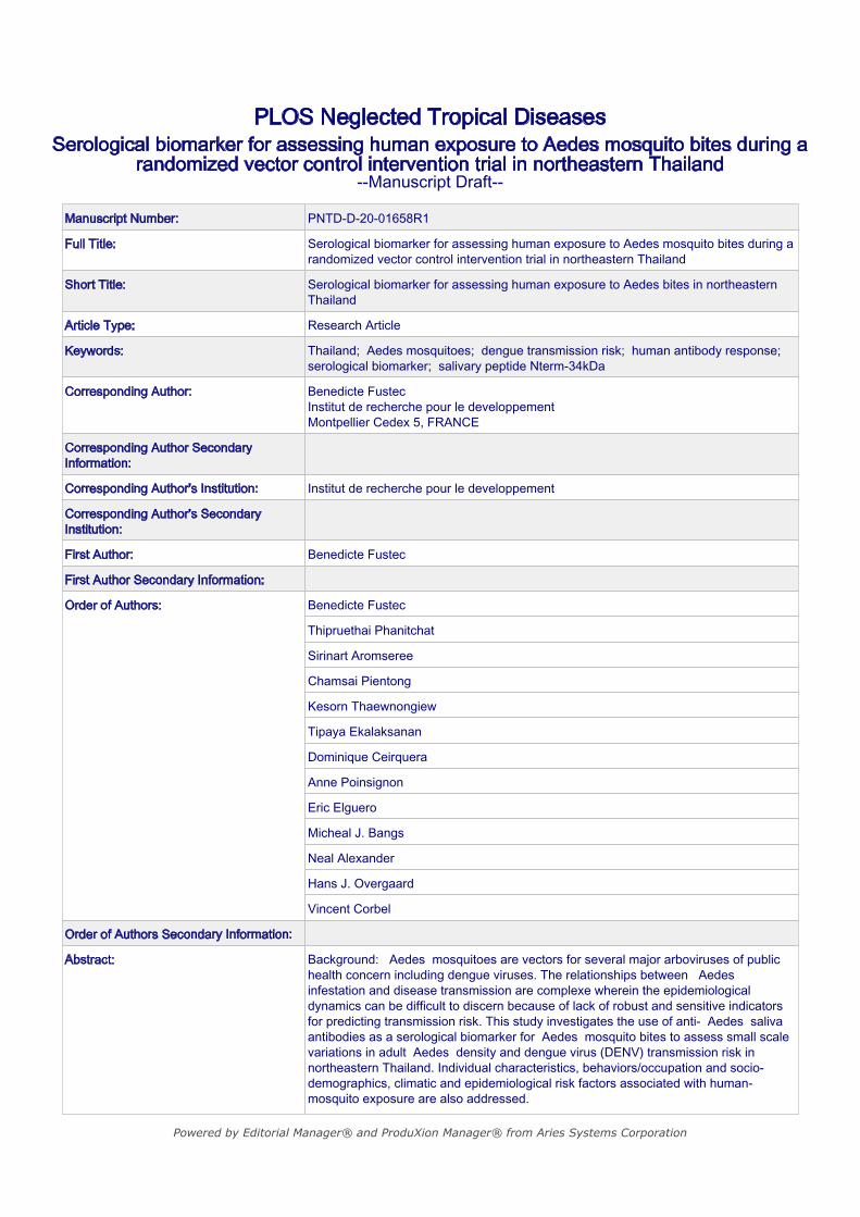
Powered by Editorial Manager® and ProduXion Manager® from Aries Systems Corporation
Powered by Editorial Manager® and ProduXion Manager® from Aries Systems Corporation
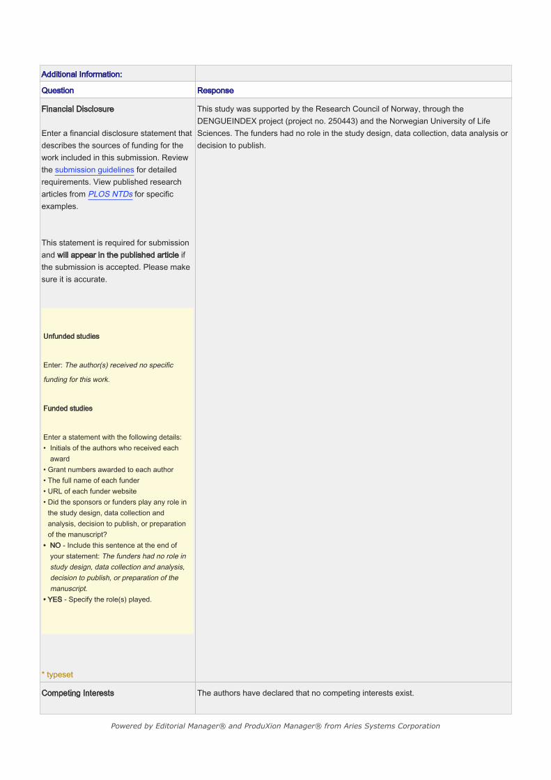
Powered by Editorial Manager® and ProduXion Manager® from Aries Systems Corporation
Powered by Editorial Manager® and ProduXion Manager® from Aries Systems Corporation

Powered by Editorial Manager® and ProduXion Manager® from Aries Systems Corporation
Powered by Editorial Manager® and ProduXion Manager® from Aries Systems Corporation

9/5/2020
Micheal J. Turell Editor at PLoS Neglected Tropical Diseases Dear Editor,
‘Serological biomarker for assessing human exposure to Aedes
mosquito bites during a randomized vector control intervention trial
in northeastern Thailand’
We believe that the results in this manuscript, in particular the strong relation between the human antibody response against Aedes saliva and the Aedes aegypti densities, will be of great interest for the readers of PLoS Neglected Tropical Diseases.
Despite the increasing burden of dengue fever in tropical and sub-tropical areas, its prevention and control still target its Aedes mosquito vectors. However, the quantitative relationships between dengue infection risk and vector mosquito infestation remain unclear despite numerous indicators used to estimate transmission risk and predict dengue outbreaks. The aim of this study is to investigate the use of an Aedes salivary biomarker to assess the small-scale variation in human exposure to Aedes bites and the risk of dengue in the context of a vector control intervention in northeastern Thailand.
Several findings of this study highlight the complexity of the human dengue vector exposure. In particular, our study demonstrates a strong positive association between the level of resting adult Aedes infestation, especially indoors Aedes infestation, and the level of specific human antibody (Ab) response against Ae. aegypti salivary peptide.
This manuscript is the first combining both entomological and immunological endpoints investigating Aedes vectors and virus transmission. Additionally, risk factors associated with human-vector contact in terms of individual human characteristics and behavior, local vector control practices, and the prevailing environmental and climatic factors are addressed. First, our findings demonstrate that human Ab response against Aedes saliva is driven by individual characteristics (i.e. age, gender) and behavior, where staying indoors was associated with a higher Ab response against Aedes bites. In addition, our results suggest an impact of the intervention on the adult Aedes densities as intervention was associated with a lower Ab response to Aedes saliva. Nonetheless, no relationship between the Ab response to Aedes saliva and dengue transmission risk (i.e., vector infection) was demonstrated.
This study demonstrated the usefulness of the Ab response to assess heterogeneity in adult Aedes infestation indices and could assist public health authorities to better address disease transmission risk and the timeliness of effective vector control interventions.
Yours sincerely,
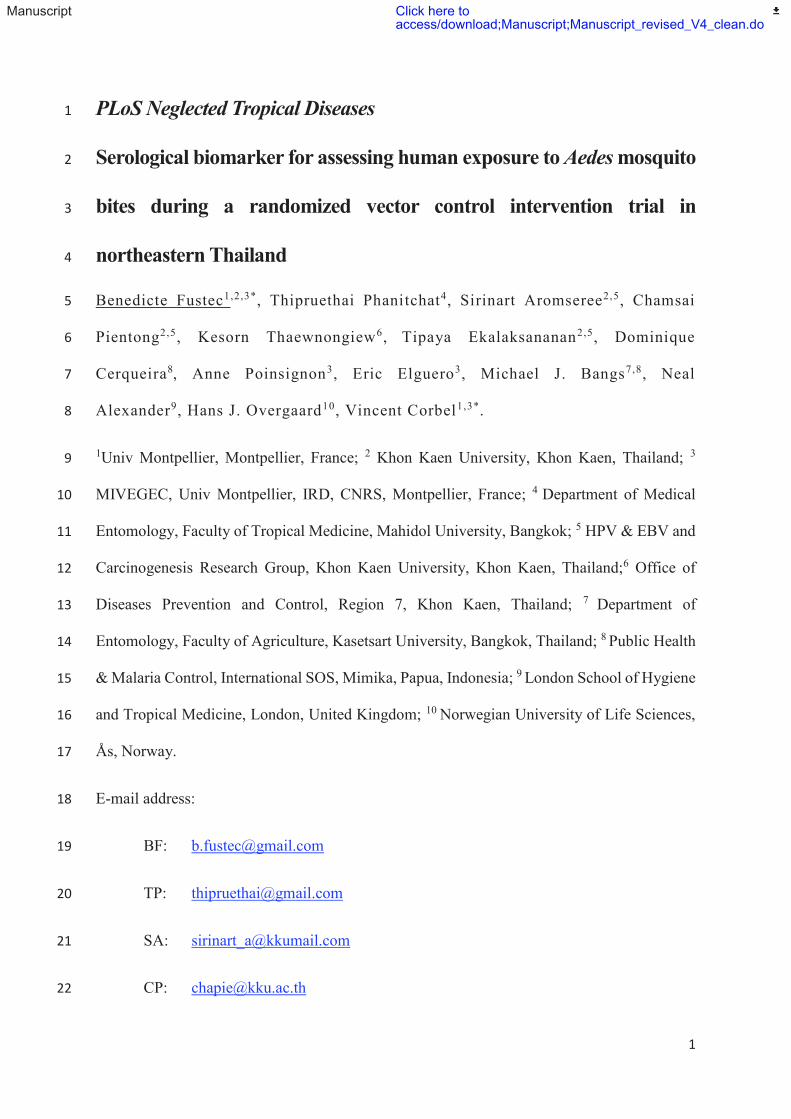
1
PLoS Neglected Tropical Diseases 1
Serological biomarker for assessing human exposure to Aedes mosquito 2
bites during a randomized vector control intervention trial in 3
northeastern Thailand 4
Benedicte Fustec1,2,3*, Thipruethai Phanitchat4, Sirinart Aromseree2,5, Chamsai 5
Pientong2,5, Kesorn Thaewnongiew6, Tipaya Ekalaksananan2,5, Dominique 6
Cerqueira8, Anne Poinsignon3, Eric Elguero3, Michael J. Bangs7,8, Neal 7
Alexander9, Hans J. Overgaard10, Vincent Corbel1,3*. 8
1Univ Montpellier, Montpellier, France; 2 Khon Kaen University, Khon Kaen, Thailand; 3 9
MIVEGEC, Univ Montpellier, IRD, CNRS, Montpellier, France; 4 Department of Medical 10
Entomology, Faculty of Tropical Medicine, Mahidol University, Bangkok; 5 HPV & EBV and 11
Carcinogenesis Research Group, Khon Kaen University, Khon Kaen, Thailand;6 Office of 12
Diseases Prevention and Control, Region 7, Khon Kaen, Thailand; 7 Department of 13
Entomology, Faculty of Agriculture, Kasetsart University, Bangkok, Thailand; 8 Public Health 14
& Malaria Control, International SOS, Mimika, Papua, Indonesia; 9 London School of Hygiene 15
and Tropical Medicine, London, United Kingdom; 10 Norwegian University of Life Sciences, 16
Ås, Norway. 17
E-mail address: 18
BF: [email protected] 19
TP: [email protected] 20
SA: [email protected] 21
CP: [email protected] 22
2
KT: [email protected] 23
DC: [email protected] 24
AP: [email protected] 25
TE: [email protected] 26
EE: [email protected] 27
MJB: [email protected] 28
NA: [email protected] 29
HJO: [email protected] 30
VC: [email protected] 31
32
*Corresponding authors 33
34

3
ABSTRACT 35
Background: Aedes mosquitoes are vectors for several major arboviruses of public health 36
concern including dengue viruses. The relationships between Aedes infestation and disease 37
transmission are complex wherein the epidemiological dynamics can be difficult to discern 38
because of a lack of robust and sensitive indicators for predicting transmission risk. This study 39
investigates the use of anti-Aedes saliva antibodies as a serological biomarker for Aedes 40
mosquito bites to assess small scale variations in adult Aedes density and dengue virus (DENV) 41
transmission risk in northeastern Thailand. Individual characteristics, behaviors/occupation 42
and socio-demographics, climatic and epidemiological risk factors associated with human-43
mosquito exposure are also addressed. 44
Methods: The study was conducted within a randomized clustered control trial in Roi Et and 45
Khon Kaen provinces over a consecutive 19 months period. Thirty-six (36) clusters were 46
selected, each of ten houses. Serological and entomological surveys were conducted in all 47
houses every four months and monthly in three sentinel households per cluster between 48
September 2017 and April 2019 for blood spot collections and recording concurrent immature 49
and adult Aedes indices. Additionally, the human exposure to Aedes mosquito bites (i.e., 50
Mosquito Exposure Index or MEI) was estimated by ELISA measuring levels of human 51
antibody response to the specific Nterm-34 kDa salivary antigen. The relationships between 52
the MEI, vector infestation indices (adult and immature stages) and vector DENV infection 53
were evaluated using a two-level (house and individual levels) mixed model analysis with one-54
month lag autoregressive correlation. 55
Results: A strong positive relationship between the MEI and the intensity of adult Aedes 56
infestation (difference in MEI mean of 0.091 p<0.0001, and 0.131, p<0.0001 for medium and 57
high levels of infestation, respectively), particularly indoor densities (difference in mean MEI 58

4
of 0.021, p<0.007, 0.053, p<0.0001 and 0.037, p<0.0001 for low, medium and high levels of 59
infestation, respectively) were found. The MEI was driven by individual characteristics, such 60
as gender, age and occupation/behaviors, and varied according to climatic, seasonal factors and 61
vector control intervention (p<0.05). Nevertheless, the study did not demonstrate a clear 62
correlation between MEI and the presence of DENV-infected Aedes. 63
Conclusion: This study represents an important step toward the validation of the specific IgG 64
response to the Aedes salivary peptide Nterm-34kDa as a proxy measure for Aedes infestation 65
levels and human-mosquito exposure risk in a dengue endemic setting. The use of the IgG 66
response to the Nterm-34 kDa peptide as a viable diagnostic tool for estimating dengue 67
transmission requires further investigations and validation in other geographical and 68
transmission settings. 69
Key words: Thailand, Aedes mosquitoes, dengue transmission risk, human antibody response, 70
serological biomarker, salivary peptide Nterm-34kDa. 71
Author summary: 72
Aedes mosquitoes and the viruses they transmit are major public health concerns for over half 73
of the global human population. However, the quantitative relationships between virus 74
transmission and vector mosquito infestation remain unclear despite numerous indicators used 75
to estimate transmission risk and predict dengue outbreaks. The aim of this study is to 76
investigate the use of a salivary biomarker to assess the small-scale variation in human 77
exposure to Aedes bites and the risk of dengue infection in the context of a vector control 78
intervention in northeastern Thailand. A cohort of 539 persons visited every four months, 79
including 161 individuals visited monthly, were recruited for routine serological and 80
concurrent household entomological surveys during 19 consecutive months follow-up. 81
Antibody response to Aedes bites was measured by enzyme-linked immunosorbent assays to 82

5
assess the mosquito exposure index (MEI) and association with the Aedes adult and immature 83
abundance as well as the presence of dengue virus (DENV) in adult mosquitoes (transmission 84
risk). Additionally, the individual (cohort), climatic, and vector control intervention risk factors 85
associated with MEI are explored. This study demonstrates that the MEI was strongly related 86
to household adult Aedes density, particularly indoors resting mosquitoes. Additionally, the 87
MEI was influenced by individual characteristics (i.e., person age, gender, staying indoors), 88
and varied according to seasons and intervention. Nonetheless, no clear relationship between 89
MEI and dengue transmission risk (i.e., vector infection) was detected. This study 90
demonstrated the potential usefulness of the MEI to assess heterogeneity in adult Aedes 91
infestation indices that could assist public health authorities to rapidly identify mosquito “hot 92
spots” and the timeliness of effective vector control interventions. 93
94

6
Introduction 95
Aedes aegypti (L) and Aedes albopictus (Skuse) are vectors of important human viral pathogens 96
including dengue, yellow, chikungunya and Zika. In Southeast Asia, dengue fever is 97
widespread and accounts for around 70% of the total clinical dengue cases reported globally 98
[1, 2]. Since the first report of dengue infection in Thailand in 1949 [3], dengue incidence has 99
dramatically increased in line with expanding urbanization. With all four virus serotypes and 100
both major mosquito vectors present in the country around 20,000 cases are reported yearly 101
[4]. Despite an affordable, universal primary health coverage system and an organized, 102
nationwide dengue prevention program, the burden of dengue in Thailand is estimated to cost 103
the equivalent of at $290 million (USD) each year [5]. 104
In northeastern Thailand, dengue fever represents major public health concerns with 105
thousands of clinical cases each year [6]. To prevent secondary transmission in communities, 106
when a dengue case is detected, insecticide treatment using adult space spray is mandated 107
within 24 hours in attempt to rapidly eliminate virus-infected vectors, surrounding its home 108
setting [7]. In parallel, basic entomological surveillance is carried regularly by one of the 22 109
regional Offices of Diseases Prevention and Control (ODPCs) to monitor Aedes vector 110
infestations [7]. In Thailand standard entomological indices are used to estimate transmission 111
risk that guide the choice of vector control interventions [8]. While, some studies have shown 112
positive associations between various entomological indices and disease transmission risk [9, 113
10], other investigations have demonstrated only weak relationships [11-13]. Most of the 114
entomological indices used to monitor dengue vector infestations are based on measuring the 115
presence of immature mosquito life stages [14]. However, immature stages typically present 116
large mortality rates during development from egg to adult stage [15], thus larval indices do 117
not provide an accurate or concurrent temporal-spatial information on the ‘productivity’ of 118
containers regards actual Aedes adult production output [16]. Conversely, pupal indices have 119

7
been proposed to assess vector infestation with higher accuracy [17, 18] as pupae generally 120
present very low mortality up to adult emergence and thus more relevant to estimate container 121
productivity [19] and adult densities in a location [16]. Operationally, pupae collections remain 122
difficult to implement on a routine basis because they are time-consuming (generally all pupae 123
must be collected and counted) that requires additional entomological staff. 124
Adult mosquito collections have been used to estimate the risk of virus transmission 125
[19, 20], but they have also their limitations. Unlike malaria vector monitoring, human land-126
catching cannot be performed to collect Aedes mosquitoes due to the inherent ethical 127
constraints and disease risks, as there is no preventive treatment nor effective vaccines for most 128
of Aedes-transmitted diseases/pathogens (except yellow fever virus). Moreover, Aedes adults 129
are most active during the day time, when most people are awake and can take some forms of 130
protection against bites. As a consequence, Aedes females are often interrupted in the course 131
of seeking a blood meal and can often feed on multiple hosts per gonotrophic cycle [21-23]. 132
Other methods to sample adult Aedes include various versions of passive and active trapping 133
devices (e.g., gravitraps, sticky traps, mechanical battery-operated aspirators, and mosquito 134
electrocuting trap) [24], each presenting differing levels of efficiency [25]. However, they do 135
not measure the inter-individual heterogeneity of exposure influenced by human attraction 136
exerted on mosquitoes and individual host behaviors (e.g., use of personal protections). 137
Nevertheless, these capture methods are used as a proxy to estimate Aedes adult density in a 138
specific area but they are not representative of actual level of contact (biting) exposure between 139
human and vector [26]. This information is yet crucial to identify host population subsets at 140
higher risk of exposure to dengue vector bites and to better estimate virus transmission risk. 141
An alternative to direct entomological indices for estimating the human exposure to 142
mosquitoes is the measure of a host’s antibody (Ab) response to mosquito saliva antigens [27-143
29]. During blood feeding process, mosquito saliva is initially injected into human skin to 144
8
facilitate the blood intake and also acts as a vehicle for transmitting pathogens to the host [30]. 145
Many salivary proteins are immunogenic and elicit an immune response including the 146
production of specific antibodies (Ab) that can be detected by simple analytic tools and 147
spectrophotometry [31-33]. Firstly developed for Anopheles, the vectors of malaria, so-called 148
biomarkers of exposure based on anti-saliva Ab response have been used successfully to 149
identify “hot spots” of vector presence and malaria transmission [34-36] along the Thailand-150
Myanmar border [34, 37]. As far as Aedes genus is concerned, several other studies have shown 151
that IgG response to salivary gland extracts from different Aedes species, such as Ae. aegypti, 152
Ae. polynesiensis, Ae. caspius are reliable indicators of human-Aedes exposure in South-153
America [38, 39], Pacific Islands [40], Africa [41] and Europe [31]. An Ae. aegypti-specific 154
salivary peptide (Nterm-34 kDa) has been identified and the human IgG response to the Nterm-155
34 kDa antigen has shown good correlation with adult Ae. aegypti infestation indices in Benin 156
[42] and Laos [43]. More recently, the Nterm-34 kDa salivary peptide successfully investigated 157
the spatial heterogeneity of Aedes exposure in several urban districts of Senegal [44]. However, 158
most of these Aedes serological studies estimated vector infestation through “indirect” 159
(relative) indicators such as immature ‘Stegomyia’ (Aedes) indices and climatic factors, thus 160
were unlikely to represent more accurate adult infestation that which is directly associated with 161
virus transmission potential. Robust evidence of the relationships between the intensity of 162
human immune response to a specific salivary biomarker, Aedes adult abundance, and dengue 163
infective bite risk is needed to assess whether small scale variations in dengue transmission can 164
be detected using this immunological tool. This is particularly relevant for measuring the 165
impact of vector control interventions where entomological indices may lack the spatio-166
temporal accuracy and sensitivity to demonstrate control effectiveness [16, 45, 46]. 167
The primary objective of this study was to assess the relationship between the intensity 168
of the human IgG response to the Nterm-34kDa Aedes salivary peptide and selected 169
9
entomological indicators of vector infestation and dengue infection risk in northeastern 170
Thailand. This study took place within the context of a randomized controlled trial 171
implemented over a consecutive 19-month period to evaluate the efficacy of an insect growth 172
regulator tool for dengue transmission prevention [47, 48]. Additionally, risk factors associated 173
with human-vector contact in terms of individual human characteristics and behavior, local 174
vector control practices, and the prevailing seasonal and climatic factors were addressed. To 175
our knowledge, this is the first longitudinal study conducted to assess dengue transmission risk 176
using a serological Aedes salivary biomarker. Hopefully, these findings will assist national 177
authorities to improve the accuracy of dengue surveillance activities and contribute to 178
strengthening the monitoring and evaluation of vector control programs in Thailand and 179
elsewhere. 180
181
Materials and Methods 182
Study sites 183
The study was conducted in six sub-districts in the city of Khon Kaen (KK), Khon Kaen 184
Province, (N16.440236, E102.828272) and in two sub-districts within the city of Roi Et (RE), 185
Roi Et Province, (N16.055637, E103.652417), in northeastern Thailand (Fig. 1). In each city, 186
18 clusters of 10 households each were randomly selected for a total of 360 households under 187
19 months of follow-up. 188
Fig. 1: Map of study sites. (A) represents Thailand and the provinces of Roi Et and Khon 189
Kaen. (B) shows the location of the 18 clusters numbered from 4001 to 4018 in the city of 190
Khon Kaen (KK Mueang District). (C) shows the location of the 18 clusters numbered from 191
4501 to 4518 in the city of Roi Et (RE Mueang District). 192

10
Study design and settings 193
This study was conducted within the framework of a randomized control intervention trial to 194
evaluate the efficacy of pyriproxyfen application (0.5% granule formulation) for dengue vector 195
control [47, 48]. The study was performed in Khon Kaen between September 2017 and March 196
2019 and between October 2017 and April 2019 in Roi Et (Fig. 2). All households were visited 197
every four months (except one time in RE between February 2018 and May 2018) to collect 198
indoor and outdoor container-breeding Aedes (both Ae. aegypti and Ae. albopictus) larvae, 199
pupae, adult resting mosquitoes, and blood samples from study volunteers living in randomly 200
selected households. In addition, three sentinel houses per cluster were visited monthly for 201
blood and entomological collections described previously. Following the initial 10 months of 202
baseline surveillance, the vector control intervention was distributed randomly in half of the 203
study clusters, in June 2018. The household selection in the cities and the randomization of the 204
intervention are described elsewhere [47, 48]. The vector control intervention was the 205
distribution of pyriproxyfen (0.5% granule formulation) into water-holding containers up to 206
0.01 mg/L active ingredient applied every four months in the treated clusters [47, 48]. 207
Fig. 2: Study design flow chart. RT-PCR: Reverse Transcriptase Polymerase Chain Reaction 208
Individual volunteer characteristics 209
In each participating household, at least one volunteer inhabitant was recruited in the study. 210
When possible, we tried to recruit inhabitants spending most of their time at home. To ensure 211
adequate representativeness of the entire target population, we recruited one adult and one child 212
per house when feasible. In addition, a pecuniary retribution (50THB) for blood sampling was 213
given to each participant. During each household visit, assigned trained Village Health 214
Volunteers (VHV) interviewed and collected blood of each participating house member. 215
Interview questions were relative to the general characteristics of the participant (i.e., age, 216
gender), occupation(s) during the weekdays and weekends (e.g., at home; at work away from 217
11
home; at school/college/university; at farm; others), in addition to normal activity and resting 218
habits (i.e., primarily indoor, outdoor or equally indoor and outdoor). The travelling history 219
within the previous 14 days and within the last three months was recorded. 220
Blood sample collections 221
Blood samples (2 blood spots per participant, 10mm diameter each, approximately 150µl) were 222
collected at the fingertips of the inhabitants recruited in the study using sterile lancets [49] and 223
spotted on filter paper Protein Saver cards (Whatman, Maidstone, UK), air-dried, individually 224
placed in plastic sealable bags and stored at room temperature at the Office of Disease 225
Prevention and Control 7 (ODPC7) until delivery to Khon Kaen University (KKU) and stored 226
at 4ºC. 227
Entomological collections 228
At each household visit, the VHVs recorded the number of inhabitants in the household at the 229
time of the survey. Houses were inspected for adult and immature Aedes both indoor and 230
outside immediately surrounding the house. The total number of containers was recorded 231
together with the number of wet containers at each household. A maximum of 20 larvae 232
(preferably late stage instar) and all pupae were collected per infested container and stored in 233
absolute ethanol at the ODCP7. Immatures and adults were identified to species-level using 234
morphological keys [50, 51], and sex was determined for adults. Aedes adult collections were 235
performed using hand-held mechanical battery-powered aspirators [52] conducted 15 min each 236
both indoors and outdoors. Adults were stored individually in labelled 1.5mL microcentrifuge 237
tubes at -20ºC and the house number and the location of collection (i.e., indoor/outdoor) was 238
recorded. 239
Entomological data were used to construct several indices as described in 240
Supplementary Table 1. At the cluster level, the Container Index (CIc) was calculated as the 241
proportion of Aedes immature-positive containers per total wet containers inspected in all 242
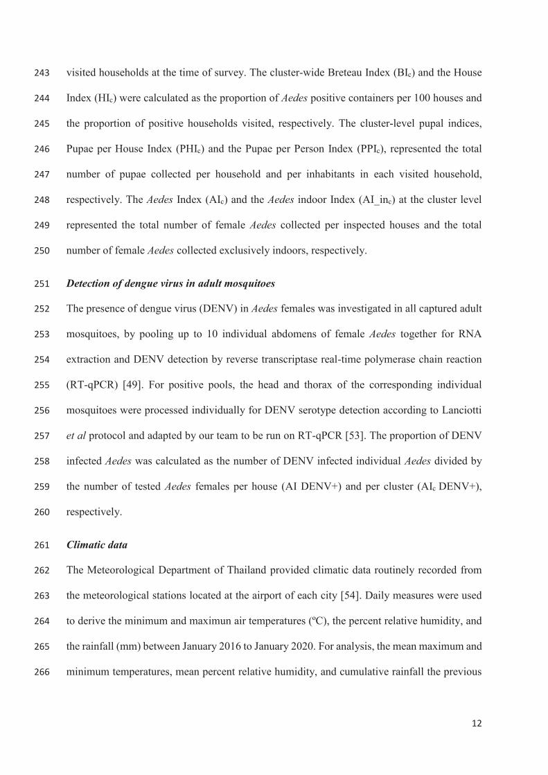
12
visited households at the time of survey. The cluster-wide Breteau Index (BIc) and the House 243
Index (HIc) were calculated as the proportion of Aedes positive containers per 100 houses and 244
the proportion of positive households visited, respectively. The cluster-level pupal indices, 245
Pupae per House Index (PHIc) and the Pupae per Person Index (PPIc), represented the total 246
number of pupae collected per household and per inhabitants in each visited household, 247
respectively. The Aedes Index (AIc) and the Aedes indoor Index (AI_inc) at the cluster level 248
represented the total number of female Aedes collected per inspected houses and the total 249
number of female Aedes collected exclusively indoors, respectively. 250
Detection of dengue virus in adult mosquitoes 251
The presence of dengue virus (DENV) in Aedes females was investigated in all captured adult 252
mosquitoes, by pooling up to 10 individual abdomens of female Aedes together for RNA 253
extraction and DENV detection by reverse transcriptase real-time polymerase chain reaction 254
(RT-qPCR) [49]. For positive pools, the head and thorax of the corresponding individual 255
mosquitoes were processed individually for DENV serotype detection according to Lanciotti 256
et al protocol and adapted by our team to be run on RT-qPCR [53]. The proportion of DENV 257
infected Aedes was calculated as the number of DENV infected individual Aedes divided by 258
the number of tested Aedes females per house (AI DENV+) and per cluster (AIc DENV+), 259
respectively. 260
Climatic data 261
The Meteorological Department of Thailand provided climatic data routinely recorded from 262
the meteorological stations located at the airport of each city [54]. Daily measures were used 263
to derive the minimum and maximun air temperatures (ºC), the percent relative humidity, and 264
the rainfall (mm) between January 2016 to January 2020. For analysis, the mean maximum and 265
minimum temperatures, mean percent relative humidity, and cumulative rainfall the previous 266

13
two weeks before entomological collections were used to account for an estimated time-lag 267
effect on vector population biology and transmission epidemiology. 268
269
Mosquito Exposure Index (MEI) 270
The specific human IgG response to the Aedes Nterm-34kDa salivary peptide (Genepep, Saint 271
Jean de Védas, France) was measured by an enzyme-linked immunosorbent assay (ELISA) as 272
described previously [48, 55]. This secreted salivary peptide was selected because it exhibits 273
high antigenic properties and it is specific to Aedes genus, therefore allowing to specifically 274
measure the immune response to Aedes bites alone [42]. Briefly, for each individual sampled, 275
dried blood spots were cut using a one cm diameter hole punch and eluted in 400µl of 276
Phosphate Buffer Saline (PBS) for 24h at 4 ºC. The resulting eluates were stored at -20ºC until 277
further processing. 96-well Maxisorp micro-assay plates (Nunc, Roskilde, Denmark) were 278
coated with the salivary peptide diluted in PBS (20µg/mL) for 180 minutes at 37ºC. Following 279
washing and blocking steps, the blood eluates were diluted at 1:160 in PBS containing 1% of 280
Tween20 (1%-PBST) and incubated overnight at 4ºC. ELISA plates were incubated with goat 281
anti-human biotin-conjugated IgG (Invitrogen, Thermo Scientific, USA) diluted at 1:6000 in 282
1%- PBST for 90 min at 37ºC, followed by streptavidin horseradish peroxidase (GE 283
Healthcare, Amersham Place, UK) diluted at 1:4000 in 1%-PBST for one hour at 37ºC. The 284
colorimetric reaction was performed using ABTS buffer (2,2’-azino-bis (3-ethylbenzthiazoline 285
6-sulfonic acid) di-ammonium) + 0.003% H2O2 and absorbance (optical density, OD) was 286
measured after 120 min at 405nm with a Sunrise spectrophotometer (Tecan, Switzerland). 287
All samples were assayed in duplicate and in a blank well (no antigen) to measure individual 288
background and antibody response (∆OD) expressed as: 289
(1) ∆OD = mean (ODAg+) - ODAg- 290
14
where "ODAg+ " represents the OD value in the well with the salivary antigen and "ODAg- " the 291
OD value in the well without the antigen. 292
To quantify the non-specific immune reactions and calculate the immune threshold, anti- 293
Nterm-34kDa IgG response was assayed in individuals (n=16) with no known exposure history 294
to Ae. aegypti bites [56] (e.g., dry blood spots collected in northern France from January to 295
March 2016 to 2018, and in Western Australia in October 2016). The specific immune 296
threshold (TR) was defined as follows at0.556. 297
(2) ! = #$%& (∆OD unexposed individuals) + 3 '* unexposed individuals 298
We also defined the Mosquito Exposure Index (MEI) for each participant as 299
(3) +,- = ∆/* − ! . 300
The MEI represents the level of specific and individual IgG response to the Aedes salivary 301
peptide. Individuals with a ∆OD value above the TR, thus with a positive MEI, were classified 302
as “immune responders” (i.e., exposed to Aedes). Individuals with a ∆OD value equal or below 303
the TR, and therefore with a null or negative MEI value, were categorized as “non-responders” 304
(i.e., non-exposed to Aedes). Individuals with negative or null MEI were considered equally 305
having a null MEI as the background immune response cannot be addressed. 306
Analysis 307
Covariates 308
The human study population was stratified into five age groups: 5-19, 20-39, ,40-59, 60-69, 309
and ≥70 years of age. Individual’s characteristics were analyzed as categorical variables to 310
estimate their influence on the MEI. Overall travel history of each subject was used as a binary 311
variable. At the village level, adult Aedes indices recorded one-month before blood collection, 312
and immatures Aedes indices recorded at the time of survey were used. Additionally, the 313
pyriproxyfen intervention was used as a binary covariate. At the province level, the mean daily 314

15
maximum and minimum air temperatures, mean percent relative humidity, and the weekly 315
cumulative rainfall two weeks before collections were treated as covariates. The estimated 2-316
week time-lag takes into account potential influence on vector population biology and 317
transmission epidemiology. Three general climatic seasons are defined according to the Thai 318
Meteorological Department [54] with 15-February to 14-May as the hot season, 15-May to 14-319
October as the rainy (wet) season, and 15 October to 14-February as the cool season. 320
Statistical approach 321
Data analysis was conducted using R software version 3.5.1 (R Core Team, Vienna, Austria) 322
and MASS, Rcmdr, nlme4, and lmerTest packages [57-59]. Figures were generated on R using 323
ggplot2 and ggpubr packages [60, 61]. Maps were built using QGIS software (version 3.10) 324
and shape files were obtained from the Humanitarian Data Exchange Project [62]. As the MEI 325
represents the specific exposition to Ae. aegypti, non-responder individuals were considered 326
with a null MEI, thus the MEI was considered as a positive continuous variable (i.e., MEI ≥0). 327
The relation between MEI and entomological indices was explored using a multivariate 2-level 328
mixed model (house, individual) with a one-month lag autoregressive correlation, assuming 329
the antibody response persisted at detectable levels between two and six weeks [33, 63]. The 330
(1) Aedes adult index (2) Aedes adult indoor index, and (3) proportion of DENV-infected Aedes 331
at the cluster level were examined in three separate analyzes. A fourth analysis was conducted 332
with the proportion of DENV-infected Aedes at the household level to assess the heterogeneity 333
of dengue transmission risk between and within study clusters. To avoid the assumption of 334
linear relationships between antibody response to Aedes bites and entomological indices, risk 335
factors were categorized into categorical variables to represent the different levels of intensity. 336
Due to the over dispersion of mosquito numbers over time, immature stages and adult 337
entomological indices at the cluster level were categorized into four classes, the null value of 338
the index, and then following the terciles. The presence of DENV-infected Aedes was used as 339

16
a binary variable (0 or >0) due to the low number of sampled DENV-infected Aedes. All 340
analyzes were performed on individuals with complete data, while individuals with missing 341
data in covariates of interest were removed. Univariable analysis using a mixed model was 342
conducted with each covariate to identify adjustment factors related to immune response to 343
Nterm-34 kDa for all models. Multivariable mixed models were performed with all covariates 344
with a p-value set at < 0.2. Subsequently, models were adjusted by backward selection and 345
removing non-significant variables at p-value < 0.05. 346
347
Ethical considerations 348
This trial was registered (ISRCTN, ISRCTN73606171) and approved by the Khon Kaen 349
University Ethics Committee (KKUEC Record No. 4.4.01: 29/2017, Reference No. HE601221, 350
1 September 2017), the London School of Hygiene and Tropical Medicine Ethical Committee, 351
UK (LSHTM Ethics Ref: 14275, 16 August 2017), and the Regional Committee for Medical 352
and Health Research Ethics, Section B, South East Norway (REK Ethics ref: 2017/1826b, 03 353
March 2018). Each participant was informed about the intent of the study and asked to 354
participate on a voluntary basis. In each household, the head of the house signed a consent form 355
to allow periodic entomological inspection inside and outside their residence. Additionally, 356
signed informed consent (or assent, if under 16 years old) were required each time blood 357
samples were taken. 358
Results 359
Population characteristics 360
The studied population, 602 individuals (318 in KK and 284 in RE), were followed-up every 361
four months up to 19 months for an average of 3.5 visits per person (Table 1) producing a total 362
of 3,919 collected dried blood spot samples. Among the 602 individuals recruited, a sub- 363
sample of 92 and 71 individuals in KK and RE, respectively, were followed-up each month in 364
17
sentinel sites with an average of 14.7 visits per person. The majority of the cohort was female 365
(65.3% and 69.0% in KK and RE, respectively). The median age of the cohort was 64 and 61 366
years in KK and RE, respectively. The majority of the study cohort stayed most of the time at 367
home during the weekdays and weekends (Table 2); although, in KK, about 30% of the cohort, 368
mostly those of younger age, indicated spending some time in schools during the weekends. In 369
KK, the vast majority of the individuals spent their weekdays indoors while in RE, about one 370
fifth spent their weekdays both indoors and outdoors (near the location where they spend their 371
time). Nevertheless, the behavioral trend was quite similar between KK and RE regarding 372
daytime activities (e.g., indoor vs. outdoor locations). Most individuals were primarily 373
sedentary with >95% declaring no travel in the previous 3 months before blood collections. At 374
the time of the study, there was no evidence of incident (new) dengue infection; therefore, 375
results presented herein is performed using entomological and immunological data only. 376
Table 1: Population description and immunological status to Nterm-34 kDa salivary 377
peptide. 378
379
Table 2: Individual participant characteristics, behavior and occupation. (NA: Not 380
available). 381
Khon Kaen Roi Et Population size, n individuals (no. dried blood spots) 318 (2003) 284 (1916) Age in years, median (range of all participants) 64 (5-90) 61 (7-92) Female proportion, % (no. females/total) 65.3 (1307/2003) 69.0 (1319/1916) Dengue cases %, (no. cases/total) 0.00 (0/2003) 0.00 (0/1916) Proportion of immune responder during the whole study, %, (no. responding/total)
All ages 57.3 (1148/2003) 60.0 (1150/1916)
Age 5-19 46.7 (14/30) 53.8 (21/39)
Age 20-39 48.9 (66/135) 64.7 (119/184)
Age 40-59 58.9 (367/623) 60.2 (415/689)
Age 60-69 58.2 (322/553) 54.0 (299/554)
Age 70+ 57.3 (379/662) 65.8 (296/450)
Khon Kaen Roi Et
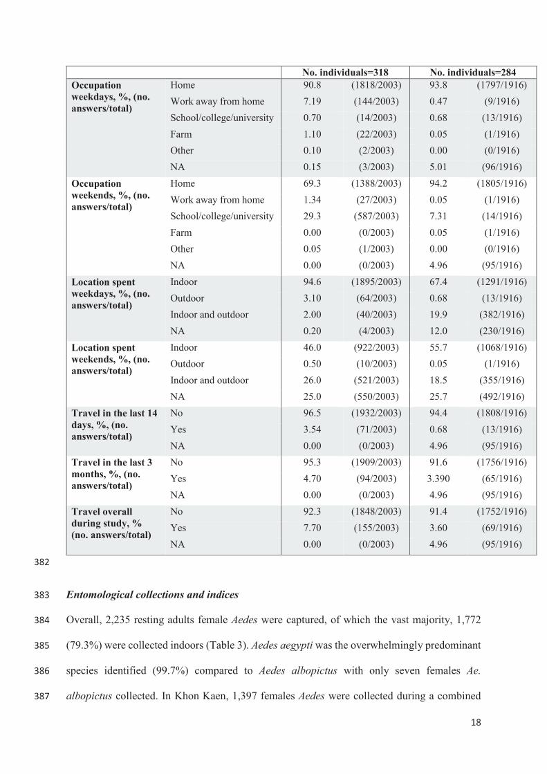
18
382
Entomological collections and indices 383
Overall, 2,235 resting adults female Aedes were captured, of which the vast majority, 1,772 384
(79.3%) were collected indoors (Table 3). Aedes aegypti was the overwhelmingly predominant 385
species identified (99.7%) compared to Aedes albopictus with only seven females Ae. 386
albopictus collected. In Khon Kaen, 1,397 females Aedes were collected during a combined 387
No. individuals=318 No. individuals=284 Occupation weekdays, %, (no. answers/total)
Home 90.8 (1818/2003) 93.8 (1797/1916)
Work away from home 7.19 (144/2003) 0.47 (9/1916)
School/college/university 0.70 (14/2003) 0.68 (13/1916)
Farm 1.10 (22/2003) 0.05 (1/1916)
Other 0.10 (2/2003) 0.00 (0/1916)
NA 0.15 (3/2003) 5.01 (96/1916)
Occupation weekends, %, (no. answers/total)
Home 69.3 (1388/2003) 94.2 (1805/1916)
Work away from home 1.34 (27/2003) 0.05 (1/1916)
School/college/university 29.3 (587/2003) 7.31 (14/1916)
Farm 0.00 (0/2003) 0.05 (1/1916)
Other 0.05 (1/2003) 0.00 (0/1916)
NA 0.00 (0/2003) 4.96 (95/1916)
Location spent weekdays, %, (no. answers/total)
Indoor 94.6 (1895/2003) 67.4 (1291/1916)
Outdoor 3.10 (64/2003) 0.68 (13/1916)
Indoor and outdoor 2.00 (40/2003) 19.9 (382/1916)
NA 0.20 (4/2003) 12.0 (230/1916)
Location spent weekends, %, (no. answers/total)
Indoor 46.0 (922/2003) 55.7 (1068/1916)
Outdoor 0.50 (10/2003) 0.05 (1/1916)
Indoor and outdoor 26.0 (521/2003) 18.5 (355/1916)
NA 25.0 (550/2003) 25.7 (492/1916)
Travel in the last 14 days, %, (no. answers/total)
No 96.5 (1932/2003) 94.4 (1808/1916)
Yes 3.54 (71/2003) 0.68 (13/1916)
NA 0.00 (0/2003) 4.96 (95/1916)
Travel in the last 3 months, %, (no. answers/total)
No 95.3 (1909/2003) 91.6 (1756/1916)
Yes 4.70 (94/2003) 3.390 (65/1916)
NA 0.00 (0/2003) 4.96 (95/1916)
Travel overall during study, % (no. answers/total)
No 92.3 (1848/2003) 91.4 (1752/1916)
Yes 7.70 (155/2003) 3.60 (69/1916)
NA 0.00 (0/2003) 4.96 (95/1916)

19
1,446 house visits, the large majority (77%) captured indoors (Table 3). Moreover, DENV 388
infection was detected among 16 females Aedes in KK. In Roi Et, 838 females Aedes were 389
sampled from 1,441 collections, of which 696 (83%) were collected indoors. Moreover, DENV 390
was detected among 14 females Aedes in RE. Additionally, 992 Aedes pupae (544 in KK and 391
448 in RE) were collected in the two cities. As with adult mosquitoes, Ae. aegypti pupae 392
represented the vast majority (95.7%) of collections. At the cluster level, the standard larval 393
indices (CIc, HI, BI) indicated significantly higher Aedes infestation in Khon Kaen compared 394
to Roi Et with an average of 16.4% and 4.11% Aedes positive containers, respectively 395
(Supplementary Table S2). Similarly, the adult Aedes indices (AIc and AI_inc) were higher in 396
KK clusters than in RE, with an average of 3.7 and 1.0 Aedes in KK and 0.79 and 0.68 Aedes 397
in RE, respectively. Only the DENV-infected adult Aedes index (AIc DENV+) was higher in 398
RE clusters than in KK with an average of 0.007 and 0.005 proportion of DENV positive Aedes 399
in RE and KK, respectively. The pupal indices were, however, slightly higher in RE than in 400
KK with 0.84 and 0.63 PHIc and 0.26 and 0.19 PPIc, respectively. 401
Table 3: Entomological collection data and indices at household and cluster level 402
Khon Kaen Roi Et Houses Visits Total Houses Visits Total
Aedes female collected 179 1446 1397 179 1441 838 Aedes female collected indoors 179 1446 1076 179 1441 696 Aedes pupae collected 179 1446 544 179 1441 448
Entomological indices Mean Std dev Range Mean Std dev Range
Household level Adult Index DENV+ 0.005 0.049 [0-1] 0.005 0.057 [0-1] Cluster level Container Indexc (CIc) (%) 16.4 14.8 [0-100] 4.11 9.17 [0-66.7] House Indexc (HIc)(%) 45.5 33.8 [0-100] 12.9 21.3 [0-100] Breteau Indexc (BIc) 60.4 55.4 [0-300] 14.2 25.0 [0-137.5] Pupae per House Indexc (PHIc) 0.63 1.40 [0-10.7] 0.84 1.99 [0-10.7]
Pupae per Person Indexc (PPIc) 0.19 0.45 [0-3.3] 0.26 0.72 [0-5.7]
Adult Indexc (AIc) 3.71 2.42 [1-15] 0.79 0.84 [0-6]
Adult Index_indoorc (AI_inc) 1.00 0.87 [0-5] 0.68 0.71 [0-4]
Adult Indexc DENV+ 0.005 0.035 [0-0.33] 0.007 0.057 [0-0.67]

20
403
Spatial and seasonal variation in mosquito exposure and vector density 404
During the study, 3,919 individual dried blood samples were collected and processed, including 405
2,003 and 1,916 in KK and RE, respectively. The seroprevalence rates for IgG reactivity were 406
57.3% and 60% in KK and RE, respectively, indicating that most individuals exhibited a 407
specific response to the Nterm-34kDa Ae. aegypti salivary peptide (Table 1). The proportion 408
of immune responders between combined RE and KK clusters was not statistically significant 409
(χ2 p=0.08) (Supplementary Table S2). 410
In both cities, Aedes density (AIc) strongly increased in May-June period corresponding to the 411
end of the hot season and the beginning of the rainy season (Fig. 3). Notably, the human IgG 412
response (ΔOD) increased a few weeks after the measured peak of mosquito density. 413
Additionally, the ΔOD decreased from the cool season until the hot season while the mosquito 414
densities were reduced during the rainy season with numbers rebounding during the hot season. 415
Collectively, the results indicated a lagged positive association between Aedes abundance and 416
human exposure to Aedes bites. Indeed, previous studies on malaria vectors showed that the 417
time-lag for human immune response was between three- to four- weeks after the vector bites 418
[64]. Additionally, univariate analysis of the intensity of MEI indicated a positive association 419
between the intensity of the human Ab response and the density of adult Aedes collected the 420
month before the blood spot collection (Supplementary Table S3). 421
Fig. 3: Seasonal variations of the human IgG response to Aedes Nterm-34kDa salivary 422
biomarker and the adult density Aedes Index (AIc), between September 2017 and April 423
2019 in Khon Kaen (A) and Roi Et (B) northeastern Thailand. The dot plots represent the 424
individual IgG immune response to the Aedes salivary peptide Nterm-34 kDa (ΔOD). The red 425
diamonds represent the median response during each survey. The solid red lines represent the 426

21
means and the grey shaded areas represent the confidence interval of the IgG response to the 427
salivary biomarker. The red dashed horizontal lines represent the specific immune threshold 428
TR. The solid blue lines represent the means and the grey shaded areas represent the 95% 429
confidence interval respectively, for the AIc at the cluster level. 430
Correlations between vector infestation, vector infectivity and human exposure risk to 431
Aedes bites. 432
Multivariate analysis was performed on a total of 539 individuals, with complete data, 433
including 378 individuals followed-up every four months, with an average number of 2.63 434
visits per person. Additionally, a sub-sample of 161 individuals, followed-up every month with 435
an average number of 12 visits per person were included in the analysis. The models showed a 436
strong positive correlation between the MEI and the Aedes adult density at the cluster level 437
when compared to the absence of Aedes for both the total adult AIc (Figure 4 B and C, mean 438
difference in MEI 0.091, p<0.0001, and 0.131, p<0.0001 for medium and high level of 439
infestation, respectively) and the adult indoor density AI_inc (Figure 4 A and C, difference in 440
mean MEI of 0.021, p<0.007, 0.053, p<0.0001 and 0.037, p<0.0001 for low, medium and high 441
levels of infestation, respectively). There was a significant positive association between the 442
individual immune response and the three categories of Aedes intensity (low, medium and 443
high), compared with the reference (no Aedes), when considering adult mosquitoes collected 444
indoors (p<0.05). 445
446
In contrast, no clear relationships were noted between MEI and vector DENV infection at the 447
cluster level (Table 4, p=0.671) nor at the household level (Table 4, p=0.764). Based on these 448
study findings, the intensity of the immune response to Aedes bite exposure was not associated 449
with a higher risk of being bitten by a DENV-infected vector (Table 4). 450
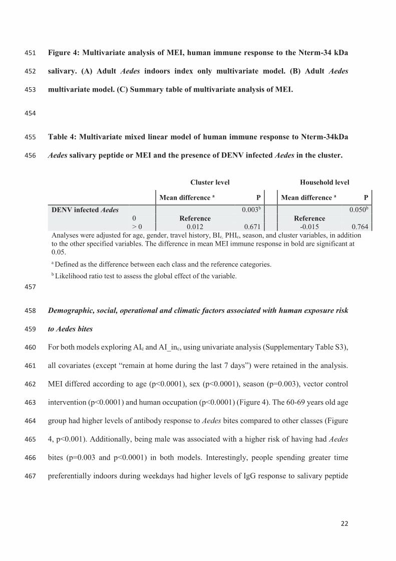
22
Figure 4: Multivariate analysis of MEI, human immune response to the Nterm-34 kDa 451
salivary. (A) Adult Aedes indoors index only multivariate model. (B) Adult Aedes 452
multivariate model. (C) Summary table of multivariate analysis of MEI. 453
454
Table 4: Multivariate mixed linear model of human immune response to Nterm-34kDa 455
Aedes salivary peptide or MEI and the presence of DENV infected Aedes in the cluster. 456
Cluster level Household level
Mean difference a P Mean difference a P
DENV infected Aedes 0.003b 0.050b 0 Reference Reference > 0 0.012 0.671 -0.015 0.764
Analyses were adjusted for age, gender, travel history, BIc, PHIc, season, and cluster variables, in addition to the other specified variables. The difference in mean MEI immune response in bold are significant at 0.05. a Defined as the difference between each class and the reference categories. b Likelihood ratio test to assess the global effect of the variable.
457
Demographic, social, operational and climatic factors associated with human exposure risk 458
to Aedes bites 459
For both models exploring AIc and AI_inc, using univariate analysis (Supplementary Table S3), 460
all covariates (except “remain at home during the last 7 days”) were retained in the analysis. 461
MEI differed according to age (p<0.0001), sex (p<0.0001), season (p=0.003), vector control 462
intervention (p<0.0001) and human occupation (p<0.0001) (Figure 4). The 60-69 years old age 463
group had higher levels of antibody response to Aedes bites compared to other classes (Figure 464
4, p<0.001). Additionally, being male was associated with a higher risk of having had Aedes 465
bites (p=0.003 and p<0.0001) in both models. Interestingly, people spending greater time 466
preferentially indoors during weekdays had higher levels of IgG response to salivary peptide 467
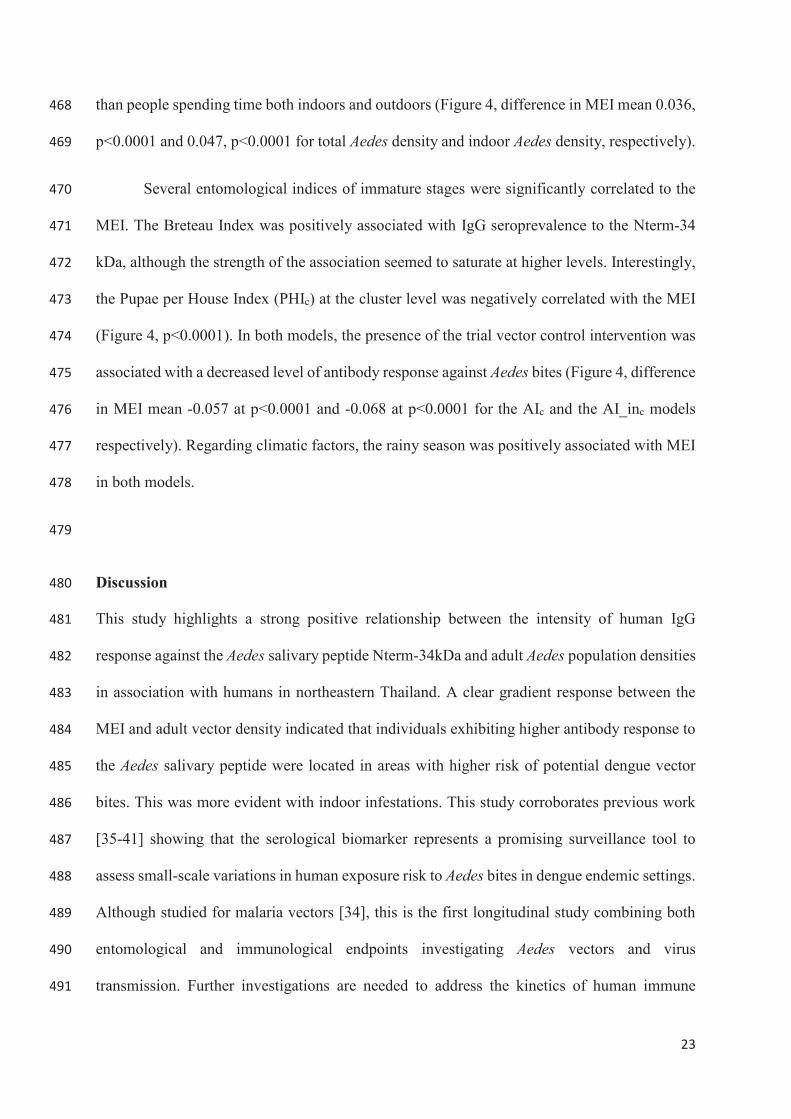
23
than people spending time both indoors and outdoors (Figure 4, difference in MEI mean 0.036, 468
p<0.0001 and 0.047, p<0.0001 for total Aedes density and indoor Aedes density, respectively). 469
Several entomological indices of immature stages were significantly correlated to the 470
MEI. The Breteau Index was positively associated with IgG seroprevalence to the Nterm-34 471
kDa, although the strength of the association seemed to saturate at higher levels. Interestingly, 472
the Pupae per House Index (PHIc) at the cluster level was negatively correlated with the MEI 473
(Figure 4, p<0.0001). In both models, the presence of the trial vector control intervention was 474
associated with a decreased level of antibody response against Aedes bites (Figure 4, difference 475
in MEI mean -0.057 at p<0.0001 and -0.068 at p<0.0001 for the AIc and the AI_inc models 476
respectively). Regarding climatic factors, the rainy season was positively associated with MEI 477
in both models. 478
479
Discussion 480
This study highlights a strong positive relationship between the intensity of human IgG 481
response against the Aedes salivary peptide Nterm-34kDa and adult Aedes population densities 482
in association with humans in northeastern Thailand. A clear gradient response between the 483
MEI and adult vector density indicated that individuals exhibiting higher antibody response to 484
the Aedes salivary peptide were located in areas with higher risk of potential dengue vector 485
bites. This was more evident with indoor infestations. This study corroborates previous work 486
[35-41] showing that the serological biomarker represents a promising surveillance tool to 487
assess small-scale variations in human exposure risk to Aedes bites in dengue endemic settings. 488
Although studied for malaria vectors [34], this is the first longitudinal study combining both 489
entomological and immunological endpoints investigating Aedes vectors and virus 490
transmission. Further investigations are needed to address the kinetics of human immune 491

24
response to Aedes salivary proteins, in particular the delay between bite exposure, and the 492
production and waning of IgG titers. 493
This study showed that the human-mosquito contact is influenced by human behavioral 494
characteristics, socio-demographic conditions, climatic factors, and trial vector control 495
interventions associated with dengue transmission risk as previously demonstrated [8, 19, 55]. 496
The relationship between human dengue infections and the intensity of the human-antibody 497
response to Aedes bites could not be ascertained because incident dengue cases were not 498
detected in the study participants during the time of longitudinal follow-up. Further analysis is 499
on-going to confirm the observation of the apparent lack (or very low) transmission during the 500
study period (to be reported elsewhere). In a recent case-control study conducted in 501
northeastern Thailand (conducted by this study team), neither the adult mosquito abundance at 502
the household level nor the degree of human exposure to Aedes bites was correlated with a 503
higher odds of acquiring dengue infection [55]. Although consistent with some previous results 504
in Southeast Asia [43, 55], the small sample size of DENV-positive Aedes might explain the 505
lack of significance between human infection and vector density seen in this study. This 506
highlights dengue virus transmission is both a multi-factorial and a complex affair that varies 507
over time and space, and the relationship between vector density and virus transmission is 508
dynamic and thus might not be adequately or accurately characterized through standard 509
methods of entomological monitoring. 510
These findings show that the MEI was significantly associated with the season and 511
prevailing climatic factors. The proportion of immune responders to Aedes bites was higher 512
during the rainy season than the drier months of the year, corresponding to the period of greater 513
adult vector densities. This is probably explained by the dramatic increase in most 514
entomological indices during this period of the year where the number of suitable larval 515
habitats increases and adult survival (longevity) is presumably enhanced [15, 65]. Similar 516

25
results were reported in Benin, where the overall anti-saliva antibody response in children 517
increased during the rainy season [42]. A recent study in Cote d’Ivoire highlighted a strong 518
relationship between human mosquito exposure, season and agricultural practices [66]. 519
Specific IgG responses remained high during both seasons in villages associated with intensive 520
agricultural compared to villages lacking agricultural practices. The authors suggest that the 521
presence of rubber and oil palm plantations, by providing a suitable environment for the 522
presence of Aedes vector species maintained a high level of human exposure to Aedes mosquito 523
bites regardless of annual seasonal changes. 524
Interestingly, the present study also suggests correlations between the MEI and Aedes 525
immature-based indices, although the association appeared weaker compared to adult 526
measures. The Breteau Index was associated with higher levels of antibody response against 527
Aedes bites but was not gradient-dependent. In contrast, the pupae per house index was 528
negatively associated with the MEI. This result might seem contradictory; however, that under 529
natural field conditions, larvae and pupae development rates are strongly influenced by climatic 530
factors, particularly ambient temperature and rainfall patterns, as well as density-dependent 531
factors of immature stages affecting resource competition [67-69]. Additionally, the presence 532
of larval stages in an aquatic habitat can inhibit further egg hatching [70]. Therefore, a decrease 533
in human immune response to Aedes bites could be the reflection of the cyclic fluctuations 534
between successive adult population densities influenced by site-specific immature mosquito 535
densities. 536
The MEI varied according to individual characteristics, such as gender, age, and 537
occupation. Interestingly, older people presented higher risk for mosquito bites than the 538
younger population. Similarly, being a male was associated with a higher exposure level to 539
Aedes bites. Similar results were found with Anopheles exposure and malaria transmission in 540
Thailand, where males were at higher risk than females, mainly due to differences in behavior 541

26
and occupational exposure [37]. Nevertheless, these results have to be viewed with caution as 542
the majority of the participants in the present study were female and the median age of the 543
cohort was 64 in KK and 61 in RE, which may have biased the outcomes. Indeed, the median 544
age of the cohort reflects the lack of representation of the younger population, which are 545
presumed more active (mobile) than older individuals. Our findings also showed that 546
individuals spending the majority of time indoors were associated with a higher exposure to 547
Aedes bites than those spending time more equally either indoors and out. An explanation is 548
that Ae. aegypti is a well-adapted species for resting and breeding inside dwellings, and is more 549
typically found indoors [22, 23]. This is also supported by the level of significance of human-550
exposure risk using the Aedes indoor index. The risk of biting (i.e., transmission) inside a 551
dwelling appears particularly important and helps explain why insecticide-treated curtains and 552
targeted indoor residual spraying were highly effective against Ae. aegypti for the control and 553
prevention of dengue outbreaks in Mexico and Australia [46, 71]. 554
This study suggests that the salivary biomarker is sensitive enough to detect small scale 555
variations in human exposure to Aedes bites over time, in particular during a vector control 556
intervention. The human IgG levels were significantly lower in treated clusters compared to 557
the control clusters. These findings would suggest an appreciable impact of pyriproxyfen 558
treatment on the density of Aedes adult populations. Similar results were observed in La 559
Réunion, where vector control intervention combining Aedes larval habitat source reduction 560
and insecticide space spray against adult mosquitoes was associated with a significant decrease 561
in human antibody response against Ae. albopictus bites [41, 63]. Investigations are on-going 562
in Thailand to assess the entomological and epidemiological impact of pyriproxyfen 563
intervention in the study area [48, 72]. 564
This study represents an important step toward the validation of using the Aedes 565
salivary peptide Nterm-34kDa as a proxy measure to assess Aedes infestation levels and 566
27
human-mosquito exposure risk in a dengue endemic area. Although promising results are 567
described, the use of the Nterm-34 kDa as a surveillance indicator for estimating dengue 568
transmission risk requires further investigations including other geographical and transmission 569
settings. 570
571
Acknowledgments 572
We thank all the cluster Village Health Volunteers for their diligence in conducting the blood 573
spot collections and the individual questionnaires. We acknowledge the principal 574
governmental officers from the ODPC7 for the entomological collections, mosquito species 575
identification, and household questionnaire sessions. We thank Panwad Thongchai for 576
assistance in DENV detection in mosquito samples. We acknowledge Franck Remoué and 577
André Sagna from the Institut de Recherche pour le Développement for their kind advice and 578
assistance on ELISA experiments. 579
Funding 580
This study was supported by the Research Council of Norway, through the DENGUE-581
INDEX project (project no. 250443). The funder had no role in the study design, data 582
collection, data analysis or decision to publish. 583
Authors contributions 584
Conceptualization: MJB NA HJO VC. 585
Data curation: BF EE. 586
Formal analysis: BF EE. 587
Funding acquisition: NA HJO. 588

28
Investigation: BF TP SA. 589
Methodology: BF SA CP TE DC AP EE MJB HJO VC. 590
Project administration: TP SA CP TE KT HJO. 591
Resources: CP TE KT. 592
Supervision: MJB NA HJO VC. 593
Visualization: BF VC. 594
Writing – original draft: BF VC. 595
Writing – review & editing: BF MJB AP NA HJO VC. 596
597
Supporting information 598
Supplementary Table S1. Defined variables 599
Supplementary Table S2. Comparison of proportion of immune responders and 600
entomological indices between Khon Kaen and Roi Et provinces using Chi square test and 601
ANOVA. 602
Supplementary Table S3. Univariate analysis of the human antibody response to the Aedes 603
salivary biomarker Nterm-34 kDa. 604
605

29
References 606
1. Bhatt, S., et al., The global distribution and burden of dengue. Nature, 2013. 607
2. World Health Organization, Dengue guidelines for diagnosis, treatment, prevention and 608
control 2009, WHO: Geneva. 609
3. Hammon, W.M., Dengue hemorrhagic fever--do we know its cause? Am J Trop Med Hyg, 610
1973. 22(1): p. 82-91. 611
4. Bureau of Epidemiology and M.o.P.H. Thailand, Annual epidemiological surveillance report 612
2018, Department of Disease Prevention, Editor. 2019. p. 808. 613
5. Shepard, D.S., E.A. Undurraga, and Y.A. Halasa, Economic and disease burden of dengue in 614
Southeast Asia. PLoS Negl Trop Dis, 2013. 7(2): p. e2055. 615
6. Limkittikul, K., J. Brett, and M. L'Azou, Epidemiological trends of dengue disease in Thailand 616
(2000-2011): a systematic literature review. PLoS Negl Trop Dis, 2014. 8(11): p. e3241. 617
7. Bureau of Emerging Infectious Disease, Thailand's National Strategic Plan For Emerging 618
Infectious Disease Preparedness, Prevention and Response 2013-2016, M.o.P.H. Department 619
of Disease Control, Editor. 2013, the War Veterans Organization of Thailand Under Royal 620
Patronage of His Majesty the King: Nonthaburi, Thailand. p. 100. 621
8. Bowman, L.R., S. Runge-Ranzinger, and P.J. McCall, Assessing the relationship between 622
vector indices and dengue transmission: a systematic review of the evidence. PLoS Negl Trop 623
Dis, 2014. 8(5): p. e2848. 624
9. Chadee, D.D., Dengue cases and Aedes aegypti indices in Trinidad, West Indies. Acta Trop, 625
2009. 112(2): p. 174-80. 626
10. Sanchez, L., et al., Aedes aegypti larval indices and risk for dengue epidemics. Emerg Infect 627
Dis, 2006. 12(5): p. 800-6. 628
11. Chadee, D.D., F.L. Williams, and U.D. Kitron, Impact of vector control on a dengue fever 629
outbreak in Trinidad, West Indies, in 1998. Trop Med Int Health, 2005. 10(8): p. 748-54. 630
12. Ooi, E.E., K.T. Goh, and D.J. Gubler, Dengue prevention and 35 years of vector control in 631
Singapore. Emerg Infect Dis, 2006. 12(6): p. 887-93. 632
13. Romero-Vivas, C.M. and A.K. Falconar, Investigation of relationships between Aedes aegypti 633
egg, larvae, pupae, and adult density indices where their main breeding sites were located 634
indoors. J Am Mosq Control Assoc, 2005. 21(1): p. 15-21. 635
14. Focks, D., A review of entomological sampling methods and indicators for dengue vectors. 636
WHO-TDR, 2004. 637
15. Focks, D.A., et al., Dynamic life table model for Aedes aegypti (Diptera: Culicidae): analysis of 638
the literature and model development. J Med Entomol, 1993. 30(6): p. 1003-17. 639
16. Roiz, D., et al., Integrated Aedes management for the control of Aedes-borne diseases. PLoS 640
Negl Trop Dis, 2018. 12(12): p. e0006845. 641
17. Focks, D.A., et al., Transmission thresholds for dengue in terms of Aedes aegypti pupae per 642
person with discussion of their utility in source reduction efforts. Am J Trop Med Hyg, 2000. 643
62(1): p. 11-8. 644
18. Focks, D.A. and D.D. Chadee, Pupal survey: an epidemiologically significant surveillance 645
method for Aedes aegypti: an example using data from Trinidad. Am J Trop Med Hyg, 1997. 646
56(2): p. 159-67. 647
19. Parra, M.C.P., et al., Using adult Aedes aegypti females to predict areas at risk for dengue 648
transmission: A spatial case-control study. Acta Trop, 2018. 182: p. 43-53. 649
20. Lau, S.M., et al., A new paradigm for Aedes spp. surveillance using gravid ovipositing sticky 650
trap and NS1 antigen test kit. Parasit Vectors, 2017. 10(1): p. 151. 651
21. Harrington, L.C., et al., Heterogeneous feeding patterns of the dengue vector, Aedes aegypti, 652
on individual human hosts in rural Thailand. PLoS Negl Trop Dis, 2014. 8(8): p. e3048. 653
22. Scott, T.W., et al., Blood-feeding patterns of Aedes aegypti (Diptera: Culicidae) collected in a 654
rural Thai village. J Med Entomol, 1993. 30(5): p. 922-7. 655

30
23. Scott, T.W., et al., Detection of multiple blood feeding in Aedes aegypti (Diptera: Culicidae) 656
during a single gonotrophic cycle using a histologic technique. J Med Entomol, 1993. 30(1): p. 657
94-9. 658
24. Ortega-López, L.D., et al., The mosquito electrocuting trap as an exposure-free method for 659
measuring human-biting rates by Aedes mosquito vectors. Parasites & vectors, 2020. 13(1): 660
p. 31-31. 661
25. Bazin, M. and C.R. Williams, Mosquito traps for urban surveillance: collection efficacy and 662
potential for use by citizen scientists. J Vector Ecol, 2018. 43(1): p. 98-103. 663
26. Barnard, D.R., et al., Measurement of landing mosquito density on humans. Acta Trop, 2014. 664
136: p. 58-67. 665
27. Doucoure, S. and P.M. Drame, Salivary biomarkers in the control of mosquito-borne diseases. 666
Insects, 2015. 6(4): p. 961-76. 667
28. Poinsignon, A., et al., Novel peptide marker corresponding to salivary protein gSG6 668
potentially identifies exposure to Anopheles bites. PLoS One, 2008. 3(6): p. e2472. 669
29. Lombardo, F., et al., The Anopheles gambiae salivary protein gSG6: an anopheline-specific 670
protein with a blood-feeding role. Insect Biochem Mol Biol, 2009. 39(7): p. 457-66. 671
30. Wasinpiyamongkol, L., et al., Blood-feeding and immunogenic Aedes aegypti saliva proteins. 672
Proteomics, 2010. 10(10): p. 1906-16. 673
31. Fontaine, A., et al., Relationship between exposure to vectors bites and antibody response to 674
mosquito salivary gland extracts. PLoS ONE, 2011. 6(12). 675
32. Remoue, F., et al., Evaluation of the antibody response to Anopheles salivary antigens as a 676
potential marker of risk of malaria. Trans R Soc Trop Med Hyg, 2006. 100(4): p. 363-70. 677
33. Orlandi-Pradines, E., et al., Antibody response against saliva antigens of Anopheles gambiae 678
and Aedes aegypti in travellers in tropical Africa. Microbes Infect, 2007. 9(12-13): p. 1454-62. 679
34. Ya-Umphan, P., et al., Use of an Anopheles salivary biomarker to assess malaria transmission 680
risk along the Thailand-Myanmar border. J Infect Dis, 2017. 215(3): p. 396-404. 681
35. Poinsignon, A., et al., Human IgG response to a salivary peptide, gSG6-P1, as a new immuno-682
epidemiological tool for evaluating low-level exposure to Anopheles bites. Malar J, 2009. 8: p. 683
198. 684
36. Marie, A., et al., The Anopheles gambiae cE5 salivary protein: a sensitive biomarker to 685
evaluate the efficacy of insecticide-treated nets in malaria vector control. Microbes Infect, 686
2015. 17(6): p. 409-16. 687
37. Ya-Umphan, P., et al., Anopheles salivary biomarker as a proxy for estimating Plasmodium 688
falciparum malaria exposure on the Thailand-Myanmar border. Am J Trop Med Hyg, 2018. 689
99(2): p. 350-356. 690
38. Londono-Renteria, B., et al., Use of anti-Aedes aegypti salivary extract antibody 691
concentration to correlate risk of vector exposure and dengue transmission risk in Colombia. 692
PLoS One, 2013. 8(12): p. e81211. 693
39. Doucoure, S., et al., Human antibody response to Aedes aegypti saliva in an urban 694
population in Bolivia: a new biomarker of exposure to dengue vector bites. Am J Trop Med 695
Hyg, 2012. 87(3): p. 504-10. 696
40. Mathieu-Daude, F., et al., Specific human antibody responses to Aedes aegypti and Aedes 697
polynesiensis saliva: A new epidemiological tool to assess human exposure to disease vectors 698
in the Pacific. PLoS Negl Trop Dis, 2018. 12(7): p. e0006660. 699
41. Doucoure, S., et al., Human antibody response to Aedes albopictus salivary proteins: a 700
potential biomarker to evaluate the efficacy of vector control in an area of chikungunya and 701
dengue virus transmission. Biomed Res Int, 2014. 2014: p. 746509. 702
42. Elanga Ndille, E., et al., First attempt to validate human IgG antibody response to Nterm-703
34kDa salivary peptide as biomarker for evaluating exposure to Aedes aegypti bites. PLoS 704
Negl Trop Dis, 2012. 6(11): p. e1905. 705
31
43. Elanga Ndille, E., et al., Human IgG antibody response to Aedes aegypti Nterm-34 kDa 706
salivary peptide as an indicator to identify areas at high risk for dengue transmission: a 707
retrospective study in urban settings of Vientiane city, Lao PDR. Trop Med Int Health, 2014. 708
19(5): p. 576-80. 709
44. Sagna, A.B., et al., Spatial assessment of contact between humans and Anopheles and Aedes 710
mosquitoes in a medium-sized African urban setting, using salivary antibody-based 711
biomarkers. J Infect Dis, 2019. 220(7): p. 1199-1208. 712
45. Bowman, L., S. Donegan, and P. McCall, Is dengue vector control deficient in effectiveness or 713
evidence?: Systematic review and meta-analysis. PLoS Negl Trop Dis, 2016. 10(3). 714
46. Erlanger, T.E., J. Keiser, and J. Utzinger, Effect of dengue vector control interventions on 715
entomological parameters in developing countries: a systematic review and meta-analysis. 716
Med Vet Entomol, 2008. 22(3): p. 203-21. 717
47. Overgaard, H.J., et al., Correction to: Assessing dengue transmission risk and a vector control 718
intervention using entomological and immunological indices in Thailand: study protocol for a 719
cluster-randomized controlled trial. Trials, 2018. 19(1): p. 703. 720
48. Overgaard, H.J., et al., Assessing dengue transmission risk and a vector control intervention 721
using entomological and immunological indices in Thailand: study protocol for a cluster-722
randomized controlled trial. Trials, 2018. 19(1): p. 122. 723
49. Roland, J.W. and G.P. Hooff, State-of-the-art dried blood spot analysis: an overview of recent 724
adavnces and future trends. Bioanalysis, 2013. 5(17). 725
50. Rueda, L.M., Pictorial keys for the identification of mosquitoes (Diptera: Culicidae) associated 726
with dengue virus transmission. Zootaxa Vol. 589. 2004, Auckland: Magnolia Press. 727
51. Bangs, M.J. and D.A. Focks, Abridged pupa identification key to the common container-728
breeding mosquitoes in urban Southeast Asia. J Am Mosq Control Assoc, 2006. 22(3): p. 565-729
72. 730
52. Vazquez-Prokopec, G.M., et al., A new, cost-effective, battery-powered aspirator for adult 731
mosquito collections. J Med Entomol, 2009. 46(6): p. 1256-9. 732
53. Lanciotti, R.S., et al., Rapid detection and typing of dengue viruses from clinical samples by 733
using reverse transcriptase-polymerase chain reaction. J Clin Microbiol, 1992. 30(3): p. 545-734
51. 735
54. Thai Meteorological Department. 19/02/2020]; Available from: https://www.tmd.go.th/en/. 736
55. Fustec, B., et al., Complex relationships between Aedes vectors, socio-economics and dengue 737
transmission— lessons learned from a case-control study in northeastern Thailand. PLoS Negl 738
Trop Dis, 2020. 739
56. Kraemer, M.U.G., et al., The global distribution of the arbovirus vectors Aedes aegypti and 740
Ae. albopictus. eLife, 2015. 4: p. e08347-e08347. 741
57. Ripley, B.D. and W.N. Venables, Modern applied statistics with S. 2002, Springer: New York. 742
58. Fox, J., The R Commander: a basic statistics graphical user interface to R. Journal of 743
Statistical Software, 2005. 14(9): p. 42. 744
59. Pinheiro, J., et al., nlme: Linear and nonlinear mixed effect models. 2020. 745
60. Wickham, H., ggplot2: elegant graphics for data analysis. 2016: Springer-Verlag New York. 746
61. Kassambara, A., ggpubr: 'ggplot2' Based publication ready plots. R package version 0.2. 747
2018. 748
62. Humanitarian Data Exchange Project, Thailand administrative boundaries common 749
operational database. 2019, United Nation Office for the Coordination of Humanitarian 750
Affairs. 751
63. Elanga Ndille, E., et al., Human IgG antibody response to Aedes Nterm-34kDa salivary 752
peptide, an epidemiological tool to assess vector control in chikungunya and dengue 753
transmission area. PLoS Negl Trop Dis, 2016. 10(12): p. e0005109. 754

32
64. Drame, P.M., et al., Human antibody responses to the Anopheles salivary gSG6-P1 peptide: a 755
novel tool for evaluating the efficacy of ITNs in malaria vector control. PLoS One, 2010. 5(12): 756
p. e15596. 757
65. Brady, O.J., et al., Modelling adult Aedes aegypti and Aedes albopictus survival at different 758
temperatures in laboratory and field settings. Parasit Vectors, 2013. 6: p. 351. 759
66. Yobo, C.M., et al., Evaluation of human exposure to Aedes bites in rubber and palm 760
cultivations using an immunoepidemiological biomarker. Biomed Res Int, 2018. 2018: p. 761
3572696. 762
67. Focks, D.A., et al., Dynamic life table model for Aedes aegypti (Diptera: Culicidae): Simulation 763
results and validation. Journal of Medical Entomology, 1993. 30(6): p. 1018-1028. 764
68. Legros, M., et al., Evaluation of location-specific predictions by a detailed simulation model of 765
Aedes aegypti populations. PloS one, 2011. 6(7): p. e22701-e22701. 766
69. Magori, K., et al., Skeeter Buster: a stochastic, spatially explicit modeling tool for studying 767
Aedes aegypti population replacement and population suppression strategies. PLoS 768
neglected tropical diseases, 2009. 3(9): p. e508-e508. 769
70. Aznar, V., et al., Modeling the complex hatching and development of Aedes aegypti in 770
temperate climates. Ecological Modelling, 2013. 253: p. 44-55. 771
71. Vazquez-Prokopec, G.M., et al., Combining contact tracing with targeted indoor residual 772
spraying significantly reduces dengue transmission. Sci Adv, 2017. 3(2): p. e1602024. 773
72. Rochon, J., Issues in adjusting for covariates arising post randomization in clinical trials. Drug 774
Information Journal, 1999. 33(4): p. 1219-1228. 775
776


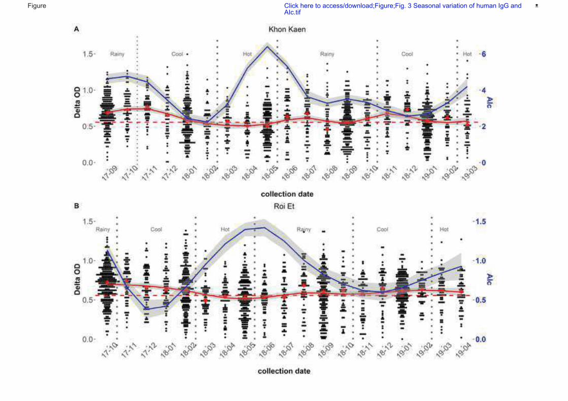


Dear Editor and Reviewers of PLoS Neglected Tropical Diseases,
We really appreciated the effort and the time spend to improve our manuscript (PNTD-D-20-00965). We
have modified the manuscript accordingly to the main comments and revisions asked. Please find below
details concerning our answers, justifications and modifications to the points raised by each referee and
the changes made in the text. Please note that the lines numbers refer to the clean version of the
revised document.
Reviewer's Responses to Questions
Key Review Criteria Required for Acceptance?
As you describe the new analyses required for acceptance, please consider the following:
Methods
-Are the objectives of the study clearly articulated with a clear testable hypothesis stated?
-Is the study design appropriate to address the stated objectives?
-Is the population clearly described and appropriate for the hypothesis being tested?
-Is the sample size sufficient to ensure adequate power to address the hypothesis being tested?
-Were correct statistical analysis used to support conclusions?
-Are there concerns about ethical or regulatory requirements being met?
Reviewer #1: (No Response)
Reviewer #2: In general, the method is adequate.
I suggest specifying the study design.
In Figure 1, it is necessary to improve the quality of the image, as it is not possible to
differentiate the color pattern from Figure 1 (A). Figure 2 needs a caption for the abbreviations
presented.
We improved the resolution of the Figure 1 and added the missing caption for Figure 2.
--------------------
Results
-Does the analysis presented match the analysis plan?
-Are the results clearly and completely presented?
-Are the figures (Tables, Images) of sufficient quality for clarity?
Reviewer #1: (No Response)
Reviewer #2: The results presentation was quite confusing.
The authors should describe the number of study participants cohesively.
For example:
Initially, in the descriptive analysis, the authors report 612 participants but, in Table 1 the total
no. is not consistent with this sample. Data on mosquito exposure report 3,989 dried blood

samples, but, the text does not specify how many individuals are represented in these samples.
Also, when presenting univariate and multivariate analyzes, the sample consists of 381
individuals.
We modified the Table 1 according to the reviewers’ comments and clarified the repartition of the
samples in the text at Lines 361-365. For the univariate and multivariate analysis, we clarified the total
number of individuals used in the analysis, being 539 individuals including 378 followed-up every four
months and 161 individuals followed-up once each month (Lines 433-436).
The confusion is also in the organization of the topics in the results section. Usually, unadjusted
estimates are presented first and then adjusted estimates. The text would be more
understandable if the authors followed this order in the presentation of the results.
In Table 1, both the title and the presentation of the results are quite confusing. The total
number presented does not refer to 612 individuals. Also, the authors do not present the
individuals' immunological status regarding the exposure biomarker.
The total number presented reflected the number of dried blood samples collected and analyzed from
the cohort, however, individuals were visited several times (from 1 to 19 visits depending on households
and persons).
Table 1 has been split into two tables, Table 1 presents the population studied, and Table 2 presents the
results from the sociodemographic questionnaire.
The immunological status of individuals is presented in Table 1 as the proportion of immune responders
according to location (province) and age groups.
We agree with the reviewers on the organization of the results. The first sections of the results present
only descriptive statistics of the data without analysis. However, we had to present the unadjusted
estimated in Supplementary material (S.Table 3), due to the large number of covariates analyzed. The
univariate analysis was used mainly to screen the significant covariates to keep in the multivariate
model. We assumed that the global message would be clearer by presenting the adjusted estimates in
the main text while the unadjusted were presented as Supplementary material, therefore avoiding
repetitions. Nonetheless, we modified the text on Lines 419-421 to mention the univariate analysis
earlier in the text.
--------------------
Conclusions
-Are the conclusions supported by the data presented?
-Are the limitations of analysis clearly described?
-Do the authors discuss how these data can be helpful to advance our understanding of the
topic under study?
-Is public health relevance addressed?
Reviewer #1: (No Response)
Reviewer #2: Line 467: authors report that human-mosquito contact may be influenced by
socioeconomic characteristics. I believe that the correct term is sociodemographic since the
analysis did not include socioeconomic variables.

We replaced the word socio-economics with sociodemographic in the abstract and the text at Lines 43
and 494.
Besides, the authors cited three references when presenting the main results of the study,
which needs to be corrected.
We modified the text presenting the main results, highlighting the similarities between our study and
the cited studies at Lines 493-495.
Line 468: I suggest removing the word “unfortunately”.
We removed the word “unfortunately” at Line 496.
I believe that the text would benefit from the inclusion of a brief discussion on the differences
observed in the analysis of the variables associated with the Aedes Index and Mosquito
exposure index in the “outdoor and indoor” and “indoors only” models.
The application of the results in entomological surveillance could be further explored in this
section.
We acknowledge the reviewers’ comments, yet, we focused our discussion on the model using “indoor
only” as the association was strictly positive and significant for all levels of indoor Aedes infestation
while in the “outdoor and indoor” model, the lower level of Aedes infestation was not significantly
associated with a higher level of Ab response. -------------------- Editorial and Data Presentation Modifications? Use this section for editorial suggestions as well as relatively minor modifications of existing
data that would enhance clarity. If the only modifications needed are minor and/or editorial, you
may wish to recommend “Minor Revision” or “Accept”.
Reviewer #1: (No Response)
Reviewer #2: Replace the socioeconomic term with sociodemographic in the abstract.
The text and tables need formatting.
The text and tables were carefully reviewed and formatted accordingly.
--------------------
Summary and General Comments
Use this section to provide overall comments, discuss strengths/weaknesses of the study,
novelty, significance, general execution and scholarship. You may also include additional
comments for the author, including concerns about dual publication, research ethics, or
publication ethics. If requesting major revision, please articulate the new experiments that are
needed.
Reviewer #1: Overall comments:
The authors present a study looking at a large suite of entomological indices for Ae. aegypti to

see how they compare to human immunological response to Ae. aegypti salivary proteins. This
was an enjoyable study to read and an impressive amount of work went into this study. I have a
few overall comments and specific comments for the authors to consider.
In the discussion the authors cite their own study (Fustec et al. 2020 PLOS NTD) which upon
review looks very similar to the current study. The regions of Thailand and the time of sampling
looks the same between that recent study and the current one but what I am not sure about is if
these exact same households and human participants are included in both studies. If they area
the same, the authors need to be more careful about articulating the difference between this
other publication and the current one. It looks like the past compared the Mosquito Exposure
Index to human dengue cases whereas the current one focuses on comparing MEI to a variety
of the entomological indices. If these data originate from the same study, it would be important
to introduce this context in the introduction section.
The data from the current study and the case-control study (Fustec et al. 2020 PLOS NTD) originated
from two different studies, although they were conducted in the same area, they didn’t involve the
same districts; or the same households/individuals and they have different objectives.
The objective of the case-control study (Fustec et al. 2020 PLOS NTD) was to determine immunological,
entomological and socioeconomic risk factors of dengue using passive hospital-based dengue detection.
The objective of the current study was to determine the relationships between the intensity of the
immune response to the Aedes salivary peptide, vector infestation indices (adult and immature stages)
and vector DENV infection though longitudinal surveys (over 2 years). In the current study, dengue cases
were investigated using active case detection.
On a similar note, this study appears to have taken place during an intervention with
pyriproxyfen. My comment below observes a reference to this intervention in the discussion
along with conclusions regarding the intervention success. I don’t see any mention of the
intervention in the methods or results. I suspect this represents a manuscript in prep that will
focus on the results of the intervention. That is fine but the only potential conflict with this
current study is if the intervention had an influence on the MEI results or any of the
entomological indices. Does the longitudinal data associated with Figure 3 include households
or communities in the intervention clusters? This could of course be a major artifact depending
on the context of the intervention so the authors need to be more transparent about this
throughout the MS.
Indeed, the results from the vector control intervention will be part of a separated publication and we
cannot provide too much information in the current manuscript.
Figure 3 includes individuals from the intervention (treated) and the control clusters, however the
current analysis aims to clarify the relationship between the intensity of the immune response to the
Nterm-34 kDa salivary peptide and the Aedes density and also to individual characteristics,
sociodemographic, climatic factors and vector control intervention.
The description of the intervention with pyriproxyfen was included in the Material and Method section
at Lines 205-207. The influence of vector control intervention with pyriproxyfen was accounted in the
models as a binary covariate at Lines 312-314.

The summary of results in the discussion concludes that there is a “strong positive relationship
between the intensity of human IgG response against the Aedes salivary peptide Nterm-34kDa
and adult Aedes infestation in northeastern Thailand.” My comments below show that it is
currently difficult to see how ‘strong’ this really is. An additional multi-panel figure might help to
show these differences better.
We removed Table 3 and included it into a multi-panel figure as Figure 4 presenting the mean difference
in MEI according to each model.
Specific comments:
Ln. 370. What about the collection of mosquitoes in genera other than Aedes? The methods
ore results don’t really discuss the ELISA’s specificity to just Ae. aegypti verses antibodies to
salivary proteins by other mosquitoes that feed on humans. I suspect the prior studies cited
discuss this topic but it is an important one worth revisiting in the current study.
Indeed, others mosquito species were collected, mainly Culex spp.; however, the salivary peptide
Nterm-34 kDa is specific to Aedes mosquito saliva (Elanga et al., 2012), therefore we do not expect a
cross reaction with other mosquito species present in the study area. We emphasized on the Aedes-
specificity of the Nterm-34kDa salivary peptide at Lines 273-275.
Ln. 417. In the results you are presenting the significance and mean difference in MEI for all
the variables. Presenting these mean differences are hard for me to digest in terms of how MEI
compares at the household or individual level. Although you already have 3 figures in the MS,
only one is a data figure. Can you somehow present the results of table 3 (or supplemental
table 3) as a figure? I see there are many significant relationships but the differences appear
small so it would be nice to see these data in another way. I could see an additional multi-panel
figure being added to main text or supplemental material.
We removed Table 3 and included it into a multi-panel figure presenting the mean difference in MEI
according to each model (Figure 4).
Ln. 468-471. You state dengue cases were not detected during follow up. However, the Fustec
et al. 2020 PLOS NTD reports many dengue cases during the 2016 to 2019 period. It looks like
the current study is 2017 to 2019 so do these represent many of the same participants?
In Fustec et al. 2020 PLOS NTD, we reported some dengue cases from hospital-based passive detection
whereas in the current study we investigated dengue cases using active case detection and fever
measurement every week. Moreover, the FUSTEC et al. 2020 PLOS NTD study was conducted in 2016
and was extended until 2019 due to the low number of incident dengue cases reported/detected.
Moreover, although the two studies were partly conducted in the same provinces, they did not include
the same districts nor the same participants. Therefore, none of the individuals from the case control
study (FUSTEC et al. PLOS NTD 2020) were included in the current longitudinal study presented here.
Ln. 526-528. You say IgG levels were significantly lower in treated clusters compared to control clusters which suggests the pyriproxyfen treatment reduced Aedes density. You are not citing these results in other studies and I don’t see any information in the methods/results regarding
the intervention or an analysis involving the intervention. These statements in the discussion sound in appropriate as it sounds like you are referring to previously published work or unpublished work. We added a sentence in the Material and Method section to describe the pyriproxyfen intervention
(Lines 205-207). We also add a sentence in the analysis section to clarify how the intervention was
accounted for in the analysis (Lines 313-314). However, the results from the intervention will be
published elsewhere and we can’t provide too much details on methods and result in this paper
(entomology/epidemiology data).
Figure 3. If I understand Fig. 3 correctly, the blue line is the Adult number per household and
the red line represents the IgG response to the salivary marker. The text says there is a delay
between when the adult numbers peak and then when the immune response to Ae. aegypti
feeding starts to increase. To me the variation in adult numbers is striking but the variation in
immune response (or delta OD) is very subtle and hard to even notice a relationship with adult
numbers. This putative lag is related to how long it takes for a person to develop these
antibodies and then how long they persist (or at least are detectable). The authors don’t appear
to bring up this topic in the discussion so this would be important to include.
We added a sentence in the Result section of Figure 3 to introduce the time-lag between the bites and
the production of antibodies (Lines 417-419) based on previous studies, mainly conducted on malaria
vectors salivary peptide which showed that this time-lag was about three- to four-weeks (Drame et al.
2010). We added a sentence in the Discussion section to highlight the need of further research on the
kinetics of IgG production and temporal waning (diminishing) antibody titres following Aedes bites (Ln.
490-492).
Supplemental Table 3. It looks like all the <.0001 should be <0.0001.
We corrected the p-values in the supplementary Table S3.
Reviewer #2: This is a study on the use of the biomarker of exposure to the mosquito bite and
its relationship with entomological indicators and individual risk factors.
The article is relevant and original. However, some changes to the text are necessary to
improve clarity in the presentation of results and discussion.
We hope that the modifications made in the article will improve the global clarity of the results and the
discussion for the reader.
--------------------


103
Chapter 4: Assessing the impact of vector control intervention on the selection
of insecticide resistance genes in dengue vectors
In the previous chapter we have shown that the levels of individual response to the Aedes
salivary biomarker were lower in the treated cluster compared to the control one’s, hence
suggesting potential reduction in Aedes density following the implementation of vector control.
Although investigations are still on-going to assess the efficacy and residual activity of PPF in the
study area, preliminary findings suggest however a lack of impact of the PPF on several
entomological indices (Overgaard personal communication). Several operational factors could
explain this outcome including an insufficient treatment coverage, inadequate frequency of
application, inadequate dose or both. These assumptions are currently tested by the DENGUE
INDEX team through complementary semi-field experimental studies. Another unaddressed
explanation is the potential selection of PPF resistance/tolerance following the deployment of the
insecticide in the treated area. Indeed, cross-resistance between PPF and PYRs has been detected
previously and this may impede the benefit of using this new molecule for vector control in
Thailand (see section for details 3.3.3.2).
Considering the above, we conducted a monitoring of insecticide resistance in the study
area before, during and after the deployment of the intervention. Briefly, PPF was deployed in half
of the clusters after ten months of follow up. The objective was to determine the baseline resistance
status of Aedes aegypti to PPF and to conventionally used public health pesticides (baseline) and
to assess change in the levels and frequency of relevant candidate markers after the deployment of
the intervention. We assumed that the subsequent use of PPF in nine clusters of the study area
(equivalent to 1,226 m2) may induce a selection pressure on resistance mechanisms already present
in the population (see section 3.3.3.2). The main findings, that have not been published yet, are
summarized in the following section.
Summary of results:
Among the eighteen clusters included in the RCT (see section 4.3), ten were randomly
selected and followed up for the resistance survey (six clusters in the treated area and four clusters
in the control area). Firstly, the study demonstrated the presence of high levels of insecticide
resistance in Ae. aegypti populations in KK. Indeed, adult mortality ranged from 0% to 37.5% and
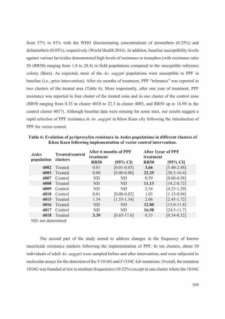
104
from 57% to 81% with the WHO discriminating concentrations of permethrin (0.25%) and
deltamethrin (0.03%), respectively (World Health 2016). In addition, baseline susceptibility levels
against various larvicides demonstrated high levels of resistance to temephos (with resistance ratio
50 (RR50) ranging from 1.8 to 28.4) in field populations compared to the susceptible reference
colony (Bora). As expected, most of the Ae. aegypti populations were susceptible to PPF in
baseline (i.e., prior intervention). After six months of treatment, PPF “tolerance” was reported in
two clusters of the treated area (Table 6). More importantly, after one year of treatment, PPF
resistance was reported in four cluster of the treated zone and in one cluster of the control zone
(RR50 ranging from 0.33 in cluster 4018 to 22.3 in cluster 4003, and RR50 up to 16.98 in the
control cluster 4017). Although baseline data were missing for some sites, our results suggest a
rapid selection of PPF resistance in Ae. aegypti in Khon Kaen city following the introduction of
PPF for vector control.
Table 6: Evolution of pyriproxyfen resistance in Aedes populations in different clusters of Khon Kaen following implementation of vector control intervention.
Aedes
population Treated/control clusters
After 6 months of PPF treatment
After 1year of PPF treatment
RR50 [95% CI] RR50 [95% CI] 4002 Treated 0.01 [0.01-0.03] 3.66 [5.40-2.48] 4003 Treated 0.00 [0.00-0.00] 22.29 [30.3-16.4] 4007 Control ND ND 0.59 [0.60-0.58] 4008 Treated ND ND 11.13 [14.2-8.72] 4009 Control ND ND 2.34 [4.25-1.29] 4010 Control 0.01 [0.00-0.02] 1.03 [1.13-0.94] 4015 Treated 1.54 [1.55-1.54] 2.06 [2.45-1.72] 4016 Treated ND ND 12.80 [13.9-11.8] 4017 Control ND ND 16.98 [24.5-11.7] 4018 Treated 3.39 [0.65-17.6] 0.33 [0.34-0.32]
ND: not determined
The second part of the study aimed to address changes in the frequency of known
insecticide resistance markers following the implementation of PPF. In ten clusters, about 30
individuals of adult Ae. aegypti were sampled before and after intervention, and were subjected to
molecular assays for the detection of the V1016G and F1534C kdr mutations. Overall, the mutation
1016G was founded at low to medium frequencies (10-52%) except in one cluster where the 1016G

105
mutation was rare (3%). Similar results were observed with the 1534C kdr mutation, which was
found at moderate prevalence (39-50%) among all clusters but one (<5% kdr frequency). Only few
populations were at Hardy-Weinberg Equilibrium for the V1016G and 1534C mutations, hence
suggesting strong selection pressure on resistant alleles. Surprisingly, no homozygote resistant
individuals for the 1534C, neither double homozygote for 1016G and 1534C, were found among
the mosquito populations, hence suggesting linkage disequilibrium between these two mutations.
Overall, no significant changes in kdr resistant allele frequency were however reported after one
year of PPF intervention.
Copy Number Variations (CNV) of six metabolic candidate genes related to insecticide
resistance (GSTE2, CYP6-like CYP6Z8, CYP9J28, CYP6BB2 and CCEAE3A) and two domestic
genes (CYP4D39 and RPS7) were investigated in seven mosquito pools per population (including
Bora) before and after PPF intervention. Gene selection was based on previous studies which found
significant correlation between CNV of these genes and PYR and/or OP resistance (Faucon et al.
2017, Cattel et al. 2020a). At baseline, the CCEAE3A gene displayed CNV comprised between 2
to 16 compared to the susceptible reference Bora strain. After one year of PPF treatment, CNV of
CCEAEA3 ranged from 1 and 8 (Figure 35), hence reflecting a decrease in CNV following PPF
intervention. However, no duplications of CYP genes previously associated with higher
detoxification of PYR or PPF were found before or after PPF intervention. Additionally, our results
suggested a simple duplication event in GSTE2 gene in field populations compared to the Bora
strain with no major changes in copy number over time. Overall, we showed that PPF did not
significantly impact on CNV selection except for the CCEAE3A gene for which the number of
copy variants decreased after treatment (i.e., 4002, 4003, 4008, 4015, 4016, 4018 populations)
(Figure 35). The reduction in CCEAE3A copy number, which is associated to OP resistance, may
reflect a strong fitness-cost associated to CCEAE3A in the absence treatment. The replacement of
temephos (OP) by other larvicides with unrelated mode of action could then negatively impact on
temephos resistance in the field.
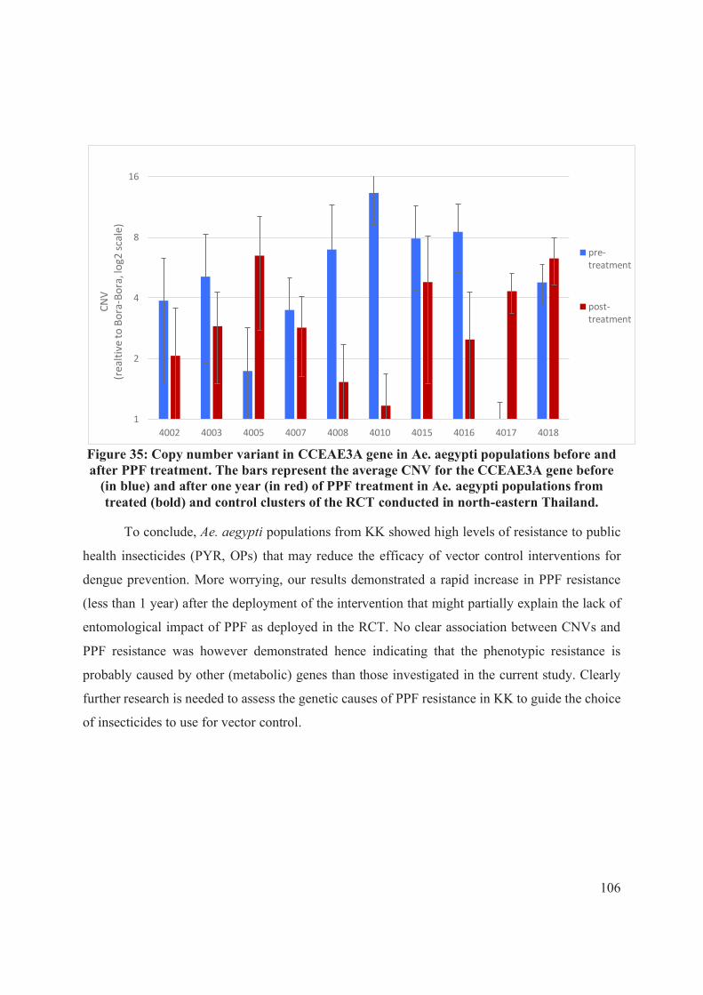
106
Figure 35: Copy number variant in CCEAE3A gene in Ae. aegypti populations before and after PPF treatment. The bars represent the average CNV for the CCEAE3A gene before
(in blue) and after one year (in red) of PPF treatment in Ae. aegypti populations from treated (bold) and control clusters of the RCT conducted in north-eastern Thailand.
To conclude, Ae. aegypti populations from KK showed high levels of resistance to public
health insecticides (PYR, OPs) that may reduce the efficacy of vector control interventions for
dengue prevention. More worrying, our results demonstrated a rapid increase in PPF resistance
(less than 1 year) after the deployment of the intervention that might partially explain the lack of
entomological impact of PPF as deployed in the RCT. No clear association between CNVs and
PPF resistance was however demonstrated hence indicating that the phenotypic resistance is
probably caused by other (metabolic) genes than those investigated in the current study. Clearly
further research is needed to assess the genetic causes of PPF resistance in KK to guide the choice
of insecticides to use for vector control.
1
2
4
8
16
4002 4003 4005 4007 4008 4010 4015 4016 4017 4018
CN
V
(rea
ltiv
e to
Bo
ra-B
ora
, lo
g2 s
cale
)
pre-treatment
post-treatment

107
Fourth part: Discussion and perspectives of the thesis
In Thailand, dengue is a major cause of hospitalisations and deaths especially among
children <15 years old. Dengue transmission occurs throughout the year yet, the rainy season is
always associated with higher dengue incidence. Major dengue major occurs every three to six
years (van Panhuis et al. 2015, Churakov et al. 2019) resulting in pluri-annual seasonal variations,
with intra and inter-epidemic periods. Large epidemic episodes are generally caused by changes
in serotype distribution in a given area but climatic factors, vector dynamic and individual
characteristics also play a role in the virus circulation’s dynamic. Due to highly seasonal and
cyclical variations of serotypes, dengue outbreaks remain difficult to predict.
In north-eastern Thailand, a clear seasonal pattern of dengue incidence was observed with
climatic factors, especially rainfall and temperature, explaining a significant part of dengue
transmission risk (Phanitchat et al. 2019). Previous studies showed positive association between
dengue and temperature and this can be explained by an increase in viral replication in the
mosquito (hence, shortening the time needed for the vector to become infective) and enhanced
vectorial capacity (Christofferson and Mores 2016, Liu et al. 2017). Although important, climatic
variations did not explain the spatial clustering of dengue cases in Thailand. In KK and RE, we
showed that dengue incidence was driven by other factors including individual age and levels of
urbanization. Unfortunately, other factors such as human population movement and/or vector
dynamics, such as seasonal changes in vector densities, that are known to influence the modalities
of transmission were not explored due to the lack of relevant entomological data. For example,
human movement is an important risk factor for dengue and rural-urban migration is common in
Thailand, with people drawn by, for example, better education, job opportunities, health facilities,
standard of living, and wages (Katewongsa 2015). Further information on human travel history
and working conditions would have been required to address the impact of travel-related infection
on dengue incidence in the study area. Our findings also highlighted the need to fill several
knowledge gaps with regards to vector dynamic and socio economic and environmental factors
that could contributed to the spatial clustering of cases dengue between rural and urban areas.
Nonetheless, such retrospective study comes with the inherent limitations of the data
collection using passive case detection (from public and private health care centres, clinics, and
hospitals) including underreporting and misreporting of symptomatic cases as well as the absence
108
of subclinical and asymptomatic infections. In Thailand, dengue diagnosis is mostly based on
clinical signs, and only severe cases are subjected to laboratory diagnosis (see section 1.6).
Consequently, only a small portion of dengue cases are submitted to dengue virus detection and/or
serotyping. Thus, it is likely that a substantial part of dengue cases is ignored hence biasing
epidemiology studies. Extensive use of RDT and systematic DENV serotyping in dengue-like
symptoms patients would beneficiate to surveillance systems to accurately diagnose dengue and
provide real time information on the circulating serotypes. Considering dengue illness time-course,
RDT should combine both NS1 and IgM/IgG detection to avoid exclusion of dengue cases.
As part of the national plan and strategies for dengue prevention and control through the
Vector-Borne Disease Bureau regulations, when a dengue case is detected at the hospital and
reported to the national dengue surveillance system (report 506), Surveillance and Rapid Response
Teams (SRRT) are deployed in dengue-positive patient households to eliminate the vector. Since
the early 1990’s, Thailand has moved toward a decentralized model of vector-borne disease control
resulting in the reorganization of disease control operations. Currently 76 provincial
administrations being aggregated into 22 regional ODPCs are carrying routine surveillance and
control operations with more or less success (Bhumiratana et al. 2014). Despite local and regional
plan for dengue vector control and surveillance, the entomological thresholds defined by the
MoPH, have not been generally adopted to address dengue transmission risk or to trigger vector
control intervention. In addition, the decentralization of vector control has led to differences in
VBDU leaderships and capacities resulting in varying efficacy in assessing dengue vector risk
(Bhumiratana et al. 2014). Differences between/within provinces may lead to differences in vector
surveillance and monitoring efficiency and in planning vector control interventions. While policies
and strategies are still decided at the national level, the implementation of prevention and control
remain under the authority of local VBDUs which may differ in terms of legislation and practices
for disease prevention and control. Additionally, policies at the local level are challenged by the
local administration, socio-political and socio-economic circumstances which differ between
districts. For instance, the number of SRRT capacity is limited per province, thus they can
intervene only for a restricted number of dengue cases, favouring the spread of the disease.
Additionally, the resources allocated for vector control are planned according to the forecasts from
MoPH and remaining chemicals for previous years (Suphanchaimat et al. 2019). Therefore, some
109
regions may lack of adequate human and financial resources to effectively control the vectors
where dengue outbreak occur.
Recent efforts have been made to improve dengue surveillance and vector control through
the WHO-GVCR which emphasises on the development of more cost-effective and practical tools
for vector surveillance. Despite extensive research and numerous prediction models, no threshold
and indicators could be clearly established to predict transmission risk and prevent outbreaks
(Lauer et al. 2018, Phanitchat et al. 2019). In Thailand, dengue incidence is heterogeneous with
distinct patterns of transmission within regions that makes dengue prediction particularly difficult
to establish (Lauer et al. 2018, Phanitchat et al. 2019). Disease transmission is complex and
involves many climatic, environmental and socio economical parameters that are rarely accounted
in dengue transmission studies. For all these reasons, outbreaks continue to occur and the overall
dengue incidence increased despite intensive efforts of national program to control the disease.
More cost-effective approaches and practical tools that can reliably measure real-time dengue
transmission dynamics are needed to enable more accurate and useful predictions of dengue
incidence and outbreaks.
The current thesis was conducted in the framework of the DENGUE INDEX project that
aimed to develop more practical and sensitive tools and indicators of dengue transmission risk in
Thailand that may be used to forecast dengue outbreaks. The scope was to address the current
challenges and limitations in dengue surveillance by exploring the potential of combined
entomological and serological tools for assessing dengue transmission risk and to identify the
determinants associated with human–Aedes relationships. This has been possible through the
integration of multiple disciplines (entomology, immunology, virology, mathematics, etc),
approaches (retrospective, case-control study, randomized controlled studies) and competences.
The expected outcome was to validate the use of serological biomarkers of human exposure to
Aedes bites as a proxy for estimating dengue transmission “hotspots” and “hot-pops” in North-
eastern Thailand facing increasing outbreaks. The following sections will discuss the strength and
weakness of entomology and immunology indicators for dengue epidemiology studies and provide
guidance on how serological biomarkers could be further incorporated into national control
programme for integrated vector surveillance. Potential applications of such serological
110
biomarkers of exposure to Aedes vector bites in the field of operational research, especially for
evaluating vector control interventions are also discussed.
Challenges in assessing dengue transmission risk using conventional entomology indices
During the case control study conducted in north-eastern Thailand, we showed that the
presence of DENV-infected Ae. aegypti in the households was positively associated with a higher
risk of dengue. This confirms previous studies demonstrating good association between vector’s
infection and dengue transmission risk (Lau et al. 2017, Parra et al. 2018). In the study area, the
prevalence of DENV-positive Ae. aegypti mosquitoes was high (13%), hence reflecting a high
dengue transmission setting. Detection of dengue serotypes in mosquito vectors could be
informative to detect the onset of an outbreak; if a serotype was absent for a long time, a larger
part of the population will be naive to it, hence intensity of transmission may be greater when re
introduced. Although vector infectivity might be a good proxy for assessing transmission risk,
DENV detection in adult mosquitoes is rarely implemented in routine surveillance as it costly and
time-consuming and it requires large sample size to get a robust estimate of virus circulation.
Recent studies in Latin America showed that the monitoring of transovarial transmission of dengue
virus in immature Aedes populations (known as xenomonitoring strategy) was a suitable strategy
to enable rapid viral monitoring in areas of difficult access and to assess the progression of dengue
disease (Arunachalam et al. 2008, Cruz et al. 2015, da Costa et al. 2017). Although promising,
more investigations are needed to assess whether DENV xenomonitoring can enable timely
identification of viral serotype's circulation and contribute to the development of more accurate
predictive models and warning systems for preventing outbreaks.
Our entomology surveys showed however that the levels of Aedes infestation based on
immature indices (HI, BI, and CI) were all negatively associated with dengue fever. Most of the
inspected household (regardless dengue status) had water-holding containers positive for Aedes
immature stages and entomology indices were higher than the “outbreak-risk” thresholds setting
up by the MoPH of Thailand. The same was true with regards to adult abundance, where more
Aedes were found in control households than in houses with a recent dengue case. Altogether these
findings emphasized the lack of sensitivity of both immature and adult indices to predict dengue
transmission risk and highlighted the need to quickly re-evaluate entomology thresholds for vector

111
surveillance and control. The lack of positive association between vector infestation
(presence/abundance) and epidemiology outcomes (DENV infections) can be explained by several
factors.
Firstly, we assumed that measurement of entomology indices may have been biased by
vector control operations in case households following onset of dengue symptoms, which would
have reduced vector infestation at the survey time point. However, mosquito collections at patient
houses were conducted within 12 hours following patient inclusion, hence limiting the risk that
SRRTs may have visited the household before our team. A higher use of household insecticide
products in the case houses was however reported through the questionnaire survey, hence
highlighting the possibility that some adult Aedes mosquitoes may have escaped the dwellings.
Further investigations are needed to assess how individual protection measures may have impacted
on vector density and thus dengue infections in the study area.
Another explanation for the lack of correlation between household-based entomological
indices and dengue cases is the possibility that dengue transmission occurred in other locations
than the household patients. Indeed, Aedes mosquitoes bite during the day, so people might be
infected when they are in workplaces, schools and shops centers as previously demonstrated
(Ratanawong et al. 2016). Information on human movement collected through social survey
showed that only 27.6% of the patients declared staying at home during the weekdays (most of
kids spending daytime at school), hence suggesting that a relatively high part of dengue
transmission may have occurred elsewhere (Ratanawong et al. 2016). Several studies demonstrated
that dengue transmission is mainly driven by human movements (Stoddard et al. 2013, Vazquez-
Prokopec et al. 2013, Reiner et al. 2014) that may favor virus dispersion from high DENV
transmission setting to low (non-immune) DENV transmission areas. Sensing human movements
using GPS tracking could help to quickly identify the routes of virus circulation and trigger timely
vector control response.
To conclude, despite the worldwide use of entomology endpoints for dengue surveillance,
the correlation between entomological indices and dengue transmission risk remains unclear
(Romero-Vivas and Falconar 2005, Bowman et al. 2014, Chang et al. 2015, Wijayanti et al. 2016b,
Lau et al. 2017, Parra et al. 2018). Differences in study design, especially the spatial unit used to
calculate entomological and epidemiological indices may also explain the lack of accuracy in
112
prediction’s (Bowman et al. 2014). For example, a large spatial unit can mask hotspots of DENV
transmission and/or vector abundance. Vector density, which varies itself over time, can be
influenced by housing density and human movement, and so, the entomological collections have
to be done quickly after clinical diagnosis (Chadee 2009). This would help reducing time-lag
between entomology and epidemiology assessment hence improving prediction accuracies.
Potential of serological biomarkers for assessing Aedes-human relationships
Results from our randomized controlled trials in the two cities of RE and KK showed a
strong and positive “dose-response” relationship between the Aedes abundance and the Ab
response to the Aedes salivary biomarker, hence indicating that individuals exhibiting higher
antibody response to the Aedes salivary peptide were also at higher risk of dengue vector bites.
Overall, our study demonstrated that the intensity of Ab response varied according to the season,
individual (gender, age, occupation) and household characteristics. Previous studies already
demonstrated good correlations between the Ab response to Aedes salivary biomarker and
entomology indices as well as with climatic and environmental factors (Elanga Ndille et al. 2012,
Sagna et al. 2018, Yobo et al. 2018). Our study highlighted the potential of using Aedes salivary
biomarker to assess fine scale variations of human-Aedes exposure that could be used to identify,
target, and prioritize vector surveillance and control operations in areas with high risk of arbovirus
transmission. This tool may be particularly relevant for invasive species such as Aedes albopictus
that is currently invading new areas and territories, and for which timely identification of mosquito
“hot-spots” could trigger locally adapted control response to prevent further establishment and
spread.
In theory, serological surveys could be easier to implement in the field compared to
entomology surveys as only a small amount of blood (< 1mL collected on filter papers as dried
blood spots) could be sufficient to assess mosquito-exposure risk. Despite easy and cheap
collection method, ELISA tests require highly qualified staff and specific equipment that is not
easily achieved by national control programmes. A promising alternative to ELISA may come
from the development of quantitative point-of-care (POC) test based on immunochromatography
to enable a rapid and easy detection of IgG Ab response to Aedes salivary antigen. The
development of POC device has been possible though public-private partnership including IRD
and DIAG4ZOO (Montpellier, France) that has validated the “proof of concept”. Briefly, the

113
principle relies on colour lines that appear after applying a finger prick of blood to the test well.
Although promising, the development of POC to stratify dengue vector exposure risk based on Ab
response thresholds will require further research and validation using samples coming various
entomological settings.
Potential of serological biomarkers for assessing dengue transmission risk
Although prosing for vector surveillance, serology biomarkers were initially designed to
identify areas (hotspot) and people (hotpops) at high risk for vector-borne diseases (Sagna et al.
2018). Additionally, our serological longitudinal study demonstrated a strong and positive
correlation between the intensity of individual immune response and Aedes densities. However,
our case-control study conducted in North-eastern Thailand didn’t show however significant
association between the levels of Ab response to Aedes salivary peptide and dengue fever. This
result suggests that Aedes salivary biomarker may be not accurate enough to assess spatio-temporal
variations in disease transmission risk. Similar results were obtained by Elanga et in Vientiane,
Lao PDR (Elanga Ndille et al. 2014) where no significant differences in the level of IgG response
to the Nterm-34 kDa peptide was seen between DENV-positive and DENV-negative individuals
using passive case detection. In this study however, only 45% of the studied population was
“immune-responder” and the median IgG response for both groups (i.e., DENV positive and
DENV negative) was below the immunological threshold hence reflecting a low Aedes-exposure
area. In our study, individuals were however located in high Aedes exposure area as demonstrated
by the high levels of entomological indices and the high seroprevalence to the Nterm-34 kDa
peptide.
Several explanations can explain the lack of relationships between Aedes-exposure risk and
dengue. First of all, in our case control study, the sample size was limited (368 dried spots were
analysed) hence limiting the power of the analysis. Moreover, the retrospective case-control design
means the temporal sequence of events cannot be determined with accuracy. In particular,
immunological (and entomology) data were collected following patient recruitment. Indeed,
symptoms of dengue fever can appear as quickly as a few days after DENV transmission (typically
incubation period between 4-7 up to 14 days), delaying the recruitment of patients and therefore
the entomological and serological surveys. Moreover, 3-4 weeks’ time are generally needed to
develop specific IgG Ab response to the salivary peptide hence the concordance between
114
serological outcomes and dengue infection is uncertain. This temporal disconnection between
acquiring an infection to time of presenting illness and testing (i.e., identification of a case) may
greatly affect attempts to link indicators of transmission with actual study design. Unfortunately,
the relationship between dengue incidence and the intensity of the human-antibody response to
Aedes bites could not be addressed through longitudinal follow up because no incident dengue
cases were detected in household participants during the randomized controlled trial.
Finally, the role of acquired immunity against dengue fever in the lack of correlation
between vector abundance or exposure and dengue infection cannot be ruled out. Indeed, the partial
acquired immunity against one or more serotypes make the relationship between the vector risk
and transmission even more complex to address, as only naïve individuals can be infected by a
defined serotype. Thus, a non-negligible proportion of infective bites, may be not followed by
dengue infections/fever in immune individuals. In our study, individuals with higher Ab response
to Aedes mosquito bites had also higher odds of being positive for DENV-IgG. This suggests than
individuals at high risk of Aedes bites were partially (or fully) protected against new dengue
infections. Information’s on herd immunity and serotype prevalence are keys to address the
complex relationships between human-vector contact and dengue in high transmission settings.
More evidence using robust experimental design will be needed to assess whether Aedes salivary
biomarker can be used to identify foci of dengue transmission in Thailand and abroad.
Prospect of serological biomarkers for assessing vector control interventions
Salivary biomarkers showed promising results to evaluate the efficacy of vector control
interventions, such as insecticide treated nets, for malaria control (Drame et al. 2010a, Drame et
al. 2010b, Drame et al. 2013). Likewise, the Nterm-34 kDa salivary peptide proved to be a sensitive
tool to evaluate the efficacy of an integrated approach combining environmental management and
space sprays with PYR against Ae. albopictus in La Reunion (Elanga Ndille et al. 2016). The
authors showed a significant reduction in the level of Ab response to the Nterm-34 salivary peptide
two weeks post-treatment and the seroprevalence among the inhabitants remained low up to four
weeks. In our randomized controlled study, we showed that the levels of IgG response to the
Nterm-34 kDa were significantly lower in PPF-treated clusters compared to control clusters.
Although speculative, these findings may reflect a reduction of vector densities after PPF treatment
that would be sufficient to reduce human-Aedes contact in treated group compared to the control.
115
Unfortunately, the entomological impact of PPF is yet unknown and statistical analysis are under
investigations by our team to fill this gap. Further evidence is needed to assess whether salivary
biomarker may complement existing tools for monitoring and evaluation of vector control
intervention. Serological biomarkers could be particularly relevant in areas where epidemiology
studies cannot be implemented for operational and/or economic constraints.
Barriers to dengue vector control in Thailand
As seen previously, only SRRTs are mandated to eliminate the mosquito vector and
interrupt dengue transmission. However, the same chemicals, especially temephos and
deltamethrin, are used in routine for vector control operations in dengue-positive households but
their efficacy to reduce or prevent dengue transmission has not been clearly demonstrated
(Phuanukoonnon et al. 2005, Bowman et al. 2016). Chemical control is also challenged by the
various operational constraints including community acceptance, lack of resources (e.g., expired
insecticides, household aerosols) and the occurrence and spread of insecticide resistance to the
main public health pesticides (Corbel et al. 2013, Suphanchaimat et al. 2019), hence representing
an additional obstacle to dengue prevention. In our study, we showed strong levels of insecticide
resistance against public health vector control insecticides in Aedes population from KK.
Moreover, we demonstrated a rise of PPF resistance within one year after the implementation of
PPF-intervention in treated clusters, hence indicating a strong selection pressure on resistance
genes. The investigation of resistance mechanisms revealed a selection pressure on kdr mutations
yet, without correlation with PPF application. Moreover, no CNVs in metabolic genes previously
associated with PPF resistance were observed hence underlying that other metabolic genes may be
involved. Interestingly, CNVs in CCE genes associated with OP resistance (Grigoraki et al. 2016,
Moyes et al. 2017, Cattel et al. 2020b) were reduced following PPF intervention, hence suggesting
that temephos resistance could be reverted in absence of temephos treatment. Unfortunately, the
resistance levels to temephos couldn’t be determined after 1 year-time due to a lack of samples for
testing. If confirmed, this would be an excellent new for vector control with the scope to preserve
the lifespan of existing public health pesticides for dengue prevention, as some genes might be
involved in resistance against different insecticides.
To conclude, this study highlights the rationale of introducing alternative vector control
methods as a part Integrated Vector Management (IVM) in Thailand and abroad. An alternative
116
would be to implement sequence or rotations of unrelated insecticides (minimum 3) to reduce the
selection pressure on public health pesticides. Spinosad, a natural neurotoxic insecticide produced
by the soil bacterium Saccharopolyspora spinosa, was initially selected together with
PPF in the DENGUE INDEX project but we didn’t receive approval by national authorities for
further deployment. Insecticides still play a key role in the prevention and control of vector borne
diseases and preserving insecticide susceptibility should be considered as a public good (Sternberg
et al. 2018). As part of IRM, non-chemical-based control strategies should be privileged. The
integration of alternative, safe and environmentally friendly methods relying on genetic or
biological control of vectors should be encouraged to improve the control of resistant mosquitoes
(Achee et al. 2019). Despite the great potential offer by new strategies for vector control to mitigate
insecticide resistance, strong evidences are still missing for most. Further investigations are still
needed to emphasize on the rationale of integrating these news strategies into IVM approaches.
Conclusion
The current thesis explored risk factors associated with Aedes-human exposure and dengue
transmission risk in Thailand. Overall, we highlighted the complex relationships between Aedes
abundance, vector infectivity, vector exposure and dengue infections in North-eastern Thailand
facing recurrent and unpredictable outbreaks. We demonstrated the potential of using salivary
biomarkers to assess fine scale variations in mosquito-exposure that could be further deployed as
part of integrated vector surveillance. In contrast, we didn’t show positive correlation between the
levels of mosquito exposure risk (as measured by entomology and immunological indices) and
dengue fever hence highlighting the limitations of existing tools for assessing dengue transmission
risk. Several factors including human movements and habits, acquired immunity and presence of
vector control partially explained that trend. Altogether, this thesis represents an important step
toward the development of serological biomarkers for assessing Aedes exposure risk and to
evaluate vector control intervention for dengue prevention. Further investigations are needed
however to develop more sensible and accurate tools to measure real-time dengue transmission
and predictive models of dengue outbreaks.
117
References
Abreu, F. V., M. M. Morais, S. P. Ribeiro and E. Eiras Á (2015). "Influence of breeding site availability on the oviposition behaviour of Aedes aegypti." Mem Inst Oswaldo Cruz 110(5): 669-676.
Achee, N. L., J. P. Grieco, H. Vatandoost, G. Seixas, J. Pinto, L. Ching-Ng, A. J. Martins, W. Juntarajumnong, V. Corbel, C. Gouagna, J. P. David, J. G. Logan, J. Orsborne, E. Marois, G. J. Devine and J. Vontas (2019). "Alternative strategies for mosquito-borne arbovirus control." PLoS Negl Trop Dis 13(1): e0006822.
Agarwal, A., S. Gupta, T. Chincholkar, V. Singh, I. K. Umare, K. Ansari, S. Paliya, A. K. Yadav, R. Chowdhary, S. Purwar and D. Biswas (2019). "Co-circulation of dengue virus serotypes in Central India: Evidence of prolonged viremia in DENV-2." Infect Genet Evol 70: 72-79.
Aida, H. N., H. Dieng, T. Satho, A. T. Nurita, M. R. C. Salmah, F. Miake, B. Norasmah and A. H. Ahmad (2011). "The biology and demographic parameters of Aedes albopictus in northern peninsular Malaysia." Asian Pacific Journal of Tropical Biomedicine 1(6): 472-477.
Akter, R., S. Naish, M. Gatton, H. Bambrick, W. Hu and S. Tong (2019). "Spatial and temporal analysis of dengue infections in Queensland, Australia: Recent trend and perspectives." PloS one 14(7): e0220134-e0220134.
Alen, M. M., K. Dallmeier, J. Balzarini, J. Neyts and D. Schols (2012). "Crucial role of the N-glycans on the viral E-envelope glycoprotein in DC-SIGN-mediated dengue virus infection." Antiviral Res 96(3): 280-287.
Alera, M. T., A. Srikiatkhachorn, J. M. Velasco, I. A. Tac-An, C. B. Lago, H. E. Clapham, S. Fernandez, J. W. Levy, B. Thaisomboonsuk, C. Klungthong, L. R. Macareo, A. Nisalak, L. Hermann, D. Villa and I. K. Yoon (2016). "Incidence of dengue virus infection in adults and children in a prospective longitudinal cohort in the Philippines." PLoS Negl Trop Dis 10(2): e0004337.
Ali, A., H. Ahmad, M. Idrees, F. Zahir and I. Ali (2016). "Circulating serotypes of dengue virus and their incursion into non-endemic areas of Pakistan; a serious threat." Virol J 13: 144.
Aliota, M. T., S. A. Peinado, I. D. Velez and J. E. Osorio (2016). "The wMel strain of Wolbachia reduces transmission of zika virus by Aedes aegypti." Sci Rep 6: 28792.
Alphey, L., A. McKemey, D. Nimmo, M. Neira Oviedo, R. Lacroix, K. Matzen and C. Beech (2013). "Genetic control of Aedes mosquitoes." Pathog Glob Health 107(4): 170-179.
Alvarado-Castro, V., S. Paredes-Solís, E. Nava-Aguilera, A. Morales-Pérez, L. Alarcón-Morales, N. A. Balderas-Vargas and N. Andersson (2017). "Assessing the effects of interventions for Aedes aegypti control: systematic review and meta-analysis of cluster randomised controlled trials." BMC Public Health 17(Suppl 1): 384.
Alyousefi, T. A., R. Abdul-Ghani, M. A. Mahdy, S. M. Al-Eryani, A. M. Al-Mekhlafi, Y. A. Raja, S. A. Shah and J. C. Beier (2016). "A household-based survey of knowledge, attitudes and practices towards dengue fever among local urban communities in Taiz Governorate, Yemen." BMC Infect Dis 16(1): 543.
Amarasinghe, A., J. N. Kuritsk, G. W. Letson and H. S. Margolis (2011). "Dengue virus infection in Africa." Emerging infectious diseases 17(8): 1349-1354.
Anderson, K. B., S. Chunsuttiwat, A. Nisalak, M. P. Mammen, D. H. Libraty, A. L. Rothman, S. Green, D. W. Vaughn, F. A. Ennis and T. P. Endy (2007). "Burden of symptomatic dengue infection in children at primary school in Thailand: a prospective study." The Lancet 369(9571): 1452-1459.

118
Anderson, K. B., R. V. Gibbons, D. A. T. Cummings, A. Nisalak, S. Green, D. H. Libraty, R. G. Jarman, A. Srikiatkhachorn, M. P. Mammen, B. Darunee, I.-K. Yoon and T. P. Endy (2014). "A shorter time interval between first and second dengue infections is associated with protection from clinical illness in a school-based cohort in Thailand." The Journal of Infectious Diseases 209(3): 360-368.
Andersson, N., J. Arostegui, E. Nava-Aguilera, E. Harris and R. J. Ledogar (2017). "Camino Verde (The Green Way): evidence-based community mobilisation for dengue control in Nicaragua and Mexico: feasibility study and study protocol for a randomised controlled trial." BMC Public Health 17(Suppl 1): 407.
Andersson, N., E. Nava-Aguilera, J. Arosteguí, A. Morales-Perez, H. Suazo-Laguna, J. Legorreta-Soberanis, C. Hernandez-Alvarez, I. Fernandez-Salas, S. Paredes-Solís, A. Balmaseda, A. J. Cortés-Guzmán, R. Serrano de Los Santos, J. Coloma, R. J. Ledogar and E. Harris (2015). "Evidence based community mobilization for dengue prevention in Nicaragua and Mexico (Camino Verde, the Green Way): cluster randomized controlled trial." BMJ 351: h3267.
Andries, A.-C., V. Duong, S. Ong, S. Ros, A. Sakuntabhai, P. Horwood, P. Dussart and P. Buchy (2016). "Evaluation of the performances of six commercial kits designed for dengue NS1 and anti-dengue IgM, IgG and IgA detection in urine and saliva clinical specimens." BMC infectious diseases 16: 201-201.
Angel, B. and V. Joshi (2009). "Distribution of dengue virus types in Aedes aegypti in dengue endemic districts of Rajasthan, India." Indian J Med Res 129(6): 665-668.
Arunachalam, N., S. C. Tewari, V. Thenmozhi, R. Rajendran, R. Paramasivan, R. Manavalan, K. Ayanar and B. K. Tyagi (2008). "Natural vertical transmission of dengue viruses by Aedes aegypti in Chennai, Tamil Nadu, India." Indian J Med Res 127(4): 395-397.
Azevedo-Santos, V. M., J. R. Vitule, F. M. Pelicice, E. García-Berthou and D. Simberloff (2017). "Nonnative fish to control Aedes mosquitoes: a controversial, harmful tool." BioScience 67(1): 84-90.
Bariami, V., C. M. Jones, R. Poupardin, J. Vontas and H. Ranson (2012). "Gene amplification, ABC transporters and cytochrome P450s: unraveling the molecular basis of pyrethroid resistance in the dengue vector, Aedes aegypti." PLoS Negl Trop Dis 6(6): e1692.
Barnard, D. R., C. Z. Dickerson, K. Murugan, R. D. Xue, D. L. Kline and U. R. Bernier (2014). "Measurement of landing mosquito density on humans." Acta Trop 136: 58-67.
Barrera-Perez, M. A., N. Pavia-Ruz, J. E. Mendoza-Mezquita, N. Torres-Arcila, R. Hernandez-Hernandez, F. Castro-Gamboa, E. Geded-Moreno, A. Cohuo-Rodriguez, A. Medina-Barreiro, E. Koyoc-Cardena, H. Gomez-Dantes, A. Kroeger, G. Vazquez-Prokopec and P. Manrique-Saide (2015). "Control of Aedes aegypti breeding sites with the program Recicla por tu bienestar in Merida, Mexico." Salud Publica Mex 57(3): 201-210.
Barrera, R., V. Acevedo, G. E. Felix, R. R. Hemme, J. Vazquez, J. L. Munoz and M. Amador (2017). "Impact of autocidal gravid ovitraps on Chikungunya virus incidence in Aedes aegypti (Diptera: Culicidae) in areas with and without traps." J Med Entomol 54(2): 387-395.
Barrera, R., A. Harris, R. R. Hemme, G. Felix, N. Nazario, J. L. Muñoz-Jordan, D. Rodriguez, J. Miranda, E. Soto, S. Martinez, K. Ryff, C. Perez, V. Acevedo, M. Amador and S. H. Waterman (2019). "Citywide control of Aedes aegypti (Diptera: Culicidae) during the 2016 zika epidemic by integrating community awareness, education, source reduction, larvicides, and mass mosquito trapping." J Med Entomol 56(4): 1033-1046.
Bartlett-Healy, K., G. Hamilton, S. Healy, T. Crepeau, I. Unlu, A. Farajollahi, D. Fonseca, R. Gaugler, G. G. Clark and D. Strickman (2011). "Source reduction behavior as an independent measurement of the impact

119
of a public health education campaign in an integrated vector management program for the Asian tiger mosquito." Int J Environ Res Public Health 8(5): 1358-1367.
Bazin, M. and C. R. Williams (2018). "Mosquito traps for urban surveillance: collection efficacy and potential for use by citizen scientists." J Vector Ecol 43(1): 98-103.
Benelli, G., C. L. Jeffries and T. Walker (2016). "Biological control of mosquito vectors: past, present, and future." Insects 7(4).
Benelli, G., A. B. B. Wilke and J. C. Beier (2020). "Aedes albopictus (Asian tiger mosquito)." Trends in Parasitology.
Bengoa, M., R. Eritja, S. Delacour, M. Miranda, A. Sureda and J. Lucientes (2017). "First data on resistance to pyrethroids in wild populations of Aedes albopictus from Spain." J Am Mosq Control Assoc 33(3): 246-249.
Bente, D. A. and R. Rico-Hesse (2006). "Models of dengue virus infection." Drug discovery today. Disease models 3(1): 97-103.
Bhatia, R., A. P. Dash and T. Sunyoto (2013). "Changing epidemiology of dengue in South-East Asia." WHO South East Asia J Public Health 2(1): 23-27.
Bhatt, S., P. Gething, O. Brady, J. Messina, A. Farlow, C. Moyes, J. Drake, J. Brownstein, A. Hoen, O. Sankoh, M. Myers, D. George, T. Jaenisch, G. Wint, C. Simmons, T. Scott, J. Farrar and S. Hay (2013). "The global distribution and burden of dengue." Nature.
Bhumiratana, A., A. Intarapuk, S. Chujun, W. Kaewwaen, P. Sorosjinda-Nunthawarasilp and S. Koyadun (2014). "Thailand momentum on policy and practice in local legislation on dengue vector control." Interdiscip Perspect Infect Dis 2014: 217237.
Biogents. (2020). "Aedes life cycle." Retrieved 01/09/2020, from https://sea.biogents.com/life-cycle-aedes-mosquitoes/.
Blacksell, S. D., R. G. Jarman, M. S. Bailey, A. Tanganuchitcharnchai, K. Jenjaroen, R. V. Gibbons, D. H. Paris, R. Premaratna, H. J. de Silva, D. G. Lalloo and N. P. Day (2011). "Evaluation of six commercial point-of-care tests for diagnosis of acute dengue infections: the need for combining NS1 antigen and IgM/IgG antibody detection to achieve acceptable levels of accuracy." Clin Vaccine Immunol 18(12): 2095-2101.
Bodinayake, C. K., L. G. Tillekeratne, A. Nagahawatte, V. Devasiri, W. Kodikara Arachichi, J. J. Strouse, O. M. Sessions, R. Kurukulasooriya, A. Uehara, S. Howe, X. M. Ong, S. Tan, A. Chow, P. Tummalapalli, A. D. De Silva, T. Østbye, C. W. Woods, D. J. Gubler and M. E. Reller (2016). "Emergence of epidemic dengue-1 virus in the southern province of Sri Lanka." PLoS neglected tropical diseases 10(10): e0004995-e0004995.
Boonklong, O. and A. Bhumiratana (2016). "Seasonal and geographical variation of dengue vectors in Narathiwat, South Thailand." Can J Infect Dis Med Microbiol 2016: 8062360.
Bourtzis, K., S. L. Dobson, Z. Xi, J. L. Rasgon, M. Calvitti, L. A. Moreira, H. C. Bossin, R. Moretti, L. A. Baton, G. L. Hughes, P. Mavingui and J. R. Gilles (2014). "Harnessing mosquito-Wolbachia symbiosis for vector and disease control." Acta Trop 132 Suppl: S150-163.
Bouzid, M., J. Brainard, L. Hooper and P. R. Hunter (2016). "Public health interventions for Aedes control in the time of zika virus- A meta-review on effectiveness of vector control strategies." PLoS Negl Trop Dis 10(12): e0005176.
Bowman, L. R., S. Donegan and M. C. Pj (2016). "Is dengue vector control deficient in effectiveness or evidence? Systematic review and meta-analysis." PLoS Negl Trop Dis 10.

120
Bowman, L. R., S. Runge-Ranzinger and P. J. McCall (2014). "Assessing the relationship between vector indices and dengue transmission: a systematic review of the evidence." PLoS Negl Trop Dis 8(5): e2848.
Boyce, R., A. Lenhart, A. Kroeger, R. Velayudhan, B. Roberts and O. Horstick (2013). "Bacillus thuringiensis israelensis (Bti) for the control of dengue vectors: systematic literature review." Trop Med Int Health 18(5): 564-577.
Bradshaw, C. J. A., B. Leroy, C. Bellard, D. Roiz, C. Albert, A. Fournier, M. Barbet-Massin, J.-M. Salles, F. Simard and F. Courchamp (2016). "Massive yet grossly underestimated global costs of invasive insects." Nature communications 7: 12986-12986.
Brady, O., P. Gething, S. Bhatt, J. Messina, J. Brownstein, A. Hoen, C. Moyes, A. Farlow, T. Scott and S. Hay (2012). "Refining the global spatial limits of dengue virus transmission by evidence-based consensus." PLoS Negl Trop Dis.
Brady, O. J., M. A. Johansson, C. A. Guerra, S. Bhatt, N. Golding, D. M. Pigott, H. Delatte, M. G. Grech, P. T. Leisnham, R. Maciel-de-Freitas, L. M. Styer, D. L. Smith, T. W. Scott, P. W. Gething and S. I. Hay (2013). "Modelling adult Aedes aegypti and Aedes albopictus survival at different temperatures in laboratory and field settings." Parasit Vectors 6: 351.
Brady, O. J., D. L. Smith, T. W. Scott and S. I. Hay (2015). "Dengue disease outbreak definitions are implicitly variable." Epidemics 11: 92-102.
Bravo, L., V. G. Roque, J. Brett, R. Dizon and M. L'Azou (2014). "Epidemiology of dengue disease in the Philippines (2000-2011): a systematic literature review." PLoS Negl Trop Dis 8(11): e3027.
Brengues, C., N. J. Hawkes, F. Chandre, L. McCarroll, S. Duchon, P. Guillet, S. Manguin, J. C. Morgan and J. Hemingway (2003). "Pyrethroid and DDT cross-resistance in Aedes aegypti is correlated with novel mutations in the voltage-gated sodium channel gene." Med Vet Entomol 17(1): 87-94.
Britch, S., H. Nyberg, R. Aldridge, T. Swan and K. Linthicum (2016). "Acoustic control of mosquito larvae in artificial drinking water containers." Journal of the American Mosquito Control Association 32: 341-344.
Brusich, M., J. Grieco, N. Penney, R. Tisgratog, W. Ritthison, T. Chareonviriyaphap and N. Achee (2015). "Targeting educational campaigns for prevention of malaria and dengue fever: an assessment in Thailand." Parasit Vectors 8: 43.
Bureau of Epidemiology and Ministry of Public Health Thailand (2011). Annual epidemiologcal surveillance report 2010. Ministry of Public Health Thailand. Nonthaburi, Ministry of Public Health Thailand,.
Bureau of Epidemiology and Ministry of Public Health Thailand (2016). Annual epidemiological surveillance report 2015. Ministry of Public Health Thailand. Nonthaburi, Ministry of Public Health Thailand, : 1000.
Bureau of Epidemiology and M. o. P. H. Thailand (2015). Annual epidemiological surveillance report 2014. M. o. P. H. Thailand. Nonthaburi, Ministry of Public Health Thailand.
Bureau of Epidemiology and M. o. P. H. Thailand (2019). Annual epidemiological surveillance report 2018. Department of Disease Prevention: 808.
Calzolari, M. (2016). "Mosquito-borne diseases in Europe: An emerging public health threat." Reports in Parasitology 5: 1.
Capeding, M. R., N. H. Tran, S. R. Hadinegoro, H. I. Ismail, T. Chotpitayasunondh, M. N. Chua, C. Q. Luong, K. Rusmil, D. N. Wirawan, R. Nallusamy, P. Pitisuttithum, U. Thisyakorn, I. K. Yoon, D. van der Vliet, E. Langevin, T. Laot, Y. Hutagalung, C. Frago, M. Boaz, T. A. Wartel, N. G. Tornieporth, M. Saville, A.

121
Bouckenooghe and C. Y. D. S. Group (2014). "Clinical efficacy and safety of a novel tetravalent dengue vaccine in healthy children in Asia: a phase 3, randomised, observer-masked, placebo-controlled trial." Lancet 384(9951): 1358-1365.
Caputo, B., A. Ienco, D. Cianci, M. Pombi, V. Petrarca and A. Baseggio (2012). "The “auto-dissemination”
approach: a novel concept to fight Aedes albopictus in urban areas." PLoS Negl Trop Dis 6.
Carrieri, M., P. Angelini, C. Venturelli, B. Maccagnani and R. Bellini (2011). "Aedes albopictus (Diptera: Culicidae) population size survey in the 2007 Chikungunya outbreak area in Italy. I. Characterization of breeding sites and evaluation of sampling methodologies." J Med Entomol 48(6): 1214-1225.
Carvalho, D. O., A. R. McKemey, L. Garziera, R. Lacroix, C. A. Donnelly, L. Alphey, A. Malavasi and M. L. Capurro (2015). "Suppression of a field population of Aedes aegypti in Brazil by sustained release of transgenic male mosquitoes." PLoS Negl Trop Dis 9(7): e0003864.
Carvalho, F. D. and L. A. Moreira (2017). "Why is Aedes aegypti Linnaeus so successful as a species?" Neotropical Entomology 46(3): 243-255.
Castro, M. G., R. M. Nogueira, H. G. Schatzmayr, M. P. Miagostovich and R. Lourenco-de-Oliveira (2004). "Dengue virus detection by using reverse transcription-polymerase chain reaction in saliva and progeny of experimentally infected Aedes albopictus from Brazil." Mem Inst Oswaldo Cruz 99(8): 809-814.
Cattel, J., F. Faucon, B. Le Peron, S. Sherpa, M. Monchal, L. Grillet, T. Gaude, F. Laporte, I. Dusfour, S. Reynaud and J. P. David (2020a). "Combining genetic crosses and pool targeted DNA-seq for untangling genomic variations associated with resistance to multiple insecticides in the mosquito Aedes aegypti." Evol Appl 13(2): 303-317.
Cattel, J., C. Haberkorn, F. Laporte, T. Gaude, T. Cumer, J. Renaud, I. W. Sutherland, J. C. Hertz, J.-M. Bonneville, V. Arnaud, C. Noûs, B. Fustec, S. Boyer, S. Marcombe and J.-P. David (2020b). "A genomic amplification affecting a carboxylesterase gene cluster confers organophosphate resistance in the mosquito Aedes aegypti: from genomic characterization to high-throughput field detection." bioRxiv: 2020.2006.2008.139741.
CDC. (2020). "Life cycle of Aedes aegypti and Ae. albopictus mosquitoes." Retrieved 10/09/2020, 2020, from https://www.cdc.gov/mosquitoes/about/life-cycles/aedes.html.
Chadee, D. D. (2009). "Dengue cases and Aedes aegypti indices in Trinidad, West Indies." Acta Trop 112(2): 174-180.
Chadee, D. D., J. C. Beier and R. T. Mohammed (2002). "Fast and slow blood-feeding durations of Aedes aegypti mosquitoes in Trinidad." J Vector Ecol 27(2): 172-177.
Chan, M. and M. A. Johansson (2012). "The incubation periods of dengue viruses." PLoS One 7(11): e50972.
Chandel, K., D. S. Suman, Y. Wang, I. Unlu, E. Williges and G. M. Williams (2016). "Targeting a hidden enemy: pyriproxyfen autodissemination strategy for the control of the container mosquito Aedes albopictus in cryptic habitats." PLoS Negl Trop Dis 10.
Chang, F. S., Y. T. Tseng, P. S. Hsu, C. D. Chen, B. Lian Ie and D. Y. Chao (2015). "Re-assess vector indices threshold as an early warning tool for predicting dengue epidemic in a eengue non-endemic country." PLoS Negl Trop Dis 9(9): e0004043.
Chansang, U. R., A. Bhumiratana and P. Kittayapong (2004). "Combination of Mesocyclops thermocyclopoides and Bacillus thuringiensis var. israelensis: a better approach for the control of Aedes aegypti larvae in water containers." J Vector Ecol 29(2): 218-226.
122
Chen, C. F., P. Y. Shu, H. J. Teng, C. L. Su, J. W. Wu, J. H. Wang, T. H. Lin, J. H. Huang and H. S. Wu (2010). "Screening of dengue virus in field-caught Aedes aegypti and Aedes albopictus (Diptera: Culicidae) by one-step SYBR green-based reverse transcriptase-polymerase chain reaction assay during 2004-2007 in Southern Taiwan." Vector Borne Zoonotic Dis 10(10): 1017-1025.
Chen, H., K. Li, X. Wang, X. Yang, Y. Lin, F. Cai, W. Zhong, C. Lin, Z. Lin and Y. Ma (2016). "First identification of kdr allele F1534S in VGSC gene and its association with resistance to pyrethroid insecticides in Aedes albopictus populations from Haikou City, Hainan Island, China." Infect Dis Poverty 5: 31.
Chiaravalloti-Neto, F., M. Pereira, E. A. Favaro, M. R. Dibo, A. Mondini, A. L. Rodrigues-Junior, A. P. Chierotti and M. L. Nogueira (2015). "Assessment of the relationship between entomologic indicators of Aedes aegypti and the epidemic occurrence of dengue virus 3 in a susceptible population, Sao Jose do Rio Preto, Sao Paulo, Brazil." Acta Trop 142: 167-177.
Christofferson, R. C. (2015). "A Reevaluation of the role of Aedes albopictus in dengue transmission." J Infect Dis 212(8): 1177-1179.
Christofferson, R. C. and C. N. Mores (2016). "Potential for Extrinsic Incubation Temperature to Alter Interplay Between Transmission Potential and Mortality of Dengue-Infected Aedes aegypti." Environ Health Insights 10: 119-123.
Chumpu, R., N. Khamsemanan and C. Nattee (2019). "The association between dengue incidences and provincial-level weather variables in Thailand from 2001 to 2014." PLOS ONE 14(12): e0226945.
Churakov, M., C. J. Villabona-Arenas, M. U. G. Kraemer, H. Salje and S. Cauchemez (2019). "Spatio-temporal dynamics of dengue in Brazil: Seasonal travelling waves and determinants of regional synchrony." PLoS Negl Trop Dis 13(4): e0007012.
Corbel, V., N. L. Achee, F. Chandre, M. B. Coulibaly, I. Dusfour, D. M. Fonseca, J. Grieco, W. Juntarajumnong, A. Lenhart, A. J. Martins, C. Moyes, L. C. Ng, J. Pinto, K. Raghavendra, H. Vatandoost, J. Vontas, D. Weetman, F. Fouque, R. Velayudhan and J. P. David (2016). "Tracking insecticide resistance in mosquito vectors of arboviruses: The Worldwide Insecticide resistance Network (WIN)." PLoS Negl Trop Dis 10(12): e0005054.
Corbel, V., C. Durot, N. Achee, F. Chandre, M. Coulibaly, J.-P. David, G. Devine, I. Dusfour, D. Fonseca, J. Griego, W. Juntarajumnong, A. Lenhart, S. Kasai, A. Martins, C. Moyes, L. C. Ng, J. Pinto, J. Pompon, P. Müller and D. Weetman (2019). "Second WIN International Conference on “Integrated approaches and
innovative tools for combating insecticide resistance in vectors of arboviruses”, October 2018, Singapore." Parasites & Vectors 12.
Corbel, V., D. M. Fonseca, D. Weetman, J. Pinto, N. L. Achee, F. Chandre, M. B. Coulibaly, I. Dusfour, J. Grieco, W. Juntarajumnong, A. Lenhart, A. J. Martins, C. Moyes, L. C. Ng, K. Raghavendra, H. Vatandoost, J. Vontas, P. Muller, S. Kasai, F. Fouque, R. Velayudhan, C. Durot and J. P. David (2017). "International workshop on insecticide resistance in vectors of arboviruses, December 2016, Rio de Janeiro, Brazil." Parasit Vectors 10(1): 278.
Corbel, V., F. Nosten, K. Thanispong, C. Luxemburger, M. Kongmee and T. Chareonviriyaphap (2013). "Challenges and prospects for dengue and malaria control in Thailand, Southeast Asia." Trends Parasitol 29(12): 623-633.
Couret, J., E. Dotson and M. Q. Benedict (2014). "Temperature, larval diet, and density effects on development rate and survival of Aedes aegypti (Diptera: Culicidae)." PloS one 9(2): e87468-e87468.

123
Crawford, J. E., D. W. Clarke, V. Criswell, M. Desnoyer, D. Cornel, B. Deegan, K. Gong, K. C. Hopkins, P. Howell, J. S. Hyde, J. Livni, C. Behling, R. Benza, W. Chen, K. L. Dobson, C. Eldershaw, D. Greeley, Y. Han, B. Hughes, E. Kakani, J. Karbowski, A. Kitchell, E. Lee, T. Lin, J. Liu, M. Lozano, W. MacDonald, J. W. Mains, M. Metlitz, S. N. Mitchell, D. Moore, J. R. Ohm, K. Parkes, A. Porshnikoff, C. Robuck, M. Sheridan, R. Sobecki, P. Smith, J. Stevenson, J. Sullivan, B. Wasson, A. M. Weakley, M. Wilhelm, J. Won, A. Yasunaga, W. C. Chan, J. Holeman, N. Snoad, L. Upson, T. Zha, S. L. Dobson, F. S. Mulligan, P. Massaro and B. J. White (2020). "Efficient production of male Wolbachia-infected Aedes aegypti mosquitoes enables large-scale suppression of wild populations." Nat Biotechnol 38(4): 482-492.
Cruz, L. C., O. P. Serra, F. A. Leal-Santos, A. L. Ribeiro, R. D. Slhessarenko and M. A. Santos (2015). "Natural transovarial transmission of dengue virus 4 in Aedes aegypti from Cuiaba, State of Mato Grosso, Brazil." Rev Soc Bras Med Trop 48(1): 18-25.
Cummings, D. A., R. A. Irizarry, N. E. Huang, T. P. Endy, A. Nisalak, K. Ungchusak and D. S. Burke (2004). "Travelling waves in the occurrence of dengue haemorrhagic fever in Thailand." Nature 427(6972): 344-347.
da Costa, C. F., R. A. Dos Passos, J. B. P. Lima, R. A. Roque, V. de Souza Sampaio, T. B. Campolina, N. F. C. Secundino and P. F. P. Pimenta (2017). "Transovarial transmission of DENV in Aedes aegypti in the Amazon basin: a local model of xenomonitoring." Parasit Vectors 10(1): 249.
David, J. P., F. Faucon, A. Chandor-Proust, R. Poupardin, M. A. Riaz, A. Bonin, V. Navratil and S. Reynaud (2014). "Comparative analysis of response to selection with three insecticides in the dengue mosquito Aedes aegypti using mRNA sequencing." BMC Genomics 15: 174.
Day, J. F., J. D. Edman and T. W. Scott (1994). "Reproductive fitness and survivorship of Aedes aegypti (Diptera: Culicidae) maintained on blood, with field observations from Thailand." Journal of Medical Entomology 31(4): 611-617.
De Paula, S. O. and B. A. L. d. Fonseca (2004). "Dengue: a review of the laboratory tests a clinician must know to achieve a correct diagnosis." Brazilian Journal of Infectious Diseases 8: 390-398.
Degallier, N., J. P. Herve, A. P. Travassos da Rosa and G. C. Sa (1988). "[Aedes aegypti (L.): importance of its bioecology in the transmission of dengue and other arboviruses. I]." Bull Soc Pathol Exot Filiales 81(1): 97-110.
Del Rio-Galvan, S. L., A. E. Flores, R. Barrera, B. Lopez-Monroy, G. Felix, M. Amador and G. Ponce-Garcia (2016). "Susceptibility to temephos and spinosad in Aedes aegypti (Diptera: Culicidae) from Puerto Rico." J Med Entomol 53(5): 1211-1217.
Delatte, H., A. Desvars, A. Bouétard, S. Bord, G. Gimonneau, G. Vourc'h and D. Fontenille (2010). "Blood-feeding behavior of Aedes albopictus, a vector of Chikungunya on La Réunion." Vector Borne Zoonotic Dis 10(3): 249-258.
Delatte, H., G. Gimonneau, A. Triboire and D. Fontenille (2009). "Influence of temperature on immature development, survival, longevity, fecundity, and gonotrophic cycles of Aedes albopictus, vector of chikungunya and dengue in the Indian Ocean." J Med Entomol 46(1): 33-41.
Delatte, H., C. Paupy, J. S. Dehecq, J. Thiria, A. B. Failloux and D. Fontenille (2008). "Aedes albopictus, vector of chikungunya and dengue viruses in Reunion Island: biology and control." Parasite 15(1): 3-13.
Dorigatti, I., S. Morrison, C. A. Donnelly, T. Garske, S. Bowden and A. Grills (2019). "Risk of yellow fever virus importation into the United States from Brazil, outbreak years 2016–2017 and 2017–2018." Scientific Reports 9(1): 20420.

124
Doucoure, S. and P. M. Drame (2015). "Salivary biomarkers in the control of mosquito-borne diseases." Insects 6(4): 961-976.
Doucoure, S., F. Mouchet, S. Cornelie, P. M. Drame, E. D'Ortenzio, J. S. DeHecq and F. Remoue (2014). "Human antibody response to Aedes albopictus salivary proteins: a potential biomarker to evaluate the efficacy of vector control in an area of chikungunya and dengue virus transmission." Biomed Res Int 2014: 746509.
Doucoure, S., F. Mouchet, A. Cournil, G. Le Goff, S. Cornelie, Y. Roca, M. G. Giraldez, Z. B. Simon, R. Loayza, D. Misse, J. V. Flores, A. Walter, C. Rogier, J. P. Herve and F. Remoue (2012). "Human antibody response to Aedes aegypti saliva in an urban population in Bolivia: a new biomarker of exposure to dengue vector bites." Am J Trop Med Hyg 87(3): 504-510.
Drame, P. M., A. Diallo, A. Poinsignon, O. Boussari, S. Dos Santos, V. Machault, R. Lalou, S. Cornelie, J. Y. LeHesran and F. Remoue (2013). "Evaluation of the effectiveness of malaria vector control measures in urban settings of Dakar by a specific anopheles salivary biomarker." PLoS One 8(6): e66354.
Drame, P. M., A. Poinsignon, P. Besnard, S. Cornelie, J. Le Mire, J. C. Toto, V. Foumane, M. A. Dos-Santos, M. Sembene, F. Fortes, F. Simondon, P. Carnevale and F. Remoue (2010a). "Human antibody responses to the Anopheles salivary gSG6-P1 peptide: a novel tool for evaluating the efficacy of ITNs in malaria vector control." PLoS One 5(12): e15596.
Drame, P. M., A. Poinsignon, P. Besnard, J. Le Mire, M. A. Dos-Santos, C. S. Sow, S. Cornelie, V. Foumane, J. C. Toto, M. Sembene, D. Boulanger, F. Simondon, F. Fortes, P. Carnevale and F. Remoue (2010b). "Human antibody response to Anopheles gambiae saliva: an immuno-epidemiological biomarker to evaluate the efficacy of insecticide-treated nets in malaria vector control." Am J Trop Med Hyg 83(1): 115-121.
Ducheyne, E., N. N. Tran Minh, N. Haddad, W. Bryssinckx, E. Buliva, F. Simard, M. R. Malik, J. Charlier, V. De Waele, O. Mahmoud, M. Mukhtar, A. Bouattour, A. Hussain, G. Hendrickx and D. Roiz (2018). "Current and future distribution of Aedes aegypti and Aedes albopictus (Diptera: Culicidae) in WHO Eastern Mediterranean Region." Int J Health Geogr 17(1): 4.
Duong, V., L. Lambrechts, R. E. Paul, S. Ly, R. S. Lay and K. C. Long (2015a). "Asymptomatic humans transmit dengue virus to mosquitoes." Proc Natl Acad Sci U S A 112.
Duong, V., L. Lambrechts, R. E. Paul, S. Ly, R. S. Lay, K. C. Long, R. Huy, A. Tarantola, T. W. Scott, A. Sakuntabhai and P. Buchy (2015b). "Asymptomatic humans transmit dengue virus to mosquitoes." Proc Natl Acad Sci U S A 112(47): 14688-14693.
Dusfour, I., V. Thalmensy, P. Gaborit, J. Issaly, R. Carinci and R. Girod (2011). "Multiple insecticide resistance in Aedes aegypti (Diptera: Culicidae) populations compromises the effectiveness of dengue vector control in French Guiana." Mem Inst Oswaldo Cruz 106(3): 346-352.
Dusfour, I., J. Vontas, J. P. David, D. Weetman, D. M. Fonseca, V. Corbel, K. Raghavendra, M. B. Coulibaly, A. J. Martins, S. Kasai and F. Chandre (2019). "Management of insecticide resistance in the major Aedes vectors of arboviruses: Advances and challenges." PLoS Negl Trop Dis 13(10): e0007615.
Dusfour, I., P. Zorrilla, A. Guidez, J. Issaly, R. Girod, L. Guillaumot, C. Robello and C. Strode (2015). "Deltamethrin resistance mechanisms in Aedes aegypti populations from three french overseas territories worldwide." PLoS Negl Trop Dis 9(11): e0004226.
Eilenberg, J., A. Hajek and C. Lomer (2001). "Suggestions for unifying the terminology in biological control." BioControl 46(4): 387-400.
125
Elanga Ndille, E., S. Doucoure, G. Damien, F. Mouchet, P. M. Drame, S. Cornelie, H. Noukpo, S. Yamadjako, A. Djenontin, N. Moiroux, D. Misse, M. Akogbeto, V. Corbel, M. C. Henry, F. Chandre, T. Baldet and F. Remoue (2012). "First attempt to validate human IgG antibody response to Nterm-34kDa salivary peptide as biomarker for evaluating exposure to Aedes aegypti bites." PLoS Negl Trop Dis 6(11): e1905.
Elanga Ndille, E., S. Doucoure, A. Poinsignon, F. Mouchet, S. Cornelie, E. D'Ortenzio, J. S. DeHecq and F. Remoue (2016). "Human IgG antibody response to Aedes Nterm-34kDa salivary peptide, an epidemiological tool to assess vector control in chikungunya and dengue transmission area." PLoS Negl Trop Dis 10(12): e0005109.
Elanga Ndille, E., A. Dubot-Peres, S. Doucoure, F. Mouchet, S. Cornelie, B. Sidavong, F. Fournet and F. Remoue (2014). "Human IgG antibody response to Aedes aegypti Nterm-34 kDa salivary peptide as an indicator to identify areas at high risk for dengue transmission: a retrospective study in urban settings of Vientiane city, Lao PDR." Trop Med Int Health 19(5): 576-580.
Endy, T. P., K. B. Anderson, A. Nisalak, I.-K. Yoon, S. Green, A. L. Rothman, S. J. Thomas, R. G. Jarman, D. H. Libraty and R. V. Gibbons (2011). "Determinants of inapparent and symptomatic dengue infection in a prospective study of primary school children in Kamphaeng Phet, Thailand." PLoS neglected tropical diseases 5(3): e975-e975.
Epelboin, Y., S. C. Chaney, A. Guidez, N. Habchi-Hanriot, S. Talaga, L. Wang and I. Dusfour (2018). "Successes and failures of sixty years of vector control in French Guiana: what is the next step?" Mem Inst Oswaldo Cruz 113(5): e170398.
Erlanger, T. E., J. Keiser and J. Utzinger (2008). "Effect of dengue vector control interventions on entomological parameters in developing countries: a systematic review and meta-analysis." Med Vet Entomol 22(3): 203-221.
Esu, E., A. Lenhart, L. Smith and O. Horstick (2010). "Effectiveness of peridomestic space spraying with insecticide on dengue transmission; systematic review." Tropical Med Int Health 15.
European Centre for Disease Prevention and Control (ECDC) (2012). Guidelines fo rthe surveillance of invasive mosquitoes in Europe. Technical Report. Stockholm, ECDC.
Facchinelli, L., L. Valerio, M. Pombi, P. Reiter, C. Costantini and A. Della Torre (2007). "Development of a novel sticky trap for container-breeding mosquitoes and evaluation of its sampling properties to monitor urban populations of Aedes albopictus." Med Vet Entomol 21(2): 183-195.
Fan, J., W. Wei, Z. Bai, C. Fan, S. Li, Q. Liu and K. Yang (2014). "A systematic review and meta-analysis of dengue risk with temperature change." Int J Environ Res Public Health 12(1): 1-15.
Faraji, A. and I. Unlu (2016). "The eye of the tiger, the thrill of the fight: Effective larval and adult control measures against the asian tiger mosquito, Aedes albopictus (Diptera: Culicidae), in North America." J Med Entomol 53(5): 1029-1047.
Farnesi, L. C., T. A. Belinato, J. S. M. Gesto, A. J. Martins, R. V. Bruno and L. A. Moreira (2019). "Embryonic development and egg viability of wMel-infected Aedes aegypti." Parasit Vectors 12(1): 211.
Faucon, F., I. Dusfour, T. Gaude, V. Navratil, F. Boyer, F. Chandre, P. Sirisopa, K. Thanispong, W. Juntarajumnong, R. Poupardin, T. Chareonviriyaphap, R. Girod, V. Corbel, S. Reynaud and J. P. David (2015). "Identifying genomic changes associated with insecticide resistance in the dengue mosquito Aedes aegypti by deep targeted sequencing." Genome Res 25(9): 1347-1359.
Faucon, F., T. Gaude, I. Dusfour, V. Navratil, V. Corbel, W. Juntarajumnong, R. Girod, R. Poupardin, F. Boyer, S. Reynaud and J. P. David (2017). "In the hunt for genomic markers of metabolic resistance to

126
pyrethroids in the mosquito Aedes aegypti: An integrated next-generation sequencing approach." PLoS Negl Trop Dis 11(4): e0005526.
Favaro, E. A., M. R. Dibo, M. Pereira, A. P. Chierotti, A. L. Rodrigues-Junior and F. Chiaravalloti-Neto (2013). "Aedes aegypti entomological indices in an endemic area for dengue in Sao Paulo State, Brazil." Rev Saude Publica 47(3): 588-597.
Faye, O., Y. Ba, O. Faye, C. Talla, D. Diallo, R. Chen, M. Mondo, R. Ba, E. Macondo, T. Siby, S. C. Weaver, M. Diallo and A. A. Sall (2014). "Urban epidemic of dengue virus serotype 3 infection, Senegal, 2009." Emerg Infect Dis 20(3): 456-459.
Ferreira-de-Lima, V. H., P. d. S. Andrade, L. M. Thomazelli, M. T. Marrelli, P. R. Urbinatti, R. M. M. d. S. Almeida and T. N. Lima-Camara (2020). "Silent circulation of dengue virus in Aedes albopictus (Diptera: Culicidae) resulting from natural vertical transmission." Scientific Reports 10(1): 3855.
Ferreira-de-Lima, V. H. and T. N. Lima-Camara (2018). "Natural vertical transmission of dengue virus in Aedes aegypti and Aedes albopictus: a systematic review." Parasit Vectors 11(1): 77.
Flacio, E., L. Engeler, M. Tonolla, P. Lüthy and N. Patocchi (2015). "Strategies of a thirteen year surveillance programme on Aedes albopictus (Stegomyia albopicta) in southern Switzerland." Parasit Vectors 8: 208.
Flasche, S., A. Wilder-Smith, J. Hombach and P. G. Smith (2019). "Estimating the proportion of vaccine-induced hospitalized dengue cases among Dengvaxia vaccinees in the Philippines." Wellcome open research 4: 165-165.
Focks, D. (2004). "A review of entomological sampling methods and indicators for dengue vectors." WHO-TDR.
Focks, D. and N. Alexander (2006). "Multicountry study of Aedes aegypti pupal productivity survey methodology: findings and recommendations." WHO-TDR.
Focks, D. A., R. J. Brenner, J. Hayes and E. Daniels (2000). "Transmission thresholds for dengue in terms of Aedes aegypti pupae per person with discussion of their utility in source reduction efforts." Am J Trop Med Hyg 62(1): 11-18.
Focks, D. A., E. Daniels, D. G. Haile and J. E. Keesling (1995). "A simulation model of the epidemiology of urban dengue fever: literature analysis, model development, preliminary validation, and samples of simulation results." Am J Trop Med Hyg 53(5): 489-506.
Focks, D. A., D. G. Haile, E. Daniels and G. A. Mount (1993a). "Dynamic life table model for Aedes aegypti (Diptera: Culicidae): analysis of the literature and model development." J Med Entomol 30(6): 1003-1017.
Focks, D. A., D. G. Haile, E. Daniels and G. A. Mount (1993b). "Dynamic life table model for Aedes aegypti (Diptera: Culicidae): Simulation results and validation." Journal of Medical Entomology 30(6): 1018-1028.
Fontaine, A., D. Jiolle, I. Moltini-Conclois, S. Lequime and L. Lambrechts (2016). "Excretion of dengue virus RNA by Aedes aegypti allows non-destructive monitoring of viral dissemination in individual mosquitoes." Sci Rep 6: 24885.
Fontaine, A., A. Pascual, E. Orlandi-Pradines, I. Diouf, F. Remoué, F. Pagès, T. Fusai, C. Rogier and L. Almeras (2011). "Relationship between exposure to vectors bites and antibody response to mosquito salivary gland extracts." PLoS ONE 6(12).
Fourié, T., L. Luciani, S. Amrane, C. Zandotti, I. Leparc-Goffart, L. Ninove and A. Nougairède (2020). "Dengue virus type 1 infection in traveler returning from Benin to France, 2019." Emerg Infect Dis 26(8): 1946-1949.

127
Fustec, B., T. Phanitchat, M. I. Hoq, S. Aromseree, C. Pientong, K. Thaewnongiew, T. Ekalaksananan, M. J. Bangs, V. Corbel, N. Alexander and H. J. Overgaard (2020). "Complex relationships between Aedes vectors, socio-economics and dengue transmission-Lessons learned from a case-control study in northeastern Thailand." PLoS Negl Trop Dis 14(10): e0008703.
Gama, R. A., E. M. Silva, I. M. Silva, M. C. Resende and A. E. Eiras (2007). "Evaluation of the sticky MosquiTRAP for detecting Aedes (Stegomyia) aegypti (L.) (Diptera: Culicidae) during the dry season in Belo Horizonte, Minas Gerais, Brazil." Neotrop Entomol 36(2): 294-302.
Gaye, A., E. Wang, N. Vasilakis, H. Guzman, D. Diallo, C. Talla, Y. Ba, I. Dia, S. C. Weaver and M. Diallo (2019). "Potential for sylvatic and urban Aedes mosquitoes from Senegal to transmit the new emerging dengue serotypes 1, 3 and 4 in West Africa." PLoS Negl Trop Dis 13(2): e0007043.
George, L., A. Lenhart, J. Toledo, A. Lazaro, W. W. Han and R. Velayudhan (2015). "Community-effectiveness of temephos for dengue vector control: a systematic literature review." PLoS Negl Trop Dis 9.
Getahun, A., A. Batikawai, D. Nand, S. Khan, A. Sahukhan and D. Faktaufon (2019). "Dengue in Fiji: epidemiology of the 2014 DENV-3 outbreak." Western Pacific surveillance and response journal : WPSAR 10(2): 31-38.
Girard, M., C. B. Nelson, V. Picot and D. J. Gubler (2020). "Arboviruses: A global public health threat." Vaccine 38(24): 3989-3994.
Gloria-Soria, A., P. M. Armstrong, J. R. Powell and P. E. Turner (2017). "Infection rate of Aedes aegypti mosquitoes with dengue virus depends on the interaction between temperature and mosquito genotype." Proc Biol Sci 284(1864).
Goindin, D., C. Delannay, A. Gelasse, C. Ramdini, T. Gaude, F. Faucon, J. P. David, J. Gustave, A. Vega-Rua and F. Fouque (2017). "Levels of insecticide resistance to deltamethrin, malathion, and temephos, and associated mechanisms in Aedes aegypti mosquitoes from the Guadeloupe and Saint Martin islands (French West Indies)." Infect Dis Poverty 6(1): 38.
Goindin, D., C. Delannay, C. Ramdini, J. Gustave and F. Fouque (2015). "Parity and longevity of Aedes aegypti according to temperatures in controlled conditions and consequences on dengue transmission risks." PLoS One 10(8): e0135489.
Golding, N., A. L. Wilson, C. L. Moyes, J. Cano, D. M. Pigott, R. Velayudhan, S. J. Brooker, D. L. Smith, S. I. Hay and S. W. Lindsay (2015). "Integrating vector control across diseases." BMC Med 13: 249.
Gonçalves, D. D. S., D. J. Hooker, Y. Dong, N. Baran, P. Kyrylos, I. Iturbe-Ormaetxe, C. P. Simmons and S. L. O'Neill (2019). "Detecting wMel Wolbachia in field-collected Aedes aegypti mosquitoes using loop-mediated isothermal amplification (LAMP)." Parasit Vectors 12(1): 404.
Gorman, K., J. Young, L. Pineda, R. Márquez, N. Sosa, D. Bernal, R. Torres, Y. Soto, R. Lacroix, N. Naish, P. Kaiser, K. Tepedino, G. Philips, C. Kosmann and L. Cáceres (2016). "Short-term suppression of Aedes aegypti using genetic control does not facilitate Aedes albopictus." Pest Manag Sci 72(3): 618-628.
Goubert, C., G. Minard, C. Vieira and M. Boulesteix (2016). "Population genetics of the Asian tiger mosquito Aedes albopictus, an invasive vector of human diseases." Heredity (Edinb) 117(3): 125-134.
Govella, N. J., D. F. Maliti, A. T. Mlwale, J. P. Masallu, N. Mirzai, P. C. Johnson, H. M. Ferguson and G. F. Killeen (2016). "An improved mosquito electrocuting trap that safely reproduces epidemiologically relevant metrics of mosquito human-feeding behaviours as determined by human landing catch." Malar J 15(1): 465.
128
Grigoraki, L., V. Balabanidou, C. Meristoudis, A. Miridakis, H. Ranson, L. Swevers and J. Vontas (2016). "Functional and immunohistochemical characterization of CCEae3a, a carboxylesterase associated with temephos resistance in the major arbovirus vectors Aedes aegypti and Ae. albopictus." Insect Biochem Mol Biol 74: 61-67.
Grisales, N., R. Poupardin, S. Gomez, I. Fonseca-Gonzalez, H. Ranson and A. Lenhart (2013). "Temephos resistance in Aedes aegypti in Colombia compromises dengue vector control." PLoS Negl Trop Dis 7(9): e2438.
Gubler, D. J. (1998). "Dengue and dengue hemorrhagic fever." Clinical microbiology reviews 11(3): 480-496.
Gubler, D. J. (2002). "The global emergence/resurgence of arboviral diseases as public health problems." Arch Med Res 33(4): 330-342.
Gubler, D. J. (2011). "Dengue, Urbanization and Globalization: The Unholy Trinity of the 21(st) Century." Tropical medicine and health 39(4 Suppl): 3-11.
Gubler, D. J. and G. Kuno (1997). Dengue and dengue hemorrhagic fever. Wallingford, Oxon, UK ; New York, CAB International.
Guo, C., Z. Zhou, Z. Wen, Y. Liu, C. Zeng, D. Xiao, M. Ou, Y. Han, S. Huang, D. Liu, X. Ye, X. Zou, J. Wu, H. Wang, E. Y. Zeng, C. Jing and G. Yang (2017). "Global epidemiology of dengue outbreaks in 1990-2015: A systematic review and meta-analysis." Frontiers in cellular and infection microbiology 7: 317-317.
Guzman, M., M. Alvarez and S. Halstead (2013). "Secondary infection as a risk factor for dengue hemmorrhagic fever/dengue shock syndrome: an historical perspective and role of antibody-dependent enhancement of infection." Arch Virol 158: 1445-14459.
Guzman, M. G., D. J. Gubler, A. Izquierdo, E. Martinez and S. B. Halstead (2016). "Dengue infection." Nature Reviews Disease Primers 2(1): 16055.
Guzman, M. G. and G. Kouri (2003). "Dengue and dengue hemorrhagic fever in the Americas: lessons and challenges." J Clin Virol 27(1): 1-13.
Hales, S., N. de Wet, J. Maindonald and A. Woodward (2002). "Potential effect of population and climate changes on global distribution of dengue fever: an empirical model." Lancet 360(9336): 830-834.
Halstead, S. (2019). "Recent advances in understanding dengue." F1000Research 8: F1000 Faculty Rev-1279.
Halstead, S. B. (2015a). Dengue Antibody-Dependent Enhancement: knowns and unknowns. Antibodies for Infectious Diseases, American Society of Microbiology.
Halstead, S. B. (2015b). "Pathogenesis of Dengue: Dawn of a New Era." F1000Res 4.
Han, W. W., A. Lazaro, P. J. McCall, L. George, S. Runge-Ranzinger, J. Toledo, R. Velayudhan and O. Horstick (2015). "Efficacy and community effectiveness of larvivorous fish for dengue vector control." Trop Med Int Health 20(9): 1239-1256.
Harrington, L. C., A. Fleisher, D. Ruiz-Moreno, F. Vermeylen, C. V. Wa, R. L. Poulson, J. D. Edman, J. M. Clark, J. W. Jones, S. Kitthawee and T. W. Scott (2014). "Heterogeneous feeding patterns of the dengue vector, Aedes aegypti, on individual human hosts in rural Thailand." PLoS Negl Trop Dis 8(8): e3048.
Harris, A. F., S. Rajatileka and H. Ranson (2010). "Pyrethroid resistance in Aedes aegypti from Grand Cayman." Am J Trop Med Hyg 83(2): 277-284.

129
Hawley, W. A., P. Reiter, R. S. Copeland, C. B. Pumpuni and G. B. Craig (1987). "Aedes albopictus in North America: probable introduction in used tires from northern Asia." Science 236(4805): 1114.
Hayes, R. J. and S. Bennett (1999). "Simple sample size calculation for cluster-randomized trials." Int J Epidemiol 28(2): 319-326.
Heintze, C., M. Velasco Garrido and A. Kroeger (2007). "What do community-based dengue control programmes achieve? A systematic review of published evaluations." Trans R Soc Trop Med Hyg 101(4): 317-325.
Helinski, M. E. H., L. Valerio, L. Facchinelli, T. W. Scott, J. Ramsey and L. C. Harrington (2012). "Evidence of polyandry for Aedes aegypti in semifield enclosures." The American journal of tropical medicine and hygiene 86(4): 635-641.
Hemingway, J., N. J. Hawkes, L. McCarroll and H. Ranson (2004). "The molecular basis of insecticide resistance in mosquitoes." Insect Biochemistry and Molecular Biology 34: 653-665.
Hii, Y. L., H. Zhu, N. Ng, L. C. Ng and J. Rocklöv (2012). "Forecast of dengue incidence using temperature and rainfall." PLOS Neglected Tropical Diseases 6(11): e1908.
Hill, S. C., J. Neto de Vasconcelos, B. G. Granja, J. Theze, D. Jandondo, Z. Neto, M. Mirandela, C. D. S. Sebastiao, A. L. M. Candido, C. Clemente, S. Pereira da Silva, T. de Oliveira, O. G. Pybus, N. R. Faria and J. M. Afonso (2019). "Early genomic detection of cosmopolitan genotype of dengue virus serotype 2, Angola, 2018." Emerg Infect Dis 25(4): 784-787.
Hirata, K., O. Komagata, K. Itokawa, A. Yamamoto, T. Tomita and S. Kasai (2014). "A single crossing-over event in voltage-sensitive Na+ channel genes may cause critical failure of dengue mosquito control by insecticides." PLoS Negl Trop Dis 8(8): e3085.
Hoffmann, A. A., A. A. Goundar, S. A. Long, P. H. Johnson and S. A. Ritchie (2014). "Invasion of Wolbachia at the residential block level is associated with local abundance of Stegomyia aegypti, yellow fever mosquito, populations and property attributes." Medical and Veterinary Entomology 28(S1): 90-97.
Huang, X., A. C. A. Clements, G. Williams, G. Devine, S. Tong and W. Hu (2015). "El Niño-Southern Oscillation, local weather and occurrences of dengue virus serotypes." Scientific reports 5: 16806-16806.
Humanitarian Data Exchange Project (2019). Thailand administrative boundaries common operational database, United Nation Office for the Coordination of Humanitarian Affairs.
Hunsperger, E. A., S. Yoksan, P. Buchy, V. C. Nguyen, S. D. Sekaran, D. A. Enria, J. L. Pelegrino, S. Vázquez, H. Artsob, M. Drebot, D. J. Gubler, S. B. Halstead, M. G. Guzmán, H. S. Margolis, C. M. Nathanson, N. R. Rizzo Lic, K. E. Bessoff, S. Kliks and R. W. Peeling (2009). "Evaluation of commercially available anti-dengue virus immunoglobulin M tests." Emerg Infect Dis 15(3): 436-440.
Hussain-Alkhateeb, L., A. Kroeger, P. Olliaro, J. Rocklöv, M. O. Sewe, G. Tejeda, D. Benitez, B. Gill, S. L. Hakim, R. Gomes Carvalho, L. Bowman and M. Petzold (2018). "Early warning and response system (EWARS) for dengue outbreaks: Recent advancements towards widespread applications in critical settings." PLOS ONE 13(5): e0196811.
Huy, R., P. Buchy, A. Conan, C. Ngan, S. Ong, R. Ali, V. Duong, S. Yit, S. Ung, V. Te, N. Chroeung, N. C. Pheaktra, V. Uok and S. Vong (2010). "National dengue surveillance in Cambodia 1980-2008: epidemiological and virological trends and the impact of vector control." Bulletin of the World Health Organization 88(9): 650-657.
Indriani, C., W. Tantowijoyo, E. Rancès, B. Andari, E. Prabowo, D. Yusdi, M. R. Ansari, D. S. Wardana, E. Supriyati, I. Nurhayati, I. Ernesia, S. Setyawan, I. Fitriana, E. Arguni, Y. Amelia, R. A. Ahmad, N. P. Jewell, S.
130
M. Dufault, P. A. Ryan, B. R. Green, T. F. McAdam, S. L. O'Neill, S. K. Tanamas, C. P. Simmons, K. L. Anders and A. Utarini (2020). "Reduced dengue incidence following deployments of Wolbachia-infected Aedes aegypti in Yogyakarta, Indonesia: a quasi-experimental trial using controlled interrupted time series analysis." Gates Open Res 4: 50.
Ishak, I. H., Z. Jaal, H. Ranson and C. S. Wondji (2015). "Contrasting patterns of insecticide resistance and knockdown resistance (kdr) in the dengue vectors Aedes aegypti and Aedes albopictus from Malaysia." Parasit Vectors 8: 181.
Ishak, I. H., J. M. Riveron, S. S. Ibrahim, R. Stott, J. Longbottom, H. Irving and C. S. Wondji (2016). "The Cytochrome P450 gene CYP6P12 confers pyrethroid resistance in kdr-free Malaysian populations of the dengue vector Aedes albopictus." Sci Rep 6: 24707.
Jaenisch, T., T. Junghanss, B. Wills, O. J. Brady, I. Eckerle, A. Farlow, S. I. Hay, P. J. McCall, J. P. Messina, V. Ofula, A. A. Sall, A. Sakuntabhai, R. Velayudhan, G. R. W. Wint, H. Zeller, H. S. Margolis, O. Sankoh and G. Dengue in Africa Study (2014). "Dengue expansion in Africa-not recognized or not happening?" Emerging infectious diseases 20(10): e140487.
Jang, W. S., S. Y. Kwak, W. L. May, D. J. Yang, J. Nam and C. S. Lim (2019). "Comparative evaluation of three dengue duo rapid test kits to detect NS1, IgM, and IgG associated with acute dengue in children in Myanmar." PLoS One 14(3): e0213451.
Jentes, E. S., R. R. Lash, M. A. Johansson, T. M. Sharp, R. Henry, O. J. Brady, M. J. Sotir, S. I. Hay, H. S. Margolis and G. W. Brunette (2016). "Evidence-based risk assessment and communication: a new global dengue-risk map for travellers and clinicians." J Travel Med 23(6).
Jirakanjanakit, N., P. Rongnoparut, S. Saengtharatip, T. Chareonviriyaphap, S. Duchon, C. Bellec and S. Yoksan (2007). "Insecticide susceptible/resistance status in Aedes (Stegomyia) aegypti and Aedes (Stegomyia) albopictus (Diptera: Culicidae) in Thailand during 2003-2005." J Econ Entomol 100(2): 545-550.
Johansson, M. A., K. M. Apfeldorf, S. Dobson, J. Devita, A. L. Buczak, B. Baugher, L. J. Moniz, T. Bagley, S. M. Babin, E. Guven, T. K. Yamana, J. Shaman, T. Moschou, N. Lothian, A. Lane, G. Osborne, G. Jiang, L. C. Brooks, D. C. Farrow, S. Hyun, R. J. Tibshirani, R. Rosenfeld, J. Lessler, N. G. Reich, D. A. T. Cummings, S. A. Lauer, S. M. Moore, H. E. Clapham, R. Lowe, T. C. Bailey, M. García-Díez, M. S. Carvalho, X. Rodó, T. Sardar, R. Paul, E. L. Ray, K. Sakrejda, A. C. Brown, X. Meng, O. Osoba, R. Vardavas, D. Manheim, M. Moore, D. M. Rao, T. C. Porco, S. Ackley, F. Liu, L. Worden, M. Convertino, Y. Liu, A. Reddy, E. Ortiz, J. Rivero, H. Brito, A. Juarrero, L. R. Johnson, R. B. Gramacy, J. M. Cohen, E. A. Mordecai, C. C. Murdock, J. R. Rohr, S. J. Ryan, A. M. Stewart-Ibarra, D. P. Weikel, A. Jutla, R. Khan, M. Poultney, R. R. Colwell, B. Rivera-García, C. M. Barker, J. E. Bell, M. Biggerstaff, D. Swerdlow, Y. T.-R. L. Mier, B. M. Forshey, J. Trtanj, J. Asher, M. Clay, H. S. Margolis, A. M. Hebbeler, D. George and J. P. Chretien (2019). "An open challenge to advance probabilistic forecasting for dengue epidemics." Proc Natl Acad Sci U S A 116(48): 24268-24274.
Johansson, M. A., N. G. Reich, A. Hota, J. S. Brownstein and M. Santillana (2016). "Evaluating the performance of infectious disease forecasts: A comparison of climate-driven and seasonal dengue forecasts for Mexico." Sci Rep 6: 33707.
Johnson, B. J., S. A. Ritchie and D. M. Fonseca (2017). "The state of the art of lethal oviposition trap-based mass interventions for arboviral control." Insects 8(1).
Johnson, B. W., B. J. Russell and R. S. Lanciotti (2005). "Serotype-specific detection of dengue viruses in a fourplex real-time reverse transcriptase PCR assay." Journal of clinical microbiology 43(10): 4977-4983.
131
Johnston, D. I., M. A. Viray, J. M. Ushiroda, H. He, A. C. Whelen, R. H. Sciulli, G. Y. Kunimoto and Y. P. S (2020). "Investigation and response to an outbreak of dengue: Island of Hawaii, 2015-2016." Public Health Rep 135(2): 230-237.
Joob, B. and V. Wiwanitkit (2016). "Fifth serotype of dengue virus: What we should prepare for?" Medical journal, Armed Forces India 72(2): 194-195.
Kamgang, B., S. Marcombe, F. Chandre, E. Nchoutpouen, P. Nwane, J. Etang, V. Corbel and C. Paupy (2011). "Insecticide susceptibility of Aedes aegypti and Aedes albopictus in Central Africa." Parasites & Vectors 4(1): 79.
Kasai, S., B. Caputo, T. Tsunoda, T. C. Cuong, Y. Maekawa, S. G. Lam-Phua, V. Pichler, K. Itokawa, K. Murota, O. Komagata, C. Yoshida, H. H. Chung, R. Bellini, Y. Tsuda, H. J. Teng, J. L. L. Filho, L. C. Alves, L. C. Ng, N. Minakawa, N. T. Yen, T. V. Phong, K. Sawabe and T. Tomita (2019). "First detection of a Vssc allele V1016G conferring a high level of insecticide resistance in Aedes albopictus collected from Europe (Italy) and Asia (Vietnam), 2016: a new emerging threat to controlling arboviral diseases." Euro Surveill 24(5).
Kasai, S., O. Komagata, K. Itokawa, T. Shono, L. C. Ng, M. Kobayashi and T. Tomita (2014). "Mechanisms of pyrethroid resistance in the dengue mosquito vector, Aedes aegypti: target site insensitivity, penetration, and metabolism." PLoS Negl Trop Dis 8(6): e2948.
Kasai, S., L. C. Ng, S. G. Lam-Phua, C. S. Tang, K. Itokawa, O. Komagata, M. Kobayashi and T. Tomita (2011). "First detection of a putative knockdown resistance gene in major mosquito vector, Aedes albopictus." Jpn J Infect Dis 64(3): 217-221.
Katewongsa, P. (2015). "Benefits of rural-urban migration for migrants’ better life: A case study in Nang
Rong, Buriram Province, Thailand." Thammasat Review 18(1): 63-81.
Katzelnick, L. C., L. Gresh, M. E. Halloran, J. C. Mercado, G. Kuan, A. Gordon, A. Balmaseda and E. Harris (2017). "Antibody-dependent enhancement of severe dengue disease in humans." Science (New York, N.Y.) 358(6365): 929-932.
Kay, B. and S. N. Vu (2005). "New strategy against Aedes aegypti in Vietnam." Lancet 365(9459): 613-617.
Kek, R., H. C. Hapuarachchi, C. Y. Chung, M. B. Humaidi, M. A. Razak, S. Chiang, C. Lee, C. H. Tan, G. Yap, C. S. Chong, K. S. Lee and L. C. Ng (2014). "Feeding host range of Aedes albopictus (Diptera: Culicidae) demonstrates its opportunistic host-seeking behavior in rural Singapore." J Med Entomol 51(4): 880-884.
Khandia, R., A. Munjal, K. Dhama, K. Karthik, R. Tiwari, Y. S. Malik, R. K. Singh and W. Chaicumpa (2018). "Modulation of dengue/zika Virus pathogenicity by Antibody-Dependent Enhancement and strategies to protect against enhancement in zika virus infection." Frontiers in immunology 9: 597-597.
Kittayapong, P., S. Ninphanomchai, W. Limohpasmanee, C. Chansang, U. Chansang and P. Mongkalangoon (2019). "Combined sterile insect technique and incompatible insect technique: The first proof-of-concept to suppress Aedes aegypti vector populations in semi-rural settings in Thailand." PLoS Negl Trop Dis 13(10): e0007771.
Kittayapong, P., P. Olanratmanee, P. Maskhao, P. Byass, J. Logan, Y. Tozan, V. Louis, D. J. Gubler and A. Wilder-Smith (2017). "Mitigating diseases transmitted by Aedes mosquitoes: A cluster-randomised trial of permethrin-impregnated school uniforms." PLoS Negl Trop Dis 11(1): e0005197.
Kittayapong, P., S. Thongyuan, P. Olanratmanee, W. Aumchareoun, S. Koyadun, R. Kittayapong and P. Butraporn (2012). "Application of eco-friendly tools and eco-bio-social strategies to control dengue vectors in urban and peri-urban settings in Thailand." Pathogens and Global Health 106(8): 446-454.
132
Koou, S. Y., C. S. Chong, I. Vythilingam, C. Y. Lee and L. C. Ng (2014). "Insecticide resistance and its underlying mechanisms in field populations of Aedes aegypti adults (Diptera: Culicidae) in Singapore." Parasit Vectors 7: 471.
Kotsakiozi, P., A. Gloria-Soria, A. Caccone, B. Evans, R. Schama, A. J. Martins and J. R. Powell (2017). "Tracking the return of Aedes aegypti to Brazil, the major vector of the dengue, chikungunya and zika viruses." PLoS Negl Trop Dis 11(7): e0005653.
Kraemer, M. U. G., R. C. Reiner, Jr., O. J. Brady, J. P. Messina, M. Gilbert, D. M. Pigott, D. Yi, K. Johnson, L. Earl, L. B. Marczak, S. Shirude, N. Davis Weaver, D. Bisanzio, T. A. Perkins, S. Lai, X. Lu, P. Jones, G. E. Coelho, R. G. Carvalho, W. Van Bortel, C. Marsboom, G. Hendrickx, F. Schaffner, C. G. Moore, H. H. Nax, L. Bengtsson, E. Wetter, A. J. Tatem, J. S. Brownstein, D. L. Smith, L. Lambrechts, S. Cauchemez, C. Linard, N. R. Faria, O. G. Pybus, T. W. Scott, Q. Liu, H. Yu, G. R. W. Wint, S. I. Hay and N. Golding (2019). "Past and future spread of the arbovirus vectors Aedes aegypti and Aedes albopictus." Nat Microbiol 4(5): 854-863.
Kraemer, M. U. G., M. E. Sinka, K. A. Duda, A. Q. N. Mylne, F. M. Shearer, C. M. Barker, C. G. Moore, R. G. Carvalho, G. E. Coelho, W. Van Bortel, G. Hendrickx, F. Schaffner, I. R. F. Elyazar, H.-J. Teng, O. J. Brady, J. P. Messina, D. M. Pigott, T. W. Scott, D. L. Smith, G. R. W. Wint, N. Golding and S. I. Hay (2015). "The global distribution of the arbovirus vectors Aedes aegypti and Ae. albopictus." eLife 4: e08347-e08347.
Kroeger, A., A. Lenhart, M. Ochoa, E. Villegas, M. Levy, N. Alexander and P. J. McCall (2006). "Effective control of dengue vectors with curtains and water container covers treated with insecticide in Mexico and Venezuela: cluster randomised trials." BMJ 332(7552): 1247-1252.
Kumaran, E., D. Doum, V. Keo, L. Sokha, B. Sam, V. Chan, N. Alexander, J. Bradley, M. Liverani, D. B. Prasetyo, A. Rachmat, S. Lopes, J. Hii, L. Rithea, M. Shafique and J. Hustedt (2018). "Dengue knowledge, attitudes and practices and their impact on community-based vector control in rural Cambodia." PLoS Negl Trop Dis 12(2): e0006268.
Kurosu, T. (2011). "Quasispecies of dengue virus." Trop Med Health 39(4 Suppl): 29-36.
Lamborn, R. H. (1890). Dragon flies Vs. mosquitoes: Can the mosquito pest be mitigated? Studies in the life history of irritating insects, their natural enemies, and artificial checks, D. Appleton.
Lambrechts, L., N. M. Fergusson, E. Harris, E. C. Holmes, E. A. McGraw, S. C. O'Neill, E. E. Ooi, S. A. Ritchie, P. A. Ryan, T. W. Scott, C. P. Simmons and S. C. Weaver (2015). "Assessing the epidemiological impact of Wolbachia deployement for dengue control." The Lancet Infectious Diseases 15(7): 862-866.
Lambrechts, L., T. W. Scott and D. J. Gubler (2010). "Consequences of the expanding global distribution of Aedes albopictus for dengue virus transmission." PLoS neglected tropical diseases 4(5): e646-e646.
Lanciotti, R. S., C. H. Calisher, D. J. Gubler, G. J. Chang and A. V. Vorndam (1992). "Rapid detection and typing of dengue viruses from clinical samples by using reverse transcriptase-polymerase chain reaction." J Clin Microbiol 30(3): 545-551.
Lane, R. S., R. B. Moss, Y. P. Hsu, T. Wei, M. L. Mesirow and M. M. Kuo (1999). "Anti-arthropod saliva antibodies among residents of a community at high risk for Lyme disease in California." Am J Trop Med Hyg 61(5): 850-859.
Lau, S. M., T. H. Chua, W. Y. Sulaiman, S. Joanne, Y. A. Lim, S. D. Sekaran, K. Chinna, B. Venugopalan and I. Vythilingam (2017). "A new paradigm for Aedes spp. surveillance using gravid ovipositing sticky trap and NS1 antigen test kit." Parasit Vectors 10(1): 151.
Lauer, S. A., K. Sakrejda, E. L. Ray, L. T. Keegan, Q. Bi, P. Suangtho, S. Hinjoy, S. Iamsirithaworn, S. Suthachana, Y. Laosiritaworn, D. A. T. Cummings, J. Lessler and N. G. Reich (2018). "Prospective forecasts

133
of annual dengue hemorrhagic fever incidence in Thailand, 2010–2014." Proceedings of the National Academy of Sciences 115(10): E2175.
Lazaro, A., W. W. Han, P. Manrique-Saide, L. George, R. Velayudhan, J. Toledo, S. Runge Ranzinger and O. Horstick (2015). "Community effectiveness of copepods for dengue vector control: systematic review." Trop Med Int Health 20(6): 685-706.
Le Goff, G., J. Revollo, M. Guerra, M. Cruz, Z. Barja Simon, Y. Roca, J. Vargas Flores and J. P. Herve (2011). "Natural vertical transmission of dengue viruses by Aedes aegypti in Bolivia." Parasite 18(3): 277-280.
Lee, C. and H. Lee (2019). "Probable female to male sexual transmission of dengue virus infection." Infectious Diseases 51(2): 150-152.
Lee, J. S., M. Carabali, J. K. Lim, V. M. Herrera, I. Y. Park, L. Villar and A. Farlow (2017). "Early warning signal for dengue outbreaks and identification of high risk areas for dengue fever in Colombia using climate and non-climate datasets." BMC Infect Dis 17(1): 480.
Legner, E. (1995). "Biological control of Diptera of medical and veterinary importance." Journal of Vector Ecology 20(1): 59-120.
Lenhart, A., Y. Trongtokit, N. Alexander, C. Apiwathnasorn, W. Satimai, V. Vanlerberghe, P. Van der Stuyft and P. J. McCall (2013). "A cluster-randomized trial of insecticide-treated curtains for dengue vector control in Thailand." Am J Trop Med Hyg 88(2): 254-259.
Li, C. X., P. E. Kaufman, R. D. Xue, M. H. Zhao, G. Wang, T. Yan, X. X. Guo, Y. M. Zhang, Y. D. Dong, D. Xing, H. D. Zhang and T. Y. Zhao (2015). "Relationship between insecticide resistance and kdr mutations in the dengue vector Aedes aegypti in Southern China." Parasit Vectors 8: 325.
Li, M., T. Yang, N. P. Kandul, M. Bui, S. Gamez, R. Raban, J. Bennett, C. H. Sánchez, G. C. Lanzaro, H. Schmidt, Y. Lee, J. M. Marshall and O. S. Akbari (2020). "Development of a confinable gene drive system in the human disease vector Aedes aegypti." Elife 9.
Lim, J. K., Y. Seydou, M. Carabali, A. Barro, D. L. Dahourou, K. S. Lee, T. Nikiema, S. Namkung, J. S. Lee, M. Y. Shin, E. Bonnet, T. Kagone, L. Kaba, T. Edwards, P. A. Somé, J. S. Yang, N. Alexander, I. K. Yoon and V. Ridde (2019). "Clinical and epidemiologic characteristics associated with dengue during and outside the 2016 outbreak identified in health facility-based surveillance in Ouagadougou, Burkina Faso." PLoS Negl Trop Dis 13(12): e0007882.
Limkittikul, K., J. Brett and M. L'Azou (2014). "Epidemiological trends of dengue disease in Thailand (2000-2011): a systematic literature review." PLoS Negl Trop Dis 8(11): e3241.
Liu-Helmersson, J., H. Stenlund, A. Wilder-Smith and J. Rocklov (2014). "Vectorial capacity of Aedes aegypti: effects of temperature and implications for global dengue epidemic potential." PLoS One 9(3): e89783.
Liu, Z., Z. Zhang, Z. Lai, T. Zhou, Z. Jia, J. Gu, K. Wu and X. G. Chen (2017). "Temperature increase enhances Aedes albopictus competence to transmit dengue virus." Front Microbiol 8: 2337.
Londono-Renteria, B., J. C. Cardenas, L. D. Cardenas, R. C. Christofferson, D. M. Chisenhall, D. M. Wesson, M. K. McCracken, D. Carvajal and C. N. Mores (2013). "Use of anti-Aedes aegypti salivary extract antibody concentration to correlate risk of vector exposure and dengue transmission risk in Colombia." PLoS One 8(12): e81211.
Lorenzi, O. D., C. Major, V. Acevedo, J. Perez-Padilla, A. Rivera, B. J. Biggerstaff, J. Munoz-Jordan, S. Waterman, R. Barrera and T. M. Sharp (2016). "Reduced incidence of chikungunya virus infection in
134
communities with ongoing Aedes aegypti mosquito trap intervention studies - Salinas and Guayama, Puerto Rico, November 2015-February 2016." MMWR Morb Mortal Wkly Rep 65(18): 479-480.
Low, J. G. H., E. E. Ooi and S. G. Vasudevan (2017). "Current status of dengue therapeutics research and development." The Journal of Infectious Diseases 215(suppl_2): S96-S102.
Lu, Y., O. Sengvilaipaseuth, A. Chanthongthip, O. Phonemixay, M. Vongsouvath, P. Phouminh, S. D. Blacksell, P. N. Newton and A. Dubot-Pérès (2019). "Comparison of two commercial ELISA kits for the detection of anti-dengue IgM for routine dengue diagnosis in Laos." Tropical medicine and infectious disease 4(3): 111.
Lukman, N., G. Salim, H. Kosasih, N. H. Susanto, I. Parwati, S. Fitri, B. Alisjahbana, S. Widjaja and M. Williams (2016). "Comparison of the hemagglutination inhibition test and IgG ELISA in categorizing primary and secondary dengue infections based on the plaque reduction neutralization test." BioMed research international 2016: 5253842-5253842.
Ly, S., C. Fortas, V. Duong, T. Benmarhnia, A. Sakuntabhai, R. Paul, R. Huy, S. Sorn, K. Nguon, S. Chan, S. Kimsan, S. Ong, K. S. Kim, S. Buoy, L. Voeung, P. Dussart, P. Buchy and A. Tarantola (2019). "Asymptomatic dengue virus infections, Cambodia, 2012-2013." Emerg Infect Dis 25(7): 1354-1362.
Macdonald, W. W. (1956). "Aedes aegypti in Malaya. II. Larval and adult biology." Ann Trop Med Parasitol 50(4): 399-414.
Maciel-de-Freitas, R., F. C. Avendanho, R. Santos, G. Sylvestre, S. C. Araujo, J. B. Lima, A. J. Martins, G. E. Coelho and D. Valle (2014). "Undesirable consequences of insecticide resistance following Aedes aegypti control activities due to a dengue outbreak." PLoS One 9(3): e92424.
Mackenzie, J. S., D. J. Gubler and L. R. Petersen (2004). "Emerging flaviviruses: the spread and resurgence of Japanese encephalitis, West Nile and dengue viruses." Nature Medicine 10(12): S98-S109.
Maïga, H., W. Mamai, N. S. Bimbilé Somda, A. Konczal, T. Wallner, G. S. Herranz, R. A. Herrero, H. Yamada and J. Bouyer (2019). "Reducing the cost and assessing the performance of a novel adult mass-rearing cage for the dengue, chikungunya, yellow fever and Zika vector, Aedes aegypti (Linnaeus)." PLoS Negl Trop Dis 13(9): e0007775.
Mains, J. W., C. L. Brelsfoard, R. I. Rose and S. L. Dobson (2016). "Female adult Aedes albopictus suppression by Wolbachia-infected male mosquitoes." Sci Rep 6: 33846.
Mammen, M. P., C. Pimgate, C. J. Koenraadt, A. L. Rothman, J. Aldstadt, A. Nisalak, R. G. Jarman, J. W. Jones, A. Srikiatkhachorn, C. A. Ypil-Butac, A. Getis, S. Thammapalo, A. C. Morrison, D. H. Libraty, S. Green and T. W. Scott (2008). "Spatial and temporal clustering of dengue virus transmission in Thai villages." PLoS Med 5(11): e205.
Maoz, D., T. Ward, M. Samuel, P. Müller, S. Runge-Ranzinger, J. Toledo, R. Boyce, R. Velayudhan and O. Horstick (2017). "Community effectiveness of pyriproxyfen as a dengue vector control method: A systematic review." PLoS Negl Trop Dis 11(7): e0005651.
Marcombe, S., F. Darriet, M. Tolosa, P. Agnew, S. Duchon, M. Etienne, M. M. Yp Tcha, F. Chandre, V. Corbel and A. Yebakima (2011). "Pyrethroid resistance reduces the efficacy of space sprays for dengue control on the island of Martinique (Caribbean)." PLoS Negl Trop Dis 5(6): e1202.
Marcombe, S., B. Fustec, J. Cattel, S. Chonephetsarath, P. Thammavong, N. Phommavanh, J. P. David, V. Corbel, I. W. Sutherland, J. C. Hertz and P. T. Brey (2019). "Distribution of insecticide resistance and mechanisms involved in the arbovirus vector Aedes aegypti in Laos and implication for vector control." PLoS Negl Trop Dis 13(12): e0007852.

135
Marcombe, S., R. B. Mathieu, N. Pocquet, M. A. Riaz, R. Poupardin, S. Selior, F. Darriet, S. Reynaud, A. Yebakima, V. Corbel, J. P. David and F. Chandre (2012). "Insecticide resistance in the dengue vector Aedes aegypti from Martinique: distribution, mechanisms and relations with environmental factors." PLoS One 7(2): e30989.
Marcombe, S., R. Poupardin, F. Darriet, S. Reynaud, J. Bonnet, C. Strode, C. Brengues, A. Yebakima, H. Ranson, V. Corbel and J. P. David (2009). "Exploring the molecular basis of insecticide resistance in the dengue vector Aedes aegypti: a case study in Martinique Island (French West Indies)." BMC Genomics 10: 494.
Marie, A., R. Ronca, A. Poinsignon, F. Lombardo, P. M. Drame, S. Cornelie, P. Besnard, J. Le Mire, G. Fiorentino, F. Fortes, P. Carnevale, F. Remoue and B. Arca (2015). "The Anopheles gambiae cE5 salivary protein: a sensitive biomarker to evaluate the efficacy of insecticide-treated nets in malaria vector control." Microbes Infect 17(6): 409-416.
Martinez-Vega, R. A., R. Danis-Lozano, F. A. Diaz-Quijano, J. Velasco-Hernandez, R. Santos-Luna, S. Roman-Perez, P. Kuri-Morales and J. Ramos-Castaneda (2015). "Peridomestic infection as a determining factor of dengue transmission." PLoS Negl Trop Dis 9(12): e0004296.
Matheus, S., X. Deparis, B. Labeau, J. Lelarge, J. Morvan and P. Dussart (2005). "Use of four dengue virus antigens for determination of dengue immune status by enzyme-linked immunosorbent assay of immunoglobulin G avidity." Journal of Clinical Microbiology 43(11): 5784.
Mathieu-Daude, F., A. Claverie, C. Plichart, D. Boulanger, F. A. Mphande and H. C. Bossin (2018). "Specific human antibody responses to Aedes aegypti and Aedes polynesiensis saliva: A new epidemiological tool to assess human exposure to disease vectors in the Pacific." PLoS Negl Trop Dis 12(7): e0006660.
Matusali, G., F. Colavita, F. Carletti, E. Lalle, L. Bordi, F. Vairo, G. Ippolito, M. R. Capobianchi and C. Castilletti (2020). "Performance of rapid tests in the management of dengue fever imported cases in Lazio, Italy 2014-2019." International Journal of Infectious Diseases 99: 193-198.
McArthur, M. A., M. B. Sztein and R. Edelman (2013). "Dengue vaccines: recent developments, ongoing challenges and current candidates." Expert Rev Vaccines 12(8): 933-953.
McBride, C. S., F. Baier, A. B. Omondi, S. A. Spitzer, J. Lutomiah, R. Sang, R. Ignell and L. B. Vosshall (2014). "Evolution of mosquito preference for humans linked to an odorant receptor." Nature 515(7526): 222-227.
McKenzie, B. A., A. E. Wilson and S. Zohdy (2019). "Aedes albopictus is a competent vector of zika virus: A meta-analysis." PLoS One 14(5): e0216794.
Medina, F., J. F. Medina, C. Colón, E. Vergne, G. A. Santiago and J. L. Muñoz-Jordán (2012). "Dengue virus: isolation, propagation, quantification, and storage." Curr Protoc Microbiol Chapter 15: Unit 15D 12.
Messina, J. P., O. J. Brady, T. W. Scott, C. Zou, D. M. Pigott, K. A. Duda, S. Bhatt, L. Katzelnick, R. E. Howes, K. E. Battle, C. P. Simmons and S. I. Hay (2014). "Global spread of dengue virus types: mapping the 70 year history." Trends in microbiology 22(3): 138-146.
Mitchell-Foster, K., B. O. Ma, S. Warsame-Ali, C. Logan, M. E. Rau and C. Lowenberger (2012). "The influence of larval density, food stress, and parasitism on the bionomics of the dengue vector Aedes aegypti (Diptera: Culicidae): implications for integrated vector management." J Vector Ecol 37(1): 221-229.
Mohd-Zaki, A. H., J. Brett, E. Ismail and M. L'Azou (2014). "Epidemiology of dengue disease in Malaysia (2000-2012): a systematic literature review." PLoS Negl Trop Dis 8(11): e3159.
136
Mondini, A., R. V. de Moraes Bronzoni, S. H. Nunes, F. Chiaravalloti Neto, E. Massad, W. J. Alonso, E. S. Lazzaro, A. A. Ferraz, P. M. de Andrade Zanotto and M. L. Nogueira (2009). "Spatio-temporal tracking and phylodynamics of an urban dengue 3 outbreak in Sao Paulo, Brazil." PLoS Negl Trop Dis 3(5): e448.
Montella, I. R., A. J. Martins, P. F. Viana-Medeiros, J. B. Lima, I. A. Braga and D. Valle (2007). "Insecticide resistance mechanisms of Brazilian Aedes aegypti populations from 2001 to 2004." Am J Trop Med Hyg 77(3): 467-477.
Montenegro, D., L. Martinez, K. Tay, T. Hernandez, D. Noriega, L. Barbosa, J. Muñoz, H. Mateus, J. Daza, A. Teherán and J. D. Ramírez (2020). "Usefulness of autocidal gravid ovitraps for the surveillance and control of Aedes (Stegomyia) aegypti (Diptera: Culicidae) in eastern Colombia." Med Vet Entomol.
Moreira, L. A., I. Iturbe-Ormaetxe, J. A. Jeffery, G. Lu, A. T. Pyke, L. M. Hedges, B. C. Rocha, S. Hall-Mendelin, A. Day, M. Riegler, L. E. Hugo, K. N. Johnson, B. H. Kay, E. A. McGraw, A. F. van den Hurk, P. A. Ryan and S. L. O'Neill (2009). "A Wolbachia symbiont in Aedes aegypti limits infection with dengue, Chikungunya, and Plasmodium." Cell 139(7): 1268-1278.
Morin, C. W., A. C. Comrie and K. Ernst (2013). "Climate and dengue transmission: evidence and implications." Environ Health Perspect 121(11-12): 1264-1272.
Morrison, A. C., K. Gray, A. Getis, H. Astete, M. Sihuincha, D. Focks, D. Watts, J. D. Stancil, J. G. Olson, P. Blair and T. W. Scott (2004). "Temporal and geographic patterns of Aedes aegypti (Diptera: Culicidae) production in Iquitos, Peru." J Med Entomol 41(6): 1123-1142.
Moyes, C. L., J. Vontas, A. J. Martins, L. C. Ng, S. Y. Koou, I. Dusfour, K. Raghavendra, J. Pinto, V. Corbel, J. P. David and D. Weetman (2017). "Contemporary status of insecticide resistance in the major Aedes vectors of arboviruses infecting humans." PLoS Negl Trop Dis 11(7): e0005625.
Müller, G. and Y. Schlein (2005). "Plant tissues: the frugal diet of mosquitoes in adverse conditions." Med Vet Entomol 19(4): 413-422.
Muraduzzaman, A. K. M., A. N. Alam, S. Sultana, M. Siddiqua, M. H. Khan, A. Akram, F. Haque, M. S. Flora and T. Shirin (2018). "Circulating dengue virus serotypes in Bangladesh from 2013 to 2016." Virusdisease 29(3): 303-307.
Mustafa, M. S., V. Rasotgi, S. Jain and V. Gupta (2015). "Discovery of fifth serotype of dengue virus (DENV-5): A new public health dilemma in dengue control." Medical journal, Armed Forces India 71(1): 67-70.
Muzari, M. O., G. Devine, J. Davis, B. Crunkhorn, A. van den Hurk, P. Whelan, R. Russell, J. Walker, P. Horne, G. Ehlers and S. Ritchie (2017). "Holding back the tiger: Successful control program protects Australia from Aedes albopictus expansion." PLoS Negl Trop Dis 11(2): e0005286.
Nathan, M. B., D. A. Focks and A. Kroeger (2006). "Pupal/demographic surveys to inform dengue-vector control." Ann Trop Med Parasitol 100 Suppl 1: S1-S3.
National Statistical Office (2010). The 2010 Population and Housing Census. N. S. Office, National Statistical Office
Ninove, L., P. Parola, C. Baronti, X. De Lamballerie, P. Gautret, B. Doudier and R. N. Charrel (2009). "Dengue virus type 3 infection in traveler returning from west Africa." Emerg Infect Dis 15(11): 1871-1872.
Niyas, K. P., R. Abraham, R. N. Unnikrishnan, T. Mathew, S. Nair and A. Manakkadan (2010). "Molecular characterization of chikungunya virus isolates from clinical samples and adult Aedes albopictus mosquitoes emerged from larvae from Kerala, South India." Virol J 7.

137
Nunes, P. C. G., R. P. Daumas, J. C. Sánchez-Arcila, R. M. R. Nogueira, M. A. P. Horta and F. B. Dos Santos (2019). "30 years of fatal dengue cases in Brazil: a review." BMC public health 19(1): 329-329.
O'Connor, L., C. Plichart, A. C. Sang, C. L. Brelsfoard, H. C. Bossin and S. L. Dobson (2012). "Open release of male mosquitoes infected with a wolbachia biopesticide: field performance and infection containment." PLoS Negl Trop Dis 6(11): e1797.
OCDE (2018). Safety Assessment of Transgenic Organisms in the Environment, Volume 8.
OhAinle, M., A. Balmaseda, A. R. Macalalad, Y. Tellez, M. C. Zody, S. Saborío, A. Nuñez, N. J. Lennon, B. W. Birren, A. Gordon, M. R. Henn and E. Harris (2011). "Dynamics of dengue disease severity determined by the interplay between viral genetics and serotype-specific immunity." Sci Transl Med 3(114): 114ra128.
Olliaro, P., F. Fouque, A. Kroeger, L. Bowman, R. Velayudhan, A. C. Santelli, D. Garcia, R. Skewes Ramm, L. H. Sulaiman, G. S. Tejeda, F. C. Morales, E. Gozzer, C. B. Garrido, L. C. Quang, G. Gutierrez, Z. E. Yadon and S. Runge-Ranzinger (2018). "Improved tools and strategies for the prevention and control of arboviral diseases: A research-to-policy forum." PLOS Neglected Tropical Diseases 12(2): e0005967.
Ooi, E. E., K. T. Goh and D. J. Gubler (2006). "Dengue prevention and 35 years of vector control in Singapore." Emerg Infect Dis 12(6): 887-893.
Organization, W. H. (2017). "Global vector control response 2017–2030. Geneva: World Health Organization; 2017. Licence: CC BY-NC-SA 3.0 IGO.".
Orlandi-Pradines, E., L. Almeras, L. Denis de Senneville, S. Barbe, F. Remoue, C. Villard, S. Cornelie, K. Penhoat, A. Pascual, C. Bourgouin, D. Fontenille, J. Bonnet, N. Corre-Catelin, P. Reiter, F. Pages, D. Laffite, D. Boulanger, F. Simondon, B. Pradines, T. Fusai and C. Rogier (2007). "Antibody response against saliva antigens of Anopheles gambiae and Aedes aegypti in travellers in tropical Africa." Microbes Infect 9(12-13): 1454-1462.
Ortega-López, L. D., E. Pondeville, A. Kohl, R. León, M. P. Betancourth, F. Almire, S. Torres-Valencia, S. Saldarriaga, N. Mirzai and H. M. Ferguson (2020). "The mosquito electrocuting trap as an exposure-free method for measuring human-biting rates by Aedes mosquito vectors." Parasites & vectors 13(1): 31-31.
Pan America Health Organization (2020). Dengue. W. H. Organisation.
Parra, M. C. P., E. A. Favaro, M. R. Dibo, A. Mondini, A. E. Eiras, E. G. Kroon, M. M. Teixeira, M. L. Nogueira and F. Chiaravalloti-Neto (2018). "Using adult Aedes aegypti females to predict areas at risk for dengue transmission: A spatial case-control study." Acta Trop 182: 43-53.
Patterson, G. M. (2016). "Looking backward, looking forward: the long, torturous struggle with mosquitoes." Insects 7(4): 56.
Paupy, C., H. Delatte, L. Bagny, V. Corbel and D. Fontenille (2009). "Aedes albopictus, an arbovirus vector: from the darkness to the light." Microbes Infect 11(14-15): 1177-1185.
Paupy, C., B. Ollomo, B. Kamgang, S. Moutailler, D. Rousset, M. Demanou, J. P. Herve, E. Leroy and F. Simard (2010). "Comparative role of Aedes albopictus and Aedes aegypti in the emergence of Dengue and Chikungunya in central Africa." Vector Borne Zoonotic Dis 10(3): 259-266.
Peeling, R. W., H. Artsob, J. L. Pelegrino, P. Buchy, M. J. Cardosa, S. Devi, D. A. Enria, J. Farrar, D. J. Gubler, M. G. Guzman, S. B. Halstead, E. Hunsperger, S. Kliks, H. S. Margolis, C. M. Nathanson, V. C. Nguyen, N. Rizzo, S. Vázquez and S. Yoksan (2010). "Evaluation of diagnostic tests: dengue." Nature Reviews Microbiology 8(12): S30-S37.

138
Perich, M. J., A. Kardec, I. A. Braga, I. F. Portal, R. Burge, B. C. Zeichner, W. A. Brogdon and R. A. Wirtz (2003). "Field evaluation of a lethal ovitrap against dengue vectors in Brazil." Med Vet Entomol 17(2): 205-210.
Phaijoo, G. R. and D. B. Gurung (2017). "Modeling impact of temperature and human movement on the persistence of dengue dsease." Comput Math Methods Med 2017: 1747134.
Phanitchat, T., B. Zhao, U. Haque, C. Pientong, T. Ekalaksananan, S. Aromseree, K. Thaewnongiew, B. Fustec, M. J. Bangs, N. Alexander and H. J. Overgaard (2019). "Spatial and temporal patterns of dengue incidence in northeastern Thailand 2006-2016." BMC Infect Dis 19(1): 743.
Phuanukoonnon, S., I. Mueller and J. H. Bryan (2005). "Effectiveness of dengue control practices in household water containers in Northeast Thailand." Trop Med Int Health 10(8): 755-763.
Pichler, V., C. Malandruccolo, P. Serini, R. Bellini, F. Severini, L. Toma, M. Di Luca, F. Montarsi, M. Ballardini, M. Manica, V. Petrarca, J. Vontas, S. Kasai, A. Della Torre and B. Caputo (2019). "Phenotypic and genotypic pyrethroid resistance of Aedes albopictus, with focus on the 2017 chikungunya outbreak in Italy." Pest Manag Sci 75(10): 2642-2651.
Plernsub, S., J. Saingamsook, J. Yanola, N. Lumjuan, P. Tippawangkosol, K. Sukontason, C. Walton and P. Somboon (2016). "Additive effect of knockdown resistance mutations, S989P, V1016G and F1534C, in a heterozygous genotype conferring pyrethroid resistance in Aedes aegypti in Thailand." Parasit Vectors 9(1): 417.
Poinsignon, A., S. Cornelie, F. Ba, D. Boulanger, C. Sow, M. Rossignol, C. Sokhna, B. Cisse, F. Simondon and F. Remoue (2009). "Human IgG response to a salivary peptide, gSG6-P1, as a new immuno-epidemiological tool for evaluating low-level exposure to Anopheles bites." Malar J 8: 198.
Poinsignon, A., S. Cornelie, M. Mestres-Simon, A. Lanfrancotti, M. Rossignol, D. Boulanger, B. Cisse, C. Sokhna, B. Arca, F. Simondon and F. Remoue (2008). "Novel peptide marker corresponding to salivary protein gSG6 potentially identifies exposure to Anopheles bites." PLoS One 3(6): e2472.
Polson, K. A., W. G. Brogdon, S. C. Rawlins and D. D. Chadee (2011). "Characterization of insecticide resistance in Trinidadian strains of Aedes aegypti mosquitoes." Acta Trop 117(1): 31-38.
Ponlawat, A., J. G. Scott and L. C. Harrington (2005). "Insecticide susceptibility of Aedes aegypti and Aedes albopictus across Thailand." J Med Entomol 42(5): 821-825.
Ramadona, A. L., L. Lazuardi, Y. L. Hii, Å. Holmner, H. Kusnanto and J. Rocklöv (2016). "Prediction of Dengue Outbreaks Based on Disease Surveillance and Meteorological Data." PLoS One 11(3): e0152688.
Rapley, L. P., P. H. Johnson, C. R. Williams, R. M. Silcock, M. Larkman, S. A. Long, R. C. Russell and S. A. Ritchie (2009). "A lethal ovitrap-based mass trapping scheme for dengue control in Australia: II. Impact on populations of the mosquito Aedes aegypti." Med Vet Entomol 23(4): 303-316.
Ratanawong, P., P. Kittayapong, P. Olanratmanee, A. Wilder-Smith, P. Byass, Y. Tozan, P. Dambach, C. A. Quinonez and V. R. Louis (2016). "Spatial variations in dengue transmission in schools in Thailand." PLoS One 11(9): e0161895.
Regional Committee for the Western, P. (2016). Dengue, Manila : WHO Regional Office for the Western Pacific.
Reich, N. G., S. A. Lauer, K. Sakrejda, S. Iamsirithaworn, S. Hinjoy, P. Suangtho, S. Suthachana, H. E. Clapham, H. Salje, D. A. Cummings and J. Lessler (2016). "Challenges in real-time prediction of infectious disease: A case study of dengue in Thailand." PLoS Negl Trop Dis 10(6): e0004761.
139
Reiner, R. C., Jr., N. Achee, R. Barrera, T. R. Burkot, D. D. Chadee, G. J. Devine, T. Endy, D. Gubler, J. Hombach, I. Kleinschmidt, A. Lenhart, S. W. Lindsay, I. Longini, M. Mondy, A. C. Morrison, T. A. Perkins, G. Vazquez-Prokopec, P. Reiter, S. A. Ritchie, D. L. Smith, D. Strickman and T. W. Scott (2016). "Quantifying the epidemiological impact of vector control on dengue." PLoS Negl Trop Dis 10(5): e0004588.
Reiner, R. C., Jr., S. T. Stoddard and T. W. Scott (2014). "Socially structured human movement shapes dengue transmission despite the diffusive effect of mosquito dispersal." Epidemics 6: 30-36.
Remoue, F., B. Cisse, F. Ba, C. Sokhna, J. P. Herve, D. Boulanger and F. Simondon (2006). "Evaluation of the antibody response to Anopheles salivary antigens as a potential marker of risk of malaria." Trans R Soc Trop Med Hyg 100(4): 363-370.
Research, U. N. U. W. B. W. S. P. f., D. Training in Tropical and I. Pediatric Dengue Vaccine (2009). Evaluation of commercially available anti-dengue virus immunoglobulin M tests. Geneva, World Health Organization.
Rezende, G. L., A. J. Martins, C. Gentile, L. C. Farnesi, M. Pelajo-Machado, A. A. Peixoto and D. Valle (2008). "Embryonic desiccation resistance in Aedes aegypti: presumptive role of the chitinized Serosal Cuticle." BMC Developmental Biology 8(1): 82.
Rezza, G. (2016). "Dengue and other Aedes-borne viruses: a threat to Europe?" Euro Surveill 21(21).
Ridde, V., I. Agier, E. Bonnet, M. Carabali, K. R. Dabire, F. Fournet, A. Ly, I. B. Meda and B. Parra (2016). "Presence of three dengue serotypes in Ouagadougou (Burkina Faso): research and public health implications." Infect Dis Poverty 5: 23.
Ritchie, S. A. and B. J. Johnson (2017). "Advances in vector control science: Rear-and-release strategies show promise… but don’t forget the basics." The Journal of Infectious Diseases 215(suppl_2): S103-S108.
Ritchie, S. A., S. Long, A. Hart, C. E. Webb and R. C. Russell (2003). "An adulticidal sticky ovitrap for sampling container-breeding mosquitoes." J Am Mosq Control Assoc 19(3): 235-242.
Ritchie, S. A., A. F. van den Hurk, M. J. Smout, K. M. Staunton and A. A. Hoffmann (2018). "Mission accomplished? We need a guide to the 'Post release' world of Wolbachia for Aedes-borne disease control." Trends Parasitol 34(3): 217-226.
Rodríguez-Barraquer, I., R. Buathong, S. Iamsirithaworn, A. Nisalak, J. Lessler, R. G. Jarman, R. V. Gibbons and D. A. Cummings (2014). "Revisiting Rayong: shifting seroprofiles of dengue in Thailand and their implications for transmission and control." Am J Epidemiol 179(3): 353-360.
Roehrig, J., J. Hombach and A. Barrett (2008). "Guidelines for plaque-reduction neutralization testing of human antibodies to dengue viruses." Viral immunology 21: 123-132.
Rohani, A., I. Zamree, R. T. Joseph and H. L. Lee (2008). "Persistency of transovarial dengue virus in Aedes aegypti (Linn.)." Southeast Asian J Trop Med Public Health 39(5): 813-816.
Rohousova, I., S. Ozensoy, Y. Ozbel and P. Volf (2005). "Detection of species-specific antibody response of humans and mice bitten by sand flies." Parasitology 130(Pt 5): 493-499.
Roiz, D., S. Duperier, M. Roussel, P. Boussès, D. Fontenille, F. Simard and C. Paupy (2016). "Trapping the tiger: Efficacy of the novel BG-Sentinel 2 with several attractants and carbon dioxide for collecting Aedes albopictus (Diptera: Culicidae) in southern France." J Med Entomol 53(2): 460-465.
Roiz, D., A. L. Wilson, T. W. Scott, D. M. Fonseca, F. Jourdain, P. Muller, R. Velayudhan and V. Corbel (2018). "Integrated Aedes management for the control of Aedes-borne diseases." PLoS Negl Trop Dis 12(12): e0006845.

140
Romero-Vivas, C., P. Arango-Padilla and A. Falconar (2006). "Pupal-productivity surveys to identify the key container habitats of Aedes aegypti (L.) in Barranquilla, the principal seaport of Colombia." Ann Trop Med Parasitol 100.
Romero-Vivas, C. M. and A. K. Falconar (2005). "Investigation of relationships between Aedes aegypti egg, larvae, pupae, and adult density indices where their main breeding sites were located indoors." J Am Mosq Control Assoc 21(1): 15-21.
Saavedra-Rodriguez, K., L. Urdaneta-Marquez, S. Rajatileka, M. Moulton, A. E. Flores, I. Fernandez-Salas, J. Bisset, P. J. McCall, M. J. Donnelly, H. Ranson, J. Hemingay and W. C. Black (2007). "A mutation in the voltage-gated channel gene associated with pyrethroid resistance in Latin American Aedes aegypti." Insect Molecular Biology 16(6): 785-798.
Sagna, A. B., D. Kassie, A. Couvray, A. M. Adja, E. Hermann, G. Riveau, G. Salem, F. Fournet and F. Remoue (2019). "Spatial assessment of contact between humans and Anopheles and Aedes mosquitoes in a medium-sized African urban setting, using salivary antibody-based biomarkers." J Infect Dis 220(7): 1199-1208.
Sagna, A. B., M. C. Yobo, E. Elanga Ndille and F. Remoue (2018). "New immuno-epidemiological biomarker of human exposure to Aedes vector bites: from concept to applications." Trop Med Infect Dis 3(3).
Salje, H., D. A. T. Cummings, I. Rodriguez-Barraquer, L. C. Katzelnick, J. Lessler, C. Klungthong, B. Thaisomboonsuk, A. Nisalak, A. Weg, D. Ellison, L. Macareo, I. K. Yoon, R. Jarman, S. Thomas, A. L. Rothman, T. Endy and S. Cauchemez (2018). "Reconstruction of antibody dynamics and infection histories to evaluate dengue risk." Nature 557(7707): 719-723.
Samuel, G. H., Z. N. Adelman and K. M. Myles (2016). "Temperature-dependent effects on the replication and transmission of arthropod-borne viruses in their insect hosts." Curr Opin Insect Sci 16: 108-113.
Samuel, M., D. Maoz, P. Manrique, T. Ward, S. Runge-Ranzinger, J. Toledo, R. Boyce and O. Horstick (2017). "Community effectiveness of indoor spraying as a dengue vector control method: A systematic review." PLoS Negl Trop Dis 11(8): e0005837.
Sanchez-Vargas, I., L. C. Harrington, J. B. Doty, W. C. t. Black and K. E. Olson (2018). "Demonstration of efficient vertical and venereal transmission of dengue virus type-2 in a genetically diverse laboratory strain of Aedes aegypti." PLoS Negl Trop Dis 12(8): e0006754.
Sanchez, L., V. Vanlerberghe, L. Alfonso, C. Marquetti Mdel, M. G. Guzman, J. Bisset and P. van der Stuyft (2006). "Aedes aegypti larval indices and risk for dengue epidemics." Emerg Infect Dis 12(5): 800-806.
Schaffner, F. and A. Mathis (2014). "Dengue and dengue vectors in the WHO European region: past, present, and scenarios for the future." Lancet Infect Dis 14(12): 1271-1280.
Scott, T. W., P. H. Amerasinghe, A. C. Morrison, L. H. Lorenz, G. G. Clark, D. Strickman, P. Kittayapong and J. D. Edman (2000a). "Longitudinal studies of Aedes aegypti (Diptera: Culicidae) in Thailand and Puerto Rico: blood feeding frequency." J Med Entomol 37(1): 89-101.
Scott, T. W., E. Chow, D. Strickman, P. Kittayapong, R. A. Wirtz, L. H. Lorenz and J. D. Edman (1993a). "Blood-feeding patterns of Aedes aegypti (Diptera: Culicidae) collected in a rural Thai village." J Med Entomol 30(5): 922-927.
Scott, T. W., G. G. Clark, L. H. Lorenz, P. H. Amerasinghe, P. Reiter and J. D. Edman (1993b). "Detection of multiple blood feeding in Aedes aegypti (Diptera: Culicidae) during a single gonotrophic cycle using a histologic technique." J Med Entomol 30(1): 94-99.
141
Scott, T. W. and A. C. Morrison (2003). Aedes aegypti density and the risk of dengue virus transmission. Ecological aspects for application of genetically modified mosquitoes. Dordrecht, The Netherlands, FRONTIS. 2: 187-206.
Scott, T. W., A. C. Morrison, L. H. Lorenz, G. G. Clark, D. Strickman, P. Kittayapong, H. Zhou and J. D. Edman (2000b). "Longitudinal studies of Aedes aegypti (Diptera: Culicidae) in Thailand and Puerto Rico: population dynamics." J Med Entomol 37(1): 77-88.
Scott, T. W. and W. Takken (2012). "Feeding strategies of anthropophilic mosquitoes result in increased risk of pathogen transmission." Trends Parasitol 28(3): 114-121.
Seet, R. C., A. M. Quek and E. C. Lim (2007). "Post-infectious fatigue syndrome in dengue infection." J Clin Virol 38(1): 1-6.
Sessions, O. M., K. Khan, Y. a. Hou, E. Meltzer, M. Quam, E. Schwartz, D. J. Gubler and A. Wilder-Smith (2013). "Exploring the origin and potential for spread of the 2013 dengue outbreak in Luanda, Angola." Global health action 6: 21822-21822.
Shepard, D. S., E. A. Undurraga and Y. A. Halasa (2013). "Economic and disease burden of dengue in Southeast Asia." PLoS Negl Trop Dis 7(2): e2055.
Shu, P. Y., S. F. Chang, Y. C. Kuo, Y. Y. Yueh, L. J. Chien, C. L. Sue, T. H. Lin and J. H. Huang (2003). "Development of group- and serotype-specific one-step SYBR green I-based real-time reverse transcription-PCR assay for dengue virus." J Clin Microbiol 41(6): 2408-2416.
Silver, J. B. and M. W. Service (2008). Mosquito ecology: Field sampling methods. Springer. Netherlands, Springer: 1473.
Simo, F. B. N., J. J. Bigna, S. Kenmoe, M. S. Ndangang, E. Temfack, P. F. Moundipa and M. Demanou (2019). "Dengue virus infection in people residing in Africa: a systematic review and meta-analysis of prevalence studies." Scientific reports 9(1): 13626-13626.
Smith, D. L., T. A. Perkins, R. C. Reiner, Jr., C. M. Barker, T. Niu, L. F. Chaves, A. M. Ellis, D. B. George, A. Le Menach, J. R. Pulliam, D. Bisanzio, C. Buckee, C. Chiyaka, D. A. Cummings, A. J. Garcia, M. L. Gatton, P. W. Gething, D. M. Hartley, G. Johnston, E. Y. Klein, E. Michael, A. L. Lloyd, D. M. Pigott, W. K. Reisen, N. Ruktanonchai, B. K. Singh, J. Stoller, A. J. Tatem, U. Kitron, H. C. Godfray, J. M. Cohen, S. I. Hay and T. W. Scott (2014). "Recasting the theory of mosquito-borne pathogen transmission dynamics and control." Trans R Soc Trop Med Hyg 108(4): 185-197.
Soo, K. M., B. Khalid, S. M. Ching and H. Y. Chee (2016). "Meta-Analysis of Dengue Severity during Infection by Different Dengue Virus Serotypes in Primary and Secondary Infections." PLoS One 11(5): e0154760.
Souza, R. S., F. Virginio, T. I. S. Riback, L. Suesdek, J. B. Barufi and F. A. Genta (2019). "Microorganism-based larval diets affect mosquito development, size and nutritional reserves in the yellow fever mosquito Aedes aegypti (Diptera: Culicidae)." Frontiers in Physiology 10: 152.
Sternberg, E. D. and M. B. Thomas (2018). "Insights from agriculture for the management of insecticide resistance in disease vectors." Evol Appl 11(4): 404-414.
Stoddard, S. T., B. M. Forshey, A. C. Morrison, V. A. Paz-Soldan, G. M. Vazquez-Prokopec, H. Astete, R. C. Reiner, Jr., S. Vilcarromero, J. P. Elder, E. S. Halsey, T. J. Kochel, U. Kitron and T. W. Scott (2013). "House-to-house human movement drives dengue virus transmission." Proc Natl Acad Sci U S A 110(3): 994-999.
Stoddard, S. T., H. J. Wearing, R. C. Reiner Jr, A. C. Morrison, H. Astete, S. Vilcarromero, C. Alvarez, C. Ramal-Asayag, M. Sihuincha, C. Rocha, E. S. Halsey, T. W. Scott, T. J. Kochel and B. M. Forshey (2014). "Long-term and seasonal dynamics of dengue in Iquitos, Peru." PLoS Negl Trop Dis 8(7).

142
Stoler, J., R. al Dashti, F. Anto, J. N. Fobil and G. A. Awandare (2014). "Deconstructing “malaria”: West
Africa as the next front for dengue fever surveillance and control." Acta Tropica 134: 58-65.
Stolerman, L. M., P. D. Maia and J. N. Kutz (2019). "Forecasting dengue fever in Brazil: An assessment of climate conditions." PLoS One 14(8): e0220106.
Strode, C., M. de Melo-Santos, T. Magalhaes, A. Araujo and C. Ayres (2012). "Expression profile of genes during resistance reversal in a temephos selected strain of the dengue vector, Aedes aegypti." PLoS One 7(8): e39439.
Styer, L. M., S. L. Minnick, A. K. Sun and T. W. Scott (2007). "Mortality and reproductive dynamics of Aedes aegypti (Diptera: Culicidae) fed human blood." Vector-Borne and Zoonotic Diseases 7(1): 86-98.
Su, X., Y. Guo, J. Deng, J. Xu, G. Zhou, T. Zhou, Y. Li, D. Zhong, L. Kong, X. Wang, M. Liu, K. Wu, G. Yan and X. G. Chen (2019). "Fast emerging insecticide resistance in Aedes albopictus in Guangzhou, China: Alarm to the dengue epidemic." PLoS Negl Trop Dis 13(9): e0007665.
Succo, T., I. Leparc-Goffart, J. B. Ferre, D. Roiz, B. Broche, M. Maquart, H. Noel, O. Catelinois, F. Entezam, D. Caire, F. Jourdain, I. Esteve-Moussion, A. Cochet, C. Paupy, C. Rousseau, M. C. Paty and F. Golliot (2016). "Autochthonous dengue outbreak in Nimes, South of France, July to September 2015." Euro Surveill. 21.
Suphanchaimat, R., P. Thammavijaya, P. Taweewigyakarn, P. Buahung, T. Boonchalermvichien, P. Pensuk, P. Niyamrattanakulsiri, M. F. Alikhan and Y. N. Aung (2019). "Systemic investigation of dengue incidence and control measures in Surin, Thailand, 2018." OSIR 12(1): 15-23.
Tan, C. H., P. J. Wong, M. I. Li, H. Yang, L. C. Ng and S. L. O'Neill (2017). "wMel limits zika and chikungunya virus infection in a Singapore Wolbachia-introgressed Ae. aegypti strain, wMel-Sg." PLoS Negl Trop Dis 11(5): e0005496.
Tangena, J. A., P. Thammavong, A. Hiscox, S. W. Lindsay and P. T. Brey (2015). "The human-baited double net trap: an alternative to human landing catches for collecting outdoor biting mosquitoes in Lao PDR." PLoS One 10(9): e0138735.
Tantowijoyo, W., B. Andari, E. Arguni, N. Budiwati, I. Nurhayati, I. Fitriana, I. Ernesia, E. W. Daniwijaya, E. Supriyati, D. H. Yusdiana, M. Victorius, D. S. Wardana, H. Ardiansyah, R. A. Ahmad, P. A. Ryan, C. P. Simmons, A. A. Hoffmann, E. Rancès, A. P. Turley, P. Johnson, A. Utarini and S. L. O'Neill (2020). "Stable establishment of wMel Wolbachia in Aedes aegypti populations in Yogyakarta, Indonesia." PLoS Negl Trop Dis 14(4): e0008157.
Teixeira, M. G. and M. L. Barreto (2009a). "Diagnosis and management of dengue." BMJ 339: b4338.
Teixeira, M. G., C. Costa Mda, F. Barreto and M. L. Barreto (2009b). "Dengue: twenty-five years since reemergence in Brazil." Cad Saude Publica 25 Suppl 1: S7-18.
Telle, O., A. Vaguet, N. K. Yadav, B. Lefebvre, A. Cebeillac, B. N. Nagpal, E. Daude and R. E. Paul (2016). "The spread of dengue in an endemic urban milieu-The case of Delhi, India." PLoS One 11(1): e0146539.
Ten Bosch, Q. A., H. E. Clapham, L. Lambrechts, V. Duong, P. Buchy, B. M. Althouse, A. L. Lloyd, L. A. Waller, A. C. Morrison, U. Kitron, G. M. Vazquez-Prokopec, T. W. Scott and T. A. Perkins (2018). "Contributions from the silent majority dominate dengue virus transmission." PLoS Pathog 14(5): e1006965.
Thai Ministry of Public Health (2013). Manual of assessment district for sustainable disease control 2013. D. o. d. control. Nonthaburi Province, Ministry of Public Health of Thailand.

143
Thenmozhi, V., J. G. Hiriyan, S. C. Tewari, P. Philip Samuel, R. Paramasivan, R. Rajendran, T. R. Mani and B. K. Tyagi (2007). "Natural vertical transmission of dengue virus in Aedes albopictus (Diptera: Culicidae) in Kerala, a southern Indian state." Jpn J Infect Dis 60(5): 245-249.
Thomas, D. D., C. A. Donnelly, R. J. Wood and L. S. Alphey (2000). "Insect population control using a dominant, repressible, lethal genetic system." Science 287(5462): 2474-2476.
Thomas, S. J., J. Aldstadt, R. G. Jarman, D. Buddhari, I. K. Yoon, J. H. Richardson, A. Ponlawat, S. Iamsirithaworn, T. W. Scott, A. L. Rothman, R. V. Gibbons, L. Lambrechts and T. P. Endy (2015). "Improving dengue virus capture rates in humans and vectors in Kamphaeng Phet Province, Thailand, using an enhanced spatiotemporal surveillance strategy." Am J Trop Med Hyg 93(1): 24-32.
Timiryasova, T. M., M. I. Bonaparte, P. Luo, R. Zedar, B. T. Hu and S. W. Hildreth (2013). "Optimization and validation of a plaque reduction neutralization test for the detection of neutralizing antibodies to four serotypes of dengue virus used in support of dengue vaccine development." The American journal of tropical medicine and hygiene 88(5): 962-970.
Tipayamongkholgul, M. and S. Lisakulruk (2011). "Socio-geographical factors in vulnerability to dengue in Thai villages: a spatial regression analysis." Geospat Health 5(2): 191-198.
Tran, T. T., A. Olsen, E. Viennet and A. Sleigh (2015). "Social sustainability of Mesocyclops biological control for dengue in South Vietnam." Acta Trop 141(Pt A): 54-59.
Tun-Lin, W., T. R. Burkot and B. H. Kay (2000). "Effects of temperature and larval diet on development rates and survival of the dengue vector Aedes aegypti in north Queensland, Australia." Med Vet Entomol 14(1): 31-37.
Udayanga, L., N. Gunathilaka, M. C. M. Iqbal, K. Lakmal, U. S. Amarasinghe and W. Abeyewickreme (2018). "Comprehensive evaluation of demographic, socio-economic and other associated risk factors affecting the occurrence of dengue incidence among Colombo and Kandy Districts of Sri Lanka: a cross-sectional study." Parasit Vectors 11(1): 478.
Uehara, A., H. A. Tissera, C. K. Bodinayake, A. Amarasinghe, A. Nagahawatte, L. G. Tillekeratne, J. Cui, M. E. Reller, P. Palihawadana, S. Gunasena, A. D. Desilva, A. Wilder-Smith, D. J. Gubler, C. W. Woods and O. M. Sessions (2017). "Analysis of dengue serotype 4 in Sri Lanka during the 2012-2013 dengue epidemic." The American journal of tropical medicine and hygiene 97(1): 130-136.
Umakanth, M. (2017). "Post dengue fatigue syndrome-Review." Saudi J. Med. Pharm. Sc 3.
van den Berg, H., M. Zaim, R. S. Yadav, A. Soares, B. Ameneshewa, A. Mnzava, J. Hii, A. P. Dash and M. Ejov (2012). "Global trends in the use of insecticides to control vector-borne diseases." Environ Health Perspect 120(4): 577-582.
van Panhuis, W. G., M. Choisy, X. Xiong, N. S. Chok, P. Akarasewi, S. Iamsirithaworn, S. K. Lam, C. K. Chong, F. C. Lam, B. Phommasak, P. Vongphrachanh, K. Bouaphanh, H. Rekol, N. T. Hien, P. Q. Thai, T. N. Duong, J.-H. Chuang, Y.-L. Liu, L.-C. Ng, Y. Shi, E. A. Tayag, V. G. Roque, Jr., L. L. Lee Suy, R. G. Jarman, R. V. Gibbons, J. M. S. Velasco, I.-K. Yoon, D. S. Burke and D. A. T. Cummings (2015). "Region-wide synchrony and traveling waves of dengue across eight countries in Southeast Asia." Proceedings of the National Academy of Sciences of the United States of America 112(42): 13069-13074.
Vanlerberghe, V., Y. Trongtokit, S. Jirarojwatana, R. Jirarojwatana, A. Lenhart, C. Apiwathnasorn, P. J. McCall and P. Van der Stuyft (2013). "Coverage-dependent effect of insecticide-treated curtains for dengue control in Thailand." Am J Trop Med Hyg 89(1): 93-98.
144
Vazquez-Prokopec, G. M., D. Bisanzio, S. T. Stoddard, V. Paz-Soldan, A. C. Morrison, J. P. Elder, J. Ramirez-Paredes, E. S. Halsey, T. J. Kochel, T. W. Scott and U. Kitron (2013). "Using GPS technology to quantify human mobility, dynamic contacts and infectious disease dynamics in a resource-poor urban environment." PLoS One 8(4): e58802.
Vazquez-Prokopec, G. M., W. A. Galvin, R. Kelly and U. Kitron (2009). "A new, cost-effective, battery-powered aspirator for adult mosquito collections." J Med Entomol 46(6): 1256-1259.
Vazquez-Prokopec, G. M., A. Medina-Barreiro, A. Che-Mendoza, F. Dzul-Manzanilla, F. Correa-Morales, G. Guillermo-May, W. Bibiano-Marin, V. Uc-Puc, E. Geded-Moreno, J. Vadillo-Sanchez, J. Palacio-Vargas, S. A. Ritchie, A. Lenhart and P. Manrique-Saide (2017a). "Deltamethrin resistance in Aedes aegypti results in treatment failure in Merida, Mexico." PLoS Negl Trop Dis 11(6): e0005656.
Vazquez-Prokopec, G. M., B. L. Montgomery, P. Horne, J. A. Clennon and S. A. Ritchie (2017b). "Combining contact tracing with targeted indoor residual spraying significantly reduces dengue transmission." Sci Adv 3(2): e1602024.
Vázquez, S., G. Lemos, M. Pupo, O. Ganzón, D. Palenzuela, A. Indart and M. G. Guzmán (2003). "Diagnosis of dengue virus infection by the visual and simple AuBioDOT immunoglobulin M capture system." Clinical and diagnostic laboratory immunology 10(6): 1074-1077.
Vincenti-Gonzalez, M. F., A. Tami, E. F. Lizarazo and M. E. Grillet (2018). "ENSO-driven climate variability promotes periodic major outbreaks of dengue in Venezuela." Sci Rep 8(1): 5727.
von Seidlein, L., H. Wood, O. S. Brittain, L. Tusting, A. Bednarz, S. Mshamu, C. Kahabuka, J. Deen, D. Bell, S. W. Lindsay and J. Knudsen (2019). "Knowledge gaps in the construction of rural healthy homes: A research agenda for improved low-cost housing in hot-humid Africa." PLoS Med 16(10): e1002909.
Vontas, J., E. Kioulos, N. Pavlidi, E. Morou, A. della Torre and H. Ranson (2012). "Insecticide resistance in the major dengue vectors Aedes albopictus and Aedes aegypti." Pesticide Biochemistry and Physiology 104(2): 126-131.
Vyas, S. and L. Kumaranayake (2006). "Constructing socio-economic status indices: how to use principal components analysis." Health Policy Plan 21(6): 459-468.
Wangdi, K., A. C. Clements, T. Du and S. V. Nery (2018). "Spatial and temporal patterns of dengue infections in Timor-Leste, 2005–2013." Parasites & vectors 11(1): 9.
Ward, T., M. Samuel, D. Maoz, S. Runge-Ranzinger, R. Boyce, J. Toledo, R. Velayudhan and O. Horstick (2017). "Dengue data and surveillance in Tanzania: a systematic literature review." Trop Med Int Health 22(8): 960-970.
Wasinpiyamongkol, L., S. Patramool, N. Luplertlop, P. Surasombatpattana, S. Doucoure, F. Mouchet, M. Seveno, F. Remoue, E. Demettre, J. P. Brizard, P. Jouin, D. G. Biron, F. Thomas and D. Misse (2010). "Blood-feeding and immunogenic Aedes aegypti saliva proteins." Proteomics 10(10): 1906-1916.
Weetman, D., B. Kamgang, A. Badolo, C. L. Moyes, F. M. Shearer, M. Coulibaly, J. Pinto, L. Lambrechts and P. J. McCall (2018). "Aedes mosquitoes and Aedes-borne arboviruses in Africa: Current and future threats." International journal of environmental research and public health 15(2): 220.
Wesson, D., A. Morrison, V. Paz Soldan, R. Moudy, K. Long, L. Ponnusamy, J. Mohler, H. Astete, L. Ayyash, E. Halsey, C. Schal, T. W. Scott and C. Apperson (2012). "Lethal ovitraps and dengue prevention: report from Iquitos, Peru." International Journal of Infectious Diseases 16: e473.
Whitehorn, J., D. T. Kien, N. M. Nguyen, H. L. Nguyen, P. P. Kyrylos, L. B. Carrington, C. N. Tran, N. T. Quyen, L. V. Thi, D. Le Thi, N. T. Truong, T. T. Luong, C. V. Nguyen, B. Wills, M. Wolbers and C. P. Simmons (2015).
145
"Comparative susceptibility of Aedes albopictus and Aedes aegypti to dengue virus infection after feeding on blood of viremic humans: Implications for public health." J Infect Dis 212(8): 1182-1190.
WHO/EMRO (2005). Newletters. R. O. f. t. E. M. World Health Organisation, Division of Communicable Disease Control. 6: 7-8.
Wijayanti, S. P., T. Porphyre, M. Chase-Topping, S. M. Rainey, M. McFarlane, E. Schnettler, R. Biek and A. Kohl (2016a). "The Importance of socio-economic versus environmental risk factors for reported dengue cases in Java, Indonesia." PLoS Negl Trop Dis 10(9): e0004964.
Wijayanti, S. P., S. Sunaryo, S. Suprihatin, M. McFarlane, S. M. Rainey, I. Dietrich, E. Schnettler, R. Biek and A. Kohl (2016b). "Dengue in Java, Indonesia: Relevance of mosquito indices as risk predictors." PLoS Negl Trop Dis 10(3): e0004500.
Wilder-Smith, A. (2012). "Dengue infections in travellers." Paediatrics and international child health 32
Suppl 1(s1): 28-32.
Wilder-Smith, A. (2019). "Can dengue virus be sexually transmitted?" J Travel Med 26(3).
Wilder-Smith A. , Q. M., Sessions O. , Rocklov J. , Liu-Helmersson J. , Franco L. , Khan K. (2014). "The 2012 dengue outbreak in Madeira: exploring the origins." Euro Surveill 19(8).
Wilson, A. L., R. C. Dhiman, U. Kitron, T. W. Scott, H. van den Berg and S. W. Lindsay (2014). "Benefit of insecticide-treated nets, curtains and screening on vector borne diseases, excluding malaria: a systematic review and meta-analysis." PLoS Negl Trop Dis 8(10): e3228.
Woon, Y. L., C. P. Hor, N. Hussin, A. Zakaria, P. P. Goh and W. K. Cheah (2016). "A Two-year review on epidemiology and clinical characteristics of dengue deaths in Malaysia, 2013-2014." PLOS Neglected Tropical Diseases 10(5): e0004575.
World Bank Group (2016). Getting back on track: Reviving growth and securing prosperity for all: Thailand systematic country diagnosis. Washigton.
World Health, O. (2016). Monitoring and managing insecticide resistance in Aedes mosquito populations: interim guidance for entomologists. Geneva, World Health Organization.
World Health, O., U. N. U. W. B. W. S. P. f. Research and D. Training in Tropical (2017). Operational guide: Early Warning and Response System (EWARS) for dengue outbreaks. Geneva, World Health Organization.
World Health Organization (1986). Dengue hemorrhagic fever, diagnosis, treatment, and control Geneva, Switwerland, World Health Organisation.
World Health Organization (2001). Report of the fourth WHOPES working group meeting : WHO/HQ, Geneva, 4-5 December 2000 : review of : IR3535; KBR3023; (RS)-Methoprene 20% EC, Pyriproxyfen 0.5% GR; and Lambda-Cyhalothrin 2.5% CS. Geneva, World Health Organization.
World Health Organization (2005). Guidelines for laboratory and field testing of mosquito larvicides. Geneva, WHO.
World Health Organization (2007a). Guidelines for plaque reduction neutralization testing of human antibodies to dengue viruses. Geneva, World Health Organization.
World Health Organization (2007b). Manual for indoor residual spraying : application of residual sprays for vector control. Geneva, World Health Organization.
World Health Organization (2009). Dengue guidelines for diagnosis, treatment, prevention and control Geneva, WHO.

146
World Health Organization (2011a). Action against dengue: dengue day campaign across Asia. Geneva, WHO.
World Health Organization (2013). Test procedures for insecticide resistance monitoring in malaria vectors mosquitoes. Geneva, WHO.
World Health Organization (2016a). Background paper on dengue vaccines, WHO.
World Health Organization (2016b). Monitoring and managing insecticide resistance in Aedes mosquito populations. Interim guidance for entomologists Geneva, WHO: 11.
World Health Organization (2016c). "Weekly epidemiological report_ Dengue vaccine : WHO position paper." 30.
World Health Organization (2018). "Weekly Epidemiological Record." Weekly Epidemiological Record 93(36): 457-476.
World Health Organization, R. O. f. S.-E. A. (2011b). Comprehensive guideline for prevention and control of dengue and dengue haemorrhagic fever. Revised and expanded edition. New Delhi, WHO Regional Office for South-East Asia.
World Health Organization. Regional Office for South-East, A. (2018). "Dengue Bulletin, Vol-40."
Xu, J., M. Bonizzoni, D. Zhong, G. Zhou, S. Cai, Y. Li, X. Wang, E. Lo, R. Lee, R. Sheen, J. Duan, G. Yan and X. G. Chen (2016). "Multi-country survey revealed prevalent and novel F1534S mutation in Voltage-Gated Sodium Channel (VGSC) gene in Aedes albopictus." PLoS Negl Trop Dis 10(5): e0004696.
Xue, L., X. Fang and J. M. Hyman (2018). "Comparing the effectiveness of different strains of Wolbachia for controlling chikungunya, dengue fever, and zika." PLoS Negl Trop Dis 12(7): e0006666.
Ya-Umphan, P., D. Cerqueira, G. Cottrell, D. M. Parker, F. J. I. Fowkes, F. Nosten and V. Corbel (2018). "Anopheles salivary biomarker as a proxy for estimating Plasmodium falciparum malaria exposure on the Thailand-Myanmar border." Am J Trop Med Hyg 99(2): 350-356.
Ya-Umphan, P., D. Cerqueira, D. M. Parker, G. Cottrell, A. Poinsignon, F. Remoue, C. Brengues, T. Chareonviriyaphap, F. Nosten and V. Corbel (2017). "Use of an Anopheles salivary biomarker to assess malaria transmission risk along the Thailand-Myanmar border." J Infect Dis 215(3): 396-404.
Yanola, J., P. Somboon, C. Walton, W. Nachaiwieng, P. Somwang and L. A. Prapanthadara (2011). "High-throughput assays for detection of the F1534C mutation in the voltage-gated sodium channel gene in permethrin-resistant Aedes aegypti and the distribution of this mutation throughout Thailand." Trop Med Int Health 16(4): 501-509.
Yauch, L. E. and S. Shresta (2014). "Dengue virus vaccine development." Adv Virus Res 88: 315-372.
Yen, J. H. and A. R. Barr (1971). "New hypothesis of the cause of cytoplasmic incompatibility in Culex pipiens L." Nature 232(5313): 657-658.
Yobo, C. M., C. A. M. Sadia-Kacou, M. A. Adja, E. Elanga-Ndille, A. B. Sagna, N. Guindo-Coulibaly, A. Poinsignon, F. Remoue and B. G. Koudou (2018). "Evaluation of human exposure to Aedes bites in rubber and palm cultivations using an immunoepidemiological biomarker." Biomed Res Int 2018: 3572696.
Yunta, C., N. Grisales, S. Nasz, K. Hemmings, P. Pignatelli, M. Voice, H. Ranson and M. J. Paine (2016). "Pyriproxyfen is metabolized by P450s associated with pyrethroid resistance in An. gambiae." Insect Biochem Mol Biol 78: 50-57.

147
Zanotto, P. M. d. A. and L. C. d. C. Leite (2018). "The challenges imposed by dengue, zika, and chikungunya to Brazil." Frontiers in Immunology 9: 1964.
Zeidler, J. D., L. O. Fernandes-Siqueira, G. M. Barbosa and A. T. Da Poian (2017). "Non-canonical roles of dengue virus non-structural proteins." Viruses 9(3): 42.
Zhang, H., Z. Li, S. Lai, A. Clements, L. Wang, W. Yin, H. Zhou, H. Yu, W. Hu and W. Yang (2014). "Evaluation of the performance of a dengue outbreak detection tool for China." PloS one 9: e106144.
148
Résumé de la thèse
Plus de 80 % de la population mondiale vit dans des zones exposées à une ou plusieurs des
sept principales maladies à transmission vectorielle. Sur ces sept maladies, quatre sont transmises
par des moustiques du genre Aedes (Golding et al. 2015). Au cours des dix dernières années, des
maladies infectieuses causées par des virus transmis par des arthropodes ("arboviroses"),
notamment les virus de la dengue (DENV), du chikungunya (CHIKV), du Zika (ZIKV) et de la
fièvre jaune (YFV), ont fait leur apparition dans le monde entier, sous l'impulsion des deux
principaux moustiques vecteurs, Aedes aegypti et Ae. albopictus (Girard et al. 2020). L'expansion
des maladies transmises par l'Aedes est attribuée à des facteurs qui favorisent la dispersion et la
prolifération des moustiques Aedes en raison du changement climatique, du commerce mondial,
de l'urbanisation non planifiée, de la mise en œuvre inefficace des programmes de contrôle des
vecteurs, et du manque d'engagement communautaire et de volonté politique (Roiz et al. 2018).
De plus, de nombreux pays ne sont toujours pas prêts à relever le défi des maladies à transmission
vectorielle, et manquent d'orientations et d'outils adéquats pour prévenir l'introduction,
l'établissement et/ou la propagation des moustiques vecteurs et des virus (Roiz et al. 2018). Par
ailleurs, les systèmes de surveillance des vecteurs d’arboviroses actuels présentent d’importantes
lacunes, notamment en Asie du Sud-Est et en Amérique latine, où les épidémies d'arboviroses sont
en augmentation (Weetman et al. 2018). En effet, les maladies transmises par les Aedes ne
présentent pas une dynamique simple et les flambées épidémiques sont particulièrement difficiles
à prévoir (Brady et al. 2015). Cela suscite des inquiétudes concernant les systèmes de détection et
d’alerte précoces des épidémies, en particulier quant à l'application de leurs lignes directrices et de
leurs indicateurs actuels. Il n'existe pas aujourd'hui d'outils de surveillance sensibles, et la plupart
des études n'ont pas réussi à démontrer de bonnes corrélations entre les indicateurs entomologiques
et les épisodes de dengue (Bowman et al. 2014), et aucun seuil entomologique ne s'est avéré
efficace pour prédire les épidémies de virus à Aedes (Bowman et al. 2016, Reiner et al. 2016).
Malheureusement, de récents modèles prédictifs basés sur les conditions climatiques et la
croissance urbaine suggèrent qu'Ae. aegypti et Ae. albopictus devraient continuer à s'étendre au-
delà de leurs distributions actuelles, ce qui étendrait le risque de transmission autochtone à de
nouveaux territoires (Kraemer et al. 2019). Des approches plus rentables et des outils pratiques
capables de mesurer de manière fiable la dynamique de la transmission de la dengue en temps réel

149
sont nécessaires pour permettre des prévisions plus utiles et plus précises des épidémies et de
l'incidence de la dengue.
Cette thèse a été menée dans le cadre du projet DENGUE INDEX, financé par le Conseil
Norvégien de la Recherche, visant à mettre au point des indicateurs entomologiques et
immunologiques de la transmission de la dengue sensibles et pratiques, pouvant être utilisés pour
prédire les épidémies de dengue. Dans ce contexte, cette thèse a exploré les déterminants associés
au risque de transmission de la dengue dans le nord-est de la Thaïlande en utilisant différentes
approches (entomologie, immunologie, virologie) et conceptions (étude rétrospective, étude cas-
témoins et un essai contrôlé randomisé), et à identifier les principaux déterminants associés à
l'exposition aux moustiques Aedes en utilisant un biomarqueur sérologique spécifique. Ainsi, cette
thèse avait les quatre objectifs spécifiques suivants : i) Évaluer la dynamique spatiale et temporelle
de la dengue dans le nord-est de la Thaïlande ; ii) Aborder la relation complexe entre les vecteurs
Aedes, la transmission de la dengue et les facteurs socio-économiques ; iii) Évaluer les variations
à petite échelle de l'exposition humaine aux piqûres de moustiques Aedes à l’aide un biomarqueur
salivaire au cours d’un essai randomisé d’intervention de contrôle vectoriel ; iv) le dernier objectif,
qui diffère des précédents, visait à évaluer l'évolution des traits de résistance aux insecticides dans
les populations locales de vecteurs de la dengue suite au déploiement de l'intervention de lutte
antivectorielle à base de pyriproxyfène (PPF).
La première partie décrit la dynamique spatiale et temporelle de l'incidence de la dengue
dans le nord-est de la Thaïlande, où la thèse a été réalisée. La deuxième partie traite des relations
complexes entre les infections de dengue, l'infestation vectorielle et le risque d'exposition humaine
aux piqûres de moustiques Aedes et évalue la précision des indices entomologiques et
immunologiques pour distinguer les maisons où la dengue est présente, des maisons témoins (non
touchées). La troisième partie étudie l'étroite association entre les niveaux d'infestation d'Aedes et
le risque d'exposition aux moustiques, mesuré par le niveau de réponse des anticorps à l’antigène
salivaire d'Aedes, afin de valider l'utilisation de biomarqueurs salivaires comme approximation
pour estimer le contact "homme-vecteur" et le risque de transmission de la dengue dans le contexte
d’une intervention de contrôle vectoriel. La dernière partie, qui diffère légèrement des trois
précédentes, aborde l'impact de l'intervention de contrôle des vecteurs sur la sélection de la

150
résistance aux insecticides afin d'orienter les stratégies de contrôles vectoriels pour la prévention
de la dengue.
L’étude préliminaire visait à évaluer les tendances saisonnières de l'incidence de la dengue
dans la province de Khon Kaen (incluant 199 sous-districts et 2 139 villages) et à identifier les
facteurs potentiels contribuant à la dispersion de la dengue à une échelle de résolution spatiale fine.
Pour ce faire, nous avons réalisé une étude épidémiologique rétrospective en utilisant l'incidence
mensuelle de la dengue et les données climatiques au niveau des sous-districts, afin de mieux
comprendre les relations entre la dengue et le climat et d'identifier les périodes et les zones à plus
haut risque de transmission de la dengue. Les cas de dengue du 1er janvier 2006 au 31 décembre
2016 ont été extraits auprès du Ministère de la Sante Publique (MoPH) et classés selon la
classification de la dengue de l'OMS pre-2009 (c'est-à-dire DF, DHF, DSS). Les données
météorologiques pour la même période ont été téléchargées à partir de la bibliothèque de données
de l'Institut international de recherche sur le climat et la société. La régression du modèle Bayésien
de Poisson a été utilisée pour évaluer les associations entre l'incidence mensuelle de la dengue au
niveau des sous-districts et les variations climatiques. La population a été utilisée comme
dénominateur dans le modèle. Pour le modèle principal, les covariables étaient la densité de
population par km2, le sexe, l'âge moyen, les précipitations moyennes et les températures
minimales et maximales. La densité de population a été incluse dans le modèle de régression en
tant que "proxy" pour l'estimation des niveaux d'urbanisation. Une structure autorégressive
conditionnelle a été utilisée comme un effet aléatoire capturant l'autocorrélation spatio-temporelle.
De plus, les indicateurs locaux d'association spatiale (LISA) ont été utilisés pour identifier les
“hotspots” de la dengue (c'est-à-dire lorsque l'incidence est plus élevée que le nombre attendu
compte tenu d'une distribution aléatoire des cas) et les "coldspots" de l'incidence de la dengue à
l’échelle des sous-districts.
Nous avons ainsi démontré un changement au cours des dix dernières années dans l'âge
des cas, le groupe d'âge des 15-29 ans étant le plus touché par la maladie corroborant la tendance
similaire dans le schéma d'infection de la dengue observée dans d'autres pays de la région SEA.
En outre, nous avons montré que l'incidence de la dengue présentait un schéma saisonnier clair,
avec environ 73% des cas de dengue survenant pendant la saison des pluies. Nos résultats ont
montré une bonne corrélation entre l'incidence de la dengue et les facteurs climatiques, en
151
particulier la température et les précipitations. Bien que la dynamique de l'incidence de la dengue
soit clairement influencée par les précipitations et la température, nos données montrent un
regroupement spatial des cas de dengue associés à des paramètres environnementaux tels que
l'urbanisation. L'analyse de régression spatiale suggère que d'autres variables que le niveau
d'urbanisation, peuvent expliquer les différences d'incidence de la dengue, car la moitié des « hot-
spots » de dengue ont été trouvés dans des zones rurales situées dans le sud-ouest de la province,
corroborant ainsi l'influence d'autres facteurs dans la transmission de la dengue. Pour conclure,
cette étude de base a clairement montré l'implication des facteurs climatiques sur la transmission
de la dengue dans la province. Le regroupement spatial des cas de dengue a été en partie associé
aux zones urbaines plus proches de la ville de Khon Kaen et aux zones rurales du sud-ouest de la
province. Toutefois, l'analyse actuelle n'a pas permis de détecter un facteur de substitution proche
pour quantifier une relation entre l'urbanisation et l'incidence de la dengue. Cette étude
préliminaire a mis en évidence la nécessité d'approfondir les recherches sur les facteurs de risque
liés à la dengue dans la zone d'étude afin de développer des systèmes d'alerte précoce de la dengue
pour guider les opérations de contrôle des vecteurs.
Pour cela, nous avons mené une première étude prospective de cas-témoin en milieu
hospitalier afin d'identifier les facteurs de risque des infections de dengue. Il s'agissait d'évaluer si
les indicateurs entomologiques et immunologiques pouvaient faire la différence entre les maisons
positives et négatives à la dengue. Au total, dix-neuf hôpitaux communautaires de district et de
sous-district dans les provinces de Khon Kaen, Roi Et, Kalasin et Maha Sarakham ont été invités
à participer à l'étude sur la base de leurs bonnes pratiques cliniques pour détecter les cas de dengue
et de leur volonté de participer à l'étude. Les collectes de l'étude ont débuté en juin 2016 et se sont
poursuivies jusqu'en août 2019. À l'hôpital, les cas présumés de dengue ont été diagnostiqués à
l'aide du SD Duo Bioline RDT. Du sang a été prélevé pour la détection et le sérotypage du DENV
(Shu et al. 2003). Le jour du recrutement, des équipes entomologiques ont été mandatées pour
visiter chaque maison de patients afin de recueillir des données sur les caractéristiques de la
maison, par exemple le nombre de membres de la famille, le sexe, l'âge, les antécédents de voyage,
le statut socio-économique, la position GPS, etc. En outre, des collectes entomologiques ont été
effectuées dans les maisons des patients cas et témoins, ainsi que dans les quatre maisons voisines.
Les collectes ont porté sur les stades immatures et adultes Aedes, capturés à l'aide d'aspirateurs à
batterie pendant 15 minutes, à l'intérieur et à l'extérieur. En outre, l'infection par le DENV a été

152
étudiée chez les femelles Aedes par RT-qPCR (Lanciotti et al. 1992). La réponse immunitaire
spécifique à l'Aedes a été évaluée chez les patients cas et témoins à partir de papiers buvard de
sang par un test indirect ELISA (Enzyme-Linked Immunosorbent Assay) utilisant le peptide
salivaire Nterm-34kDa (Genepep, St Jean de Vedas, France), spécifique aux Ae. aegypti. L’indice
d'exposition aux moustiques (MEI) a été défini comme la réponse immunitaire spécifique au
peptide salivaire de chaque échantillon. L’analyse des facteurs de risque de la dengue a été
analysée par régression logistique multivariée a été effectuée en utilisant toutes les variables (c'est-
à-dire les caractéristiques individuelles, les caractéristiques de la maison, le statut socio-
économique, les indices entomologiques et immunologiques) et le meilleur modèle a été
sélectionné sur le critère d'information d'Akaike (AIC).
Nos résultats ont ainsi montré que l'âge du patient était associé à un risque plus élevé de
dengue. Bien que la dengue touche normalement les jeunes enfants, nous avons constaté que les
personnes de 10 à 25 ans étaient plus à risque que les personnes plus jeunes ou plus âgées. Bien
que d'autres études aient constaté un risque plus élevé de transmission de la dengue dans les
familles à faibles revenus (Telle et al. 2016, Wijayanti et al. 2016, Udayanga et al. 2018), aucune
association de ce type n'a été trouvée dans notre étude. Cependant, nous avons montré que la
construction des maisons peut jouer un rôle dans le risque de transmission de la dengue, car les
personnes vivant dans des maisons à deux étages avaient un risque plus élevé d'infection par la
dengue.
Bien que cela ne soit pas surprenant, notre étude a confirmé que les indices entomologiques
traditionnels n'étaient pas de bons indicateurs de la transmission de la dengue, car ils étaient
statistiquement plus élevés dans les maisons "témoins" que dans les maisons "cas" de dengue. En
effet, les indices d'infestation par des vecteurs basés sur des stades immatures (HI, BI et CI) étaient
tous associés négativement à la dengue en utilisant une analyse univariée. Il convient de
mentionner que la plupart des foyers inspectés (témoins et cas) présentaient des indices
d'immaturité supérieurs aux seuils de "risque d'épidémie" fixés par le MoPH (à savoir CI<1%,
BI<50 et HI<10%) (ministère thaïlandais de la santé publique 2013). De même, les indices de
pupaison (PHI et PPI) n'étaient pas significativement différents entre les maisons de cas et les
maisons témoins, tout comme un nombre encore plus important d'adultes Aedes a été trouvé dans
les maisons témoins. Bien que surprenant, cela pourrait s'expliquer par des efforts plus importants

153
de lutte vectorielle après l'apparition des symptômes de la dengue dans les maisons "cas", ce qui
aurait réduit l'infestation vectorielle au moment des enquêtes. Néanmoins, nos résultats ont montré
que la présence d'Ae. aegypti infecté par le DENV dans les ménages était positivement associée
aux infections de dengue (p=0,018). En effet, la proportion d'Aedes infectés par le DENV était
plus élevée dans les maisons de patients (≈8%) que dans les maisons témoins (3%), ce qui suggère
que l'infectiosité des vecteurs serait un indicateur plus fiable que l'abondance vectorielle pour
évaluer le risque de transmission de la dengue.
Il est intéressant de noter que les individus du groupe de contrôle présentaient un niveau
de réponse anticorps (Ac) plus élevé au Nterm-34 que les individus du groupe de cas de dengue,
ce qui corrobore les résultats de l'entomologie. Néanmoins, ni l'abondance d'adultes Aedes dans le
foyer des patients, ni le niveau d'exposition humaine aux piqûres de moustiques Aedes n'ont été
corrélés avec l'incidence de la dengue. Cela souligne le fait que la transmission du virus de la
dengue est complexe et varie dans le temps et l'espace, et que la relation entre la densité/agressivité
du vecteur et le risque d'infection humaine n'est ni statique ni linéaire.
En conclusion, cette première étude met en évidence la relation complexe entre les vecteurs
Aedes, les facteurs socio-économiques et le risque de transmission de la dengue, et souligne les
défis à relever pour mettre en place des indicateurs d'alerte précis pour la prévention de la dengue.
Une étude longitudinale randomisée et contrôlée (RCT) menée dans le cadre du projet DENGUE
INDEX a ensuite été réalisée pour mieux évaluer la relation étroite entre les niveaux de contact
entre l'homme et l'Aedes, les niveaux d'infestation par l'Aedes et le risque de transmission de la
dengue dans le nord-est de la Thaïlande.
Afin de déterminer si la réponse de l'Ac contre le peptide salivaire Nterm-34 pouvait être
un bon indicateur pour évaluer les variations à petite échelle de l'abondance de l'Aedes, là où la
dengue est endémique, nous avons donc mené une enquête sérologique dans le cadre du RCT dans
les deux villes RE et KK du nord-est de la Thaïlande. Les facteurs individuels tels que le sexe,
l'âge, la profession, ainsi que les interventions socio-économiques, environnementales,
épidémiologiques et de contrôle des vecteurs qui pourraient influencer la réponse de l'anticorps
aux piqûres de moustiques ont été étudiés.
Pour cette seconde étude, une cohorte de 563 individus a été recrutée parmi les habitants
du RCT et a fait l'objet d'un suivi sérologique et entomologique concomitant pendant 19 mois. La

154
fièvre a été enregistrée chaque semaine pour détecter précocement les symptômes de la dengue
chez les participants à l'étude. Des gouttes de sang séché ont été prélevées sur des habitants
sélectionnés afin d'évaluer l'exposition humaine aux piqûres de moustiques. Parallèlement à
l'enquête sérologique, des collectes entomologiques, comprenant les Aedes adultes, les pupes et
les larves, ont été effectuées dans les 180 maisons tous les quatre mois. De plus, des collectes
entomologiques et des collectes de tâches de sang séché ont été effectuées tous les mois dans trois
maisons sentinelles par village. De plus, dans le cadre du RCT, une intervention de contrôle des
vecteurs consistant en une distribution tous les quatre mois de PPF (appliqué sous forme de
granules à 0,5 %) dans des gîtes larvaires permanents (dose cible de 0,01 mg/L selon la
recommandation de l'OMS) a été initiée, après une période de référence de dix mois, dans la moitié
des villages, sélectionnés aléatoirement (Overgaard et al 2018). Les villages contrôles n'ont pas
bénéficié de l'intervention du PPF.
Plus de 3 980 échantillons de sang ont été prélevés sur papier buvard et analysés par
ELISA. Le niveau de réponse Ac au peptide salivaire Nterm-34 a été utilisé pour développer un
indice d'exposition des moustiques (MEI) reflétant le niveau de réponse IgG spécifique et
individuelle au peptide salivaire Aedes. Les relations entre le MEI et les indices d'Aedes ainsi que
l'infectiosité des vecteurs ont été évaluées au niveau des maisons et des villages à l'aide d'un
modèle mixte multivarié à deux niveaux (maison, individu) avec une corrélation autorégressive
avec un décalage d'un mois, en supposant que la réponse des anticorps persistait à des niveaux
détectables entre deux et six semaines (Orlandi-Pradines et al. 2007, Elanga Ndille et al. 2016).
Cette étude longitudinale a démontré un taux de séroprévalence IgG élevé chez les
habitants du nord-est de la Thaïlande, 57,3 % et 60 % des individus de KK et de RE
respectivement, étant des répondeurs au peptide salivaire Nterm-34. De plus, dans ces deux villes,
la réponse IgG a augmenté quelques semaines après le pic de densité d'Aedes (AIc) qui s'est produit
au début de la saison des pluies. De plus, la réponse Ac a diminué entre la saison froide et la saison
chaude, tandis que les densités de moustiques ont chuté après la saison des pluies et ont augmenté
à nouveau pendant la saison chaude. Nos résultats ont donc corroboré les résultats précédents dans
d'autres contextes de transmission où une réponse Ac plus élevée contre les antigènes salivaires de
l'Aedes a été observée avec l'occurrence des pluies (Elanga Ndille et al. 2012, Yobo et al. 2018).

155
Dans l'ensemble, les résultats suggèrent une association positive décalée entre l'abondance de
l'Aedes et la réponse de l'homme à la piqûre de l'Aedes.
Ensuite, l'analyse multivariée a démontré pour la première fois une association dose-
réponse forte et positive entre la réponse individuelle de l'Ac au peptide salivaire Nterm-34 et les
niveaux d'abondance de l'Aedes, en particulier si l'on considère la densité intérieure de l'Aedes.
Dans notre étude, un total de 2 235 femelles adultes Ae. aegypti ont été collectées, la grande
majorité d'entre elles (70 % du total) se trouvant à l'intérieur. Par conséquent, le biomarqueur
sérologique semble prometteur pour détecter les petites variations de l'exposition humaine aux
piqûres de moustiques Aedes. Bien que cela ait déjà été démontré pour les vecteurs du paludisme
(Ya-Umphan et al. 2017), c'est la première fois que nous démontrons une telle tendance pour les
vecteurs de la dengue.
Malheureusement, aucune relation claire n'a été observée entre l'intensité de la réponse de
l'Ac aux piqûres de l'Aedes et l'infectiosité des vecteurs (ni au niveau des villages ni au niveau des
maisons). Cela confirme que la transmission du virus de la dengue est une affaire complexe qui
varie dans le temps et l'espace, et la relation entre la densité des vecteurs et la transmission du virus
n'est pas facile à traiter par des enquêtes entomologiques successives. À l'inverse, la relation entre
les infections de dengue humaine et l'intensité de la réponse Ac humaine aux piqûres d'Aedes n'a
pas pu être traitée car aucun cas de dengue n'a été détecté au cours de l’étude longitudinale. Des
analyses supplémentaires sont en cours par l'équipe du DENGUE INDEX pour confirmer
l'apparente absence d'infection de dengue pendant la période d'étude.
Par ailleurs, l'analyse multivariée révèle que le contact entre les vecteurs et les humains,
tel que mesuré par le MEI, varie en fonction de caractéristiques individuelles telles que le sexe et
l'âge, les personnes plus âgées étant plus exposées au risque de piqûres d’Aedes. De même, le fait
d'être un homme était associé à un risque d'exposition aux Aedes plus élevé. De plus, les personnes
passant la plupart de leur temps à l'intérieur étaient associées à une réponse Ac plus élevée au
peptide salivaire, confirmant ainsi la forte préférence endophagique des Ae. aegypti (Scott et al.
2000b).
Enfin, nos résultats ont montré que les niveaux d'IgG humaines de l'antigène salivaire
d'Aedes étaient significativement plus faibles dans les villages traités (ayant reçu 0,01 mg/L de
PPF) que dans les villages témoins. Bien que spéculatifs, ces résultats suggèrent que le PPF

156
pourrait avoir réduit les densités d'Aedes sous un certain seuil qui était suffisant pour réduire le
contact humain-Aedes dans les villages traités par rapport aux groupes de contrôle.
Malheureusement, l'impact opérationnel du PPF sur la transmission de la dengue est encore
inconnu. Des analyses statistiques sont menées par notre équipe afin de combler cette lacune et
d'évaluer si les biomarqueurs salivaires peuvent compléter les outils et indicateurs existants pour
le suivi et l'évaluation de l'intervention de contrôle des vecteurs.
Ainsi, cette étude représente une étape importante vers la validation de l'utilisation du
peptide salivaire d'Aedes Nterm-34kDa comme mesure de substitution pour évaluer les niveaux
d'infestation par l'Aedes et le risque d'exposition de l'homme aux moustiques dans une zone
d'endémie de la dengue. Malheureusement, aucun cas de dengue n'a été détecté au cours du suivi,
de sorte que la relation entre la transmission de la dengue et l'exposition à l'Aedes n'a pas pu être
examinée.
Dans l'étude longitudinale précédente, nous avons montré que les niveaux de réponse
individuelle au biomarqueur salivaire de l'Aedes étaient plus faibles dans les villages traités que
dans les villages témoins, ce qui suggère une réduction potentielle de la densité de l'Aedes suite à
la mise en œuvre de la lutte antivectorielle. Bien que les recherches soient toujours en cours pour
évaluer l'efficacité et l'activité résiduelle du PPF dans la zone d'étude, les résultats préliminaires
suggèrent cependant un manque d'impact du PPF sur plusieurs indicateurs entomologiques
(communication personnelle Overgaard). Plusieurs facteurs opérationnels pourraient expliquer ce
résultat et certains sont actuellement testés par l'équipe DENGUE INDEX par le biais d'études
complémentaires. Une autre explication non traitée est la sélection potentielle de la
résistance/tolérance au PPF suite au déploiement de l'insecticide dans la zone traitée. En effet, des
résistances croisées entre le PPF et les PYR ont été détectées précédemment, ce qui pourrait
entraver l'utilisation de cette nouvelle molécule pour la lutte antivectorielle en Thaïlande. Compte
tenu de ce qui précède, nous avons effectué un suivi de la résistance aux insecticides dans la zone
d'étude avant, pendant et après le déploiement de l'intervention. L'objectif était de déterminer le
statut de résistance de base de l'Aedes aegypti au PPF et aux pesticides de santé publique utilisés
de manière conventionnelle (niveau de base) et d'évaluer les changements dans les niveaux et la
fréquence des marqueurs candidats pertinents après le déploiement de l'intervention. Nous avons
supposé que l'utilisation subséquente du PPF dans neuf villages de la zone d'étude (équivalent à 1
157
226 m2) pourrait induire une pression de sélection sur les mécanismes de résistance déjà présents
dans la population.
Dans cette étude, nous avons montré de forts niveaux de résistance aux insecticides de lutte
contre les vecteurs de santé publique dans la population d'Aedes de KK. De plus, nous avons
démontré une sélection rapide de la résistance au PPF, dans l'année suivant la mise en œuvre de
l'intervention PPF dans les villages traités. L'étude des mécanismes de résistance a révélé une
pression de sélection sur les mutations du Kdr, sans corrélation avec l'application du PPF. Par
ailleurs, aucune variation du nombre copie de gènes n'a été associé à la résistance au PPF, ce qui
sous-tend que d'autres gènes de résistance métabolique pourraient être impliqués. Il est intéressant
de noter que les CNV dans les gènes CCE associés à la résistance à l'OP (Grigoraki et al. 2016,
Moyes et al. 2017) ont été réduits à la suite de l'intervention du PPF, ce qui suggère que la
résistance au téméphos pourrait être inversée sans application de téméphos. Malheureusement, la
sensibilité au téméphos n'a pas pu être déterminée après un an en raison du manque d'échantillons
de moustiques pour les tests. Si cela se confirme, ce serait une excellente nouvelle pour la lutte
contre les vecteurs, qui permettrait de préserver la durée de vie des pesticides de santé publique
existants.
En conclusion, la présente thèse a exploré les facteurs de risques associés à l'exposition
humaine à l'Aedes et le risque de transmission de la dengue en utilisant divers indicateurs
épidémiologiques, entomologiques et immunologiques. Dans l'ensemble, nous avons mis en
évidence les relations complexes entre l'abondance de l'Aedes, l'infectiosité des vecteurs,
l'exposition des vecteurs et les infections de dengue dans le nord-est de la Thaïlande, confronté à
des épidémies récurrentes et imprévisibles. Nous avons démontré le potentiel de l'utilisation de
biomarqueurs salivaires pour évaluer les variations à petite échelle de l'exposition aux moustiques,
qui pourraient être déployés dans le cadre d'une surveillance intégrée des vecteurs. En revanche,
nous n'avons pas montré de corrélation positive entre les niveaux d'exposition aux moustiques
(mesurés par des indices entomologiques et immunologiques) et la transmission de la dengue, ce
qui met en évidence les limites des outils existants pour prédire le risque de transmission de la
dengue. Plusieurs facteurs, dont les mouvements et les habitudes de l'homme, l'immunité acquise
et la présence d'un contrôle vectoriel, expliquent en partie cette tendance. Dans l'ensemble, cette
thèse représente une étape importante vers le développement de biomarqueurs sérologiques pour

158
évaluer le risque d'exposition à l'Aedes et pour évaluer l'intervention de contrôle des vecteurs pour
la prévention de la dengue. Des recherches supplémentaires sont toutefois nécessaires pour
développer des outils plus sensibles et plus précis pour mesurer en temps réel la transmission de
la dengue et prévenir les épidémies.

159
Exploring the potential of serological biomarkers to assess the risk of dengue transmission in north-Eastern Thailand
In Thailand dengue epidemiology is seasonal and cyclical, yet outbreaks are particularly difficult to predict. Various epidemiological and entomological indices have been used for surveillance but they lack of reliability and accuracy for assessing dengue transmission risk. This thesis aims to develop more practical and sensitive tools and indicators of dengue transmission risk that may be used to forecast dengue outbreaks. A first retrospective epidemiological study showed that dengue incidence spatio-temporal pattern is strongly guided by climatic factors and urbanization. Serology surveys conducted through a randomized controlled trial evidenced a strong and positive “dose-response” association between Aedes adult abundance and the intensity of Ab response to Aedes salivary peptide, hence demonstrating the capacity of salivary biomarkers to assess fine-scale variations in Aedes-exposure risk. A case-control study conducted in the same area showed however that neither the level of Aedes infestation nor the intensity of Ab response to Aedes were good predictors of dengue and risk factors associated with dengue were age, house characteristics and the presence of DENV-infected Aedes at the patient house. This thesis highlighted the complex interactions between Aedes vectors, climatic and socioeconomic factors and dengue transmission risk in Thailand and discussed the implications for the development of more efficient warning indices to prevent outbreaks.
Key words: Aedes, dengue, Thailand, transmission, serology biomarkers, case-control study, RCT, human-vector contact, vector control.
Étude du potentiel des biomarqueurs sérologiques pour évaluer le risque de transmission de la dengue dans le nord-est de la Thaïlande.
En Thaïlande, l'épidémiologie de la dengue est saisonnière et cyclique, mais les épidémies sont particulièrement difficiles à prévoir. Divers indices épidémiologiques et entomologiques ont été utilisés pour la surveillance, mais ils manquent de fiabilité et de précision pour évaluer le risque de transmission de la dengue. Cette thèse vise à développer des outils et des indicateurs de risque de transmission de la dengue plus pratiques et plus sensibles qui peuvent être utilisés pour prévoir les épidémies de dengue. Une première étude épidémiologique rétrospective a montré que le schéma spatio-temporel de l'incidence de la dengue est fortement guidé par les facteurs climatiques et l'urbanisation. Des enquêtes sérologiques menées dans le cadre d'un essai contrôlé randomisé ont mis en évidence une association "dose-réponse" forte et positive entre l'abondance des adultes atteints d'Aedes et l'intensité de la réponse de l'Ab au peptide salivaire de l'Aedes, démontrant ainsi la capacité des biomarqueurs salivaires à évaluer les variations à petite échelle du risque d'exposition à l'Aedes. Une étude cas-témoins menée dans la même région a toutefois montré que ni le niveau d'infestation par l'Aedes ni l'intensité de la réponse de l'Ab à l'Aedes n'étaient de bons prédicteurs de la dengue et que les facteurs de risque associés à la dengue étaient l'âge, les caractéristiques de la maison et la présence d'Aedes infecté par le DENV au domicile du patient. Cette thèse a mis en évidence les interactions complexes entre les vecteurs Aedes, les facteurs climatiques et socio-économiques et le risque de transmission de la dengue en Thaïlande et a discuté des implications pour le développement d'indices d'alerte plus efficaces pour prévenir les épidémies. Mots-clés : Aedes, dengue, Thaïlande, transmission, biomarqueurs sérologiques, étude cas-témoin, RCT, contact homme-vecteur, control vectoriel.