Embed Size (px)
DESCRIPTION
tumor
Citation preview

Brain metastasesAndrew D. Nordena,b,c, Patrick Y. Wend,e and Santosh Kesarid,e
Purpose of review
Brain metastases occur in 10–30% of cancer patients, and
they are associated with a dismal prognosis. Radiation
therapy has been the mainstay of treatment for patients
without surgically treatable lesions. For patients with good
prognostic factors and a single metastasis, surgical
resection is recommended. The management of patients
with multiple metastases, poor prognostic factors, or
unresectable lesions is, however, controversial. Recently
published data will be reviewed.
Recent findings
Radiation therapy has been shown to substantially reduce
the risk of local recurrence after surgical resection of brain
metastases, although this does not translate into improved
survival. Recently, stereotactic radiosurgery has emerged
as an increasingly important alternative to surgery that
appears to be associated with less morbidity and similar
outcomes. Other potentially promising therapies under
investigation include interstitial brachytherapy, new
chemotherapeutic agents that cross the blood–brain
barrier, and targeted molecular agents.
Summary
Patients with brain metastases are now eligible for a number
of treatment options that are increasingly likely to improve
outcomes. Randomized, prospective trials are necessary to
better define the utility of radiosurgery versus surgery in the
management of patients with brain metastases. Future
investigations should address quality of life and
neurocognitive outcomes, in addition to traditional outcome
measures such as recurrence and survival rates. The
potentially substantial role for chemotherapeutics that cross
the blood–brain barrier and for novel targeted molecular
agents is now being elucidated.
Keywords
brain metastases, chemotherapy, stereotactic
radiosurgery, surgery, whole brain radiation therapy
Curr Opin Neurol 18:654–661. � 2005 Lippincott Williams & Wilkins.
aPartners Neurology, bDepartment of Neurology, Brigham and Women’s Hospital,cDepartment of Neurology, Massachusetts General Hospital, dDivision of CancerNeurology, Department of Neurology, Brigham and Women’s Hospital and eCenterfor Neuro-Oncology, Dana-Farber/Brigham and Women’s Cancer Center, Boston,Massachusetts, USA
Correspondence to Santosh Kesari, Brigham and Women’s Hospital, Division ofCancer Neurology, Department of Neurology, 75 Francis Street, Boston, MA02115, USATel: +1 617 632 3929; fax: +1 617 632 4773; e-mail: [email protected]
Current Opinion in Neurology 2005, 18:654–661
Abbreviations
BBB blood–brain barrierKPS Karnofsky performance statusMRI magnetic resonance imagingNSCLC non-small cell lung cancerPCI prophylactic cranial irradiationRPA recursive partitioning analysisRTOG Radiation Therapy Oncology GroupSRS stereotactic radiosurgeryWBRT whole brain radiation therapy
� 2005 Lippincott Williams & Wilkins1350-7540
IntroductionDespite major treatment advances in recent decades,
almost 25% of deaths in the United States are cancer-
related, and cancer remains the second leading cause of
death [1]. Brain metastases are among the most feared
complications of cancer because they often cause pro-
found neurologic symptoms that severely impair quality
of life [2�]. They represent a common complication,
occurring in 10–30% of cancer patients. The prevalence
of brain metastases in cancer patients has been rising over
the past three decades. Factors contributing to this
increase include improved survival of cancer patients
as a result of more effective systemic therapy, the aging
of the US population, and enhanced detection of clini-
cally silent lesions with magnetic resonance imaging
(MRI). Among adults, the most common origins of brain
metastasis include lung cancer (50%), breast cancer
(15–20%), and melanoma (10%). The next most fre-
quent sources include renal cancer, colorectal cancer,
lymphoma, and tumors of unknown primary [2�–4�,5].
Metastases from breast, colon, and renal cell carcinoma
are often single, while melanoma and lung cancer have a
greater tendency to produce multiple metastases [6�,7�].
MRI studies suggest that single metastases account for
one third to one quarter of patients with brain metastases
[8��]. This is important because stereotactic radiosurgery
(SRS), an increasingly valuable therapeutic modality, is
effective only in patients with a limited number of
metastases.
Because physical factors contribute to the deposition of
tumor cells, the distribution of metastases generally
occurs in proportion to blood flow. Thus, about 80% of
metastases are located in the cerebral hemispheres, 15%
in the cerebellum, and 5% in the brainstem. As a brain
metastasis grows and edema develops, the majority of
patients present with a progressive focal neurological
deficit such as hemiparesis, aphasia, or visual field defect.
654

Other typical features include headache, seizure, and
cognitive dysfunction. Notably, as many as one third of
brain metastases may escape detection during life [5,9�].
Treatment goals and optionsBrain metastases are associated with a poor prognosis.
Depending on the patient’s age, functional status, extent
of systemic disease, and number of metastases, median
survival ranges from 2.3 to 13.5 months [10]. Manage-
ment consists of supportive care and definitive therapy.
Supportive care addresses brain edema, seizures, deep
venous thrombosis, gastrointestinal complaints, psychia-
tric complications, and side-effects of treatment. This
important topic is comprehensively reviewed elsewhere
[9�]. The remainder of this review will focus on definitive
therapy.
Definitive therapy is intended to restore neurological
function, improve quality of life, and extend survival.
Therapeutic modalities that may be used singly or in
combination include surgery, stereotactic radiosurgery
(SRS), whole brain radiotherapy (WBRT), and che-
motherapy. The optimal combination of therapies for
each patient depends on careful evaluation of various
factors including the location, size, and number of brain
metastases; patient age, general condition, and neuro-
logical status; extent of systemic cancer; and the tumor’s
response to past therapy and its potential response to
future treatments.
SurgeryThe goals of surgery are to provide immediate relief of
neurological symptoms due to mass effect, to establish a
histological diagnosis, to provide local control of the
metastasis, and if possible, to prolong survival. Thanks
to advances in surgical technique including image-guided
surgery and improved localization, surgical morbidity and
mortality have improved significantly [6�,11�]. In one large
series, overall in-hospital mortality for patients under-
going surgical resection of brain metastases was 3.1%.
Data from this series suggest that high-volume surgical
centers are associated with substantially lower mortality
rates than low-volume centers (1.8% versus 4.4%) [12�].
Single metastasis
In general, surgery should be considered for patients with
good prognostic factors when there is a single metastasis
in an accessible location, especially if the tumor is produc-
ing mass effect. This approach is based on the results of
two prospective randomized trials [8��,13]. In both stu-
dies, reasonably functional patients with a single brain
metastasis and well-controlled extracranial disease were
randomized to receive needle biopsy of the metastasis
followed by WBRT versus surgical resection followed by
WBRT. Patients in the surgery plus WBRT group had
fewer local recurrences, improved survival (40 weeks
versus 15 weeks, and 10 months versus 6 months), and
better Karnofsky performance status (KPS) than patients
who received WBRT alone. Studies have been unable to
replicate these results in patients with active extracranial
disease and lower KPS [14]. A recent meta-analysis
published by the Cochrane collaboration concluded that
surgery may improve functionally independent survival
but has not been shown to have a statistically significant
impact on overall survival [15�]. Across multiple studies, a
trend toward decreased proportion of deaths due to
neurological causes was observed. Small numbers of
patients in the published trials, as well as highly selected
patient populations, rendered the results difficult for the
Cochrane investigators to interpret. Similar results were
obtained in a Canadian meta-analysis [16�]. Although
these recent studies did not confirm a significant survival
benefit, most neuro-oncologists feel that resection of a
single metastasis is probably beneficial in carefully
selected patients. It deserves mention that the fraction
of patients who have a single metastasis on imaging
depends on the modality used. As high-resolution MRI
techniques continue to advance, one can expect the
frequency of single metastases to steadily decline.
Multiple metastases
The role of surgery in patients with multiple brain
metastases is usually limited to resection of a large,
symptomatic or life-threatening lesion or to obtain a
tissue diagnosis. Retrospective trials of WBRT versus
WBRT plus surgery for patients with multiple metastases
have produced conflicting results that are reviewed else-
where [11�]. Large retrospective series recently pub-
lished in the neurosurgical literature suggest that
resection is a viable option for patients with good prog-
nostic features and two or three metastases [17,18]. This
remains to be assessed in a prospective, controlled study.
Radiation therapyMany patients are deemed poor surgical candidates
because of multiple or inaccessible lesions or poor per-
formance status. In contrast to surgery, radiation therapy
can be delivered to most patients with relatively modest
morbidity. As such, radiation therapy has been the cor-
nerstone of treatment for brain metastases for more than
50 years. Radiation has traditionally been viewed as a
palliative modality intended primarily to relieve neuro-
logical symptoms, with only a modest impact on survival.
Whole brain radiotherapy
WBRT produces symptomatic improvement in 75–80%
of patients with brain metastases [5]. Only one trial has
ever compared WBRT with supportive care, and al-
though median survival was better in the WBRT group,
statistical significance of the findings was not reported
[19]. A large number of studies performed by the Radia-
tion Therapy Oncology Group (RTOG) and others since
Brain metastases Norden et al. 655
1971 have compared various WBRT dose-fractionation
schedules. These uniformly failed to show any signifi-
cant differences in outcome and are reviewed in detail
elsewhere [16�]. At present, the most frequently used
regimen delivers 30 Gy in 10 fractions over 2 weeks.
Despite interest in improving WBRT outcomes with
radiosensitizing agents such as gemcitabine [20], lonida-
mine, metronidazole, misonidazole, bromodeoxyuridine,
motexafin gadolinium, and efaproxiral (RSR-13), most
results have thus far been disappointing [16�]. Promising
phase II results for efaproxiral [21�] were partially con-
firmed in an international phase III trial which suggested
a possible survival benefit in patients with non-small cell
lung cancer (NSCLC) or breast cancer [22]. ENRICH
(Enhancing Whole Brain Radiation Therapy In Patients
with Breast Cancer and Hypoxic Brain Metastases) is
another phase III trial of this agent, which enhances
tumor oxygenation by an allosteric effect on hemoglobin,
that will enroll up to 360 women with brain metastases
from breast cancer; results are expected in early 2006
(NCT-00083304; Allos Therapeutics). Celecoxib, a cyclo-
oxygenase-2 inhibitor, is currently under investigation for
its radiation sensitizing properties [23]. A novel agent,
motexafin gadolinium, is being tested as a radiation
sensitizer and as an anti-tumor agent [24]. In one trial,
it appeared to improve cognitive function in patients with
brain metastases from NSCLC treated with WBRT [25].
A compelling recent study [26�] suggested that diffusion-
weighted MRI may be useful in predicting the response
of primary and metastatic brain tumors to radiotherapy.
Palliative whole brain radiation therapy
Some investigators advocate the use of the RTOG recur-
sive partitioning analysis (RPA) prognostic classes in
defining WBRT candidates (Table 1) [10,27��]. Recent
literature suggests that non-surgical candidates in RPA
classes 2 and 3 may not benefit from WBRT [28]. Unfor-
tunately, investigators have not yet succeeded in pre-
cisely defining the subset of patients who are likely to die
before realizing any benefit of WBRT [29]; this infor-
mation is relevant because older studies suggest that as
many as 40% of high-risk patients live fewer than 2
months [27��]. Furthermore, the acute side effects of
WBRT are unpleasant and include hair loss (88%),
fatigue (95%), memory impairment (72%), poor concen-
tration (61%), and depression (54%) [30].
Postoperative whole brain radiation therapyAs compared with surgery alone, WBRT after surgical
resection of a single brain metastasis leads to a marked
reduction in recurrence rate (18% versus 70%) and in the
rate of death due to neurologic causes (14% versus 44%).
An overall survival benefit has not, however, been
demonstrated [31��]. Recent data conclude that the
benefits of postoperative WBRT may be realized in
patients regardless of RPA prognostic class [32].
Late toxicity
As increasing numbers of patients survive after treatment
for brain metastases, late complications are a mounting
problem. These include neurocognitive decline, hydro-
cephalus and its associated symptoms, and neuroendo-
crine dysfunction. Although few data are available to
guide management decisions, patients in a favorable
prognostic category are generally treated with daily
fraction doses of less than 3 Gy so as to minimize neuro-
toxicity [3�]. Increasingly, studies are including neuro-
cognitive outcome evaluations as part of the patient
assessment [33�].
Prophylactic cranial irradiation
Patients with locally advanced NSCLC have a particu-
larly high incidence of brain recurrence. Current therapy
for NSCLC patients includes chemotherapy, radiation,
and surgery, and results in median survival rates of 15–25
months. Despite the improving efficacy of treatment for
extracranial disease, these modalities are inadequate to
prevent central nervous system recurrences, which ulti-
mately develop in 21–54% of patients. A number of in-
vestigators have used PCI with various radiation doses
and regimens to treat patients with locally advanced
NSCLC and no evidence of metastasis. Although a
survival benefit has not been demonstrated, the majority
of these studies show a decreased incidence of brain
metastasis in patients who receive PCI [34�]. A recent
Cochrane meta-analysis concluded that PCI should not
be used outside of clinical trials until better data regard-
ing efficacy, survival, and quality of life outcomes are
available [35]. The RTOG has an ongoing phase III study
in which patients are randomized to PCI (30 Gy in 15
fractions) or close observation. The study is powered to
demonstrate a survival advantage, and it includes cogni-
tive and quality of life assessments [34�].
Stereotactic radiosurgery
SRS is a technique of external irradiation that utilizes
multiple convergent beams to deliver a high single dose
of radiation to a discrete treatment volume. Radiosurgery
can be performed with high energy x-rays produced by a
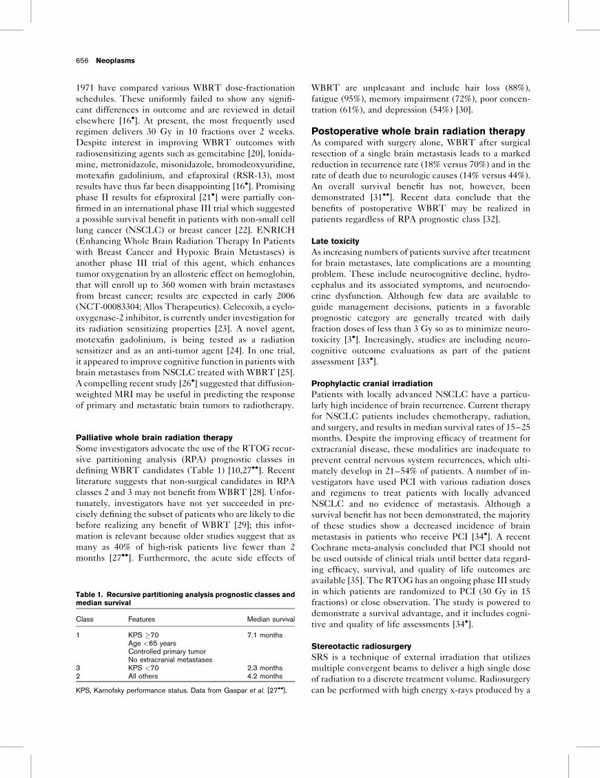
656 Neoplasms
Table 1. Recursive partitioning analysis prognostic classes and
median survival
Class Features Median survival
1 KPS �70 7.1 monthsAge <65 yearsControlled primary tumorNo extracranial metastases
3 KPS <70 2.3 months2 All others 4.2 months
KPS, Karnofsky performance status. Data from Gaspar et al. [27��].

linear accelerator, with gamma radiation (gamma knife),
and less frequently with charged particles such as protons
produced by cyclotrons. All of the stereotactic radiation
techniques produce a rapid fall-off of dose at the edge of
the target volume resulting in a clinically insignificant
radiation dose to normal non-target tissue. Because most
metastases are small, spherical, discrete, and sensitive to
single fraction radiotherapy, they serve as ideal targets for
stereotactic radiotherapy [36��]. Ample data have shown
that SRS is responsible for local tumor control rates on the
order of 73–94% [5]. Numerous recent analyses indicate
that SRS may effectively treat brain metastases [37–41].
Even neoplasms that are resistant to fractionated radia-
tion therapy such as melanoma, renal cell carcinoma, and
NSCLC, usually respond to single fraction SRS [42].
Complications of SRS include nausea, brain edema,
seizures, and later, radiation necrosis; these are reviewed
elsewhere [36��].
Stereotactic radiosurgery versus surgery
There is an emerging view that SRS may serve as an
alternative to surgical resection for small metastases not
producing mass effect. SRS can also be used to treat
lesions in the brainstem or eloquent areas with much
less risk than surgery. Additionally, because of the non-
invasive, outpatient nature of SRS, it is associated with
less morbidity and may be more cost-effective than
conventional surgery [43]. There may be a lower risk
of leptomeningeal disease dissemination in patients
with posterior fossa metastases treated with SRS [44].
As is the case for conventional surgery, careful selection
of patients is critical; patients without good prognostic
factors are unlikely to benefit [45]. Although the precise
role for SRS remains to be defined by a randomized,
prospective trial, many retrospective studies suggest that
SRS outcomes for appropriately selected patients are
equivalent to those achieved with conventional surgery
[36��], and long-term survival among patients with good
prognostic factors is possible [37]. The most recent of
these reviewed the Mayo Clinic experience of 74 patients
with solitary brain metastases treated with surgery com-
pared with 23 patients treated with SRS. Outcomes were
similar with 1-year survival of 56% for the SRS group and
62% for the surgery group (P ¼ 0.15). Local control was
significantly better in the radiosurgery group (no recur-
rences compared with 58% in the surgery group) [46�]. A
prospective trial comparing SRS to surgery is much
needed. Unfortunately, previous attempts at such a study
have been unsuccessful due to poor accrual, primarily as a
result of patient or physician preference for one of the
treatment modalities.
Stereotactic radiosurgery with or without whole brain
radiation therapy
The role of WBRT in patients treated with SRS is
controversial, especially for patients with relatively radio-
resistant tumors. While recent data established that the
addition of WBRT to SRS significantly improves local
tumor control [47�], an overall survival benefit has not
been demonstrated [48�]. Patients report that the addi-
tion of WBRT causes more memory impairment, depres-
sion, poor concentration, and hair loss than SRS alone
[30]. Much needed randomized studies comparing SRS
and the combination of SRS and WBRT are underway to
assess survival, quality of life, and cost-effectiveness in
patients with newly diagnosed brain metastases.
Whole brain radiation therapy with or without
stereotactic radiosurgery
In 2004, Andrews et al. [49��] published the first random-
ized trial comparing SRS combined with WBRT to
WBRT alone (RTOG 95-08). For patients with a single
unresectable metastasis, SRS was found by intention-to-
treat analysis to confer a significant survival benefit (mean
survival 6.5 months versus 4.9 months; P ¼ 0.039). Addi-
tionally, the SRS group showed a significant improve-
ment in KPS and decreased steroid use at 6 months.
There was no significant survival benefit for patients with
multiple metastases. No difference in efficacy was
observed between linear accelerator or gamma knife
SRS. Trials are needed to further assess the role of
SRS for patients with multiple metastases. A study is
currently underway to investigate the value of combin-
ing temozolomide or the epidermal growth factor
receptor inhibitor, gefitinib with SRS to improve its
efficacy (RTOG 0320).
Interstitial brachytherapy
This technique involves the implantation of radioactive
nuclides into the wall of the surgical cavity to deliver a
dose of radiation to the residual tumor while limiting
radiation exposure to the surrounding brain. Thus far,
brachytherapy remains an experimental treatment
modality. GliaSite (Proxima Therapeutics, Alpharetta,
Georgia, USA) is a novel brachytherapy system cur-
rently under investigation. An inflatable balloon cathe-
ter is placed in a resection cavity following debulking or
resection of a brain tumor. The balloon is filled with an
aqueous solution of 125I that delivers a low, continuous
dose of radiation to the margins of the resection cavity.
Preliminary results for primary brain tumors are promis-
ing [50]. Studies of GliaSite for the treatment of meta-
stases are ongoing.
ChemotherapyChemotherapy has generally been used in patients who
have failed other treatment modalities. Although che-
motherapy may occasionally be useful in patients with
chemosensitive tumors such as small cell lung cancer,
choriocarcinoma, and breast cancer, the results of most
chemotherapy trials have been disappointing. The
primary reasons for chemotherapeutic failure include
Brain metastases Norden et al. 657

inability of the agent to cross the blood–brain barrier
(BBB) and insensitivity of the tumor to the particular
agent. Some of the new chemotherapeutic agents that
cross the BBB hold promise as treatment options for brain
metastases. Preliminary studies suggest that topotecan,
an inhibitor of topoisomerase I that crosses the BBB, may
effectively treat brain metastases from small cell lung and
breast cancer [51]. Temozolomide, an oral alkylating
agent approved for use in the treatment of malignant
gliomas, is well-tolerated and also crosses the BBB. It has
been studied in phase II trials and appears to have modest
activity against brain metastases from lung cancer, breast
cancer, and melanoma [52�,53].
Experimental approaches
An area of intense research involves targeted molecular
agents. A promising recent finding is that gefitinib has
activity against brain metastases from NSCLC [54��,55].
Gefitinib is an oral tyrosine kinase inhibitor of the
epidermal growth factor receptor, which is effective
against a subset of NSCLC. In a prospective trial, gefi-
tinib controlled brain metastases in 27% of patients, with
a median duration of 4 months (Fig. 1) [54��]. Additional
molecular agents in development are reviewed elsewhere
[56]. One provocative idea currently being studied in
mice with intracerebral human breast tumors involves the
intracarotid administration of a genetically engineered
oncolytic virus [57�]. This approach has produced a
survival benefit in mice and warrants additional investi-
gation.
GuidelinesCurrent guidelines published by the National Compre-
hensive Cancer Network recommend management
similar to that which has been detailed herein [58].
For patients with one to three metastatic lesions on brain
658 Neoplasms
Figure 1. Radiological response of metastatic lung cancer to brain with high dose gefitinib
A 54-year-old male with non-small celllung cancer and multiple small brainmetastases (arrows), whichprogressed through whole brainradiation therapy (August 2004, toprow). The patient was then treated withhigh dose gefitinib with reduction insize of parenchymal nodular lesions(October 2004, bottom row).

MRI, aggressive management is generally recommended
so long as systemic disease is limited or controllable.
Options include resection and SRS. Either resection or
SRS may be followed by WBRT in an attempt to prevent
local recurrence. If the lesions are deemed unresectable,
WBRT or SRS should be considered. In cases of highly
radiosensitive tumors such as small cell lung cancer or
lymphoma, or when there is disseminated systemic dis-
ease with poor treatment options, WBRT is recom-
mended. In all cases, surgery should be considered for
relief of symptomatic mass effect or hydrocephalus.
When brain metastases initially present as more than
three lesions, surgery is again recommended if a diagnosis
has not been established or if there is symptomatic
mass effect. Surgery should be followed by WBRT with
or without SRS. The same treatment regimen is recom-
mended for patients with multiple metastases who do
not have surgery. After treatment for brain metastases,
patients should be followed with MRI approximately
every 3 months for 1 year and then as clinically indi-
cated. Local recurrences may be treated with surgery,
SRS or occasionally chemotherapy. In cases of distant
recurrence, multiple treatment modalities can be con-
sidered.
PrognosisThe median survival of patients with untreated brain
metastases is approximately 1 month. The addition
of steroids increases survival to 2 months, while WBRT
further improves survival to 3–6 months [5]. Patients
with single brain metastases and limited extracranial
disease who are treated with surgery and WBRT have
a median survival of approximately 10–16 months
[8��,13]. Prognostic data for patients treated with SRS
or novel chemotherapy is not yet available. In review-
ing prognostic information for various treatment modal-
ities, though, one is clearly struck by the degree to
which interventions developed in recent decades have
had an impact on the survival of patients with brain
metastases.
ConclusionIn the last decade, the emergence of SRS as a primary
treatment modality for patients with good prognostic
factors and one or a few small metastases has been a
significant development. Additional data will be necess-
ary to validate the view that SRS is a viable alternative
to surgery in certain situations. Future investigations
should address quality of life and neurocognitive out-
comes in addition to traditional outcome measures such
as recurrence and survival rates. Promising therapies
currently under investigation include chemotherapeu-
tics that effectively cross the BBB, targeted mole-
cular agents, radiation sensitizing agents, and oncolytic
viruses.
References and recommended readingPapers of particular interest, published within the annual period of review, havebeen highlighted as:� of special interest�� of outstanding interestAdditional references related to this topic can also be found in the CurrentWorld Literature section in this issue (pp. 756–757).
1 Cancer facts and figures. Statistics for 2005 [online resource]. Atlanta:American Cancer Society; 2005. http://www.cancer.org/downloads/STT/CAFF2005f4PWSecured.pdf. [Accessed 5 January 2005]
2
�Lassman AB, DeAngelis LM. Brain metastases. Neurol Clin 2003; 21:1–23.
This comprehensive review addresses all aspects of brain metastases with aparticular focus on therapy.
3
�Kaal EC, Niel CG, Vecht CJ. Therapeutic management of brain metastasis.Lancet Neurol 2005; 4:289–298.
The authors of this comprehensive review propose an algorithm for management ofpatients with brain metastases.
4
�Bradley KA, Mehta MP. Management of brain metastases. Semin Oncol 2004;31:693–701.
This is an excellent comprehensive review of current treatment for brain metastases.
5 Plotkin SR, Wen PY. Brain metastases. In: Samuels MA, Feske SK, editors.Office practice of neurology, Edition 2. Philadelphia: Churchill Livingstone;2003. pp. 1101–1106.
6
�Shaffrey ME, Mut M, Asher AL, et al. Brain metastases. Curr Probl Surg 2004;41:665–741.
This is an excellent and exhaustive review of the subject with a focus on surgicalmanagement.
7
�Tosoni A, Ermani M, Brandes AA. The pathogenesis and treatment of brainmetastases: a comprehensive review. Crit Rev Oncol Hematol 2004;52:199–215.
This is a detailed review that critically evaluates recent literature.
8
��Patchell RA, Tibbs PA, Walsh JW, et al. A randomized trial of surgery inthe treatment of single metastases to the brain. N Engl J Med 1990; 322:494–500.
This randomized trial was the first to demonstrate improved outcomes in patientswith single brain metastases treated with surgery and radiation as opposed toradiation alone.
9
�El Kamar FG, Posner JB. Brain metastases. Semin Neurol 2004; 24:347–362.
This is an outstanding review of supportive care for brain metastases.
10 Gaspar LE, Scott C, Murray K, Curran W. Validation of the RTOG recursivepartitioning analysis (RPA) classification for brain metastases. Int J RadiatOncol Biol Phys 2000; 47:1001–1006.
11
�Black PM, Johnson MD. Surgical resection for patients with solid brainmetastases: current status. J Neurooncol 2004; 69:119–124.
This is a detailed review of the evidence regarding surgical management of brainmetastases.
12
�Barker FG 2nd. Craniotomy for the resection of metastatic brain tumors in theU.S., 1988–2000: decreasing mortality and the effect of provider caseload.Cancer 2004; 100:999–1007.
This is an interesting retrospective analysis that considers the role of hospital andsurgeon volume in determining treatment outcomes.
13 Vecht CJ, Haaxma-Reiche H, Noordijk EM, et al. Treatment of single brainmetastasis: radiotherapy alone or combined with neurosurgery? Ann Neurol1993; 33:583–590.
14 Mintz AP, Cairncross JG. Treatment of a single brain metastasis: therole of radiation following surgical resection. JAMA 1998; 280:1527–1529.
15
�Hart MG, Grant R, Walker M, Dickinson H. Surgical resection and wholebrain radiation therapy versus whole brain radiation therapy alone forsingle brain metastases. Cochrane Database Syst Rev 2005; (1):CD003292.
This well-executed meta-analysis considers whether the combination of surgeryand WBRT is more effective than WBRT alone.
16
�Tsao MN, Lloyd NS, Wong RK, et al. Radiotherapeutic management of brainmetastases: A systematic review and meta-analysis. Cancer Treat Rev 2005;31:256–273.
This is a thorough review of the literature concerning WBRT and SRS in themanagement of brain metastases.
17 Paek SH, Audu PB, Sperling MR, et al. Reevaluation of surgery for thetreatment of brain metastases: review of 208 patients with single or multiplebrain metastases treated at one institution with modern neurosurgical tech-niques. Neurosurgery 2005; 56:1021–1034.
Brain metastases Norden et al. 659

18 Stark AM, Tscheslog H, Buhl R, et al. Surgical treatment for brain metastases:prognostic factors and survival in 177 patients. Neurosurg Rev 2005;28:115–119.
19 Horton J, Baxter DH, Olson KB. The management of metastases to the brainby irradiation and corticosteroids. Am J Roentgenol Radium Ther Nucl Med1971; 111:334–336.
20 Maraveyas A, Sgouros J, Upadhyay S, et al. Gemcitabine twice weekly as aradiosensitiser for the treatment of brain metastases in patients with carci-noma: a phase I study. Br J Cancer 2005; 92:815–819.
21
�Shaw E, Scott C, Suh J, et al. RSR13 plus cranial radiation therapy in patientswith brain metastases: comparison with the Radiation Therapy OncologyGroup Recursive Partitioning Analysis Brain Metastases Database. J ClinOncol 2003; 21:2364–2371.
This phase II trial was one of the first to evaluate the safety and efficacy ofefaproxiral (RSR-13) as a radiation sensitizing agent for patients with brainmetastases undergoing WBRT.
22 Suh JH, Stea B, Nabid A, et al. Standard whole brain radiation therapy plussupplemental oxygen with or without efaproxiral (EFAPROXYNTM) in patientswith brain metastases: updated survival results of the randomized REACH(RT-009) study. Ann Oncol 2004; 15:iii207.
23 Cerchietti LC, Bonomi MR, Navigante AH, et al. Phase I/II study of selectivecyclooxygenase-2 inhibitor celecoxib as a radiation sensitizer in patients withunresectable brain metastases. J Neurooncol 2005; 71:73–81.
24 Evens AM. Motexafin gadolinium: a redox-active tumor selective agent for thetreatment of cancer. Curr Opin Oncol 2004; 16:576–580.
25 Meyers CA, Smith JA, Bezjak A, et al. Neurocognitive function and progres-sion in patients with brain metastases treated with whole-brain radiation andmotexafin gadolinium: results of a randomized phase III trial. J Clin Oncol2004; 22:157–165.
26
�Mardor Y, Roth Y, Ochershvilli A, et al. Pretreatment prediction of braintumors’ response to radiation therapy using high b-value diffusion-weightedMRI. Neoplasia 2004; 6:136–142.
This interesting study considers whether MRI might be useful in predicting a braintumor’s response to radiation therapy.
27
��Gaspar L, Scott C, Rotman M, et al. Recursive partitioning analysis (RPA) ofprognostic factors in three Radiation Therapy Oncology Group (RTOG) brainmetastases trials. Int J Radiat Oncol Biol Phys 1997; 37:745–751.
This large retrospective analysis of patients with brain metastases used recur-sive partitioning analysis to identify three prognostic groups; this classificationscheme has allowed subsequent studies to focus on more homogeneous popula-tions.
28 Morris SL, Low SH, A’Hern RP, et al. A prognostic index that predictsoutcome following palliative whole brain radiotherapy for patients with meta-static malignant melanoma. Br J Cancer 2004; 91:829–833.
29 Lock M, Chow E, Pond GR, et al. Prognostic factors in brain metastases: canwe determine patients who do not benefit from whole-brain radiotherapy? ClinOncol (R Coll Radiol) 2004; 16:332–338.
30 Kondziolka D, Niranjan A, Flickinger JC, Lunsford LD. Radiosurgery withor without whole-brain radiotherapy for brain metastases: the patients’perspective regarding complications. Am J Clin Oncol 2005; 28:173–179.
31
��Patchell RA, Tibbs PA, Regine WF, et al. Postoperative radiotherapy in thetreatment of single metastases to the brain: a randomized trial. JAMA 1998;280:1485–1489.
This randomized trial convincingly demonstrates that the use of postoperativeWBRT decreases the risks of recurrence in the brain, death due to neurologicalcauses as compared to surgery alone.
32 Regine WF, Rogozinska A, Kryscio RJ, et al. Recursive partitioning analysisclassifications I and II: applicability evaluated in a randomized trial for resectedsingle brain metastases. Am J Clin Oncol 2004; 27:505–509.
33
�Regine WF, Schmitt FA, Scott CB, et al. Feasibility of neurocognitive outcomeevaluations in patients with brain metastases in a multi-institutional coopera-tive group setting: results of Radiation Therapy Oncology Group trialBR-0018. Int J Radiat Oncol Biol Phys 2004; 58:1346–1352.
This important study demonstrated that neurocognitive evaluations of patients withbrain metastases may be incorporated into large multicenter trials.
34
�Gore E, Choy H. Non-small cell lung cancer and central nervous systemmetastases: should we be using prophylactic cranial irradiation? SeminRadiat Oncol 2004; 14:292–297.
This is an excellent comprehensive review of the evidence for and againstprophylactic cranial irradiation in patients with locally advanced NSCLC.
35 Lester JF, Macbeth FR, Coles B. Prophylactic cranial irradiation for preventingbrain metastases in patients undergoing radical treatment for non-small-celllung cancer: A Cochrane review. Int J Radiat Oncol Biol Phys 2005; 23 May2005 [Epub ahead of print].
36
��Warnick RE, Darakchiev BJ, Breneman JC. Stereotactic radiosurgery forpatients with solid brain metastases: current status. J Neurooncol 2004;69:125–137.
This is a thorough current review of SRS for patients with brain metastases.
37 Jagannathan J, Petit JH, Balsara K, et al. Long-term survival after gamma kniferadiosurgery for primary and metastatic brain tumors. Am J Clin Oncol 2004;27:441–444.
38 Radbill AE, Fiveash JF, Falkenberg ET, et al. Initial treatment of melanoma brainmetastases using gamma knife radiosurgery: an evaluation of efficacy andtoxicity. Cancer 2004; 101:825–833.
39 Muacevic A, Kreth FW, Tonn JC, Wowra B. Stereotactic radiosurgery formultiple brain metastases from breast carcinoma. Cancer 2004; 100:1705–1711.
40 Koc M, McGregor J, Grecula J, et al. Gamma knife radiosurgery for intracranialmetastatic melanoma: an analysis of survival and prognostic factors.J Neurooncol 2005; 71:307–313.
41 Selek U, Chang EL, Hassenbusch SJ 3rd, et al. Stereotactic radiosurgicaltreatment in 103 patients for 153 cerebral melanoma metastases. Int J RadiatOncol Biol Phys 2004; 59:1097–1106.
42 Gerosa M, Nicolato A, Foroni R, et al. Regional treatment of metastasis: role ofradiosurgery in brain metastases: gamma knife radiosurgery. Ann Oncol2004; 15 (Suppl 4):iv113–iv117.
43 Mehta M, Noyes W, Craig B, et al. A cost-effectiveness and cost-utilityanalysis of radiosurgery vs. resection for single-brain metastases. Int J RadiatOncol Biol Phys 1997; 39:445–454.
44 Siomin VE, Vogelbaum MA, Kanner AA, et al. Posterior fossa metastases: riskof leptomeningeal disease when treated with stereotactic radiosurgery com-pared to surgery. J Neurooncol 2004; 67:115–121.
45 Lorenzoni J, Devriendt D, Massager N, et al. Radiosurgery for treatment ofbrain metastases: estimation of patient eligibility using three stratificationsystems. Int J Radiat Oncol Biol Phys 2004; 60:218–224.
46
�O’Neill BP, Iturria NJ, Link MJ, et al. A comparison of surgical resection andstereotactic radiosurgery in the treatment of solitary brain metastases. Int JRadiat Oncol Biol Phys 2003; 55:1169–1176.
This retrospective review compares outcomes of brain metastasis patients treatedwith surgery or SRS. Local tumor control is significantly better in the SRS group,but survival is not significantly different.
47
�Shehata MK, Young B, Reid B, et al. Stereotatic radiosurgery of 468 brainmetastases < or ¼2 cm: implications for SRS dose and whole brain radiationtherapy. Int J Radiat Oncol Biol Phys 2004; 59:87–93.
This excellent retrospective review evaluates the role of WBRT in patients whoreceive SRS for brain metastasis. Patients who receive both treatments havesuperior local tumor control rates to patients who receive SRS alone.
48
�Sneed PK, Suh JH, Goetsch SJ, et al. A multi-institutional review of radio-surgery alone vs. radiosurgery with whole brain radiotherapy as the initialmanagement of brain metastases. Int J Radiat Oncol Biol Phys 2002;53:519–526.
This large retrospective review demonstrates that post-radiosurgical WBRT doesnot confer a survival advantage as compared to SRS alone.
49
��Andrews DW, Scott CB, Sperduto PW, et al. Whole brain radiation therapywith or without stereotactic radiosurgery boost for patients with one to threebrain metastases: phase III results of the RTOG 9508 randomised trial. Lancet2004; 363:1665–1672.
This randomized trial was the first to demonstrate a survival advantage for patientswith a single unresectable brain metastasis treated with SRS and WBRT ascompared to WBRT alone.
50 Chan TA, Weingart JD, Parisi M, et al. Treatment of recurrent glioblastomamultiforme with GliaSite brachytherapy. Int J Radiat Oncol Biol Phys 2005;62:1133–1139.
51 Wong ET, Berkenblit A. The role of topotecan in the treatment of brainmetastases. Oncologist 2004; 9:68–79.
52
�Agarwala SS, Kirkwood JM, Gore M, et al. Temozolomide for the treatment ofbrain metastases associated with metastatic melanoma: a phase II study.J Clin Oncol 2004; 22:2101–2107.
This phase II trial demonstrates that temozolomide is active against brain meta-stases from melanoma.
53 Christodoulou C, Bafaloukos D, Linardou H, et al. Temozolomide (TMZ)combined with cisplatin (CDDP) in patients with brain metastases from solidtumors: a Hellenic Cooperative Oncology Group (HeCOG) Phase II study.J Neurooncol 2005; 71:61–65.
54
��Ceresoli GL, Cappuzzo F, Gregorc V, et al. Gefitinib in patients with brainmetastases from non-small-cell lung cancer: a prospective trial. Ann Oncol2004; 15:1042–1047.
This small prospective study of gefitinib for the treatment of NSCLC brainmetastases demonstrated that gefitinib can control metastatic disease in nearlyone-third of patients.
660 Neoplasms

55 Roggero E, Giancarla B, Antonella P, Augusto P. Gefitinib (‘Iressa’, ZD1839)is active against brain metastases in a 77 year old patient. J Neurooncol 2005;71:277–280.
56 Hsu SH, Yung W-.A. Chemotherapy for brain metastases. In: Sawaya R,editor. Intracranial metastases. Malden: Blackwell Futura; 2004. pp. 183–195.
57
�Liu R, Martuza RL, Rabkin SD. Intracarotid delivery of oncolytic HSV vectorG47Delta to metastatic breast cancer in the brain. Gene Ther 2005; 12:647–654.
This intriguing study of mice with intracerebral human breast tumors demonstratesimproved survival after treatment with a genetically engineered oncolytic virus.
58 Brem SS, DeAngelis LM, Maor MH, et al. Central nervous system cancers.Clinical practice guidelines in oncology version 1. Jenkintown: NationalComprehensive Cancer Network; 2005.
Brain metastases Norden et al. 661