Embed Size (px)
Citation preview
Tumours ..Retinal and optic nerve head tumours …
RETINOBLASTOMA
It is the most common PRIMARY intraocular malignancy of childhood , even so it is rare and occurring about 1:20 000
live births and accounts for 3% of all childhood cancers .
Genetics :
• Retinoblastoma results from malignant transformation of primitive retinal cells BEFORE final differentiation .
• Because these cells disappear within the first few years of life , the tumour is rarely seen after 3 years age .
• Retinoblastoma may be heritable or non-heritable .•
Heritable retinoblastoma ( germline )
- Accounts for 40% .
- Only one allele of RPE1(tumour supressor gene) has mutated in all body cells .
- when a further mutogenic event (second hit )affects the 2nd allele , the cell undergoes malignant transformation .
-Because all retinal precursor cells contain the initial mutation ,these children develop bilateral and multifocal tumours .
-Familial cases also carry a predisposition to non ocular cancers most common pinealoblastoma (trilateral retinoblastoma ) and osteosarcoma
Non heritable retinoblastoma (somatic ) :
- Accounts for 60% .
- Unilateral .
- Not transmissible.
- Does not predispose the patient to increased risk of second non ocular cancers .
- 85% of pts with unilateral retinoblastoma fall into this category .
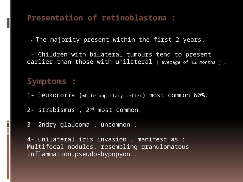
Presentation of retinoblastoma :
- The majority present within the first 2 years.
- Children with bilateral tumours tend to present earlier than those with unilateral ( average of 12 months ) .
Symptoms :
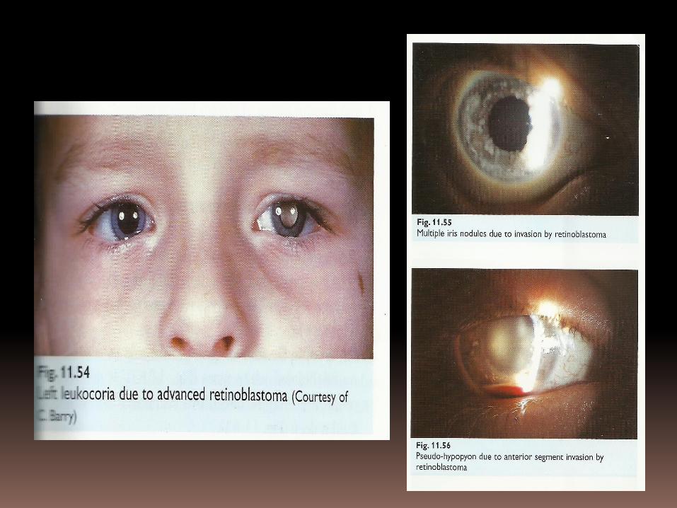
1- leukocoria (white pupillary reflex) most common 60%.
2- strabismus , 2nd most common.
3- 2ndry glaucoma , uncommon .
4- unilateral iris invasion , manifest as :Multifocal nodules, resembling granulomatous inflammation,pseudo-hypopyon .
Therefore, it’s important to consider retinoblastoma in the differential diagnosis of unusual chronic uveitis in children .
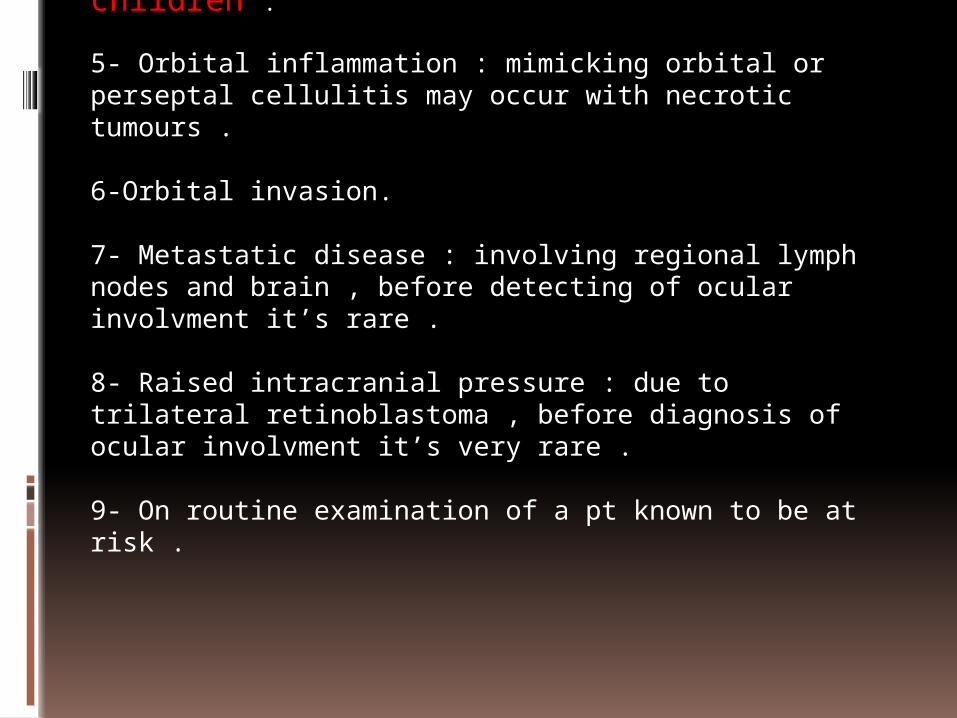
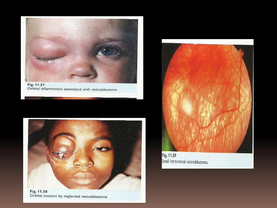
5- Orbital inflammation : mimicking orbital or perseptal cellulitis may occur with necrotic tumours .
6-Orbital invasion.
7- Metastatic disease : involving regional lymph nodes and brain , before detecting of ocular involvment it’s rare .
8- Raised intracranial pressure : due to trilateral retinoblastoma , before diagnosis of ocular involvment it’s very rare . 9- On routine examination of a pt known to be at risk .
Signs :
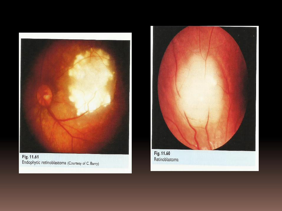
1- An early intraretinal tumour is a placoid white lesion .
2- An endopathic tumour grows inwards TOWARDS the vitreous , projectin from the retinal surface as a white cottage cheese-like mass, with surface blood vessels.
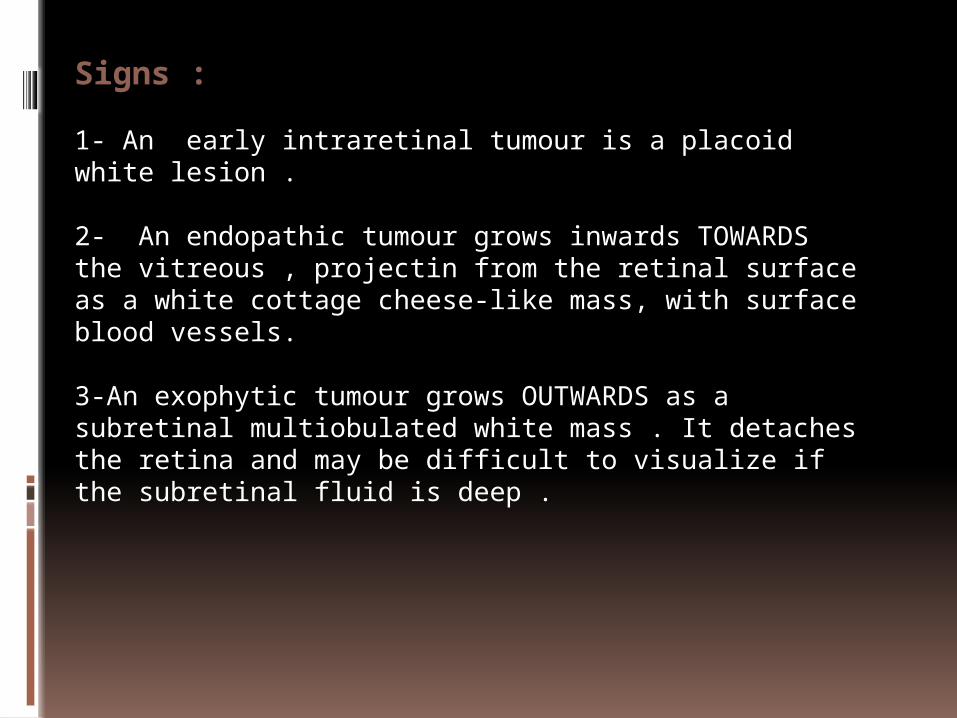
3-An exophytic tumour grows OUTWARDS as a subretinal multiobulated white mass . It detaches the retina and may be difficult to visualize if the subretinal fluid is deep .
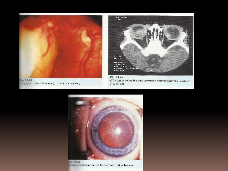
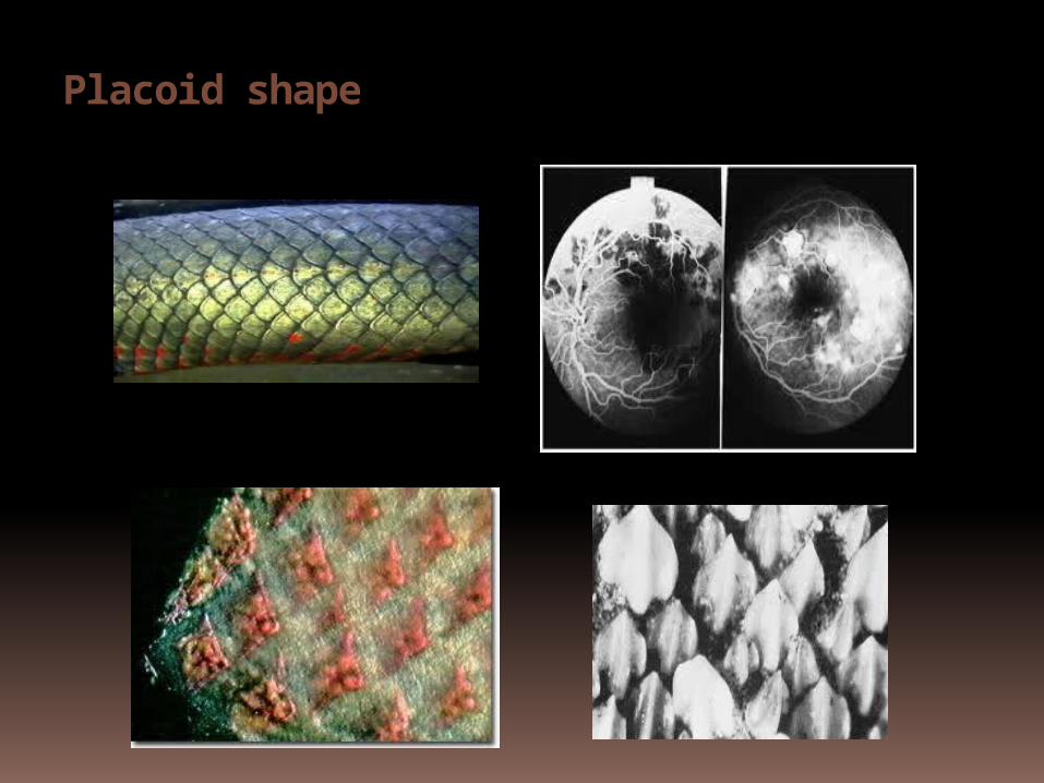
Placoid shape
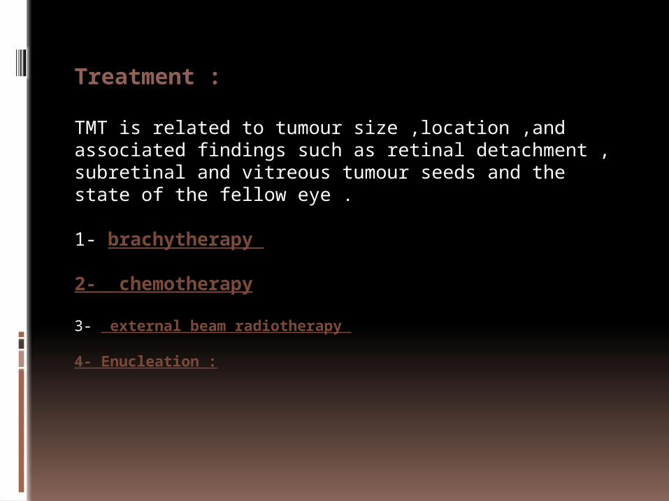
Treatment :
TMT is related to tumour size ,location ,and associated findings such as retinal detachment , subretinal and vitreous tumour seeds and the state of the fellow eye .
1- brachytherapy
2- chemotherapy
3- external beam radiotherapy
4- Enucleation :
Prognostic factors :
The overall related mortality is 2-5% and is related to the follwoings :
1- tumour size and location small posterior tumours do best but there is no significant difference between endophytic and exophytic type .
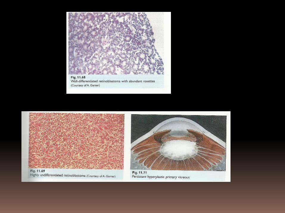
2- cellular differentiation : the mortality rate of pts whose tumours have abundant rosettes much less than in those with highly undifferentiated tumours .
3- optic nerve involvement : beyond the point of surgical transection is associated with high mortality .
4- invasion of the choroid or vortex veins facilitates haematogenous spread and therefore of adverse prognostic significance .
5- extrascleral spread carries a grave prognosis .