Embed Size (px)
Citation preview
Turkish Society of Physiological Sciences
42nd National Physiology Congress
September 2016 • Volume 218 • Supplement 709
Turkish Society of Physiological Sciences
42nd National Physiology Congress
05-08 September 2016
Düzce University, Turkey
Conferences ......................................................................................................................1
Symposia ......................................................................................................................4
Panels ......................................................................................................................7
Oral Communica>ons ......................................................................................................................8
Poster Communica>ons ....................................................................................................................26
Turkish Society of Physiological Sciences
42nd National Physiology Congress
05-08 September 2016
Düzce University, Convention Center, Düzce - TURKEY
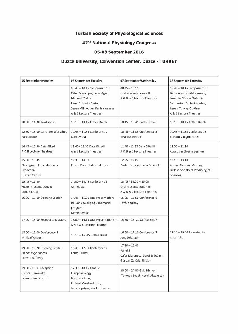
05 September Monday 06 September Tuesday 07 September Wednesday 08 September Thursday
08.45 – 10.15 Symposium 1:
Cafer Marangoz, Erdal Ağar,
Mehmet Yıldırım
Panel 1: Narin Derin,
Sezen Milli Avtan, Fa>h Karaaslan
A & B Lecture Theatres
08.45 – 10.15
Oral Presenta>ons – II
A & B & C Lecture Theatres
08.45 – 10.15 Symposium 2:
Deniz Atasoy, Bilal Kerman,
Yasemin Gürsoy Özdemir
Symposium 3: Sadi Kurdak,
Kerem Tuncay Özgünen
A & B Lecture Theatres
10.00 – 14.30 Workshops 10.15 – 10.45 Coffee Break 10.15 – 10.45 Coffee Break 10.15 – 10.45 Coffee Break
12.30 – 13.00 Lunch for Workshop
Par>cipants
10.45 – 11.35 Conference 2
Cenk Ayata
10.45 – 11.35 Conference 5
(Markus Hecker)
10.45 – 11.35 Conference 8
Richard Vaughn-Jones
14.45 – 15.30 Data Blitz-I
A & B Lecture Theatres
11.40 - 12.30 Data Blitz-II
A & B Lecture Theatres
11.40 - 12.25 Data Blitz-III
A & B & C Lecture Theatres
11.35 – 12.10
Awards & Closing Session
15.30 – 15.45
Photograph Presenta>on &
Exhibi>on
Gürkan Öztürk
12.30 – 14.00
Poster Presenta>ons & Lunch
12.25 - 13.45
Poster Presenta>ons & Lunch
12.10 – 13.10
Annual General Mee>ng
Turkish Society of Physiological
Sciences
15.45 – 16.30
Poster Presenta>ons &
Coffee Break
14.00 – 14.45 Conference 3
Ahmet Gül
13.45 / 14.00 – 15.00
Oral Presenta>ons – III
A & B & C Lecture Theatres
13.10 – 19.00 Excursion to
waterfalls
16.30 – 17.00 Opening Session 14.45 – 15.00 Oral Presenta>ons
Dr. Banu Ocakçıoğlu memorial
program
Me>n Baştuğ
15.05 – 15.50 Conference 6
Tayfun Uzbay
17.00 – 18.00 Respect to Masters 15.00 – 16.15 Oral Presenta>ons – I
A & B & C Lecture Theatres
15.50 – 16. 20 Coffee Break
18.00 – 19.00 Conference 1
M. Gazi Yaşargil16.15 – 16. 45 Coffee Break
16.20 – 17.10 Conference 7
Jens Leipziger
19.00 – 19.20 Opening Resital
Piano: Ayşe Kaptan
Flute: Eda Özdiş
16.45 – 17.30 Conference 4
Kemal Türker
17.10 – 18.40
Panel 3
Cafer Marangoz, Şeref Erdoğan,
Gürkan Öztürk, Elif Şen
19.30 - 21.00 Recep>on
(Düzce University,
Conven>on Center)
17.30 – 18.15 Panel 2:
Europhysiology
Bayram Yılmaz,
Richard Vaughn-Jones,
Jens Leipziger, Markus Hecker
20.00 – 24.00 Gala Dinner
(Turkuaz Beach Hotel, Akçakoca)
Turkish Society of Physiological Sciences
42nd National Physiology Congress
05-08 September 2016
Düzce University, Turkey
All abstracts have been reviewed by the following list of referees.
Prof. Dr. Aysel Ağar
Prof. Dr. İnci Alican
Prof. Dr. Ahmet Ayar
Prof. Dr. Sami Aydoğan
Prof. Dr. Nure?n Aydoğdu
Prof. Dr. Mehmet Aydın
Prof. Dr. Şeref Erdoğan
Prof. Dr. Hasan Erdoğan
Prof. Dr. Filiz Basralı
Prof. Dr. Metehan Çiçek
Prof. Dr. Nazan Dolu
Prof. Dr. Nurcan Dursun
Prof. Dr. Nilüfer Erkasap
Prof. Dr. Numan Ermutlu
Prof. Dr. Ersin Fadıllıoğlu
Prof. Dr. Hakkı Gökbel
Prof. Dr. Ümmühan İşoğlu
Prof. Dr. Nevzat Kahveci
Prof. Dr. Sacit Karamürsel
Prof. Dr. Muammer Kayatekin
Prof. Dr. Haluk Keleş>mur
Prof. Dr. Naim Khan
Prof. Dr. Ertuğrul Kılıç
Prof. Dr. Sadi Sanlı Kurdak
Prof. Dr. Selim Kutlu
Prof. Dr. Melek Bor Küçükatay
Prof. Dr. Nilsel Okudan
Prof. Dr. Güler Öztürk
Prof. Dr. Süleyman Sandal
Prof. Dr. Güldal Süyen
Prof. Dr. Gülderen Şahin
Prof. Dr. V. Nimet Uysal
Prof. Dr. Alex Verkhratsky
Prof. Dr. Mehmet Yıldırım
Prof. Dr. Bayram Yılmaz
Conferences
Conference 1The Impact of Scientific Evolution on the NeurosciencesM. Gazi YaşargilYeditepe University, Medical School Hospital, Department of BrainSurgery, İstanbul
It is not surprising that the invention of motor cars and airplaneswas not considered 2000 or even 200 years ago, due to the abs-cence of their socio-economic necessity. However, it is surprisingthat inventions for optic glasses and microscopes remained fairlydelayed, not only to advance scientific research, but also to helpthose people with inherited or acquired weakness of their visualfunctions.It is indeed astonishing that Ibn al-Haytham (Alhazen 915-1039), as-tronomer and mathematician, initially an engineer for hydraulics,who examined such phenomena as the rainbow, atmospheric ref-raction, mirror and lentiform crystals, and, notably, the anatomyand physiology of the eye, and wrote the famous book on optics(Kitab el-Menazir), which became widely influential in Europe, buthe failed to consider developing an optical aid. The first single lens“spectacles” were manufactured in Florence, Italy (1303-1313), butthe demand for them remained low until the invention of cheapbooks in the 17th century. The manufacture of sophisticated eyeglasses with individual diopters, the availability of contact lenses,and finally laser surgery of the cornea, required 600 years of dili-gent research.At the end of the 16th century and beginning of the17th century simple microscopes and telescopes were constructedin the Netherlands and Italy. Interestingly, periods of devastatingepidemics and several wars in Europe did not inhibit the incessantendeavors of manufacturers, glass grinders, mathematicians, as-tronomers, scientists, and even philosophers (Descartes, Spinoza).The upheaval of the industrial revolution in England between 1770and 1825 culminated in the production of achromatic microscopeswithout spherical aberration. The formulation of the equation “an-gular aperture” in 1880 by Ernst Abbe at Zeiss Company in Jena,Germany, allowed the serial production of high quality microsco-pes and telescopes. Advances in the entire field of neuroscien-ces, particularly in integral neurophysiology, EEG, MEG,angiography, CT, MRI and ultrasound technologies constitutetoday the prime and essential components of neuro-diagnosisand neuro-therapies. Within the past 50 years, neurosurgery hasexperienced a great number of technological innovations, suchas the introduction of stereotactic technology, operating micros-cope, bipolar coagulation technology, bipolar forceps, differentsizes and shapes of temporary and permanent vessel and ane-urysm clips, microsutures, ultrasound devices to identify thedeep localized lesions, ultrasound microflowmeter, ultrasonicsuction, ultrasonic microdrill apparatus, high speed drill appara-tus, flexible and rigid endoscopy technology, intraoperative sti-mulation and monitoring technology, intraoperative tractographyusing diffusion tensor mapping, and anisotropic diffusion weightedMRI which provides spatial and directional information of the neu-
ronal fibers, gradient respondent gamma surgery, and intensity mo-dulated radiation therapy. Modern operating rooms offer hithertounimagined technologies for the accurate targeting of the lesions,and their complete elimination without endangering adjacent nor-mal structures and functions. A great number of lesions localizedin so-called eloquent areas of the CNS, and defined as inoperable,are, at the present time, explored on a routine basis and success-fully treated in many centers. The above qualification of a fullyequipped department with a team of expert personnel representideal working conditions.One of the axioms guiding open societiesis the issue of ethics, desiring us to respect the rights, freedom anddignity of each individual and, in cases of illness, requiring us to ful-fill each patient’s claim to equal benefit from advances in medicineand surgery. Our professional goal is to further improve diagnosticand therapeutic procedures, and to ensure their availability globallyto each and every individual.
Conference 2Spreading Depolarization Waves in Injured BrainCenk AyataHarvard Medical School, Massachusetts General Hospital,Neurovascular Research Lab &Department of Neurology, Boston,MA, USA
Spreading depolarization are intense, slowly propagating waves ofneuronal and glial pandepolarization that emerge in an apparentlyspontaneous fashion in injured brain regardless of the mode of in-jury (ischemic, hemorrhagic, traumatic). Starting at the onset of in-jury, recurrent spreading depolarizations continue to occur formany days after injury with very high frequency. They worsen theoxygen supply-demand mismatch by increasing the metabolic de-mand and decreasing the blood flow. Therefore, spreading depola-rizations are considered a clinically relevant therapeutic target invarious types of brain injury. However, pharmacological inhibition ofspreading depolarizations has clinically proven to be difficult to ac-hieve without unwanted side effects. Instead, recent efforts haveturned to the origins of spreading depolarizations. If we develop abetter understanding of how and why spreading depolarizationsare triggered in the first place, we can perhaps implement measu-res in neurocritical care that might minimize their occurrence. Inmy talk, I will present data showing that oxygen supply-demandmismatch transients trigger spreading depolarizations in suscep-tible peri-infarct “hot zones.” Such supply-demand mismatch tran-sients can be precipitated by transient reduction in oxygen supply,such as during brief hypoxic or hypotensive events that are clini-cally exceedingly common, or by transient increase in oxygen de-mand, such as during functional activation, which is part of life. Inaddition, I will present data showing that brief intracranial pressurespikes, which are also very common in neurocritical care, are alsocapable of triggering spreading depolarizations. I will finish by sum-marizing the clinical implications of the data, and how it can be tes-ted clinically.
1
Turkish Society of Physiological Sciences42nd National Physiology Congress
Conference 3Asia Minor, Genes, and FeverAhmet Gülİstanbul University, İstanbul Faculty of Medicine, Department ofInternal Medicine, Division of Rheumatology, İstanbul, Turkey
Interaction with environment plays a critical role in shaping our ge-netic variations. Especially, polymorphisms in the genes involved ininflammatory response show regional variations and affect ouradaptation to the local environment by protecting integrity of ourbody. Fever is one of the most important components of inflam-matory response, and it is usually induced by pathogen- or danger-associated danger signals during microbial infections. However,mutations in some innate immune response genes are also res-ponsible for hereditary fever syndromes, which are characterizedby recurrent and self-limited inflammatory episodes without an ob-vious trigger. These conditions are now known as autoinflamma-torydisorders. Familial Mediterranean fever is the most commonform of autoinflammatory disorders. It is associated with autoso-mal recessively inherited MEFV gene variations, and prevalence ofthe carriers of these variations is quite high in Eastern Mediterra-nean populations, including those living in Asia Minor, reaching20%. MEFV gene exon 10 variations result in increased activationof interleukin-1 beta (IL-1β) in inflammatory cells by proteolyticcleavage of its precursor by caspase 1 enzyme following triggers.IL-1β is known as the strongest endogenous pyrogen and respon-sible for fever and inflammatory response. Increased prevalence ofMEFV mutation carriers in the Eastern Mediterranean may resultfrom its accumulation through ages because of a selective hete-rozygous advantage. Higher frequency of sickle cell anemia mutati-ons in certain regions of Africa is considered to be associated witha heterozygous advantage against malaria. Similarly, the MEFV va-riants might have provided a selective advantage to populations li-ving in Asia Minor and neighboring regions by protecting themagainst pandemic infections with high mortalitysuch as smallpoxthrough overcoming specific immune evasion ways of microbeswith a higher inflammasome activity and IL-1production.
Conference 4How Do We Masticate Without Eating Our Own Tongue?Kemal S. TürkerKoç University School of Medicine, Department of Physiology,İstanbul, Turkey
Mastication has two fundamental control mechanisms: the centralpattern generator (CPG) that sets the pattern of mastication by al-ternately sending action potentials to jaw opening and closingmuscles; and the peripheral controller that modulates the output ofthe CPG and jaw muscle motoneurons so that optimum bite forcesare developed between the jaws. The peripheral control mecha-nism includes the cutaneous and mucosal receptors that innervatethe lips and the oral mucosa, periodontal mechanoreceptors thatinnervate the support tissues of the tooth root, and muscle spindlesin the jaw muscles. These receptors monitor chewing forces and mo-dify the activity of muscles in the jaw, tongue and cheeks in order tofacilitate mastication and prevent damage to oral tissues. To inves-tigate their connections to motoneurons that innervate jaw muscles,we stimulate these receptors electrically and/or mechanically in con-senting adult volunteers. The responses of the jaw muscles to the sti-muli are recorded using intramuscular fine wire and surface
electromyography (EMG) electrodes. These studies contribute to abetter understanding of the neuronal circuitry of the masticatorysystem and provide a scientific baseline from where the neurophy-siological consequences of interventions that change the physicalrelationship of the masticatory elementscan be investigated.ReferencesLobbezoo F, Sowman PF, Türker KS (2009). Modulation of humanexteroceptive jaw reflexes during simulated mastication. Clin Neu-rophysiol 120(2):398-406.Türker KS (2002). Reflex control of human jaw muscles. Crit Rev OralBiol Med 13(1):85-104.Türker KS, Brinkworth, R.S.A., Abolfathi, P., Linke, I.R., Nazeran, H.(2004). A device for investigating neuromuscular control in thehuman masticatory system. J Neurosci Methods 136:141-149.
Conference 5Epigenetic Control of Endothelial Nitric Oxide Synthase Expressionand Its Role in Coronary Heart DiseaseMarkus HeckerDepartment of Cardiovascular Physiology, Heidelberg University,Germany
A single nucleotide polymorphism (SNP) within the promoter of theendothelial nitric oxide (NO) synthase (NOS3) gene (T-786C,rs2070744) adversely affects the response of endothelial cellsfromCC-genotypeindividuals to shear stressor the prototypic anti-type 1 T-helper (Th1) cell cytokine interleukin-10. Homozygosity forthe C-allele, whichoccurs in about 12% of Caucasians, is a strongpredictor for coronary heart disease (CHD), polymyalgia rheumat-ica or rheumatoid arthritis (RA).We have previously identified acompensatory mechanisminvolving manganese-dependent super-oxide dismutase that helps to maintain the bioavailability of en-dothelial cell-derived NO. With the shear stress-dependentincreased release of 15-deoxy-Δ12,14-prostaglandin J2 (15d-PGJ2)from CC- but not TT-genotype endothelial cells, we have charac-terised anothercompensatory mechanism that may not only sup-port the anti-inflammatory capacity of the CC-genotype endothelialcells but also act as a novel general defence mechanism againstchronic inflammation. Correlating plasma 15d-PGJ2 levels with theseverity of the disease in patients with CHD or RA support this no-tion.Moreover, data acquired quite recently point to a possible epige-netic control of NOS3 expression through chromatin remodellingthat may differ in at least two aspects between CC- and TT-geno-type endothelial cells. Having generated knock-in mice harbouringthe human C- or T-type NOS3 promoteron a disease-susceptible ge-netic background, we will verify that the T-786C SNP of the humanNOS3 gene, if present,boosts the development of arteriosclerosisand/or arthritis in these animals. The reason for this complex ex-perimental approach is that instead of the 5’-GGC(TŽC)GG-3’ motiffound in humans the murine Nos3promoter contains a 5’-GGCCAT-3’motif so that the suspected critical CpG-dinucleotide cannot form.Finally, we have begun to corroborate in an in vitro transmigrationmodel mimicking shear stress conditions at arteriosclerosispredilection sites that the T-786C SNP of the human NOS3 gene notonly differentially affects monocyte-endothelial cell but also Th1and Th17 cell-endothelial cell interaction, respectively, through 15d-PGJ2. In a nutshell, due to the acquired compensatory mechanismsthe formerly adverse SNP in the promoter of the human NOS3 genemay be viewed as a largely neutral mutation.
Turkish Society of Physiological Sciences42nd National Physiology Congress
2
3
Turkish Society of Physiological Sciences42nd National Physiology Congress
Conference 6Neuroplasticity in Substance AddictionTayfun UzbayÜsküdar University, Neuropsychopharmacology Application andResearch Center (NPARC), İstanbul, Turkey
Substance abuse and addiction is increasingly understood as a neu-robiological disorder where continuously abuse of addictive sub-stances corrupts the normal circuitry of rewarding and adaptivebehaviors generating fromadaptive changes in central nervous sys-tem (CNS) induced by these substances.Neuroplasticity can brieflybe defined as adaptive changes against internal and external stim-uli in the brain’s neurons, and structural and functional alterationsin synapses formed by these neurons.If the changes are not con-fined to a single neuron but reach the level of a synapse, the adap-tive response formed may also be called “synaptic plasticity” (1).Finally, neuroplasticity is a flexible re-organization, adaptation orremodeling of mammalian brain carried out by changes in synap-ticformation and elements. It is affected by endogenous, exogenousand environmental stressful factors. While normal synaptic plas-ticity is involved in several vital functions such as mind, memory,learning, psychomotor performance and healthy behaviors, abnor-mal neuroplasticity may cause some serious CNS disorders such asaddiction, depression, autism and schizophrenia. Long term poten-tiation (LTP) formation in neurons is necessary for learning and it isan adaptive response associated with synaptic plasticity. Becauselearning and memory is an important part of brain neuroplasticity,we can interpret the addiction as a negative neuroplasticity or anegative neuronal adaptation appearing under a heavy stress de-pending on toxic effects of continuously abused drugs in brain.In amolecular and cellular level, addiction is a negative neuroadaptationin some specific structures or pathways such as dopaminergic systemin brain (2). This statement may be related to neurodegeneration orgenerates unusual, mutant or zombie (terrorist) neurons or both ofthemin these specific brain regions (3). The process is directly gen-erated and/or supported by chronic use of addictive agents.A treat-ment that inhibits neurodegeneration andstimulates healthyneurogenesis in damaged areas, and/or prevents to generate thezombie/terrorist neurons or convertingthem to normal neuronsmayprovide a radical solution for this serious public health problem.
References1. Uzbay IT. A New Approach to Etiopathogenezis of Depression: Neu-
roplasticity (Editor) NOVA Publishers, New York, USA, 2011.2. O’Brian CP. Neuroplasticity in addictive disorders. Dialogues Clin
Neurosci 11:350-353, 2009.3. Uzbay İT. Madde Bağımlılığı. İstanbul Tıp Kitabevi, İstanbul, 2015.
Conference 7Extracellular ATP Signaling in Kidney Functions MAKE SENSEJens LeipzigerInstitute of Biomedicine, Physiology, Aarhus University, Denmark
I will take you along a trail of own discoveries that have outlined anauto- and paracrine signaling system that is necessary for properrenal function. This system uses extracellular ATP, which is released
from renal epithelia to then stimulate membrane receptors (puri-nergic P2X and P2Y). Multiple physiological stimuli exist that lead tocellular ATP release and these will be discussed. Most intriguingly,the flow of pre-urine along the renal tubule can be sensed by theprimary cilium. Its bending causes an increase of [Ca2+]i and activa-tes ATP secretion. Stimulation of purinergic receptors has prono-unced effects on the ability to transport ions and water along theentire renal tubule. These intra-renal purinergic signals work as en-dogenous “diuretics”, i.e. they favor the excretion of urine. Impor-tantly, the tubular lumen, i.e. the urinary space is an active signalingcompartment, from which renal functions are regulated. Eventu-ally, evidence accumulates indicating that aberrant activation ofintra-renal purinergic signals are danger signals for the activationof immune responses and are involved in renal damage, fibrosis andloss of organ function.
Conference 8Ca2+-H+ Coupling in the Heart: A Driver of Contractility,Arrhythmia and Nuclear SignallingRichard D Vaughan JonesBurdon Sanderson Cardiac Science Centre, Department of Physio-logy Anatomy & Genetics, Oxford, UK
H+ ions are universal products of metabolism. In myocytes of theheart, the intracellular H+ concentration ([H+]i) is kept close to 60nM(pHi 7.20) by means of sarcolemmal H+ extrusion on Na+-coupledtransporters such as Na/H exchange (NHE1). Extrusion is essentialbecause of the H+ ion’s high chemical reactivity, although significantfluctuations in [H+]ido occur physiologically, and during clinicalevents such as myocardial ischaemia/reperfusion. In the ventricu-lar myocyte, there is an intimate positive coupling between Ca2+ sig-nalling and [H+]I, which can help to protect contractility duringperiods of intracellular acidosis. If pronounced, acidosis can alsodrive intracellular Ca2+ overload, triggering pro-arrhythmogenic Ca2+
waves. I will describe how myocardial [H+]i is regulated, emphasisingthe importance of cytoplasmic histidyl dipeptides (HDPs) for diffu-sively shuttling H+ ions within individual cells. I will then focus onthree distinct mechanisms of myocardial Ca2+-H+ coupling. One mec-hanism, relies upon a rise of intracellular Na+, driven by [H+]i sti-mulation of NHE1. This rise modulates sarcoplasmic reticulum (SR)Ca2+-loading, thereby regulating Ca2+ signalling and the triggeringofCa2+ waves. A second mechanism relies upon the exchange ofH+for Ca2+ ions at intracellular buffers, particularly the HDPs. Thissecond mechanismmay be important for maintaining Ca2+-acti-vated processes in the face of competitive inhibition by H+ ions.A third mechanism is associated with the counter transport byHDPs of Ca2+ and H+ ions through nuclear pores, resulting in a co-upling between nuclear Ca2+ signalling and nuclear pH, a pheno-menon that may be important for modulating gene transcription.Overall, the combination of NHE1 activity andthe diffusive trans-port of intracellular Ca2+ and H+on HDPs, forms a fundamentalsystem in ventricular myocytes that controls the amplitude andspatial distribution of Ca2+ within the SR and cytoplasm, and helpsto determine the pH of the nucleus.I will speculate on how thissystem may become distorted in disease processes such as myo-cardial ischaemia and heart failure.
4
Symposia
Symposium 1: Current Studies in Experimental Epilepsy
S1.1 The Kindling Model of EpilepsyCafer MarangozRetired from the Department of Physiology, Medical Faculty,Ondokuz Mayis University, Samsun, Turkey
Epilepsy is a very important disease manifested by recurrent pa-roxysmal bursts of abnormal electrical brain activity. More than 50million people worldwide suffer from epilepsy, and about 30% ofthose affected have seizures that are resistant to treatment withthe currently available antiepileptic drugs. For development of newand more effective drugs, and the study of mechanisms of the epi-lepsies requires appropriate experimental models. A good modelfor epilepsy should have certain features. Kindling has been exten-sively studied as an epilepsy model of complex partial seizures withsecondary generalization. Kindling model of epilepsy iscreated bythe repeated subthreshold electrical stimuli or repeated subthres-hold chemical stimuli. The aim one of the studies in our laboratorywas to reveal the effects of nitrendipine, levetiracetam and pheno-barbital on kindling model. In order to induce kindling, three daysof the week (Mondays, Wednesdays and Fridays)35 mg/kg penty-lenetetrazole was administered intra¬peritoneally (i.p) until thea-nimals kindled. Electrocorticographic activities were obtained fromawake animals and their convulsive behaviors were classified. Nit-rendipine (5 mg/kg), 20 mg/kg levetiracetam and 10 mg/kg phe-nobarbital significantly suppressed spike frequency in fully kindledrats (p<0.05). Coadministrationof effective doses of 5 mg/kg nit-rendipine and 20 mg/kg levetiracetam reduced epileptiform acti-vityeffectively (p<0.05). Our aim in the second study was toinvestigate the effect of carbenoxolone, a gap junction blocker, onthe anticonvulsant effect of phenytoin in pentylenetetrazole kin-dled rats. Phenytoin decreased generalized seizure duration, totalspike number and seizure severity score (p<0.05). Carbenoxoloneand phenytoin have antiseizure effects in PTZ kindled rats. Therewas no significant difference between the carbenoxolone + pheny-toin combination and phenytoin in terms of generalized seizure du-ration,total spike number and seizure stage (p>0.05). Our thirdstudy showed a decrease in the CA1 hippocampal pyramidal neu-rons numberof kindled rats (p<0.05).
S1.2 The Effects of Cannabinoids on Epileptiform ActivityErdal AğarDepartment of Physiology Faculty of Medicine University ofOndokuz Mayis, Samsun, Turkey
Cannabinoids are a heterogeneous group of compounds, which canbe separated into three different groups: phytocannabinoids, en-dogenous compounds known as endocannabinoids and syntheticcompounds. Cannabinoids involve many physiological and patho-logical conditions in mammals. Cannabinoids exert protective ef-fects in different neurodegenerative disorders, including epilepsy.The cannabinoid system also plays a key role in regulating seizureactivity in brain through the activation of cannabinoid CB1 recep-tors.Epilepsies are the most prevalent neurological disorder; they
are characterized by recurrent, unprovoked seizures. Epilepsies arecomplex syndromes due to their multifactorial origins and manife-stations. An abundant series of cannabinoid agonists and antago-nists has been tested in in vivo and in vitro experimental models ofepileptic activity. Δ9tetrahydrocannabinoid (THC) andcannabimi-metic compounds are anticonvulsant in various experimental mo-dels of epilepsy. Cannabidiol (CBD) inhibited epileptiform activityin vitro and reduced the severity and lethality of seizures in a penty-lenetetrazole model of generalized seizures in vivo and tonic–clonicseizures in acute pilocarpine and penicillin models of temporal lobeand partial seizures. CBD, even at high doses, did not induce exci-tatory effects or convulsions in rats. Despite the extensive accountsof the antiepileptic effects of CBD, the mechanisms underlyingthese effects are not well understood. The CB1receptor antagonistAM251 disrupts the endocannabinoid tone by blocking CB1recep-tor activation, which caused development of status epilepticus inpopulations of epileptic neurons in the hippocampal neuronal cul-ture and penicilinin duced epilepsy models whereas the CB1 re-ceptor agonist, ACEA exhibits antiepileptic activity in the most ofexperimental model of epilepsy. There are a few reports on the cli-nical use of cannabinoid extracts as antiepileptics have been pub-lished; it is unclear whether cannabinoids might be efficacious andsafe in the treatment of epilepsy.
S1.3 The Effect of Adenosine on Epileptiform ActivityMehmet YıldırımDepartment of Medical Physiology, Faculty of Medicine, HealthSciences University,İstanbul, Turkey
Adenosine is an inhibitory neuromodulator that suppress excitatorysynaptic communication between neurons. Because it is not a clas-sical neurotransmitter, adenosine is neither stored in presynapticvesicles nor it affects only on synaptic area. It controls neuronal ac-tivity through activation of A1, A2A, A2B and A3 receptors. Adeno-sine and its receptors are involved in different diseases anddisorders such as hypoxia, ischemia, inflammation, pain, Parkinsonand epilepsy.Epilepsy is a major public health problem affecting atabout 1% of the population. At the basis of epileptic seizures, thereis a loss of balance in neuronal excitability. It has been reported thatmany neurotransmitters and neuromodulators can be associatedwith this change in the neuronal excitability. In this context, it hasbeen suggested that adenosine may act as an endogenous anti-convulsant. Many studies have reported a protective effect of ade-nosine against epileptic seizure and epileptiform activity. Forexamples, the daily systemic administration of adenosine preventsthe epileptic behavioral induced by pentylenetetrazole. Adenosinealso inhibits epileptiform activity induced by bicuculline in the rathippocampus. In our laboratory, we have compared the effects offocal and intracerebroventricular (i.c.v.) administered adenosine (1,10 and 100 μg/rat) on the penicillin-induced epileptiform activity inrats. Our findings suggest that focal adenosine is more effectivethan i.c.v. adenosine. In another study, we also found that adeno-sine (100 μg/rat, i.c.) and nitric oxide may decrease penicillin-indu-ced epileptiform activity in rats and that nitric oxide, at least in part,may mediate the anticonvulsant effect of adenosine.Consistent withthe literature, our results indicate that adenosine has an inhibitoryeffect on epileptiform activity.
Turkish Society of Physiological Sciences42nd National Physiology Congress
Symposium 2: Role of Neuronal and Non-neuronal CellTypes in CNS Function in Health and Disease
S2.1 Optogenetic Manipulation of Neuronal Circuits RegulatingFeeding BehaviorDeniz Atasoyİstanbul Medipol University, Medical School, Department ofPhysiology, İstanbul, Turkey
New tools for mapping and manipulating molecularly defined neu-ral circuits have improved the understanding of how the central ner-vous system regulates appetite.tion of starvation-sensitiveagouti-related peptide (AGRP) neurons can rapidly elicit behavioralstate similar to food deprivation, which present an entry point forreverse-engineering neural circuits for hunger. We mapped func-tional synaptic interactions of AGRP neurons with multiple cell po-pulations in mice and probed the contribution of these distinctcircuits to feeding behaviour using optogenetic and pharmacoge-netic techniques. We have also developed tools for detailed struc-tural analysis of AGRP neuronal connections using serial-sectionelectron microscopy. Our results characterized some basic featuresof functional and anatomical circuit organization for AGRP axon pro-jections.
S2.2 Comparison of Central and Peripheral Myelination DynamicsBilal Ersen KermanIstanbul Medipol University, REMER, İstanbul, Turkey
Myelin is an insulating material that accelerates the electrical im-pulse propagation along axons and that provides trophic supportto neurons.Dysmyelination or demyelination (improper develop-ment or loss of myelin, respectively) occurs in many neurologicaldisorders, such as multiple sclerosis, Pelizaeus–Merzbacher diseaseand other leukodystrophies leading to disruption of electrical im-pulse conductivity, atrophy of neurons, and permanent functionaldeficits.Myelin is produced by specialized glial cells: oligodendro-cytes and Schwann cells. In the peripheral nervous system, Schwanncells myelinate axons in one internode (myelinated region) and cellratio. Conversely, in the central nervous system, oligodendrocytessend out processes to myelinate several internodes. Common mo-lecular pathways and mechanical properties as well as significantdifferences are observed for myelination within both systems. Inorder to study myelin formation in detail, we are observing wrap-ping of axons by both oligodendrocytes and Schwann cells in vitrothrough live imaging.We observed that at the early stages of myeli-nation, oligodendrocytes surveyed the environment and theirprocesses anchored on the axons. Next, these processes wrappedaround the axons and expanded to cover the entire internode.These findings lead us to propose a novel myelination model, whichwas named SARAPE. Previously, it was proposed that Schwann cellswrapped the axons like a rolling carpet. In our initial observations,Schwann cells interacted with axons in a dynamic fashion. We be-lieve that comparing and contrasting myelination processes in bothsystems will lead to better understanding of basic mechanisms ofmyelination. We believe that this information will aid in develop-ment of novel therapies against myelin disorders.
S2.3 The Role of Pericytes in Central Nervous System and Neuro-degenerative DiseasesYasemin Gürsoy-ÖzdemirKoç University, Medical School, Department of Neurology and Neu-roscience, İstanbul, Turkey
Pericytes are the cells of the blood brain barrier. They are localizedespecially in the precapillar arterioles, capillaries and post capillaryvenous system. They function in brain activity dependent bloods-tream regulation (neurovascular coupling), maintenance of theblood brain barrier integrity and angiogenesis. These cells expressgenes associated with contractility similar to the smooth musclecells and have a role in the regulation of bloodstream in the vascu-lar bed. They prevent full reperfusion in ischemic strokes (especiallyin the case of reperfusion insult) by disrupting microcirculation asa result of oxydative-nitrative effect. Their role in neurodegenera-tive diseases like Alzheimer’s disease is well documented with re-cent studies. Especially, loss of pericytes and increment of PDGFwhich is a downstream product of pericytes in cerebrospinal fluidand blood implies pericytes are involved in the development of neu-rodegenerative diseases. In this talk, pathophysiology of pericytesin central nervous system and neurodegenerative diseases will bediscussed.
Symposium 3: Thermal Stress and Physical Activity
S3.1 Thermal Stress and Physical ActivitySanlı Sadi KurdakUniversity of Çukurova, Faculty of Medicine Department ofPhysiology, Division of Sports Physiology, Adana, Turkey
It’sknown that in today’s world there are very serious campaignsthat inspire an active life style. Additionally, due to their intensecompetitive calendar, athletes are obliged to train and compete atall times throughout the year. During physical activity, approxi-mately 25% of the energy released with metabolic reactions is usedto do active work. The remaining 75% is released as heat and thisenergy causes the body temperature to increase. Because of this,body temperature tends to proportionally increase with respect tothe type and intensity of the exercise during various types of phys-ical activity. In warm blooded organisms that are programmed tomaintain life at around 37°C, the stability of the body temperatureis sustained through activation of some unique physiological mech-anisms. Beside the changes in skin blood flow and sweating rate,the entirety of the defense mechanisms that orient towards main-taining body water electrolyte balance force the organ systems thatplay a part in this process to work actively. The increase in meta-bolic need during physical activity creates a significant stress fore-most on the cardiovascular, respiratory and skeletal, but also on allother organ systems. Besides the uncontrolled increase of bodytemperature and the water-electrolyte imbalance, the addition ofproblems in blood flow makes the maintenance of homeostatic con-ditions harder during physical activity in high temperatures. Anytype of problem that may occur in this fragile balance, in the mostoptimistic case, will decrease athletic performance and in somecases cause life threatening issues. Deriving from this point, re-searchers conducted studies to assess different strategies to over-
5
Turkish Society of Physiological Sciences42nd National Physiology Congress
come the problem of hyperthermia during physical activity. Amongthese, active cooling strategies, sweating and proper hydrationmethods and studies that address the treatment of problems thatarise after instances of hyperthermia and acclimatization are theones that stand out. The importance of having fundamental knowl-edge on hyperthermia, exercise and thermoregulation mechanismsto all bodies that participate in sportive activities in different scalesin maintaining athletic performance as well as protecting humanhealth, should not be forgotten.
S3.2 Football in Hot and Humid Conditions - Potential Threats andPrecautionsKerem Tuncay ÖzgünenUniversity of Çukurova, Faculty of Medicine Department ofPhysiology, Division of Sports Physiology, Adana, Turkey
In the world different sports are is played regardless of age, genderand physical capacity. Even though most people perform sports forrecreational purposes, it can really be physically demanding at theprofessional level. When we make a worldwide list of sports ac-cording to their popularity, 8 of 10 post popular sports are playedoutdoor and most of them are played either whole year or betweenspring and autumn. Most popular sport of the list football, is gen-
erally played in hot seasons when the preseason preparation pe-riod is included.Most energy liberated during physical activity ap-pear as heat and must be dissipated from body surface to preventthermal damage to homeostasis. Even though under mild environ-mental conditions excess heat can be liberated via evaporation, adrastic increase in surrounding environmental temperature and es-pecially humidity causes the body to gain heat instead of losing. Itis well known that the sweating rate almost increase instanta-neously following the initiation of physical activity. It is important toremember that there is a positive correlation between the sweat-ing rate and the intensity of the physical activity. Both informationlead to a simple conclusion that throughout a high level physical ac-tivity just as in a professional football match, fluid loss by evapora-tion is inevitable. One of the threats to player’s health is if thesurrounding environment is humid enough then evaporation mightlead to vastly fluid loss rather than excess heat dissipation which inturn causes an extra increase in body core temperature.It is knownthat in hot and humid environments during a football match, someplayers’ body core temperature might increase to levels that is de-fined as profound hyperthermia. Such high values definitely raiseconcerns for the well-being of the players. To minimize the adverseeffects of hot and humid conditions some preventive measuresmust be taken. Proper hydration of the player, acclimation to envi-ronment and cooling interventions before and during exercise aresome of the topics that will be discussed as prophylactic interven-tions.
Turkish Society of Physiological Sciences42nd National Physiology Congress
6
Panels
Panel 1: Medical Simulation and Mathematical ModellingNarin Derin1, Fatih Karaaslan2, Sezen Milli Avtan2
1Akdeniz University, Medical School, Department of Biophysics, An-talya, Turkey2İstanbul Sağlık Müdürlüğü, SİMMERK Medikal Simülasyon Merkezi,İstanbul, Turkey
Mathematical models in physiology are used to test hypothesis, tounderstand any system as a whole and to predict/estimate the va-lues of variables cannot be measured technically. Besides scientificstudies, mathematical models and simulators built by using thesemodels are also performed for medical education. The history ofmedical simulation, the first medical simulation center in our co-untry (Simmerk), contribution of simulation to medical educationand use of mathematical modelling in physiology will be discussedin Medical Simulation and Mathematical Modelling titled panel in42nd National Physiology Congress.
Panel 2: EurophysiologyBiennial Joint Physiology Meetings in EuropeBayram Yılmaz1, Richard Vaughan-Jones2, Jens Leipziger3 and Mar-kus Hecker4
1Yeditepe University, Medical School, Department of Physiology, Is-tanbul2Oxford University, Burdon Sanderson Cardiac Science Centre, De-partment of Physiology Anatomy & Genetics, Oxford, UK3Aarhus University, Institute of Biomedicine, Physiology, Denmark4Heidelberg University, Department of Cardiovascular Physiology,Germany
Earlier this year, the Physiological Society, the Scandinavian Physio-logical Society, the Deutsche Physiologische Gesellschaft and theFederation of European Physiological Societies (FEPS) initiated anagreement to co-host a series of biennial joint meetings. The serieswill begin in London, UK in 2018 and will subsequently be organisedin Berlin, Germany (2020) and Copenhagen, Denmark in 2022. TheEurophysiology meetings will replace the national meetings of therespective societies. FEPS will always be a joint organiser of thesebiennial meetings.The aim of the Europhysiology meetings is tobring together the broader European physiology community andpromote collaboration among scientists. In the last two decadesphysiology meetings have been challenged by evolving sub-discip-linary and thematic meetings. In this exciting initiative, it is hopedthat participation of large numbers of physiologists accross Europe
will enable organizers to hold specific sessions and thematic sympo-sia to represent all aspects of physiology. It is also expected thatthese joint biennial meetings will attract scientists from other partsof the world.In this panel, former President of the Physiological So-ciety (Richard Vaughan-Jones), Representative of the ScandinavianPhysiological Society (Jens Leipziger) and President (Markus Hec-ker) and Secretary General (Bayram Yılmaz) of FEPS will discussperspectives, challenges and opportunities of the Europhysiologymeetings with the Turkish physiologists.
Panel 3: Research University Identity, Quality Problems in PhD andResidency Education and Search of Contemporary University inTurkeyCafer Marangoz1, Şeref Erdoğan2, Gürkan Öztürk3,Elif Şen4
1 Ondokuz Mayıs University, Medical School, Department of Physio-logy, Emeritus, Samsun, Turkey2 Çukurova University, Medical School, Department of Physiology,Adana, Tukey3 İstanbul Medipol University, Medical School, Department ofPhysiology, İstanbul, Turkey4 Ondokuz Mayıs University, Medical School, Department ofPhysiology, Samsun, Turkey
In the last 20 years, several countries have restructured their highereducation system and made new university legislation. EuropeanUnion member countries are continuing their efforts to increasetheir number of world-class universities and to compete with thebest universities in the United States for global higher education.In the process of restructuring higher education in Europe, thereare five main factors. They are autonomy, expansion of higher edu-cation, market sensitivity (entrepreneurial and innovative univer-sity),harmony and quality control. There have been a dramaticincrease in the number of new state and foundation universities inTurkey in order to meet the increasing demand for higher educa-tion, and this process is ongoing. Most of the newly established uni-versities prioritize education, while setting infrastructure forscientific research becoming a second priority. In addition, need andemployment of high profile researchers appears to be an importantproblem. There has long been a quality concern in PhD education.Residency training and education is usually programmed to educatespecialist doctors, and yet the efficiency of residency training to-wards fostering research scientists is discussed. Particularly, therehave been problems in employment of residency trained doctors inbasic medical sciences. In this panel, problems and reorganizationof Turkish higher education system in line with trends in developedcountries and suggestions for improvement will be discussed.
7
Turkish Society of Physiological Sciences42nd National Physiology Congress

Oral Communications
OC01The Effect of Coenzyme Q10 on Absence Seizures and the Role ofNitric Oxide Pathway in this EffectHandan Güneş, Ercan Özdemir, Sefa Gültürk, Gökhan ArslanCumhuriyet University Medical Faculty, Department of Physiology,Sivas, Turkey
AIM: It is thought that, a dietary supplement Coenzyme Q10(CoQ10) influence the nitric oxide pathway and decreased genera-lized epileptic seizures. In this study, we have investigated effects ofCoQ10’s role on absence seizures and the role of nitric oxide path-way in this disorder.METHODS: Six months old WAG/Rij male rats with spontaneous sei-zure activities were randomly divided into 12 groups (n=72). Oneweek after placing the electrodes for EEG recording, animals wereconnected to the Powerlab data acquisition unit for observing thespontaneous seizure activities. Three hours after the EEG recording,the solvent of CoQ10 soybean oil (control group), CoQ10 (25, 50,100 ve 200 mg/kg), L-arginine (500 ve 1000 mg/kg), 7-Nitroinda-zole (25 ve 50 mg/kg) and combination of these drugs were appliedintraperitoneally (i.p.). EEG recording was started again after 15 minand three more hours of recording was obtained. Seizure activitiesrecorded (total number of seizures, total seizure duration, totalnumber of spikes, the number of spikes per seizure, the averageduration per seizure) after the drugs were analyzed by comparisonwith the pre-drug seizure activities.RESULTS: CoQ10 (50 mg/kg and higher doses) and nitric oxide pre-cursor L-arginine (500 ve 1000 mg/kg) increased the absence sei-zures compared with the control group (p<0.05). On the otherhand, neuronal nitric oxide synthase inhibitor 7-Nitroindazole (25and 50 mg/kg) reduced the seizures (p<0.01). The combination ofCoQ10 (200 mg/kg) + L-arginine (1000 mg/kg) was not significant incomparison with the L-arginine which was administered alone(p>0.05). CoQ10 repressed the effect of 7-Nitroindazole and wasbrought closer to the control group values in the combination ofCoQ10 (200 mg/kg) + 7-nitroindazole (50 mg/kg) group.CONCLUSIONS: We suggest that absence epileptic patients shouldavoid the use of food supplements that have CoQ10 and/or causenitric oxide release.
OC02The Role of T-Type Calcium Channels in Proconvulsant Effect ofApelin-13 on Penicillin-Induced Epileptiform ActivityDurmuş Uçar1, Gökhan Arslan2, Sabiha Kübra Alıcı1,Mustafa Ayyıldız3, Erdal Ağar3
1Department of Physiology, Medical School, GaziosmanpaşaUniversity, Tokat, Turkey2Department of Physiology, Medical School, CumhuriyetUniversity, Sivas, Turkey3Department of Physiology, Medical School, Ondokuz MayısUniversity, Samsun, Turkey
AIM: Epilepsy is characterized by recurrent seizures that involvesabnormal, excessive and hypersynchronized electrical discharges of
neurons. It has been shown that the endogenous neuropeptide,apelin-13 affect intracellular calcium level, suggesting apelin-13 mayact as a partial/weak agonist of Ca²⁺ influx. The aim of this studywas to investigate the interaction of apelin-13 and a selective T-typecalcium channel blocker NNC 55-0396inpenicillin-induced epilepti-formactivity.METHODS: 35 male albino Wistar rats (180-240 g) were divided into5 groups. Groups: 1-Control, 2-Apelin-13 (15 µg, i.c.v.), 3-NNC 55-0396 (30 µg, i.c.), 4-Apelin-13 (15 µg, i.c.v.) + NNC 55-0396 (30 µg,i.c.), and 5-NNC 55-0396 (30 µg, i.c.) + apelin-13 (15 µg, i.c.v.) (n=7per group). Rats were placed in the stereotaxic frame after anest-hetized by 1.25 g/kg urethane (i.p.). After drilling two holes on theskull using a hand drill, the recording electrode were placed intothe holes and connected to the PowerLab data acquisition system.Substances were applied 30 min after penicillin (500 IU, 2.5 μl, i.c.)injection. The data obtained were compared by One-Way ANOVAwith Post-Hoc Tukey test. p≤0.05 was considered statistically signi-ficant.RESULTS: Apelin-13 increased the mean frequency of epileptiformactivity in 20th minuntil theend of theexperiments. NNC 55-0396decreased the spike frequency in the 40th minute. The administra-tion of NNC 55-0396 after apelin-13 reversed the proconvulsant ef-fect of apelin-13 and caused a decrease in the spike frequency in140th min. Interestingly, when apelin-13 was administrated 10 mi-nutes after NNC 55-0396 injection, the proconvulsant effect of ape-lin-13 was not observed. Even though, the mean spike frequencywas significantly decreased in the 60th min.CONCLUSIONS: The proconvulsant effect of apelin-13 was blockedby NNC 55-0396. Therefore, it is assumed that the proconvulsantaction of apelin-13 might be mediated by T-type calcium channelsin the experimental penicillin model of epilepsy.
OC03Effects of Agomelatineon Penicillin-Induced Epilepsy withElectrocorticogram and Blood PressureSinem Muhsine Ethemoğlu1, Burcu Şeker1, Selim Kutlu2,Bayram Yılmaz1
1School of Medicine, Department of Physiology, YeditepeUniversity, İstanbul, Turkey2School of Medicine, Department of Physiology, NecmettinErbakan University, Konya, Turkey
AIM: Agomelatine is an antidepressant which has been in use since2009. It is a synthetic analog of melatonin, so it binds to melatoninreceptors MT1 and MT2. In addition, agomelatine is an antagonistof serotonin receptor 5-HT2c. Thus, it causes antidepressant effectsby increasing noradrenalin and dopamine concentrations. In thisstudy, we investigated the effects of agomelatine on epileptic acti-vity and blood pressure changes in penicilin- induced experimentalepilepsy model.METHODS: Forty adult male Spraque-Dawley rats were used in fourgroups (n=10 /group). Control (tap water), 10 mg/kg Agomelatine,50 mg/kg Agomelatine and 100 mg/kg Agomelatine (n=10). Ago-melatine was administered by oral gavage for 14 days. After admi-nistration of the last dose, animals were anesthesized with
8
Turkish Society of Physiological Sciences42nd National Physiology Congress
urethane (1,25 g/kg). Femoral artery was catheterized for bloodpressure measurement. Then, penicillin (500IU/2,5μl) was appliedintracortically to induce epileptic seizures. Electrocorticography wasrecorded for 90 min through electrodes located on the somotomo-tor cortex. Spike frequencies and amplitudes recorded were analy-zed by One-Way ANOVA with LSD. P<0.05 was considered to bestatistically significant. RESULTS: Agomelatine at 50 mg/kg dose sig-nificantly decreased spike fequency (p<0.001) at the end of 14 dayapplication period. On the other hand, amplitudes of spikes did notsignificantly differ in any of the groups. Agomelatine administrationhad no significant effect on mean blood pressure values recordedfrom the femoral artery.CONCLUSIONS: The most important finding of the present study isthat agomelatine (50 mg/kg) treatment for 14 days can have anti-convulsant effects. These result suggest that Agomelatine may alsobe used as an important antidepressant in epilepsy patients.Thisproject was supported by Necmettin Erbakan University ResearchFund (BAP #141318006).
OC04The Combination of Nesfatin-1 and Phenytoin Has a SynergisticEffect in Improving Seizure-induced Neuronal Damage andMemory Dysfunction in RatsSevil Arabacı Tamer1, Türkan Koyuncuoğlu1, Ayça Karagöz2,Dilek Akakın2, Can Erzik3, Meral Yüksel4, Berrak Ç. Yeğen1
1Marmara University School of Medicine, Department ofPhysiology, İstanbul, Turkey2Marmara University School of Medicine, Department of Histologyand Embryology, İstanbul, Turkey3
Marmara University School of Medicine, Department of MedicalBiology, İstanbul, Turkey4Marmara University Vocational School of Health Service, İstanbul,Tukey
AIM: Based on anti-inflammatory and anti-apoptotic effects of nes-fatin-1 (NES) reported in several inflammatory models, we aimedto investigate neuroprotective effects of nesfatin-1 on seizure-in-duced neuronal injury.METHODS: Following passive avoidance test (PAT), male Wistaralbino rats were divided into control (n=12) and pentylenetetra-zole (PTZ)-induced seizure (n=72) groups. Thirty minutes prior toPTZ (45 mg/kg; intraperitoneal, i.p.), NES (0.3, 1 or 3µg/kg/day),phenytoin (PH, 40 mg/kg/day), PH + NES (0.3µg/kg/day) or sa-line was injected i.p, and seizures were scored using Racine’sscale. Treatments were repeated at 24th and 48th h of seizures.At 72nd hour, PAT was repeated to evaluate memory function,and rats were decapitated. Oxidative parameters, histological andimmunohistochemical evaluations were determined in brain tis-sues. Statistical analysis was performed by ANOVA and Student’st tests.RESULTS: Among PTZ groups, high percentage of rats with tonic-clo-nic seizures and high average seizure-scores were reduced only inPH+NES-treated group (p<0.05). Control rats avoided entrance todark-chamber in 5 min, while rats in PTZ groups entered in shortertimes, indicating memory dysfunction. NES (3 µg/kg/day) increa-
sed delay in entrance (p<0.05). Seizure-induced elevations in ma-londialdehyde, nitric oxide and chemiluminescence levels weredepressed by NES- (0.3 µg/kg), and PH+NES-treated groups, whileall doses of NES, but not PH, elevated depleted glutathione level(p<0.001). Neuronal degeneration in cerebral cortex, hippocampalCA3 and dentate gyrus of saline-treated PTZ group (p<0.001) wasreduced in NES- (0.3 µg/kg), PH- and PH+NES-treated rats. Increa-sed TUNEL-positive cells were determined in cortices of PTZ-admi-nistered rats, but no significant difference was observed amongtreatments. Increased glial fibrillary acidic protein immunolabellingdetermined in cortex and hippocampal CA3 of PTZ-administeredgroup was reduced in NES- (0.3 µg/kg) and PH+NES-treated groups(p<0.05).CONCLUSIONS: Nesfatin-1 has a synergistic effect with phenytoin,potentiating its anti-seizure effect along with an additional neu-roprotection on seizure-induced oxidative brain injury.
OC05The Connection Between Sleep Spindles and Seizures inSchizencephaly: A Case ReportMurat Kayabekir1, Mustafa Ceylan2, Ahmet Yalçın3,Ömercan Topaloğlu4
1Regional Training and Research Hospital, Sleep Disorders Center,Electrophysiology Laboratory, Erzurum, Turkey2Neurology Clinic, Erzurum, Turkey3Radiology Clinic, Erzurum, Turkey4İnönü University Medicine Faculty, Department of theEndocrinology, Malatya, Turkey
AIM: The aim of this case report is to define the interaction bet-ween sleep and epilepsy (and, sleep spindles and seizures), also theimportance of electrophysiological follow up in sleep laboratory andcorrect diagnostic approach.CASE: Sleep spindles are generated from the thalamus. Sleep spin-dles and seizures originate from the same type of dynamical system.It has been shown that antagonizing manipulations made on sleepspindles had the same effect on seizures. Schizencephaly is a rarecongenital disorder of cell migration with defect in sulcation. It ischaracterized by gray matter lined clefts.RESULTS: 23 years old male patient was a student attending uni-versity. He had complaints of short-term paresthesia in right arm atdaytime and morning. Brain MRI showed us “Cortical Heterotopia”at left temporoparietal area. In polisomnography (PSG) analysis,there were no sleep spindles, mostly there were delta wave oscil-lations together with repeated seizures.CONCLUSIONS: The absence of sleep spindles in PSG, presenceof epileptic seizures and somatic complaints of the patient to-gether with the appearance of heterotopic area on brain MRI ledus to think about the influenced of associative thalamocorticaltracts. Subcortical heterotopic neurons due to schizencephalymay be shown by MR tractography. As a result; this case is belie-ved to be of importance with regard to understand the physio-pathological basis of the relationship between sleep spindle andepilepsy.
9
Turkish Society of Physiological Sciences42nd National Physiology Congress
OC06Effects of Cannabinoid Receptor Blockers on SerumNesfatin-1/Nucb2 Levels in REM sleep Deprived MiceOktay Kaya1, M. Elif Yılmaz1, Şinasi Bayram2, Özgür Gündüz3,Gülnur Kızılay Özfidan2, Levent Öztürk1
1Department of Physiology, Trakya University Faculty of Medicine,Edirne, Turkey2Department of Histology, Trakya University Faculty of Medicine,Edirne, Turkey3Department of Pharmacology, Trakya University Faculty ofMedicine, Edirne, Turkey
AIM: Recently, the effects of both nesfatin-1 and cannabinoid re-ceptors on sleep, metabolism, and control of food intake have beendemonstrated (1,2). No previous study investigated the relationshipbetween cannabinoid receptor blockers and nesfatin-1 levels. Inthis study, nesfatin-1, insulin and glucose levels were investigated in72-hour REM sleep deprived balb/c mice after intraperitoneal ad-ministration of a cannabinoid, WIN55,212,2 and cannabinoid re-ceptor blockers (CB1 blocker AM251 and CB2 blocker SR144528).METHODS: In total, 60 balb/c mice were exposed to 72 hours sleepdeprivation on modified flowerpot method. Groups and intraperi-toneal drug administrations were as follows: Group 1 (control) re-ceived injection of vehicle (78% saline+1% Ethanol+1% Tween80+20% DMSO). Group 2 received WIN 55,212,2, Group 3 receivedAM251 followed by WIN 55,212,2. Group 4 received SR144528 fol-lowed by WIN 55,212,2 injection. Group 5 received only AM251.Group 6 received only SR144528. Blood samples were collected 1 hafter drug administrations and prepared for biochemical measure-ments. Glucose levels were measured by glucometer, whereas in-sulin and nesfatin-1 levels were measured by ELISA.RESULTS: There was no significant (>10%) weight change after 72-hour REM sleep deprivation. Glucose and insulin levels were hig-her in CB2 receptor antagonist groups compared to controls.Nesfatin-1 levels were comparable among the groups.CONCLUSIONS: We suggest that CB2 receptor antagonist led to in-crease in blood glucose and insulin levels. Nesfatin-1 levels werenot affected from administration of cannabinoid agonist or anta-gonists. This study was supported by TUBAP (2015/230).
OC07Effect of Resveratrol on Diabetic NephropathyDuygu Altın1, Çiğdem Özer2, Aydan Babül2, Sevim Ercan3,Ergin Dileköz3, Gülnur Take Kaplanoğlu4
1Department of Physiology, Ufuk University Shool of Medicine,Ankara, Turkey2Department of Physiology,Gazi University School of Medicine,Ankara,Turkey3Department of Pharmacology, Gazi University School ofMedicine,Ankara, Turkey4Department of Histology and Embryology, Gazi University School ofMedicine,Ankara, Turkey
AIM: Resveratrol is known with its anticancer, anti-inflammatory,antioxidative effects. The aim of present study was to investigatemorphologic, biochemical and functional effects of resveratrol ondiabetic nephropathy in streptozotocin diabetic rat model.METHODS: Ethical Committee for the Use and Care of LaboratoryAnimals of Gazi University approved the procedures used in this
study. 30 adult male Wistar albino rats weighing 250-300 g wereused in experiments. The rats were divided into 4 groups as follows:1.Control, 2.Resveratrol, 3.Diabetes, 4.Diabetes+Resveratrol. Strep-tozotocin(65 mg/kg in 0,1M in citrate buffer )was administered todiabetes groups, citrate buffer was administered to control groupsintraperitoneally as a single dose. Two weeks after streptozotocinadministration, a basal blood glucose level above 250 mg/dl wereconsidered diabetic. Application of resveratrol (10/mg/kg/day dosedissolved in 0.1M ethanol) or vehicle wasstarted 2 weeks after dia-betes formation and continued through 8 weeks by using oral ga-vage. At the end of experiments, rats were anaesthetized withRompun+ketamine(50+60-100mg/kg), and sacrificed by cardiacpuncture. During the study: Fasting blood glucose levels, renalfunctions were analyzed and renal vascular responses and im-munohistochemically, morphological research performed byTEM, proinflammatory cytokines TGF-β, iNOS, eNOS, fibronectin le-vels were measured. Kidney tissue oxidant (malondialdehyde,MDA)and antioxidants (glutathione, GSH) parameters were studied, totalnitric oxide (NOx)levels were also determined. Results were statis-tically analyzed by using Kruskal-Wallis and Mann-Whitney tests.P<0.05 was considered to be statistically significant.RESULTS: Fasting blood glucose, fluid intake, urine volume, ALP, ALT,BUN and creatinine levels in blood were determined to increase indiabetic groups and diminution in sodium level (p<0.05). But, app-lication of resveratrol did not have any significant effect on theseparameters. In diabetic and resveratrol applied groups; AngiotensinII and Phenilephrine on perfussion pressure were not significantwhen compared with the control. However both Angiotensin II andPhenilephrine effects were significantly decreased in Diabetes+Res-veratrol(p<0.05). In the same group, vasodilator responses ofacetylcholine were found to significantly decreased (p<0.05). By res-veratrol treatment, diabetes induced increased kidney MDA levelswere decreased (p<0.01) and GSH levels were increased (p<0.01),but NOx levels were not significantly changed. Examination of TEM:application of resveratrol significantly preserved diabetes-relateddeteriorating renal tissue.CONCLUSIONS: Increment of TGFβ,fibronectin and iNOS immunre-activity were partially decreased; however, decreased eNOS levelis increased with treatment of resveratrol.This study was supported by Gazi University Scientific Research Pro-jects Unit (01/2011-75).
OC08Investigation of Possible Effects of Exendine During Exposure toModerate Chronic Stress in Dehydroepiandrosterone-inducedPolycystic Ovary Syndrome in RatsBurcu Köksal1, Ergül Alçin2, Tuba Özgöçer1, Azibe Yıldız3,Nigar Vardı3, Özlem Barutçu1, Sedat Yıldız1
1Department of Physiology, Inonu University, Malatya, Turkey2Department of Physiology, Hacettepe University, Ankara, Turkey3Department of Histology, Inonu University, Malatya, Turkey
AIM: Polycystic ovary syndrome (PCOS) is a disease leading to en-docrine, metabolic and reproductive problems. Chronic stress inwomen with PCOS causes to increase insulin resistance by affectinghypothalamic-pituatary-gonadal and hypothalamic-pituatary-adrenal axises. Nowadays insulin hormone analogs (Glucagon-likepeptide 1 (GLP) agonists) are investigated to decrease insulin resis-tance. Exenatide (Exe-4) molecule is the best natural GLP-1 agonist
Turkish Society of Physiological Sciences42nd National Physiology Congress
10
and its affinity to GLP-1 receptors is high. It contributes to decreaseinsulin resistance associated with PCOS by increasing pancreatic cellmass, feeling of fullness and loss of weight. Despite the fact thattarget of PCOS cure is to decrease insuline resistance and to adjustandrogen levels, stress situation of women with PCOS is not consi-dered during the cure. For contributing to this problem and inves-tigating effects of stress on PCOS, possible effects of Exe inmoderate chronic stress on cure of rats with PCOS induced by DHEAwas investigated.METHODS: PCOS model; DHEA (6mg/100 g) in 0.2ml sesame oil wasapplied subcutaneously to 21-days Sprague-Dawley rats until theirvaginal opening was observed. 0.2 ml sesame oil was injected sub-cutaneously to rats in solution groups. Then vaginal smear wastaken from rats having vaginal opening and their cycles were ob-served to check PCOS. Blood samples from tails of rats having cycledeficits were taken and HOMA-IR values was calculated.Restric-ted movement field for stress was applied to the rats with insulinresistance during one hour in each day of 4 weeks. Exe-4(10μg/kg/day) was applied intraperitoneally to the rats in stressmedium for four weeks. Effects of Exe-4 and chronic mild stress app-lication on HOMA-IR, LH, FSH and corticosterone were investigatedafter taking blood samples from the rats at the end of the applica-tions. Moreover ovariums werehistologically investigated.RESULTS: Administration of Exe-4 in stress situation increasedHOMA-IR, corticosterone level and duration of completing eustruscycle but it decreased number of total healthy folicules. NormalExe-4 dose in PCOS treatment during moderate chronic stress didnot have significant effect on insulin resistance and ovarium deficits.CONCLUSIONS: Chronic stress has harmful effects on metabolic di-seases like PCOS and limits effects of treatment agents in PCOS.
OC09The Effect of Tadalafil on Renal Fibrosis Induced by UreteralObstructionSelma Cırrık1, Erdal Benli1, Sema Nur Ayyıldız1, Ersagun Karagüzel2,Ayper Kaçar1, Tevfik Noyan1 Ali Ayyıldız1, Sibel Köktürk1
1Department of Physiology, Faculty of Medicine, Ordu University,Ordu, Turkey2Department of Urology, Faculty of Medicine, Karadeniz TechnicalUniversity, Trabzon, Turkey
AIM: It has been reported that the cGMP increasing approachesmay improve kidney functions during acute and chronic renal fai-lure. This study aimed to evaluate the possible therapeutic effectsof tadalafil, a specific inhibitor of cGMP degrading phosphodieste-rase-5 (PDE5) enzyme on renal fibrosis induced by unilateral urete-ral obstruction(UO).METHODS: Male Sprague-Dawley rats (277.6±9.7 g) were dividedinto sham-operated, UO, and tadalafil treated (10 mg/72 h, ig)(UO+T). UO was induced by complete ligation of left urether andthe animals were sacrificed 14 days after the surgery. The kidneysamples were stained with hematoxylin-eosin and Masson’s trich-rome stains. Both alpha-sma (smooth muscle actin) and TGF-Beta(Transforming growth factor-beta) levels in kidney were determi-ned by ELISA in addition to urinary cGMP level. Serum and urinarycreatinine were determined and creatinine clearance was calcula-ted. Data were expressed as mean ± SD and the statistical compa-risons were performed by one-way ANOVA and Newman Keuls as apost-hoc test.
RESULTS: An increased collagen deposition, tubular dilation,lymphocyte infiltration and necrosis was observed in UO group inaddition to increased alpha-sma level (from 0.41±0.06 to 1.21±0.12mg/g protein; p<0.001). Tadalafil treatment decreased the collagendeposition and alpha-sma level when compared to UO (p<0.05).Tissue TGF-Beta content increased from 0.16±0.01 to 0.42 ±0.02ng/mg protein in UO group (p<0.01) and significantly decreased inUO+T group (p<0.01). Urinary cGMP levels decreased in UO group(p<0.05) and tadalafil treatment restored it to the range of controls.Creatinine clearance decreased in UO group (p<0.001) and did notchange in treated animals.CONCLUSIONS: Our results suggest that tadalafil treatment ame-liorates renal fibrosis in UO by reducing TGF-Beta expression. Sincefibrosis was not completely prevented, no significant improvementsin kidney function were observed. However, tadalafil may have aclinical importance to treat fibrosis induced by urinary stones.
OC108-br cADPR Suppresses the Renal Ischemia-Reperfusion InjuryAyhan Tanyeli1, Ersen Eraslan1, Elif Polat2, Elif Polat3
1Department of Physiology, Faculty of Medicine, Atatürk University,Erzurum, Turkey,2Department of Biochemistry, Faculty of Medicine, Atatürk University,Erzurum, Turkey,3Department of Histology and Embryology, Faculty of Medicine,Atatürk University, Erzurum, Turkey,
AIM: Renal ischemia-reperfusion (I/R) injury is a significant clinicalproblem resulting in acute renal insufficiency. During I/R, increasedcytokines, reactive oxygen types and calcium accumulating causeskidney injury. In this study, we examined the effects of nifedipine,non-specific calcium channelantagonist, and 8-br-cADPR, TRPM2ion channel antagonist, on renal I/R injury by using biochemical andhistopathological methods.METHODS: A total of 60 Wistar albino rats were used in the study.Group 1 is the control group. Group 2 sham; right kidney has beendissected. Group 3 I/R; right kidney has been dissected, ischemia for1 hour and reperfusion for 24 hours was applied to the left kidney.The surgical processes in the 3rd group were applied to the animalsin Group 4, 5 and 6,respectively 4 mg/kg nifedipine, 4 mg/kg nifedi-pine+40 µg/kg 8-br-cADPR and 4 mg/kg nifedipine+400 µg/kg 8-br-cADPR were applied intraperitoneally before the commencement ofreperfusion. The differences were compared with the Tukey PostHoc Analysis following the OneWayANOVA test (P<0.05).RESULTS: When compared to the control group; the levels of CD38,cADPR, TNF-α, IL-1β, MPO and MDA increasing in I/R group havesignificantly decreased in the groups to which especially 8-br cADPRwas applied. The rennin level decreasing when compared to thecontrol group has meaningfully increased with the application ofhigh dosage 8-br cADPR. Significant changes did not occur in EPO le-vels. In the histological examinations; the renal injury increasingwith I/R, caspase-3 expression andTRPM2 ion canal distribution were decreased with the applicationof calcium channel antagonists.CONCLUSIONS: The oxidative injury, cytokine levels, caspase-3,TRPM2 ion channel distribution and TRPM2 ion channelantagonistincreasing in I/R were suppressed with the application of 8-br cADPR.This study has been supported by Ataturk University BAP(2014/146).
11
Turkish Society of Physiological Sciences42nd National Physiology Congress
OC11Individual Variation of Leptin, Nesfatin-1 and Irisin Levels DuringAerobic Exercise Performed Morning and Night in Trained and Un-trained Male SubjectsSermin Algül1, Oğuz Özçelik2
1Yüzüncü Yıl University, Faculty of Medicine, Department ofPhysiology, Van, Turkey2Firat University, Faculty of Medicine, Department of Physiology,Elazığ, Turkey
AIM: Exercise is an important factor to regulate energy homeosta-sis. However, it is unclear if exercise modify or have an effect onenergy homeostasis via stimulating energy-related hormones. Ske-letal muscle and fat tissue are endocrine acting organs that produ-ced leptin, nesfatin-1 and irisin hormones which regulate vitalphysiological processes in energy-metabolic systems. We have in-vestigated effects of acute aerobic exercise performed morning andnight on leptin, nesfatin-1 and irisin levels in trained and untrainedsubjects.METHODS: Thirty trained (18.3±0.1 years) and 30 untrained(18.6±0.1 years) male subjects performed two aerobic running exer-cise (3 days between test) to subjects’ 50-85% of maximal heartrate for 30 min. The written informed consent approved by the ins-titutional ethics committee were taken. Pre-post exercise venousblood samples were taken and analyzed for leptin, nesfatin-1 andirisin using a ELISA. Paired and unpaired t-tests used to analyse dataRESULTS: We observed increase in irisin levels in all subjects(p<0.001). Significant increases observed in nesfatin-1 levels in nightexercise in both groups (p<0.05). Importantly, leptin and nesfatin-1 levels varied among subjects. In trained and untrained groups,leptin and nesfatin-1 levels increased in 4 (13%) vs 12 (40%) sub-jects in morning and 9 (30%) vs 10 (33%) subjects in night exercise,while they decreased in 5 (16%) vs 7 (23%) subjects in morning and6 (20%) vs 3 (10%) subjects in night exercise, respectively.CONCLUSIONS: Exercise may provide additional effects on increaseenergy consumption via alteration of irisin levels. However, increasein leptin and nesfatin-1 levels on reduction of food intake may notbe applicable due to individual variation. Interestingly, exercise-in-duced energy imbalance was caused to be a decrease leptin andnesfatin-1 levels that may increase in food intake in some subjects.Consequently, exercise has strong beneficial effects on increasedenergy consumption but not on appetite regulation.
OC12Investigation of Hemorheological Parameters and Response toOxidative Stress Before and After the Treatment in Newly-Diagnosed Patients with Chronic Obstructive Pulmonary DiseaseErhan Uğurlu1, Emine Kılıç Toprak2, İlknur Can1, Yusuf Ekbiç2,Vural Küçükatay2, Göksel Altınışık1,Melek Bor Küçükatay2
1Pamukkale University, Faculty of Medicine, Department of ChestDisease, Denizli, Turkey2Pamukkale University, Faculty of Medicine, Department ofPhysiology, Denizli, Turkey
AIM: Chronic obstructive pulmonary disease (COPD) is characteri-zed by progressive airflow limitation associated with increased in-flammatory response. Lungs are highly susceptible to oxidativedamage because of rich vascular bed and large surface area. The
aim of the current study was to determine alterations in hemor-heology (erythrocyte aggregation and deformability) and oxidativestress (total oxidant/antioxidant status [TOS/TAS], oxidative stressindex [OSI]) in response to treatment in newly-diagnosed COPD pa-tients.METHODS: The study comprised 13 newly-diagnosed COPD pati-ents (58.46±2.69 years), 12 controls (54.16±3.29 years). The seve-rity of COPD was determined as stage I (n=3), stage II (n=10)according to Global Initiative for Chronic Obstructive Lung Disease(GOLD) guidelines. Short-acting beta-2 agonists was applied to stageI COPD patients, stage II patients were treated with long-acting an-ticholinergics for 1 month. Hemorheological parameters were mea-sured by an ektacytometer. TOS and TAS were determined using akit andOSI was calculated.RESULTS: FEV1 and MEF 25/75 were increased after treatment(p=0.046, p=0.045, respectively). TOS (30.67±3.84) and OSI of COPDpatients (2.25±0.29) were higher than controls (8.84±1.69,0.58±0.13, respectively). TAS decreased after treatment. Differen-ces were statistically significant (p=0.0001, p=0.033, respectively).No alteration was observed in erythrocyte aggregation and defor-mability.CONCLUSIONS: According to the current treatment protocol forstage I-II COPD, only symptomatic treatment is applied and ste-roids are not used for suppressing the inflammation. This treat-ment does not seem to provide adequate contribution to thereduction of oxidative stress and further decreases antioxidantstatus as well. Based on the results of our study, it can be specu-lated that usage of treatment methods which will decrease oxi-dative stress and improve hemorheology might be a suitableapproach in patients with COPD.
OC13Investigation of Protective Effects of Alpha Crystallin AgainstIschemia-Reperfusion Induced Acute Lung InjurySeda Koçak1, İbrahim Güner1, Onur Yaman1, Nermin Yelmen1,Elif Güzel2, Tuba Ekiz2, Emine Gülderen Şahin1
1Department of Physiology, İstanbul University, Cerrahpaşa Facultyof Medicine, İstanbul, Turkey2Department of Embryology and Histology, İstanbul University,Cerrahpaşa Faculty of Medicine, İstanbul, Turkey
AIM: This study aimed to investigate the protective effect of α-crystallin against acute lung injury via oxidative stress parameters.Antioxidant capacity of alpha crystallin, which is one of the mainlens proteins in vertebrates has shown in previous studies.THODS:In our study, 24 Sprague-Dawley male rats were divided into threegroups: control, ischemia-reperfusion (IR), and alpha-crystalline+IR.After anesthesia, infrarenal abdominal aorta (IAA) of control groupanimals was dissected by closing incision and ischemia-reperfusionwas not applied. IAA of IR group animals were clamped for 90 mintoinduce ischemia. Then, clamp was removed for 180 min of reper-fusion. IR protocol was administered by giving alpha-crystalline(50mg/100 g body weight) one hour before IR. To obtain bronchoal-veolar lavage fluid (BALF), 5 ml physiological saline was administe-red intratracheally. The liquid was withdrawn from the trachea until90% or more of the given amount of saline solution was obtained.This was applied three times. Extracted lung samples, blood, andBALF were stored in -80˚C for biochemical analysis. The findings ob-
Turkish Society of Physiological Sciences42nd National Physiology Congress
12
tained from biochemical studies were analyzed using GraphPadPrism version 5.0 for Windows statistical software package. One-Way ANOVA test followed by Bonferroni post hoc tests was used.RESULTS: When we examined the ELISA analysis of lungtissues,blood and BALF, oxidative parameters TOC and MDA of IRgroup increased whereas SOD and TAC decreased. When alpha-crystalline + IR was compared with the IR, MDA and TOC levels ofalpha crystalline+IR group decreased, SOD and TAC levels increa-sed.When we compared lung tissues of IR group to alpha- crystal-line+IR group, MDA (26.12±1.39 nm/100 mg protein; 18.54±0.71nm/100 mg protein, p<0.001) and TOS levels (7.70±0.34 nm/ 100mg protein, 5.62±0.30 nm/100 mg protein, p0,01) decreased sig-nificantly while SOD (20 25±1.55 ng/100 mg protein; 26.66 ± 0.73ng/100 mg protein, p<0.01) and TAC levels (14.91± 1.46 U/100 mgprotein; 17.31±0.73 U/100 mg protein, p<0.01) increased signifi-cantly.CONCLUSIONS: All in all, our biochemical results concluded thatalpha crystalline may prevent acute lung injury induced by IR.
OC14The Effects of Exercise on Vascular Responses in Type I DiabeticRatsZihni Can1, Cengiz Ünsal2
1Ministry of Food, Agriculture and Livestock, County Directorate ofInegol, Bursa, Turkey2Department of Physiology, Adnan Menderes University Faculty ofVeterinary Medicine, Aydın, Turkey
AIM: Diabetes Mellitus (DM) is a metabolic disorder with multi-ethiologic nature characterized by the defects in insulin produc-tion and the effects of insulin or both, leading disorders incarbohydrate, fat, and protein metabolism. In the long-term, dia-betes causes dysfunctions and damages in different tissues. It isknown that exercise has beneficial effects on the pathologies as-sociated with DM. This study aims to evaluate the effects of mo-derate swimming exercise on vascular responses in type-1diabetic rats.METHODS: The totally 36 male adult Wistar Albino rats were usedin this study. The groups in the experiment were generated as dia-betes, diabetes + exercise, exercise and control. Streptozotocin (STZ)(50 mg/kg, intraperitoneally) was given to induce T1D. The rats ingroup diabetes + exercise and exercise only were subjected to 1 hswimming exercise for 4 weeks after STZ injection. The initial andfinal blood glucose levels and weekly body weight were measured.In vitro thoracic aorta responses were recorded at the end of the ex-periment.RESULTS: A reduction in body weight of type-1 diabetic rats was de-termined from week 1 to week 4 (p<0.001). Blood glucose levelswere significantly (p<0.001) greater in both type-1 diabetic groupsthan in controls and group exercise, but swimming exercise didnot affect blood glucose levels of type-1 diabetic rats. No diffe-rences between groups were found in the response of thoracicaorta to norepinephrine and sodium nitroprusside. It was deter-mined that highest relaxation response to acetylcholine was ob-served in control and this response gradually decreased in groupdiabetes + exercise, group exercise, and group diabetes. These
differences were significant in the concentration of 10⁻⁷, 10 ⁻⁶ and10⁻⁵ mmol of acetylcholine (p=0.003, p<0.001, p=0.001, respecti-vely).CONCLUSIONS: It was observed that 4-week moderate swimmingexercise regimen corrected endothelium-dependent relaxation res-ponses in type-1 diabetic rats.
OC15What are the Mechanisms Mediating the Neuroprotective Effectsof Ozone in Diabetic Neuropathy?Haydar Ali Erken1, Gülten Erken1, Raziye Akçılar2,Fatma Emel Koçak3, Arzu Yay4, Hasan Şimşek2,Gözde Özge Önder4, Aydın Akçılar5, Osman Genç2
1Balikesir University, Faculty of Medicine, Department ofPhysiology, Balıkesir, Turkey2Dumlupinar University, Faculty of Medicine, Department ofPhysiology, Kütahya, Turkey3Dumlupinar University, Faculty of Medicine, Department ofBiochemistry, Kütahya, Turkey4Erciyes University, Faculty of Medicine, Department of Histologyand Embryology, Kayseri, Turkey5Dumlupinar University, Faculty of Medicine, ExperimentalResearch Unit, Kütahya, Turkey
AIM: Ozone, which has therapeutic effect in many pathologies, par-tially prevented diabetic neuropathy in rats in our previous study. Inthe present study, it was investigated that the mechanisms media-ting the neuroprotective effects of ozone in diabetic neuropathy.mlydivided into 6 groups (n=7): control (C), ozone (O), diabetic (D),ozone-treated diabetic (DO), insulin-treated diabetic (DI), andozone- and insulin-treated diabetic (DOI). Diabetes was induced bya single injection of streptozotocin (60 mg/kg, i.p.), after which in-sulin was administered (3 IU, i.p., once a day) to the DI and DOI gro-ups for 6 weeks, and 1.1 mg/kg (50 µg/ml, i.p., once a day) ozonewas given to the O, DO, and DOI groups for 6 weeks. 6 weeks afterthe induction of diabetes, the electrophysiological, biochemical andhistopathological tests were made. Data were analysed using theone way ANOVA and post hoc Tukey tests. p<0.05 was consideredstatistically significant.RESULTS: While the blood glucose, HbA1c, plasma total oxidant sta-tus, oxidative stress index and apoptotic cell number of the D groupwere significantly higher than the C and O groups, the same valuesof DO, DI and DOI groups were significantly lower than the D group.While the nerve conduction velocity, amplitude of compound ac-tion potential, axon number and endoneural capillary number ofthe D group were significantly lower than the C and O groups, thesame values of DO, DI and DOI groups were significantly higher thanthe D group.CONCLUSIONS: Consistent with previous studies, ozone reducedthe blood glucose and HbA1c levels, and partially prevented diabe-tic neuropathy in this study. These findings indicate that the neu-roprotective effect of ozone in diabetic neuropathy are mediatedthrough oxidative stress, vascular and apoptotic mechanisms. Webelieve that ozone, as a potential therapeutic agent for diabeticneuropathy, a focus on ozone and a molecule to be investigated indetail.
13
Turkish Society of Physiological Sciences42nd National Physiology Congress
OC16Silencing HMGB1 Expression Inhibits Myocardial Apoptosis-induced by Doxorubicin via TLR4 Dependent Manner ThroughMAPK Signal TransductionEylem Taşkın Güven1, Celal Güven2, Melda Sarıman3,Zeliha Emrence3, Sema Abacı3, Neslihan Sirma Ekmekçi3,Handan Akçakaya4
1The Department of Physiotherapy and Rehabilitation, School of He-alth Sciences, Istanbul Bilim University, İstanbul, Turkey2The Department of Biophysics, Faculty of Medicine, University ofAdiyaman, Adiyaman, Turkey3The Department of Genetics, Institute of Experimental Medicine,University of Istanbul, İstanbul, Turkey4The Department of Biophysics, Faculty of Medicine, University ofIstanbul, İstanbul, Turkey
AIM: Doxorubisin (DOX) istoxic on many tissuesincluding the heart,but is an anticancer drug widely used for cancer treatment. It is notstill completely understood the mechanism of DOX-induced heartfailure. It, therefore, has not been developed a treatment for DOX-induced heart failure. High mobility group box 1 (HMGB1) which isa chromatin protein highly conservative among the species andAMP-activated kinase (AMPK) have connection between cell’s sur-vival and death pathways. The aim of the study was to investigatewhether DOX -induced heart failure mediates HMGB1 to initiatethe apoptosis through (AMPK-α1) or not.METHODS: It was created 4 groups as a control, HMGB1 inhibition(HMGB1), DOX, HMGB1+DOX by using H9c2 cell line. SilencingHMGB1 expression was performed by using spesific small interferingRNA (10 nM). DOX was used at 2 µM concentration for 36 and 48hours. The silencing HMGB1 and DOX were performed cotreatment.Western blot and qRT-PCR identify protein and genes expressions re-lated to apoptosis. Apoptosis was determined by TUNEL and FITC-IETD-FMK. Statistical analysis was performed by one-way-ANOVA.RESULTS: ERK1/2 and AMPK gene expressions were down by DOX,although P38 expression was high by DOX. HMGB1+DOX inhibitioncaused to low expression of ERK1/2, AMPK but not P38 gene exp-ression. Although DOX gave rise to decrease AMPK, P-AMPK,ERK1/2, PERK1/2, P38, JNK protein expressions, increase caspase-3 protein expression, HMGB1+DOX inhibition led to increase AMPK,P-AMPK, ERK1/2, PERK1/2, P38, JNK and decrease caspase-3 pro-tein expression. The number of TUNEL positive and active caspase8 cells at ADR group was higher than control and HMGB1 (p<0.01).However, the number of TUNEL positive and active caspase-8 cellsat HMGB1+DOX was lower vs DOX group (p<0.01).CONCLUSIONS: HMGB1 plays important role as amplifying on DOXtoxicity on heart by TLR4 via MAPK signal transduction.This study was supported by TUBITAK (114S118).
OC17The Effect of Resveratrol and Glibenclamide on Ischemia/Reperfusion Induced Arrhythmias in STZ-induced Diabetic RatsSalih Tunç Kaya1, Oğulcan Talat Özaslan2, Didem Ekşioğlu2,Firdevs Erim2, Selçuk Yaşar2, Ömer Bozdoğan2
1Faculty of Arts and Science, Biology Department, Duzce University,Duzce, Turkey2Faculty of Arts and Science, Biology Department, Abant İzzetBaysal University, Bolu, Turkey
AIM: Diabetes mellitus is highly associated with cardiovascular com-plications, which is one of the major causes of morbidity and mor-
tality in developing countries.The present study was aimed to in-vestigate the effect of resveratrol alone and its combination withglibenclamide on ischemia and reperfusion induced arrhythmias inSTZ-induced diabetic rats.METHODS: In this study, 37 male Sprague Dawley rats, 4-5 monthsof age, were used. Diabetes mellitus was induced by intraperito-neal injection of STZ (65 mg/kg). Blood glucose levels above 250mg/dl were accepted as diabetic. Resveratrol (5 mg/kg, ip) and gli-benclamide (5 mg/kg, ip) were freshly prepared and daily adminis-trated for 6 week. Heart rate, blood pressure incidence ofarrhythmias and arrhythmias score were determined. The arrhyth-mias was identified in accordance with the Lambeth Conventions(3). Data was analyzed by one-way ANOVA with LSD post hoc test.RESULTS: Resveratrol and its combination with glibenclamide re-sulted in significantly decreased arrhythmia score at reperfusion(p<0.05). Arrhythmic period, duration of other types of arrhyth-mia and incidence of other types of arrhythmias during reperfu-sion were found to be significantly decreased in diabetic ratstreated with resveratrol and its combination with glibenclamide(p< 0.05).CONCLUSIONS: Arrhythmia score and incidence of arrhythmiasduring reperfusion were decreased in diabetic rats receiving res-veratrol and its combination with glibenclamide. Further studiesare needed to understand the underlying mechanism of cardioprotective effect of resveratrol in diabetes mellitus. This study wassupported by the Abant İzzet Baysal University Research Fund.
OC18Effects of Quercetin on Cyclophosphamide-induced Cardiotoxicityin RatsVolkan Gelen1, Emin Şengül2, Semin Gedikli3, Seçkin Özkanlar4,Cihan Gür4, Adem Kara3, Fikret Çelebi2, Ali Çınar2
1Kafkas University Veterinary Faculty Department of Physiology,Kars, Turkey2Atatürk University Veterinary Faculty Department of Physiology,Erzurum, Turkey3Atatürk University Veterinary Faculty Department of Histology,Erzurum, Turkey4Atatürk University Veterinary Faculty Department of Biochemistry,Erzurum, Turkey
AIM: Cyclophosphamide (CYP) is an anti-carcinogenic substance andcauses cardiotoxicity and disorders of vascular contraction-relaxa-tion mechanism. The study aimed to investigate protective effectsof Quercetin(Q) on cardiac injury and thoracic aorta smooth musclecontraction-relaxation responses in the CYP-induced rat model.METHODS: In our study, we used 40-male Sprague-Dawley rats(200-250g).Rats were assigned to five groups(8 each group): Con-trol, CYP, Q50+CYP, Q100+CYP and Q100. For seven days, the con-trol group was given intragastric(i.g.) the cornoil(1ml) as solvent ofQ. CYP group was given cornoil for seven days and given intraperi-toneal(i.p.) single-dose of CYP(200mg/kg) on the seventh day. Ratsin groups Q50+CYP and Q100+CYP were given 50 and 100mg/kgdose of Q(i.g.) dissolved in cornoil for 7 days. These two groups ratswere given a single dose ofCYP(200mg/kg,i.p.) on the seventh day. Rats in Q100 group wasgiven 100mg/kg dose of Q for seven days. On the eighth day of ex-periment, blood and tissue samples were collected from all groupsof rats after sacrification. In blood samples, the levels of Troponin-
Turkish Society of Physiological Sciences42nd National Physiology Congress
14
I, CK and CK-MB were measured.Heart tissues were used for bioc-hemical and histopathological evaluation. Aortic tissues were usedin the isolated-organ bath system to examinephenylephrine(PH)-induced contraction and acetylcholine(ACh)-induced relaxation.Collected data were analyzed using Tukey Test in ANOVA.RESULTS: Level of MDA in CYP and Q100+CYP groups were signifi-cantly increased compared with control group(p<0.0001). SOD ac-tivity and GSH levels were significantly decreased in CYP groupcompared to control and Q100+CYP groups(p<0.0001). In isolated-organbath system, the contraction and relaxation responses of aor-tic tissue were evaluated according to PH(10-9-10-5M) andACh(10-6M) doses of and inducing sequences. Responses comingfrom CYP group were decreased significantly compared to othergroups(p<0.05). Serum Troponin-I, CK-MB and CK levels significantlyincreased in CYP group compared to other groups(p<0.05). The hi-stopathological examination of cardiac tissue showed that CYPgroup has considerably myositis and myofibril degeneration in ad-dition to dramatically increasing apoptotic cell density comparedto other groups (p<0.05).CONCLUSIONS: The present findings revealed that especially hig-her dose of Q has preventive effect on degenerative and toxic ef-fects of CYP of the heart and thoracic aorta tissues.
OC19The Effect of Phenoxybenzamine to Decreased Heart MuscleContractility After in vitro Constituted IschemiaZ Işık Solak Görmüş1, Hatice Solak1, Raviye Özen Koca1,Zafer Şahin1, Selim Kutlu1, Niyazi Görmüş2
1Department of Physiology, Necmettin Erbakan University,Meram Faculty of Medicine, Konya, Turkey2Department of Cardiovascular Surgery, Necmettin ErbakanUniversity, Meram Faculty of Medicine, Konya, Turkey
AIM: An ischemia-reperfusion injury may occur in heart myocar-dium following open heart surgery. This may cause mortality aftercardiac surgery due to diminished contractility of the heart. Phe-noxybenzamine is an agent causing vascular dilatation and has po-sitive inotropic and chronotropic effects. In this study effects ofphenoxybenzamineon heart muscle has been investigated by an invitro study designed with ischemia-reperfusion injury model onhuman atrium.METHODS: Patients were divided into 4 groups: Phenoxybenzaminecontrol, ischemia hypotermia control, ischemia-hypotermia phe-noxybenzamine (treatment), phenoxybenzamine ischemia-hypo-termia (prophylactic). In all groups atrium tissues are placed intoisolated organ bath and washed for 3 hto diminish the anaestheticagents. Cumulative dose of H2O2 was introduced to establish isc-hemia. Adrenaline 0.5mg/ml was put in tissue cabs for producingisovolumetric contractions. Contraction width measurementswere used as contraction parameters. Friedman and Kruskal Wal-lis tests were used for statistical evaluation.RESULTS: Inhibitionof contraction was statistically significant at the first dose of Phe-noxybenzamine following the initial administration of adrenalinein control group. In ischemia hypotermia control group H2O2 di-minished the contractions occured by administration of first dose
adrenaline which was aggravated by second adrenaline. In treat-ment group diminished atrium contractions caused by ischemia-hypotermia was significantly elevated after phenoxybenzamine. Inprophylactic group contractions were almost normal and statisti-cally meaningful after initial phenoxybenzamine administration be-yond cooling and ischemia caused by all doses of H2O2. Adrenalineinduced contractions were also elevated after ischemia-hypoter-mia.CONCLUSIONS: Results of this study showed that preoperative ad-ministration of phenoxybenzamine may prevent negative inotropiceffect of ischemia induced injury in human heart. By the help of ad-ditional vasodilator effect at splanchnic vasculature phenoxyben-zamine may be useful in patients with low cardiac output aftercardiac surgery.
OC20The Effect of an Angiotensin II AT1 Receptor Antagonist, L-158,809,on Baroreflex Sensitivity in Conscious Two-kidney, One-clipHypertensive RatsBesim Özaykan, Zehra Gül KoçaklıDepartment of Physiology, Faculty of Medicine, University ofÇukurova, Adana, Turkey
AIM: The mechanism by which baroreflex sensitivity decreases in2K1C hypertensive rats is not clear. In this study, we aimed to de-termine if angiotensin II (ANG II) contributes to decrease the baro-reflex sensitivity in 2K1C hypertension, by using L-158,809, an ANGII type 1 (AT1) receptor antagonist.METHODS: 2K1C rats or their sham operated (SO) controls weregiven L-158,809 (0.3 mg/kg/d, oral gavage) or vehicle for 7 days,two weeks after clipping or sham operation. Then, baseline bloodpressure was measured and the last dose of L-158,809 or vehiclewas given (i.v.) one day after femoral artery and vein cannulation.Later, baroreflex sensitivity was measured by pharmacological (Ox-ford) method before and after β1 blockade by atenolol (1 mg/kg).Statistical analysis was performed by using either two-way ANOVAor Mann-Whitney U test with alpha correction. The significancelevel was considered as p<0.05.RESULTS: L-158,809 decreased blood pressure in 2K1C rats (p<0.05).1) The baroreflex sensitivity before β1 blockade: L-158,809 increa-sed bradycardic response of baroreflex in the rats [F(1,38)=5.166,p<0.05]. There was an interaction between clipping and L-158,809for their effects on tachycardic response of baroreflex [for the inte-raction F(1,42)=11.870, p=0.001)]. Tachycardic response was grea-ter in 2K1C rats than in SO rats under L-158,809 treatment (p<0.05).2) The baroreflex sensitivity after β1 blockade: Clipping decreasedparasympathetic bradycardic response of baroreflex (p<0.05). L-158,809 increased this bradycardic response in 2K1C rats (p<0.05).CONCLUSIONS: Our findings show that ANG II contributes to dec-rease the baroreflex sensitivity in 2K1C hypertensive rats by type1 (AT1) receptors. This is because an ANG II AT1 antagonist L-158,809 increased the tachycardic response of baroreflex by inver-ting the decreasing tendency of the response and the bradycardicresponse of parasympathetic baroreflex in clipped rats.Acknowledgement: This study was supported by Merck Sharp &Dohme, Co., Inc., whichsupplied L-158,809 as a gift.
15
Turkish Society of Physiological Sciences42nd National Physiology Congress
OC21Effect of Caffeine on Vascular Smooth Muscle Cell Proliferationand Calcium LevelAyşe Doğan, Zehra Gül Koçaklı, Zehra Çiçek, Kübra Akıllıoğlu,Halil Şener, Osman SağlamDepartment of Medical Physiology, Cukurova University,Adana, Turkey
AIM: Caffeine has been reported to reduce vascular smooth musclecell proliferation. Increasing intracellular calcium suggests the roleof vascular smooth muscle cell proliferation. In this study, we aimedto investigate caffeine’s effects of the vascular smooth muscle cellproliferation and intracellular calcium levels.METHODS: In our study, Wistar male rats (180-200 g) were used foranimal experiments. Primary culture cells were obtained from aor-tic smooth muscle tissue of rats. The number of cells per milliliterwas determined by counting the cells obtained on thoma countingchamber. The cultivation was homogeneously made into ninety-sixculture vessels in equal numbers. After 24 h of incubation, the cellswere treated with 10 µM, 100 µM and 1000 µM caffeine. At theend of 24 h of incubation period, cell proliferation and viability wereassessed spectrophotometrically by MTT (tetrazolium salt 3-[4,5-dimethythiazol-2-yl]-2,5-diphenyltetrazolium bromide). Intracellu-lar calcium level was measured by cell microspectroflorometricmethod. Before measurement the cells were plated on coverslips of24 mm diameter. Calcium indicator Fura 2- AM was incubated for 1h at 37 °C. The cells were administered 1000 µM caffeine duringmeasurement. The data which were analyzed by ANOVA and pairedt-tests were handled with SPSS.RESULTS: Caffeine administered in 100 µM (p<0.001) and 1000 µM(p<0.01) concentrations caused a significant reduction in cell proli-feration when compared to the control group. Caffeine with 1000µM concentration caused higher reduction in the cell proliferation,when compared to the concentrations of 10 µM (p<0.05) and of100 µM (p<0.001). Intracellular calcium level significantly decreasedwith 1000 µM caffeine administration (p<0.001).CONCLUSIONS: According to these findings, caffeine causes a re-duction in cell proliferation and this reduction depends on the dose.This finding can be considered to be associated with reduced intra-cellular calcium reduction.
OC22The Effects of Reactive Oxygen Species and TRPA1 Receptors onNociceptive Firing and Calcitonin Gene-related Peptide Release inRat MeningesErkan Kılınç1, Fatma Töre2, Rashid Giniatullin3
1Department of Physiology, Faculty of Medicine, Abant İzzet BaysalUniversity, Bolu, Turkey2Department of Physiology, Faculty of Medicine, Sanko University,Gaziantep, Turkey3A.I.Virtanen Institute, University of Eastern Finland, Kuopio,Finland
AIM:It was shown that oxidative stress was increased in migrainepatients in clinical studies. In studies with experimental migrainemodels were reported that migraine is associated with oxidativestress in trigeminal nociceptive system. Migraine headache likelyinvolves redox sensitive TRPA1 receptor ion channels in sensory
neurons. Pro-calcitonin has a function as partial agonist for calcito-nin gene-related peptide(CGRP) receptor and reactive oxygen spe-cies can activate pro-calcitonin expression from the CGRP gene. Inthe present study, we aimed to investigate the actions of redox ac-tive TRPA1 specific agonists including acrolein, methylglyoxal andH2O2 on nociceptive firing and CGRP release in rat meninges.METHODS: ecorded,for this, redox agents methylglyoxal(300μM,n=8) or acrolein(300 μM,n=6) was applied. In different experi-ments, using ELISA assay, we tested the action of redox agents ac-rolein(300 μM,n=4), methylglyoxal(300 μM, n=8) and H2O2(300μM,n=8) on release of the main migraine mediator CGRP from me-ningeal preparation. Data were analyzed with paired t-test usingOrigin 8.5 software.RESULTS: Both methylglyoxal and acrolein from redox active TRPA1specific agonists induced nociceptive firing in rat meninges whichare very sensitive to pain, respectively(p<0.05). Moreover, acroleinfrom these specific agonists increased significantly release of CGRPwhich is established as main mediator of migraine in meningeal ner-ves where is considered to be onset of migraine pain(p<0.05), butboth methylglyoxal and H2O2 didn’t change CGRP release(p>0.05).CONCLUSIONS: Methylglyoxal and acrolein increased the nocicep-tive firing and moreover acrolein increased CGRP release in menin-geal nerves showing that redox active agents cause migraineheadache by inducing oxidative stress. Our findings suggest thatoxidative stress caused by redox active agents show its the effect viaTRPA1 receptors in meningeal nerves.This project was supported by Cost Action BM1203.
OC23Role of Normobaric Oxygen Treatment on NewbornHypoxia-IschemiaTaha Keleştemur1, Mustafa Çağlar Beker1, Ahmet Burak Çağlayan1,Esra Yalçın1, Serdar Altunay2, Arman Dalay2, Elif Sertel2,Aysun Dilden2, Berrak Çağlayan1, Ertuğrul Kılıç1
1Department of Physiology, İstanbul Medipol University, İstanbul,Turkey2Regenerative and Restorative Medicine Research Center, İstanbulMedipol University, İstanbul, Turkey
AIM: Hypoxic-ischemia (HI) is a widely used animal model to mimicthe preterm or perinatal sublethal hypoxia. It causes diffuse neu-rodegeneration in the brain and results in mental retardation, ce-rebral palsy and epilepsy. Normobaric oxygen (NBO) enhancespost-ischemic tissue re-oxygenation and promotes neuronal sur-vival. However, its therapeutic effect is compromised byreactiveoxygen species which are formed in response to oxygen.METHODS: In this contex, we investigated effects of NBO combinedwith free radical scavenger melatonin after newborn hypoxic-isc-hemia. For this aim we anesthetized 7 days old rat with 1% isoflu-rane (30% O2; reminder N2O) and exposed to 8% oxygen for 2hours after right carotid artery ligation, evaluated effects of nor-mobaric oxygen (70% or 100% over 120 min), administered eitheralone or in combination with melatonin (4 mg/kg, i.p.) on apopto-tic cell death, neuronal survival, infarct volume, brain swelling andcell signalling. One-way ANOVA was used for statistical analysis.RESULTS: Combination of oxygen and melatonin treatment decrea-sed infarct volume, neuronal injury, brain swelling more stronglythan oxygen or melatonin alone. As compared with oxygen and me-
Turkish Society of Physiological Sciences42nd National Physiology Congress
16
latonin treatment, phosphorylation of SAPK/JNK-1/2 was reducedand ERK-1/2, CREB phosphorylation were increased in NBO/mela-tonin combined animals.CONCLUSIONS: Here, we provided evidence that NBO treatment isbeneficial after newborn HI, which was associate with improvedneuronal survival and ERK-1/2 and CREB activities. This data en-courages proof-of- concept studies in human hypoxia-ischemiatre-atment.
OC24Anti-obesity and Anti-diabetic Effects of Ifenprodil, a SelectiveNMDA-GluN2B Receptor AntagonistAykut Göktürk Üner1, Young-Bum Kim2
1Department of Physiology, Faculty of Veterinary Medicine, AdnanMenderes University, Aydın, Turkey2Department of Medicine, Division of Endocrinology andMetabolism, Beth Israel Deaconess Medical Centre and HarvardMedical School, Boston, Massachusetts, USA
AIM: N-methyl-D-aspartate receptors (NMDARs) play critical roles inthe regulation of neuronal development and survival, and areamongst the most important proteins in the post-synaptic densityof excitatory synapses. NMDARs also have effects on energy ba-lance. Deletion of NMDA-GluN1 receptors from agouti-related pep-tide (AgRP) neurons leads to a reduction in body weight and foodintake. We have recently shown that genetic loss of NMDA-GluN2Breceptors specifically in AgRP neurons reduces body weight, food in-take, neuronal development, and dendritic plasticity. We have alsoshown that the deletion of NMDA-GluN2B receptors from AgRPneurons prevents the development of diabetes in ob/ob mice. The-refore, we hypothesized that the administration of ifenprodil, ahighly selective NMDA-GluN2B receptor antagonist, into the ar-cuate nucleus of the hypothalamus will correct the metabolic phe-notype of diabetic ob/ob mice.METHODS: We injected ifenprodil (25 nM/day) into the arcuate nuc-leus by stereotaxically implanting a guide cannula for 5 days. Thechanges in body weight, food intake, and blood glucose levels weremonitored daily for 10 days. At the end of the experiment, a greenfluorescent protein (GFP) marker (AAVeGFP) was injected to verifythe accuracy of the injection sites under a confocal fluorescencemicroscope. Data were evaluated by repeated measures 2-wayanalysis of variance.RESULTS: Here we show that administration of ifenprodil into the ar-cuate nucleus markedly (P<0.001) reduces food intake and bodyweight within 3 days and this rebounds quickly after ifenprodil in-jection ceases. In addition, the administration of ifenprodil leads tonormalization of hyperglycemia in diabetic ob/ob mice (P<0.001).Of note, the normalization of hyperglycemia is dependent on the re-duction in food intake.CONCLUSIONS: NMDA-GluN2B receptor action in the central ner-vous system plays an important role in regulating fuel homeostasisand ifenprodil may have a therapeutic potential for treatment/pre-vention of diabetes and obesity.
OC25The Therapeutic Effects of Catalase-PEG on the DisruptedBlood-brain Barrier Integrity by Hyperosmolar Mannitol Infusionin RatsNilüfer Aytürk1, Nurcan Orhan2,Canan Uğur Yılmaz3, Nadir Arıcan4,Mehmet Kaya5, İmdat Elmas4, Mutlu Küçük6, Bülent Ahıshalı7
1Department of Histology and Embriology, Medipol University,Turkey2Department of Neuroscience, Institute of Experimental Medicine,İstanbul University, Turkey3Department of Physiology, Faculty of Istanbul Medicine, İstanbulUniversity, Turkey4Department of Forensic Medicine, Faculty of İstanbul Medicine,İstanbul University, Turkey5Department of Physiology, Faculty of Medicine, Koç University,Turkey6Department of Laboratory Animals Science, Institute ofExperimental Medicine, İstanbul University, Turkey7Department of Histology and Embriology, Faculty of İstanbulMedicine, İstanbul University, Turkey
AIM: Several methodological approaches including the administra-tion of hyperosmolar solutions have been described to temporarilyincrease paracellular permeability of theraupetic drugs into thebrain in pathological conditions such as epilepsy and brain tumors.The limitations of this methodological approach include certain sideeffects sourced by the inevitable exposure of the brain tissue to po-tentially neurotoxic circulatory substances. Therefore, definingmeans of modulation of hyperosmolar blood-brain barrier (BBB)opening may serve both to elucidate the mechanisms of BBB dis-ruption and to reduce the potential brain damage in this setting. Inthis context, the aim of this study was to explore whether catalase-polyethylene glycol (PEG) as an antioxidant agent may exert pro-tection against BBB disruption induced by hyperosmolar mannitolin rats.METHODS: In this study, 84 adult male Wistar albino rats (10-12weeks) weighing 230-280 g were used. In order to open the BBB, ahyperosmolar mannitol solution (25%; 1.6 M) was infused througha catheter inserted into the right internal carotid artery for 30 s ata rate of 0.25 mL, kg−1 s−1. Catalase-PEG (2.500 IU/0.2 mL) wasadministered intravenously 2 min before mannitol treatment. Evansblue (EB) dye and horseradish peroxidase (HRP) were used as mar-ker of BBB disruption.RESULTS: A significant increase of EB extravasation was observedunilaterally in the right cerebral cortex, right hippocampus and ce-rebellum by mannitol infusion compared with sham animals(p<0.01), and preconditioning with catalase-PEG significantly redu-ced the increased EB extravasation in these regions (p<0.01). Simi-larly, a significant coloration of HRP extravasation was observedunilaterally in the right cerebral cortex, right hippocampus and ce-rebellum by mannitol infusion compared with sham animals, andpreconditioning with catalase-PEG significantly reduced the HRPextravasation in these regions.CONCLUSIONS: Our results indicate that regulation of oxidant/an-tioxidant status modulates the barrier function in brain microvesselsand catalase-PEG could be useful for the improvement of BBB in-tegrity against hyperosmolar BBB disruption.
17
Turkish Society of Physiological Sciences42nd National Physiology Congress
OC26TRPM2 Antagonist ACA Ameliorates Okadaic Acid-inducedOxidative Stress and Neuroinflammation in RatsMurat Çakır, Halil Düzova, Suat Tekin, Tuba ÖzgöçerDepartment of Physiology, İnönü University, Medical School,Malatya, Turkey.
AIM: Okadaic acid (OKA) is a specific protein phosphatases inhibi-tor and induced impairments in cognitive function and free radicalhomeostasis. Transient receptor potential melastatin 2 (TRPM2) isan oxidative stress sensing calcium-permeable channel that is tho-ught to contribute to calcium dysregulation associated with neuro-degenerative diseases, including Alzheimer’s disease. This studyaimed to investigate the oxidative stress and neuroinflammationeffect of TRPM2 inhibitor N-(p-Amylcinnamoyl) anthranilic acid(ACA) in a neurodegenerative model induced by OKA.METHODS: Male Sprague-Dawley rats (n=50) were randomly divi-ded into five groups; i) control, ii) sham: rats were injected icv withartificial cerebrospinal fluid-(aCSF) and treated with vehicle for 13days, iii) ACA: rats were treated with ACA intraperitoneally (ip) 25mg/kg/day for 13 days, iv)OKA: OKA was dissolved in aCSF and in-jected icv (200 ng) in a volume of 10 μl bilaterally and v) OKA+ACA:rats were injected icv with OKA (200 ng) and treated with ACA ip25 mg/kg/day for 13 days.At the end of the experiment, the ratswere sacrificed by taking blood of their hearts, then the hippo-campus and cerebral cortex of rats were removed. SOD, GSH-PXenzyme activation, and MDA and GSH, TNF-α, IL-1β levels in tissueswere measured.RESULTS: While the MDA, IL-1β, TNF-α levels of tissues were signi-ficantly increased in OKA group, SOD, GSH-PX enzyme activationand GSH levels were decreased compared to all other groups (P<0.05). OKA+ACA treatment increased SOD and GSH-PX enzyme ac-tivation and GSH levels, and conversely decreased the levels ofMDA, TNF-α and IL-1β in comparison to OKA group.CONCLUSIONS: Our results suggested that ACA supplementationprevented oxidative stress and neuroinflammation in OKA-inducedneurodegeneration.This study was supported İnönü University BAP(2015/105).
OC27The Effects of Caffeine Application Before Psychological Stress onAnxiety Level and Cognitive FunctionsHülya Keleş1, Rabia Hamamcı1, Nurfitnat Ellek1, Nabila Salehin1,Dilek Özbeyli2, Meral Yüksel3, Dilek Akakın4, Damla Gökçeoğlu4,Özgür Kasımay Çakır2
1School of Medicine, Marmara University, İstanbul, Turkey2Department of Physiology, Marmara University, İstanbul, Turkey3Department of Biochemistry, Marmara University, İstanbu, Turkey4Department of Histology and Embryology, Marmara University,İstanbul, Turkey
AIM: Caffeine is an addictive substance which is found in food anddrinks. Psychological stress affects cognitive functions negatively.Caffeine can be protective in stressful conditions; but there is noclear information about that. This study aimed to investigate theeffects of acute and chronic coffee application before acute andchronic stress on anxiety-like behaviour and working memory.METHODS: Acute and chronic stress were applied to male Sprague
Dawley rats (200-250 g, n=42). Acute and chronic caffeine (3 mg/kg)were given rats intraperitonaelly 5 min before stress. Anxiety wasevaluated with Holeboard and cognitive functions was evaluatedby Object Recognition Test. The measurements of glutathione(GSH), malondialdehide (MDA), nitric oxide (NO), myeloperoxidase(MPO), superoxide dismutase and catalase, luminol and lucigeninchemiluminescence in brain tissue and histological evaluationswere done. Statistical analyses were done with GraphPad prismProgram by t-test, ANOVA and Tukey test. p<0.05 is accepted as sig-nificant.RESULTS: Caffeine increased cognitive function in chronic stress(p<0.05-0.01) Anxiety increased in stress groups. Caffeine decrea-sed anxiety (p<0.05-0.01). Chronic caffeine decreased MPO (p<0.05)which was increased by acute stress (p<0.05). Caffeine blocked theincrease which was done by chronic stress (p<0.05-0.01) Acutestress increased MDA (p<0.001) and lucigenin (p<0.001). Chroniccaffeine decreased them (p<0.01). Acute caffeine decreased MDA(p<0.01). Acute stress increased NO (p<0.05) and caffeine applica-tions decreased them p<0.05-0.001). Acute caffeine decreased SOD(p<0.001) which was increased by acute stress (p<0.001). Acute caf-feine increased GSH(p<0.001) and decreased luminol(p<0.05). Neu-ronal injury which was increased in stress histologically wasdecreased by caffeine. It is more prominant in chronic caffeine.CONCLUSIONS: It is seen that acute and chronic caffeine decreasedanxiety and increased cognitive function which was decreased es-pecially by chronic stress. It is thought that caffeine decreased theoxidative injury which was increased by stress in brain tissue.
OC28Investigation of the Anti-inflammatory and Anti-oxidant Effects ofPhosphodiesterase-5 Inhibitor Tadalafil on Rat Adjuvant-inducedModel of ArthritisElif F Bahadır1, Kutay M: Köroğlu2, Meral Yüksel3, Feriha Ercan2,İnci Alican1
1Marmara University Shool of Medicine, Department of Physiology,İstanbul, Turkey2Marmara University, Shool of Medicine, Department of Histologyand Embryology, İstanbul, Turkey3Marmara University, Vocational School of Health Services,İstanbul, Turkey
AIM: Rheumatoid arthritis is a chronic, progressive, inflammatoryautoimmune disease associated with articular, extra-articular andsystemic effects. Inflammation and altered oxidant/antioxidant ba-lance influence the prognosis of the disease. There are numerousexperimental studies which studied the anti-inflammatory and anti-oxidant effects of phosphodiesterase-5 inhibitors.This study aimedto investigate the anti-inflammatory and anti-oxidant effects ofphosphodiesterase-5 inhibitor tadalafil on rat adjuvant- inducedarthritis model.METHODS: 56 Male Sprague-Dawley rats (300-450g) were includedin the study. Arthritis was induced by intradermal injection of Com-plete Freud’s adjuvant(0.1ml) into the plantar surface of right handpawwhile control groups received the vehicle (paraffin oil;0.1 ml).Beginning from the 5th day, groups were treatedwith tadalafil orsaline (10 mg/kg/per oral) for 11 days. On day-15, rats with arthri-tis received soluble guanylylcyclase inhibitor ODQ (10mg/kg), non-selective nitric oxide inhibitor L-NAME (25 mg/kg) or non-selective
Turkish Society of Physiological Sciences42nd National Physiology Congress
18
cyclooxygenase inhibitor indomethacin (10mg/kg). Animals weredepitated on day-16, Metatarsophalangeal joint,gastrocnemiusmuscle, liver and trunk blood were sampled.Joint and muscle sam-ples were evaluated histolopathologically and scored.Malondial-dehyde (MDA) level, glutathione content, myeloperoxidase (MPO)activity and chemiluminesce levels were measured in muscle andliver. Trunkblood was usedfor total oxidant status (TOC) and totalantioxidant capacity (TAC) assays...Statistical analysis was perfor-med by ANOVA and Student’s t-tests.RESULTS: Arthritis group revealed markedly increased paw edema(34.76±0.93 mm2, p<0.01), increased muscle MDA (6.60±1.26nmol/g, p<0.01) and glutathione levels (0.75±0.08 mmol/g;p<0.001) compared to control. High microscopic score showing jointinjury and increased muscle luminolandlucigenin-enhanced che-miluminesce levels in the arthritis group were attenuated by tada-lafil (4.00±0.69; 13.17±2.47 rlu/mg and 10.69±1.43 rlu/mg,p<0,01-0,001). ODQ, L-NAME and indomethacin did not change theeffects of tadalafil on these parameters.CONCLUSIONS: Inhibition of PDE-5 enzyme by tadalafil decreasesthe extent of the histopathological damage in joints and generationof reactive oxygen metabolites in the muscle in a rat model of ex-perimental rheumatoid artritis via mechanisms that do not seemto interfere with guanlyl cyclase, nitric oxide or cyclooxygenase.
OC29The Effects of Dental Bleaching Agent and α-Tocopherol onDental Pulp Stem CellMukaddes Mergen Dalyanoğlu1, Mücahit Seçme2, Canan Eroğlu2,Melek Tunç Ata1, Yusuf Ekbiç1, Vural Küçükatay1, Yavuz Dodurga2,Sebahat Turgut1
1Pamukkale University, Faculty of Medicine, Department ofPhysiology, Denizli, Turkey2Pamukkale University, Faculty of Medicine, Department of MedicalBiology, Denizli, Turkey
AIM: Tooth bleaching agents contain peroxide and lead to cellulardamage. Therefore, our study aims to find the effects of applica-tion of hydrogen peroxide (HP) on dental pulp stem cell (DPSC) cul-tures and whether vitaminE has any protective effects against thepotential damage to occur.METHODS: Study groups were: Control (C), 2Hydrogen Peroxide(2HP) (2 μg/ml), 6Hydrogen Peroxide (6HP) (6 μg/ml), vitaminE (100μM)+2HP (E2HP), vitaminE (100 μM)+6HP (E6HP) groups. DPSCtreated according to assigned groups, the cells were removed fromtheir media at 0, 24, 72 hours. DNA damage was measured usingcomet assay. TNF-alpha, IL-6, TOS, TAS levels were measured usingELISA method. Kruskal -Wallis variance of analysis and Mann Whit-ney-U Test with Bonferroni correction were used for the analysis ofthe data.RESULTS: Tail intensity was observed to significantly increase in onlyHP groups compared to control group at all time points (p=0.000).Analyses performed at 24 h showed significant decrease in vitaminEgroups compared to 2HP group (p=0.000). Tail intensity showed sig-nificant decreases in vitaminE groups compared to 6HP group at 0h and 72 h (p=0.000). Tail moments increased significantly in 2HPand 6HP groups at all time points compared to control group(p=0.000). Measurements performed at 24 h showed significant
decreases in averages of vitamin E groups compared to 2HP group(p=0.000). Tail moment was found significantly decreased in vita-min E groups compared to 6HP group at 0 h and 72 h, respectively(p=0.015 and p=0.000). A comparison of TNF-alpha, IL-6, TAS, TOSand OSI levels of the study groups didnot reveal significant diffe-rences at all time points.CONCLUSIONS: As a result, we suggest that HP dosages used leadsto genotoxicity and vitamin E decreases HP-induced DNA damage.Our results indicate that the HP dosages used had no effect on theoxidative and inflammatory processes.
OC30Effects of Alpha-Lipoic Acid on Urotensin II and TGF-β1 onSteroid-induced Osteonecrosis in RatsRecep Dokuyucu1, Okan Tutuk1, Hatice Doğan1, Nilüfer Bilgiç2,Zeynel Abidin Taş3, Fatih Sefil1, Bülent Gögebakan2, Cemil Tümer1
1Department of Medical Physiology, School of Medicine, MustafaKemal University, Hatay, Turkey2Department of Medical Biology, School of Medicine, MustafaKemal University, Hatay, Turkey3Department of Pathology, Hospital of Adana Numune Educationand Research, Adana, Turkey
AIM: It was known that systemic glucocorticoids which are using invariety immunological, inflammatory and rheumatological disea-ses, cause oxidative stress and also osteonecrosis. In our study, weinvestigated effects of alpha lipoic acid (ALA) in glucocorticoid-in-duced osteonecrosis in rats.METHODS: Totally 40 male Wistar rats were equally divided into 4groups which were control (C; n=10), Metilprednisolon Asetat(MPA; 15 mg/kg/twice a week for 2 weeks, n=10), Alfa Lipoic Acid(ALA; 100 mg/kg/per day for 4 weeks, n=10), ALA and MPA (n=10)groups. While ALA was administered intraperitoneally to rats fromthe age of 13 weeks, MPA was administered subcutaneously fromage of 15 weeks. Animals were sacrificed and their femur and bloodsamples were collected for biochemical and histopathological analy-sis. Osteonecrosis model was diagnosed with histopathological exa-mination. Urotensin-II (U-II) and transforming growth factor(TGF-β1) were assayed. The data were analyzed by one-way ANOVAfollowed by post hoc Bonferroni multiple comparison test and exp-ressed as mean ± standard error of the mean.RESULTS: In histopathological evaluation, it was determined thatfatty degeneration and osteonecrosis significantly decreased intreatment group (ALA+MPA) when compared to MPA group(p<0.05). It was observed that myeloid necrosis and osteocytenecrosis increased in MPA group but these were not observed inother groups. As compared to the controls, both U-II and TGF-β1levels significantly increased in MPA group (p<0.05). U-II and TGF-β1 levels significantly decreased in treatment group (ALA+MPA)as comparison with MPA group (p<0.05).CONCLUSIONS: Previous studies showed that osteonecrosis couldbe induced with administration of a single dose of glucocorticoidandnecrotic lesions could be seen after administration of MPAfor two weeks. Therefore, the results of our study with adminis-tration of MPA for 2 weeks showed that ALA treatment whichstarted before 2 weeks of glucocorticoid injection, amelioratedthe experimental glucocorticoid-induced osteonecrosis.
19
Turkish Society of Physiological Sciences42nd National Physiology Congress

OC31Antioxidant Effects of Melatonin on Ovarian Ischemia-Reperfusion Injury of RatsBeril Yüksel1, Bilal Altan2, Ramazan Yüksel3, Ali Seven1,Uğur Keskin4, Murat Özekinci5, Bülent Uysal6
1Department of Gynecology and Obstetrics, DumlupinarUniversity, Medical Faculty, Kutahya, Turkey2Department of Pediatric Surgery, Gulhane Military Medical Faculty,Ankara, Turkey3Deparment of Physiology, Yildirim Beyazit University, MedicalFaculty, Ankara, Turkey4Department of Gynecology and Obstetrics, Gulhane MilitaryMedical Faculty, Ankara, Turkey5Department of Gynecology and Obstetrics, Akdeniz University,Medical Faculty, Antalya, Turkey6Department of Physiology, Gulhane Military Medical Faculty,Ankara, Turkey
AIM: Ovarian torsion is a rare gynecological emergency that canlead to serious problems as infertility. In this study, we aimed to in-vestigate the possible beneficial effects of melatonin on oxidativestress injury in ovarian ischemia-reperfusion model.METHODS: A total of 35 female Wistar-Albino rats in 5 groups (n=7)was presented. First two groups constituted sham (Group I) and de-torsion control groups (Group II). Group III: the rats received 10 mg/kgmelatonin 30 minutes before torsion; Group IV: melatonin was in-jected 30 minutes before detorsion; Group V: melatonin was injectedtwice, before torsion and detorsion. As markers of oxidative stress,the total antioxidant status (TAS), total oxidant status (TOS), oxida-tive stress index (OSI), myeloperoxidase (MPO), interleukin 1beta(IL1β) and malondialdehyde (MDA) levels were investigated. Tissuesamples were stained with hematoxylin-eosin and histopathologicalchanges (edema, hemorrhage, inflammation, necrosis) were scored;and the tissue inducible nitric oxide synthase (iNOS) expressions werealso assessed by immunohistochemistry. The Kruskal-Wallis and theMann-Whitney U tests were used for the statistical analysis.RESULTS: When compared to control group, in all treatment groups(Group III, IV, V) the TAS levels were increased, whereas TOS and MDAwere decreased statistically significant (p<0.05). In addition, in GroupV, when compared to controls, OSI and MPO levels were also decrea-sed significantly (p<0.05). The scores of histopathological changeswere increased in Group II; decreased in Group III and V significantly(p<0.05). iNOS staining results were also supportive of these findings.CONCLUSIONS: Melatonin reduced the severe oxidative stress andincreased antioxidant capacity in the ovarian ischemia-reperfusionmodel. In addition, reduction of OSI levels especially in Group Vsupports the opinion that melatonin injection may has a prophy-lactic and therapeutic importance in ovarian torsion. However, furt-her studies are needed to understand the underlying mechanismof melatonin administration in ovarian torsion.
OC32Effects of Circadian Rhythm on Brain Injury and RelatedMolecular MechanisimsMustafa Çağlar Beker, Ahmet Burak Çağlayan, Taha Keleştemur,Berrak Çağlayan, Esra Yalçın, Arman Dalay, Serdar Altunay,Elif Sertel, Ertuğrul KılıçDepartment of Physiology, Istanbul Medipol University, Istanbul,Turkey
AIM: In addition to daily physiological functions circadian rhythmcan also affect pathophysiological processes. Various studies, es-
pecially clinical ones have been showing the probable relationshipbetween ischemia and circadian rhythm since 1970s. However,as most of those studies remained restricted with clinical obser-vations, current experimental researches focusing on molecularmechanisms are insufficient. This investigation aimed to find outeffects of circadian rhythm on ischemia induced neuronal da-mage at molecular level after middle cerebral artery occlusion(MCAO).METHODS: Male Balb/c mice (8-10 weeks) were exposed to 30minof MCAO following 72 h ofreperfusion at four distinct time po-ints (06:00 a.m., 12:00 a.m., 06:00 p.m. and 12:00 p.m.) selectedaccording to their circadian rhythms. Post-ischemic lesion, neu-ronal survival, apoptotic cell death and activation of intracellularsignaling molecules were analyzed at each time point for investi-gating the role of biological clock on the pathophysiology ofstroke.RESULTS: Neuronal survival was increased significantly in ischemicmice operated at 00:00 a.m. time point, compared to 06:00 a.m.while apoptotic cell death was decreased. Protein levels of stresskinases JNK, GSK-3, PTEN and iNOS also showed a decline in 00:00a.m. group. However, expression of circadian rhythm related pro-teins, BMAL-1, Clock, PerI and PerII, and eNOS were increased atthe same time point.CONCLUSIONS: In conclusion, activational changes of proteins bycircadian rhythm regulations directly influence brain injury follo-wing ischemia. Depending on circadian rhythm, protein expressionat 12 p.m. especially BMAL-1, Clock, PerI, PerII and eNOS, increasedneuronal survival and decreased apoptotic cell death after MCAO.These results contribute to identification of new pharmacologicaltherapeutic target molecules after neuronal injury.
OC33Induction of Behavioral and Histological Alterations in Rats byChronic Consuming of Artificial Sweeteners: A Comparative StudyMümin Alper Erdoğan1, Oytun Erbaş2, Asghar Khalilnezhad1,Fulya Tuzcu Gürkan1, Gürkan Yiğittürk3, Dilek Taşkıran1
1 Department of Physiology, Ege University School of Medicine,İzmir, Turkey2 Department of Physiology, Istanbul Bilim University School of Me-dicine, İstanbul, Turkey3Department of Histology and Embryology, Ege University School ofMedicine, İzmir, Turkey
AIM: Previous studies have reported the negative health outcomes(obesity, type 2 diabetes mellitus and metabolic syndrome) ofsugar-sweetened beverages. Therefore, many people turned to ar-tificial sweeteners to lessen the risk of these diseases. However, ithas been suggested that artificial sweeteners may also have harm-ful or unclear effects with regard to regulation of energy balanceor other metabolic activities. In our study, we aimed to comparethe neurobehavioral effects of most widely used artificial sweete-ners including aspartame, sucralose, and saccharin in rats.METHODS: Twenty-four adult male Sprague-Dawley rats were in-cluded in the study. All animals were housed with unlimited accessto regular chow. Control group (n=6) received regular tap waterwhile other groups received aspartame (3 mg/kg/day, n=6,) or sacc-harin (3 mg/kg/day, n=6) or sucralose (1.5 mg/kg/day, n=6) in thedrinking water. Following 6 weeks, passive avoidance learning (PAL)test was performed to evaluate the neurobehavioral effects ofsweeteners. Then, brains were removed and assessed for hippo-
Turkish Society of Physiological Sciences42nd National Physiology Congress
20
campal neuronal (CA1-CA3) density and GFAP immunohistoche-mistry.RESULTS: Our results demonstrated that chronic consumption ofsweeteners significantly impaired PAL performance in all groupscompared to control (p<0.0001). Furthermore, histological evalua-tion of hippocampal CA1-CA3 areas revealed significantly lowerneuronal cell numbers and increased GFAP expression in aspartame(p<0.0001) and sucralose-treated (p<0.05) groups compared to con-trol.CONCLUSIONS: It can be concluded from these data that long-termconsumption of artificial sweeteners may have harmful effects onlearning and memory performance and hippocampal neuronal den-sity in rats.
OC34Lignosus Rhinocerotis Protects Axotomized Sensory Neuronesin vitroRamazan Üstün1, Elif Kaval Oğuz2
1Yüzüncü Yıl University, Faculty of Medicine, Department ofPhysiology, Van, Turkey2Yüzüncü Yıl University, Faculty of Education, Department of ScienceEducation, Van, Turkey
AIM: To test whether hot water extract (HWE) and cold water ext-ract (CWE) of Lignosus rhinocerotis sclerotimaffect the axon lengthand the survival of axotomized sensory neurons in an in vitromodel.METHODS: Sensory neurons were cultured by dissociation of mousedorsal root ganglia. The neurons were treated with CWE (25μg/ml)and HWE (25μg/ml) one hour prior to axotomy. The nerve growthfactor(NGF), (50ng/ml) as a positive control and phosphate buf-fered saline (PBS), a negative control were used in our experi-ments. Axon transections were performed with a lasermicrodissections system equipped with UV laser unit. Exact po-ints of transection was 150 μm distance from soma. Totally 327neurons were axotomized in the all groups(64 in CWE, 55 in HWE,51 in NGF, 71 in control). After the microdissection procedure,the preparations were transferred to time-lapse microscopysystem. To visualize the death and survival of the neurons, pro-pidium iodide(7.5μM) and calcein AM(1μM) was added to theculture medium. Phase contrast and fluorescence images of cellswere captured digitally with a 40X objective with 5 min intervalsover the course of 24 h.RESULTS: Our experiments demonstrated a statistically significant(p<0.01) difference in the axon length following the treatmentwith CWE (59.68μm) or NGF (68.65μm) compared to the controltreatment (27.10μm). However the difference between CWE(59.68μm), HWE (45.78μm) or NGF (68.65μm) were not statisti-cally significant. In addition, we determined the neuron viabilityfollowing axotomy. A highest number of viable neurons werefound in CWE (81.25%) treatment groups that was followed bythe NGF (70.59%) and HWE (56.36%) as compared to the controlgroup (29.58%). The differences among the groups were statisti-cally significant and also all treatment groups compared to thecontrol showed a statistically significant higher rate of neuronviability.CONCLUSIONS: Our study for the first time demonstrated that thecold water extract of Lignosus rhinocerotis sclerotim prevents neu-ron death after axotomy damage.
OC35The Effect of Melatonin on the Exosome Release and Content inin vitro Amyloid-beta Toxicity ModelMehmet Ozansoy1, Muzaffer Beyza Çetin Ozansoy1, Fikrettin Şahin3,Ülkan Kılıç2
1Department of Physiology, İstanbul Medipol University, istanbul,Turkey2Department of Medical Biology, İstanbul Medipol University,İstanbul, Turkey3Department of Genetics and Bioengineering, Yeditepe University,İstanbul, Turkey
AIM: Alzheimer’s Disease (AD) is the most common and progres-sive neurodegenerative disorder. The main pathology of AD is thepresence of extracellular senile plaques and intracytoplasmic neu-rofibrillary tangles (NFTs). Senile plaques are comprised of amyloidbeta peptide (Aβ), whereas NFTs contain primarily hyperphospho-rylated tau. Knowing the increasing involvement of exosomes in thespread of molecular pathology of AD, our aim was to investigatethe possible role of melatonin treatment in the releasing of exo-somes and exosomal tau content in an in vitro AD model.METHODS: In this study, SH-SY5Y cell line was used and the opti-mum concentration of Aβ has been determined colorimetrically bycell viability (Lactate Dehydrogenase Kit) and cell proliferation tests(WST-1 Kit). Melatonin (100µM) was applied before and after Aβapplication. After 48 h of Aβ application, cell culture medium wasobtained and exosome isolation wasperformed by Total ExosomeIsolation Kit. Isolated exosomes were immunoprecipitated withmarker antibodies. The amount of released exosomes and their taucontent were analyzed by Western blot technique. Data weresta-tistically evaulated by using ANOVA.RESULTS: Our results showed that the application of Aβ led to a de-crease in exosome release (56.68%, p<0.001), whereas both pre-and post-treatment of melatonin caused an increase (60.2%;p<0.001 and 139%; p<0.001). Pre-treatment of melatonin led to anincrease in exosomal total tau (15.22%; p<0.01), but its post-treat-ment caused a decrease of total tau carried by exosomes (83.66%;p<0.05).CONCLUSIONS: Melatonin treatment clearly affects the release ofexosomes in our in vitro SH-SY5Y cell line model, and the amount oftotal tau carried by exosomes seems to depend on whether mela-tonin is applied before or after the Aβ application. The effect ofmelatonin on exosomes would shed light on the spreading mecha-nism(s) of neurodegeneration.
OC36The Effect of Melatonin on the Alzheimer’s Disease: Relationshipwith FEZ1 Gene ExpressionMehmet Demir1, Ümit Yılmaz1, Süleyman Sandal1, Cemil Çolak2,Yılmaz Çiğremiş3
1İnönü University, Faculty of Medicine, Department of Physiology,Malatya, Turkey2İnönüUniversity, Faculty of Medicine, Department of Biostatisticsand Medical Informatics, Malatya, Turkey3İnönüUniversity, Faculty of Medicine, Department of MedicalBiology and Genetics, Malatya, Turkey
AIM: Alzheimer’s Disease (AD) the most common neurodegenera-tive disorder, which affects more than 35 million people worldwide.Melatonin may play a role in the neuro-protective mechanism inthe AD. The present study was designed to determine the relation
21
Turkish Society of Physiological Sciences42nd National Physiology Congress
between the Fasciculation and elongation protein zeta-1 (FEZ1) andAD in melatonin deficiency.METHODS: 30 Male rats weighing 220-280 g were used in the study.The rats were divided into 3 groups: sham pinealectomy (PnX),PnX+streptozotocin (STZ) and melatonin+STZ+PnX groups (n=10).The PnX process was performed to the rats before the STZ applica-tions and the intracerebroventricularly STZ injections were appliedto the rats on the 1st and 3rd days as 3 mg/kg. Melatonin applica-tions (intraperitoneally 10 mg/kg/day) were started 1 h before thefirst dose of STZ administration and continued for 14 days. 14 daysafter the first STZ application, the behavioral tests were performedfor 5 days. In the end of behavioral tests, the rats were sacrificedand their hippocampus tissues were collected. Protein levels ofFEZ1 were measured by Western Blot method.RESULTS:While thelevels of FEZ1 in STZ+PnX group were similar to sham groups, the le-vels of FEZ1 in the STZ+PnX+melatonin group were found to belower than both STZ+PnX and sham groups (p<0.05).CONCLUSIONS:Exogenous melatonin administration reduced levelsof FEZ1 and contributed to the prevention of the development ofAD. However, results of the study showed that absence of melato-nin did not have any effect on the development of AD.This studywas supported by TUBITAK (Project no:214S410).
OC37The Effect of Alpha Lipoic Acid on the Hypothalamic-pituitary-adrenal Axis and Cholinergic System in an ExperimentalAlzheimer’s ModelMehmet Öz1, Enver Ahmet Demir2, Hasan Serdar Gergerlioğlu3
1Department of Physiology, Faculty of Medicine, Bozok University,Yozgat, Turkey2Department of Physiology, Faculty of Medicine, Mustafa KemalUniversity, Hatay, Turkey3Department of Physiology, Faculty of Medicine, Selçuk University,Konya, Turkey
AIM: The aim of this was to investigate the effect of alpha-lipoicacid (ALA) on the learning and memory performance, cholinergicfunction, and hypothalamic-pituitary-adrenal (HPA) axis in an ex-perimental sporadic Alzheimer’s-type dementia model in rats.METHODS: The study was conducted with 24 male Wistar rats in 3groups. The control group received only tap water (p.o.) and physio-logic saline (i.p.). In AI group, aluminum chloride (1600 mg/L) wasprepared and added to drinking water daily to generate dementiamodel. In treatment group, ALA (100 mg/kg, i.p.) was administereddaily alongside in-drinking-water aluminum. Learning and memoryperformances were evaluated by Morris’ water maze test 8 weekslater. Rats were sacrificed following the behavioral test. ACTH andCORT were measured from serum for evaluating HPA axis activitywhereas hippocampal acetylcholine and acetylcholinesterase wereestimated for evaluating cholinergic activity by ELISA method. Non-parametric data (platform-latency) were assessed with Kruskal-Wallis post hoc Dunn’s tests whereas parametric data (all other)were analyzed with one-way-ANOVA post hoc Tukey’s tests.RESULTS: No significant difference for body weight was observedbetween groups (CON: 409.9±23.86;AL:392.6±24.14; AL+ALA:398.9±27.12; p=0.3954). The escape latency was prolonged in theAI group compared to the other groups in the last two days of thetraining trials (p<0.05). In the probe trial, the time elapsed in targetquadrant (CON:25.43±6.6;AL:13.43±5.4;AL+ALA:23.1±5.2;p<0.05)and the platform crossings (CON:7.12±1.9; AL:2.87±1.6;AL+ALA:
6.9±3.5;p<0.05) were found to be lower in the Al group. The hip-pocampal acetylcholine level was decreased; acetylcholinesteraseactivity was increased in the Al group (CON:1.089±0.38;AL:1.713±0.32;AL+ALA:1.015±0.52;p<0.05). Serum ACTH (CON:186.8±40.8; AL:138,7±13.2;AL+ALA:191.6±40.5;p<0.05) and CORTlevels (CON:163.5±4.6; AL:130.8±30.1;AL+ALA: 181.1±16.3; p<0.05)were found to be lower than others in Al treated ones.CONCLUSIONS: In-drinking-water aluminum treatment diminishesthe learning and memory performance in rats while ALA attenuatesthis disruption. The beneficial effect of ALA on the learning and me-mory activity is associated with both the HPA axis and cholinergictransmission.This study was supported by Selçuk University Research Founda-tion (BAP project # 15401131).
OC38Effect of RFRP-3 on Anxiety-like Behaviour and HippocampalNeurogenesis in RatsYasemin Böyükçam1, Zafer Şahin1, Aynur Koç1, Canan Eroğlu2,Işık Solak Görmüş1, Ercan Kurar2, Selim Kutlu1
1Department of Physiology, Necmettin Erbakan University, Konya,Turkey2Department of Medical Biology, Necmettin Erbakan University,Konya, Turkey
AIM: RFamide related peptide-3 (RFRP-3) is homolog of gonadot-ropin inhibitory hormone and plays role in hypothalamic regulationof reproductive functions. RF9 is a RFRP-3 receptor (GPR147) anta-gonist. There is no evidence regarding the effect of RFRP-3 on hip-pocampal behavioural processes and neurogenesis. The aim ofpresent study is to investigate possible effects of RFRP-3 andGPR147 signalling on neurogenesis in rat hippocampus and anxi-ety-like behaviours.METHODS: 18Adult male rats were divided four groups as control,RFRP-3 (1nmol/day), RF9 (10nmol/day) and RFRP-3+RF9 in presentstudy. Osmotic brain minipumps were implanted to rats and druginfusions into lateral ventricles were performed for 15 days. Beha-vioural tests associated with anxiety were applied to rats. Individualanxiety parameters were evaluated. mRNA levels of neurogenesismarkers in hippocampus tissues were determined by RT-PCR analy-sis.RESULTS: RFRP-3 administration reduced the percent time spent inopen arms of elevated plus maze test (p<0.01). RFRP-3 decreasednumbers of entries into open arms and increased numbers of en-tries into closed arms (p<0.05). Significant reduction in time spentin centre of open field test was solely found in RFRP-3 group com-pared to others (p<0.05). RFRP-3 enhanced time spent in the corner(p<0,05) and diminished rearing behaviour (p<0.01). As for RT-PCRanalysis we determined an important reduction on nestin expres-sion in RFRP-3 group compared to control (p<0.05). Expressions ofnestin, doublecortin, calbindin and neuron specific nuclear antigenwere decreased in RFRP-3 group compared to RF9 administeredgroup (p<0.05). Nestin expression in RFRP-3+RF9 group was dec-reased compared to control (p<0.05). All of genes in the same groupwere also decreased compared to RF9 group (p<0.01).CONCLUSIONS: RFRP-3 showed anxiety like effect and suppres-sed neuronal progenitor proliferation in adult rat hippocampus.These results suggested that adult hippocampal neurogenesismay play a mediator role for the mechanism of anxiogenic effectof RFRP-3.
Turkish Society of Physiological Sciences42nd National Physiology Congress
22
OC39Role of Calcium Sensing Receptors on GLP-1 and PYY SecretionAfter Acute Intraduodenal L-tryptophan Administration in Ratsİpek Acar1, Mehmet Alper Çetinkaya2, İncilay Lay3, Esin İleri Gürel1
1Hacettepe University, Faculty of Medicine, Physiology Department,Ankara, Turkey2Hacettepe University, Laboratory Animals Research andApplication Center, Ankara, Turkey3Hacettepe University, Faculty of Medicine, BiochemistryDepartment, Ankara, Turkey
AIM: L-tryptophan, which is an essential amino acid, increases in-sulin level and decreases food intake. It is thought that L-tryptophancauses release of glucagon-like peptide-1 (GLP-1) and peptide YY(PYY) hormones from intestinal enteroendocrine L cells and cal-cium-sensing receptors (CaSR) play a role in this effect. In this study,we aimed to investigate the effect of acute intraduodenal L-tryptop-han administration on GLP-1 and PYY levels and the role of CaSR inthis effect in rats.METHODS: Male Wistar rats (n=21) were divided into L-tryptophan,L-tryptophan+CaSR antagonist and L-tryptophan+vehicle groups.All solutions were directly given to the intestines with intraduode-nal catheter. Before (min 0) and after (min 30, 60, and 90) intra-duodenal infusions, blood samples were taken from tail vein forplasma glucose, L-tryptophan, insulin, GLP-1 and PYY measure-ments. Blood glucose was measured by test stripes. L-tryptophanlevel was measured by HPLC and GLP-1, PYY and insulin levels weremeasured by ELISA kits. Area under the curves (AUCs) were calcu-lated for glucose, insulin, GLP-1 and PYY levels obtained throughout90 min.RESULTS: In all groups, plasma L-tryptophan levels were maximumon the 30th min (P<0.05). Plasma insulin and GLP-1 secretions werelower in the antagonist group compared to other groups (P<0.05).PYY levels were not different between groups and time points. Alt-hough plasma glucose levels were higher in the antagonist group,there was no significant difference between groups.CONCLUSIONS: This study demonstrates that L-tryptophan causesintestinal GLP-1 release and CaSRs play a role in this effect. As a re-sult, targeting these pathways in L-cells can be used to increase GLP-1 secretion, so it could be possible to develop new treatments toprevent or treat diseases like diabetes and obesity.
OC40Sinapic Acid Ameliorates Gut Inflammation Induced byTrinitrobenzene Sulfonic Acid in RatsAyşe Melis Günal1, Berat Yüksel1, Berk Barış Özmen1, Cansu Ünal1,İffet Beril Gökmen1, Merve Açıkel Elmas2, Serap Arbak2,Meltem Kolgazi3
1Acıbadem University School of Medicine Sophomore, İstanbul,Turkey2Acıbadem University School of Medicine Histology andEmbryology, İstanbul, Turkey3Acıbadem University School of Medicine Department ofPhysiology, İstanbul, Turkey
AIM: Inflammatory bowel disease is the inflammation of gastroin-testinal system concomitant intense immune response associatedwith mucosal barrier dysfunction. Recruitment of inflammatorycells and subsequent release of inflammatory mediators all causetissue damage and contribute many of the clinical features of colo-nic inflammation. Sinapic acid (SA), a cinnamic acid derivative, isobtained from various fruits and vegetables. Recently, anti-inflam-
matory effects of SA have been shown in several in vitro and in vivoexperimental models; however, there is no report for the effects inthe gastrointestinal system. The present study was undertaken toexamine the anti-inflammatory effects of SA on trinitrobenzene sul-fonic acid (TNBS)- induced colitis in rats.METHODS: The colonic inflammation was induced in Wistar-Albinorats (250-300 g; n=9-10/group) by intrarectal administration of 1ml of30 mg/ml TNBS in 40% ethanol under light isoflurane anesthesia. Con-trol rats received the same volume of saline by the same route. In thetreatment group, rats were treated with SA (20 mg/kg in olive oil/peroral) 10 min after induction of colitis and continued for 3 consecutivedays. On the 4th day, rats were decapitated and distal colon were re-moved for the macroscopic damage scoring, determination of tissuewet weight index (WI), malondialdehyde (MDA), glutathione (GSH) le-vels and myeloperoxidase (MPO) activity. Values were compared byANOVA.RESULTS: The macroscopic score and WI of colitis group wassignificantly higher compared with control group (p<0.001) and SAtreatment (p<0.01) reduced these parameters. Increase in colonicMDA levels and MPO activity in rats with colitis were attenuated by SAtreatment (p<0.05 and p<0.01, respectively). In addition, the GSH dep-letion in colitis group was prevented by SA treatment (p<0.05).CONCLUSIONS: Results demonstrate that SA exerts anti-inflamma-tory effects by inhibiting tissue neutrophil infiltration, suppressingthe activation of release of oxygen free radicals and preventing lipidperoxidation.
OC41The Role of Central Oxytocin in Chronic Homotypic Stress-inducedGastric Motor AdaptationOsman Sinen, Mehmet Bülbül, İlknur Birsen, V. Nimet İzgüt UysalAkdeniz University, Faculty of Medicine, Department of Physiology,Antalya, Turkey
AIM: In modern societies, individuals experience various types ofstressors in daily life. Chronic exposure to a repeated stressor canlead gastric motor dysfunction due to the permanent changes incentral stress circuitry and brain-gut axis, while some individualscan adapt without developing gastric symptoms. The mechanismof the relevant adaptation is not fully investigated, however the roleof hypothalamic oxytocin (OXT)-induced inhibition on corticotropinreleasing factor (CRF) has been reported. Besides its well-knownendocrine-fashion effects such as uterine contraction and milk ejec-tion, OXT plays a pivotal role as a neurotransmitter and a neuro-modulator in regulation of stress response and stress-relatedbehaviours. Furthermore, OXT-ergic neurons in hypothalamic para-ventricular nucleus (PVN) projecting on dorsal vagal complex inbrain stem are known to regulate gastric motor functions. The aimof the present study is to elucidate the role of the central OXT ingastric motor adaptation following chronic homotypic stress (CHS).METHODS: For the adaptive stress model CHS, adult male ratswere exposed restraint stress (RS) 90 min/day for 5 consecutivedays. Microdialysis samples were collected every 30 min fromparvocellular region of hypothalamic PVN during pre-RS, RS andpost-RS periods on 1st and 5th days of CHS. OXT levels in sam-ples were assayed by EIA method. Solid gastric emptying (GE) wasmeasured following acute and CHS. To evaluate the role of en-dogenous OXT in CHS-induced gastric motor adaptation, selec-tive OXT receptor antagonist L-371257 was daily administeredthroughout the CHS. One-way (GE) or two-way (microdialysis)ANOVA with repeated measures followed by an appropriate post-hoc analysis were used to determine the significance among thegroups (n=8).
23
Turkish Society of Physiological Sciences42nd National Physiology Congress
RESULTS: CHS-loading for 5 days significantly (p<0.05) increased theOXT levels in RS and post-RS samples. Following CHS, acute RS-in-duced delayed gastric emptying was restored which was abolishedby chronic central administration of L-371257.CONCLUSIONS: Hypothalamic endogenous OXT plays a role in gas-tric motor adaptation occurred following CHS. OXT receptor anta-gonists seem to be a candidate for treatment of stress-relatedgastric motility disorders.
OC42Central Apelin Inhibits Gastric Motility via APJ Receptor-mediatedVagal Efferent Pathwayİlknur Birsen1, Mehmet Bülbül1, Osman Sinen1, Özlem Özsoy1,Gökhan Akkoyunlu2, Vecihe Nimet İzgüt Uysal1
1Akdeniz University, Faculty of Medicine, Department of Physiology,Antalya, Turkey2Akdeniz University, Faculty of Medicine, Department of Histologyand Embryology, Antalya, Turkey
AIM: Apelin is the endogenous ligand of G-protein coupled recep-tor APJ. In brain, expression of APJ mRNA in autonomic centers in-cluding hypothalamus, amygdala, nucleus tractus solitarius (NTS)and rostral ventrolateral medulla (RVLM) suggests that apelin maybe a central regulator of gastrointestinal (GI) motor functions. Gas-tro-inhibitory effect of central exogenous apelin was reported in ro-dents indicating that apelin may alter gastric motility through APJreceptor in brain. The aim of the present study is to investigatewhether (1) centrally administered apelin alters gastric motility and(2) apelin exerts its relevant effects through APJ receptor expres-sed on stomach-projecting neurons of dorsal motor nucleus ofN.vagus (DMV).METHODS: Seven days prior to the experiments, a 26G guide can-nula was stereotaxically implanted into the lateral ventricle of maleadult Wistar rats. A neuronal florescent retrograde tracer, fast bluesolution was injected into antrum. For recording of gastric postp-randial contractions, a strain gage transducer was implanted ontothe antral surface in vagatomized or sham animals. After a 7-dayrecovery, gastric contractions were monitored in conscious stateusing a Wheatstone bridge bioamplifier and a digital data acquisi-tion system for 3-4 hours. The changes in motility index were eva-luated before and after apelin administrations. After euthanization,brainstems were removed and fixed in 10% formalin solution. APJand c-Fos co-expressions on gastro-projecting vagal efferent cellsin DMV were monitored using immunofluorescence method on 50µm coronal brainstem sections.RESULTS: Antral contractions were inhibited following central ad-ministration of apelin which was abolished by truncal vagotomy.The gastro-projecting neurons of DMV were marked by florescentfast blue. In apelin-treated rats, c-Fos expression on APJ-positiveDMV cells was more remarkable compared with vehicle-injectedrats.CONCLUSIONS: Apelinergic system is widely produced in brain. Ape-lin may be a central regulator of GI motor functions through APJ re-ceptor-mediated vagal efferent pathways.
OC43A Novel Effective Myokine in the Control of ReproductionBehaviour; IrisinSuat Tekin1, Yavuz Erden2, Asiye Beytur1, Yılmaz Çiğremiş3,Süleyman Sandal1, Yusuf Türköz4, İbrahim Tekedereli3, Ali Beytur5
1Department of Physiology, Faculty of Medicine, İnönü University,Malatya, Turkey2Department of Molecular Biology and Genetics, Faculty of Science,Bartin University, Bartın, Turkey3Department of Medical Biology and Genetics, Faculty of Medicine,İnönü University, Malatya, Turkey4Department of Medical Biochemistry, Faculty of Medicine,İnönüUniversity, Malatya, Turkey5Department of Urology, Faculty of Medicine, İnönüUniversity,Malatya, Turkey
AIM: Irisin was defined as a myokine with a peptide structure in2012. The demonstration of the existence of irisin in cerebrospinalfluid and testis shows that this peptide may have roles in the hypot-halamus-hypophysis-testicular axes. This study aims to examine theinfluences of the irisin on reproduction function in molecular andbiochemical levels.METHODS: 40 Wistar-Albino male rats were used in the study, andthe rats were evenly distributed into 4 groups (n=10): Control groupdid not receive any applications. Osmotic mini pump was implantedto the sham group, and infusion of artificial cerebrospinal fluid (sol-vent) was applied for 7 days at 10µl/h. The study groups received in-fusion of irisin as physiological (10nM) and pharmacological(100nM) doses for 7 days at 10 µl/h. At the end of the infusion, theanimals were decapitated, and the brain (hypothalamus), testis andblood tissues were collected. The serum LH, FSH and testosteronelevels were determined by ELISA method. The GnRH mRNA levels inthe hypothalamus were determined with RT-PCR method. The exa-minations were performed from the testis tissues with spermanalyze.RESULTS: Both concentrations of the irisin decreased the GnRHmRNA and protein levels in the hypothalamus (p<0.05). In addi-tion, it was also determined that the serum LH, FSH and testos-terone levels decreased depending on the irisin infusion (p<0.05).It was also observed upon the sperm analyses that the spermdensity and activity decreased depending on the irisin applica-tion (p<0.05).CONCLUSIONS: Our results have shown that the irisin may play ac-tive roles in regulating the reproduction funtions in a central man-ner; and meanwhile, may also suppress the LH and FSH release, anddecrease the testosterone level. These results show that the irisinreceptor antagonists may be beneficial in the treatment of inferti-lity.This work is supported by TUBITAK (Project#214S567).
Turkish Society of Physiological Sciences42nd National Physiology Congress
24
OC44Effect of Metformin on the MyometriumPelin Özdemir, Meltem Tuncer, Bilge Pehlivanoğlu, Hilal Korkmaz,Z. Dicle BalkancıDepartment of Physiology, Hacettepe University Faculty ofMedicine, Ankara, Turkey
AIM: Myometrium; smooth muscle of uterus, ready to labor duringpregnancy and it is very active during labor, also it is functional innon-pregnant women. Therefore, it is important to understand con-traction and relaxation mechanisms of myometrium. Metformin isa biguanide derivative oral antidiabetic drug, which is the firstchoice in the treatment of type II Diabetes Mellitus patients espe-cially if they are obese. In recent years, metformin extensivelystudied due to its smooth muscle relaxant, antioxidative and an-tineoplastic activities. Also, metformin acts as an adenosine mo-nophosphate activated protein kinase (AMPK) and nitric oxidesynthase (NOS) activator. It has been shown that by using NO met-formin causes vasodilation. Its effect on the genital system is alsowidely investigated but still there is only very little information com-piled on the effect of metformin on myometrium. Since, it is clini-cally important to understand these effects on myometriumcontractility we conducted present study. Firstly; we aim to investi-gate effect of metformin both on spontaneous and evoked myo-metrial contractions. Secondly; we aim to investigate role of AMPKand NO pathway on presumed response to metformin.METHODS-RESULTS: Myometrial strips are obtained from adult fe-male rats and our results has shown that metformin decreases fre-quency of spontaneous contractions and amplitude of KCl andKCl+oxytocin induced contractions (P<0.05). Dorsomorfin (AMPKinhibitor) attenuates effect of metformin on the spontaneous con-tractions but could not alter any other parameter. L-NAME (NOS in-hibitor) decreases frequency of spontaneous contractions,amplitude; area under the curve of KCl and oxytocin induced con-tractions, amplitude of KCl+oxytocin induced contractions (P<0.05).CONCLUSIONS: In conclusion; our findings suggest that metforminmay have a relaxant effect on the myometrium and this effect couldbe mediated via AMPK pathway.
OC45Nesfatin-1 may Interfere with Kisspeptin/kiss1 System in GonadalActivity of Female RatsZafer Şahin1, Gökhan Cüce2, Mete Özcan3, Sinan Canpolat4,Zülfikare Işık Solak Görmüş5, Selim Kutlu5,Haluk Keleştimur4
1Necmettin Erbakan University, Faculty of Health Sciences, Konya,Turkey2Necmettin Erbakan University, Faculty of Meram Medicine,Department of Histology and Embryology, Konya, Turkey3Firat University, Faculty of Medicine, Department of Biophysics,Elazığ, Turkey4Firat University, Faculty of Medicine, Department of Physiology,Elazığ, Turkey5Necmettin Erbakan University, Faculty of Meram Medicine,Department of Physiology, Konya,Turkey
AIM: Nesfatin-1, a hypothalamic anorexigenic neuropeptide, is sug-gested to have a role for central reproductive functions. Althoughspecific receptor(s) of nesfatin-1 are still not to be identified, it isknown that its putative receptor autoradiography areas are locatedin the brain and peripheral organs. The aim of this study was to de-termine the role of nesfatin-1 on gonadotropin levels, ovarian fol-licle counts and its interaction with peptide 234 (p234, anantagonist of kiss1 receptors) in rats.
METHODS: Female Sprague-Dawley rats were weaned on post-natalday (pnd) 21. The animals were intracerebroventricularly cannula-ted under general anesthesia on pnd 23. Groups of female rats weredaily administrated with nesfatin-1 (25 pmol/rat), p234 (1nmol/rat), and nesfatin-1 plus p234. Blood samples and ovarian tis-sues were obtained from all animals after pnd 60th day in diestrus,which was determined by vaginal smears.vations of LH levels com-pared to controls (P<0.001, 7.23±0.33ng/ml and 4.31±0.34ng/ml, res-pectively). Administrations of nesfatin-1 plus P234 (4.82±0.31ng/ml)significantly decreased LH levels compared to nesfatin-1 group(P≤0.001). FSH levels did not differ among groups. Although the co-unts of small, medium, antral follicles and corpus luteum did not dif-fer between nesfatin-1 and control groups, their counts significantlydecreased in nesfatin-1 plus P234 group compared to the nesfatin-1 administered group.tin/kiss1r system in the female rats.
OC46The Effects of Urotensin II Agonist and Antagonist on the Testicu-lar Damage of Streptozotocin-induced Diabetic RatsEmin Şengül1, Volkan Gelen2, Fikret Çelebi1, Ali Çınar1,Hasan Tarık Atmaca3
1Department of Physiology, Faculty of Veterinary, AtatürkUniversity, Erzurum, Turkey2Department of Physiology, Faculty of Veterinary, Kafkas University,Kars, Turkey3Department of Pathology, Faculty of Veterinary, KırıkkaleUniversity, Kırıkkale, Turkey
AIM: Diabetes mellitus (DM) is one of the most common chronicdiseases. Besides, DM causes testicular damage, decrease in spermcount and abnormal spermatogenesis as well as many systems da-mages. Urotesin-II and Urotesin-II receptors are widely available intestis tissue. The aim of this study is to investigate the therapeuticeffects of UTR agonist and antagonist treatment on streptozotocin(STZ) induced diabetes in testicular tissue.METHODS: In our study, 50 Sprague Dawley male rats weredividedinto 5groups (control, DM, DM+Ago, DM+Anta, DM+Ago+Anta). Allrats except for the control group were induced diabetes by admi-nistration of 60mg/kg STZ intraperitoneally (IP) and the control andDM groups after proof of diabetes 0.09% NaCl solution ip daily for6 weeks. Human Urotensin II (10 nmol/kg: ip) was given to DM+Agogroup for 6 weeks. Urantide (0.6 mg/kg; ip) was given to DM+Antagroup for 6 weeks. Human Urotensin II (10 nmol/kg; ip) and Uran-tide (0.6-mg/kg; ip) were given to DM+Ago+Anta group for 6 weeks.At the 6-weeks later, all rats were euthanized under anesthesia (ti-yopental-sodyum, 50mg/kg). The collected testis tissues from ratswere used for analysis of gene expressions and histopathology. Thegene expression analysis results were evaluated by ANOVA, Tukeytest. P<0.05 was considered significant.RESULTS: mRNA expression level of Cox-2, Tnf-α and Urotensin II re-ceptor (UTR), the antagonist administration significantly decreased(P<0.05) and significantly increased DM groups (P<0.05), while ago-nist administration did not (P>0.05). In addition, expression level ofthese gene decreased with the treatment of agonist and antago-nist doses in DM+Ago+Anta groups (P<0.05). The testicular pa-renchyma was observed as normal structure with in control groupanimals, but severe intertubular edemas were observed in DMgroup. However, the edema and disorganization of seminiferous tu-bules were decreased in DM+Anta and DM+Ago+Anta groups.CONCLUSIONS: This study revealed that administration of the Uro-tensin-II antagonist decreased the DM-mediated testicular dama-ges.
25
Turkish Society of Physiological Sciences42nd National Physiology Congress
Poster Communications
PC001Bradycardia in Hypothyroidism is Caused by Intrinsic Remodel-ling of the Sinus NodeSunil Jit R.j. Logantha1, Dilek Ozbeyli2, Joseph Yanni1,Alicia D’souza1, Halina Dobrzynski1, Ozgur Kasımay Cakir2,Mark R. Boyett1
1Institute of Cardiovascular Sciences, University of Manchester, UK2Marmara University School of Medicine, Departments ofPhysiology, İstanbul, Turkey
AIM: To investigate characterise the hypothyroid (HT) sinus node andidentified the important mechanisms responsible for the bradycardia.METHODS: Rats (n=18) were made HT with 6-n-propyl-2-thio-uracil (10 mg/kg/day for 15 days). Control cohort (n=20) receivedvehicle. ECGs were recorded in anesthetised animals and after com-plete autonomic block with propranolol (2 mg/kg) and atropine (1mg/kg). Hearts extracted and Langendorff perfused and intrinsicheart rate measured. In sinus node, atrial and ventricular voltagechannel components mRNA abundance was measured using qPCR.Data are presented as mean±SEM and one-way ANOVA was usedfor statistical comparison.RESULTS: After complete autonomic block the intrinsic heart rate re-corded in vivo was 223±9 bpm in HT and 404±19 bpm in control ani-mals. In vitro, the intrinsic rate measured in HT and control hearts was186±9 and 238±7 bpm (P<0.01), respectively. In the HT sinus node,transcripts proprortion for key voltage-clock components were signi-ficantly downregulated: funny channel (HCN4 down by 79%, P<0.05),L-type Ca2+ channel ( Cav1.2 down by 66%, P<0.05) and T-type Ca2+channel ( Cav3.1 down by 77%, P<0.05). Similar downregulation wasobserved in Ca2+-clock components: sarcoplasmic reticulum Ca2+ AT-Pase (SERCA2a, down by 90%, P<0.01) and ryanodine receptor (RyR2,down by 77%, P<0.05). The plasma membrane Ca2+ ATPase (PMCA1)responsible for Ca2+ extrusion out of the cell was downregulated by85% (P<0.05) in the HT sinus node. Consistent with these findings, weidentified potential thyroid hormone response elements in the 10 kbpromoter region of HCN4 using in-silico analysis.CONCLUSIONS: In the HT sinus node, downregulation of HCN4,Cav1.2 and Cav3.1 coupled with significant loss of SERCA2a andRyR2 is likely to compromise the voltage- and Ca2+-clock pacema-ker mechanisms. Thus, ion channel remodelling intrinsic to the sinusnode is the likely cause of bradycardia in hypothyroidism.
PC002The Effect of Apelin on the Expression of Atrial NatriureticPeptide in Cardiac TissueVecihe Nimet İzgüt Uysal1, Nuray Acar2, İlknur Birsen1, Filiz Özcan3,Özlem Özbey2, Hakan Soylu2, Sema Avcı2, Filiz Tepeköy2,Gökhan Akkoyunlu2, Gültekin Yücel3, İsmail Üstünel2
1Department of Physiology, Faculty of Medicine, Akdeniz University,Antalya, Turkey2Department of Histology and Embriology, Faculty of Medicine,Akdeniz University, Antalya, Turkey3Department of Biochemistry, Faculty of Medicine, AkdenizUniversity, 07070 Antalya, Turkey
AIM: Stress is an important etiologic factor in cardiovascular disea-ses. It has been shown that stressors lead to the increased expres-
sions of apelin and atrial natriuretic peptide (ANP) in cardiac tissue.The aim of the present study was to investigate whether stress-in-duced apelin has an effect on the expression of ANP in cardiac tissue.METHODS: A total of 24 adult male Wistar albino rats were used inthis study. The rats were divided into control (n=8), stress (n=8) andF13A+stress (n=8) groups. In the stress group, the conscious ratswere restrained individually in rectangular polypropylene cages(28x8x8 cm) and immersed up to the depth of the xyphoid processin a 23oC water to induce stress for 6 hours. In the F13A+stressgroup, apelin receptor antagonist F13A was injected intravenouslyimmediately before application of water immersion and restraintstress. The plasma corticosterone levels using enzyme immunoas-say kit and the plasma ANP levels using enzyme-linked immuno-sorbent assay kit were measured. The samples obtained from atriawere used for immunohistochemistry and western blot analysis.Kruskal-Wallis and Mann-Whitney U tests were used for statisticalanalysis.RESULTS: Stress application increased the expression of apelin, HIF-1α and ANP in atrial cardiomyocytes. F13A prevented the stress-in-duced increase of HIF-1α and ANP expression in cardiac tissue.CONCLUSION: The increased expression of apelin under stress con-ditions to be effective in ANP production, which is demonstratedby the use of apelin receptor antagonist F13A and apelin may playa role in cardiovascular homeostasis by increasing ANP expression.
PC003Growth Hormone Affects PI3K/AKT/mTOR Signaling Pathways inthe Rat Heart Subjected to ExerciseOrkide Palabıyık1, Pınar Tayfur2, Ebru Taştekin3,Zeynep Banu Doğanlar4, Selma Arzu Vardar2
1Department of Biophysics, Trakya University Faculty of Medicine,Edirne, Turkey2Department of Physiology, Trakya University Faculty of Medicine,Edirne, Turkey3Department of Pathology, Trakya University Faculty of Medicine,Edirne, Turkey4Department of Medical Biology, Trakya University Faculty ofMedicine, Edirne, Turkey
AIM: Recombinant hGH(r-hGH) is self-administered by athletesin the belief that it increases performance. Although it has beenknown that r-hGH administration increases in cardiac hypert-rophy the molecular mechanisms have not been indicated com-pletely. The study aimedto examine (1) the role of r-hGH andexercise on cardiac PI3K/AKT/mTOR and ERK signalling pathwaysand (2) the role of miR21 and miR133 expression on the hearttissue.METHODS: Adult male Sprague-Dawley rats were divided into se-dantary control (SC, n=9), swimming exercise (SE, n=8), r-hGH (GH,n=10), swimming exercise-r-hGH (SE-GH, n=9) groups. r-hGH wa-sadministered at a dose of 0.3 mg/kg/day during 8-weeks subcuta-neously. Exercise groups wore caudal dumbells weighing 5% andcompleted 1-h swimming exercise 5 times a week during 8-weeks.PI3K, AKT, ERK, miR21 and miR133 gene expressions wereperformed by real-time PCR in the left ventricle muscle. Proteinexpression of PI3K, AKT, ERK and mTOR determined with immu-nohistochemistry technique. Immunoreactivities were scored as
26
Turkish Society of Physiological Sciences42nd National Physiology Congress
mild, moderate, strong and very strong. Statistical differences bet-ween mean values were analyzed using two-way or one-wayANOVA followed by Tukey’s post hoc test.RESULTS: PI3K and AKT gene expressions were increased whenSE-GH group compared with SC group (4.45 and 3.6 fold, p=0.006,p=0.008,respectively). mTOR protein expression was higher forGH and SE-GH groups compared with the SC group (p=0.003,p=0.001respectively,). ERK gene/protein expression were similarin all groups. miR21 was up-regulated 2.10 fold in GH group whencompared with SC group (P=0.010). miR133 expression was sho-wed increase in GH, SE and SE-GH (2.61,2.30, 1.44 foldchange,respectively,) groups when compared with SC group.CONCLUSIONS: Growth hormone and swimming exercise appearto affect PI3K/AKT/mTOR signalling pathway in the left ventricu-lar tissue in rats. Furthermore, r-hGH administration resulted inincreased miR21 gene expression.This study was supported byTrakya University Scientific Research Projects Unit (TUBAP-2015-36).
PC004Lithium’s Effect on Streptozotocin-induced Experimental DiabetesRats of Vascular Smooth Muscle Cell ProliferationZehra Çiçek, Zehra Gül Koçaklı, Kübra Akıllıoğlu, Ayşe DoğanÇukurova University, Medicine Faculty, Medical PhysiologyDepartment, Adana,Turkey
AIM: Lithium which is used to treat bipolar disorders, is also repor-ted to have detractive effect on abnormal vascular smooth musclecell proliferation in addition to its neuroprotective effects. It isknown that, vascular smooth muscle cell proliferation is increasedin diabetes mellitus. Therefore, in this study on experimental dia-betes model (STZ-DM) induced by streptozotocin, we aim to inves-tigate that lithium’s effect on vascular smooth muscle cellproliferation.METHODS: In our study, Wistar male rats weighing 180-200 g wereused for animal experiments. Streptozotocin (45 mg/kg) was intra-venously administered into the tail vein to generate the STZ-DMgroup. Streptozotocin-treated rats were followed up for bloodglucose levels for 8 weeks. Primary culture cells were obtainedfrom aortic smooth muscle tissue of normal healthy and STZ-DMrats. The number of cells per milliliter was determined manuallycounting the cells obtained on thoma counting chamber. The cul-tivation was homogeneously made into ninety-six culture vesselsin equal numbers. After 24 hof incubation, the cells in both gro-ups were treated with 15 mM lithium chloride. At the end of 24h of incubation period, cell proliferation and viability were as-sessed spectrophotometrically by MTT (tetrazolium salt 3-[4,5-dimethythiazol-2-yl]- 2,5-diphenyltetrazolium bromide). SPSS wasused to analyze all the data which was analyzed by Student’s t-test.RESULTS: Streptozotocin treated rats blood glucose measurementduring 8 weeks was significantly increased compared to normalhealthy rats (12.5 ±2.8 mg/dL; 398.0±2.5 mg/dL,respectively;p<0.001). Lithium application, while increasing cell proliferationin normal healthy rats (p<0.01) reduced in STZ –DM (p < 0.05).CONCLUSIONS: Lithium can be an agent to prevent STZ -DM com-prising vascular smooth muscle cell proliferation.
PC005Effect of Nicotine on The Smooth Muscle Cell Proliferation andIntracellular Calcium LevelsZehra Gül Koçaklı, Zehra Çiçek, Kübra Akıllıoğlu, Halil Şener,Osman Sağlam, Ayşe DoğanÇukurova University, Medicine Faculty, Medical PhysiologyDepartment, Adana,Turkey
AIM: Studies reported that nicotine both increase and reduce theproliferation of vascular smooth muscle cells (VSMC) through the ni-cotinic acetylcholine receptors. It is suggested that intracellular cal-cium concentrations increase the effect of stimulants forproliferation. Therefore, in our study, we aimed to investigate theeffects of nicotine on vascular smooth muscle cell proliferation andfree cytoplasmic calcium level.METHODS: Wistar male rats weighing 180-200g were used for ani-mal experiments. Primary culture cells were obtained from aorticsmooth muscle tissue of rats. The number of cells per milliliter wasdetermined by manually counting the cells obtained on thoma co-unting chamber. The cultivation was homogeneously made into ni-nety-six culture vessels in equal numbers. After twenty four hoursincubation, the cells were treated with 10 µM, 100 µM ve 1000 µMnicotine. At the end of 24 h of incubation period, cell proliferationand viability were assessed spectrophotometrically by 3-[4,5-dimethythiazolyl-2]-2,5-diphenyltetrazolium bromide. Intracellularcalcium levels were measured by micro spectrofluorimetric met-hod. The cells were plated on 24 mm diameter coverslips beforemeasurement and treated for 1 h with the calcium indicator Fura-2 AM. The 10 µM, 100 µM nicotine and solution of high concentra-tion KCl was acutely applied to the cells during measurement. Alldata were analyzed with SPSS using paired t-test and ANOVA met-hods.RESULTS: The 10 µM nicotine concentration did not change cell pro-liferation. The 100 µM and 1000 µM concentration of nicotine rai-sed cell proliferation significantly (p<0.05). In addition, 10 µM and100 µM nicotine administration significantly increased intracellularcalcium when compared to the controls (p<0.001).CONCLUSIONS: It may be suggested that administration of nicotineresults in increased healthy VSMC proliferation and is associatedwith increased intracellular calcium.
PC006Effects of L-NAME, DEXA and L-NAME+DEXA on Systemic BloodPressure of Spontaneous Hypertensive Pregnant and Non-Preg-nant Wistar Albino RatsGülsüm Bacak1, Muharrem Balkaya2
1Ege University Training and Research Hospital, Obstetrics andGynecology Department, İzmir, Turkey2Adnan Menderes University, Faculty of Veterinary Medicine,Department of Physiology, Aydın, Turkey
AIM: Chronic L-NAME does not always cause a sustainable NO-in-hibition, but addition of dexamethasone (DEXA) strengthens its ef-fect, and in hypertensive subjects, endothelium-derived relaxantsubstances-NO-releasing response to stimuli is decreased. Thisstudy aimed to determine effects of L-NAME, DEXA and L-NAME+DEXA on systemic blood pressure of spontaneous hyper-tensive Wistar albino rats.
27
Turkish Society of Physiological Sciences42nd National Physiology Congress
METHODS: Approximately 3-moths old 62 rats were used in two si-multaneous experiments. Sperm positive (n=33) and negative(n=29) animals were each divided randomly into 4 groups. Bloodpressures were recorded at 15-day of pregnancy/experiment bynon-invasive method, then animals were given physiologic saline(Control), L-NAME (150mg/kg/day), DEXA (100µg/kgBW/day) or L-NAME+DEXA (150mg and 100µg per kg BW/day) for consequent 5days. At 19 day of pregnancy, blood pressures were measured againbefore applications. 24-h urines were collected in individual meta-bolic cages. Body weights, 24-h urine, urinary proteins, blood glu-cose and weights of some organs and fetuses were determined.Data were analyzed by ANOVA and ANOVA for repeated measures.RESULTS: Compared to L-NAME, DEXA and L-NAME+DEXA causedremarkable decreases in body weights of animals (P=0.021 andP=0.012, respectively). In non-pregnant animals, only the weights ofDEXA-given animals was significantly reduced when compared withcontrols (P=0.042). Systolic and diastolic blood pressures of the po-pulation varied between 145-170mmHg vs 118-135mmHg, respec-tively. Interventions influenced only the diastolic blood pressure ofpregnant animals (P<0.043). The difference between DEXA and L-NAME+DEXA was significant (P=0.044). Other variables were notaffected by interventions in both experiments.CONCLUSIONS: Animals used in this study showed resistance aga-inst both L-NAME and DEXA in regarding hypertension.
PC007Effects of Phenoxybenzamine after Hypothermic and Normother-mic Ischemia Injury in Rat AortaIşık Solak Görmüş1, Raviye Özen Koca1, Hatice Solak1, Zafer Şahin1,Adnan Karaibrahimoğlu2, Niyazi Görmüş3, Selim Kutlu1
1Department of Physiology, Necmettin Erbakan University, MeramFaculty of Medicine, Konya, Turkey2Department of Medical Training and Informatics, NecmettinErbakan University, Meram Faculty of Medicine, Konya, Turkey3Department of Cardiovascular Surgery, Necmettin ErbakanUniversity, Meram Faculty of Medicine, Konya, Turkey
AIM: Paraplegia can develop due to spinal cord ischemia during tho-racoabdominal aortic surgery. In this study, effects of phenoxyben-zamine on spinal cord blood flow wereinvestigated by dilatation ofaorta after in vitro rat aorta ischemia.METHODS: In this study we used 48 Wistar albino rats in 4 groups(n=12): Control- phenoxybenzamine group, ischemic normother-mia-hypothermia group, ischemic normothermia-hypothermic phe-noxybenzamine (treatment) group, after phenoxybenzamineischemic normothermia-hypothermia (prophylaxis) group. Organbath temperature was reduced to 32°C, H2O2 10-8-10-4M was cu-mulatively administered in 20 min and ischemia was made in Group2-3-4. After 10 min following last dose, temperature was raised to37°C again and phenylephrine (PE 10-6M) was applied. Phenoxy-benzamine (10mg/ml) 10-10-10-4M was given cumulatively in Group1-3. 10 minafter phenoxybenzamine’s last dose PE (10-6M) was ad-ministered. Phenoxybenzamine (10-6M) was given after sponta-neous contractions in Group 4. PE was administered 10 min laterand contractions were recorded. When contractions were stable,temperature was reduced to 32°C within 20 min, H2O2 was addedcumulatively and ischemia was made. Then temperature was rai-sed to 37°C, second dose PE (10-6M) was given.
RESULTS: After phenoxybenzamine, first PE contractions were inhi-bited increasingly in Group 1. After ischemic hypotermia, H2O2 (10-
4,10-5,10-6M) revealed significant inhibition of spontaneouscontraction in Group 2 (p<0.05). Inhibition of first PE contractionscontinued after ischemia hypotermia with phenoxybenzamine.There was no significant contractions according to last PE dose inGroup 3. After phenoxybenzamine application, we found significantcontractions both in the first PE and second PE doses following isc-hemic hypothermia in Group 4 (p<0.05).CONCLUSIONS: Increasing blood flow mediated hypothermia andphenoxybenzamine vasodilation may prevent ischemia injury in rataorta. Therefore, this method can be considered as an alternativetreatment for the prevention of spinal cord ischemia during thora-coabdominal aortic surgery.
PC008The Effects of Increasing Load During Exercise on Heart RateRecovery and the Position Change Maneuver During Rest on QTDispersionElif Yaman1, Ümit Şener2, Şeref Alpsoy3, Pınar Çelebioğlu1,Hasan Erdoğan2
1Department of Cardiovascular Physiology, Institute of HealthSciences, Namik Kemal University, Tekirdag, Turkey2Department of Physiology, Faculty of Medicine, Namik KemalUniversity, Tekirdag, Turkey3Department of Cardiology, Faculty of Medicine, Namik KemalUniversity, Tekirdag, Turkey
AIM: Heart Rate Recovery (HRR) is procured by deducting the heartrates at 1st, 2nd, and 3rd min of recovery from the heart rate atmaximal exercise. Subnormal values of HRR is found correlated todiseases like myocardial infarction and diabetes mellitus. The diffe-rence between the longest QT interval and the shortest QT intervalon ECG is called QT dispersion. QT dispersion is the indicator of ven-tricular arrhythmia of heart. It is shown that the QT dispersion iscorrelated to increased cardiovascular morbidity and mortality inischemic heart disease, dilated and hypertrophic cardiomyopathy,and hypertension. Our study aims to research into the effects ofgradually increased load on HRR time and how movements depen-ding on position affect the QT dispersion in healthy individuals.METHODS: In our study, healthy male individuals (n=20, 19.9±2.1years old, 75.1±9 kg) were practiced on treadmill a)free effort, b)10and c)20kg loaded effort tests at different times, and by deductingthe 1st, 2nd, 3rd minutes from maximum reached heart rates ofeach; indexes of HRR were found. How movements depending onposition affects ((a)lying right, b)lying left, c)lying back, d)sitting,e)upright positions) the QT dispersion of healthy individuals wascalculated based on Bazett’s formula by measuring the QT intervalsof ECG recordings. Tukey test was used as the statically method.RESULTS: Practicing effort tests by gradually increasing loads in he-althy adult individuals showed that the raise of load did not changethe HRR (1st min. HRR; i=25.59±7.60, ii=25.27±6.15, iii=26.09±10.19beats/min.). Since the research findings of movements dependingon position of healthy adult individuals did not cause any signifi-cant difference on QT dispersion (a=39,91±4,60, b=41,70±5,19,c=41,17±4,92, d=40,35±4,44, e=42,70±4,20). At the same time,there is no difference seen at the P dispersions that are the indica-tor of atrial arrhythmia.
Turkish Society of Physiological Sciences42nd National Physiology Congress
28
CONCLUSIONS: Results of effort tests practiced by gradually in-creased loads to healthy individuals showedthat there is no corre-lation between increasing load and HRR. It is detected in our studythat any of these movements depending on position did not causeany significant difference on QT dispersion; therefore it does notcause ventricular arrhythmias.
PC009Probing the Mediation of Central Cyclooxygenase andLipoxygenase Pathways in Cardiovascular Responses FollowingCentral Injection of Orexin AGökçen Güvenç, Leman Gizem İlhan, Burçin Altınbaş,Begüm Aydın, Nasir Niaz, Murat YalçınDepartment of Physiology, Faculty of Veterinary Medicine,Uludağ University, Bursa, Turkey
AIM: Central administration of Orexin A (OA), a neuropeptide inhypothalamus, is known to induce pressor response not only thro-ugh its self-receptors, but also through central histaminergic andcholinergic systems. Experiment performed in our laboratory de-monstrated that arachidonic acid and its metabolites induced car-diovascular responses which were identical to those generated byOA. Based on this comparability, the fundamental purpose of theexperiment was to demonstrate the mediation of central cyclooxy-genase (COX) reaction products like prostaglandin (PG) E,D and F2alipoxygenase (LOX) pathways in OA-induced cardiovascular respon-ses.METHODS: 85 Male Sprague-Dawley rats were used in the experi-ment. Initially, the cardiovascular responses were investigated byadministrating different doses of OA given intracerebroventricu-larly. Then, to demonstrate the mediation of COX, PGE, PGD,PGF2a and LOX pathways in OA-induced cardiovascular reponses,pretreatments with non-selective COX inhibitor ibuprofen, PGEand PGD receptorantagonist AH6809, PGF2a receptor antagonistPGF2a dimethylamine (PGF2aDMA) and non-selective LOX inhi-bitor NDGA were performed on separate groups and cardiovas-cular parametres were recorded upto 60 min following OAadministration.The data obtained from 5 animals were providedas average ±standard error. To perform statistical evaluation,posthoc Benforoni test of RM-ANOVA was employed. p<0.05 wasconsidered to be statistically significant. Groups were as follows(n=5/group): 0.9% Saline; Orexin (0.75, 1.5, 3nmol); 30% DMSO;Saline+ Saline; Saline+ Orexin; DMSO+ Saline; DMSO+ Orexin;Ibuprofen+ Saline; Ibuprofen+ Orexin; NDGA+ Saline; NDGA+ Ore-xin; AH6809+ Saline; AH6809+ Orexin; PGF2aDMA+ Saline;PGF2aDMA+ Orexin.RESULTS: Centrally injected OA elicited a rise in blood pressure andheart rate. Pretreatment with ibuprofen blocked OA-induced car-diovascular responses while, AH6809, PGF2aDMA and NDGA pret-reatments inhibited OA-inducedcardiovascular responses partially.CONCLUSIONS: The present findings show pressor and tachycardiaccardiovascular responses of OA and complete mediation of COXpathway and partial mediation of PGE, PGD, PGF2a and LOX path-ways in these responses. This study was supported by TÜBİTAK(214O728) grant.
PC010Atherogenic Indices and Lipid Profile in Acute Ischemic StrokePatientsAli Ziya Karakılçık1, Gül Şahika Gökdemir2, Hakim Çelik1,Mehmet Tahir Gökdemir3
1Department of Physiology, Medical Faculty, Harran University,Sanlıurfa, Turkey2Mehmet Akif Inan Research Hospital, Şanlıurfa, Turkey3Departments of Emergency Medicine, Faculty of Medicine,Harran University, Şanlıurfa, Turkey
AIM: Stroke is one of the important cerebrovascular diseases leadto morbidity and mortality. Free radicals produced in ischemicstroke may affect lipid and lipoproteins via oxidative damage, andlead to deterioration of cerebral blood flow. Increasing of plasmalow-density lipoprotein cholesterol is an important risk for cardio-vascular system. Recent studies suggest that atherogenic indicesare also associated with vascular damages. The study was carriedout to investigate the values of atherogenic indices and lipidprofilein patients with acute ischemic stroke.METHODS: This study were performed on 51 acute ischemic strokepatients and 49 healthy controls. Mean age of the patients was68.37±12.72 andof controls was 65.04±10.79 years. The values ofsystolic and diastolic blood pressure, and lipid profile such as trigli-serid (TG), total cholesterol (CHOL), high density lipoprotein-cho-lesterol (HDL-C), low density lipoprotein-cholesterol (LDL-C), andrates of the TG/HDL-C and TCHOL/HDL-C were determined in allsamples. Data were analysed by SPSSsoftware program.RESULTS: The values TG (p=0.049), CHOL (p=0.023), LDL (p=0.050),atherogenic indices such as TG/HDL-C (p=0.004) ve TCHOL/HDL-C(p=0.004) were in creased in patient with acute ischemic stroke, butHDL (p=0.042) levels was significantly decreased in these patients.On the other hand, there were not differences (p>0.05) betweenthe values of the blood pressure in patients and control groups.CONCLUSIONS: The levels of HDL were significantly decreased whilethe values of CHOL, LDL, TG, TG/HDL-C and TCHOL/HDL-C were in-creased in the patients with acute ischemic stroke. Based uponthese results, the decreasing of HDL and the increasing of CHOL,LDL, TG, and atherogenic indices (TG/HDL-C and TCHOL/HDL-C) maybe important risk factors for atherothrombotic cerebrovascular di-seases such as ischemic stroke. However, further studies are neces-sary in order to evaluate the atherogenic index in ischemic strokepatients.
PC011Investigation of the Effect of Nifedipine on Heart GlutathioneReductase Enzyme of Ischemia/Reperfusion Injured RatErsen Eraslan, Deniz Ekinci, Ayhan Tanyeli, Murat Şentürk1Department of Physiology, Faculty of Medicine, Atatürk University,Turkey2Ondokuz Mayıs University, Department of Agricultural Biotechno-logy, Samsun, Turkey3Department of Biochemistry, Faculty of Medicine, Agri IbrahimCecen University Agri, Turkey
AIM: The purpose of this study was to investigate the effect of ni-fedipine drug on rat heart tissue glutathione reductase (GR) enzymein ischemia/reperfusion (I/R) and I/R injured rats.
29
Turkish Society of Physiological Sciences42nd National Physiology Congress
METHODS: Control, sham, I/R and nifedipine + I/R are as follows: Atotal of 30 male Wistar albino rats weighing 250-300 g were equallyand randomly divided into four groups. In the last group, nifedipinewas administered at the 4 mg/kg dose before intraperitoneal isc-hemia period. Heart tissues were removed after bilateral I/R pro-cess. Tissue levels of enzyme activity of glutathione reductase (GR)were measured. Enzymatic activity was measured by using Beut-ler’s method by using a spectrophotometer. The assay system con-tained 100 mM Tris-HCl buffer pH 8.0, including 0.5 mM EDTA, 3.3mM GSSG and 0.1 mM NADPH.RESULTS: Specific activity values were determined for GR enzyme atfour different experimental groups. Groups were determined as fol-lows: Control group 0.187 ± 0.006 EU/mg protein, sham group0.162 ± 0.004 EU/mg protein, I/R group 0.220 ± 0.008 EU/mg pro-tein, and nifedipine + I/R group 0.160 ± 0.005 EU/mg protein.(p≤0.05).CONCLUSION: As a result, the activity of the GR enzyme was deter-mined to be mostly inhibited in nifedipine + I/R group among allapplications.
PC012The Effect of Exenatide Treatment on Myocardial Alterations in aRat Polycystic Ovary Syndrome ModelBurcu Kırımhan1, Nigar Vardı2, Azibe Yıldız2, Sedat Yıldız1,Ergül Alçin3
1İnönü University, Medical School, Department of Physiology,Malatya, Turkey2İnönü University, Medical School, Department of Histology andEmbryology, Malatya, Turkey3Hacettepe University, Medical School, Department of Physiology,Ankara, Turkey
AIM: Metabolic characteristics of polycystic ovarium syndrome(PCOS) are composed of insulin resistance, obesity, type II diabetesand lipid anomalies. Especially insulin resistance is an important riskin tendency to cardiovascular diseases. Exenatide (Exe-4) is a natu-ral GLP-1 agonist and it contributes to decrease insulin resistance.The current study was designed to evaluate the effect of PCOS onmyocardial tissue and therapeutic effect of Exe-4 on PCOS-relatedmyocardial damage.METHODS: Rats were assigned into four groups as control, solution,PCOS and PCOS+Exe-4. For PCOS model, DHEA (6mg/100g) in 0.2 mlsesame oil was applied subcutaneously to 21-day Spraque-Dawleyrats while 0.2ml sesameoil was applied subcutaneously to therats in solution group. In PCOS group increase in weight andHOMA-IR, deficiency in eustrus cycles and cystic structures wereobserved. During the following four weeks, Exe-4 was applied in-traperitoneally (10μg/kg/day) to the rats representing insulin re-sistance. Assessment of cardiac injury was performed using thefollowing criteria; cardiomyocytes with eosinophilic cytoplasmand pyknotic nuclei, cytoplasmic vacuolization, and disorganiza-tion of cardiomyocytes.The histological changes and caspase-3 im-munoreactivity were graded according to the extent of the lesionfrom 0 to 3.RESULTS: In the histological examination, disorganization of cardi-omyocyte and intracytoplasmic vacuolization were observed in
PCOS group. Moreover eosinophilic cells with pyknotic and hete-rochromatic nucleus among normal myocardial fibers were also ob-served. Although administration of Exe-4 restored the myocardialstructures, the lesions did not completely alleviated. There was nostatistically significant difference between PCOS group andPCOS+Exe-4 in terms of histological alterations(p>0.05). Immuno-histochemical analysis showed that PCOS significantly(p<0.005) in-creased the expression of caspase-3 protein expression in themyocardium when compared to control group. By the treatmentof Exe-4, the expression of caspase-3 protein didn’t change sta-tistically when Exe-4 group was compared with PCOS group(p>0.05). treatment partially ameliorated PCOS induced myocar-dial damage.
PC013Effect of Salusin-α and Salusin-β on Heart Damage Following theRenal Ischemia/ReperfusionMurat Çakır1, Aslı Taşlıdere2, Halil Düzova1, Güler Orhan1
1Department of Physiology, İnönü University Faculty of Medicine,Malatya, Turkey.2Department of Histology and Embryology, İnönü University Facultyof Medicine, Malatya, Turkey.
AIM: The ischemia-reperfusion (I/R) damage is known as the da-mage as a result of the re-oxygenation of the hypoxic organ. Toget-her with the local tissue damage, it may cause the damage of theorgan at distant. Acute kidney injury (AKI) is associated with highmortality resulting from extra-renal organ damage, particularly theheart. In this study, we investigated whether salusin-α and salusin-β which have been found in the kidney, heart tissues and demons-trated to have protective against cardiac I/R damage features haveprotective effect of salusin-α and salusin-β against remote cardiacand damage induced by renal I/R.METHODS: Sprague-Dawley rats were divided into six groups(control group and 5 I/R groups (1 h ischemia followed by 23 h re-perfusion). The treatment groups were subcutaneously adminis-tered 1 and 10 μg/kg salusin-α and, 1 and 10 μg/kg salusin-β atthe beginning of ischemia. At the end of the study, heart tissuesamples were taken from rats. The heart samples were processedby routine tissue techniques and embedded in paraffin. 5µmthick sections of tissues were cut, mounted on slides, stainedwith Hematoxylin-Eosin (H-E) and examined under a LeicaDFC280 lıght microscope by Leica Qwin and Image AnalysisSystem.RESULTS: In control group, heart tissue showed normal histolo-gical appearance. In I/R group, we detected some histologicalchanges. These histopathological changes are eosinophilic cytop-lasm and pyknotic nuclei cells, mononuclear cell infiltration, in-tracytoplasmic vacuolisation, vascular congestion, necrosis andhemorrhage. These histological changes significantly decreasedin I/R+salusin-α 1 μg/kg, I/R+salusin-α 10 μg/kg, I/R+salusin-β 1μg/kg, I/R+salusin-β 10 μg/kg groups, especially salusin-β gro-ups.CONCLUSION: It is concluded that salusin-α and salusin-β exertedprotective effects on renal IR-induced heart injury as a remoteorgan.
Turkish Society of Physiological Sciences42nd National Physiology Congress
30
PC014Protective Effect of Alpha-Lipoic Acid on Methotrexate-inducedCardiac InjuryAzibe Yıldız1, Kevser Tanbek2, Elif Özerol2, Nigar Vardı1
1İnönü Universty, Medical School, Department of Histology andEmbryology, Malatya, Turkey2İnönü Universty, Medical School, Department of MedicalBiochemistry, Malatya, Turkey
AIM: Methotrexate (MTX) is used as a cytotoxic agent in the treat-ment of a variety malignancies and autoimmune diseases.Unfortu-nately, the curative potential of MTX is accompanied by multi-organtoxicity.However, the mechanisms of MTX-induced toxicity have notbeen completely determined. Alpha-lipoic acid (ALA) is a powerfulantioxidant that protects tissues against oxidative damage and in-flammation.This study was designed to investigate protective effectof ALA on cardiac damage induced by MTX.METHODS: In this study, Wistar albino female rats, aged 10-12weeks and weighing 200-300 gwere divided into 4 groups; controlALA, MTX, MTX+ALA. ALA (50mg/kg) was performed intraperito-neally daily during the experimental period (24 days). MTX(20mg/kg) was given intraperitoneally as a single dose on day 21. Atthe end of the experiment, myocardial tissue was evaluated for his-tological changes and in terms of caspase-3 immunoreactivity. MDAlevels were also measured.RESULTS: Control group showed a normal histological appearance.InMTX group, we observed intensive eosinophilic cytoplasm-pykno-tic nuclei with cardiomyocytes and disorganization of cardiom-yocytes.The damage score in the MTX group was statistically higherthan control group (P<0.05).Although, the myocardial injury wasmarkedly reduced in MTX+ALA group, histological alterations werestill present in some areas.In the immunohistochemical evaluation,the control group showed slight expression of caspase-3.On theother hand, caspase-3 expression was significantly increased in MTXgroup in comparisonto control group (P<0.05).ALA administrationsignificantly reduced the expression of caspase-3 (P<0.05).Malon-dialdehyde (MDA) was found to be increased in MTX group com-pared to the control group.However, this increase was notstatistically significant.ALA administration did notaffect MDA level.CONCLUSIONS: While giving ALA decreases the cardiac tissue da-mage histologically caused by MTX, it didn’t change oxidativestress.Findings of the study give rise to the thought that protectiveeffects of ALA on myocardial tissue take place over a different mec-hanism rather than antioxidant mechanism.
PC015Novel in vivo Angiogenesis Model:“Pecten Oculi”Sedat Develi1, Yasin Ilgaz2
1GMMA, Department of Anatomy, Ankara, Turkey2GMMA, Department of Histology and Embryology, Ankara, Turkey
AIM: Pecten oculi, located in bulbus oculi of birds, is a vascular sacformed by vessel folds. In this study, we aimed to investigate avai-lability of pecten oculi as an invivo angiogenesis model.METHODS: Development of pecten oculi in fertilized chicken eggsbegins at 5th-day and completed in 17-18th-days. Therefore,agentswhich their effects on angiogenesis proven previously, were injec-ted into albumen on 6th-day of development. Previously proveneffects of agents were investigated in development of sac, its
availability as invivo model was evaluated.Na-nitrit (0.5ml 80mMstocksolution), nitroglycerin (6mg/0.5ml), adenosine(2.5mg/0.5ml), ethanol (0.5ml-98%) were used as (+)angiogenicagents. Heparin (1500IU/0.5 ml), Heparin+corticosteroid combina-tion (2500IU/0.5 ml+5 mg/0.5 ml) and vitamin-D3 (1666IU/0.5ml)as (-)angiogenic agents. No agent was injected into control group.8 fertilized eggs were used in each group. Sacrification was doneon 19th-day. Pecten was examined histologically. Angiogenesis wasevaluated according to pecten development, vessel diameter, vas-cular index and basalmembrane thickness.RESULTS: In terms of embryo development, pecten oculi presence,number of folds, vessel diameters, vascular index, basalmembranethickness and pigmentation; well-developed embryos, similar num-ber of folds were noticed in (+)angiogenic groups compared to con-trol. Vascular index, basal membrane thickness, vessel diametersand pigmentation was higher (p<0.05). No embryo developmentwas seen in heparin+corticosteroids group. Compared to control,underdeveloped embryos were observed in low-dose-heparin andvitamin-D3 groups. Number of folds, pigmentation, vessel size andnumber were low (p<0.05).CONCLUSIONS: Although sample size is low, our data suggests pec-ten may be useful as an invivo angiogenesis model. Since pecten isonly composed of vessels; development, maturation and diametersof vessels, thickness of basal membrane, vascular surface index areassessable factors. Compared to CAM-model, application is easier.Since given agents are metabolized by embryo, we believe that ourfindings are more significant and pecten is a useful anatomicalstructure for angiogenesis studies.
PC016The Effect of Long Term and Acute Administration of Genistein onIschemia-reperfusion-induced ArrhythmiaDidem Ekşioğlu1, Firdevs Erim1, Ömer Bozdoğan1,Oğulcan Talat Özarslan1, Hayriye Soytürk Orallar3,Salih Tunç Kaya2, Selçuk Yaşar1
1Abant İzzet Baysal University, Biology Department, Bolu, Turkey2Düzce University, Biology Department, Düzce, Turkey3Abant İzzet Baysal University, Faculty of Agriculture and NaturalSciences, Poultry Science, Bolu, Turkey
AIM: Genistein is a phytoestrogen that has been used to decreasesymptoms of menapose in estrogen replacement therapy inwomen. The resistance against the cardiovascular disease and thesudden death due to coronary artery occlusion increases in womenafter menapose. In this study, we investigated effects of genisteinon the ischemia-reperfusion-induced arrhythmia in rats.METHODS: 64 Female, 12 male rats (6-7 months old) were used inthis study. In one group, genistein were intraperitoneally given in100ug/kg dose daily for 4 weeks. In another group, 1mg/kg dosegenistein were given intravenously just before reperfusion. Sixminof myocardial ischemia was produced by ligation of left coro-nary artery (LAD) and reperfusion by releasing of this artery. Thetype and duration of arrhythmia and blood pressure were analysedby using one way ANOVA. All operations on the animals were app-roved by the Abant İzzet Baysal University Animal Research EthicalCommittee (protocol number: 2012/49, 2014/30).RESULTS: Reperfusion- induced arrhythmias decreased in overiec-tomised group that genistein was administered just before reper-fusion in respect to sham and overiectomised groups (P<0,05). Long
31
Turkish Society of Physiological Sciences42nd National Physiology Congress
term genistein administration was not effective on the ischemia orreperfusion induced arrhythmia. The blood pressure and heart ratedid not significantly change when genistein administered in the longterm or just before reperfusion.CONCLUSIONS: Lesser ventricular arrhythmia observed during re-perfusion in overiectomised rats that genistein administered justbefore reperfusion in respect to sham or only overiectomised ani-mals shows that this effect might be depend on preconditioning ofearly surgical operation but not on genistein.This study was supported by Abant İzzet Baysal University ResearchFund.
PC017Inhibition of AMPK by Doxorubicin Exacerbates CardiomyocytesDeath: Role of TLR4Celal Güven1, Eylem Taşkın Güven2, Melda Sarıman3,Zeliha Emrence3, Sema Sırma Ekmekçi3, Neslihan Abacı3,Handan Akçakaya4
1Department of Biophysics, Faculty of Medicine, University of Adı-yaman, Adıyaman, Turkey2Department of Physiotherapy and Rehabilitation, School of HealthSciences, İstanbul Bilim University, İstanbul, Turkey3Department of Genetics, Institute of Experimental Medicine,University of İstanbul, İstanbul,Turkey4Department of Biophysics, Faculty of Medicine, University ofİstanbul, İstanbul, Turkey
AIM: Doxorubicin (DOX) is an anticancer drug widely used for can-cer treatment, but has toxic effects on many tissue including theheart. DOX may exacerbate the defect of energy production by dec-reasing mitochondrial energy production, blocking the AMP-acti-vated kinase and trigger apoptosis known as programmed celldeath pathway. AMPK hasbeen associatedbetween cell’s survivaland death pathways. DOX inhibits AMPK. However, the mechanismof AMPK by DOXis not completely understood. The aim of the studywas to investigate whether inhibition of AMPK in heart failure-in-duced by DOX is through TLR4 and/or mTOR or not. METHODS: 5groups were: control, DOX, DOX+TLR4 (Resatorvid, DT), DOX +mTOR(Rapamicin, DR), and DOX+mTOR+TLR4 (DRT) by using H9c2 cellline. At different time points, western blot and qRT-PCR were per-formed to identify the protein and gene expressions related toapoptosis. Using the same time points, apoptosis was determinedby using TUNEL and FITC-IETD-FMK methods. One-way-ANOVA wasused for evaluating statistical analysis.RESULTS: TLR4 protein exp-ression was decreased in DOX group, but high at DR and DT groups.AMPK and PAMPK protein expressions were decreased, AMPK ele-vated at DR. PAMPK was high at DR, DT and DRT groups. The pro-tein expression of cytochrom-c increased at DOX group, butdecreased at DR, DT and DRT groups. The number of TUNEL positiveand active caspase 8 cells at DOX group was higher than control.However, the number of TUNEL positive and active caspase-8 cellsat DR, DT, DRT was lower vs DOX group.CONCLUSIONS: AMPK inhibition through TLR4 receptor, resultingfrom energy deficiency via MAPK at heart failure-induced by DOXplays an important role for triggering apoptosis. HMGB1 plays animportant role as amplifying DOX toxicity in the heart by TLR4 viaMAPK signal transduction.This study was financially supported byTUBITAK (114S118).
PC018Evaluation of Blood Flow Heterogeneity During Reperfusion byUsing Laser Speckle Imaging techniqueGülsüm Karduz1, Kübra Vardar1, Uğur Aksu1, Fevzi Toraman2
1Departman of Biology, İstanbul University, İstanbul, Turkey2Departmant of Anesthesiology and Reanimation, AcıbademUniversity, İstanbul, Turkey
AIM: Reperfusion process following ischemia leads to impairmentof local microvascular control mechanisms. Impaired control mec-hanisms leads to perfusion heterogeneity. Previous studies relatedto perfusion heterogeneity using Laser Speckle Imaging (LSI) tech-nique has only been experimentally performed on kidney. We in-vestigated the link between prolonged period of ischemia andreperfusion heterogeneity by using LSI technique in human skinmicrocirculation.METHODS: Healthy men and women volunteers were included(n=11). Ischemic processes were performed with pressure cuff con-nected to the right arm. Ischemia was confirmed with the pulsetransducer simultaneously connected to finger. Thirty-45 and 60 se-conds ischemic episodes were performed in groups. One-minutereperfusion period was provided following each ischemic period.All perfusion images from the outer-surface of the hand were re-corded by using LSI device. Moreover, perfusion heterogeneity wasdetermined with histogram analysis. All data were presented asmean±SD. All datawere analyzed using Student’s t-test and ANOVA.P<0.05 was considered to be statistically significant.RESULTS: Baseline perfusion value was found to be as 355.2±30.1PU. While pulse is not received in the process of ischemia, perfusionvalues were measured as 28.7±1.7 PU (p<0.001). The maximumperfusion values after 30-45-60 seconds ischemic episodes weremeasured respectively as 408.3 ± 60.5 (p>0.05), 422.2±60.8(p<0.05) and 479.2±86.0 PU (p>0.05). However, when comparedto baseline, the percentage changes of maximum perfusion afterischemia were found to be as 61.8±17.3, 21.8±8.5 (p<0.05; com-pared to 30s ischemic group) and 20.8±7.4 (p<0.05; compared to30s ischemic group). At the end of one min of reperfusion, per-centages of changes were significantly difference found as41.8±12.1, 8.9±6.4 in 30 and 45-second ischemic episodes(p<0.05). In addition, a normal distribution histogram at the ba-seline was found around 1,000 perfusion unit, while an asym-metric histogram distribution around 700 PU, shifted to left wasmeasured in one minutes of reperfusion.CONCLUSIONS: Prolonged ischemic period leads to decrease in re-active hyperemia response and results to perfusion heterogeneity.
PC019The Effects of Ghrelin and Orexin Antagonists in LPS-inducedSepsis ModelSümeyye Çilingir1, Evren Kılınç2, Gamze Kırıkçı3, Meltem Kolgazi1
1Acıbadem University School of Medicine Department ofPhysiology, İstanbul, Turkey2Acıbadem University School of Medicine Department ofBiophysics, İstanbul, Turkey3Acıbadem University School of Medicine Department of Anatomy,İstanbul, Turkey
AIM: Sepsis is a generalized inflammatory response, which involvesorgan systems remote from the locus of the initial infectious insult,
Turkish Society of Physiological Sciences42nd National Physiology Congress
32
involves the release of cytokines and the subsequent formation ofreactive oxygen and nitrogen species. In recent years, there is in-creasing evidence implicating that appetite-regulating peptideshave anti-inflammatory effects. We aimed to investigate the roles ofghrelin and orexin in LPS induced endotoxemia by using antagonistsof these peptides. METHODS: Sprague Dawley rats (male=female)(250-300g; n=8/group) were randomly divided into 4 groups: (1)Control group was treated with saline for 4 days; (2) Endotoxemia(E) group was treated with saline for 3 days and on 4th day was in-jected with LPS (E. coli serotype 0111:B4; in saline; 10 mg/kg ip);(4) E + ghrelin antagonist group was treated with D-Lys3-GHRP-6 (6mg/kg ip) for 3 days and on 4th day was injected with LPS; (5) E+ orexin antagonist group was treated with almorexant (30 mg/kgip) for 3 days and on the 4th day was injected with LPS. All ratswere decapitated 6 h after LPS injection and their duodenum,stomach, liver and colon samples were collected. Tissue sampleswere analyzed for myeloperoxidase (MPO) activity as the indica-tor of neutrophil infiltration. Student’s t test was used for statis-tical analysis. RESULTS: LPS injection increased MPO activity inall tissues (p<0.05-0.01). While MPO activitiesin duodenum, sto-mach and colon tissues were decreased with ghrelin antagonist,MPO levels increased in kidney and liver tissues compared withendotoxemia group. Besides, orexin antagonist caused decreasesin MPO activity in duodenum and increases in colon and kidneytissues (p<0.05).CONCLUSIONS: The results of our research give rise to thethought that ghrelin and orexin may have different effects onLPS-induced sepsis. Further experiments are needed aboutthe effects of these peptides in different inflammatory conditi-ons.
PC020The Importance of Acetylcholine Esteraseand Nitric Oxide Changesin Monosodium Glutamate (MSG)-induced Oxidative Damage: TheRole of MelatoninMuhammed Aktoprak, Sami AydoğanErciyes University, Medical Faculty, Department of Physiology,Kayseri, Turkey
AIM: In this study, the effects of monosodium glutamate (MSCG)-in-duced oxidative damage on liver and erythrocytes and related chan-ges in acetylcholine esterase and nitric oxide (NO) levels, and alsoin hematological parameters and possible role of melatonin as pro-tective were investigated.METHODS: Sixty Wistar albino rats were used (male, 4-5 monthsold) in the experiments. One ml saline to control group rats, 4mg/kg ve 8 mg/kg monosodium glutamate to MSG group rats wereadministered by gavage for 21 days. The other three MLT group ratsreceived 10 mg/kg melatonin by ip, one day before MSG feeding,during the same period. Hematological parameters in blood sam-ples (erythrocyte count, % hematocrit, hemoglobin, MCV, MCH andMCHC values) by automated analyzer, liver and erythrocyte acetylc-holine esterase (AChE)levels and plasma and liver malondialdehyde(MDA) and NO amounts were measured by ELISA method.Compa-risons between groups were performed by using one-way ANOVA.
RESULTS: Erythrocyte count, hemoglobin amount and MCHC valueswere decreased, but MCV values were increased in MSG adminis-trated rats. There was no significant difference in % hematocrit andMCH values.When compared with control group values, plasma andliver MDA and NO amounts were found to be significantly higher,whereas erythrocyte and liver AChE levels have decreased in MSGtoxicity groups (p<0.05). However, in MSG+MLT groups, melatonintreatment partially suppressed all these changes.CONCLUSIONS: Consequently, MSG causes oxidative damage in liverand erythrocytes and also leads to a decrease in the levels ofacetylcholinesterase enzyme and increase in nitric oxide levels.These changes may negatively affect erythrocyte function andmicrocirculation This means that the oxygen demand of the tis-sues cannot be efficiently provided. However, melatonin, as anantioxidant, partly prevents these negative changes.This workwas supported by Erciyes University Research Fund (Project # TYL-2013-4825).
PC021Effect of Thymoquinone Administration on Erythrocyte Fragilityin Diethylnitorsamine Administered RatsHawar Ahmad Muhammed Amin1, Okan Arıhan2,Murat Çetin Rağbetli1
1Yüzüncü Yıl University, Faculty of Medicine, Department ofMedical Histology and Embryology, Van, Turkey2Yüzüncü Yıl University, Faculty of Medicine, Department ofPhysiology, Van, Turkey
AIM: Diethylnitrosamine is used as preservative in foods but foundto exert carcinogenic effects. Nigella sativa plant has anti-tumoral,anti-inflammatory and antidiabetic activity. Aim of this study wa toinvestigate the effect of thymoquinone which is one of the active in-gredients of Nigella sativa on erythrocyte fragility in diethylnitro-samne administered rats.METHODS: 28 male Wistar-albino rats were divided into four gro-ups; Control group (Saline/5 days, i.p.), thymoquinone group (4mg/kg/5 days/p.o.), diethylnitrosamine group (SF/5 days /i.p. anddiethylnitrosamine 200 mg/kg/i.p. for two consecutive days) anddiethylnitrosamine + thymoquinone group (4 mg/kg/5 days thymo-quinone p.o. and diethylnitrosamine 200 mg/kg/i.p. for two con-secutive days). Erythrocyte fragility, hemoglobin and hematocritcounts were performed. Kruskal-Wallis and Tukey tests were per-formed for statistical analysis.RESULTS: Number of erythrocytes was increased in diethylnitrosa-mine administered groups significantly compared with other gro-ups (p<0.05). Hematocrit value was found to be higher indietilnitrosamine group compared to control and thymoquinonegroups (p<0.05). Hemoglobin value was found to be higher in die-tilnitrosamine administered groups than groups without diethyl-nitrosamine administration (p<0.05). No significant change inerythrocyte fragility was observed.CONCLUSIONS: Augmentation in erythrocyte, hemoglobin andhematocrit count due to diethylnitrosamine administration andan opposite impact of thymoquinone administration were ob-served.
33
Turkish Society of Physiological Sciences42nd National Physiology Congress
PC022Evaluation of the Prevalence of Thyroid Autoantibodies inPatients with Alopecia AreataFethullah Gerin1, Hülya Albayrak2, Zekeriya Soydan3, Ümit Şener3,Mehmet Emin Yanık2, Mustafa Erboğa4, Ahmet Gürel1
1Department of Biochemistry, Faculty of Medicine, Namik KemalUniversity, Tekirdag, Turkey2Department of Dermatology, Faculty of Medicine, Namik KemalUniversity, Tekirdag, Turkey3Department of Physiology, Faculty of Medicine, Namik KemalUniversity, Tekirdag, Turkey4Department of Histology and Embryology, Faculty of Medicine,Namik Kemal University, Tekirdag, Turkey
AIM: Alopecia Areata (AA) is a disease characterized by hair lossfrom the body. Although some factors such as genetic, atopy, oto-immunity, endocrine dysfunctions, stress, and infections play role inpathogenesis of AA, the etiology of AA is not fully understood. Pre-velance of thyroid dysfunctions and autoantibody positivity aremore common compared to general population. We aimed to in-vestigate the prevelance of thyroid dysfunctions and autoantibodypositivity among the AA patients.METHODS: AA patients who presented our dermatology clinic bet-ween January 2009 and August 2015 were reviewed. The patientswho had thyroid function tests (TSH, FT3, FT4) and thyroid autoan-tibodies (anti-TG, anti-TPO) were enrolled in the study. Pregnantpatients and the patients who had not these tests results were exc-luded from the study.RESULTS: 448 AA patients were detected during the period.Among these patients, 195 patients had TSH, 153 patients hadFT4, 132 patients had FT3, 81 patients had anti-TG, and 130 pa-tients had anti-TPO test results. The thyroid function and auto-antibody test result revealed that the prevalence of FT4, FT3deficiency and anti-TG, anti-TPO positivity were 8.5%, 1.5%,14.8%, 18.5%, respectively.CONCLUSIONS: Thyroid dysfunctions and autoantibody positivitymay occur concurrently with AA. The thyroid function tests andautoantibody levels should be followed-up in AA patients.
PC023The Levels of CD35, CD46 on Peripheral Leukocyte and Erytrocytein Turkish Population: The Sample of Antalya Education andResearch HospitalBelkıs Salman Koçtekin1, Ayşegül Uğur Kurtoğlu2
1Blood Transfusion Centre, Antalya Education and ResearchHospital, Antalya,Turkey2Department of Biochemistry, Antalya Education and ResearchHospital, Antalya,Turkey
AIM: The complement system and complement regulatory prote-ins (CRP) play important roles in the physiopathology of many di-seases. CD35 (complement receptor 1,CR1) and CD46 (membranecofactor protein, MCP) are CRPs that are related to diseases. Manystudies reported infections, thrombosis, and autoimmune diseasesdeveloping due to genetic variations of these proteins. In this study,we aimed to measure the expression levels of CD35 and CD46, onperipheral leucocytes and erythrocytes in healthy Turkish popula-tion.
METHODS: The study included in 20 healthy volunteers whose agesranged from 20 to 40 years. Blood samples were analysed by flowcytometry by using CD35 (PE Mouse Anti-Human), CD46 (FITCMouse Anti-Human) antibodies. The data were evaluated by IBMSPSS Statistics 20 (SPSS/IBM, Chicago,IL,USA).RESULTS: CD35 level was found to be 92% on neutrophils, 84% onmonocytes, 34% on erythrocytes and 17% on lymphocytes. CD35levels were significantly lower on erythrocytes and lymphocytes,compared to monocytes and neutrophils (p<0.05). The CD46 levelwas detected approximately 99% on lymphocytes, monocytes andneutrophils whereas no expression was detected on erythrocytes.CD46 levels of erythrocytes were significantly lower compared tolymphocytes, monocytes, and neutrophils (p<0.05).CONCLUSIONS: Reason of genetic variations, CRP levels of special tothe population should be kept in mind in CRP-related diseases suchas infection, cancer, autoimmune diseases and Alzheimer’s Disease.We think large scale studies in Turkish population are essential to in-vestigate CRP and diseases relationship.
PC024Protective Effect of Polydatin on Spleen Ischemia-reperfusionInjury in RatsFiliz Özyiğit1, Ayşe Nur Değer2, Sayit Altıkat3, Sezgin Zeren4,Zülfü Bayhan4, Mustafa Kavasoğlu5, Hasan Şimşek6, Özlem Arık7,Hakkı Değer8
1Dumlupınar University Faculty of Medicine Dept of Pharmacology,Kütahya,Turkey2Dumlupınar University Faculty of Medicine Dept of Pathology,Kütahya,Turkey3Dumlupınar University Faculty of Medicine Dept of Biochemistry,Kütahya, Turkey4Dumlupınar University Faculty of Medicine Dept of GeneralSurgery, Kütahya,Turkey5Dumlupınar University, Faculty of Arts and Sciences Dept ofBiochemistry, Kütahya,Turkey6Dumlupınar University Faculty of Medicine Dept of Physiology,Kütahya,Turkey7Dumlupınar University Faculty of Medicine Dept of MedicalBioistatistics, Kütahya,Turkey8Dumlupınar University Evliya Çelebi Hospital, Neurosurgery Clinic,Kütahya, Turkey
AIM: Polydatin (3,4’,5-trihydroxystibene-3-β-mono-D-glucoside),isolated from the Polygonum cuspidatum, is widely used in tradi-tional Chinese medicine (1). Polydatin is a compound with antioxi-dant and anti-inflammatory effects. The aim of this study was toinvestigate whether there is a protective effect of polydatin inspleen ischemia-reperfusion injury.METHODS: Fifty adult male Sprague-Dawley rats (250-300gr) wererandomly divided into 5 groups as: control, sham, ischemia (I), isc-hemia/reperfusion (I/R), and polydatin+I/R. Polydatin was admi-nistered intraperitoneally 1 h before I/R. Splenic artery and veinwere ligated. 30 min of ischemia and 60 min of reperfusion was per-formed. The spleen was excised. MDA, SOD, CAT as biochemical pa-rameters; caspase-3 for apotosis and proliferating cell nuclearantigen (PCNA) for cell proliferation as immunohistochemical para-meters were determined. Descriptive statistics, normality test, Krus-kal-Wallis, one-way ANOVA and post hoc Tukey tests wereused.P<0.05 was considered statistically significant.
Turkish Society of Physiological Sciences42nd National Physiology Congress
34
RESULTS: Caspase-3 level showed a significant decrease in polydatin+ I/R group compared to I/R group. PCNA levelshowed a significantinrease in polydatin + I/R group compared to I/R group. Tissue MDAlevel showed a significant increase in polydatin + I/R group compa-red to I/R group. CAT level showed a significant increase in polyda-tin + I/R group compared to I/R group. SOD level showed a significantincrease in polydatin + I/R group compared to control group.CONCLUSIONS: Our results indicate that 20 mg/kg dose of polyda-tin can prevent I/R-induced spleen injury.
PC025Mean Platelet Volume and Platelet Distributed Width Levels inABO Blood GroupsHakim Çelik1, Mehmet Aslan2, Adnan Kirmit3,İbrahim Halil Altıparmak4, Erdal Kara5, Ali Ziya Karakılçık1
1Department of Physiology, Medical Faculty, Harran University,Şanlıurfa, Turkey2Department of Internal Medicine, Medical Faculty, Yüzüncü YılUniversity, Van, Turkey3Department of Clinical Biochemistry, Medical Faculty, HarranUniversity, Şanlıurfa, Turkey4Department of Cardiology, Medical Faculty, Harran University,Şanlıurfa, Turkey5Department of Hematology, Medical Faculty, Yüzüncü YılUniversity, Van, Turkey
AIM: Although there are many researches to indicate the relati-onship between ABO blood groups and atherosclerosis and throm-bosis, there is no data explaining these relations precisely. Most ofprevious studies have shown the lower risk of O blood group thanothers in terms of the severity of atherosclerosis and acute throm-botic events. We aim to investigate if there are differences in pla-telet counts (PLT), mean platelet volume (MPV), platelet distributionwidth (PDW), and plateletcrit (PCT) among ABO blood groups.METHODS: We prospectively recruited 301 healthy subjects (99women and 202 men, mean age; 33±8 years) who were followedup in the Internal Medicine and Cardiology Outpatient Clinic, Har-ran University and Yüzüncü Yıl University Medical Faculty becauseof dyspeptic complaints and atypical chest pain. Previously, sub-jects with known blood groups were included in the study. ThePLT, MPV, PDW and PCT values of all subjects were evaluated bycomplete blood counts. All statistical analyses were performedusing IBM SPSS 20.0 (Chicago, USA).RESULTS: There were no statistically significant differences bet-ween the ABO blood groups with regard to age, gender and Rhfactor (p>0.05). MPV levels were found to be significantly lowerin the O and A blood group subjects than B and AB blood groupsubjects (p<0.05). Similarly PDW levels were found to be signifi-cantly lower in the O and A blood group subjects than B bloodgroup subjects (p<0.05). In terms of hematological parameters,there were no any significant difference between Rh+ and Rh-factors.CONCLUSIONS: These findings speculate that the lower MPV levelsin O and A groups and lower PDW levels in O group may be protec-tive factors against acute thrombosis and atherosclerosis. However,further prospective studies with larger number of participants areneeded to gain a better understanding of ABO blood groups’ effecton platelet functions.
PC026The Effect of Gonadectomy, Exercise and Borax Application onSerum Immunoglobulin Levels Among Male and Female RatsErtuğrul Keklik1, Bekir Çoksevim1, Umut Bakkaloglu1, Merve Yuruk2
1Department of Physiology, Faculty of Medicine, Erciyes University,Kayseri, Turkey2Department of Parasitology, Faculty of Medicine, Erciyes University,Kayseri, Turkey
AIM: We investigated the effects of gonadectomy, exercise amongrats and borax added to their normal diet on immunoglobulin le-vels.METHODS: Eight-week-old Wistar albino rats (64 males, 60 fema-les) were used in the experiments. Male and female rats were ran-domly divided into 16 groups. 62 of the rats were applied withgonadectomy, 62 of them were applied with sham- gonadectomy.Borax in the amount of 0.7gr/L was added into drinking waters ofthe groups to be applied with borax for a month. The exercise gro-ups were exercised in forced swimming exercise for a month. In-tracardiac blood samples were taken from all groups beforesacrification. The serum was separated, and IgG, IGM, IGA levelswere evaluated using the ELISA method.RESULTS: IgG levels in the male gonadectomy-exercise group werehigher than the levels in the gonadectomy, gonadectomy-borax andexercise groups. IgG levels in the gonadectomy-borax-exercisegroup were higher than the levels in the control group, similar toother groups. IgG levels in the female group fed with borax and app-lied with ovariectomy were higher than the levels in the group fedwith borax and applied with ovariectomy. Similar IgM values werefound in the male and female groups. IgA levels were higher in thegonadectomy-borax-exercise group than the control group amongfemale groups.CONCLUSIONS: Consequently gonadectomy, exercise, and borax,when applied alone, were not effective in immunoglobulin levelsin both sexes among rats. It was seen that gonadectomy-exerciseand gonadectomy-borax applications caused an increase in IgG le-vels. While IgA levels increased among female rats applied with allthree applications, IgM levels did not change in both sexes. We sug-gest that borax preparations and exercise might make positive con-tributions to the immune system in case of gonadal hormonedeficiency.
PC027Comparison of Cytotoxic Activity of Some Flavonoids on K562 andHL60 Human Leukemia Cell LinesFerdane Danışman1, Hüsniye Birman1, Eda Çandöken2,Serap Kuruca1, Gülay Melikoğlu3, Işıl Albeniz4
1Department of Physiology, İstanbul Faculty of Medicine, İstanbulUniversity, İstanbul, Turkey2Department of Biochemistry,Faculty of Pharmacy, İstanbul University,İstanbul, Turkey3Department of Pharmacognosy, Faculty of Pharmacy, İstanbulUniversity, İstanbul, Turkey4Department of Biophysic, Faculty of Medicine, İstanbul University,İstanbul, Turkey
AIM: Flavonoids are natural plant compounds occuring widely inthe human diet. It has been suggested that flavonoids have cytoto-xic effects in vitro in the various types of cancer. Some epidemiolo-
35
Turkish Society of Physiological Sciences42nd National Physiology Congress
gic studies have been reported the association between dietary fla-vonoid intake and reduced some cancer types. In our previous stu-dies, we showed the cytotoxic effects of some flavonoid compoundson K562 leukemia cell line. The present study was designed to com-pare of cytotoxic effects of apigenin, luteolin and the recentlysynthesized 5-desmethyl sinensetin (6-hydroxyluteolin 6,7,3‘,4’-tet-ramethyl ether) on K562 and HL60 human myeloid leukemia celllines.METHODS: Cell proliferation was detected by MTT assay. K562 andHL60 cells were incubated with apigenin, luteolin, 5-desmethyl si-nensetin (3-(4,5-dimethylthiazol-2-yl)-2,5-diphentyltetrazolium bro-mide) flavonoids, ranging from 50 to 200 µM concentrations for72h.RESULTS: IC50 values detected by MTT assay. The results indicatedsignificant cytotoxic activities with apigenin IC50: 140 µM, luteolinIC50: 100 µM, 5- desmethyl sinensetin >200 µM concentration forthe same incubation period, against K562 leukemia cells and IC50values for HL60 apigenin IC50: 180 µM, luteolin IC50: 150 µM, 5-desmethyl sinensetin IC50>200 µM.CONCLUSIONS: While apigenin and luteolin have significant cyto-toxic effect on acute myeloid (HL60) and chronic myeloid (K562)leukemic cell lines, 5-desmethyl sinensetin does not. We concludedthat apigenin and luteolin are more sensitive for cytotoxic activityon K562 cells than HL-60 cell lines, but 5-desmethyl sinensetin isnot effective for these cancer types.
PC028The Effects of Dental Bleaching Agent and Crithmum MaritimumL. on Dental Pulp Stem Cell (DPSC)Mukaddes Mergen Dalyanoğlu1, Sebahat Turgut1, Yusuf Ekbiç1,Ahmet Ermiş2, Ali Çelik2, Vural Küçükatay1
1Pamukkale University, Faculty of Medicine, Department ofPhysiology, Denizli, Turkey2Pamukkale University, Faculty of Science, Department of Biology,Denizli, Turkey
AIM: Tooth bleaching agents contain peroxide are asserted to causepotential cellular damage. For that reason, our study aimed to findthe effects of application of hydrogen peroxide on dental pulp stemcell (DPSC) cultures and whether CM has any protective effects aga-inst the potential damage to occur.METHODS: Five study groups were performed, control (C) group, 2Hydrogen Peroxide (2HP) (2 μg/ml), 6 Hydrogen Peroxide (6HP) (6μg/ml), CM (2 μg/ml)+2HP, CM (2 μg/ml)+6HP groups. ReproducedDPSC in culture media and treated according to the assigned gro-ups, the cells were removed from their media at 0, 24, 72 hours,and DNA damage was measured using comet assay method andTNF-alpha, IL-6, TOS, TAS levels were measured using ELISA met-hod. Kruskal Wallis variance analysis was used for independent sam-ples and Mann Whitney-U Test with Bonferroni correction was usedfor post hoc comparisons.RESULTS: Tail intensity was observed significantly increase in gro-ups treated with only HP compared to the control group (p=0.000).Measurements performed at 24 hours showed significant decreasein CM groups versus 2HP group. Tail intensity in CM groups at all
time was found lower significantly than the 6HP group. Tail momentwas observed significant increase in 2HP and 6HP groups at all timecompared to the control group. At 24 hours, it was found signifi-cant decrease in CM groups compared to the 2HP group. Tail mo-ment was observed significantly different between CM groups andthe 6HP groups at all time points. We have not observed a signifi-cant difference between TNF-alpha, IL-6, TAS, TOS and OSI levels ofthe groups.CONCLUSIONS: As a result, our findings demonstrate that HP leadsto genotoxicity, and that CM decreases HP-induced DNA damage.Our results indicated that the HP dosages used have not effect onthe oxidative and inflammatory processes.
PC029Effect of Wireless Networks on the Lymphocyte DNA and the Roleof Vitamin COğuzhan Kavrık1, Mustafa Saygın1, Halil Aşçı2, Rahime Aslankoç3,Selçuk Çömlekçi4, Nurhan Gümral1, Fatma Nihan Cankara2,Sümeyra Kayan1
1Department of Physiology, Süleyman Demirel University, Isparta,Turkey2Department of Pharamacology, Süleyman Demirel University,Isparta, Turkey3Department of Midwifery, Süleyman Demirel University, Isparta,Turkey4Department of Electronics and Communications Engineering,Süleyman Demirel University, Isparta, Turkey
AIM: In this study, we investigated the effect of 2450 GHz electro-magnetic radiation (EMR) on the lymphocyte DNA and the role ofvitamin C.METHODS: A total of 18 female Sprague-Dawley rats (6-8 monthsold) were used in this study. These animals were separated intothree groups; control, EMR, EMR+vitamin C. Control group: 1hour/30 day, it was incubated at restrictive cage and was given-saline by gavage. EMR group: 1 hour/30 day, 2.45 GHz frequency(SAR: 2, 24 kW / kg) was applied electromagnetic radiation.EMR+vitamin C group: 1 hour/30 day EMR and vitamin C(100mg/kg) was administered by gavage. Blood samples werecollected for biochemical analysis. DNA damage was investigatedby the comet assay method (DNA damage in the cells were studiedwere numbered 0,1,2,3,4, and each pair of samples. Scoring wasperformed by counting 100 cells). Also, MDA and complete bloodcount were investigated.RESULTS: MDA was not statistically significant between in groups(p>0.05). Comet score of EMR group was significantly higher thanthe control group (p<0.005). Comet score of EMR and vitamin Cgroup was significantly decreased compared to the EMR group(p<0.05). Platelet large cell ratio (P-LCR) in the EMR group than thecontrol group increased significantly (p<0.05).CONCLUSIONS: As a result of this study, it was demonstrated thatDNA damage was generated by 2.45 GHz electromagnetic radia-tion. Also, vitamin C was detected to protect the lymphocyte DNAagainst electromagnetic radiation.
Turkish Society of Physiological Sciences42nd National Physiology Congress
36
PC030Investigation of the Effect of CAPE on Urotensin II andTGF-β1 Levels on Experimental Osteonecrosis Rat ModelRecep Dokuyucu1, Okan Tutuk1, Bülent Göğebakan2,Hatice Doğan1, Nilüfer Bilgiç2, Zeynel Abidin Taş3, Fatih Sefil1,Cemil Tümer1
1Department of Medical Physiology, School of Medicine, MustafaKemal University, Hatay, Turkey2Department of Medical Biology, School of Medicine, MustafaKemal University, Hatay, Turkey3Department of Pathology, Hospital of Adana Numune Educationand Research, Adana, Turkey
AIM: Urotensin II (UII) which has a role with its profibrotic featureson fibrosis diseases is known the most vasoconstrictor agent. It isthought that these profibrotic features can be formed through trans-forming growth factor (TGF-β1) and Rho-kinase. In our study, weai-medtoinvestigatetheeffect of CAPE on UII and TGF-β1 levels onsteroid-induced osteonecrosis in rats.METHODS: 30 male Wistar albino rats were classified into 4 groupsas groupI: control (C) (n=7); group II: methylprednisolone acetate(MPA) (n=8); group III: CAPE (n=7) and grup IV: CAPE with methylp-rednisolone acetate (CAPE+MPA) (n=8). Group III andIVreceivedCAPEintraperitoneally at a concentration of 10 μmol/kg for two weeks.After treatment of CAPE, group IV and II received a single dose of 15mg/kg MPA subcutaneously once a week for 2 weeks and so osteo-necrosis model was induced. At the end of study, femur and bloodsamples were collected for biochemical and histopathological analy-sis by sacrificing rats. Hematoxylin-eosin and immunohistochemicalstains (4 –HNE, 8-OHdG) were used for histopathological evaluation.RESULTS: In histopathological evaluation, it was found that the fattydegeneration increased significantly in the MPA group as compari-son with the other groups (p < 0.05). Although there were myeloidnecrosis and osteocyte necrosis in the MPA group, myeloid necro-sis and osteocyte necrosis were not observed in the CAPE andCAPE+MPA groups. At the same time plasma UII and TGF-β1 levelswere statistically higher in MPA group than control group (p < 0.05).But plasma UII and TGF-β1 levels were significantly decreased intreatment group (CAPE+MPA) than the MPA group (p < 0.01). Mo-reover, it was found that there was a significant correlation bet-ween UII and TGF- β1 (p=0.003, r: 0.920).CONCLUSIONS: The result of this study supported that CAPE maybe an effective agent on steroid-induced osteonecrosis by decrea-sing UII and TGF-β1 levels.
PC031Cytotoxic Effects of Ursolic Acid and its Derivative on BreastCancer Cell LinesBurcu Çulhaoğlu1, Leyla Türker Şener1, Hüsniye Birman2,Işıl Albeniz3, Gülaçtı Topçu4
1Bezmialem Vakıf University, Faculty of Pharmacy, Department ofPharmaceutical Chemistry, İstanbul, Turkey2Istanbul University, Faculty of Medicine, Department of Biophysics,İstanbul, Turkey3Istanbul University, Faculty of Medicine, Department of Physiology,İstanbul, Turkey4Bezmialem Vakıf University, Faculty of Pharmacy, Department ofPharmacognosy, İstanbul, Turkey
AIM: Terpenoids are one of the important secondary metabolites ofthe plants. Triterpenoids have skeleton with 30 carbons, consisting
of different pharmacological features due to diversity of theirchemical structures. They are antimicrobial, anti-inflammatory,antihyperlipidemic, anti-ulcer, hypoglycemic, antifertility, anticar-cinogenic, cytotoxic and protect against toxicity activities. The pre-sent study was designed to investigate their cytotoxic effects onMDA-MB 231 cell line.METHODS: Cytotoxic activity measurement wasdone with xCELLi-gence System. MDA-MB-231 breast cancer cells were reproduced ina suitable medium and incubator. Antiproliferative effect of ursolicacid and 3-hydroxyimino ursolic acid were measured using a real-time cell analysis system (xCELLingence System). Cell were culturedin 96-wellplates with 10000 cells in each well. We evaluated thecytotoxic effects of these terpenoids at 0 μM dimethyl sulfoxide(DMSO)50,100,500,1000 μM concentrations for 72 h of incubationwith cells. Antiproliferative effect was evaluated for 72 h after de-termining the number of cells. IC50 values of compounds weremeasured with a special quantitative value software. Statisticalanalysis was performed using one-way ANOVA. P≤0.05 was consi-dered to be statistically significant.RESULTS: In this study, ursolic acid and 3-hydroxyimino ursolic acidwere analysed at 0,50, 100, 500, 1000 μM concentrations againstMDA-MB-231. 3-hydroxyimino ursolic acid showed cytotoxic effectat 25 μM and higher doses and did not show any significant cyto-toxic effect at 6.25 and 12.5 μM. There was no cytotoxic effect onhuman umbilical vein endothelial cells (HUVECs).CONCLUSIONS: These terpenoids can be possible candidates for an-ticancer activity in breast cancer treatment. Our studies are stillgoing on other natural and synthetic triterpenoids.
PC032Effect of Tetracycline Antibiotic Minocycline onOxidant/antioxidant Parameters on Burn-induced RemoteOrgan Damage in the RatMurat Sancar1, Gazi Giray Tarhan1, Emre Koçak1,Zeynep Budancamanak1, Seda Karaaslan1, Elif Bahadır2,Elif Kervancıoğlu Demirci3, Meral Yüksel4, Feriha Ercan3,İnci Alican2
13rd YearStudent, Marmara University School of Medicine,İstanbul, Turkey2Department of Physiology, Marmara University School ofMedicine, İstanbul,Turkey3Department of Histology and Embryology, Marmara UniversitySchool of Medicine, İstanbul, Turkey4Marmara University Vocational School of Health Services, İstanbul,Turkey
AIM: Systemic inflammation due to thermal injury is an importantcause of morbidity and mortality. Second generation, semi-synthe-tic tetracycline analogue minocycline has anti-inflammatory andanti-apoptotic effects in addition to its antibacterial action. Thisstudy aimed to evaluate the effect of minocycline on burn-inducedremote organ damage.METHODS: Thirty-two female Sprague-Dawley rats (250-300 g)were used. Burn and sham burn groups were immersed in 90oC and25oC waterbaths for 10 s under ether anesthesia, respectively. Tre-atment groups received minocycline (20 mg/kg; twice daily; orally)or inducible nitric oxide synthase (iNOS) inhibitor aminoguanidine(100 mg/kg/day; intraperitoneally) following burn. Trunk blood,liver, lung and kidney samples were collected 24 h after burn or
37
Turkish Society of Physiological Sciences42nd National Physiology Congress
sham burn. Liver and renal function tests, total oxidative status(TOS) and total antioxidant capacity (TAC) were measured in theblood. Tissue samples were used for the measurement of malondi-aldehyde (MDA), glutathione, myeloperoxidase (MPO) levels andoxidant production via chemiluminescence. Student’s t test orMann-Whitney U test were used for statistical analysis.RESULTS: Burn caused deterioration of liver (ALT and AST: p<0.001)and renal function tests (BUN and urea: p<0.01; creatinine:p<0.05) and increased TOS (p<0.001) compared to sham burn.Minocycline reversed the increase in TOS (p<0.01). Burn increa-sed liver and kidney MDA (p<0.001), glutathione (p<0.05 andp<0.001, respectively) and luminol chemiluminescence (p<0.01)compared to sham burn. Minocycline reversed luminol chemilu-minescence levels back to control in liver (p<0.001) and kidney(p<0.01). Burn-induced severe microscopic tissue damage wasslightly decreased by minocycline in the liver (p<0.01). Amino-guanidine decreased luminol chemiluminescence in both liverand kidney (p<0.01), and liver damage microscopically (p<0.05)due to burn; but was not effective when other tissue injury para-meters were considered.CONCLUSIONS: Treatment with minocycline caused partial impro-vement in liver and kidney injury due to oxidant stress in a rat modelof burn injury.
PC033The Healing Effect of Resveratrol on Excisional WoundMehmet Yalçın Günal1, Şule Ayla2, Mehmet Ozansoy1,Nejda Bedri2, İsmail Aslan3, Cenk Ersavaş4,Muzaffer Beyza Çetin Ozansoy1, Erdem Yeşilada5, Ertuğrul Kılıç6
1Department of Physiology, School of Medicine, İstanbul MedipolUniversity, İstanbul, Turkey2Department of Histology and Embryology, School of Medicine,İstanbul Medipol University, İstanbul, Turkey3Department of Pharmaceutical Technology, Faculty of Pharmacy,Yeditepe University, İstanbul, Turkey4Department of General Surgery, School of Medicine, İstanbulMedipol University, İstanbul, Turkey5Department of Pharmacognosy, Faculty of Pharmacy, YeditepeUniversity, İstanbul, Turkey6Regenerative and Restorative Medical Research Center (REMER),İstanbul Medipol University, İstanbul, Turkey
AIM: We aimed to investigate the healing effect of a cream contai-ning 1% and 5% of resveratrol in excisional wound model in rats.METHODS: Wistar albino male rats (250-300 g) were divided intofour groups.Each group contained 8 rats. Rats were anaesthesizedby using ketamine/xylazine and with 5-mm punch biopsy apparatusexcisional wounds were made on their dorsal regions. Madecassol-base cream, as a vehicle in madecassol, was applied to the animalsin the control group. Madecassol was applied to the second group(group M), madecassol base containing 1% resveratrol and made-cassol base containing 5% resveratrol were applied third and fourthgroups, respectively. Applications were done topically each day for5 days. Wounds were photographed and calculated by ImageJ soft-ware. In the twelfth day all rats were sacrificied and tissue sampleswere histologically analyzed.RESULTS: Wound surface areas at 2nd and 8th days did not showany significant difference with respect to control group. On the 4th
day a significant difference was observed between the group M andcontrols (P=0.048), and on the 6th day significant differences weredetermined in third (P=0.005) and fourth groups (P=0.004) with res-pect to control group. Statistically significant differences were alsoobserved between control group and group M (P=0.01), third group(P=0.026) and fourth group (P=0.000) on the 10th day of the study;at the end of the study (twelfth day) the only significant differenceswere in the group M (P=0.037) and the fourth group (P=0.003) withrespect to controls. In the histological examination, there were nosignificant differences in the third and fourth groups with respect tocontrol group in terms of epidermal, dermal regeneration, granu-lation and angiogenesis,CONCLUSIONS: Creams containing 1% and 5% resveratrol may havepositive effects on excisional wound healing.
PC034Effect of Hyperbaric Oxygen and Ozone Therapy on WoundHealing in Diabetic RatsMehmet Özler, Turgut Topal, Bülent Uysal, Şükrü Öter,Ömer Aykutluğ, Ahmet KorkmazDepartment of Physiology, Gülhane Military Medical Academy,Ankara, Turkey
AIM: Although Hyperbaric Oxygen (HBO) therapy is commonly usedto increase the oxygenation of the wound area in clinical practise,the use of ozone therapy is limited. In this study, our aim is compa-ring of HBO therapy and the effects of the ozone therapy on wouldhealing processes.METHODS: One week after 50mg/kg intraperitoneal streptozoto-cin was administered to the experimental animals, a full-thicknessskin wound (3x3cm) was created on the experimental animals’ backunder anesthesia. After this step, the experimental animals weresplit into 3 groups at random. HBO was administered for 90 minonce a day under 2.5 ATA pressure in treatment group, while ozonetreatment was administered intraperitoneally once a day for 6 daysat the dose of 1mg/kg. On the 7th day, surface areas of the woundsof the experimental animals were measured and wound tissueswere removed under anesthesia. VEGF, TGFβ1 and MMP-1 measu-rements were made on the wound tissues with commercial ELISAkits. SPSS.15.0 program was used for statistical evaluation and sig-nificance was set at p<0.05.RESULTS: Wound surface areas of the treatment groups weresmaller (p<0.05) than control group. It was found that there wasno significant difference between HBO and ozone groups. Interms of VEGF levels, both of the treatment groups were signifi-cantly higher (p<0.05) than control group, but it was seenthat the VEGF levels of the HBO group were higher (p<0.05) thanthe ozone group. In addition, it was detected that HBO groupTGFβ1 levels were significantly (p<0.05) higher than the othergroups.CONCLUSIONS: Our study shows that both ozone therapy andHBO therapy accelerate wound healing. According to the presentresults, main effect of HBO therapy is dependent on VEGF, TGFβ1and oxygenation increase. On the other hand, it is thought thatpositive contribution of ozone therapy to the wound healing pro-cesses may be explained by immunomodulation like differentmechanisms.
Turkish Society of Physiological Sciences42nd National Physiology Congress
38
PC035Revealing the Epigenetic Mechanisms on the Pathogenesis of LungDamage Caused by Mustard Analogue MechlorethamineÖmer Aykutluğ1, Ahmet Korkmaz1, Turgut Topal1, Kahraman Ateş2,Bülent Uysal1, Fatih Kalkan1, Şükrü Öter1
1Department of Physiology, Gülhane Military Medicine Faculty,Ankara, Turkey2Department of Biophysics, Gülhane Military Medicine Faculty,Ankara, Turkey
AIM: Besides the lack of antidote, the popularity of mustard gas re-sults from its easy produce, low cost and long storage time. Mustardgas causes DNA alkylation, nitrooxidative stress and inflammatoryreactions by binding to macromolecular structures on DNA. “Mus-tard lung” can occur years after mustard exposure, suggesting thatepigenetic changes may cause damage in the chronic phase.METHODS: In this study, 300-330g weight, 138 Sprague-Dawleymale rats were used. Mustard toxicity was established as a trans-dermal application of 25mg/kg dose of mechlorethamine to theshaved back of the rats. The rats were divided into 5 groups, inclu-ding the Sham, Control (Mechlorethamine), Mechlorethamine+Valproic acid (VA), Mechlorethamine+Azacitidine (AZ) and Mechlo-rethamine+Valproic acid+Azacitidine (VA+AZ) by a simple randomsampling method.RESULTS: At the end of the study, the average weights of rats weredecreased significantly in all groups compared to the sham groupbut only average weights in the VA group increased significantlycompared to the control group. Also highest survival rate was ob-tained for VA group (%65). Urine NOx levels, tissue apoptosis, oxi-dant-antioxidant, cytokine, Nrf2 and COX2 levels were analyzed.Analysis showed that VA group is superior to the other treatmentgroups.CONCLUSIONS: The study compared Histone Deacetylase (HDAC)inhibitor VA and DNA Methyltransferase inhibitor AZ. VA showedan obvious superiority in many parameters. Inhibition of histonedeacetylation, probably paved the way for a group of repair enzy-mes or pathway. When all the results are evaluated, inhibition ofHDAC enzyme with VA, runs an important epigenetic mechanismin mustard damage. Anti-inflammatory agent combination with VAat the next stage is thought to be the ideal treatment option. Thisstudy also “112S628” coded TUBITAK project.
PC036Comparison of the Effectiveness of Various Antioxidants on LungDamage Caused by Sulfur Mustard And Presenting Their Effectson Serum ParametersFatih Kalkan1, Ömer Aykutluğ1, Turgut Topal1, Bülent Kurt2,Emin Özgür Akgül3, Ahmet Korkmaz1
1Department of Physiology, Gülhane Military Medicine Faculty,Ankara, Turkey2Department of Pathology, Gülhane Military Medicine Faculty,Ankara, Turkey3Department of Biochemistry, Gülhane Military Medicine Faculty,Ankara, Turkey
AIM: Mustard as an important chemical warfare agent causes da-mage in body through DNA alkylation, nitrooxidative stress and in-flammatory reactions. After mustard gas exposure some symptoms
appear in the body, especially in lung, eyes and skin, in a dose de-pendent manner. Lots of antioxidants were tried in the treatmentof mustard damage, which is still incurable. We targeted at thisstudy presenting the effectiveness of the various antioxidants aga-inst lung damage caused by sulfur mustard.METHODS: 60 male rats were used in our study. By setting themodel “2 chloroethyl sulfide”(CEES), which is known as a sulfurmustard agent and is half-mustard, is applied as 1 mg/kg singledose via intracheal route. The animals were separated into 6 gro-ups as Sham, Control (CEES), CEES+N-Acetyl Cysteine (NAC),CEES+Lipoic acid (LA), CEES+Mesna, CEES+ Amifostine (AMF).Within 15 min after setting of toxicity model,the curative thera-peutic agents were started to be applied. Treatments were app-lied for 3 days. Then, the rats were sacrificed; blood and lungtissue samples were taken for histopathological examination andbiochemical analysis.RESULTS: When we look into results of death rates, we observedthat 60%of death rate in CEES group was decreased only in AMFand LA applied groups (p<0.05). At histopathological examination,increased edema, hemorrhage and inflammation were decreasedin AMF and LA applied groups (p<0.05). There were no significantresults in biochemical analysis.CONCLUSIONS: In the light of these data, it is understood that itshould be tested with an more appropriate model if the measuredparameters in serum were used or not in assessment of situationand follow up of acute lung toxicity and besides we concluded thatantioxidants like Amifostine and α-lipoic acid promises hope inacute lung toxicity caused by sulfur mustard.
PC037Assessment of Respiratory Function Test Parameters for Workersand Officers Who Work in Various Business LinesYildiz Aktaş1, Nuran Ekerbiçer2, Beyhan Cengiz Özyurt3,Ercan Ergin4
1Savaştepe State Hospital, Balıkesir, Turkey2Celal Bayar University, Medical Faculty, The Department ofPhysiology, Manisa, Turkey3Celal Bayar University, Medical Faculty, The Department of PublicHealth, Manisa, Turkey4Health Sciences University Tepecik Training and Research Hospital,Internal Diseases Clinic, İzmir, Turkey
AIM: It is thought that the microparticles adversely affect thehuman lung mechanics in many studies on asbestosis, silicosis andcoal worker pneumoconiosis. We aimed to compare PulmonaryFunction Tests (PFT) that the clay, wood and poultry (chicken) dustin exposed workers with office staff on living at the same altitude.METHODS: In order to separete factors which can affect lung mec-hanics, face-to-face interviews and questionnaires were applied be-fore pulmonary function test. After Ethics Commissions permissionand approval of related work place, the pulmonary function testswere repeated 3 times and the results are obtained for working atleast for 5years of 25 workers of clay package, 18 workers of po-ultry farm, 27 workers exposed to wood dust and 40 office workersas a control group. PEF(I/min), FEV1(I), FVC(I), Ratio(%),FEF25 75(I/sec) results were examined by expert counselor and appropriateresults were used for evaluation.For the statistical analysis of ob-tained data SPSS 16.0 program was used. The data were evaluated
39
Turkish Society of Physiological Sciences42nd National Physiology Congress
independent samples t, Mann-Whitney U test and chi-square test.RESULTS: There were statistically significant no differences age, gen-der, income, residential areas, smoking and alcohol use patterns.PEF(I/min), FEV1(I), FVC(I), Ratio(%),FEF25 75 (I/sec) values weredifferent between working groups compared with control group(p<0.005).CONCLUSIONS: It was found to be effective on pulmonary mecha-nics of encountered at any time of the powder in the atmosphe-ric air, except of irritants also. On occupational health and safety,it should be aimed to increase training on wearing mask and glas-ses and increase safety measures for employees’ health at workp-lace.
PC038Influence of Exercise Intensity on Ventilation During Constant LoadExercise Test in Sedentary Male SubjectsFethi Ahmet Uğur1, Sermin Algül2, Oğuz Özçelik1
1Fırat University, Faculty of Medicine, Department of Physiology,Elazığ, Turkey2Yüzüncü Yıl University, Faculty of Medicine, Department ofPhysiology, Van, Turkey
AIM: During exercise, increased metabolic demand must be ac-companied with an increased ventilation (VE). Anaerobic threshold(AT) and critical power output (CPO) describe two important exer-cise intensity associated with specific metabolic set points. In thepresent study, influence of exercise intensity corresponded to ATand (CPO) on VE were evaluated.METHODS: Sedentary (n=11, 20.8±0.6 yr, 74.9±1.7 kg) male sub-jects, initially performed an incremental exercise test using acycle ergometer to determine AT, CPO. Then, each subjects per-formed three constant load exercise tests: work load 25% belowAT (97.8±4.7 W), at the AT (130.4±6.4 W) and at the CPO(156.7±7.2 W) for 15 min. The study protocol was approved byLocal Ethics Committee. Ventilatory and pulmonary gas exchangeparameters were evaluated breath-by-breath using metabolicgas analyser. Values are expressed as means±S.E.M., comparedby ANOVA test and p < 0.05 was accepted as statistically signifi-cant.RESULTS: VE were found to be 43.68±1.7 L/min (25% below AT),59.65±2.9 L/min (AT) and 74.12±4.7 L/min (CPO). Tidal volume (VT)were 1.628±0.1 L (25% below AT), 1.875±0.1 L (AT) 2.192±0.1 L(CPO). Despite significant differences between VE values among thetests (p<0.05), there were no statistically significant (p>0.05) diffe-rences between VE values normalised to body weight (BW) andapplied work load (W): 5.96±0.30 ml/min/BW/W, 6.10±0.35ml/min/BW/W 6.31±0.39 ml/min/BW/W.CONCLUSIONS: VE response to the work load 25% below AT (lowintensity), AT (highest work load without increased metabolic aci-dosis, moderate intensity) and CPO (highest sustainable work loadwithout fatigue, high intensity) were coincided approximately withthe value of 6.00 ml/min/BW/W. Increased VT is an important fac-tor on VE despite altered metabolic intensity. Importantly, furtherstudies with higher number of subjects with different fitness status
need to be performed to support 6.00 ml/min/BW/W values couldbe use as an criteria to evaluate ventilatory efficiency to appliedexercise work load.
PC039FEF25–75/FVC Measurements in COPD, Asthma and SarcoidosisPatientsNihal Enşen1, Tunçalp Demir2
1İstanbul University Cerrahpaşa Faculty of Medicine, Department ofPhysiology, İstanbul, Turkey2İstanbul University Cerrahpaşa Faculty of Medicine, Departmentof Lung Diseases, İstanbul, Turkey
AIM: It was suggested in 1970s for the first time that the variablesof maximal flow caused by unequal growth between lung pa-renchyma and airway. It was indicated that FEF25-75/FVC rate(DYSANAPSIS) is the best parameter, which shows the relationshipbetween airway and parenchyma (1). Also, it was stated that FEF25-75/FVC rate is associated to the hypersensitivity of the airway aswell (2). It was indicated that FEF25-75 and FEF25-75/FVC values ofthe non-smoker, immediate relatives of the early-onset KOAH pati-ents are lower than the control subjects (3). Presence of the nega-tive correlation between HCRT consolidations and FEV1, FEV1%,FEF25-75, FEF25-75 % of sarcoidosis patients, despite the lack ofsignificant changes in the respiratory functions, the importance ofindividual changes were argued. In another study that is conduc-ted with juveniles, no such correlation was observed.METHODS: Our study was especially conducted for the investiga-tion of the link between FEF 25-75/FVC ratio and respiratory func-tions in different disease groups that includes airway disease orparenchymal involvement. In our study, the relationship betweenFEF 25-75/FVC rate and pulmonary functions on 623 patients intotal, who are treated at Istanbul University, Cerrahpasa MedicalFaculty for KOAH (GOLD, 22 Women/196 Men), asthma (GINA, 129Women/57 Men) and sarcoidosis (162 Women/57 Men) was analy-zed. The pulmonary function tests of patients were performed viaSensorMedics Vmax22 spirometer. Data was analysed by SPSS20.RESULTS: Although a correlation was found in the FVC and FEF 25-75/FVC in KOAH and asthma cases, it was not observed in the casesof sarcoidosis. FEV1, FEV1%, FEV1/FVC, FEV1/FVC%, FEF25, FEF25%,FEF50, FEF50%, FEF25-75, FEF25-75%, FEV25-75/FVC parameterswas found different as istatistically in all groups(P<0.001). Further,although a correlation was found in the FVC and FEF25-75/FVC inKOAH and asthma cases, it was not observed in the cases of sarcoi-dosis.CONCLUSIONS: Our findings indicate that patients’ obstruction pa-rameters (FEV1, FEV1%, FEF25-75, FEF25-75%) worsen, as long asFEF25-75/FVC rate decreases and it supports Green and his friends’hypothesis (1) that ‘the changes on airway’s radius affect pulmo-nary functions independentlyon lung parenchyma’.References1. Green M et al. J Appl Physiol 1974; 37:67-74.2. Augusto A et al. Am J Respir Crit Care Med 1999; 159: 1574-9.3. DeMeo DL et al. Thorax 2004; 59: 396–400.
Turkish Society of Physiological Sciences42nd National Physiology Congress
40
PC040Comparison of Peak Expiratory Flow (PEF), Vital Capacity (VC) andFEV1/FVC values of Healthy People Considering Their ExerciseStatusSadettin Demirel, Fadıl ÖzyenerDepartment of Physiology, Uludağ University, Bursa, Turkey
AIM: Respiratory capacities of exercising people had been shownto be higher than those who do not. PEF, VC and FEV1/FVC are fun-damental parameters to interpret pulmonary functions. The risingmuscles’ O2 requirement during exercise forces cardiac, circulatoryand respiratory systems to adapt to the changing metabolism. Tolook into respiratory adaptation, therefore, this study aimed to com-pare the expected PEF%, VC% and FEV1/FVC% of pulmonary func-tion tests (PFT) of sedentary individuals and exercising ones.METHODS: Thirty-four non-smoking healthy men and women volun-teers (22-65 years of age) with similar social characteristics volun-teered after signing informed consent forms. PFT was performed withspirometer (Spirolab III; USA) according to ATS/ERS criteria. Data col-lection forms used to determine the participants exercise (walking,running, swimming, ball games and similar physical activities) fre-quency (never,1-2 times a week,or every day). Expected PEF%, VC%and FEV1/FVC% values of participants who grouped according totheir physical exercise frequency, were compared. SPSS 20.0 softwarewas used for statistical analysis. p<0.05 was considered significant.RESULTS: VCs of who exercise are higher than who do not; VCs of whoexercise every day are higher than VCs of who exercise 1-2 times aweek. Between the VCs of who exercise every day and who do notexercise, there was a significant difference statistically (p=0.018). Ad-ditionally, PEF values of who exercise are higher than who do not.However, based on the exercise frequency the difference for PEF va-lues between groups was not significant. Similarly, any significant dif-ference was not detected for FEV1/FVC parameter between groups.CONCLUSIONS: Increasing muscle strength and physiological adap-tations with physical activity can positively affect respiratory func-tions such as VC, FVC, FEV1 and PEF. Sedentary people havingsignificantly lower PEF and VC values than those who exercise maybenefit from exercising regularly. Starting physical training, there-fore, may strengthen respiratory muscles and lead to improveddynamic and static pulmonary functions.This work was supportedby Erciyes University Scientific Reserarch Projects with TDK-2015-6077 registration number.
PC041The Protective Role of Erdosteine Pre-treatment onOleic Acid-induced Acute Lung InjuryAyşen Erdem1, Esra Gedikli1, Nilgün Yersal2, Serkan Karaismailoğlu1,Meltem Tuncer1, Sevda Müftüoğlu2, Ersin Fadıllıoğlu1
1Hacettepe University, Faculty of Medicine, Department ofPhysiology, Ankara, Turkey2Hacettepe University, Faculty of Medicine, Department ofHistology and Embryology, Ankara, Turkey
AIM: Acute lung injury is a condition characterized by acute respi-ratory failure with high mortality rate. Erdosteine is a mucolyticagent with antioxidant and anti-inflammatory effects. It reduces theseverity of injury by preventing neutrophil accumulation. It is par-ticularly used in chronic lung diseases. However, there are few stu-dies on its acute effects. Therefore, we aimed to investigate the
protective effect of erdosteine pre-treatment on oleic acid (OA)-in-duced acute lung injury.METHODS: 24 male Wistar albino rats were assigned into four gro-ups: Control group received oral saline and one hour later 50 mic-roliters of i.v. saline. OA group was administered oral saline and onehour later 50 microliters of i.v. OA. Erdosteine group received a sin-gle dose of erdosteine (150 mg/kg) orally and one hour later 50 mic-roliters of i.v. saline. OA + Erdosteine group received oral erdosteine(150 mg/kg) and one hour later 50 microliters of i.v. OA. Tail veinwas used for all i.v. injections. Four hours later following OA injec-tion, rats were anesthetized. Lung tissues were excised to evaluatethe tissue malondialdehyde and protein carbonyl levels, catalaseactivity, and histopathological changes. Data were analyzed byANOVA followed by Tukey’s test.RESULTS: OA administration increased lung weight (P<0.05), tissuemalondialdehyde and protein carbonyl levels (P<0.05) and decrea-sed catalase activity (P<0.01). Erdosteine pre-treatment amelioratedall these changes and OA-induced alveolar fluid, capillary congestion,interstitial edema, macrophage and neutrophil accumulation.CONCLUSIONS: Erdosteine pre-treatment reduces OA-induced in-flammation and oxidant stress and protects the lung tissue againstacute lung injury.This work was supported by Hacettepe UniversityScientific Research Coordination Unit. Project Number:THD-2015-7586.
PC042Investigation of the Relationship Between Pulmonary FunctionTest and Different SomatotypesMahmut Çay1, Nesibe Yılmaz1, Deniz Şenol1, Furkan Çevirgen1,Cihat Uçar2, Davut Özbağ1
1İnönü University, Faculty of Medicine, Department of Anatomy,Malatya, Turkey2İnönü University, Faculty of Medicine, Department of Physiology,Malatya, Turkey
AIM: Pulmonary function tests (PFT) are useful tests used for diag-nosis and monitoring of diseases that affect the lungs. Somatotypeis convenient shorthand description of all physician terms that isindependent of body size in terms of body shape and composition.This study, putting out aerobic capacity variables between soma-totype, aimed to identify how PFT changes in which body type inhealthy individuals.METHODS: 120 people ranging from 20-26 years old, using no al-cohol, cigarette and tobacco materials, without having any pulmo-nary disease and surgical operation were included in this study. Carterand Heath somatotype determination technique was used for de-termining somatotype. Except those, anthropometric measurementsthat impact on lung capacity, such as biachromial diameter, chestdepth, chest width, neck circumference, chest circumference andwaist circumference were measured. In PFT application, applicationform was explained to the patient within the framework of rules andwas made at sitting position as rested, in compliance with all com-mands by Minispir device. Evaluation of the data was performedusing the Kruskal-Wallis H test and correlation analysis.RESULTS: No statistically significant difference was determined bet-ween different somatotypes and PFT values (FVC,FEV1, FEV1/FVC,PEF, FEF25-75) (p>0.05). Statistically significant differences werefound between anthropometric measurements that effect on lungcapacity and different somatotypes (p<0.05). A positive significant
41
Turkish Society of Physiological Sciences42nd National Physiology Congress
relationship between PFT and anthropometric measurements wasdetermined by using correlation analysis (p<0.05).CONCLUSIONS: This study conducted on healthy individuals sho-wed that PFT was not associated with body type, but it waslikelybe determined by measurements taken from the body. The signifi-cant difference was determined between expected and observedvalues and as a result, that was considered to the participants werenot use their lung capacity sufficiently.
PC043The Effects of Medical Ozone on Oxidative Stress in Lung Tissueof Diabetic RatsGülten Erken1, Fatma Emel Koçak2, Figen Aslan3, Hasan Şimşek4,Haydar Ali Erken1
1Balıkesir University, Faculty of Medicine, Deparment of Physiology,Balıkesir, Turkey2Dumlupınar University, Faculty of Medicine, Deparment ofBiochemistry Kütahya, Turkey3Balıkesir University, Faculty of Medicine, Deparment of MedicalPathology, Balıkesir, Turkey4Dumlupınar University, Faculty of Medicine, Deparment ofPhysiology, Kütahya, Turkey
AIM: Ozone is a trivalent form of oxygen. Medical ozone is a mixtureof ozone and oxygen. Relationship between oxidant stress and dia-betic complications has been shown previously. In this study, it wasaimed to investigate the effects of medical ozone on oxidativestress/antioxidant balance in the lung tissue of diabetic rats.METHODS: 42 adult male Sprague-Dawley rats were randomly divi-ded into 6 groups (n=7): Control (C), ozone (O), diabetic (D), ozone-treated diabetic (DO), insulin-treated diabetic (DI), and ozone- andinsulin-treated diabetic (DOI). Diabetes was induced by a single in-jection of streptozotocin (60 mg/kg, i.p.), after which insulin was ad-ministered (3 IU, i.p., once a day) to the DI and DOI groups for 6weeks, and 1.1mg/kg (50µg/ml, i.p., once a day) ozone was given tothe O, DO, and DOI groups for 6 weeks. 6 weeks after the inductionof diabetes, the oxidative stress tests (Total Oxidant Status, Total An-tioxidant Status, Oxidative Stress Index) were made in lung tissuesamples. Data were analysed using One-way ANOVA and post hocTukey tests. p<0.05 was considered statistically significant.RESULTS: Oxidative stress index values of D group were higherthan the C, DO and DOI groups (p<0.01). There were no signifi-cant differences among the other groups in terms of oxidativestress index.CONCLUSIONS: In conclusion, medical ozone therapy alone or incombination with insulin can reduce the development of com-plications induced by oxidative stress in diabetic rats.
PC044Comparing the Levels of IL-6, Oxidant and Antioxidants on Kidneyof Trained and Untrained Rats After an Exhausting ExerciseEmine Merve Ersever1, Arzu Aral2, Tayfun Göktaş1, Lamia Pınar1
1Department of Physiology, Gazi University Faculty of Medicine,Ankara, Turkey2Department of Immunology, Gazi University Faculty of Medicine,Ankara, Turkey
AIM: We tried to find out whether an exercise training has a pro-tective effect on kidney damage, which can be caused by exhausting
exercise. We examined the cytokine IL-6 and the free oxygen ra-dicals (TOS) which are thought to be responsible to this damage,and the antioxidants (TAS) in exercise trained and untrained rats.METHOD: Forty Wistar albino rats were divided into groups as;sedentary controls (Con), acutely forced to exhausting exercise(Acute) group, and exercise trained group (Chronic) which had runfor 3 months, 30 min/day, 15 m/min speed and15° slope in the tre-admill. Exhaustion was determined by lying down of the rats, onthe electrically-shocked-given grid for a few minutes. Acute andchronic groups; immediately after exhaustion (Acute-Immediate,Chronic-Immediate), and a day after exhaustion (Acute 1 day andChronic 1 day) were sacrificed. The TOS and TAS in the kidney tis-sue are examined with Rel Assay Diagnostic kits, and IL-6 was as-sesed with Eliza kits. As statistical analysis, SPSS program wasused and the p<0.05 value was accepted as significant.RESULTS: TOS levels increased both in the Acute immediate andChronic immediate groups, comparing the controls (p<0.05). IL-6increased in the Acute 1 day group, without increasing in the Acuteimmediate group (p<0.05), and it is found enhanced in the Chronicimmdiate group, comparing to Acute group and Controls (p<0.05).TAS, was found in higher levels in the Acute 1 day group, comparingthe Acute immediate group (p<0.05), but it was higher in the Chro-nic immediate group (p<0.05).CONCLUSIONS: Although there was an increase of oxidant stress inall Acute and Chronic groups after exhaustion, the antioxidant mec-hanism, and the IL-6, which has an antiinflammatory effect on exer-cise, were higher in the Chronic immediate group, probably to startthe regenerative processes.This study was supported by the Gazi University Committee of Sci-entific Researches. Project No: 01/2015-21
PC045Non-invasive Determination of Physiological and PsychologicalStress Level of Cortisol Hormone which Comes out of FootballMatchYıldırım Kayacan1, Ahmet Mor2, Tuba Tapan3, Cihat Uçar3,Sedat Yıldız3
1Ondokuz Mayıs University, Yaşar Doğu Faculty of Sports Science,Samsun, Turkey2Sinop University, Faculty of Sports Sciences, Sinop, Turkey3İnönü University, Faculty of Medicine, Department of Physiology,Malatya, Turkey
AIM: A high level of physiological and psychological endurance isrequired in sports branches performed under high pressure like fo-otball. The aim of this survey was to determine the stress level ofathletes, via measuring the possible changes in spit as non-invasive,at match day’s waking up level and pre match, through match andpost match levels of football playerws.METHODS: Male football players, from 18 to 27 age range (n=14),attended this survey. Spit samples were taken at regular season pe-riod in 3 different days (the day before match, match day and theday after match as waking up, 0, 30, 45 and 60th minutes) so as todetermine waking up responses. Besides, the spit samples weretaken for cortisol analyses in match day 30th and 15th minutes be-fore competition, half time and after the game. The spit cortisol le-vels of athletes were analysed with ELISA method. The findingswere analysed with Mann Whitney-U test and P<0.05 values wereconsidered as significant.
Turkish Society of Physiological Sciences42nd National Physiology Congress
42
RESULTS: It was appeared as a result of analyses that waking up res-ponses of spit cortisol level increase in game day and decrease theday after. It is seen in the samples taken in match day, cortisol levelsof athletes increase 30 minutes before competition and decrease inthe samples taken in half time. It is determined that the samplestaken the day before match and the day after match are similar toeach other. No significant difference is appeared statistically consi-dering the different days (P>0.05).CONCLUSIONS: According to the findings, no significant differencewas observed in the cortisol levels of waking up responses consi-dering different days. Moreover, it was observed that athletes areable to control their stress levels particularly according to the sam-ples taken match day.
PC046The Relationship Between Some Respiratory Functions andMaximal Oxygen Consumption in Prepubescent Girl FootballPlayersBircan Dokumacı, Hayriye Çakır AtabekDepartment of Coach Training, Faculty of Sports Science, AnadoluUniversity, Eskişehir, Turkey
AIM: The purpose of this study was to examine the relationshipbetween various respiratory parameters and maximal oxygenconsumption determined by YO-YO test in football trainedgirls.METHODS: Twenty two girls who were engaged in football trai-ning (mean age: 12.95 ± 1.96 years; mean height: 151.20 ± 9.35cm; mean weight: 42.99 ± 10.32 kg) volunteered to participatein the study. All subjects had trained at least 90 minutes per day,4-5 days a week and were members of the same team that gainedsuccess at the national level. Forced vital capacity (FVC), forcedexpiratory volume in one second (FEV1), maximum expiratoryflow (PEF), forced expiratory flow rate medium (FEF25-75) andmaximum voluntary ventilation (MVV) measurements were takento determine the respiratory function using the Spirolab III spi-rometry (Medical International Research). Maximal oxygen con-sumption (VO2max) were calculated using the “(Distance x0.0084) +36.4” formula where the distance was determined byYO-YO test. The study was approved by the Anadolu UniversityEthical Committee.RESULTS: The mean values of distance and the VO2max determi-ned by YO-YO test were 420.00 ± 127.24 (m) and 39.98 ± 1.10(ml.kg.min-1), respectively. Additionally, the mean values of FVC,FEV1, FEV1/FVC, PEF, FEF25-75 and MVV were 2.97 ± 0.60 (L), 2.42± 0.66 (L), 82.46 ± 14.19 (%), 3.68 ± 1.31 (L/sec), 2.77 ± 0.90 (L/sec)and 90.88 ± 25.08 (L), respectively. A statistically significant relati-onship was determined between the FVC values and YO-YO test dis-tance and VO2max (r = 0.662 and r = 0.583; p<0.01). Furthermore,a statistically significant relationship was determined between theMVV values and YO-YO test distance and VO2max (r = 0.520 and r= 0.500; p<0.05).
CONCLUSIONS: The results of the present study are in concordancewith studies that put forth the positive effects of exercise programson the respiratory functions of individuals who have not yet com-pleted their developments. On the other hand, because of the FCV,FEV1 and PEF variables have been shown to be associated with age,height and weight, this effect may be a natural consequence of thegrowth.
PC047Investigation of NRF-1 Genotypes and ACE Gene Polymorphism inElite AthletesCemil Tümer1, Gülay Gülbol Duran2, Yunus Yıldırım3, Enver AhmetDemir1, Okan Tutuk1, Fatih Sefil1, Nizami Duran4
1Department of Medical Physiology, School of Medicine, MustafaKemal University, Hatay, Turkey2Department of Medical Biology, School of Medicine, MustafaKemal University, Hatay, Turkey3School of Physical Education and Sports, Mustafa Kemal University,Hatay, Turkey4Department of Medical Microbiology, Mustafa Kemal University,Hatay, Turkey
AIM: The present study aimed to investigate the genetic poly-morphisms in Nuclear Respiratory Factor-1 (NRF-1) and angiotensinconverting enzyme (ACE) in elite athletes who have diverse profes-sions.METHODS: 240 male athletes from diverse disciplines and 250 malecontrols were included in this study. Athletes were assigned into 4groups (n=60 in each) as “football”, “basketball”, and “volleyball”which are endurance sports, and “wrestling” which is a strengthsport. Blood samples were obtained and stored at -20 °C until thePCR analyses. Following the DNA extraction, NRF-1 and ACE generegions were amplified by using the primers in the PCR. The acqui-red products were cleaved by the restriction enzymes MFE-1 andRSAand the polymorphisms for the mentioned genes were investi-gated by using the RFLP-PCR assay.RESULTS: Of the athletes, ACE DD genotype was detected in 27.5%(66/240), ACE ID genotype in 32.9% (79/240), and ACE II genotypein 36.5% (88/240), whereas those three genotypes were found tosignificantly lower than the athletes in the control group (p<0.05).In regard to the professions, a significant difference between foot-ballers and wrestlers was determined for ACE DD and ACE ID ge-notypes (p<0.01) while no statistical significance was foundbetween volleyball and basketball players for ACE gene polymorp-hism (p>0.05). We assume that this result may depend on thatthose two sports require higher physical performance than volley-ball and basketball. Considering the NFR-1 gene polymorphism, astatistical significance was found in wrestlers, football and basket-ball players as compared to the controls (p<0.01).CONCLUSIONS: Our results suggest that the distributions of the ACEand NRF-1 genotypes may be used as a genetic factor for electingathletes in early ages.
43
Turkish Society of Physiological Sciences42nd National Physiology Congress
PC048Effect of Mesenchymal Stem Cell Applications on the Rat MuscleDamageSevil Kestane1, Bekir Çoksevim1, Tuğba Ayverdi2
1Departmant of Physiology, Erciyes University, Kayseri, Turkey2Departmant of Stem Cell, Erciyes University, Kayseri, Turkey
AIM: The aim of the present study was to investigate the apoptoticeffects of mesenchymal stem cell (MSCs) transplantation on the ratmusle tissue after exhaust swimming exercise. METHODS: A totalof 30 Spraque-Dawley male rats (250-280g/12 weeks) wereused..They were divided randomly into five groups: Control (C), Me-senchymal Stem Cell (SC), Exercise (E), Mesenchymal Stem Celland Exercise (ESC1) and Exercise and Mesenchymal Stem Cell(ESC2). The rats swam every day during one month until exhaust,resting two days a week. The exhaust point will be accepted whenthe rats still remain 8-10 sec. Isotonic saline was givenas placeboto C and E groups and 1X10⁶MSC/1.5ml/dose of MSCs was app-licated by intramuscular injection in equal doses tofive parts ofrats’ bodies (upper and lower extremities (right-left) and tailmuscle) at 4th week two times per 24 hours. Muscle samples-were taken for apoptotic assay after sacrifiation of rats. The levelof apoptosis was detected by using TUNEL method. The studywas approved by Erciyes University Animal Experimentation Et-hics Committee. Data were evaluated with ANOVA and Student Ttest in computer environment and the significance level wastaken as p<0.05.RESULTS: MSC and swimming exercise applications were found tobe substantially effective both morphologically and physiologically.The apoptotic cells number decreased significantly by MSC appli-cations to tissue damage derived after exercise (p<0.05). Swimmingtime of rats were increased significantly between two groups(p<0.05).CONCLUSIONS: The MSC and swimming exersice applications in-creased the rats’ swimming performance and cell damage inducedby exhausted swimming exercisewas significantly decreased. Thecommon use of MSC in many clinical disciplines for treatment cancontribute significantly to increase the athletic performance, to pre-ventsport’s injuries and tissue damage induced by exercise.Thisstudy was supported by Erciyes University SRP (Project Number:TYL-2014-5270)
PC049The Effects of Probiotic VSL#3, on Zonulin and Some Inflamma-tory and Oxidative Parameters in Rats Subjected to Moderate andIntensive Swimming ExercisesHümeyra Ünsal, Cengiz Ünsal, Aykut Göktürk Üner, Ece Koç Yıldırım,Mehmet Ekici, Muharrem Balkaya, Ferda BelgeAdnan Menderes University, Faculty of Veterinary Medicine, De-partment of Physiology, Aydın,Turkey
AIM: The disruption of intestinal barrier and concomitant inflam-matory and oxidative processes may cause some gastrointestinaland upper respiratory tract complaints after intensive exercise pe-riods, but moderate exercises improve immunity and oxidative ba-lance. We tested that the effects of moderate and intensiveswimming exercise on zonulin, an intestinal barrier marker, andsome cytokines and oxidative parameters and also tested the ef-
fects of probiotic administration in both exercise regimens.METHODS: Twenty-eight rats were randomly divided into 4 equalgroups: Control-C, Probiotic-P, Exercise-E, Probiotic+Exercise-PE.The rats in group E and PE underwent moderate swimming exer-cise 1 h/day, 5 days per week for 5 weeks. Following moderateswimming period, the same rats were subjected to intensive swim-ming exercise (1 h) with 150 min interval, 3 times/day for 5 days.Group C and group P were sedentary which were not subjected toswimming exercise. Probiotic VSL≠3 was given to group P and groupPE in tap water. At the end of the experiments, serum zonulin, TNF-α, IL-6, IL-10, TGF-β, MDA, and protein carbonyl levels were deter-mined. Data were analysed with oneway ANOVA and post-hocDuncan tests.RESULTS: It was determined that serum zonulin and MDA levels dec-reased after moderate exercise in group E (P=0.01). TNF-α(P=0.003), IL-6 (P=0.022), TGF-β (P=0.04), and MDA (P=0.01) levelsdecreased, but the reduction in zonulin levels was not statisticallysignificant in group PE.CONCLUSIONS: Moderate swimming exercise improves intestinalbarrier integrity and reduces oxidative stress. During this period,probiotic VSL≠3 supplementation also improves inflammatory res-ponse. On the other hand, intensive exercise does not stimulateany inflammatory response and/or oxidative stress, but beneficialresponses induced by moderate exercise disappeared possibly dueto intense exercise-induced mild stress.
PC050The Effect of Full Body Massage on the Excretion of Creatinine,Uric Acid and Microprotein Levels in UrineYalçın Kaya1, Yıldırım Kayacan2, Yucel Makaracı2
1Selçuk University Faculty of Sports Sciences, Konya, Turkey2Ondokuz Mayıs University Yaşar Doğu Faculty of Sports Sciences,Samsun, Turkey
AIM: The purpose of this study was to determine the effect of mas-sage on some biochemical parameters excreted by urine.METHODS: The study was carried out on with 42 attendees (21study-21 control group). Both groups performed submaximal exer-cise, whereas the study group was given massage following the sub-maximal exercise. During the study group’s massage, the controlgroup was allowed for passive resting and was not given any mas-sage. Urine samples were collected twice from the study group-both before and after the massage (following the 35 minutes ofmassaging and the waiting period)-and analyzed. Massage mani-pulations were done by eflorage, friction, petrisoge, pression, deepfriction and vibrations. Creatinine, uric acid and microproteins inurine were analysed using Olympos AU 5200 kit with strict adhe-rence to the company’s recommendations. In the statistical analy-sis within groups, we used paired t test, in between groups andindependent samples t test was for comparison. There were no sig-nificant differences in the comparison of the experimental and con-trol groups. Uric acid, creatinin and microproteins datas wererespectively analysed before and after massage. Data are presentedas mg/dl, mean +/- SD.RESULTS: The analysis results suggest that massage the treated ex-perimental group received urine creatinine and uric acid value weredetected increased significantly (P<0.01). There were no significant
Turkish Society of Physiological Sciences42nd National Physiology Congress
44
differences in the comparison of the experimental and control gro-ups.CONCLUSIONS: In the light of results obtained, massage hassome effects on the parameters measured by this study. Howe-ver, the actual mechanism by which such effects occur is notclear. We suggest that massage may cause some alterations byaffecting anti-oxitan protection system. It has been suggestedthat creatinine, uric acid and microprotein levels in urine aftermassage might be taken into consideration to increase perfor-mance and to regulate and balance the relaxation time of ath-lete.
PC051Negative Effects of the Stimulant of Light on Performance andElimination of this Effect Through Hypnotic SuggestionYalçın Kaya1, Yıldırım Kayacan2, Ahmet Selim Kaya3, Kadir Ekin1
1Selçuk University Faculty of Sports Sciences, Konya, Turkey2Ondokuz Mayıs University Yaşar Doğu Faculty of Sports Sciences,Samsun, Turkey3Kemerburgaz University, Student, İstanbul, Turkey
AIM: A great number of hallucinations can be generated to affectsportive performance in humans by means of hypnotic suggestion.The light that comes to the eyes in critical moments may distractthe sportsmen and affect their performance negatively. This studywas carried out in order to investigate the physiological responsecaused by light stimulants of all the stimulants that negatively affectthe sportsmen’s performance on strength performance and to de-termine the effects of these hypnotic suggestions on those res-ponses.METHODS: This study was carried out on 28 volunteer nationalmale athletes in the international standards. The subjects weremade to use strength twice: In the first, the responses to direct lightstimulants were determined during strength application. In the se-cond, the same subjects were hypnotised and given the same sti-mulants, but before giving the stimulants, they were inculcated that“A light will come to your eyes. When you see it, you will feel quitestronger, be able to squeeze what you hold in your hand, and seethat it will nearly be crushed and disappear.” Then the physicalchanges in the strength re-entered. The changes in the strengthwere determined as paw strength in TMR EST 1000 isometricmuscle exercise measurement device. During strength application;100, 150, and 200 W lights was sent as stimulant to the eyes from50 cm distance with 450 angle. As a result, the changes caused byextra light stimulants affecting the strength during the suggestionwith and without hypnosis were statistically evaluated. The findingswere analysed with paired t test, and the values at the level ofP<0.05 were considered significant.RESULTS: It was determined that there was statistically significantdifference between the strength values (100 W: p<0.001, 150 W:p<0.001, 200 W: p<0.001).CONCLUSIONS: It was found that the light that came to the eyes du-ring strength application affected the performance negatively, butit could be eliminated with hypnotic suggestion; and even it affec-ted the strength positively. Therefore, it is suggested that thehypnosis will be effective in performance loss caused by distractionin exercise forms related with competition and struggle.
PC052Investigation of the Relationship Between the Reaction Time andHand Grip Strength in BoxingYıldırım Kayacan1, Kazım Bıyık1, Anzel Bahadır2, Vedat Erim1, YücelMakaracı1, İzzet İslamoğlu1
1Ondokuz Mayıs University, Faculty of Yaşar Doğu Sports Science,Samsun, Turkey2Düzce University, Faculty of Medicine, Department of Biophysics,Düzce, Turkey
AIM: Mostly, the motor ability of hands is often evaluated by measu-rements of hand grip strength. Reaction time is defined as the timebetween stimulus and response and the time is also the most impor-tant component taken as a criterion for sports performance becauseits one of the indicators of the neuromuscular performance. Thus, thepurpose of the present study is to examine the relationship betweenreaction time with hand grip strength of the professional boxers.METHODS: An elite-level struggling twenty-eight male boxers, bet-ween the ages of 16 and 20 years, participated in this study. TheMOART (Lafayette USA) measurements equipment was used in re-action time measurements of the boxers. The measurements werecarried out in two ways as simple and multiple visual reaction mea-surements. The hand grip strength mesaurements were performedby hand dynamometer grip-D (Takei, Japan) measuring force wit-hin the range of 0 to 100 kg. The findings from this study wereanalyzed by Pearson’s correlation test and P<0.05 values were con-sidered statistically significant.RESULT: It was determined that there were statistically positive cor-relations among the basic auditory reaction time with simple visualreaction time, multiple visual and auditory reaction times with sim-ple visual reaction, left hand grip strength with right hand gripstrength (P<0.05). There were no found significant relationshipsamong reaction time parameters with the left and right hand gripstrength (P> 0.05).CONCLUSIONS: According to these findings, it has been observedthat the visual and auditory reaction times have a coordinatedstructure with each other in boxers. In this respect, it has been de-termined that hand grip strength an indicator of the multiple bodyfunction is not related with reaction time. Results obtained fromthese parameters suggest the forms of training are effective in bo-xing which came into prominence of the time dependent struggle.
PC053The Reliability of Ultrasonographic Measurement ofBiceps Brachii Muscle Thickness and StiffnessÖmer Batın Gözübüyük1, Bernard Tahirbegolli2, Orkun Akkoç3, Meh-met Akman4, Bülent Bayraktar5
1Istanbul University, İstanbul Faculty of Medicine, Department ofSports Medicine, Medipol Başakşehir Football Club, İstanbul, Turkey2Istanbul University, İstanbul Faculty of Medicine, Department ofSports Medicine, Istanbul University, Cerrahpaşa Faculty of Medi-cine, Public Health Department, İstanbul, Turkey3Istanbul University, Faculty of Sports Science, Movement and Trai-ning Science Department, İstanbul, Turkey4Medipol Başakşehir Football Club, İstanbul, Turkey5Istanbul University, İstanbul Faculty of Medicine, Department ofSports Medicine, Istanbul University, Faculty of Sports Science, Mo-vement and Training Science Department, İstanbul, Turkey
AIM: Estimation of muscle thickness from anthropometric measu-rements are not precise so further utilization of advanced methods
45
Turkish Society of Physiological Sciences42nd National Physiology Congress
are recommended to measure muscle thickness. Muscle stiffness,an important measure in exercise science can be objectively mea-sured using a recent ultrasound technology, shear-wave elastog-raphy(SWE). Aim of our study is to measure the precise thicknessand stiffness of biceps brachii muscle non-invasively with a cost-ef-fective method, ultrasonographic imaging and calculate its reliabi-lity.METHODS: The sample size is calculated as 11 participants, atANOVA axis with 3 repeated measurements, 0.5 effect size, 95%confidence and 90% power. Thickness measurements were doneon 17(11 male, 6 female) volunteers with brightness mode(B-mode)ultrasound by an experienced Sports Physician in 3 consecutive daystriple times from 60% distal point of the upper arm of their non-dominant side. Participants were sitting, relaxed and elbow flexedat 90 degrees. Also stiffness of biceps brachii was measured from 3different randomly chosen points at a shear-wave cycle using a di-agnostic ultrasound device. Friedman test was used in order to eva-luate differences. Intraclass Corelation Coefficient(r) and 95%Confidence Interval(CI) were calculated for reliability measures bet-ween sessions.RESULTS: There was no statistically significant difference betweenthickness measurements inside sessions, and in between sessions(p>0.05). Inter-sessions thickness measurements reliability wasfound very high r=0.993 (CI: 0.985–0.997). No statistically significantdifference was found among stiffness measurements of intra-ses-sion (p=0.307, p=0.529 and p=0.234 for day 1, 2 and 3 respectively)and of inter-sessions (p=0.529). However day-to-day stiffness mea-surements reliability was moderate, r=0.678 (0.13–0.91).CONCLUSIONS: B-mode ultrasonographic imaging can reliably beused in order to measure biceps brachii muscle thickness and SWEcan reliably be used to quantify human biceps brachii muscle stiff-ness. Repeating measurements 3 times inside a session yields bet-ter reliability.
PC054The Relationship Between Incidence of Deformity in the LowerLimbs of Cases with Cerebral Palsy with Ambulation and withoutAmbulationÖzlem Kutlu İshakoğlu1, Recep Dokuyucu1, Hatice Doğan1, OkanTutuk1, Cemil Tümer1, Fatma Duman2
1Department of Medical Physiology, School of Medicine, MustafaKemal University, Hatay, Turkey2School of Physical Therapy and Rehabilitation, Mustafa Kemal Uni-versity, Hatay, Turkey
AIM: Contractures, bone or articular deformities which accompanyto cerebral palsy (CP) greatly restrict the movement of the child.Besides dynamic deformities and movement disorders are beco-ming more pronounced with ambulation and activities. Therefore,we aimed to compare the incidence of lower extremities deformitybetween CP patients with ambulation and CP patients without am-bulation.METHODS: This study was doneon CP patients who have outpatientphysiotherapy in Hatay Drops of Hope Special Education and Reha-bilitation Center. 23 patients (12Females-11Males) whose avarageof ages are 18.91±6.24 were assigned to 2 groups as groupI: CP withambulation (n=13); groupII: CP without ambulation(n=10). Infor-
mation of patients as demographic information, clinical type of di-sease, disease involvement and level, lower limb deformities andpresence of epilepsy were written down. Gross Motor FunctionalClassification System (GMFCS) and Functional Mobility Scale (FMS)were used for evaluation and posture analysis was done. The analy-ses were performed using SPSS-V19. The data were analyzed by chi-square test for correlation between categorical variables. Statisticalsignificance was considered to be p<0.05.RESULTS: In the study, knee flexion deformity was frequently ob-served (30.43% of CP with ambulatin, 34.78% of CP without ambu-lation). But there was no significance between the incidence ofknee flexion deformity and ambulation(p>0.05). There was no sig-nificant difference between ambulation and pes planus, pes cavus,equinovarus, equinovalgus, flexion in the knee, hallux valgus, ham-mer finger, tibial torsion, calcaneovarus, calcaneovalgus and genuvarum(p>0.05). However it was observed that genu valgum defor-mity was higher in CP with ambulatin than CP without ambula-tion(p<0.05).CONCLUSIONS: In patients which were evaluated in our study, themost common observed lower extremities deformity was the kneeflexion deformity. The statistically insignificant results are associa-ted with the limited number of cases. We think that this study maybe concluded with definitive results when studies included morepatients will perform.
PC055Examination of PTX-3, IL-6 and CRP Levels in a Rat Model of Shortand Long-Term ExerciseMelek Tunç Ata, Günfer Turgut, Mukaddes Mergen Dalyanoğlu, Se-bahat TurgutPamukkale University, Physiology Department, Denizli, Turkey
AIM: Different types of exercise may occurs damage in the muscleswith different rate ratio in the cellular level. Muscle damage cau-sed by exercise is determined by increasing rate of creatine kinase,myoglobin, and acute phase protein and interleukin levels in blood.The purpose of this study was to investigate the levels of PTX-3(Pentraxin-3), IL-6 (interleukin-6) and CRP (C-Reactive Protein) inrats that underwent acute or chronic exercise.METHODS: Thirty Wistar Albino male rats were grouped in to threeequal groups as control, long-term and short-term. The short-termexercise group was performed 3 days/week, 10 min/day, 20 m/dkfor a week. In long-term exercise groups was performed 7days/week, 60 min/day for 4 weeks. At the end of the experiment,plasma PTX-3, IL-6 and CRP levels were measured by ELISA method.The results were evaluated with SPSS 17.0 software program byusing Mann-Whitney U, Kruskal-Wallis, Wilcoxon and pearson cor-relation tests.RESULTS: No significant difference between the levels of PTX-3, IL-6 and CRP was observed among the control, short and long-termexercise groups. The levels of IL-6 and CRP were not significantlydifferent in short and long-term exercise groups. However, the levelof PTX-3 was found higher in the long-term exercise group (3.04±0.37) compared to the short-term exercise group (2.27± 0.58).CONCLUSIONS: The level of PTX-3 increases in the muscle damagecaused by long-term exercise compared to short-term exercisemuscle damage. This increase shows that PTX-3 may have a pro-tective role on muscle damage.
Turkish Society of Physiological Sciences42nd National Physiology Congress
46
PC056Comparing Accelerometer and International Physical ActivityQuestionnaire for Assessing Physical Activity LevelsZeynep Altınkaya1, Uğur Dal1, Figen Dağ1, Merve Türkegün2
1Mersin University, Department of Physiology, Mersin, Turkey2Mersin University, Department of Biostatistics and Medical Infor-matics, Mersin, Turkey
AIM: Physical activity (PA) may contribute to the development ofhealthy lifestyle. The importance of increasing PA for preventingobesity and diseases is known in literature. Many methods are usedfor assessing PA levels. Two of them are triaxial accelerometer andquestionnaire. In our study it was aimed to assess the PA levels ofgroups according to body mass index (BMI) and to investigate theconsistency between accelerometer and International Physical Ac-tivity Questionnaire (IPAQ).METHODS: 64 participants between 18-30 years of age were inclu-ded in our study in terms of their BMI values as underweight, nor-mal, overweight and obese. Before accelerometer was used byparticipants, device was programmed with the participant’s dataand set to capture movements continuously in 1-minute intervals.Accelerometer was worn by the participant on the right or left hipwhich is used actively and then asked them to use consecutive 7days through their daily life. After following 7 days, participantscompleted IPAQ. ANOVA, Tukey, LSD, Intraclass Correlation Coeffi-cient and confidence interval were used for statistical analysis.RESULTS: According to questionnaire’s data, mean value of light in-tensity time in obese group was significantly higher than normalgroup (p=0.031). In normal (p=0.034), overweight (p=0.006) andobese (p=0.023) groups, mean value of light intensity time obtai-ned from accelerometer was significantly higher than underweightgroup. There was no consistency between light (ICC=0.04; [-0.2092]-[0.2784]), moderate (ICC=-0.04; [-0.3198]-[0.2425]) and vi-gorous (ICC=-0.01; [-0.5407]-[0.5240]) intensity time obtained fromquestionnaire and accelerometer.CONCLUSIONS: According to accelerometer data, normal, overwe-ight and obese individuals spent more time in light intensity activitythan underweight ones. According to questionnaire, obese indivi-duals spent more time in light intensity activity than normal indivi-duals. Because of being objective method, accelerometer may bepreferred for assessing PA levels rather than questionnaire.
PC057Effects of Physical Activity on Metabolic and Cardiac Hemodyna-mic Alterations Related with Fructose-rich DietPınar Tayfur1, Orkide Palabıyık2, Nurşen Uzun1, Necdet Süt3, SelmaArzu Vardar1
1Department of Physiology, Trakya University Medical Faculty,Edirne, Turkey2Department of Biophysics,Trakya University Medical Faculty,Edirne, Turkey3Department of Biostatistics, Trakya University Medical Faculty,Edirne, Turkey
AIM: Fructose-rich diet induces obesity and metabolic changes. Thisstudy was to investigate the effect of voluntary physical activity onmetabolic, cardiac hemodynamic and inflammatory alterations re-sult from the fructose-rich diet.
METODS: Male rats were divided as control (C; n=7), fructose groupwhich was fed 10% fructose to drinking water (F; n=7) and fructose-active group (FA; n=7) housed with a running wheel during 10weeks. Daily fluid intake and body weight of rats were measuredweekly. Serum glucose, triglycerides, total cholesterol, HDL, LDLwere assessed using enzymatic method. Insulin, TNFα and IL6 le-vels were determined by ELISA method. Heart and liver weight wasdetermined following the blood samples collection. Left ventriculardeveloped pressure, maximum and minimum rate of change andheart rate were recorded by Langendorff apparatus. Kruskal-Wallisand Mann-Whitney U test were used for statistical analysis.RESULTS: Weight gain between the first and 10th weeks were sig-nificantly lower for FA group (95.1±14.3 g), in comparison to F(109.0±6.6 g) and C (113.4±10.9 g) groups (p=0.04 and p=0.03).Liver weight was significantly higher for F group (11,8±1,0 g), incomparison to C (9,7±1,3 g) and FA (10,2±0,7g) groups (p=0.01 andp=0.01). Serum levels did not significantly differ between the gro-ups. The maximum rate of change was found higher in FA group(2351.6±442.2) than F (1320±542.2) and C groups (1756±468.7)(p=0.01 and p=0.05).CONCLUSIONS: The present study indicated that voluntary physicalactivity decreased weight gain and increased cardiac contractilitywithout affecting metabolic and inflammatory parameters in ratswhich were fed with fructose-rich diet.This study was supported by TÜBAP (2015/27).
PC058Effects of Central FGF21 Infusion on TRH and TSH LevelsÜmit Yılmaz1, Suat Tekin1, Mehmet Demir1, Süleyman Sandal1, Yıl-maz Çiğremiş2
1Inönü University, Faculty of Medicine, Department of Physiology,Malatya, Turkey2Inönü University, Faculty of Medicine, Department of Medical Bio-logy and Genetics, Malatya, Turkey
AIM: Fibroblast growth factor 21 (FGF21) is an important endocrinehormone regulating multiple metabolic pathways. For all of theseeffects, FGF21 requires FGFR1c receptor and β-klotho co-receptorand they are expressed in the brain and especially pituitary glandand suprachiasmatic and paraventricular nucleus in the hypothala-mus. Thyrotropin releasing hormone (TRH) is synthesized in the pa-raventricular nucleus of the hypothalamus and regulates thesynthesis and bioactivity of thyroid stimulating hormone (TSH) inthe pituitary gland. The aim of the study was to evaluate the effectof intracerebroventricular chronic FGF21 infusion on hypothalamic-TRH gene expression and serum TSH hormone levels.METHODS: A total of 30 male rats were used in this study. Theywere divided randomly into three groups: FGF21, sham and con-trol. FGF21 (0.72 µg/day) and vehicle (artificial cerebrospinal fluid)were infused intracerebroventricularly,in lateral ventricle of rats for7 days, except control group. The all animals were sacrificed at theend of 7days, and blood and hypothalamus were collected fromanimals. Serum TSH levels were measured with ELISA and TRH geneexpressions in hypothalamus were determined by Real-Time PCR.RESULTS: FGF21 treatment group was compared to the control andsham group in terms of hypothalamic TRH gene expression andserum TSH hormone levels. TRH gene expression and serum TSHhormone levels of FGF21 group were significantly higher comparedto control and sham groups (p<0.05).
47
Turkish Society of Physiological Sciences42nd National Physiology Congress
CONCLUSIONS: These results indicate that intracerebroventricularFGF21 administration may play important roles in centrally regula-tion of thyroid hormones.This study was supported by the Inonu University-BAP (Project no:2014/16).
PC059The Effect of Visceral Fat Amount on Anxiety Scores in DifferentAge Groups: The Role of GenderHilal Korkmaz1, Atakan Savrun2, Deniz Önal1, Bilge Pehlivanoğlu1
1Hacettepe University, Department of Physiology, Ankara, Turkey2Dr. Nafiz Körez Sincan Hospital, Emergency Medicine Department,Ankara, Turkey
AIM: It is known that increased body weight and obesity especiallyvisceral fat amount becomes a public health problem and is asso-ciated with various diseases. The incidence of anxiety and relateddiseases are increasing and modulated by body weight. Based onthese data, we aimed to investigate the relationship of body we-ight and anxiety in men and women between 18-60 years of age.METHODS: 220 women, 160 men volunteers were participated inthis study (n=380). The ages, heights, weights and waist circumfe-rences of these individuals were recorded and anxiety scores werecalculated out of the “Beck Anxiety Inventory” applied. The datawas grouped on the basis of gender, age-group, waist circumferenceand analyzed with SPSS 22.0 software. p<0.05 considered statisti-cally significant.RESULTS: The comparison according to age groups (18-29, 30-49,50-60) and waist circumference (102 cm in men, 80 cm in women)revealed higher anxiety scores in women (10.8±5.8) with more vis-ceral fat than corresponding men (7.6±5.0). All the other compari-sons did not show any significant differences between groups forage, gender or waist circumference.CONCLUSIONS: The results obtained in this study, pointing out thedifference between women and men in 30-49 age-group may helpto explain the higher incidence of some gender dependent diseasese.g autoinflammatory conditions. The observation of higher anxi-ety especially in wider waist circumference group also supports therole of body weight in this difference. But the insignificant diffe-rence in other groups makes us to consider to repeat the study withother scales. It will be helpful to use state and trait inventories infurther studies.
PC060Protective Effect of 1,25 dihydroxyvitamin D3 onHCl/Ethanol-induced Gastric Injury in RatsHasan Hüseyin Şahin, İnci AlicanDepartment of Physiology, Marmara University School of Medicine,İstanbul, Turkey
AIM: Beyond its role in calcium and phosphate homeostasis, calcit-riol - active form of 1,25 dihydroxyvitamin D3 (vitD) - is known as animportant modulator of cellular proliferation, differentiation, in-flammation, and immune systems. The aim of study was to deter-mine if vitamin D3 has protective effect against tissue injury in ratmodel of HCl/ethanol-induced gastric ulcer.
METHODS: Sprague-Dawley rats of both sexes (250-300 g; n=8)were fasted for 24 hours. Gastric injury was induced by acidifiedethanol solution (0.3 M HCl/60% ethanol) per os (0.2 ml). Controlgroup received saline (0.2 ml, per os). Ulcer groups were treatedwith VitD (0.25 μg/kg; intraperitoneally) for 14 days alone oralong with nitric oxide synthase inhibitor L-NAME, the inhibitorof sulfhydryl groups N-ethylmaleimide (NEM), ATP-sensitiveK+ channel blocker glibenclamide or cyclooxygenase inhibitorindomethacin (Indo). The rats underwent ulcer induction and de-capitated 60 min later. The stomachs were examined macrosco-pically. Stomachs and trunk blood were sampled for biochemicalassays. Values are means±SEM, compared using ANOVA and Stu-dent’s t-tests.RESULTS: Gastric macroscopic lesion score of untreated ulcer group(33.13±5.09) was decreased by pretreatment with VitD(19.00±4.34; p<0.05) and this effect was augmented by L-NAME(0.11±0.05; p<0.01) and attenuated by Indo (45.33±6.04; p<0.01)given along with VitD. Ulcer group revealed increased gastric ma-londialdehyde (MDA) (15.20±1.64 nmol/g; p<0.001), reduced en-dogenous antioxidant glutathione (GSH) levels (0.69±0.09 μmol/g;p<0.05) and increased myeloperoxidase (MPO) activity (20.96± 0.86U/g; p<0.001) in comparison to control. Changes in these parame-ters were effectively suppressed by VitD (7.63±0.31 nmol/g; p<0.01,1.33±0.16 μmol/g; p<0.01 and 8.82±0.41; p<0.001 U/g, respecti-vely). Indo reversed effect of VitD on MDA, GSH and MPO levels(p<0.05-p<0.001) whilst NEM had slighter effect on gastric MDA andMPO levels achieved by VitD (p<0.05-p<0.001); but no differencein catalase and SOD.CONCLUSIONS: VitD protected gastric tissue against oxidant injuryvia mechanisms possibly involving cyclooxygenase system.
PC061Effect of Intravenous Glucose Administration on CatecholamineConcentrations in Hypothalamic Ventromedial Nucleus in Rats:A Microdialysis StudyHatice Solak1, Raviye Özen Koca1, Z. Işık Solak Görmüş1, Zafer Şahin1,Aynur Koç1, Adnan Karaibrahimoğlu2, Selim Kutlu1
1Necmettin Erbakan University Meram Faculty of Medicine Depart-ment of Physiology, Konya, Turkey2Necmettin Erbakan University, Meram Faculty of Medicine,Department of Medical Education and Informatics, Konya,Turkey
AIM: Hypothalamic ventromedial nucleus (VMN) have role for sati-ety signal and termination of feeding. Catecholaminergic projecti-ons to VMN involve in regulation of feeding. It is not knownwhether catecholamine levels in VMN change according as bloodglucose concentration. Purpose of present study is to determine ef-fect of intravenous glucose administration on catecholamine con-centrations in VMN in adult male rats after hungry period for 24hours and during normal feeding.METHODS: Adult male Wistar rats feeding normally (two groups)and exposed to hunger (two groups) were used for this study. Mic-rodialysis probes were implanted into VMN according to stereota-xic coordinates after anaesthesia and artificial cerebrospinal fluidwas infused via microdialysis pump. Physiologic saline and 50% glu-cose solution were intravenously administered to groups (1.4ml/kg). Microdialysis supernatants were collected before (control)
Turkish Society of Physiological Sciences42nd National Physiology Congress
48

and after (for three times) application for 20min periods. Catecho-lamine concentrations in microdialysis samples were detected withliquid chromatography as pg/ml. Wilcoxon test was used for statis-tical evaluation.RESULTS: Concentrations of norepinephrine and its’ metabolite,dihydroxyphenylglycol were not different compared to control insaline and glucose treatment groups. Dopamine concentrations didnot change in physiologic saline groups and glucose treatment hun-ger group, while there was a slight increase in normal feeding groupafter glucose administration (Values as mean±SEM in control:0.16±0.02 and after application 0.30±0.11, 0,26±0.07 and0.27±0.08, respectively). DOPAC (dopamine metabolite) concen-tration was not differ in glucose administered hunger group, whe-reas its’ levels after glucose treatment in normal group werestatistically higher than control (p<0.05, control values asmean±SEM: 0.69±0.07 and after glucose treatment: 1.76±0.48,1.54±0.48 and 1.73±0.67, respectively).CONCLUSION: These results demonstrate that dopaminergic neu-rotransmission in VMN may change due to blood glucose concen-tration. Hyperglycaemia after feeding may contribute to satietysense by increasing dopamine concentration in VMN. Noradrener-gic innervation in VMN does not seem to be affected by blood glu-cose level.
PC062In vitro Effects of Linuron on Luteal Progesterone SynthesisFatih Sultan Bilmen1, Şevket Arikan1, Özkan Şimşek1,Nurgül Atmaca1, Hakan Kalender2, Dinç Eşsiz3, Ruhi Kabakçı1
1Kırıkkale University, Veterinary Faculty, Department of Physiology,Kırıkkale, Turkey2Kırıkkale University, Veterinary Faculty, Department of Obstetricand Gyneacology, Kırıkkale, Turkey3Kırıkkale University, Veterinary Faculty, Department of Pharmaco-logy and Toxicology, Kırıkkale, Turkey
AIM: Linuron is a widely used herbicide in agriculture which humansand animals have a risk of exposure to. The aim of this study was toassess the invitro effects of linuron on luteal progesterone synthe-sis.METHODS: Midluteal bovine ovaries were used as luteal cell sour-ces. The cells were dissociated in oxygenated culture medium con-taining collagenase, DNase, bovine serum albumin and antibioticsolution by shaking at 37°C. The cells were incubated without tre-atment for the first 24 h. Subsequently, the cells were cultured withserum free media containing Linuron (0 µM, 50 µM, 100 µM and200 µM) for the remaining 96 h. After starting the treatment, spentmedium was replaced with fresh medium every 48 h. Progesteroneconcentrations in the culture media were measured via radioim-munoassay (RIA) method. Statistical analysis was assessed withANOVA followed by Duncan test for multiple comparison.RESULTS: Incubation of the cells with 100 µM and 200 µM linuronresulted in significant reduction (P<0.01) on progesterone produc-tion both on days 3 and 5. On the contrary, treatment of cells with50 µM Linuron did not effected luteal progesterone synthesis thro-
ughout 96 h incubation. Treatment of cells with 200 µM Linuron re-sulted in 41% and 45% inhibition in progesterone production on day3 and 5 respectively.CONCLUSIONS: Conclusively, certain doses of Linuron have ad-verse effects on the invitro synthesis of luteal progesterone. Rep-roductive health is important for animal breeding and livestockeconomy. Present results indicate that maximum precautionmust be taken to avoid exposure of Linuron to living organism.This research was supported by Scientific and Technological Re-search Council of Turkey (TUBITAK) (Project No: TOVAG-213O174).
PC063Does Irisin Affect Uncoupling Protein Levels in Fat and MuscleTissues?Suat Tekin1, Süleyman Sandal1, Yavuz Erden2, Ebru Etem Önalan3,Cemil Çolak4
1Department of Physiology, Faculty of Medicine, İnönü University,Malatya, Turkey2Department of Molecular Biology and Genetics, Faculty of Science,Bartın University, Bartın, Turkey3Department of Biology, Faculty of Science, Fırat University, Elazığ,Turkey4Department of Biostatistics and Medical Informatics, Faculty ofMedicine, İnönü University, Malatya, Turkey
AIM: Irisin is a hormone which mediates the beneficial effects ofexercise and releases from muscle tissue during exercise. Uncoup-ling proteins are considered as an indicator of energy metabolismin peripheral tissues and the increased levels of these proteins inthe fat and muscle tissue use the energy. In this study, the effectsof the irisin application were determined on UCP1 in white andbrown adipose tissue and UCP3 gene expression in muscle tissuein rats.METHODS: 40 Wistar albino rats were used in this study and therats were divided into 4 groups (n=10 per group). No applicationwas administrated to the rats in the control group. For 7 days, 10and 100 nm of irisin in experimental group and the solvent (artifi-cial cerebrospinal fluid) infusion in sham group were performed in-tracerebroventricularly by means of osmotic mini-pumps (10 ml/h).End of the experiment the animals were killed and the interscapu-lar brown, white adipose tissue and the biceps muscle tissue sam-ples were collected. UCP1 from the white and brown adipose tissueand UCP3 mRNA expression from muscle tissue were determinedby RT-PCR.RESULTS: Both concentrations of irisin increased the levels of UCP1in white and brown adipose tissue and UCP3 gene expression inmuscle tissue when compared to the control and sham groups (p<0.01). No significant changes between the concentrations wereidentified.CONCLUSIONS: Irisin regulates UCP1 and UCP3 mRNA expressionswhich are the indicator of peripheral energy consumption and me-diates the beneficial effects of exercise on the energy metabolism.This study was supported by TUBITAK Project No. 214S640.
49
Turkish Society of Physiological Sciences42nd National Physiology Congress
PC064Effects of Endocrine Disrupting Compounds on Fetal DevelopmentDuring PregnancyLeyla T. Şener1, Gürcan Albeniz2, Yasin Çelikok3, Ferdane Danışman4,Müfide A Ahbab5, Işıl Albeniz1
1Department of Biophysics, İstanbul University İstanbul Faculty ofMedicine, İstanbul, Turkey2Department of General Surgery, İstanbul University Cerrahpaşa Fa-culty of Medicine, İstanbul, Turkey3Department of Biophysics, Yeni Yüzyıl University, İstanbul, Turkey4Department of Physiology, İstanbul Universitiy İstanbul Faculty ofMedicine, İstanbul, Turkey5Department of Biology, University of Hacettepe, Ankara, Turkey
AIM: Chemicals that inhibit normal function of the endocrinesystem are generally known as endocrine disrupting compounds.Most recent studies have shown that endocrine disrupting compo-unds affect mammals during fetal and post-natal life periods morethan in adulthood. The aim of the present study was to investigatethe effects of exposure of rats during maternity to various doses ofdiethylhexyl phthalate (DEHP) and di-n-butyl phthalate (DBP) onfetal skeletal development.METHODS: İstanbul University animal experiments ethics commit-tee approval was obtained. Pregnant Sprague-Dawley rats (n=4)used in the study of lipid application in control-prenatal, low doseprenatal, high dose prenatal groups (3 groups). Between the 6thand 19th days of pregnancy, 61 µg/kg/day and 61 mg/kg/day DEHPand DBP were administered to the rats by gavage feeding each dayat the same time. The rats were anesthetized using ether on the20th day of pregnancy and cervical dislocation was performed. TheX-ray images of the fetuses were taken on 20th day of pregnancy. X-rays were performed at 42 kVp, 160 mA, and 0.01 s rotationRESULTS: The X-ray images revealed that bone density levels werelow in the group exposed to DEHP and DBP in high and low dose gro-ups compared with in-lipid application control-prenatal group. Thebone and embryonic development was not complete. Results of thestatistical analysis demonstrated that there was an advanced level ofdifference between the control and the groups (p<0.001).CONCLUSIONS: We investigated the skeletal development of fetu-ses removed from pregnant rats. We found that the frequently usedphtalates DEHP and DBP delayed the bone development. In addi-tion, the present study showed that a low dose of 61 µg/kg/dayDEHP and DBP was effective on the skeletal system of fetuses.
PC065The Effects of Tumor Necrosis Factor Alpha Inhibition onApoptotic Cell Death In Β-Cell with Type-I Diabetes-Induced byStreptozotocinCelal Güven1, Eylem Taşkın Güven2, Pelin Yazgan3, Aylin Rezvani4,Sayad Kocahan5
1Department of Biophysics, Faculty of Medicine, University of Adı-yaman, Adıyaman, Turkey2Department of Physiotherapy and Rehabilitation, Istanbul BilimUniversity School of Health Sciences, İstanbul, Turkey3Department ofPhysical Medicine and Rehabilitation, İstanbul YeniYuzyıl University Medical Faculty, İstanbul, Turkey4Departmen ofPhysical Medicine and Rehabilitation,Bezmi AlemUniversity Medical Faculty, İstanbul, Turkey5Department of Physiology, Adıyaman University, Medical Faculty,Adıyaman, Turkey
AIM: Patients with rheumatoid arthritis (RA) suffer from co-morbi-dities because inflammation is important for the development of
some disease, for example, diabetes mellitus. Type 1 diabetes is oneof the most common chronic autoimmune diseases characterizedby loss of insulin-producing beta cells. Tumor necrosis factor alpha(TNF-α) promotes the inflammation response and apoptotic celldeath. The approach of targeting TNF-α has considerably improvedthe success in the treatment of RA. TNF inhibitors may improve theglycemic control in patients with concomitant RA and diabetes mel-litus. The aim of the study was to investigate whether TNF-α inhi-bition by adalimumab, etanercept or golimumab can be achieved tomodulate beta cell loss by apoptosis or not.METHODS: The current study was used to human β-cell line. Totalof 5 groups were created as control (C) Diabetes (D), Diabetes+Ada-limumab (10 µg/ml; DA), Diabetes+Etanercept (5 µg/ml; DE), Dia-betes+Golimumab (10 µg/ml; DG). After diabetes was created byusing streptozotocin (20 mM), TNF-α inhibitors were incubated for24 h. Protein was isolated to measure some apoptotic proteins forWestern blot analysis.RESULTS: Diabetes gave rise to increasing energetic stress by dimi-nishing AMP-kinase (AMPK) protein level and initiate to apoptosisby alteration of p53 protein level, resulting in Smac/DIABLO effluxfrom mitochondria to cytosol that occurs downstream of cytoch-rome c release. Pharmacological inhibition of TNF-α could modulatethe energetic stress in a beta cell by increasing AMPK protein levelin DA, DE, DG groups. Hence, all TNF-α inhibition groups restored tocell loss through apoptosis by rising of p53 protein level comparedto the diabetic group.CONCLUSIONS: Although type-1 diabetes leads to losing β-cellby apoptosis, resulting from inflammation response, TNF-αinhibitions might prevent to β-cell by decreasing apoptotic pro-teins.
PC066Thymoquinone Reduces the Inflammatory Response in AdipocytesMehmet Berköz1, Oruc Allahverdiyev2, Metin Yıldırım3,Serap Yalın3
1Yüzüncü Yıl University, Faculty of Pharmacy, Department of Phar-maceutical Biotechnology, Van, Turkey2Yüzüncü Yıl University, Faculty of Pharmacy, Department of Phar-macology, Van, Turkey3Mersin University, Faculty of Pharmacy, Department of Bioche-mistry, Mersin, Turkey
AIM: In response to inflammatory signals, adipocytes have beenshown to induce the expression of several acute phase proteins andmediators of inflammation. Thymoquinone, the main bioactivecomponent of the black seed oil has been reported to have anti-in-flammatory effects. The aim of this study was to investigate the mo-dulation of inflammation by thymoquinone in vitro using 3T3-L1mouse adipocytes.METHODS: 3T3-L1 adipocytes were pretreated with thymoquinoneone hour before lipopolysaccharide (LPS) treatment. The geneexpression of tumour necrosis factor-alpha (TNF-α), plasminogenactivator inhibitor-1 (PAI-1), monocyte chemoattractant protein1 (MCP-1), leptin, interleukin-1β (IL-1β), IL-6 and IL-10 were mea-sured by qRT-PCR. All data are expressed as means ± SD fromthree independent experiments. SPSS v.10 was used for statisti-cal analyses. Differences between the mean values of multiplegroups were analyzed by one-way analysis of variance followedby Tukey’s post hoc test. Statistical significance was considered tobe p<0.05.
Turkish Society of Physiological Sciences42nd National Physiology Congress
50
RESULTS: Our data demonstrated that thymoquinone significantlyreduced the expression of TNF-α, IL-1β, IL-6, MCP-1 and leptin(p<0.05), as well as suppressed the overexpression of PAI-1 indu-ced by LPS (p<0.05) but thymoquinone treatment didn’t show anyeffect on IL-10 expression in 3T3-L1 cell line (p>0.05).CONCLUSIONS: Our findings suggested that thymoquinone coun-teracted the stimulatory effect of LPS on expression of pro-inflam-matory adipokines, implying a potential anti-inflammatory effectduring the inflammatory process in adipocytes.
PC067Investigation of Anti-cancer Properties of 2,2,4,4-Tetra(4΄-oxy-substituted-chalcone)-6,6-diphenylcyclotriphosphazeneDerivatives Against Human Ovarian (A2780) and Prostate (PC-3)Cancer Cell LinesÇiğdem Tekin1, Kenan Koran2, Suat Tekin3, Süleyman Sandal3,Ahmet Orhan Görgülü2
1Department of Public Health, Faculty of Medicine, İnönü University,Malatya, Turkey2Department of Chemistry, Faculty of Science, Fırat University,Elazig, Turkey3Department of Physiology, Faculty of Medicine, İnönü University,Malatya, Turkey
AIM: Phosphazenes, which consist of repeating units of –P=N– intheir structure, are compounds having linear or cyclic structure con-nected to the two organic or inorganic side groups (R) in eachphosphorus atom. Due to the reactivity of -Cl atom in their struc-ture, the type of organic or inorganic group bonded as side groupsto the phosphazene compounds changes physical, biological andchemical properties of these compounds. Recently, phosphazenesderivatives were found to be effective on various cancer cells. Inthis study, the chalcone-phosphazene compounds bearing –CH3/OCH3/Cl/F side groups at ortho position were conducted toinvestigate the effects on human ovarian (A2780) and prostate (PC-3) cancer cell lines.METHODS: In the present study, 2,2,4,4-tetra(4΄-oxy-substituted-kalkon)-6,6-diphenylcyclotriphosphazene compounds (chemical for-mula: compound 1;2,2,4,4-tetra(4΄-oxy-2-methylchalcone)-6, 6-diphenylcyclotriphosphazene (C76H62O8N3P3), compound 2;2,2,4,4-tetra(4΄-oxy-2-methoxychalcone)-6,6-diphenylcyclo-triphosphazene (C76H62O12N3P3), compound 3; 2,2,4,4-tetra(4΄-oxy-2-chlorochalcone)-6,6-diphenylcyclo-triphosphazene(C72H50O8Cl4N3P3) and compound 4; 2,2,4,4-tetra (4΄-oxy-2-f l u o r o c h a l c o n e ) - 6 , 6 - d i p h e n y l c y c l o t r i p h o s p h a z e n e(C72H50O8F4N3P3)) were synthesized. Anti-tumor properties ofthese chalcone-phosphazene compounds in the different concentra-tions (1, 5, 25, 50 and 100 µM) were determined on human ovarianand prostate cancer cell lines by using [3-(4,5- dimethylthiazol)-2-yl]-2,5-diphenyl-2H-tetrazolium bromide] (MTT) assay method. TheLogIC50 values of chalcone-phosphazene compounds on PC-3 andA2780 cell lines were calculated by using inhibition % values bythe GraphPad Prism 6 program on a computer.RESULTS: As a result, the chalcone-phosphazene compoundscontaining the methyl (1), methoxy (2), chloro (3), fluoro (4)side groups at ortho position were found to be effectiveagainst A2780 and PC-3 cancer cell lines (p<0.05). At 1, 5, 25,50 and 100 µM concentrations of all the compounds signifi-
cantly reduced the percentage of viability of A2780 cells(p<0.05).CONCLUSIONS: These results displayed that cyclophosphazene bea-ring chalcone and phenyl groups may be useful for anticancer drugdevelopment in the future.This work is supported by TUBITAK (Project Number: 115Z101).
PC068The Effects on the Thyroid Hormones of the Application ofPeripheral-Adropin in RatsTuncer Nacar1, Ayhan Tanyeli1, Ersen Eraslan1, Mustafa Can Güler1,Elif Polat2, Elif Polat3
1Department of Physiology, Faculty of Medicine, Atatürk University,Erzurum, Turkey2Department of Biochemistry, Faculty of Medicine, Atatürk Univer-sity, Erzurum, Turkey3Department Histology and Embryology, Faculty of Medicine, Ata-türk University, Erzurum, Turkey
AIM: Adropin has been defined as a metabolic hormone respon-sible for the regulation of the lipid metabolism having a molecularweight of 4499.9 Da and having 76 aminoacids isolated from theliver and brain tissues. It is regulated with the nutrition amountsand ensures the sustainment of energy homeostasis. In our study,we have examined the effects of adropin hormone in different do-sages on the thyroid hormones.METHODS: A total of 40 Wistar albino rats were used in this study.Four groups were formed each included 10 animals. First group con-trol, second group sham, adropin was administered intraperitone-ally to third and fourth groups 4µg/kg and 40µg/kg, respectively,for seven days. At the end of the experiment, the animals were eut-hanized. The blood samples were kept at -80°C until serum levels ofhormones were analyzed. The levels of the thyroid stimulating hor-mone (TSH), triiodothyronine (T3) and thyroxin (T4) were measuredby using ELISA method. The statistical analysis was performed usingBonferroni posthoc analysis following one-way ANOVA test. All theresults arepresented as mean ±SD.RESULTS: When compared to control group, the increased TSH andT4 levels in the groups which adropin was applied havenot beenfound significant; but, T3 level has been increased significantly inthe group which high dose of adropin was applied when comparedto control group (P<0.05).CONCLUSIONS: It has been shown that adropin hormone that is re-lated to the energy homeostasis could have an effect on the thyroidhormones by increasing T3 level.This study was supported by Ata-turk University BAP (Project No:2015/281).
PC069Is there Any Relationship Between Obesity and FGF21?Ümit Yılmaz, Suat Tekin, Mehmet Demir, Süleyman SandalInönü University, Faculty of Medicine, Department of Physiology,Malatya, Turkey
AIM: Fibroblast growth factor 21 (FGF21), a 208 amino acid protein,is predominantly expressed in liver and also expressed in multiplemetabolic tissues such as skeletal muscle, adipose tissue and pan-creas and plays very important roles in glucose uptake and oxida-
51
Turkish Society of Physiological Sciences42nd National Physiology Congress
tion, lipid metabolism and energy balance. The present study wasdesigned to determine the effects of chronic intracerebroventricu-lar (icv) FGF21 infusion on food consumption and body weight chan-ges.METHODS: In the study, 30 Wistar albino male rats were dividedthree groups: control, sham and FGF21 (n=10, for all groups).Brain infusion kits were implanted into lateral ventricle and os-motic mini pumps were subcutaneously implanted to sham andFGF21 groups. The animals in the experiment group were infu-sed with FGF21 (0.72 µg/day) for 7 days, while rats in the shamgroup were infused with artificial cerebrospinal fluid (vehicle;0.72 µg/day) for 7 days. Food consumption and body weights ofanimals were daily recorded. All statistical analyses were carriedout using SPSS version 22.0 and Wilcoxon test followed by Bon-ferroni correction were performed.RESULTS: Food consumptions of FGF21 infused animals were notfound different from control and sham groups. Infusion of FGF21caused significantly decreases in the body weights of rats com-pared to sham and control groups (p<0.05).t affect to food con-sumption. This study was supported by the Scientific ResearchFunds of Inonu University (Project no: 2014/16).
PC070Determination of Anticancer and Antimicrobial Properties ofAlcea calvertiiSerhat Keser1, Suat Tekin2, Ömer Kayğılı3, Fatma Keser1,İsmail Türkoğlu4, Semra Türkoğlu5, Sevda Kırbağ6,Süleyman Sandal2
1Department of Chemistry, Faculty of Science, Fırat University,Elazığ, Turkey2Department of Physiology, Faculty of Medicine, İnönü University,Malatya, Turkey3Department of Physics, Faculty of Science, Fırat University, Elazığ,Turkey4Department of Biology Education, Faculty of Education, Fırat Uni-versity, Elazığ, Turkey5Department of Nutrition and Dietetics, Faculty of Health Sciences,Fırat University, Elazığ, Turkey6Department of Biology, Faculty of Science, Fırat University, Elazığ,Turkey
AIM: For long years, the plant derived drugs are used for anti-cancer and antimicrobial purposes due to have low cost and lessside effects by the majority of the World’s instead of syntheticdrugs. Alcea calvertii L. is belonging to Malvaceae family, and itis an endemic perennial plant. Previous studies have been shownthat Alcea species have anti-viral, anti-inflammatory, diureticand antimicrobial properties. In this study, it was investigatedthat the antimicrobial and anticancer properties of endemic A.calvertii flowers aqueous, ethanol, methanol and acetone ext-racts.METHODS: The anticancer activities of extracts in different con-centrations were determined on the prostate (LNCaP), colon (HCT-116) and breast (MCF-7) cell lines by using MTT assay. Theantimicrobial properties of extracts were determined against diffe-rent microorganisms may caused infection, such as Bacillus mega-terium, Bacillus subtilis, Escherichia coli, Pseudomonas aeroginosa,Listeria monocytogenes, Klebsiella pneumonia, Proteus vulgaris,
Staphylococcus aureus bacteria and Candidia albicans yeast byusing inhibition zone method.RESULTS: It was observed that A. calvertii aqueous, ethanol, met-hanol and acetone extracts were caused a significantly reductionon the cell viability of prostate (LNCaP), colon (HCT-116) and breast(MCF-7) cancer cell lines when compared to control values(p<0.001). A. calvertii methanol extracts have been shown higherantimicrobial activity than standard antibiotic on E. coli, B. subtilis,L. monocytogenes and C. albicans microorganisms.CONCLUSIONS: Consequently, A. calvertii aqueous, ethanol, met-hanol and acetone extracts were significantly reduced the cell via-bility of prostate (LNCaP), colon (HCT-116) and breast (MCF-7)cancer cells, and A. calvertii methanol extract has potent antimic-robial activity.This study was supported by TUBITAK (Project number: 114Z124)
PC071The Effect of Hypericum scabrum on the Human Prostate, Colonand Breast CancerSerhat Keser1, Suat Tekin2, Ömer Kayğılı3, Fatma Keser1,Süleyman Sandal2
1Department of Chemistry, Faculty of Science, Fırat University,Elazığ, Turkey2Department of Physiology, Faculty of Medicine, İnönü University,Malatya, Turkey3Department of Physics, Faculty of Science, Fırat University, Elazığ,Turkey
AIM: It is considered that plants are the ancient drugs used for tre-atment of cancer. Especially after 60s and 70s, it was determinedthat the compounds obtained from plants have a significant ef-fect in the treatment of some cancers. Hypericum scabrum L. isbelonging to Hypericaceae family, and it is a perennial herbace-ous plant. Previous studies have been shown that H. scabrumhave liver and kidney protective, wound healing, anti-inflamma-tory, anticonvulsant, antioxidant, antiradical, antimicrobial, anti-bacterial, insecticidal and DNA damage inhibitory effects. In thisstudy, we aimed to determine the effects of H. scabrum flowersaqueous and ethanol extracts on the cell viability of human pro-state (LNCaP), human colon (HCT-116) and human breast (MCF-7)cancer.METHODS: In this study, three cancer cell lines [prostate (LNCaP),colon (HCT-116) and breast (MCF-7)] were used. Changes occurringin the viability of cancer cells were determined by 3-(4,5-dimethylthiazol)-2-yl]-2,5-diphenyl-2H-tetrazolium bromide (MTT)assay. Statistical analyses were performed by SPSS 17.0 softwarepackage. p<0.05 value was considered statistically significant. Ac-cording to MTT results, logarithmic inhibitory concentration value(LogIC50) was calculated by Graphpad prism 6 software.RESULTS: It was found that H. scabrum flower extracts administra-tion was caused a very significant reduction on the cell viability inthe prostate (LNCaP), colon (HCT-116) and breast (MCF-7) cell lineswhen compared to the control values (p<0.001), and it was obser-ved that this reduction was dose dependent.CONCLUSIONS: Consequently, it was determined that H. scabrumaqueous and ethanol extracts have potent anticancer activity onthe prostate (LNCaP), colon (HCT-116) and breast (MCF-7) cancercells.
Turkish Society of Physiological Sciences42nd National Physiology Congress
52
PC072Salivary Cortisol Levels in Hungry Fasting Individuals at the FirstWeek of RamadanTuba Özgöçer, Cihat Uçar, Hilal Yıldırım, Pınar Çakan, Özlem Barutçu,Sedat Yıldızİnönü University, Medical School, Department of Physiology,Malatya, Turkey
AIM: Fasting from sunrise to sunset in the month of Ramadan is aMuslim prayer. A breakfast is eaten before the sunrise and nothingis drunk or eaten until the dinner at the sunset. Effect of fasting oncircadian rhythm of the stress axis, namely hypothalamo-pitiutary-adrenal axis, is not known. Therefore the aim of the current studywas to assess the changes in salivary cortisol levels in the fastingindividuals in the first week of the Ramadan.METHODS: Women (n=51) and men (n=39) (age= 18-50) fasting inthe first week of Ramadan participated to this study. Saliva sampleswere collected before morning breakfast (about 02.00 am, respec-tively), and at before and after dinner (about 19.00 pm and 21.00pm, respectively). Additionally, another saliva sample was taken at60 min following awakening (approx. 09.00 am). Some participantsdid not sleep before breakfasts while all slept after the breakfast.Mood scales were filled and dietary preferences were recorded. Cor-tisol were analyzed by ELISA method. Non-parametric tests wereused to statistical analyze the non-normally distributed cortisol data.RESULTS: Gender did not affect cortisol concentrations (p>0.05).Cortisol levels at morning following awakening (136.0±64.1) washighest than all other times (p<0.005). Moreover, cortisol levels washigher at breakfast (116.4±59.2) than before (17.0±2.2) and afterdinner (79.3±44.9) time (p<0.005). Cortisol levels at breakfast werehigher in participants (n= 52) who slept before breakfast (p<0.05).However, cortisol concentration in the following morning was notdifferent between the participants who slept or did not sleep in thepreceding night (p>0.05). Cortisol levels at all times were higher inthe participants (n= 18) preferring high sugar food (p<0.005).CONCLUSIONS: Fasting for approximately 16 hours did not disruptnormally profile of circadian cortisol secretion during the 1st weekof Ramadan. Sleeping in the preceding night does not appear to af-fect morning cortisol levels. Preferring high sugar foods might bedue to homeostatic effects of cortisol on blood glucose levels.
PC073Examining the Effect of Stress-induced Increases in Cortisol Levelson the Dynamic Balance Scores of Medical StudentsDeniz Şenol1, Cihat Uçar2, Mahmut Çay1, Furkan Çevirgen1,Davut Özbağ1, Sedat Yıldız2
1Inönü University, Faculty of Medicine, Department of Anatomy,Malatya, Turkey2Inönü University, Faculty of Medicine, Department of Physiology,Malatya, Turkey
AIM: Test anxiety is the main factor that hampers the achievementof students. Stress affects the hypothalamic-pituitary-adrenal (HPA)axis, leading to an increase in the cortisol level. This study aims toexamine the effect of stress-induced increases in cortisol levels onthe dynamic balance scores of medical students.METHODS: This study was conducted with the participation of 107students from Faculty of Medicine. To examine the dynamic balanceof the students, the Star Excursion Balance Test (SEBT) was perfor-
med to test both right and left feet of the students during the pe-riod of relax a month before the committee exams. Saliva sampleswere taken and the State-Trait Anxiety Inventory (STAI-1) was per-formed to analyze the stress levels. The same tests were repeatedon the day of the committee exam. Saliva samples were obtainedand STAI-1 was performed again. Cortisol concentrations in the sa-liva samples were analyzed.RESULTS: The statistical analysis shows that there is a statisticallysignificant relationship between the results obtained from theanalysis of cortisol as a stress indicator and the dynamic balancetest conducted during the period of relax and the results of thedynamic balance test performed during the period of stress(p<0.05). The correlation analysis shows that balance scores dec-rease with increased level of stress.CONCLUSIONS: The present study has shown that stress-inducedincreases in cortisol levels have a negative effect on the dynamicbalance scores of the medical students.
PC074The Effect of Stress and Exercise on BehaviourEmine Kaçar1, Zübeyde Ercan1, Fatih Tan1, Gökhan Zorlu2,Özgür Bulmuş1, İhsan Serhatlıoğlu2, Ayşe Sümeyra Dereli3,Haluk Keleştimur1
1Fırat University Faculty of Medicine, Department of Physiology,Elazığ, Turkey2Fırat University Faculty of Medicine, Department of Biophysics,Elazığ, Turkey3Zirve University Faculty of Medicine, Gaziantep, Turkey
AIM: While stress is known to have a negative effect on the brainand exercise has positive effects. The aim of this study was to ob-serve the effect of stress and exercise on animal behaviour.METHODS: A total of 40 adult male Sprague-Dawley rats were usedand the study lasted 45 days. Rats underwent sexual behaviour testand the ones who showed 2-4 ejaculations a day were included inthe study. Animals were separated in 4 groups according their we-ight: control, exercise, stress+exercise, stress. Animals in the stressgroup were exposed to stress in a cabin where their movement wasrestricted. Stress exposure was increased periodically (starting at 1hour to a maximum of 3 hours). On the other hand, animals in theexercise group underwent exercise on a treadmill. The speed of theexercise starting from 15 m/min up to 25 m/min. The duration ofthe exercise was increased from 15 min to 50 min. In thestress+exercise group animals were exposed to both stress andexercise. 3 days interval from the twentieth day with applied beha-vioral tests for animals of all groups.RESULTS: According to forced-swimming depression test, floating(immobility) time increased significantly in animals under stress.Tail-suspension tests revealed that exercise significantly decreasesstress-related depression. Light/dark transition test was used to de-termine anxiety in animals and showed that in animals under stress,the time spent under the dark was significantly different to theirpreference to stay under light. In the open-area test, that indirectlymeasures locomotor activity, all parameters except for defecation,disclosed significantly important differences between the groups.CONCLUSIONS: It can be concluded that stress, created as a resultof chronic physical restriction, leads to depression. Exercise has avery important role in decreasing stress-related depressive effectin animals exposed to stress.
53
Turkish Society of Physiological Sciences42nd National Physiology Congress
PC075Role of Centrally Injected Leptin on Prostaglandin Release fromPosterior Hypothalamus and Nucleus Tractus SolitariusBurçin Altınbaş1, Murat Yalçın1, Begüm Aydın1,Duygu Udum Küçükşen2, Gökçen Güvenç1, Nasir Niaz1,Leman Gizem İlhan1
1Department of Physiology, Faculty of Veterinary Medicine, UludağUniversity, Bursa, Turkey2Department of Biochemistry, Faculty of Veterinary Medicine,Uludağ University, Bursa, Turkey
AIM: Leptinergic and prostaglandinergic systems, located withincentral nervous system, possess neuromodulatory activity. Boththese systems play their role in the regulation of cardiovascularsystem within the CNS. Recently, the experiment performed in ourlaboratory on leptin depicted that leptin is implicated in the regu-lation of cardiovascular system through the mediation of centralhistaminergic system. In the recent past, another experiment per-formed on arachidonic acid which is a component of prostaglandi-nergic system demonstrated that there is an interaction betweencentral arachidonic acid and central cholinergic nervous system inthe brain. The fundamental purpose of this study was to show theassociation between centrally injected leptin and extracellular totalprostaglandin (T-PG) concentration in posterior hypothalamus (PH)and nucleus of tractus solitarius (NTS) and its involment in the re-gulation cardiovascular system,METHODS: 20 male Spraque-Dawley rats were utilized in the expe-riment. Microdialysis analysis was employed for measuring extra-cellular concentration of T-PG in PH and NTS. A dose of 20 µg ofleptin was administered intracerebroventricularly. ELISA methodwas utilized to measure T-PG in the microdialyzate samples. Dataare given as mean ± SEM of five measurements. Statistical analysiswas performed using RM-ANOVA with posthoc Bonferroni test.p<0.05 was considered significant.RESULTS: Following intracerebroventricular administration andcomparison of the test groups with salt water control groups, Lep-tin induced a decline in the concentration of T-PG by 26% / 10% at20th minute, 19% / 15% at 40th minute and 29% / 3% at 60th mi-nute in PH and NTS, respectively.CONCLUSIONS: In conclusion, prostaglandinergic system found inthe PH and NTS, important centres for cardiovascular regulation,could play a role as a mediator in the central leptinergic systembased control of cardiovascular system.
PC076Decreased Sleep Duration Reduces Hypothalamo-pituitary-adrenal Axis Activity Without Affecting Autonomic NervousSystemCihat Uçar, Tuba Özgöçer, Sedat YıldızDepartment of Physiology, Faculty of Medicine, İnönü University,Malatya, Turkey
AIM: Changes in sleep duration have been reported to affect hypot-halamic-pituitary-adrenal axis (HPA) and the autonomic nervoussystem (ANS) activities. However, the number of studies on the ef-fect of sleep duration on HPA activity is very scarce and also, thereare no studies on the effects of sleep duration on ANS activity. Aimof the current study, was, therefore, to examine effects of changesin sleep duration on cortisol awakening response as indicator of
HPA and heart rate variability (HRV) as indicator of ANS activity.METHODS: In this study, two groups were formed consisting of se-cond year medical students. Sleep duration the first group (n=27)was reduced (sleep at 02:00 p.m., waking up at 06:30 a.m.), the se-cond group (n=25) was allowed to sleep in normal duration (sleepat 11:00 p.m., waking up at 06:30 a.m.). In each day, sleep dairieswere filled; salivary samples were taken at 0, 15, 30 and 60 minpost-awakening for measurement of CAR; and electrocardiogramwas recorded for 5 min for determination of HRV. Cortisol concen-trations were measured in the salivary samples by enzyme immu-noassay. Mann-Whitney U test was used for the data distributednon-normally and p<0.05 denoted statistical significance.RESULTS: Restricted sleep duration decreased cortisol concentrati-ons 15 and 30 minutes following awakening (p<0.05). Changes insleep duration did not affect time- and frequency-domain parame-ters of HRV (p>0.05). Daily awakening problems score were higherbut daily disturbed sleep score was lower in the restricted sleep(p<0.05).CONCLUSIONS: Sleep restriction decreases cortisol concentrationand this, in turn, is associated with awakening problems. On theother hand, autonomic nervous activity as measured by heart ratevariability was not affected by sleep restriction.This study was supported by İnönü University BAP (Project #2015-96)
PC077Investigation of Biphosphonate and Melatonin CombineTreatment on Bone Formation/Resorption Rate and Morphologyin a Rat Model of OsteoporosisEsra Bihter Gürler1, İrem Peker2, Özlem Tuğçe Çilingir Kaya3, Mus-tafa Akkiprik2, Feriha Ercan3, Berrak Çağlayan Yeğen1
1Marmara University, School of Medicine, Dept. of Physiology, İs-tanbul2Marmara University, School of Medicine, Dept. of Medical Biology,İstanbul3Marmara University, School of Medicine, Dept. of Histology andEmbryology, İstanbul
AIM: First line therapy for osteoporosis based on pharmaceuticalswhich maintains bone mass such as bisphosphonate alendronate(ALN). There are evidences that melatonin reduces inflammationand has a regulator role on bone physiology. The aim was to eva-luate the possible anti-osteoporotic effect of melatonin.METHODS: Under anaesthesia, Sprague-Dawley rats (n=56) under-went bilateral ovariectomy (OVX), while control group had sham-sur-gery (n=8). OVX rats were treated with saline, alendronate (70µg/kg/week, subcutaneously), melatonin (25 mg/kg/day, orally), me-latonin+alendronate, luzindole (10 µg/kg/day, intraperitoneally) oralendronate + melatonin + luzindole for 8 weeks. Rats were euthani-zed at the end of 4th week (n=8) and 12th week (n=56). Transcriptionfactor associated with osteoblast differentiation was determinedusing real-time polymerase chain reaction of Runx2 expression. Ti-biae were stained with TUNEL kit and Masson’s trichrome. Statisticalanalysis was performed using Kruskal-Wallis and ANOVA tests.RESULTS: Runx2 expression was depressed in all OVX groups. Serumoestrogen level in the saline-treated groups was decreased atboth the 4th and 12th weeks (p<0.05), while melatonin abolis-hed this reduction. In melatonin- or alendronatetreated groups,trabecular bones were mostly calcified, with new bone forma-
Turkish Society of Physiological Sciences42nd National Physiology Congress
54
tion in some regions and less separated lamellae. In alendro-nate+melatonin-treated group, quite regular, mostly calcified tra-becular bones were present, while trabecular thickness was similarto sham-operated group. Moderate decreases in calcified areasand in trabecular thickness, increased decalcified areas with se-vere separation of lamellae in trabecular bones were observedin melatonin + luzindole-treated group, while these were milder inmelatonin+ luzindole+alendronate-treated group. TUNEL analysisrevealed significant decreases in both alendronate-treated and me-latonintreated groups.CONCLUSIONS: Similar to alendronate, melatonin has direct anti-apoptotic and bone-mass-preserving effects without any additiveaction. The stimulatory effect of melatonin on trabecular thicknessappears to be receptor-mediated.Supported by Marmara University Research Fund.
PC078The Moleculer Mechanism of Squalen on Cisplatin-inducedNefrotoxicityAyşe Arzu Sayın Şakul1, Mehmet Ozansoy2, Şule Ayla3,Mehmet Yalçın Günal2, Yasemin Yozgat4, Rümeysa Kazancıoğlu5,Kazım Şahin6, Ülkan Kılıç7
1Medipol University School of Medicine, Department of MedicalPharmacology, İstanbul, Turkey2Medipol University School of Medicine, Department of Physiology,İstanbul, Turkey3Medipol University School of Medicine, Department of Histologyand Embryology, İstanbul, Turkey4Medipol University- REMER, İstanbul, Turkey5Bezmi Alem University School of Medicine Department of InternalMedicine, İstanbul, Turkey6Firat University School of Veterinary Medicine, Department of Ani-mal Nutrition, Elazığ, Turkey7Medipol University School of Medicine Department of Medical Bio-logy, İstanbul, Turkey
AIM: Cisplatin, a platinum compound, is used as a first-line agentagainst various forms of solid cancers. The clinical use of Cisplatinis highly restricted, because of its nephrotoxicity. Nephrotoxicity isan important adverse effect of Cisplatin therapy, which involves in-creased oxidative stress, inflammation, apoptosis, and activation ofthe mitogen-activated protein kinase (MAPK) pathway. Squalene isa natural organic compound originally obtained from shark liver oil.In this study, the molecular effects of Squalen against Cisplatin-in-duced oxidative stress and nephrotoxicity was investigated in mice.METHODS: Studies were performed on male BALB/c mice (n=7).Group1was administered a single dose of Cisplatin i.p (7 mg/kg, dis-solved in %0.9 SF), Group2 was administered a single dose SF i.p (7mg/kg), Group3 was administered corn oil by orogastric gavage, (10days), Group4 was administered 100 mg/kg Squalen (by orogastricgavage, 10 days) and Group5 was administered both a single doseand 100 mg/kg Squalen by orogastric gavage (10 days). After 10days, the experiments were terminated. Oxidative stress parame-ters were measured colorimetrically. Interferon-gamma, IL6, IL10,IL17 and TNF-alpha were evaluated by luminometric analysis. Thedifferences in the levels of Akt, p-Akt, GSK-3B, p-GSK-3B, mTOR, p-mTOR and Nrf2 were investigated by Western blot. Kidney damage
was evaluated using PAS staining. Alldata were evaluated byANOVA.RESULTS: Application of squalene combined with Cisplatin was dec-reased oxidative stress parameters. Interferone-gamma (47.73 %;p<0.05), phospho-Akt (34.45 %; p<0.05) and phospho-mTOR (51.63%; p<0.05) levels increased. GSK-3B and Nrf2 levels did not changesignificantly. Histopathologically glomerular/tubular degenerationand mesengial matrix ratio were found to be increased in Cisplatingroup, whereas these parameters decreased in Cisplatin with Squa-len group significantly (p<0.05).CONCLUSIONS: It is considered that Squalen might amelioratethe nephrotoxic effect of cisplatin via fosfo AKT and mTOR path-ways.
PC079The Effects of Nifedipine in Liver Injury Induced byRenal Ischemia-ReperfusionMustafa Can Güler1, Ayhan Tanyeli1, Ersen Eraslan1, Elif Polat2, ElifPolat3, Nezahat Kurt2
1Department of Physiology, Faculty of Medicine, Atatürk University,Erzurum, Turkey2Department of Biochemistry, Faculty of Medicine, Atatürk Univer-sity, Erzurum, Turkey3Department Histology and Embryology, Faculty of Medicine, Ata-türk University, Erzurum, Turkey
AIM: Acute renal injury developing due to ischemia-reperfusion(I/R) damage may lead to dysfunction in extra-renal organs inclu-ding liver and brain. Reperfusion borne injury might occur due tothe increase and activation of leucocytes, release of reactive oxygentypes like hydrogen peroxide (H2O2), and intracellular calcium(Ca2+) increase. In our study, we examined the effects of nifedipine,which is a nonspecific calcium channel antagonist, in liver injury in-duced by renal I/R by determining some oxidative stress markersand the CD38 and cyclic adenosine diphosphate ribose (cADPR) le-vels that have roles in intracellular calcium regulation.METHODS: 24 Wistar Albino male rats weighing 240-260g wereused in our study. 4 groups were formed each of which had 6 ani-mals. The 1st Group was the Control Group (C). In the 2nd Group,the Sham (S) Group; right kidney was dissected. In the 3rd Group(I/R), 1hour ischemia 24 hour reperfusion were applied to theleft kidney after the right kidney was dissected. In the 4th Group(N), the same surgical procedures were applied as in the 3rdGroup, and 4mg/kg nifedipine was administered intraperitone-ally before the reperfusion started. The statistical analyses andthe results are given as mean±SD. The differences were compa-red with the Tukey Post Hoc Analysis following the one-wayANOVA test.RESULTS: It was observed that applying nifedipine in liver injury oc-curring due to renal I/R decreased the MDA, GSH, MPO and H2O2levels in the group which received nifedipine, when compared withthe I/R Group, and increased the SOD, Cat, CD38 and cADPR levels;however, these changes are not significant. In the histological exa-minations; the renal injury increasing with I/R, Caspase-3 expres-sion have decreased with the application of calcium canalantagonists.This study was supported by Atatürk University SRP (2014/146).
55
Turkish Society of Physiological Sciences42nd National Physiology Congress
PC080Effects of Hypericum perforatum on Renal Ischemia-ReperfusionInjury in the RatMurat Çakır, Halil DüzovaDepartment of Physiology, İnönü University Faculty of Medicine,Malatya, Turkey.
AIM: Acute renal failure (ARF) is a common clinical problem withhigh rates of mortality and morbidity. Renal ischemia/reperfusion(I/R) is one of the most common causes of ARF. Renal I/R causesthe occurrence of acute inflammatory response and formation ofreactive oxygen species. Hypericum perforatum (HP) popularly cal-led St. John’s wort, is one of the most important pharmaceuticalherbs and its extract contains flavonoids and phenolic acids, whichhave been demonstrated exerting very efficient anti-inflammatoryeffects and a free radical scavenging activity in animal model ofacute inflammation. In this study, we investigated whether HP mayhave a protective effect against renal I/R damage.METHODS: Male Sprague-Dawley rats (n=24) were divided intothree groups, each including 8 animals: (1) Control group, in whichthe rats only underwent right nephrectomy; (2) I/R group, rightnephrectomy and left kidney ischemia (45 min ) and reperfusion (3h); (3) I/R+HP group, right nephrectomy and left kidney ischemia(45 min ) and reperfusion (3 h), HP administrated 50 mg/kg intra-peritoneally at the beginning of ischemia. Changes in the ratkidney were observed by measuring the tissue levels of malondi-aldehyde (MDA), glutathione (GSH), and superoxide dismutase(SOD), glutathione peroxidase (GSH-PX), catalase (CAT) enzyme ac-tivity and serum levels of blood urea nitrogen (BUN), creatinine(Cre).RESULTS: In the I/R group, serum levels of BUN and Cre, kidney tis-sue MDA level increased and kidney tissue GSH-PX, CAT, SODenzyme activity levels decreased significantly compared to the con-trol group (P<0.05). In the IR+HP groups’ serum levels of BUN andCre increased, and kidney tissue SOD enzyme activity levels dec-reased significantly compared to the control group (P<0.05). In theIR+HP groups’ kidney tissue GSH-PX, CAT, SOD enzyme activity levelsincreased significantly compared to the I/R group (P<0.05).CONCLUSIONS: This study showed that HP prevents increasing ofMDA levels and decreasing of GSH-PX, CAT, SOD enzyme activity le-vels in renal induced by I/R but the other changes didn’t the im-provement.This study was supported by İnönü University BAP (2016/40).
PC081Effects of Maternal Nicotine Exposure on the Caspase-3Expression in the Kidney Tubule CellsAzibe Yıldız1, Nigar Vardı1, Merve Gökşin Karaaslan2, Birgül Yiğitcan1
1Inönü University, Faculty of Medicine,Medical School, Departmentof Histology and Embryology, Malatya, Turkey2Inönü University, Arts and Sciences Faculty, Department ofChemistry, Malatya, Turkey
AIM: Maternal exposure to cigarette smoke has been shown to havedetrimental effects on the development of fetuses and infants. Ni-cotine is an important component of cigarette smoke and offspringhave been exposed to nicotine via the placenta and mother’s milk.Nicotine has been reported to induced apoptosis in offspring tis-sue. Nicotine shows its effects through activating nicotinic acetylc-
holine receptors (nAChRs). For that reason, it has been thought thatincreased expression of apoptotic markers in the cells associatedby activating of nAChRs. In the present study, we aimed to investi-gate the apoptotic effect of nicotine in renal tubule cells of offs-pring of rats exposed to nicotine during gestation and lactationperiod.METHODS: Male and female Wistar albino rats weighing 200-250 grwere housed in cages throughout the study in a well ventilatedroom with a 12 h light/12 h dark cycle at 21 °C. Animals were fedstandard rat chow ad libitum. After confirming pregnancy with thevaginal smear method, pregnant rats were randomly assigned intotwo groups; nicotine (3 mg/kg, intraperitoneally during pregnancyand lactation period) and control (0.5 cc, intraperitoneally nicotinesolvent-normal saline). At the end of the experiment, renal tissueevaluated in terms of caspase 3 immunoreactivity. Mann-WhitneyU test was used for comparison between groups. Values of P<0.05were considered significant.RESULTS: In histological evaluations, caspase 3 expression was ob-served locally and weak in the tubules of control rats. On the otherhand, there was an increase in caspase 3 expression in the nicotinegroup. Moreover, intensity of caspase 3 staining has increased inthis group. In the nicotine group, increase of caspase 3 staining inthe tubules was determined as statistically significant when com-pared with control group (p=0.001).CONCLUSIONS: This study showed that, maternal nicotine exposureincreases the caspase 3 expression in renal tubule cells. As a result,this exposure induces the apoptotic cell death due to caspase 3 ac-tivation. Apoptosis of renal tubule cells has an important role in de-velopment of renal failure.
PC082Importance of Urinary Platelet-derived Growth Factor in Early Di-agnosis of Diabetic Nephropathy and the Relation Between Dia-betic Neuropathy in Patients with Type 2 Diabetes MellitusSelin Merih Urlu1,Saime Özbek Şebin2, Engin Şebin3, Abdullah Uya-nık4, Hülya Aksoy3
1Department of Hematology, Numune Training and Research Hos-pital, Ankara, Turkey2Department of Physiology, Atatürk UniversityFaculty of Medicine,Erzurum, Turkey3Department of Medical Biochemistry, Atatürk UniversityFaculty ofMedicine, Erzurum, Turkey4Department of Internal Medicine, Atatürk UniversityFaculty of Me-dicine, Erzurum, Turkey
AIM: Diabetes mellitus (DM) is the most important reason of endstage renal failure in the most of the developed countries and Tur-key. But, early diagnosis and treatment of this disease are delayedsince nephropathy does not show any symptom or sign in earlystage. Today, biomarkers are required for early diagnosis even be-fore microalbuminuria. In this study, the relationship of PDGF-BBwith the degree of albuminuria and neuropathy in diabetic neph-ropathy was investigated.METHODS: 54 diabetic patients and 20 healthy volunteers were in-cluded in the study. Diabetic patients were divided into 3 groupssuch as normoalbuminuric, microalbuminuric and macroalbuminu-ric according to their levels of urinary microalbumin; and into 2 gro-ups according to the existence of diabetic neuropathy andretinopathy. Urinary PDGF-BB, microalbumin, serum HDL, LDL,
Turkish Society of Physiological Sciences42nd National Physiology Congress
56

triglyceride, total cholesterol, blood HbA1c levels were analyzed oneach groups. Electromyography was done for the evaluation of dia-betic neuropathy.RESULTS: No significant difference was determined in the comparisonof normo, micro, macroalbuminuric groups in terms of urinary PDGF-BB levels. The levels of cholesterol in macroalbuminuric group weresignificantly higher than the microalbuminuric (p=0.008), normoal-buminuric (p=0.001) and control group (p<0.001). The concentrationof urinary PDGF-BB in neuropathic group was higher than in the non-neuropathic group (p<0.001). The levels of HbA1c in retinopathicgroup were higher than in the non-retinopathic group (p<0.001).CONCLUSIONS: The results of this study revealed that urinary PDGF-BB cannot be used as a biomarker for the early diagnosis of diabeticnephropathy. In this study the association between the urinary PDGF-BB and diabetic neuropathy has been found to be significant for thefirst time. This work may inspire future studies on this subject.
PC083Investigation of the Effects of Some Catecholamines on theActivity of Carbonic Anhydrase Enzyme Purified from BovineKidney TissueHilal Urçar1, Esra Şentürk2, Murat Şentürk3, Mustafa Gül4,Serap Yıldırım4
1Artvin Çoruh University, Faculty of Health Sciences, Artvin, Turkey2Ağrı İbrahim Çeçen University, School of Health Services, Ağrı,Turkey3Agrı İbrahim Çeçen University, Department of Chemistry, Ağrı,Turkey4Atatürk University, Faculty of Medicine, Department of Physiology,Erzurum, Turkey
AIM: Carbonic anhydrase (CA, EC 4.2.1.1) is known as a very im-portant enzyme regulating CO2 levels in living organisms. CA whichexists usually in mammalian tissues catalyzes interconversion hydra-tion whereby carbon dioxide is transported to hydrogen ion. SinceCA inhibitors decreases pressure in the eye they lead developing nu-merous medicines against glaucoma, antitumor, pain reliever, epi-lepsy, and neurological diseases.Therefore, the inhibition mechanismof CA enzymes must be explored clearly in order to synthesize newcrucial compounds.It was aimed in this study to purify of CA enzymefrom healthy bovine kidney tissue and investigate of inhibitory ef-fects of catechol, tyrosine, noradrenaline, and adrenaline.METHODS: Kidney tissue was fractionated by liquid nitrogen. 50mM K- phosphate, (pH 7.5), buffer was used to prepare homoge-nate. It was centrifuged at 10,000 g for 30 min. Bovine kidneyenzyme was isolated by means of cellulose-phenyl-sulphonamideaffinity column. CA activity was determined by the esterase met-hod which follows the formation of 4-nitrophenylacetate to 4-nit-rophenol at 348 nm. Activit%-[Inhibitor] graphs were drawn and I50values were calculated.RESULTS: Bovine kidney CA enzyme was purified 43.35 fold with813.33 EU/mg protein and 42.5% yield using affinity chromatog-raphy. Most of these compounds were observed to inhibit theseenzymes at low concentrations. CA enzyme IC50 values of catechol,L-tyrosine, and adrenaline were 9.85 ± 0.07, 27.72 ± 0.11, and 30.13± 0.12 µM, respectively.CONCLUSIONS: Bovine kidney CA enzyme was purified and inhibi-tion effects of some catecholamines on the enzyme was investiga-ted. The molecules were found to inhibit CA enzyme. Thus, use of
these molecules in treating certain diseases can be beneficial dueto the inhibition effects of these molecules on CA enzymes.This study was financed by Turkish Research Council-TÜBİTAK (KBAG114Z731).
PC084Investigation of Inhibitory Effects of Some Sulfonamides onAcethylcholinesterase EnzymeKübra Çavuşoğlu1, Habip Çelik1, Murat Şentürk1, Deniz Ekinci2
1Ağrı Ibrahim Çeçen University, Department of Chemistry, Ağrı,Turkey2Ondokuz Mayıs University, Department of AgriculturalBiotechnology, Samsun, Turkey
AIM: Acetylcholinesterase (AChE) hydrolyses the neurotransmitteracetylcholine to acetic acid and choline. It was aimed in this studyto investigate inhibitory effects of benzenesulfonamide, p-toluene-sulfonamide, sulfanilamide, mafenide, and 4-chloro benzenesulfo-namide on AChE enzyme.METHODS: The AChE inhibition assay was determined using thespectrophotometric Ellman’s method. The absorbance was read at412 nm and 5,5΄-dithiobisbis nitrobenzoic acid (DTNB), and acetylt-hiocholine iodide (ATChI) were used as substrates. Activity%-[Inhi-bitor] graphs were drawn and IC50 values were calculated.RESULTS: Benzenesulfonamide, p-toluenesulfonamide, sulfanila-mide, mafenide, and 4-chloro benzenesulfonamide tested were ob-served to inhibit this enzyme at low concentrations.CONCLUSIONS: Inhibitoryeffects of some sulfonamides on AChEenzyme was investigated. It was observed that IC50 values of ben-zenesulfonamide, p-toluenesulfonamide, sulfanilamide, mafenide,and 4-chloro benzenesulfonamide were 0.512, 0.543, 0.36, 0.457,and 0.243 µM, respectively. % Activity - [inhibitor] plots by plottingthe calculated IC50 values were found to be significant when com-pared with literature. The error margin in the study was found to bein the range of 1-3%.
PC085Purification of Carbonic Anhydrase Enzyme from Bovine LiverTissue and Investigation of yhe Inhibitory Effects of BischalconeDerivativesLütfi Karagöz1, Tayfun Arslan2, Deniz Ekinci3, Murat Şentürk4,Nurettin Yaylı5, Fikriye Uras1
1Marmara University, Faculty of Pharmacy, İstanbul, Turkey2Giresun University, Technical Sciences Vocational School, Giresun,Turkey3Ondokuz Mayıs University, Department of AgriculturalBiotechnology, Samsun, Turkey4Ağrı İbrahim Çeçen University, Department of Chemistry, Ağrı,Turkey5Karadeniz Technical University, Department of Pharmacy, Trabzon,Turkey.
AIM: It was aimed in this study to purify carbonic anhydrase II (bCAII) enzyme from healthy bovine liver tissue by means of newlysynthesized affinity gel and to investigate inhibitory effects of fivedifferent bischalcone, catechol, resorcinol, progallol, and acetazo-lamide (AZA).
57
Turkish Society of Physiological Sciences42nd National Physiology Congress
METHODS: Bovine liver CA II isoenzyme was isolated by meansof cellulose-phenyl-sulphonamide affinity column. Molecular we-ight and purity of the enzyme was determined by SDS-PAGE. CAactivity was determined by the esterase method which followsthe formation of 4-nitrophenylacetate to 4-nitrophenol at 348nm. Activit%-[Inhibitor] graphs were drawn and IC50 values werecalculated.RESULTS: Bovine liver CA II enzyme was purified 59.4 foldwith 1160 EU/mg protein and 40.14% yield. Most of these com-pounds were observed to inhibit this enzyme at low concentrati-ons.CONCLUSIONS: bCA II enzyme was purified and inhibitory effectsof bischalcon derivative compounds, catechol, resorcinol, pyrogal-lol, and AZA on the enzyme were investigated. Inhibition effects onthe enzyme wheras bischalcon derivatives, phenolic compoundsmoderate and AZA had strong inhibitory actions. The margin oferror for the IC 50 values found in this study between 1-3%.This study was financed by Turkish Research Council-TÜBİTAK (KBAG114Z731).
PC086Bovine Liver Tissue on Glutathione Reductase EnzymeDetermination of Effects of Thiamine, Tyrosine, Dopamineand AdrenalineEsra Şentürk1, Hilal Urçar3, Murat Şentürk4, Serap Yıldırım2,Mustafa Gül2
1Ağrı İbrahim Çeçen University, School of Health Services, Ağrı,Turkey2Atatürk University Faculty of Medicine, Department of Physiology,Erzurum, Turkey3Artvin Çoruh University, Department of Nursing, Artvin, Turkey4Ağrı İbrahim Çeçen University, Department of Chemistry, Ağrı,Turkey
AIM: It was aimed in this study to partially purify glutathione re-ductase (GR) enzyme from healthy bovine liver and to investigate in-hibitory effects of thiamine pyrophosphate, L-tyrosine, dopamine,and adrenaline.METHODS: Liver tissue used broken down into by liquid nitrogen. 50mM K- phosphate, 1 mM EDTA (pH 6.0), buffer was prepared ho-mogenates. It was centrifuged at 10,000 g for 30 min. (NH4)2SO4precipitation and dialysis were performed purification. GR enzymepurified by 2’, 5’-ADP Sepharose-4B affinity chromatography. GR ac-tivity was determined as substrates NADPH and GSSG at 340 nm.Activit %-[Inhibitor] graphs were drawn and IC50 values were cal-culated.RESULTS: Used different purification method bovine liver GRenzyme was purified 41.89 fold with 12.944 EU/mg protein and62.33% yield. Most of these compounds were observed to inhibitthis enzyme at low concentrations.CONCLUSIONS: Bovine liver GR enzyme was purified and inhibitioneffects of some natural amines on the enzyme was investigated.Liver GR enzyme IC50 values of thiamine pyrophosphate 6.79 µM,L-tyrosine 4.03 µM, dopamine 5.21 µM, and adrenaline 3.31 µMwere determined. Values of inhibition obtained was determined tobe statistically significant. The error margin in the study was foundto be in the range of %1-3.
PC087Investigation of Degeneration Process by Thiol-disulphideHomeostasis in Surgical and Natural MenopauseVakkas Korkmaz1, Zehra Kurdoğlu1, Murat Alışık2, Hilal Korkmaz3,Ezgi Turgut1, Bilge Pehlivanoğlu3, Özcan Erel2
1Ankara Training and Research Hospital, Department of Obstetricsand Gynecology, Ankara, Turkey2Yıldırım Beyazıt University, Faculty of Medicine, Department ofBiochemistry, Ankara, Turkey3Hacettepe University, Faculty of Medicine, Department ofPhysiology, Ankara, Turkey
AIM: Thiol-disulphide homeostasis indicates overall degenera-tion/regeneration status of the body and a shift towards thiol de-notes the dominance of dejeneration. We aimed to investigatethiol-disulphide homeostasis in naturally or surgically induced post-menapausal women.METHODS: After obtaining the informed consent, a total of 107 par-ticipants, who applied to the Ankara Training and Research Hospi-tal, Department of Obstetrics and Gynecology, were divided intonatural menopause (n=75) and surgical menopause (n=32) groups.The groups were compared for clinical findings determined by pa-tient history and physical examination, and lipid profile, albumin,total protein, lipoprotein a, seruloplasmin, catalase, myeloperoxi-dase and thiol-disulfide balance measured in blood samples.RESULTS: The study groups were similar for age, body mass indexand the duration of the menopause. The serum thiol level was sig-nificantly higher in surgical menopause group (207.1±75.5;282.6±86.5), (p <0.001) and the serum disulfide level was signifi-cantly higher in natural menopause group (24.8±7.7; 21.0±8.3),(p=0.03). Thiol-disulfide balance was shifted towards disulfide insurgical menopause group (Disulphide/thiol % ratio was 15.9±13.2and 8.9±6.3 in natural and surgical menopause groups, respecti-vely) (p=0.001). The serum thiol level was moderately correlatedwith lipoprotein a (r=-0.342, p=0.001), albumin (r=0.483, p<0.001)and total protein (r=0.468, p<0.001).The serum disulphide level wasnegatively correlated with albumin (r=-0.318, p=0.001) and totalprotein (r=-0.303, p=0.002).CONCLUSIONS: The level of serum thiol-disulphide and their corre-lations with serum lipid and protein levels indicates a slower dege-neration process in surgical menopause group. It can be suggestedthat compensation mechanisms in women experiencing surgicalmenopause, before completing the natural degeneration process,was stronger. The metabolic cost of the menopausal period is less.
PC088Time- and Dose-dependent Effects of Lead on the Number ofCultured Granulosa Cells Showing Steroidogenic ActivityFatih Sultan Bilmen, Özkan Şimşek, Ruhi Kabakçı, Şevket ArıkanKirikkale University, Faculty of Veterinary Medicine, Department ofPhysiology, Kirikkale, Turkey
AIM: It is well known that lead has adverse effects on biologicalsystems. The aim of this study was to determine the time- and dose-dependent effects of lead acetate trihydrate on the number of cul-tured granulosa cells showing steroidogenic activity.METHODS:Cattle ovaries were collected from the local abattoir.After dissecting the follicle from the ovaries, it (4-8 mm) was placed
Turkish Society of Physiological Sciences42nd National Physiology Congress
58

in a petri dish. Granulosa cells were gently scraped from the innersurface of the follicular with an inoculation loop. The cells (25.000cell / well) were incubated without treatment for the first 24 h. Sub-sequently, granulosa cells were incubated in media containing leadacetate trihydrate (500, 1000 and 2000 µM) for an additional 12,24 or 48 h. After each incubation period, cells growing on the bot-tom surface of the plates were stained for 3beta-Hydroxysteroiddehydrogenase(3β-HSD)activity. Stained cells were counted on aninverted microscope.RESULTS:Incubation of the cells with 3 different doses of lead for12 hours did not induce any effect on the granulosa cells. In con-trast, treatment of cells with increased lead doses for 24 hours re-sulted in significant decline (p<0.001) in the cell number only at thehighest application. Additionally, incubation of the cells with 1000and 2000 µM lead for 48 hours also induced significant (p<0.01) re-duction. Rate of reduction changed between 28% and 57% depen-ding on the doses of the lead used and duration of the incubation.CONCLUSIONS: As a result, adverse effects of lead acetate trihydrateon the number of cultured granulosa cells were observed depen-ding on the dose and incubation duration.
PC089Cortisol Awakening Response, Ovarian Steroids and PremenstrualSymptoms in Healthy Premenopausal WomenTuba Özgöçer, Cihat Uçar, Sedat Yıldız, Özlem Barutçuİnönü University, Faculty of Medicine, Department of Physiology,Malatya, Turkey
AIM: Premenopause, characterized by menstrual irregularities, en-docrine changes and abnormal menses, coincides with psychologi-cal distress, instability, anxiety and mood disorders. Disruption incortisol, estradiol and progesterone secretions may play crucialroles in manifestation of these symptoms. Thus, our aim was to as-sess progesterone and estradiol secretions and cortisol awakeningresponse (CAR) at the menstrual and premenstrual stages in pre-menopausal women.METHODS: CAR was assessed by measuring salivary cortisol at 0,15, 30 and 60 min immediately following awakening (n=15, age=41-50) at menstrual and premenstrual stages of the menstrualcycle. Saliva sample taken at 60 min was also used for determina-tion of progesterone and estradiol. DRSP (Daily Records of Severityof Problems), STAI II (trait anxiety inventory) and VAS (visual analogscale) were used to assess presence of premenstrual syndrome, an-xiety and pain, respectively.RESULTS: CAR was lower during menstruation (p<0.01). Progeste-rone was significantly elevated during premenstrual period (p<0.01)but estradiol was unchanged (p>0.05). Positive significant correla-tions were found between cortisol and progesterone (R2= 0.471;p<0.02) at menstrual period; cortisol and estradiol (R2= 0.418;p<0.05) at premenstrual period. Negative significant correlationswere found between menstrual cortisol and STAI II (R2= -0.627;p<0.02) or menstrual pain (R2= -0.577; p<0.02). Premenstrualsyndrome scores were similar (p>0.05).CONCLUSIONS: Decreased CAR during menstrual stage was asso-ciated with lower progesterone but higher anxiety and pain in pre-menopausal women. This suggests that CAR might be an importantcorrelate of ovarian dynamics and behavior in menstrual cyclephysiology.Supported by TÜBİTAK (Project # 115S949).
PC090The Effects of Adropin Application on the Hormones Having a Rolein the Reproduction FunctionMustafa Can Güler1, Ersen Eraslan1, Ayhan Tanyeli1, Elif Polat2,Tuğba Bal3, Nezahat Kurt2
1Department of Physiology, Faculty of Medicine, Atatürk University,Turkey2Department of Biochemistry, Faculty of Medicine, Atatürk University,Erzurum, Turkey3Department of Histology and Embryology, Faculty of Medicine,Atatürk University, Erzurum, Turkey
AIM: Adropin, determined by Kumar et al in hypothalamus andmany tissues in 2008, is a novel metabolic hormone. In our study,we examined the effects of different dosages of adropin hormoneon hypothalamus hypophysis gonadal axis, modulating reproduc-tive functions.METHODS: A total of 40 Wistar albino rats were used in thisstudy. 4 groups were formed each included 10 animals. Firstgroup control, second group sham. Adropin was administered in-traperitoneally to third and fourth groups 4µg/kg and 40µg/kg,respectively, for 7 days. The hormone levels of follicle stimula-ting hormone (FSH), luteinizing hormone (LH), inhibin, activin andtestosterone have been measured by using ELISA method. Thestatistical analysis was performed using Bonferroni posthocanalysis followed byone-way ANOVA test. All the results werep-resented as mean±SD.RESULTS: When compared to the control group; while FSH leveldoes not change in the groups to which high dose adropin was app-lied, activin and LH levels were decreased (P<0.05), testosteroneand inhibin levels were increased (P<0.05).CONCLUSIONS: Administration of adropin may cause to the dec-rease of LH level due to the negative feedback by increasing thelevel of testosterone. In addition, we think that it may cause to thestability of FSH level due to the effects on inhibin and activin.This study was supported by Atatürk University BAP (Projectno:2015/39).
PC091Maternal Viral Infection in the Late Gestation Shortens Time toPuberty without Affecting Body Weight in Rat OffspringPınar Çakan, Tuba Özgöçer, Sedat Yıldızİnönü University, Medical School, Department of Physiology,Malatya, Türkiye
AIM: Viral infections during pregnancy result in neurological disor-ders in rat offspring. As reproduction is also controlled by neuro-endocrine mechanisms, we postulated that viral infections mightalso perturb reproductive functions including timing of puberty inrat offspring. For that purpose, we injected a viral mimetic to preg-nant rats and assessed the time to puberty in the offspring.METHODS: Pregnant Sprague-Dawley rats were injected (i.p.) withsaline or poly i:c, a viral mimetic, at the late pregnancy (day 17) andfemale and male offspring born were taken into the experiment. Fi-nally, a total of four groups were formed: male poly i:c (n=10), malecontrol (n=4) female poly i:c (n=5) and female control (n=4). Theirbody weight was followed from weaning to puberty. Vaginal ope-ning and preputial separation was used to assess timing of pubertyin female and male offspring. Data was analyzed using ANOVA wit-
59
Turkish Society of Physiological Sciences42nd National Physiology Congress
hin MINITAB statistical software. P<0.05 was accepted as statisti-cally different.RESULTS: Body weight at weaning and puberty were indifferentbetween the groups (P>0.05). Both male and female offspring hadsimilar body weight increases from weaning to puberty. Pubertywas earlier in the poly i:c injected groups than the control groups(45.1±1.3 vs. 51.5±0.8 days; P=0.004).CONCLUSIONS: As a preliminary study, it has been shown for thefirst time that viral infections in the late gestation might hasten pu-berty in the next generation. This finding might add new dimensi-ons to the unresolved issue of precocious puberty observed inhumans.Supported by İnönü University, Scientific Research Projects Unit(Project # 2012/41)
PC092Effect of the Incubation Time and High Dose Lead Application onSteroidogenic Luteal Cell NumbersFatih Sultan Bilmen, Ruhi Kabakçı, Şevket Arıkan, Özkan ŞimşekDepartment of Physiology, Kırıkkale University, Veterinary Faculty,Kırıkkale, Turkey
AIM:According to recent data, heavy metals have detrimental ef-fects on human and animal reproduction. Present study was desig-nated to investigate effect of the incubation time and high doselead application on the number of bovine luteal cells showing ste-roidogenic activityMETHODS:Luteal cells were isolated from midluteal bovine corporalutea. Luteal tissue was decomposed into the cells in oxygenatedculture media containing collagenase, bovine serum albumin,DNase, and antibiotic/antimycotic solution. Luteal cells were incu-bated without treatment to induce the adhesion of cells to the bot-tom of the plate for 24 h. Afterwards, cells were exposed to leadacetate trihydrate (500, 1000 or 2000 µM) for 12, 24, or 48 h. Atthe end of the incubation, cells were assessed with regard to the ac-tivity of 3β-HSD, and counted, via sampling technique according to“the equal opportunity rule” on the bottom of the plate under aninverted microscope.RESULTS:All three doses of lead induced a significant reduction(P<0.05), depending on dose and time, in the number of luteal cellsshowing steroidogenic activity. Treatment of the cells with 500,1000 or 2000 µM lead resulted in 23-46%, 33-58%, and 40-72% dec-rease in cell numbers respectively, at 12, 24, or 48 h of incubation.CONCLUSIONS:Consequently, it was observed that even the lowestdose of the lead caused negative effects on the luteal cell count at12 h incubation. Moreover, reduction in cell numbers was in paral-lel with the applied dose and time.
PC093Effects of Regular Exercise on Endogenic Reproductive Hormonesin MenMuhammed Öniz, Yıldırım KayacanOndokuz Mayıs University, Yaşar Doğu Faculty of Sports Sciences,Samsun, Turkey
AIM: The effects of regular exercise on hormones have been subjectof many research and the idea that hormonal changes answered
physical activity by metabolic and endocrine adaptation has gainedweight. Also it has been reported that there is a relation betweenphysical activity and FSH and LH of pituitary gonadotropins, estro-gen and testosteron of reproductive hormones and prolaktin whichis also pituitary. However, it is observed that these works had focu-sed on females and works on males has not been enough. The pur-pose of the study is to examine in the blood the effects of regularexercise on the levels of FSH, LH, estradiol, total testosteron, TSHand prolactin hormones.METHODS: In this work, approximately 10 ml of venousblood wastaken from 40 males between 18-25 years old (20 control, 20 exer-cise) in the laboratory of Kayseri Medical Palace Hospital in the mor-ning at 09:00 and was centrifuged at +4C, in 4000 rev/min for 5 min.Results were compared with each other as ng/mL. Obtained fin-dings wereanalysed by independent t test and values which are atP<0.05 level were accepted as significantly different. All procedureswere approved by The Clinical Ethics Committee of Ondokuz MayısUniversity.RESULTS: According to resulsts of analysis, averages of all parame-ters, except for TSH, were determined as high level in the sedan-tery group. Also, in the sedantery group, the levels of estradiol, LHand testosteron were determined as statistically significant. Thebody mass index of participants were significantly different.CONCLUSIONS: According to these findings, we suggest that regu-lar exercise programmes can affect some male reproductive hor-mones and would reduce body mass index. However, it is thoughtthat to specify this affect, measurements should be made by testingthe protocols of acute/chronic trainings and different energysystems (aerobic-anaerobic). Because, the present findings contra-dict with the hypothesis that regular exercise increases reproduc-tive hormones.
PC094Gestational Viral Mimetic Administration in Rats does not AlterLeptin and Corticosterone Levels in Male OffspringPınar Çakan, Sedat Yıldızİnönü University, Medical School, Department of Physiology,Malatya, Turkey
AIM: Resveratrol (RSV) is knownwithitsanticancer,anti-inflamma-tory, antioxidativeeffects.Theaim of presentstudywastoinvestigethemorphologic,biochemicalandfunctionaleffects of RSV on diabe-ticnephropathy (DN) in streptozotocin (STZ)-diabeticrat model.METHODS: EthicalComitteefortheUseandCare of Laboratory Ani-mals of Gazi Universityapprovedtheproceduresused in thisstudy. 30adultmale Wistar albino ratssweighing 250-300g wereused in ex-periments.Theratsweredividedinto 4 groups as follows:1.Control,2.Resveratrol, 3.Diabetes, 4.Diabetes+Resveratrol.STZ (65 mg/kg0.1M incitratebuffer) were administered to diabetes groups, citra-tebuffer were administered to control groups intraperitoneally as asingledose.Twoweeksafter STZ administration,abasalbloodglucose-levelabove 250 mg/dl were considered diabetic. Application of RSV(10/mg/kg/daydose (dissolved in 0.1M ethanol) orvehicle wasstar-ted 2 weeksafterdiabetesformationandcontinuedthrough 8 we-eksbyusing oral gavage.Attheend of experiments, rats wereanaesthetized with Rompun+ketamine (50+60-100mg/kg), and sac-rificed by cardiac puncture. During the study:Fasting blood glucoselevels, renal functions were analyzed and renal vascular response-sand immunohistochemically, morphological research performed
Turkish Society of Physiological Sciences42nd National Physiology Congress
60
by TEM, proinflammatory cytokines TGF-β, iNOS, eNOS, fibronec-tin levels were measured. However, kidney tissue oxidant (Malon-dialdehyde/MDA) andantioxidants (glutathione/GSH) parameterswere studied, total nitricoxide (NOx) levels were also determined.Results in SPSS, Kruskal-Wallis and Mann-Whitney tests were com-pared. p<0.05 was considered significant.RESULTS: and CONCLUSIONS: Fasting blood glucose, fluidintake,urine volume, ALP, ALT, BUN and creatinine levels in blood were de-termined to increase in diabetic groups and diminution in sodium-level (p<0.05). But, application of RSV doesnt have any effect onthat parameters. Indiabetic and RSV applied groups; Angiotensin II(AngII) and Phenilephrine (Phe) on perfussion pressure weren’t sig-nificant when compared with the control. However, both Ang II andPhe effects were significantly decreased in Diabetes+Resveratrol(p<0.05). In the same group, vasodilator responses of acetylcholine(ACh) were found to significantly decrease (p<0.05). By RSV treat-ment, diabetes induced increased kidney MDA levels were decrea-sed (p<0.01) and GSH levels were increased (p<0.01) but NOx levelsweren’t affected. Examination of TEM: application of RSV, diabetes-related deteriorating renal tissue was significantly preserved. In-crement of TGFβ,fibronectinandiNOSimmunreactivityarepartially-decreased, however, decreasede NOS level is increased withtreatment of RSV.This study was supported by Gazi University Scientific Research Pro-jects Unit. (01/2011 75).
PC095Comparison of Bioactivity of Dioxins in Breast Milk withBody Mass Index in Lactating MothersSami Ağuş1, Özge Atasayan1, Süleyman Sandal2, Fatih Mete3,Siğnem Eyuboğlu1, Bayram Yılmaz1
1Yeditepe University, Medical School, Department of Physiology,İstanbul, Turkey2Inonu University, Medical School, Department of Physiology,Malatya, Turkey3Kanuni Sultan Süleyman Research & Training Hospital, Departmentof Paediatrics, İstanbul, Turkey
AIM: Organochlorinated chemicals are persistent pollutants thatpose a threat to the environment and human health. The aim ofthis study was to investigate bioactivity of dioxins and dioxin-likechemicals and a possible relationship with Body Mass Index (BMI)of breastfeeding mothers and fat content of donated milk.METHODS: Milk samples were collected from 200 healthy lactatingmothers who live in İstanbul. Sample collection procedure was app-roved by the local ethics committee. Each mother was asked to filla questionnaire form including personal information, medical re-cords and nutritional behavior. Mothers donated about 10 ml bre-ast milk. 5 ml of milk samples was shaken for 10 min with 5 ml ofisopropanol and 10 ml of n-Hexane. The upper phase was transfer-red to a glass tube and evaporated in a nitrogen-evaporator at 45oC. Remained fat was weighted and suspended with n-Hexane in-cluding 3% diethyl ether. The solution was cleaned-up with a silicacolumn. Dioxin bioactivity in the extract was analyzed by reportergene assay (DR-CALUX). The hepatoma cells which have luciferasegene on Aryl Hydrocarbon promoter region were exposed to theextract for 24 hours. Dioxin bioactivity was determined by lumino-metric method.
RESULTS: Mean BMI values of the mothers studied was 26.8(SD=4.9), and average of the fat percentage of the milk samples was4.7 (SD=1.8). Dioxin activity was represented as “Toxic Equivalency”(TEQ) and calculated as 1.7 (SD=3.6) As a result of analysis a signi-ficant correlation was seen between DR-CALUX results and BMI andfat content (p<0.001).CONCLUSIONS: High level of dioxins was observed in the sampleswith high fat content milk samples. This positive correlation is takento reflect bioaccumulation of dioxins (that are lipophilic) in fat tissue.These findings obtained from a high number of breastfeeding mot-hers provide important information about VKI and nursing-health.This study was supported by TUBITAK (Project # 113S115).
PC096The Role of Central Arginine Vasopressin in Colonic Motor Mala-daptation Induced by Chronic Heterotypic StressMehmet Bülbül, Osman Sinen, İlknur Birsen, V. Nimet İzgüt UysalAkdeniz University, Faculty of Medicine, Department of Physiology,Antalya, Turkey
AIM: Stress is known to play a role in etiopathogenesis andsymptomatic exacerbation of irritable bowel syndrome (IBS). Ar-ginine vasopressin (AVP) is mainly produced in paraventricularnucleus (PVN) and supraoptic nucleus (SON) of hypothalamus.Besides its well-defined physiological effects such as regulation ofbody water content and peripheral vascular resistance, alongwith corticotropin-releasing factor (CRF), AVP mediates stressresponse as a neurotransmitter and a neuromodulator in brain.Acute stress is known to activate hypothalamic AVP-producingneurons and AVP receptor-expressing cells in dorsal motor nuc-leus of N.vagus (DMV). In rodents, chronic heterotypic stress(CHeS) causes long-lasting increases in hypothalamic CRF pro-duction and colonic motility, however the role of AVP in relevantmechanism is not well-known. Using rat CHeS model, the aim ofthe present study is to clarify (1) the alterations in hypothalamicAVP production and (2) the role of central endogenous AVP in co-lonic motor maladaptation.METHODS: The non-adaptive stress model CHeS was utilized inadult male rats by loading a combination of different stress mo-dels for 7 consecutive days. Microdialysates were obtained every30 min from parvocellular region of PVN during pre-RS, RS andpost-RS periods on 1st and 7th days of CHeS. AVP levels were mea-sured using EIA method. In order to elucidate role of central endo-genous AVP in CHeS-induced colonic motor maladaptation,selective AVP receptor antagonist SR-49059 was daily administeredintracerebroventricularly (icv) throughout CHeS. CT was measuredby determination of the geometric center using spectrophotometry.One-way (CT) or two-way (microdialysis) ANOVA with repeatedmeasures followed by an appropriate post-hoc analysis wereused to determine the significance among the groups.RESULTS: CHeS significantly (p<0.05) increased AVP in RS and post-RS periods. At the end of CHeS, acute stress-induced acceleratedcolon transit was not adapted and still measured high. However, thismaladaptation was abolished (p<0.05) by chronic icv administrationof SR-49059 and colon transit was restored to control levels.CONCLUSIONS: AVP plays a role in CHeS-induced colonic hypermo-tility and central AVP receptor antagonism seems to be a candidatefor treatment of IBS.
61
Turkish Society of Physiological Sciences42nd National Physiology Congress
PC097Effects of Melatonin on High-Fat Diet-Induced Inflammation andDelayed Intestinal Transit: Involvement of Gut MicrobiotaSevil Arabacı Tamer1, Alper Yıldırım1, Duran Şahin2,Fatma Bağrıaçık2, Merve Meriç Kahraman2, Nilsu Damla Onur2,Yusuf Burak Çayırlı2, Özlem Tuğçe Çilingir3, Burak Aksu4,Meral Yüksel5, Şule Çetinel3, Berrak Ç. Yeğen1
1Marmara University School of Medicine, Department of Physio-logy, İstanbul, Turkey2Marmara University School of Medicine, İstanbul, Turkey3Marmara University School of Medicine, Department of Histologyand Embryology, İstanbul, Turkey4Marmara University School of Medicine, Department of Microbio-logy, İstanbul, Turkey5Marmara University Vocational School of Health Service, İstanbul,Turkey
AIM: Antibiotics-induced dysbiosis affects metabolism by means offat accumulation. We aimed to elucidate the effects of melatonin ongut microbiota and gastrointestinal changes induced by short-termingestion of high fat diet (HFD) and antibiotics.METHODS: Six-week-old male Sprague Dawley rats (n=64) were ran-domly divided in to 8 groups; pair-fed with chow or HFD(45% fat),and were given either antibiotics (neomycin+ampicillin+metroni-dazole;1 g/L each) or melatonin (4 mg/kg/day) or melatonin+anti-biotics(MA) in drinking water. Fecal output was used as an index forcolonic motility and intestinal transit was assessed using charcoalpropagation. Liver, epididymal and perirenal fat weight, plasmatriglyceride, alanine aminotransferase, glucose levels and inflam-matory parameters(MPO, MDA,NO, GSH) in liver and ileum weremeasured. Histologically ileal villus heights and fecal bacterial co-lonies were counted. Statistical analysis was made by ANOVA andStudent’s t tests.RESULTS: HFD increased perirenal fat weight(p<0.05), but melato-nin prevented fat accumulation. In MA-treated HFD group, increa-ses in perirenal fat and plasma glucose were abolished, whiletriglyceride levels were further elevated(p<0.05). Reduced colonicand intestinal transits in HFD were further delayed with melatonin,while addition of antibiotics abolished this delay(p<0.001). Villusheights were decreased significantly in HFD-rats with MA treat-ment(p<0.01). Hepatic and ileal NO and chemiluminescence le-vels that were elevated by HFD were lower in melatonin-andMA-treated rats. HFD increased ileal and hepatic GSH contentwithout changing MPO and MDA levels, while MA combinationdepleted GSH(p<0.001). Total fecal bacterial count was reducedby antibiotics, but total bacteria was increased by melatonin andHFD.CONCLUSIONS: The inflammation and fat accumulation observedfollowing two-week HFD were reduced with melatonin, and thedelayed gut transit associated with fat ingestion was further de-celerated. Reversal of GSH and motility responses by antibiotics-induced dysbiosis suggest that melatonin-modulated gutmicrobiota are involved in altered GSH metabolism andgastrointestinal motility due to acute exposition to pro-oxidantlipids.
PC098The Effects of Salusin Alpha and Salusin Beta on Cold RestrictedStress-induced Gastric InjuryTuğba Bal1, Ersen Eraslan2, Ayhan Tanyeli2, Elif Polat3
1Department of Histology and Embryology,Atatürk University, Faculty of Medicine,Erzurum, Turkey2Department of Physiology, Atatürk University,Faculty of Medicine,Erzurum, Turkey3Department of Biochemistry, Atatürk University,Faculty of Medicine,Erzurum, Turkey
AIM: Salusin-α (sal-α) and salusin-β (sal-β) are recently discoveredbioactive endogenous peptides with both haemodynamic and mi-togenic activity. Sal-α and sal-β immunoreactivity have been de-tected in the stomach and intestine. Our study is designed toexamine the oxidative stress, cytokines and histological effects ofadministration of sal-α and sal-β on cold-restricted stress (CRS)-in-duced gastric ulcers.METHODS: A total of 32 Sprague-Dawley, male rats were dividedinto four groups (n=8/group) randomly. Group1: Control; Group2:CRS (Rats were placedindividually in the restriction chamber andwere subjected to the cold restricted stress at 4°C for 4 hours);Group3: CRS+5nmol/kg Sal-α; Group4: CRS+5nmol/kg Sal-β. Ani-mals were euthanized 4h after CRS administered and their sto-machs and blood tissues were immediately collected. Wedetermined malondialdehyde (MDA), myeloperoxidase (MPO),superoxide dismutase (SOD), salusin-α and salusin-β levels fromstomach tissue and tumor necrosis factor-α (TNF-α) level fromserum. Multiple comparison tests of Kruskal-Wallis and Dunn we-reused for the analysis of the data. All the results were presentedas mean±SD.RESULTS: When compared to the control group; while sal-αlevel (P<0.,001) significantly increases in the group to whichCRS has been applied, Sal-β has shown a slight increase. MDA,MPO ve TNF-α levels increased and SOD activity decreased in theCRS group, while MDA (P<0.05), MPO (P<0.001) ve TNF-α(P<0.05) significantly decreased, SOD activity increased withadministration of sal-α ve sal-β. The mucosal injury and caspase-3 expression increasing with the application of CRS in thehistological examinations decreased with the application of sa-lusins.CONCLUSIONS: The suppression of Sal-α and Sal-β on caspase-3expression by means of their effects on oxidative injury and TNF-αlevels shows that these two hormones could be an anti-ulcerativeagent.This study was supported by Atatürk University BAP(2014/145).
Turkish Society of Physiological Sciences42nd National Physiology Congress
62
PC099Investigation of the Effects of Olive Leaf Extract on Liver Enzymesin Streptozotocin-induced Diabetic RatsZehra Yarım1, Recep Dokuyucu1, Okan Tutuk1, Hatice Doğan1,İrem Hüzmeli2, Oğuzhan Özcan3, Cemil Tümer1,Duygu Aktar Reyhanioğlu4
1Department of Medical Physiology, School of Medicine, MustafaKemal University, Hatay, Turkey2School of Physical Medicine and Rehabilitation, Mustafa Kemal Uni-versity, Hatay, Turkey3Department of Medical Biochemistry, School of Medicine, MustafaKemal University, Hatay, Turkey4Physiotherapy and Rehabilitation Department, Faculty of Health,İzmir University, İzmir, Turkey
AIM: The aim of this study was to investigate effects of olive leafextract (OLE) which is effective in glucose metabolism on liver enzy-mes in rats with induced diabet with streptozotocin (STZ).METHODS: Thirty two male Wistar albino rats were randomly divi-ded into four groups (for each n=8); Group 1 (control); Group 2 (STZinduced diabet); Group 3 (STZ+OLE); Group IV (OLE+STZ). Two dia-betic groups (STZ+OLE, OLE+STZ) fed for 6 weeks with 0.5g/kg OLEby oral gavage. Blood glucose levels measured before surgery. He-xokinase (HK), pyruvate kinase (PK), glucose-6-phosphatase (G6P),fructose-1,6-bisphosphatase, glucose-6-phosphate dehydrogenase(G6PD) enzymes which are effective in glucose metabolism measu-red by Eastbiopharm ELISA kits. One-way Anova test (posthoc Bon-ferroni and Student’s t-test) and Kruskal-Wallis test (posthoc Dun’stest) were used for statistical analysis. p<0.05 values was conside-red statistically significant.RESULTS: There was a significant decrease in only Group 4 betweenpre and post experiment outcomes in blood glucose values(p<0.05). HK levels increased when comparing treatment groupsaccording to the diabetic group in intra-group measurement. Sta-tistically significant increase was found in PK levels in Group 3. Itwas calculated that G6PD levels significantly rising between treat-ment groups. There was no significant differences in inter grouprates in G6P and FBP1.CONCLUSIONS: The present findings show that streptozotozin-in-duced experimental diabetes have caused several alterations in glu-cose metabolism enzymes. We suggest that administration of OLEmay have beneficial effects on diabetic status and returning ofenzyme levels to the control group values.
PC100The Protective Effect of Taurine on Doxorubicin-inducedHepatotoxicityEsra Gedikli1, Nilgün Yersal2, Davut Singer1, Veysel Özgür Barış1,Sevda Müftüoğlu2, Ersin Fadıllıoğlu1, Ayşen Erdem1
1Hacettepe University, Faculty of Medicine, Department of Physio-logy, Ankara2Hacettepe University, Faculty of Medicine, Department of Histo-logy, Ankara
AIM: Doxorubicin (DOX) is a chemotherapeutic agent, widely usedagainst different tumors. Its clinical use has been limited due to itstoxic effects on several organs particularly the liver. Taurine, a sulp-hur-containing intracellular free β aminoacid is found in variousmammalian tissues. It mediates physiological functions including
calcium transport, osmoregulation and membrane stabilization. Itsantioxidant effect by scavenging free radicals diminishes liver in-jury. This study was planned to examine the possible protective ef-fect of taurine against DOX-induced hepatotoxicity.METHODS: Sprague-Dawley male rats were used. Control group(n=7) received i.p. saline for 14 days, Taurine group (n=8) was ad-ministered i.p. taurine (150 mg/kg body weight/day) for 14 days,Doxorubicin group (n=8) received a total dose of 25 mg/kg bodyweight divided into 3 days (days 12, 13 and 14, i.p.).Taurine+Do-xorubicin group (n=8) received the same doses of taurine andDOX with the same protocol. On the 15th day, having the ratsanesthetized, blood samples were collected to determine the ALT,AST and ALP levels, and liver tissues were excised to evaluate theoxidative stress parameters and histopathological findings. Datawere analyzed by Kruskal-Wallis followed by Dunn test. (Thisstudy was supported by scope of “Teaching Staff Training Prog-ram”)RESULTS: DOX administration increased tissue malondialdehyde(MDA) (P<0.05) and protein carbonyl (P<0.05), serum ALT (P<0.01),AST (P<0,01) levels and decreased ALP (P<0.05) level, but not af-fected catalase activity. Taurine pre-treatment ameliorated theDOX-induced tissue degeneration, congestion, cell infiltration, nuc-lear membrane pore enlargements. ALT, ALP, MDA, but not AST andprotein carbonyl levels approached to control by taurine pre-treat-ment.CONCLUSION: Taurine pre-treatment protects the liver tissue aga-inst DOX-induced injury by reducing lipid peroxidation and stabili-zing the membranes.
PC101The Effects of Menthol-nicotine Usage on Gastric Mucosal Damageand the Investigation of Possible MechanismsSerdar Şahintürk1, Zülfiye Gül2, Betül Çam1, Deniz Bağdaş3,Naciye İşbil Büyükcoşkun1, Kasım Özlük1
1Department of Medical Physiology, Faculty of Medicine, UludağUniversity, Bursa, Turkey2Department of Medical Pharmacology, Faculty of Medicine,Uludağ University, Bursa, Turkey3Experimental Animals Breeding and Research Center, Faculty ofMedicine, Uludağ University, Bursa, Turkey.
AIM: Since the limited area for smoking tobacco products, the useof orally taken nicotine products increases. Some flavors such asmenthol are added into those products to cover the bitter taste ofnicotine. Previously, it has been shown that menthol has gastrop-rotective effects via its anti-apoptotic, anti-oxidant, anti-inflamma-tory properties. In this study we aimed to investigate the effects ofmenthol-nicotine combination on experimental gastric ulcer da-mage in stomach, intestine, liver and kidney.METHODS: Sprague-Dawley, 63-male rats were used in the study.Voluntary two-bottle choice drinking was performed during 14days. The bottles for water and drug solutions were separately pla-ced on cages and swiched with the other daily. The dose of drugsolutions were prepared as menthol 1g/L, nicotine 60mg/L. Subg-roups were planned as vehicle, menthol, nicotine, menthol+nico-tine-combination for main groups of ulcer-created and noulcer-created. Gastric damage was created via orogastric admi-nistration of 1ml absolute-ethanol. Mucosal damage was scoredmacroscopically. Malondialdehyde(MDA), glutathione(GSH), mye-
63
Turkish Society of Physiological Sciences42nd National Physiology Congress
loperoxidase (MPO), and superoxide dismutase(SOD) levels weremeasured in stomach, intestine, liver and kidney tissues.RESULTS: Mucosal ulcer scores were found on average 4.5±5 for allgroups. No statistical diffference wasfound in MDA values betweengroups. MPO levels were higher in stomach, intestine, liver and kid-ney in ulcer-created groups, however, GSH levels were lower. Ment-hol-nicotine combination decreased MPO levels in all tissues butliver in ulcer-created group. This combination has no affect on GSHlevels. SOD levels were found in all tissues of ulcer-created groupfollowing menthol+nicotine use.CONCLUSIONS: The voluntary choice drinking of menthol+nicotinecombination did not affect gastric damage, but altered the GSH,MPO, SOD levels.
PC102The Effects of 2.45 GHz Electromagnetic Radiation on the Rat LiverTissue: Role of MelatoninNurhan Gumral1, Duygu Kumbul Doguc2, Tahir Catalbas2,Rahime Aslankoc3
1Department of Physiology, Süleyman Demirel University, Facultyof Medicine,Isparta, Turkey2Department of Biochemistry, Süleyman Demirel University, Facultyof Medicine,Isparta, Turkey3Department of Midwifery, Süleyman Demirel University, Isparta,Turkey
AIM: The biological effects and the human health considerationsof electromagnetic radiation (EMR) have been the subject of sci-entific investigations for most of this century. The present studywas carried out to evaluate the effects of 2.45 GHz EMR exposurein liver of rat and possible ameliorative effects of melatonin.METHODS: Thirty two male Wistar Albino rats were divided intofour equal groups: Cage-Control Group; [dimethyl sulfoxide (DMSO)10 mg/kg/day, i.p., without stress and EMR], Shame-Control Group;[rats stayed in restrainer without exposure to EMR and DMSO (10mg/kg/day, i.p.)], rats exposed to 2.45 GHz EMR (EMR Group) (60minutes/day/30 days), 2.45 MHz EMR exposed (60 minutes/day/30days)+melatonin (10 mg/kg/day, i.p.) treated group (EMR+Melato-nin Group). At the end of the study period, liver tissues were takenfor oxidant-antioxidant examinations. Data was evaluated withMann Whitney U ve ANOVA Test.RESULTS: In the EMR group, MDA level were observed significantlyincreased when compared with Sham Group (p=0.019). Also, therewas statistically significant difference between EMR Group andEMR+Melatonin Group. MDA level decreased in the EMR+Melato-nin Group (p=0.006). Moreover, there was statistically significantdifference between Sham Group and EMR Group in the CAT, SOD,GSH-Px enzyme activities. The enzyme activities were reduced inthe EMR Group (respectively; p=0.005, p=0.028, p=0.009). In addi-tion to, there was statistically significant difference between EMRGroup and EMR+Melatonin Group. The same enzyme activities in-creased in the EMR+Melatonin Group (respectively; p=0.006,p=0.002, p=0.001).CONCLUSIONS: Our results showed that 2.45 GHz EMR made chan-ges on the rat liver tissue’s oxidative-antioxidative parameters andadministration of melatonin significantly ameliorated these chan-ges.ported by Süleyman Demirel University Scientific Research Pro-ject Coordinator (2531-M-10)..
PC103The Role of Lidocaine on Transient Receptor Potential Melastatin(TRPM) 6,7 Channels in Hepatic Ischemia/Reperfusion Rat ModelRecep Dokuyucu1, Okan Tutuk1, Hatice Doğan1, Müzeyyen İzmirli2,Nilüfer Bilgiç2, Hasan Gökçe3
1Department of MedicalPhysiology, School of Medicine, MustafaKemal University, Hatay, Turkey2Department of Medical Biology, School of Medicine, MustafaKemal University, Hatay, Turkey3Department of Pathology, School of Medicine, Inonu University,Malatya, Turkey
AIM: Lidocaine is a common local anesthetic that blocks sodiumchannels, inhibits platelet aggregation and suppresses mitochon-drial functions of neutrophils. Therefore, we aimed to investigatethe effects of lidocaine on TRPM6,7 gene expressions levels in he-patic ischemia-reperfusion rat model by thinking of lidocaine willbe effective in recovery after reperfusion injury.METHODS: 40 male Wistar albino rats were randomly divided into5 groups; group I: Control (n=8); group II: Sham (n=8); group III: Li-docaine (Lid; 2 mg/kg, n=8); group IV: 45 min Ischemia+60 min Re-perfusion (IR, n=8); group V: Lid+IR (Lid; 2 mg/kg,45 minischemia+60 min reperfusion, n=8). Single dose lidocaine (2 mg/kg)was intraperitoneally given to the rats of the treatment group(Lid+IR) before10 min of reperfusion period. At the end of reperfu-sion, rats were sacrificed and liver tissues, blood samples were col-lected for molecular, histopathological and biochemical analyses.The data was analyzed by Kruskal-Wallis test. Statistical significancewas considered to be p<0.05.RESULTS: Polymorphonuclear leukocytes (PNL), congestion, apop-tosis, cellular swelling and the total oxidant status (TOS) increasedsignificantly in IR group as compared to other groups. But total an-tioxidant status (TAS) decreased in I/R group as compared to othergroups (p<0.05). While TAS increased in Lid+IRgroup as comparisonwith IR group; PNL, congestion, apoptosis, cellular swelling and TOSdecreased significantly in Lid+IR group as comparison with IR group(p<0.05). Additionally TRPM6,7 genes expression levels were signi-ficantly higher in IR group when compared to Lid+IR group (p=0.01;p=0.02, respectively).CONCLUSIONS: Based on these results, it was suggested that lido-caine may give beneficial results by effecting the apoptotic processand reducing TRPM6,7 genes expression levels as a sodium channelblocker for treatment of hepatic ischemia-reperfusion injury.
PC104The Effect of Caffeic Acid Phenethyl Ester (CAPE) on TRPM 2, 8Channels in Hepatic Ischemia-Reperfusion ModelRecep Dokuyucu1, Hatice Doğan1, Okan Tutuk1, Fatih Sefil1,Bülent Göğebakan2, Hasan Gökçe3, Cemil Tümer1
1Department of Medical Physiology, School of Medicine, MustafaKemal University, Hatay, Turkey2Department of Medical Biology, School of Medicine, MustafaKemal University, Hatay, Turkey3Department of Pathology, Hospital of Adana Numune Educationand Research, Adana, Turkey
AIM: Ischemia-reperfusion (I/R) injury causes life-threatening prob-lems in many organs and tissues especially in liver. It is the mosttype of cell injury in clinical medicine. Intracellular Ca+2 concen-
Turkish Society of Physiological Sciences42nd National Physiology Congress
64

tration increases in I/R injury. It causes some physiopathologic prob-lems which mechanism is not fully understood. Transient ReceptorPotential Melastatin (TRPM) 2, 8 are important channels which havehigh permeability of Ca+2. Thus, we aimed to investigate the effectsof CAPE on TRPM channels in experimental hepatic ischemia andreperfusion rat model.METHODS: 32 male Wistar albino rats were divided into 4 groups.GroupI: sham; groupII: I/R (60 min ischemia+60 min reperfusion);groupIII: CAPE (10 μmol/kg of CAPE was given intraperitoneally for7 days); groupIV: CAPE+I/R (after treatment of intraperitoneally 10μmol/kg of CAPE for 7 days, I/R injury was administered). Then hi-stopathological examinations and gene expression levels of TRPM2, 8 were evaluated for determining the severity of I/R injury.RESULTS: In physiopathologic examination, it was observed thatpolymorphonuclear leukocytes (PNL), congestion, apoptosis andcellular swelling increased significantly in I/R group as compared tosham and CAPE groups (p<0.05). It was observed that PNL, conges-tion, apoptosis and cellular swelling decreased significantly in CAPE+İ/R group as compared to I/R group. It was found that TRPM8 geneexpression decreased significantly in CAPE+İ/R group as comparedto I/R group (P<0.001). TRPM2 gene expression level decreased sig-nificantly only in CAPE group as compared to other other groups(p<0.05).CONCLUCIONS: In conclusion, we showed that CAPE effectsCa+2entery into cell by reducing TRPM 8 gene expression level.Thus, we think that CAPE pretreatment attenuates the I/R injury.
PC105Investigation of the Effect of Verapamil on Hepatic IschemiaReperfusion InjuryRecep Dokuyucu1, Hatice Doğan1, Müzeyyen İzmirli2,Nilüfer Bilgiç2, Hasan Gökçe3
1Department of Medical Physiology, School of Medicine, MustafaKemal University, Hatay, Turkey2Department of Medical Biology, School of Medicine, MustafaKemal University, Hatay, Turkey3Department of Pathology, School of Medicine, İnönü University,Malatya, Turkey
AIM: Intracellular Ca+2 concentration increases in I/R injury.TRPM is important channels which have high permeability ofCa+2 and Mg+2. Verapamil, a calcium channel blocker, is an ef-fective cytoprotective agent. The study aimed to investigate theeffects of the verapamil on hepatic ischemia/reperfusion(I/R) in-jury in rats.METHODS: A total of 44 male Wistar albino rats were classified 5groups as group I: control (C,n=7); groupII: sham (n=10); groupIII:verapamil (5mg/kg, n=7); groupIV: ischemia/reperfusion (I/R,n=10);groupV: ischemia/reperfusion with verapamil (Ver+I/R,n=10). Ani-mals of groups IV and V were subjected to 45 min hepatic ischemiafollowed by 60 min reperfusion. 5 mg/kg/day verapamil was intra-peritoneally administered to treatment group(Ver+I/R) before 10min of reperfusion period. At the end of reperfusion, rats were sac-rificed. Liver tissues and blood samples were collected for molecu-lar, histopathological and biochemical analyses. Total oxidantstatus(TOS), total antioxidant status(TAS), polymorphonuclear leu-kocytes(PNL), congestion, apoptosis and cellular swelling were eva-luated. Kruskal-Wallis test was used for statistical analysis. Statisticalsignificance was considered to be p<0.05.
RESULTS: In the analyses, PNL, congestion, apoptosis, cellular swel-ling and TOS levels were significantly higher in I/R group than theother groups but the TAS level was lower in I/R group than the othergroups(p<0.05). PNL, congestion, apoptosis, cellular swelling para-meters and TOS levels were significantly decreased in the treatmentgroup when compared to I/R group but also TAS levels were signi-ficantly higher in the treatment group(Ver+I/R) than the I/Rgroup(p<0.05). The expression level of TRPM2,6,7,8 gene were sig-nificantly higher in I/R, verapamil, Ver+IR groups compared to shamgroup (p<0.0024,p<0.0001, p<0.0002, p<0.0060, respectively).CONCLUSIONS: This is the first study reporting the effects of vera-pamil on the expression level of TRPM2,6,7,8 and hepatic I/R in-jury. Our results need to be confirmed with clinical studies in termsof introduction of use of verapamil in treatment of hepatic ische-mia-reperfusion injury.
PC106The Effect of Antifibrotic Drug Halofuginone on CausticOesophageal Injury in RatsKıvılcım Karadeniz Cerit1, Berna Karakoyun2, F. Elif Bahadır3,Meral Yüksel4, Nurdan Bülbül5, Feriha Ercan5, E. Tolga Dağlı1,Berrak Ç. Yeğen3
1Marmara University Shool of Medicine Department of PediatricSurgery, İstanbul, Turkey2Marmara University, Health Sciences Faculty, Basic Health Science;Istanbul, Turkey3Marmara University Shool of Medicine Department of Physiology,İstanbul, Turkey4Marmara University, Vocational School of Health Related Professi-ons, İstanbul, Turkey5Marmara University, Shool of Medicine Department of Histologyand Embryology, İstanbul, Turkey
AIM: Halofuginone, a specific inhibitor of collagen type 1 synthe-sis, is beneficial in ameliorating oxidative damage in different ratmodels. The aim of this study is to evaluate the putative anti-in-flammatory effects of halofuginone in caustic oesophageal injuryin rats.METHODS: Caustic oesophageal injury (COI) was produced in maleWistar albino rats under anesthesia by applying NaOH (37.5%) ontodistal oesophagus (n=40), while only saline was instilled in controlgroup (n=8). Until the rats were decapitated on 3rd day (earlygroup) or 28th day (late group) of COI induction, rats were treatedintraperitoneally with saline or halofuginone (100 µg/kg/day) oneach day (early group) or on alternate days (late group). Nitric oxide(NO), peroxynitrite, nuclear factor (NF)-kB, caspase-3 and luminol-and lucigenin-enhanced chemiluminescence (CL) levels were mea-sured in the oesophageal tissues. Extra tissue samples were eva-luated histopathologically. Statistical analysis was performed byANOVA and Student’s t-tests.RESULTS: NFkB and caspase-3 levels were not different among gro-ups. Microscopic damage scores were elevated in both early andlate COI groups (p<0.001), while halofuginone treatment reducedthe microscopic damage scores in both groups. NO, peroxynitriteand CL levels, which were elevated in the saline-treated early COIgroup (p<0.05-0.001), were suppressed by halofuginone treatment(p<0.05). COI-induced elevations in NO and peroxynitrite levelswere reduced in late period of saline-treated group, while these le-vels were increased by halofuginone (p<0.001). COI -induced high
65
Turkish Society of Physiological Sciences42nd National Physiology Congress
CL levels were not changed in late groups treated with either salineor halofuginone.CONCLUSIONS: In the early period, halofuginone alleviated causticoesophagus injury by reducing release of oxygen/nitrogen-derivedfree radicals. Despite that halofuginone was efficient in alleviatingCOI in the chronic phase, its oxidant scavenging effect was repla-ced by enhanced production of nitrogen radicals, suggesting thecontribution of other anti-inflammatory mechanisms.
PC107Comparison of Inflammatory Signal Responses of Monocytes andHepatocytes in Mono and Co-culture ModelsNuray Yazıhan1, Ezgi Ermiş2, Cihan Yurdaydın3, Mehtap Kaçar4,Özden Uzunalimoğlu5
1Pathophysiology Department, Ankara University, Faculty of Medi-cine, Ankara, Turkey.2Molecular Biology R&D Unit, Ankara University, Faculty of Medi-cine, Ankara, Turkey.3Gastroenterology Department, Ankara University, Faculty of Me-dicine, Ankara, Turkey4Physiology Department, Yeditepe University, Faculty of Medicine,İstanbul, Turkey5Turkish Hepatology Foundation, Ankara, Turkey
AIM: Bacterial infection may have a detrimental effect on the co-urse of acute and chronic liver diseases. Leukocytes are a vital partof the innate immune response to microbial infections and tissuetrauma in many organs including the liver. Bacterial products espe-cially endotoxin or lipopolysaccharide (LPS) can trigger inflamma-tory responses and cause further damage in liver. An excessiveinflammatory response can lead to a dramatic aggravation of theexisting injury.METHODS: To elucidate the importance of the hepatocyte/immunecell interaction in liver inflammation and the effect of of bacterial in-fection on liver inflammation, we investigated the human hepato-cellular cell line HepG2 in coculture with monocytic cells (THP-1)for survival and apoptotic activity. Inflammation level was measu-red by TNF-α expression/secretion. NFkb, IkB and mitogen activatedkinase related pathways (p38, JNK/c-Jun, ERK1/2, akt) were eva-luated. The cells were grown in monolayer single cultures or cocul-tures in transwell inserts and stimulated by LPS (1 μg/ml) for 24h.RESULTS: Our results showed that THP-1 cells were more sensitiveto inflammatory damage (p<0.01). LPS stimulation caused increasedLDH secretion and caspase-3 activity (p<0.01), the effect was moreprominent in coculture conditions. Coculture of THP-Hep G2 cells in-duced significant NFkb activation mainly in Hep G2 cells and TNF-αexpression in both cell types (p<0.01). Total IkB levels were unc-hanged but phophoIkB levels were significantly decreased in LPSstimulated cocultured compared to monocultured HepG2 cells.Both total/Phospho Akt levels decreased and JNK/c-Jun levels in-creased in cocultured cells (p<0.05).CONCLUSIONS: We found that NFkappaB related pathways and ata lesser extend ERK are important for the LPS-stimulated cytokineproduction in hepatocytes, whereas MAPK pathways involving p38and JNK contribute to the exacerbation of the inflammation in mo-nocytes. The data indicate that monocyte/hepatocyte coculturestudies will supply new insights into the mechanisms of inflam-mation-mediated liver damage.
PC108The Role of Solute Carrier Oatp1a4 in Brain InjuryPharmacotherapyEsra Yalçın1, Ahmet Burak Çağlayan1, Mustafa Çağlar Beker1,Taha Keleştemur1, Berrak Çağlayan1, Serdar Altunay2,Arman Dalay2, Aysun Dilden2, Elif Sertel2, Ertuğrul Kılıç1
1Istanbul Medipol University, Faculty of Medicine, Department ofPhysiology, İstanbul, Turkey2Istanbul Medipol University, Regenerative and Restorative Medi-cine Research Center (REMER), İstanbul, Turkey
AIM: Selective permeability characteristic of blood brain barrier(BBB) prevents pharmacological therapies following central nervoussystem injury. This restrictive role takes place by either blocking theentrance of pharmaceutical reagents and their accumulation or byactive and passive transport systems found on the BBB that carrytheir substrates inside and outside of the brain. In this study, weaimed to investigate the role of one of these systems, the role of so-lute carrier Oatp1a4 following middle cerebral artery occlusion.METHODS: In the first part of the study animals were divided into6 groups (SF/DMSO, SF/Rosuvastatin 2 mg, SF/Rosuvastatin 20mg,Glutatyon/DMSO, Glutatyon/Rosuvastatin 2mg, Glutatyon/ Rosuv-statin 20mg) and evaluated in terms or neuronal survival, numberof apoptotic cell death and activational status of intracellular sig-naling pathways after 30 min of middle cerebra artery occlusion fol-lowing 72 hr reperfuison. The activity of Oatp1a4 was evaluatedthrough effects of one of rosuvastatin substrates which have beenpreviously shown to improve neuronal plasticity after cerebral isc-hemia. In the second part, time-dependent expressional profile ofOatp1a4 was investigated. Animals were divided into three groupswith 30 min middle cerebral artery occlusion and varying reperfui-son time points (12,24, 72hr). Upon sacrifice, the brain microvesselsare isolated and protein level of Oatp1a4 was investigated.RESULTS: Administration of 2mg/kg and 20mg/kg rosuvastatin has im-proved the neuronal survival and decreased the number of apoptoticcells whereas the inhibition of Oatp1a4 by glutathione has significantlyreversed the favorable effects of 2mg/kg rosuvastatin and slightly re-duced the effect of 20mg/kg rosuvastatin in a dose dependent manner.CONCLUSIONS: Inhibition of Oatp1a4 solute carrier following ce-rebral artery occlusion has reversed the favorable effects of its ownsusbtrate. Thus, it can be concluded that they can be targeted inbrain pharmacotherapies.
PC109c-Abl Tyrosine Kinasemayhave Regulatory Effect on EstrogenRecetor-β and mTOR in GT1-7 CellsSami Ağuş1, Sena Aksoy1, Aylin Yaba Uçar2, Mehtap Kaçar1,Bayram Yılmaz1
1Yeditepe University, Medical School, Department of Physiology,İstanbul, Turkey2Yeditepe University, Medical School, Department of Histology andEmbryology, İstanbul, Turkey
AIM: c-Abl is a member of non-receptor tyrosine kinase family andactivated by DNA double-strand breaks and proteins involved in therepair of these lesions. It is shown that c-Abl has direct phosphory-lation effect on estrogen receptor-β (ER-β). Estradiol is involved inregulation of gonadotropin-releasing hormone (GnRH) release thro-ugh GnRH neurons expressing ER-β. The mammalian target of ra-
Turkish Society of Physiological Sciences42nd National Physiology Congress
66
pamycin (mTOR), modulates GnRH production and regulates pu-berty onset and fertility. The relationship between mTOR and c-Ablwas explained that c-Abl has inhibitory effect on mTOR in case ofDNA damage. We hypothesized that c-Abl may have regulatory ef-fect on ER-β and mTOR so we aimed to show whether c-Abl has aneffect on ERβ and mTOR in GnRH neurons.METHODS: GT1-7 cells were cultured in medium containing DMEM,10% fetal bovine serum and 0.1% antibiotics. GT1-7 cells were trea-ted with 10 μM ds-siRNA to silence c-Abl expression combining withsiGLO green transfection indicator in serum-free medium. Cyclop-hilin-B was used as a control. After 24-hour-incubation, siRNA-trans-fected cells were examined under fluorescence microscope. Proteinquantification was done after trypsinization. c-Abl, ER-β and mTORwas evaluated by using Western Blot.RESULTS: Western Blot results presented that there is a decrease inER-β level and increase in mTOR expression in c-Abl-siRNA-transfected GT1-7 cells.CONCLUSIONS: GT1-7 cells provide a useful model system for stud-ying the regulation of hypothalamo-pituitary axis. This study is thefirst examining the effect of c-Abl on ER-β and mTOR in GT1-7 cells.
PC110Clopidogrel Exerts Neuroprotective Effect in ExperimentalTraumatic Brain InjuryMehmet Yalçın Günal1, Mehmet Ozansoy1, Burak Yuluğ2,Ülkan Kılıç3, Özlem Gök3, İlknur Keskin4, Muzaffer Beyza ÇetinOzansoy1, Ayşe Arzu Şakul5, Ertuğrul Kılıç6
1Department of Physiology, School of Medicine, University ofİstanbul Medipol, İstanbul, Turkey2Department of Neurology, School of Medicine, University of Istan-bulMedipol, İstanbul, Turkey3Department of Medical Biology and Genetics, School of Medicine,University of İstanbul Medipol, İstanbul, Turkey4Department of Histology and Embryology, School of Medicine, Uni-versity of İstanbul Medipol, İstanbul, Turkey5Department of Pharmacology, School of Medicine, University of İs-tanbul Medipol, İstanbul, Turkey6Regenerative and Restorative Medical Research Center (REMER),University of İstanbul Medipol, İstanbul, Turkey
AIM: To observe the possible neuroprotective effect of clopidogrelin the context of traumatic brain injury.MATERIAL-METHODS: In this study, four groups of mice were used.Traumatic brain injury was performed by used cryogenic traumamodel for all groups. For each group different concentrations of clo-pidogrel were applied intraperitoneally. After sacrification, brainswelling and infarct volumes were calculated and evaluated. In ad-dition, oxidative stress index (OSI) was determined.RESULTS: Measured infarct volumes indicate that there is no statis-tically significant difference between experimental groups. Howe-ver, we observe statistically significant decreases in group 3 (10mg/kg clopidogrel) and group 4 (30 mg/ kg clopidogrel) with res-pect to control group. OSI value in group 2 (3 mg/kg clopidogrel)has been found to be decreased significantly.CONCLUSIONS: Our study may provide important evidence for theneuroprotective effect of a well-known antiplatelet agent in an ex-perimental brain trauma model. This suggests that in case of clini-cal acute traumatic haemorrhage and related hematoma volumeexpansion after brain injury, the increased bleeding tendency of clo-pidogrel may be outweighed by its neuroprotective effects thatcould be mediated via its antioxidant activity.
PC111Evaluation of Retina and Retinal Nerve Fiber Layer Thicknesseswith Vitamin B12 DeficiencyÜmit Doğan1, Şule Aydın Türkoğlu2, Fatih Ulaş1, Adem Soydan3,Abdülgani Kaymaz4, Tarık Eroğlu2, Sümeyra Ağca1
1Abant Izzet Baysal University, Faculty of Medicine, Department ofOphthalmology, Bolu, Turkey2Abant Izzet Baysal University, Faculty of Medicine,Department ofNeurology, Bolu, Turkey3Gaziosmanpaşa University, Faculty of Medicine,Department ofOphthalmology, Tokat, Turkey4Karabük University, Faculty of Medicine, Department of Ophthal-mology, Karabük, Turkey
AIM: To evaluate the retina and peripapillary retinal nerve fiberlayer (RNFL) thicknesses of patients with vitamin B12 deficiencyusing optical coherence tomography (OCT) and compare them withhealthy subjects. included in this study. Retinal thickness measure-ments taken at the central retina (fovea) and at 2 points that were1500 μm nasal and temporal to the fovea using OCT. Independentsamples t-test was used for the statistical analysis of the data.RESULTS: Serum vitamin B12 levels of the healthy subjects and pa-tients with vitamin B12 deficiency was 406.74±26.12 and160.71±20.06 pg/ml, respectively.The mean central retinal thick-ness of healthy subjects and patients with vitamin B12 deficiencywere 216.06±14.64 and 213.50±13.39 μm, respectively and this dif-ference did not reach to statistical significance (p=0.448). The meanglobal peripapillary RNFL thickness of healty subjects and patientswith vitamin B12 deficiency were 103.00±10.24 and 100.15±6.42μm, respectively and this difference did not reach to statistical sig-nificance (p=0.217).CONCLUSIONS: Patients with vitamin B12 deficiency had thinnercentral retina and peripapillary RNFL. As a preliminary report, oursample size might not to be sufficient to determine the statisticalsignificance for RNFL and retinal thickness.
PC112Plasticity-Promoting Effect of Melatonin After ExperimentalCerebral IschemiaÜlkan Kılıç1, Birsen Elibol2, Mustafa Çağlar Beker3,Ahmet Burak Çağlayan3, Burcugül Altuğ Tasa4, Merve Beker2,Mustafa Özen5, Bayram Yılmaz6, Ertuğrul Kılıç3
1Istanbul Medipol University, Faculty of Medicine, Department ofMedical Biology, İstanbul, Turkey Medipol University, Regenerativeand Restorative Medicine Research Center (REMER), İstanbul, Tur-key2Bezmialem Vakıf University, Faculty of Medicine, Department ofMedical Biology, İstanbul, Turkey3Istanbul Medipol University, Faculty of Medicine, Department ofPhysiology, İstanbul, Turkey4Anadolu University, Faculty of Science, Department of Biology, Es-kişehir, Turkey5Biruni University, Faculty of Medicine, Department of Medical Ge-netic, İstanbul, Turkey6Yeditepe University, Faculty of Medicine, Department of Physio-logy, İstanbul, Turkey
AIM: Stroke remains the leading cause of serious motor disabilitiesin adults. Stroke recovery is associated with reorganization of neu-
67
Turkish Society of Physiological Sciences42nd National Physiology Congress
ronal circuits both at the cortical and subcortical level. In the isc-haemic boundary zone, a cascade of events including neurogenesis,angiogenesis and inhibition of astrogliosis contribute to the remo-delling of brain tissue. Previous studies have examined ipsilesionaland contralesional recovery processes independent of each other.There are no studies evaluating how both processes are coordina-ted by melatonin.METHODS: To elucidate melatonin’s effects in the post-acute isc-haemic brain, we examined how melatonin influences functionalneurological recovery, perilesional tissue remodelling (angiogenesis,neurogenesis) and axonal sprouting in both hemisphers, when ad-ministered intraperitoneally (4 mg/kg/day) starting 3 days after 30min. of middle cerebral artery occlusion. The behavioral and braintissue examinations were performed on two groups of adult maleC57BL/6 mice including four subgroups of animals (n=6-10 ani-mals/group) that underwent30 min of left-sided middle cerebral ar-tery occlusion (MCAO) only, and MCAO plus melatonin that aresacrified in four different (3, 14, 30, and 55 days) post-ischemic timepoints. Plasticity-promoting effect of melatonin was examined bybehavioral tests, immunofluorescence staining, RT-PCR and Wes-tern blot analysis.RESULTS: In the post-acute ischemic brain, melatonin increased gripstrength of the paretic forelimb, improved motor coordination andexploration behavior. Neurological recovery by melatonin was as-sociated with structural remodeling of ischemic brain tissue, ref-lected by increased angiogenesis and decreased reactiveastrogliosis that resulted in reduced scar formation. Melatoninchanged expression of pro/anti-plasticity genes (Gap43, Basp1, cntf,etc.) and genes responsible for angiogenesis (EPO, Hif1a etc.) aswell as proteins related to inhibition of axonal sprouting, sugges-ting an improvement in neurogenesis and angiogenesis in both he-mispheres in a different manner.CONCLUSIONS: For the first time, these data establishe how mela-tonin coordinates plasticity in both hemispheres after brain ische-mia.This study was supported by TUBITAK (111S418).
PC113The Investigation of the Protective Effect of Glucagon-like peptide(GLP-1) Analogue Exenatide on Glucose and Fructose-inducedNeurotoxicityAsghar Khalilnezhad1, Oytun Erbaş2, Dilek Taşkıran1
1Ege University School of Medicine Department of Physiology, İzmir,Turkey2Istanbul Bilim University School of Medicine Department of Physio-logy, İstanbul, Turkey
AIM: Diabetes mellitus is one of the most common metabolic di-sorders characterized by hyperglycemia due to insufficiency of in-sulin and/ or insulin resistance. Clinical studies have revealed ahigher risk of neurodegenerative disorders such as Alzheimer’sdi-sease or Parkinson’s disease in diabetic patients. Recently, gluca-gon-like peptide (GLP-1) is an attractive potential treatmentmodality for various neurodegenerative diseases. In our study, weaimed to investigate the toxic effects of both high glucose and fruc-tose on neuronal cells. Also, we aimed to assess whether exena-tide, a GLP-1 analogue, has neuroprotective effects against glucoseand fructose-induced toxicity.
METHODS: SHSY5Y neuroblastoma cells were used to evaluate thetoxic effects of glucose and fructose. Neurotoxicity was induced byincubating SHSY5Y cells with different doses (0-100 mM) of glucoseand fructose for 24 hours. Following determination of the signifi-cant toxic doses of glucose and fructose, various doses of exena-tide (0-100 nM) were added in the cultures to assess its protectiveeffect. XTT (2,3-bis[2-methoxy-4-nitro-5-sulfophenyl]-2H-tetrazolium-5-carboxanilide) assay was used to measure cell viabi-lity in the cultures. The data were analyzed using independentt-test. P values <0.05 were accepted as statistically significant.RESULTS: Both glucose and fructose treatments reduced cell viabi-lity dose-dependently in neuronal cells. The cell viability was signi-ficantly decreased in 50 mM glucose (71.6 ± 9.04%, p<0.0001) andin 50 mM fructose (66.42 ± 4.88%, p<0.0001) compared to controls.However, addition of exenadite in growth medium significantly pre-vented cell death in 50 mM glucose and fructose-treated culturesin a dose-dependent manner. The most prominent effect was ob-served in 100 nM exenatide-treated cultures (p<0.05).CONCLUSIONS: Our results suggest that high doses of glucose andfructose lead to neurotoxicity and GLP-1 may have neuroprotectiveeffects against neuronal damage.This study was supported by TUBITAK (project no: 115S136).
PC114Effects of Riboflavin on Traumatic Brain Injury Induced in RatsTürkan Koyuncuoğlu1, Emin Mehmet Eminoğlu2, MehmetErşahin2, Özge Çevik3, Ömercan Albayrak4, Berrak Ç. Yeğen1,Göksel Şener4
1Marmara University, School of Medicine, Department of Physio-logy, İstanbul, Turkey2Istanbul Medeniyet University, Göztepe Education and ResearchHospital, Neurosurgery Clinic, İstanbul, Turkey3Cumhuriyet University, School of Pharmacy, Department of Bioc-hemistry, Sivas, Turkey4Marmara University, Faculty of Pharmacy, Department of Pharma-cology, İstanbul, Turkey
AIM: Presenting with high mortality and morbidity, traumatic braininjury (TBI) results from a head trauma due to a sudden strike. Ri-boflavin, also known as vitamin B2, is a water-soluble essential vi-tamin found in various nutrients. This study was aimed toinvestigate the possible protective effects of riboflavin on trauma-induced oxidative brain damage.METHODS: Anesthetized male Sprague Dawley rats (n = 36) weredivided into 3 groups: control group with scalp incision and vehicle(carboxymethyl cellulose; 1 ml/kg, intraperitoneally, i.p.) injection;traumatic brain injury (TBI) group with weight-drop following scalpincision and vehicle treatment; and TBI+Riboflavin (30 mg/kg/day;i.p.) group. Treatments were continued for 3 days and at 72nd h oftrauma rats were decapitated. In brain tissues, levels of malondial-dehyde (MDA), superoxide dismutase (SOD), glutathione (GSH),neuron-specific enolase (NSE), human soluble protein-100β(S100β), caspase-3 activity, caspase-3 protein expression were mea-sured and reactive oxygen metabolites (ROM) were determinedusing chemiluminescence (CL). Data were analyzed by ANOVA andStudent’s t-test.RESULTS: In TBI group, antioxidant GSH and SOD levels were dec-reased as compared to control (p<0.001), but were increased in ri-
Turkish Society of Physiological Sciences42nd National Physiology Congress
68
boflavin-treated TBI group (p<0.001). MDA levels, caspase-3 acti-vity and cleaved (activated) caspase-3 protein expression, luminoland lucigenin CL levels were increased in TBI group (p<0.001), whilethese elevations were reversed in riboflavin-treated TBI group(p<0.01). Compared to control group, elevated NSE and S100β pro-tein levels in brain tissue and serum due to brain damage were re-duced by riboflavin-treatment (p<0.01).CONCLUSIONS: Riboflavin ameliorated trauma-induced oxidativebrain damage by stimulating antioxidant mechanisms and by supp-ressing ROM production and apoptosis.
PC115Effects of Black Cumin Seeds and Certain Species of Fungi Extractson the Number and Degranulation of Mast Cells in Dura Mater inRatsErkan Kılınç1, Yaşar Dağıstan2
1Department of Physiology, Faculty of Medicine, Abant İzzet BaysalUniversity, Bolu, Turkey2Department of Neurosurgery, Faculty of Medicine, Abant İzzet Bay-sal University, Bolu, Turkey
AIM: It has been postulated that mast cell degranulation in duramater plays key role in migraine pathophysiology by causing neu-rogenic inflammation and activating meningeal nociceptors. It wasreported that certain species of fungi caused allergy and headache,but black-cumin seeds have antinociceptive and antihistaminic ef-fects. In the present study, it was aimed to investigate effects ofblack cumin seeds and certain species of fungi extracts on numberand degranulation states of mast cells in dura mater.METHODS: Rats were divided into eight groups(n=5,male-Wistar,200-250g).Control and C-48/80 groups were fed with tap-water for three days,but at the end of third day C-48/80 group wasinjected with compound-48/80(2 mg/kg,i.p.),Nigella+C-48/80 groupwas fed with tap-water with extract ofNigella sativa(black cumin)seeds(4 mg/ml) for three days and at the end of third day,they wereinjected with compound-48/80. Condensata, Salmonicolor, Pipera-tus and Terreum groups were fed with tap-water including theirown extracts (4 ml/kg), respectively. Because only Tricholoma ter-reum caused effects on mast cells, Nigella+Terreum group wasfed with tap-water with their combination for three days. Mastcells were counted and evaluated their granulation/degranula-tion states. Data were analyzed with one-way ANOVA test usingSPSS 17.0.RESULTS: Compound-48/80 and Tricholoma terreum caused deg-ranulation of dural mast cells, respectively (p<0.01 and p<0.05).Tricholoma terreum increased total number of mastcells(p<0.05). Nigella sativa inhibited mast cell degranulation in-duced by both compound-48/80 and Tricholoma ter-reum(p<0.05), respectively, and decreased increased mast cellnumbers caused by Tricholoma terreum (p<0.05). Probably,Tric-holoma terreum caused these effects on mast cells via an antigenwhich is absent in other fungi.CONCLUSIONS: Inhibition of dural mast cell degranulation byblack cumin seeds showed that black cumin may probably revealanalgesic, antihistaminic and anti-inflammatory effects by stabili-zing mast cells. Black cumin seeds may prevent neurogenic inflam-mation may be responsible for migraine pathophysiology bystabilizing dural mast cells. However, this needs to be further in-vestigated.
PC116Investigation of Plasma PTX-3, IL-6 and CRP Levels in RatTraumatic Brain InjuryMelek Tunç Ata, Günfer Turgut, Mukaddes Mergen Dalyanoğlu,Sebahat TurgutPamukkale University, Faculty of Medicine, Department of Physio-logy, Denizli, Turkey
AIM: The prevalence and high cost of traumatic brain injury (TBI)increases with each passing day. Therefore, the severity of diseaseand the early diagnosis is very important in order to define the di-sease. The purpose of this study is to research the levels of PTX-3,IL-6 and CRP on the rats with TBI.METHODS: During our research period, 8 months old (200-250 g)twenty eight Wistar albino male rats were used for testing. Beforethe implementation of traumatic brain injury, cardiac blood sam-ples were taken from the each rat. TBI was implemented to allanimals under deep anesthesia with weight-drop method. AfterTBI, the rats were grouped into 4 groups; in order to take cardiacblood samples in 1st, 3th, 5th and 7th hours. After that, the levelsof PTX-3, IL-6 and CRP in plasma of the blood samples taken fromeach rat were measured by ELISA method. The results wereevaluated with SPSS 17.0 software program by using Mann-Whitney U, Kruskal-Wallis, Wilcoxon and pearson correlationtests.RESULTS: No significant difference was seen in PTX-3, IL-6 and CRPlevels among these four groups as a finding of these experiments.It was observed that CRP levels in 7th hour group was statisticallysignificantly different according to before and after the damage. Inaddition, the positive correlation was found between the CRP andPTX-3 levels after the damage.CONCLUSIONS: The CRP level in blood sample, which is taken at 7thhour, increased, on the other hand the PTX-3 and IL-6 levels did nothave a significant differance according to different hours of THI. Thepresent studyshowed that CRP is a more sensitive biomarker thanPTX-3 ve IL-6 in determination of early brain injury.
PC117The Effects of Sirtuin 1 and Oxidative Stress on OligodendrocyteDeath in Cuprizone-induced Model of DemyelinationVeysel Haktan Özaçmak1, Hale Sayan Özaçmak1, Figen Barut2
1Department of Physiology, Medical School, Bülent Ecevit Univer-sity, Zonguldak, Turkey2Department of Medical Pathology, Medical School, Bülent EcevitUniversity, Zonguldak, Turkey
AIM: Cuprizone (CPZ)-induced demyelination in rodents has beenwidely used as a model for multiple sclerosis (MS). The copper che-lator cuprizone induces selective oligodendrocyte death, followedby myelin disruption, astrogliosis and microglia- and macrophageactivation. Sirtuins are NAD-dependent deacetylases that counteraging and have a wide spectrum of metabolic and stress-tolerancefunctions. Of the seven mammalian sirtuins, the SIR2 ortholog SIRT1deacetylates numerous regulatory proteins, such as PGC-1α, p53,FOXO, HSF and HIF-2α to trigger resistance to metabolic, oxidative,heat and hypoxic stres. SIRT1 has been directly implicated in neu-ronal protection against ischemia. SIRT1 has also been linked toneurodegenerative diseases, such as Huntington’s disease and Alz-heimer’s disease, and MS. The present study aims to determine the
69
Turkish Society of Physiological Sciences42nd National Physiology Congress
relationship between the tissue oxidative stress and SIRT-1 level inrat model of MS.METHODS: In the current study, the rat model of chronic demyeli-nation was constructed by continuous ingestion of 0.6% cuprizonefor 2 weeks. Animals were divided into two groups each including10 rats: CPZ group receiving CPZ for 14 days at 21st day of postna-tal period; sham control group fed with standard pellet. Expressionsof nitrothyrosine, NOGO A, and SIRT-1 as well as the tissue contentsof malondialdehyde (MDA) and superoxide dismutase (SOD) weremeasured via immunohistochemical and spectrophotometricaltechniques, respectively.RESULTS: Tissue levels of lipid peroxidation and reduced glutathionein both groups were statistically indifferent (p>0.01) while SOD ac-tivity in CPZ group was three times more than that in control(p<0.01). Demyelination plates at 1st and 2nd degree of severitywere observed in CPZ group, whereas normal myelin structureswere evident in control. Intense increase in Nogo-A expression (par-ticularly around plates) was evident in CPZ group compared to con-trol. No expression of 3-Nitrotyrozine was detected in any group,supporting biochemical findings. However, level of Sirt-1 was signi-ficantly reduced in CPZ group when compared with control.CONCLUSIONS: MS may not induce oxidative stress in central ner-vous system of rats but trigger a significant decrease in SIRT-1 withelevated SOD level.The present study was funded by a project of Bülent Ecevit Univer-sity (BAP 2012-20-00-25).
PC118Effects of Neurotrophic Factors CDNF and MANF on Brain Plasticityand Repair After Brain IschemiaAhmet Burak Çağlayan1, Mustafa Çağlar Beker1, Berrak Çağlayan1,Serdar Altunay1, Arman Dalay1, Aysun Dilden1, Elif Sertel1,Taha Keleştemur1, Esra Yalçın1, Ülkan Kılıç2, Mart Saarma3,Ertuğrul Kılıç1
1Department of Physiology, İstanbul Medipol University, Faculty ofMedicine, İstanbul, Turkey2Department of Medical Biology, İstanbul Medipol University, Fa-culty of Medicine, İstanbul, Turkey3Institute of Biotechnology, University of Helsinki, Helsinki, Finland
AIM: Neurotrophic factors play essential roles in development ofneurons and the maintenance of neuronal synaptic function. Dec-rease or absence of neurotrophic factor levels play important rolesin pathobiology of several neurodegenerative disorders. In thisstudy, effects of MANF (Mesencephalic Astrocyte-Derived Neurot-rophic Factor) and CDNF (Cerebral Dopamine Neurotrophic Factor)neurotrophic factors which differ from the other known neurode-generative factors on cellular survival, apoptotic cell death, neuro-genesis, gliogenesis, atrophy in striatum and corpus callosum andsignal transduction pathways were investigated.METHODS: 8-12 weeks old male C57/Bl6 mice were anesthetizedwith 1% isoflurane (30% O2, remainder N2O) and submitted to 30minutes of middle cerebral artery occlusion, and reperfused for Day3, 14, 28 and 55. Miniosmotic pumps that were randomly filled withvehicle (0.01 M PBS) or MANF or CDNF (for both 1 mg/kg/day dilu-ted in 0.01 M PBS) were placed into the left lateral ventricle (n = 10animals/group). These miniosmotic pumps were left in place du-ring reperfusion. Apoptotic cell death, neurogenesis, gliogenesis,and atrophy were identifed by immunofluorescence staining on fro-
zen brain slice and protein expressions were measured with Wes-tern blot.RESULTS: In this study, MANF and CDNF treatments were shown todecrease DNA fragmentation, trigger neurogenesis and gliogene-sis, alleviate glial scar formation and atrophy in striatum and corpuscallosum in long term compared to the control group. IncreasedeNOS and decreased iNOS and time dependent changes in the ac-tivation of p38 and p21 were observed.CONCLUSIONS: The obtained results demonstrated that MANFand CDNF treatments exerted their effects on programmed celldeath, neurogenesis and gliogenesis after brain ischemia thro-ugh increased eNOS and decreased iNOS, and through p38 andp21 proteins. To this end, these results will contribute to newtargets and/or treatments that can direct brain ischemia treat-ments.This study was supported by TUBITAK 114S402 project.
PC119The Effect of Glutathione Isopropyl Ester on 6-Hydroxydopamine-induced Model of Parkinson’s DiseaseÖzlem Özsoy1, Mutay Aslan2, Gamze Tanrıöver3, Sinan Yürüker4,Necdet Demir3, Aysel Ağar1
1Akdeniz University, Faculty of Medicine, Department of Physiology,Antalya, Turkey2Akdeniz University, Faculty of Medicine, Department of Bioche-mistry, Antalya, Turkey3Akdeniz University, Faculty of Medicine, Department of Histologyand Embryology, Antalya, Turkey4Hacettepe University, Faculty of Medicine, Department of Histo-logy and Embryology, Ankara, Turkey
AIM: Parkinson’s disease is characterized by selective degenerationof dopaminergic neurons in substantia nigra (SN) and progressivemotor disabilities. The earliest known indicator of nigral degenera-tion is decrement of glutathione (GSH) which is one of the most im-portant antioxidant in cell. It has been suggested that attempts thatwill be result in the increment of GSH synthesis or the inhibition ofcatabolism may slowdown the progression of the disease. The pur-pose of this study is to determine the possible effect and mecha-nism of YM737 (N-(N-T-I:glutamyl-L-cysteinyl) glycine l-isopropylester sulfate monohydrate), the GSH analogue which is known tocross the blood brain barrier (BBB), on experimental Parkinsonmodel.METHODS: Three month old male Wistar rats were randomly divi-ded into four groups: control (Control), YM737-treated (YM737), 6-hydroxydopamine (6-OHDA)-injected (6-OHDA), 6-OHDA-injectedand YM737-treated (6-OHDA+YM737). Rats received a unilateralstereotaxic injection of 6-OHDA (3x4µg/µl) into the right medial fo-rebrain bundle (MFB). YM737 was administered intraperitoneally, ata single dose of 300mg/kg when experimental PD was created.Three days after the 6-OHDA injection, the rats were tested formotor activity (locomotor activity, catatonia and apomorphine-in-duced rotation tests). The dopaminergic cell death in SN were de-termined by immunohistochemical and stereologic estimation oftyrosine hydroxylase (TH)-immunopositive cells. Nigral GSH and 4-Hyroxynonenal (4-HNE) levels were determined spectrophotomet-rically. Differences in data were analyzed by ANOVA followed byTukey’s Post Hoc Test or Kruskal Wallis followed up Mann Whitney
Turkish Society of Physiological Sciences42nd National Physiology Congress
70
U test. Significance levels were set at p < 0.05. Values are given asthe mean ± standart error.RESULTS: The deficits in motor activity due to 6-OHDA lesioningwere significant however were not restored by YM737 treatment.Lesioning was followed by a decreased dopaminergic neuron num-ber in SN (8125±721; 3469±377) which was not prevented withYM737 treatment. 6-hydroxydopamine injection significantly dec-reased nigral GSH levels (56±9; 13±1) and increased 4-HNE levels(16±0.9; 44±9) however YM737 treatment had no effect on theseparameters.CONCLUSIONS: The results demonstrated no beneficial effect ofYM737 treatment in 6-OHDA lesioned rats by means of behaviou-ral, histological and biochemical parameters.
PC120The Protective Effect of Central Exogenous Neuropeptide-S onParkinson’s DiseaseMehmet Bülbül1, Osman Sinen1, V. Nimet İzgüt Uysal1,Özlem Özsoy1, Gökhan Akkoyunlu2, Hande Parlak1, Ayşe Özkan1,İlknur Birsen1, Aysel Ağar1
1Akdeniz University, Faculty of Medicine, Department of Physiology,Antalya, Turkey2Akdeniz University, Faculty of Medicine, Department of Histologyand Embryology, Antalya, Turkey
AIM: Parkinson’s disease (PD) is a neurodegenerative disordercharacterized by the loss of dopaminergic neurons from the subs-tantia nigra (SN). Its major clinical symptoms are tremor, rijidity,bradykinesia and postural instability. Neuropeptide-S (NPS), anovel brain peptide, selectively binds to its receptor NPSR exp-ressed in several brain regions mediating locomotor activity, au-tonomic network and behavior. NPSR was shown to be expressedin SN, while NPS has been reported to protect neurons againstapoptosis by decreasing lipid peroxidation in certain brain regi-ons. The aim of the present study is to investigate the effect ofchronic central exogenous NPS on reduced nigral dopaminergiccell population and disturbed motor functions in mouse experi-mental PD model.METHODS: In this study, 3-month old male C57BL/6 mice were ran-domly divided into 3 groups as: Control, MPTP-induced PD andchronic central exogenous NPS-treated PD. MPTP was intraperito-neally injected at dose of 4x20mg/kg at 12-hr intervals. Intracereb-roventricular NPS administration started on day of MPTP treatmentand was performed for 7 days at dose of 0.1 nmol. In all groups,motor activity tests were performed before and on 7th day of thePD model. At the end of the 7-day period, brains were collected andtyrosine hydroxylase (TH) immunoreactive cells in SN and ventraltegmental area (VTA) were analyzed by immunohistochemistry.RESULTS: Compared to control, MPTP decreased total locomotoractivity and time exhibited on the rotating mill at 20, 30 and 40 rpmwhich were significantly (p<0.05) improved following chronic cen-tral NPS treatment. Furthermore, the reduced number of TH-posi-tive cells in SN and VTA were remarkably increased by exogenousNPS.CONCLUSIONS: The present findings suggest that central exoge-nous NPS might restore MPTP-induced impaired locomotion andTH-positive cell loss through nigral NPSR. Central NPSR agonismmay contribute to the novel approaches for the clinical treatmentof PD.
PC121Neurotransmitters Levels in Experimentally Produced AlzheimerModel in RatsMehmet Demir1, Ümit Yılmaz1, Süleyman Sandal1, Cemil Çolak2,Fatma Özyalın3
1Inönö University, Faculty of Medicine, Department of Physiology,Malatya, Turkey2Inönö University, Faculty of Medicine, Department of Biostatisticsand Medical Informatics, Malatya, Turkey3Inönö University, Faculty of Medicine, Department of Medical Bi-ochemistry, Malatya, Turkey
AIM: Alzheimer’s disease (AD) is a progressive neurological disorderwith memory dysfunction. There are studies that show that thechanges in neurotransmitter systems, including noradrenaline, do-pamine and serotonin, accompany the cholinergic inefficiencies inAlzheimer. It has been argued that the irregular noradrenergic in-nervations may be responsible for the reduced melatonin levels inthe AD and in the melatonin rhythm loss. This study was designedto determine the possible relation between the AD, melatonin andneurotransmitters (serotonin, noradrenaline and dopamine) havean important role in axonal transfer and regulating the controlledbalance of the excitator/inhibitor signals.METHODS: 30 male rats weighing 220-280 gr were used in thestudy. The rats were divided into 3 groups (n=10): sham, intrace-rebroventricular streptozotocin and melatonin+streptozotocin. Theintracerebroventricular streptozotocin injections were applied tothe rats on the 1st and 3rd days as 3 mg/kg. Melatonin applications(as i.p.10 mg/kg/day) were started one hour before the first dose ofSTZ administration, and were continued for 14 days. The rats weresacrificed and blood samples were collected. The serum serotonin,noradrenaline and dopamine levels were determined with the ELISAmethod.RESULTS: While no statistically significant differences were observedbetween the groups in terms of serum dopamine and serotonin le-vels, noradrenaline levels of all experiment groups were found to belower than that of sham group (p<0.05).CONCLUSIONS: The present findings suggest that deficiencies in thenoradrenergic neurotransmitter system may play an important rolein the development of neurodegenerative diseases such as AD.This study was supported by TUBITAK (Project no: 214S410).
PC122Effect of Different Exercise Methods on Anxiety-Like Behavior andCerabral Plasticity in An Experimental Model of Alzheimer’sDiseaseDilek Özbeyli1, Gülce Sarı2, Naziye Özkan3, Betül Karademir2,Özlem Tuğçe Çilingir3, Meral Yüksel4, Özgür Kasımay Çakır1
1Departments of Physiology, Marmara University School ofMedicine, İstanbul, Turkey2Departments of Biochemistry, Marmara University School ofMedicine, İstanbul,Turkey3Departments of Histology and Embryology,Marmara UniversitySchool of Medicine, İstanbul,Turkey4Vocational School of Health Service Medical Laboratory TechnicsProgram, İstanbul,Turkey
AIM: To investigate the probable improving effects of aerobic, re-sistance and combined exercise methods on anxiety-like behavior
71
Turkish Society of Physiological Sciences42nd National Physiology Congress
and cerebral plasticity in an experimental Alzheimer’s disease (AD)model.METHODS: An AD model has been demonstrated with a long-term D-galactose injection combined with ovariectomy (D-GAL+OVX). Female Wistar rats (250-340) g were divided into twomain groups as sham operated and D-GAL+OVX. D-GAL (100 mg/kg)or saline was administered by intraperitoneally to ovariectomized(OVX) and sham-operated rats (n=8) respectively for 6 weeks. Ae-robic (AE; swimming) (1 hour/day), resistance (RE; climbing withweight on vertical ladder/8-12 sets) and combined exercises (CE;aerobic+resistance/ alternately) were performed for 3 times a weekfor 6 weeks. Some of the rats were sedentary. Anxiety via holebo-ard test, Nerve Growth Factor (NGF), Brain Derived NeurotrophicFactor (BDNF), Insulin Like Growth Factor-I (IGF-I) and Amyloid Beta(Aβ) immunoreactive neurons were evaluated via immunocytoc-hemical technique in hippocampus and cortex.RESULTS: Freezing time was increased in OVX+D-GAL sedentarygroup (p<0.01) compared with sham sedentary group, and decrea-sed back with AE (p<0.05). Increased Aβ immunocytochemical sco-res in hippocampus in OVX+D-GAL sedanter group (p<0.001-0.05)were decreased via RE and CE (p<0.01). Hippocampal NGF immu-nocytochemical scores were increased in sham AE, RE group com-pared to sham sedentary (p<0.05). Decreased cortex NGFimmunocytochemical score of OVX+D-GAL sedentary group(p<0.01) was increased by CE (p<0.05). BDNF immunocytoche-mical scores of sham operated AE (p<0.01) and CE (p<0.05) gro-ups were elevated compared to sham sedentary group.CONCLUSIONS: RE, CE via decreasing hippocampal Aβ score, CEvia improving neuroplasticity and AE via decreasing anxiety-likebehaviour may have protective effects in development stage ofAlzheimer’s disease.
PC123Investigation of Oxidant/Antioxidant Balance in AlzheimerPatientsYusuf Ekbiç1, Fatma Demirkaya Miloğlu2, Gülşah Gündoğdu3,Gülfizar Varma4, Hasan Herken4, Vural Küçükatay1
1Departmentof Physiology, Pamukkale University, Faculty of Medi-cine, Denizli, Turkey2Department of Analytical Chemistry, Atatürk University, Faculty ofMedicine,Erzurum, Turkey3Department of Psysiology, Atatürk University, Faculty of Medicine,Erzurum, Turkey4Department of Psychology, Pamukkale University, Faculty of Me-dicine,Denizli, Turkey
AIM: Alzheimer’s disease (AD) is an incurable neurodegenerativeillness with progressive deterioration in cognitive functions accom-panied with feelings and behavior change. Many mechanisms forneurodegeneration including oxidant stress has been presented.Harmful effects of oxidative stress is well-known and the body isprotected by endogenous and exogenous antioxidants. There istightly regulated balance between oxidants and antioxidants in thebody. In our study, we aimed to investigate Total Oxidant Level(TOL), Total Antioxidant Level (TAL), and Oxidative Stress Index (OSI)in serum of Alzheimer patients and compared those parameterswith healthy groups.METHODS: The study was performed on AD (32) and healthy vo-lunteers (31). Cognitive function of individuals in the study was eva-
luated with the Mini-Mental State Examination. The average ageand gender of the patient and control groups has been noted to beclose to each other. 8 ml of blood was taken from individuals andthe serum TOL and TAL levels were evaluated by using of a com-mercial kits. The OSI value was calculated through TOL and TAL le-vels.RESULTS: The TOL value in AD group was statistically found less thanthat of control groups (p<0.05). There was no significant differencein TAL and OSI values between the groups.CONCLUSIONS: Our results showed that there was a statisticallysignificant decrement in oxidative stress levels in AD group.This can be explained with some systemic diseases of indi-vidual’s in healthy control group in addition to advance age ofthem.
PC124The Effects of Ethyl Pyruvate on Cognitive Function, OxidativeStress, and Na-K ATPase Levels in Vascular Dementia ModelHale Sayan Özaçmak, Ercan Gencer, Veysel Haktan ÖzaçmakBülent Ecevit University Medical School, Depatment of Physiology,Zonguldak, Turkey
AIM: Vascular dementia, the second most common type of de-mentia, is mainly caused by alteration of blood supply to the brain.Chronic cerebral hypoperfusion (CCH) has been implicated to causeneurodegeneration and cognitive impairment through multiplemechanisms, including induction of oxidative stress, amyloid-β ac-cumulation, tau hyperphosphorylation, neuronal loss, and neuro-inflammation. Ethyl pyruvate (EP) has been shown to conferprotective effects in various disease models. For example, EP ad-ministration improved survival in mice with established lethalsepsis and systemic inflammation and diminished ischemia-in-duced myocardial injury and infarct volumes in postischemicbrain. The present study was, therefore, designed to evaluate theeffects of EP treatment on the oxidative stres status by measuringthe levels of malondialdehyde (MDA) and reduced glutathione(GSH), Na-K ATase levels and cognitive function in chronic cereb-ral hypoperfusion-induced neurodegeneration in two vessels occ-luded rats.METHODS: Wistar Albino male rats were randomly dividedinto three groups each including 10 animals (sham control, CCH,and CCH with EP treatment). CCH was induced by bilateral caro-tid artery occlusion in rats. EP was given to rats daily (i.p., 10mg/kg) for 15 days. 15 days later, contents of Na-K ATPase,MDA,and GSH were measured by methods of ELISA and spectropho-tometry, respectively. For evaluation of cognitive function, Mor-ris water maze was applied. Hippocampal memory was evaluatedby escape latency, which is the time it takes to find hidden plat-form.RESULTS: MDA content in CCH group was more than those in othergroups after 15 days later (p<0.05). Treatment with EP significantlyreduced MDA content compared to that of CCH group(p<0.05). Le-vels of Na-K ATPase and GSH in all groups were indifferent fromeach other. Escape latecy time in treatment group was significantlyless than that in CCH group.CONCLUSIONS: These results may suggest that EP treatment im-prove cognitive function by reducing oxidative stress in chronic ce-rebral hypoperfusion.
Turkish Society of Physiological Sciences42nd National Physiology Congress
72
PC125Evaluation of Spatial Learning Performance in SeleniumSupplemented RatsBurak Tan, Ercan Babur, Ebru Erol, Cem Süer, Nurcan DursunDepartment of Physiology, School of Medicine, Erciyes University,Kayseri, Turkey
AIM: In our previous work we have shown that hypothyroidism de-velops hippocampal function deterioration and this deteriorationimproves with selenium supplementation. In this study, sele-nium(Se) deficiency and Se supplementation in rats learning andmemory functions was evaluated.METHODS: Male Wistar rats were divided into 4 groups; 1. Control,2. Se supplementation (10 ppm, Se+10), 3. Se supplementation (20ppm, Se+20), 4. Se deficiency (0.7 ppm, Se(-)).The rats was fed dietscontaining different dietary selenium. Learning and memory per-formance of rats evaluated in the Morris water maze and behaviourof animals during trial was recorded with NOLDUS monitoringsystem.RESULTS: It was found a significant difference between the valuesof the hippocampal selenium amoung groups. Se supplementation(given in particular 20 ppm) and Se deficiency has adversely affec-ted the learning functions. In Se(-) group, distance moved to findthe platform on 2. and 3. days, escape latency on 2., 3. and 4. dayssignificantly increased compared to the control group. The meandistance to platform of Se(-) group on 2., 3. and 4. days significantlyincreased compared to the control group. Se+20 group remainedthe least time in the target quadrant and this value is significantlydifferent from the other groups.CONCLUSIONS: Selenium deficiency impaired hippocampal functi-onsignificantly, in selenium supplemented group, in contrast to in-crease in hippocampal function, it was decreased. We believe thatexcess or deficiency of selenium adversely affects synaptic plasti-city formation by affecting antioxidant stress. Selenium supple-mentation in pathological conditions, it has a significantly curativeeffect in neuronal cells but it could adversely affect cell functions inphysiological conditions.
PC126The Effect oF MK-801 and Dexmedetomidine on Spatial Learningand MemoryDeniz Barç1,Kübra Akıllıoğlu2, Mehmet Bertan Yılmaz1
1Department of Medical Biology, Çukurova University, Faculty ofMedicine, Adana, Turkey2Department of Medical Physiology, Çukurova University, Faculty ofMedicine, Adana,Turkey
AIM: The negative effects of N-methyl-D-aspartate receptor(NMDAR) antagonist, MK-801, on learning and memory are wellknown. It has recently been argued that alpha-2 adrenergic recep-tor agonist, dexmedetomidine affects learning and memory. In thisstudy was evaluated the effects of the NMDAR system in combina-tion with the alpha-2 adrenergic receptor system on spatial lear-ning and memory.METHODS: In this study, the spatial learning and memory skills ofmice were tested using the Morris water maze. The mice were eightto ten week old adult male Balb/c mice. Before the Morris watermaze testing, MK-801 (0.25 mg/kg), dexmedetomidine (10 mcg/kg)and MK-801+ dexmedetomidine (0.25 mg/kg+10 mcg/kg) were
given to the experimental group intraperitoneal twice a day for aweek. The control group received 0.9% NaCl (0.1 ml/10g body we-ight) in the same method. The data was analyzed by using Statisti-cal Package for the Social Sciences (SPSS) program and the repeatedmeasurements were analyzed by ANOVA.RESULTS: In the Morris water maze test, MK-801 caused a signifi-cant increase in time taken to reach the platform, compared to thecontrol group, on the second, third, fourth and fifth days (respecti-vely p<0.05, p<0.05, p<0.001, p<0.001), while causing a decreasein time to stay on the target quadrant. Groups that were treatedwith dexmedetomidine and MK-801+dexmedetomidine had no sig-nificant difference in the time taken to reach the platform and thetime spent on the target quadrant. MK-801+dexmedetomidine cau-sed a significant decrease in time taken to reach the platform, com-pared to the MK-801 group, while causing a increase in time to stayon the target quadrant (p<0,05).CONCLUSIONS: According to the findings, the negative effects ofMK-801 on spatial learning and memory may be recovered withDexmedetomidine application. NMDAR system and alpha-2 adre-nergic receptor system may play a role on spatial learning and me-mory performance.
PC127Effects of Coenzyme Q10 Supplementation on DoxorubicinTreatment-induced Cognitive Dysfunction in RatsNilsel Okudan, Muaz Belviranlı, Tuğba SezerDivision of Sports Physiology, Department of Physiology, MedicalFaculty of Selçuk University, Konya,Turkey
AIM: Doxorubicin is an anthracycline derivative antibiotic which iscommonly used in various cancer therapies. Adverse effects of anti-cancer agents are a major problem. It has been specified that thedeterioration of cognitive functions can be observed particularly inpatients with breast cancer after the chemotherapy. It is believedthat the doxorubicin derived toxicity can be due to the oxidativestress. CoenzymeQ10 is a fat soluble vitamin, located on the innerphospholipid layer of the cell membrane. It is known as a strong an-tioxidant. In this study, we aimed to examine whether coenzymeQ10supplementation had protective role doxorubicin induced cognitivedysfunction in rats and its relation with oxidative stress markers.METHODS: The ethical approval was obtained from the Local EthicsCommittee of Selçuk University. A total of 32 female Wistar albinorats were divided into four groups: Control (n=6), CoenzymeQ10(n=6), Doxorubicin (n=10) and Doxorubicin-CoenzymeQ10 (n=10).From the first day of the study, CoenzymeQ10 treated groups weregiven 200 mg/kg CoenzymeQ10 via oral gavage for 21 days. On the7th and 14th day of study, single dose of 4 mg/kg doxorubicin (to-tally 8mg/kg) were injected intraperitoneally. From the 21th day ofstudy, open field and elevated plus maze tests were applied. At theend of the study brain samples were collected under anesthesiaand oxidative stress markers were analyzed.RESULTS: In the doxo-rubicin group locomotor activity was decreased, anxiety level wasincreased compared to the control group (p<0.05). There was nosignificant difference among the Doxorubicin-CoenzymeQ10, con-trol and CoenzymeQ10 groups (p>0.05). MDA and PC levels tendedto increase and SOD activity tended to decrease in the doxorubicingroup (p>0.05). CoenzymeQ10 supplementation led these valuesto be similar to the control group (p>0.05).CONCLUSIONS: Coenzy-meQ10 supplementation may improve doxorubicin-induced cogni-tive dysfunction independent from the oxidant system in rats.
73
Turkish Society of Physiological Sciences42nd National Physiology Congress
PC128Investigation of Anxiolytic Effects of Meprobamate forPharmaceuticals Determination of a New Derivative ofMepronarilmate ComposesŞule Aydın Turkoğlu1, Hayriye Orallar2, Öznur Demir Ordu3,Ayhan Çetinkaya4, Hamit Coşkun5, Erol Ayaz6
1Abant İzzet Baysal University Faculty of Medicine, Department ofNeurology, Bolu, Turkey2Abant İzzet Baysal University Faculty of Agriculture and NaturalScience, Poultry Secience, Bolu, Turkey3Abant İzzet Baysal University Faculty Of Arts And Sciences,Department Of Chemistry, Bolu, Turkey4Düzce University Faculty of Medicine, Department of Physiology,Düzce, Turkey5Abant İzzet Baysal University Faculty Of Arts And Sciences,Department of Psychology, Bolu, Turkey6Abant İzzet Baysal University Faculty of Medicine Department ofParasitology, Bolu, Turkey
AIM: Compounds containing carbamate group are known to haveanxiolytic effects. The aim of this study was to investigate the anxi-olytic effects of newly synthesized 11 different meprobamate deri-vatives.METHODS: Male wistar albino rats (4-6 months old) was used inthis study. 11 diferent meprobamate derivatives were synthesi-zed in AİBU department of chemistry for this study. These deri-vatives were named as 500, 501,502,503,504,505,506,507,508,509,513. Single dose was selected for all the derivativesas 50 mg/kg. Animals were divided into three main groups Thefirst group was called experimental group that was applied sepa-rately on 11 new derivatives of meprobamate. Second Group wascalled positive control group and received diazepam (1mg/kg).Third Group which received DMSO was called negative controlgroup. 30 min before each tes,t the derivatives were applied in-traperitoneally (50 mg/kg). After 30 minutes, the anxiolytic ef-fect of the derivatives was determined by open field and elevatedplus maze tests.RESULTS: Firstly, in the open field test 505,501,508 and diazepamadministrated rats took more total distance than control grouosrats (p<0.05). 505, 508,506,507,500 and diazepam andministra-ted rats spend more time at the side than control groups(p<0.05). 505,513,506,509,502 and diazepam admistrated ratstook more total distance than control groups (p<0.,05). 506,503,505,501,509, 508,513 and diazepam administrated rats spent lesstime at closed arm than control groups (p<0.05). 504 and 507 ad-ministrated rats spent more time at closed arm than control gro-ups (p<0,05). 506,503,501 adminestrated rats spend more timeat open arm than diazepam adminestrated groups and controlgroups( p<0,05).CONCLUSIONS: In this study, 505,501,509,508,513 and 501substances can be said effective in open field also it reducedanxiety which is similar to diazepam (p<0.05). 506,503,505,501,509,508, 513 substances can be said effective in ele-vated plus maze. These substance may be effective in closedarm and they reduces anxiety. These newly synthesisedagents were planned to be used in further phase studies morecomprehensivly for medical use in the treatment of anxiety di-sorders.
PC129The Effects of Leptin Administration to Amygdala Region onAnxiety, Depression and Learning Behaviors: Change in Seretonineand Glutamate LevelsHayriye Orallar1, Fatma Pehlivan Karakaş2, Bihter Gökçe Bozat3,Hamit Coşkun4
1Department of Poultry Science&Technology, Abant İzzet BaysalUniversity, Bolu,Turkey2Depertmant of Field Crops, Abant İzzet Baysal University, Bolu,Turkey3Department of Biology, Abant İzzet Baysal University, Bolu,Turkey4Department of Psychology, Abant İzzet Baysal University, Bolu,Turkey
AIM: Leptin is a hormone secreted by adipose tissue and involvedin modulation of food intake. In addition to these functions, it playsa role in various physiological events such as anxiety, depressionand learning. The aim of this study was the investigation of effectsof leptin administration to amygdala on anxiety, depression and le-arning behaviors. In addition, changing of serotonin and glutamateneurotransmitter levels interaction between behavior.METHODS: Present study used Wistar albino rats. Animals were se-parated 3 groups such as low dose (0.1µg/kg) and high dose (1µg/kg)leptin and saline (control) intraamigdalar administration group. Inthis study, we planned two procedures. Firstly, a special cannula wasimplanted into amygdala region of rats and leptin and saline were in-jected in this region by infusion pump. After leptin injections, beha-vioral tests were performed. In the second study, the extracellularfluid was collected by microdialysis from the amygdale region.Thelevels of serotonin and glutamate were measured by HPLC.RESULTS: The first experiment demonstrated that in the open field,low dose leptin administrated rats spent more time at the center(F(2,57)= 3.97 p=0.02), showed more zone transition than controlones (F(2,57)= 3.22 p=0.04). In the elevated plus maze, high andlow doses leptin administrated rats spent more time in the openarms (F(2,55)= 6.77 p=0.002) and also high dose leptin injected ratsshowed more frequency of entering open arms (F(2,55)= 9.97p=0.001). In Porsolt test, low dose leptin administrated rats showedhigh mobility in center than sham group (F(2,55)= 4.30 p=0.02). Inthe Morris water maze, low dose leptin administrated rats spentmore time over the platform (F(2,45)= 3.50 p=0.04). The second ex-periment demonstrated that serotonin and glutamate levels in-creased in the first hour after the leptin injections (p<0.05).CONCLUSIONS: Present study suggests that both high and low lep-tin administration to rat brain playsa role as anxiolytic and anti-depression agents in rats.
PC130The Effects of Lycium barbarum L. Polysaccharides on LearningBehaviors of Young Ovariectomized Female RatsBihter Gökçe Bozat1, Fatma Pehlivan Karakaş2, Hayriye Orallar3,Hamit Coşkun4
1Department of Biology, Abant İzzet Baysal University, Bolu, Turkey2Field Crops, Abant İzzet Baysal University, Bolu, Turkey3Department of Poultry Science & Technology, Abant İzzet BaysalUniversity, Bolu, Turkey4Department of Psychology, Abant İzzet Baysal University, Bolu, Turkey
AIM: Lycium barbarum Linnaeus is also known as goji berry, wolf-berry or super fruit. The red-orange and sweet fruits of goji berry
Turkish Society of Physiological Sciences42nd National Physiology Congress
74
have been used in herbal medicine and health food for the thou-sand years. L. barbarum fruits have polysaccharides which havemany beneficial effects for human such as ocular neuroprotective,antioxidant, immunomodulator, hepatic pretection and antitumoreffects. In the present study, the aim was to investigate the effectsof the L. barbarum polysaccharides (LBP) on learning behavior inovariectomized young female rats (two months age) using the Mor-ris water maze test.METHODS: Two weeks after ovariectomy applications, rats were di-vided into five groups: control (distile water 3 mL/kg, gavage, perday), low dose LBP (20 mg/kg, 3 mL/kg, gavage, per day), high doseLBP (200 mg/kg, 3 mL/kg, gavage, per day), estrogen (1 mg/kg, 3mL/kg, gavage, per day) and donepezil (1 mg/kg, 3 mL/kg, gavage,per day) and two subgroup within the each group: sham (pseudoovariectomized rat) and overiectomized groups. After all treatmentswere applied for thirty consecutive days, behavioral test was app-lied. Blood serum samples of all rats were collected and levels ofantioxidant enzymes (SOD, CAT, GPX, MDA and E2) of the sampleswere detected by ELISA method. All animals weights were measu-red weekly.RESULTS: The findings of the present experiment demonstrated thatplatform finding time and travelled distance of the LBP administredovariectomized groups were less than estrogen and donepezil ad-ministred groups.CONCLUSIONS: In conclusion, LBP enhances the learning perfor-mance and antioxidant enzyme activity of ovariectomized femalerats.
PC131Evaluation of Cognitive Decline in Rheumatoid Arthritis:Predictive Value of Joint Destruction and Disease SeverityDerya Güzel1, Sinem Sağ2, Mustafa Serdar Sağ3, İbrahim Tekeoğlu2,Ayhan Kamanlı2, Kemal Nas2
1Sakarya University, Medical Faculty, Department of Physiology,Sakarya, Turkey2Sakarya University, Medical Faculty, Department of PhysicalMedicine and Rehabilitation, Division of Rheumatology, Sakarya,Turkey3Sakarya Training & Research Hospital, Department of Physical Me-dicine and Rehabilitation, Sakarya, Turkey
AIM: Cognitive process of deterioration in Rheumatoid Arthritis (RA)are previously investigated in a few studies and to the best of ourknowledge there have been no study about the correlation bet-ween radiological progression and cognitive decline in patients di-agnosed with RA. The aim of the present study is to indicate thecorrelation between joint destruction and cognitive functions in RA.METHODS: This cross-sectional study included forty five patientsand forty healthy controls. Predictors of radiological progression(≥0.5 the modified Sharp/van der Heijde scores- MTSs), disease ac-tivity scores (DAS-28 and DAS-44) were evaluated in patients withRA. Mini Mental State Examination (MMSEs) and Hospital AnxietyDepression Scale (HADS) were evaluated and the results were com-pared between patients and control groups to determinate cogni-tive functions.
RESULTS: There was no statistically significant difference among gro-ups in terms of age, sex and educational parameters (p>0.05). Therewas a statistically significant difference between groups in terms ofMMSE scores (p<0.01). There weren’t significant differences interms of anxiety levels between both groups. MMSEs was correla-ted with MTSs and DAS-44(p<0.05). Depression score was also cor-related with DAS-44(p<0.05). No relationship was found betweendisease duration, RF levels, anti-CCP levels and the cognitive func-tion tests.CONCLUSIONS: Although the brain has highly selective permeabilitythat separates the circulating blood, inflammatory disorders such asRA cause cognitive problems including inability to concentrate,fuzzy and sluggish thought process via released mediators. The dataobtained indicate that exposure to inflammatory mediators duringlong standing RA may lead to deterioration on central nervoussystem as long as affecting joints. Cognitive deterioration was cor-related with disease severity and joint destruction. Thus, the radi-ographic joint destruction can be a positive predictor of reflectingthe extent of neuronal damage and cognitive deterioration duringthis chronic process in rheumatoid arthritis.
PC132Investigation of Selenium Effect on Long-term PotentiationErcan Babur, Ebru Erol, Cem Süer, Burak Tan, Nurcan DursunDepartment of Physiology, School of Medicine, Erciyes University,Kayseri, Turkey
AIM: Selenium has been recognized as a vital trace element of ourdiet with numerous beneficial effects on health. Se is required forthe expression of Se-dependent enzymes such as glutathione pe-roxidase, thioredoxin reductase, iodothyronine deiodinases whichare essential for several known major metabolic pathways, inclu-ding thyroid hormone metabolism and antioxidant defensesystems. In this study, we investigated the effect of selenium defi-ciency and excess in learning and memory function in rats.METHODS: The Wistar male rats were divided into 4 groups;1-Con-trol,2-Se supplement group(10ppm Se), 3-Se supplementgroup(20ppm Se), 4-Se deficient group(0.07ppm Se).For the evalu-tion of long-term potentiation, high frequency stimulus giving theperforant path and the field potentials of dentate gyrus was recor-ded.After HFS 55-60 min, excitatory postsynaptic (EPSP) and popu-lation spike (PS) records analysis were evaluated and commentedon the significance of the ANOVA test. In the plasma of animals,and the hippocampus tissue SA levels were measured.RESULTS: Significant differences were found between the groups ofthe hippocampus Se values. Induction and maintenance period ofpopulation spike (PS) -Long Term Potentiation size were found lowerthan the control group. Excitatory postsynaptic potential(EPSP) ofSe + 10ppm group decreased compared with the control group.CONCLUSIONS: Long-term potentiation was impaired in Se deficientgroup.This impairment are thought to be due to reduction of an-tioxidant and deiodinase enzymes activity in hippocampus.We alsoobserved reduction of hippocampal functions in Se supplementgroup.This shows that excess of Se affects the healthy neuronne-gatively.
75
Turkish Society of Physiological Sciences42nd National Physiology Congress
PC133Comparing the Short-term Effect of Drinking Caffeinated Coffeeon Hand Skill Rate Performance Ability Due to its Hotness,Smelling and Caffeine IngredientNimet Ünay Gündoğan1, A. Şebnem İlhan2, Ersin Öğüş2,Anıl Cenk Olşen3, Buse Gürcan3, Doğancan Özer3, Mine Koru3,Nazenin Durak3, Duygu Uğur1
1Başkent University, Faculty of Medicine, Department of Physiology,Ankara, Turkey2Başkent University, Faculty of Medicine, Department of Biostatis-tics, Ankara, Turkey3Başkent University, Faculty of Medicine, Ankara, Turkey
AIM: Comparingthe short term effect of drinking caffeinated coffeeon hand skill rate performance due to its hotness, smelling and caf-feine ingredients with non-invasive methods.METHODS: 34 healthy; 19 female(56%) and 15 maleright-handed(44%)students were participated. Average of age was 18–22y. Handpreference was assessed by Oldfield Test. Hand skill rate was as-sessed by Annett’s Peg Moving Task-10. Each participant repeatedthe task five times for each hand before and after drinking bevera-ges. Four applications performed (a-I/a-IV) within 3-4 days interval(a-I: control (no beverage), a-II:caffeinated coffee. a-III:hot water;a-IV decaffeinated coffee). Hand skill was measured twice before andafterdrinking beverage.Data were analyzed by statistical softwarepackage SPSS and p<0.05 was considered to be significant. Side-ef-fects were lost in a short-time.RESULTS: Hand skill rateperformance was not associated with thegender difference. Base on this result statistical evaluation was per-formed without gender discrimination. When the hand skill rate-performance values were compared for each hand,it was found thathand skill rate for both right and left hands were decreased afterdrinking beverage (a-II-IV)(p <0.05).CONCLUSIONS: This study may suggest that hotness, smelling andcaffeine ingredient of coffee have equal effects on central nervoussystem.
PC134The Role of Finger Tapping Task in the Assessment of CentralFatigueLeyla Aydın1, Erhan Kızıltan1, Ersin Öğüş2, Bahadır Azizağaoğlu3,Arda Büyükkaraman3, Selen Doğan3, Gizem Ertürk3, Cansel Kuş3
1Başkent University Medical Faculty, Physiology Department,Ankara, Turkey2Başkent University Medical Faculty, Biostatistics Department,Ankara, Turkey3Başkent University Medical Faculty, Undergraduate Student,Ankara, Turkey
AIM:Fatigue is a general symptom of neuromuscular diseases andis described as progressive decrease observed in voluntary muscleactivity during exercise. For formation of fatigue, 2 distinct levels ofimplications are mentioned as peripheral and central fatigue (CF).In the studies aiming the assessment of CF several technologicalmethods have been used. Some studies also include the simple testof finger-tapping task (FTT) as additional method. The aim of thisstudy was to investigate whether FTT could be used in the assess-ment of CF alone or not.
METHODS:Right-handed, male, first and second year students whohave no declared neuromuscular disorders from Başkent UniversityFaculty of Medicine participated in the study. CF was induced by“short-lasting repetitive maximal-rate activity model” and the as-sessment was made on FTT of 20 s. Task performances were analy-sed by linear regression, statistically. Independent groups werecompared by “Student’s t-test” in the case of satisfying parametrictest’s assumptions otherwise by “Mann-Whitney U Test”. α=0.05was set as level of significancy.RESULTS:Mean tapping rate was lower in CF group (n=44) com-pared with control group (n=58) (p<0.05). Non-linear temporalchanges occured during the task and the performance patternswere found to be different in certain periods of the task. The bo-undaries of these periods were defined objectively by the nu-merical method of “sum of square of the difference”. There weretwo distinguished points in both groups indicating three differentsub-divisions in the task. Initial sub-division lasted shorter, fluc-tuations and intertap intervals increased significantly in threesub-divisions of CF group (p<0.05, p<0.001 ve p<0.001, respecti-vely).CONCLUSIONS:The results of this preliminary study suggest that thevariations in temporal behavior of FTT may be used as an objectivemeasure in the assessment of CF.
PC135Autistic Feature and 2D: 4D Finger Ratio Relations Children andAdolescents with Congenital Adrenal HyperplasiaGizem Melissa Kocaman1, Recep Özmerdivenli1, Çiğdem Yektaş2,İlknur Arslanoğlu4, Esra Özdemir5, Semih Bolu4,Yunus Emre Haskılıç6, Ayşegül Akaltun3, Handan Ankaralı7,Ayten Erdoğan8
1Düzce University Medical Faculty Physiology Department, Turkey2Düzce University Medical Faculty Child and Adolescent Depart-ment, Turkey3Düzce University Medical Faculty Pediatry Department, Turkey4Düzce University Medical Faculty Pediatric Endocrinology Depart-ment, Turkey5Erciyes University Medical Faculty Child and Adolescent Depart-ment, Turkey6Gazi University Medical Faculty Medical Genetics Department,Turkey7Düzce University Medical Faculty Biostatistics Department, Turkey8Beykent University Psychology Department, Turkey
AIM: Because congenital adrenal hiperplazi (CAH) and Autism spect-rum disorders (ASD) is rarely seen, it is difficult to assess whetherthe increased incidence of ASD in girls with CAH. ASD behaviors and2D: 4D finger ratio changes in patients with androgen overproduc-tion have been reported. The aim of this study is determining the2D:4D finger ratios and ASD features in girls with CAH and normalcontrol group.METHODS: The study group will consist of thirty female childrenand adolescents between the ages of 3 and 15 who are diagnosedwith classic CAH who were followed at the Pediatric EndocrinologyDepartment of x and y Universites Medical Faculty Hospital. For thecontrol group, 30 healthy children and adolescents equalized withCAH patients in terms of age and gender will be taken. All patientsand controls were examined for psychiatric disorders by clinicians.
Turkish Society of Physiological Sciences42nd National Physiology Congress
76
Patients and control groups filled Autism Behavior Checlist and se-mistructured sociodemogrphic form that evaluated the gender, age,pregnancy and birth complications, history of mothers’ cigaretteand alcohol use. 2D:4D ratios were measured in both of control andCAH groups.RESULTS: CAH group right and left hand 2D:4D ratios were statisti-cally lower compared to controls and also lower 2D:4D ratios hadassociation with high ABC scores. When compared with controlgroup, ABC scores were significantly higher in CAH group. In KAHgroup there was a positive correlation between 2D:4D ratios andABC scores.CONCLUSIONS: The present findings suggest that high and-rojen may have an effect on low 2D:4D ratios and high ASDsymptoms. Further research with a large sample is needed in thisfield.
PC136Laboratory Animal Tracking in Behavioral Experiments:An Alternative Low Cost SolutionErhan Kızıltan, Leyla AydınBaşkent University Medical Faculty, Department of Physiology,Ankara, Turkey
AIM: Behavioral animal experiments are widely used methods inneurophysiological and neuropharmacological studies. Animal trac-king and analysis in such experiments are performed by either ma-nually or automatically. There are several commercial toolspermitting accurate automatic analysis within short time. However,relatively higher initial cost of these tools may prevent promisinglow budgeted projects to be initiated. In order to introduce alter-native shareware solution for such a problem we developed ourown system and the results were discussed in accordance with thereliability and the validity aspects.METHODS: The system includes the software program whichanalyses set of image frames, the arena where the animal movesand standard webcam. The software extracts the coordinates ofanimal’s instantaneous location, calculates previously defined ex-perimental measures, displays on the screen and saves perma-nently for further analysis. The system was tested for twodifferent arenas namely, Morris Water Maze (MWM) and OpenField (OF). The results were compared with that of manual met-hod. Cronbach’s alpha test was used for the internal consistencyof the data sets. Accuracy of the results was evaluated by pairedsamples t-test and Pearson correlation. Statistical level of signifi-cance was set to p<0.01.RESULTS: Video files of 25s and 138s were recorded during MWMand OF tests, respectively. Instantaneous travelled distances (ITD)that the animal moved within the two successive picture’s timewere computed by both method and, were compared by pairedsamples t-test. Statistical analysis didn’t reveal any significant dif-ference between the methods (r=0.954,p=0.792 forMWM test andr=0.996,p=0.024 forOF test).CONCLUSIONS: The results suggest that the system consists of reli-able and valid software algorithm for locating the instantaneous co-ordinates of experimental animal and may be evaluated as an highresolution low cost tool for the behavioral experiments. Therefore,we believe that this study may contribute to initiate low budgetedprojects.
PC137The Effect of P2X7 Receptor Antagonist A-438079 on Penicillin-induced Epileptiform ActivityGökhan Arslan1, Süleyman Emre Kocacan2, Mustafa Ayyıldız2,Emil Rzayev3, Bahattin Avcı3, Erdal Ağar2
1Cumhuriyet University Medical Faculty, Department of Physiology,Sivas, Turkey2Ondokuz Mayıs University Medical Faculty, Department of Physio-logy, Samsun, Turkey3Ondokuz Mayıs University Medical Faculty, Department of MedicalBiochemistry, Turkey
AIM: P2X7 receptor has been recently discovered member of theP2X receptor family, which is one of the three subgroups of puri-nergic receptors. It is present in almost all cells in the brain. The aimof this study was to determine the role of P2X7 receptor on peni-cillin induced epileptiform activity.METHODS: 16-18 weeks of age male Wistar rats (n=35) were anest-hetized with 1,25 mg/kg urethane and fixed to the stereotacticframe. Bipolar electrode was placed with surgery in occipitofrontaldirection and was connected to the Powerlab data acquisition unit.Then, two more holes were drilled for intacortical (i.c.) and intra-cerebroventricular (i.c.v.) injections. Epileptic seizure was createdwith the injection of Penicillin-G potassium (i.c.) and interictal spikeactivities were observed through the Chart-7 software. A-438079(5, 10, 20, 40 µg; i.c.v.) was administered after 30 minutes from be-ginning the spike activity and the results were analysed.RESULTS: A-438079, a selective antagonist of the P2X7 receptor, didnot change the spike frequency at a dose of 5 µg when comparedto the control group (p> 0.05). On the other hand, the means of thespike frequency were decreased at a dose of 10, 20, 40 µg in the100th (p<0.05), 60th (p<0.01) and 60th (p<0.01) minutes after A-438079 injection, respectively. There was no significant differencein the amplitudes between groups.CONCLUSIONS: Releasing of ATP from neurons and neuroglias, ac-tivates the P2X7 receptor and causes calcium influx into the cells.These lead to increase the spike wave discharges, the exacerbationof seizures and neuronal deaths. In the present study, the suppres-sion of the P2X7 receptor decreased the penicillin-induced spikewave discharges. After this stage, the effects of P2X7 receptors inthe brain will be studied from a biochemical point.This study is supported by TUBITAK (Project number: 115S361)
PC138The Effect of Low Dose Memantine on Epileptic Activity inWAG/Rij RatsElif Şen1, Hatice Aygün3, Gökhan Arslan4, Bahattin Avcı2,Mustafa Ayyıldız1, Erdal Ağar1
1Ondokuz Mayıs University, Faculty of Medicine, Physiology De-partment, Samsun, Turkey2Ondokuz Mayıs University, Faculty of Medicine, Biochemistry De-partment, Samsun, Turkey3Gaziosmanpaşa University, Faculty of Medicine, Physiology De-partment, Tokat, Turkey4Cumhuriyet University, Faculty of Medicine, Physiology Depart-ment, Sivas, Turkey
AIM: Absence epilepsy is a non-convulsive type of epilepsy that ischaracterized with spike-and-wave discharges (SWDs) on electro-
77
Turkish Society of Physiological Sciences42nd National Physiology Congress
encephalogram (EEG) and behavioral pause at the same time.A non-competitive NMDA receptor antagonist, memantinehas been known anti-convulsant in various experimental epilepsymodels. The aim of present study is to investigate the effectof low dose of memantine on the absence epilepsy in Wag/Rijrats, which is a genetically determined model for absence epi-lepsy.METHODS: Six adult male Wag/Rij rats were used in this study.Tripolar electrodes were placed on the skull. Animals wereallowed to recovery after electrode implantation for a week.After basal electrocorticogram (ECoG) activity recording at 10:00a.m. for 3 hours, memantine, at a dose of 1mg/kg (i.p.), wasadministered then ECoG activity was recorded for another3 hours.RESULTS: Low dose of memantine significantly decreased the clus-ter number, cluster duration and SWDs numbers compared to basalECoG recordings (p<0.05). However, low dose of memantine didn’taffect the mean amplitude of SWDs (p>0.05).CONCLUSIONS: Although memantine, at dose of 1 mg/kg, was inef-fective in convulsive experimental epilepsy models, it was anti-con-vulsant in non-convulsive genetic epilepsy model of WAG/Rij ratsin this study. The pathophysiology of absence epilepsy is not exactlyknown. The molecular mechanism of this effect is needed to behighlighted by other analysis methods.
PC139The Effects of Very Low Frequency Electromagnetic Fields onMorphine Analgesia and Tolerance in RatsErcan Özdemir1, Ayşe Demirkazık Çançalar2,Ahmet Şevki Taşkıran1, Olca Kılınç2, Gökhan Arslan1
1Cumhuriyet University School of Medicine, Department of Physio-logy, Sivas, Turkey2Cumhuriyet University School of Medicine, Department of Bi-ophysics, Sivas, Turkey
AIM: Morphine is one of the most important drugs used in clinicalsevere and chronic pain. However, prolonged use of morphine leadsto the development of tolerance to its analgesic effect. Several stu-dies have demonstrated that the electromagnetic fields produceanalgesic activity. The aim of this study was to investigate the ef-fects of very low frequency electromagnetic field on morphine anal-gesia and tolerance in rats.METHODS: This study used 78 adult male Wistar albino rats (app-roximately 240±12 g). The application of 50 Hz magnetic field, eachday the same times for 30 min for 15 days, and a total of four timesevery 15 min intervals. To constitute morphine tolerance, high doseof morphine (50 mg/kg) was administered for 3 days in rats and to-lerance was evaluated on the 4th day. The analgesic effect measu-rement was performed by tail-flick and hot-plate test equipment.Prior to analgesia tests, the effective dose (5 mg/kg) of morphinewas injected in rats. The data was converted to % analgesic effect(%MPE). In the statistical analyzes of the data, analysis of variance(two-way ANOVA) was used and the multiple comparison determi-ned by Tukey tests.
RESULTS: The maximum analgesic effect of the 5 militesla (mT) mag-netic field was determined on 7 days (tail-flick: 26,11±3,06 and hot-plate: 62.24±3.16). Administration of morphine (5 mg/kg) in ratsexposed to a magnetic field the analgesic effect (tail-flick:72.32±5.08 and hot-plate: 85.43±5.54) was significantly higher com-pared to the magnetic field group (p <0.05). Morphine tolerant ani-mals exposed to a magnetic field, the analgesic effect was foundsignificantly higher than morphine tolerance group rats (p <0.05).CONCLUSIONS: Analgesia test data demonstrated that applicationof low frequency electromagnetic field to rats increases the morp-hine analgesia and reduces morphine tolerance.
PC140Effects of Leontice leontopetalum and Bongardia chrysogonumExtracts on Pentylenetetrazole Kindling Epilepsy Modelin RatsÖzlem Ergül Erkeç1, Okan Arıhan1, Gülay Bulut2, Ersin Karataş3,İsmail Meral4, Fevzi Özgökçe5
1Department of Physiology, Faculty of Medicine, Yüzüncü Yıl Uni-versity, Van, Turkey2Department of Pathology, Faculty of Medicine, Yüzüncü Yıl Uni-versity, Van, Turkey3Department of Medical Biochemistry, Bezmialem Vakıf University,İstanbul, Turkey4Department of Physiology, Bezmialem Vakıf University, İstanbul,Turkey5
Department of Biology, Faculty of Science, Yüzüncü Yıl University,Van, Turkey.
AIM: We investigated possible antiepileptic effects of aqueous ext-racts of roots of Leontice leontopetalum used for treatment of epi-lepsy in folk medicine and roots of Bongardia chrysogonum whichbelongs to the same family.METHODS: 28 Wistaralbino male rats were divided into four gro-ups; Control group (saline, 0.5 ml), pentylenetetrazole group (PTZ,35mg/kg, 18 injections; 19th injection 75mg/kg), Leontice group(200mg/kg extract p.o. + PTZ, 35mg/kg, i.p., 18 injections; 19th in-jection 75mg/kg, i.p.) and Bongardia group (200mg/kg extract p.o.+ PTZ, 35mg/kg, i.p., 18 injections; 19th injection 75mg/kg, i.p.).Bcl-2 and Cyclin-B1 levels were assessed with Western-blot met-hod in hippocampus and whole brain. Expression of GABAA recep-tor was analyzed immunohistochemically with specific antibodies.RESULTS: Bcl-2 level was decreased whereas cyclin-B1 level was in-creased in whole brain and hippocampus in PTZ group comparedto control significantly (p<0.01). An increase in Bcl-2 level and a dec-rease in cyclin-B1 level in whole brain and hippocampus were ob-served in plant administered groups compared to PTZ group(p<0.01). Number of GABAA positive stained cells was significantlylower in plant administered groups compared to PTZ group, bothin whole brain and hippocampus (p<0.01).CONCLUSIONS: Plant administration ameliorates alterations indu-ced by PTZ administration. Therefore, we suggest that aqueous ext-ract of plant may exert neuroprotective effect against PTZ inducedcell death.This study was supported by YYU Scientific Research Projects Unit.
Turkish Society of Physiological Sciences42nd National Physiology Congress
78
PC141The Effects of Cichorium intybus Extract on Cerebellar Apoptosisand Oxidative Stress in Pentylenetetrazole-induced KindlingModel in Ratsİsmail Meral1, Ebru Gürel Gürevin2, Savaş Üstünova1,Murat Demirtaş1, Nilgün Öztürk3, Hülya Tuba Kıyan3,Elif Ilkay Armutak4, Ibrahim Söğüt5, Özlem Ergül Erkeç6
1Bezmialem Vakıf University, Faculty of Medicine, Department ofPhysiology, İstanbul, Turkey2Istanbul University, Faculty of Science, Department of Biology,İstanbul, Turkey3Anadolu University, Faculty of Pharmacy, Department of Pharma-cognosy,Eskisehir, Turkey4Istanbul University, Faculty of Veterinary Medicine, Department ofHistology and Embryology, İstanbul, Turkey5Istanbul Bilim University, Vocational School of Health Services,Department of Medical Services and Techniques, İstanbul, Turkey6Yüzüncü Yıl University, Faculty of Medicine, Department ofPhysiology, Van, Turkey
AIM: Epilepsy is among the most common neurological disorderswhich affects about 1% of general population. Current therapiesused for epilepsy are symptomatic and have anti-convulsive ratherthan anti-epileptic effect. Roots of Cichorium intybus plant havebeen used for the traditional treatment of epilepsy in Europe aswell as in Eastern Anatolia region. Pentylenetetrazol (PTZ) kindlingmodel is widely used in antiepileptic drug discovery and has greatimportance in understanding the pathophysiology of epilepsy. Theaim of this study is to investigate aqueous root extract of Cichoriumintybus plant on cerebellar apoptosis and oxidative stress.METHODS: Total phenolic content of the C. intybus aqueous rootextract was determined by using a Folin-Ciocalteau colorimetricmethod. Also the determination of six phenolic acids [(Chlorogenicacid (ChA), Syringic acid (SA), Ferulic acid (FA), o-Coumaric acid (o-COU), p-Coumaric acid (p-COU), tr-Cinnamic acids (trCIN)] were ac-hieved by using a modified Reverse phase-High pressure liquidchromatography (RP-HPLC) method. The amounts of the relevantphenolic acids were calculated by using their calibration equations.Twenty seven male Wistaralbino rats were randomly divided into 3groups of control, epileptic and plant extract-treated (200 mg/kg,orally) epileptic group. Following PTZ kindling; endothelial nitricoxide synthase (eNOS), inducible NOS (iNOS), neuronal NOS (nNOS),caspase-3 levels were investigated by immunohistochemically andapoptotic cells were determined by TUNEL technique in cerebel-lum. Total antioxidant status (TAS), total oxidant status (TOS) andoxidative stress index (OSI) levels were also examined biochemi-cally. The statistical significance was established by one-way ANOVAfollowed by Tukey post-tests. A value of p<0.05 was considered sta-tistically significant.RESULTS: The extraction yield as a percentage of plant material was14.5% (wt/wt), and total phenolic content was 83.86 ± 3.24 mg ofgallic acid equivalents/g of extract. According to the HPLC analysis;FA was most abundant (45.64 µg/g), followed by ChA (42.47 µg/g),SA (21.93 µg/g), p-COU (12.12 µg/g), o-COU (10.56 µg/g), and tr-CIN (9.25 µg/g) in the extract. Although PTZ treatment increased(p<0.001) the eNOS and iNOS levels, it decreased (p<0.001) thenNOS levels in cerebellum. Cichorium intybus treatment did notchange eNOS levels, but decreased (p<0.001) the iNOS and increa-sed (p<0.001) the nNOS levels when compared to PTZ. Cichoriumintybus decreased (p<0.001) the PTZ-induced increase in caspase-3 level. In the TUNEL assay, the apoptotic index results were paral-
lel to caspase-3 levels in cerebellum. Although PTZ treatment in-creased the TOS (p<0.01) and OSI (p<0.05) levels, Cichorium intybustreatment decreased (p<0.05) these parameters.CONCLUSIONS: It was concluded that Cichorium intybus plant tre-atment might ameliorate the PTZ-induced neurodegeneration inthe cerebellum regarding to apoptosis and oxidative stress.
PC142Anticonvulsant Effect of Ramelteon on the Seizures Induced byPentylenetetrazole in RatsMukaddes Pala1, Ramazan Kozan2, Hayrullah Köse3
1Bezmialem Vakıf University Faculty of Medicine Department ofPhysiology, İstanbul, Turkey2Istanbul Medeniyet University Faculty of Medicine Department ofPhysiology, İstanbul, Turkey3Bezmialem Vakıf University Faculty of Medicine Department ofBiophysics, İstanbul, Turkey
AIM: There are many difficulties involved with the treatment of epi-lepsy. Thus, there is a need for new approaches in order to controlepileptic seizures. Melatonin is an endogenous hormone that is sec-reted from the pineal gland. Anticonvulsant effects of melatoninwere shown in a variety of experimental models. Ramelteon is anagonist that acts solely on melatonin receptors. In this study, weaimed to investigate the effects of ramelteon as a melatonin agonistin PTZ-induced epilepsy in rats.METHODS: The rats were divided into six groups. All animals weresubjected to cortical electroencephalographic (EEG) recordings. Thefirst group was the control. The second group was 60 mg/kg PTZapplied. The third group was DMSO+PTZ injected. The fourth groupwas given 150 mg/kg VPA+PTZ. The fifth group was given 30 mg/kgRamelteon+PTZ. The sixth group was given Ramelteon+VPA+PTZ.EEG traces, Racine’s convulsion stages and the time of onset of thefirst myoclonic jerk were compared between the groups.RESULTS: There were significant differences between the PTZ andRamelteon+VPA+PTZ groups in terms of the Racine’s convulsion sta-ges, the onset of the first myoclonic jerk and the rate of the spikesin the EEG traces (p<0.001).CONCLUSIONS: The selective melatonin receptor agonist ramelteonshowed anticonvulsant properties in the PTZ model. It is necessaryto elaborate which of the melatonin receptors is used by ramelteonin order to suppress epileptic seizures.This study was supported by the Research Fund of Bezmialem VakıfUniversity (Project No.12.2011/15)
PC143Investigation of Possible Therapeutic and Preventive Effect ofOmentin onto Absance EpilepsyHüsniye Bilge1, Bedri Selim Benek1, Aysel Kükner2,Kenan Gümüştekin1
1Department of Physiology, Abant İzzet Baysal University Faculty ofMedicine, Bolu, Turkey2Department of Histology and Embryology, Abant İzzet BaysalUniversity Faculty of Medicine, Bolu, Turkey
AIM: Epilepsy is one of the frequently observed central nervoussystem disorder. Ethosuximide (ESX) is a drug of choice for the
79
Turkish Society of Physiological Sciences42nd National Physiology Congress
symptomatic treatment of absence seizures. Omentin is an adi-pocytokine that is abundantly expressed in visseral fat tissue.Omentin increases AMP-activated protein kinase (AMPK) phosp-horylation, GABAB receptors are intimately associated with 5’AMP-dependent protein kinase (AMPK). GABAB receptors are heterodi-meric G protein-coupled receptors composed of R1 and R2 subu-nits. GABAB receptors mediate slow prolonged inhibition in thebrain by activating postsynaptic inwardly rectifying K+ channels(GIRKs) and inactivating presynaptic voltage-gated Ca++ channels.METHODS: For this experiment male ( 250- 350 gr) WAG/ Rij (n=16)and Wistar albino (n=40) rats were used. After stereotaxic surgicalprocedures, all rats were returned ready for direct measurementof cortical EEG. Animals were seperated into seven groups. GroupA1: Control, Group A2: Pentilantetrazol (PTZ), Group A3: ESX+ PTZ,Group A4: ESX + PTZ combined with Omentin, Group A5: Omentin+PTZ, Group A6: PTZ+ Omentin as a result of the implementation ofsix groups (n=8) in Wistar albino rats. ESX (125mg/ kg/ 3 day),Omentin (8 μg/ kg/ 3 day) and PTZ (40 mg/ kg) were administeredintraperitoneally.RESULTS: DDD group treated with ESX, number, latens period andaverage duration of the reduction (p<0. 05). Omentin and ETX cti-veness of the DDD group (p<0..05). Group B1(WAG/Rij) (n=8)wasaccepted as the control group and Group B2 (WAG/Rij) was usedfor Omentin experiment (p> 0.05).CONCLUSIONS: As a result, ESX led to a decrease in the effectiveabsence seizure. Omentin administered with ESX treatment wasstrenghened.
PC144The Effect of Maternal Deprivation on Penicillin-induced EpilepsyGülçin Günyıldız1, Süleyman Emre Kocacan2, Erdal Ağar2,Hamit Özyürek3, Mehmet Emirzeoğlu4
1Department of Neuroscience, Faculty of Medicine, Ondokuz MayısUniversity, Samsun, Turkey2Department of Physiology, Faculty of Medicine, Ondokuz MayısUniversity, Samsun, Turkey3Department of Neurology, Medical Park Hospital, Ankara, Turkey4Department of Anatomy, Faculty of Medicine, Ondokuz Mayıs Uni-versity, Samsun, Turkey
AIM: Epilepsy is a neurological disorder manifested by recurrent sei-zures originated from abnormal electrical activity of the brain. Manyepilepsy models were created specifically to reproduce particulartypes of epilepsy. Penicillin-induced epileptiform activity is a widelyused experimental model of focal epilepsy. Maternal deprivationcan be defined as loss of the care of the mother to the infant whichmay have intense or long continued even irreversible effects on thebaby’s brain activity. The aim of this study is to investigate the ef-fects of short-term maternal deprivation on penicillin induced epi-leptiform activity in rats.METHODS: In this study, total of 20 male Wistar rat puppies, aging2 months were used. First group (stress group) (n=10) the rat’s pup-pies were separated from their mother. Second group (controlgroup) (n=10) the rat’s puppies were not separated from their mot-her. The left cerebral cortex was exposed by craniotomy under uret-hane anesthesia (1.25 g/kg). Silver ball electrodes placed over thecortex and connected to a data acquisition system for the recordingof electrocorticography (ECoG). The epileptiform activity was indu-
ced by microinjection of penicillin G (500 IU/2.5 μl) into the left cor-tex.RESULTS: The maternal deprivation did not cause any change in themean frequency and amplitude of penicillin induced epileptiformactivity (p>0.05). The mean frequency and amplitude of epilepti-form activity were 39.52 ± 5.23, 38.36 ± 6.90 the number ofspike/minute ± standard error of mean, 711.75 ± 88.28, 663.76 ±75.10 spike amplitude microvolt (µV)/minute ± standard error ofmean between 45th and 50th minutes after injection in the controland stress group, respectively.CONCLUSIONS: The stress of separation from mother has no effecton the penicillin induced epileptic activity. This result needs to besupported by the biochemical analyses.
PC145Quercetin Decreased Penicillin-induced Epileptiform Activity inRatsBurhanettin Baysal1, Durmuş Uçar2, Faruk Bağırıcı1
1Department of Physiology, Faculty of Medicine, Ondokuz MayısUniversity, Samsun, Turkey2Department of Physiology, Faculty of Medicine, GaziosmanpaşaUniversity, Tokat, Turkey
AIM: Epilepsy is a common chronic neurological disorder that cha-racterized with synchronized abnormal electrical discharges. Quer-cetin is a flavonoid that widely present in tea, red wine, fruits andvegetables; and frequently ingested in daily diet. Studies show thatquercetin has anticonvulsant effect. Also, some studies suggest thatquercetin acts as proconvulsant. Moreover, these studies are per-formed with behavioral methods that base on observation in animalmodels of epilepsy. The aim of this study is to research the effect ofquercetin on epileptiform activity in experimental penicillin modelof epilepsy by using electrophysiological methods.METHODS: In this study, 30 Wistar albino female rats (180-240 g)were used in five groups as following (n=6): Control group (penicil-lin G), vehicle group (penicillin+dimethyl sulfoxide(DMSO)), quer-cetin 5, 20 and 50 mg/kg groups (penicillin+quercetin 5, 20, and50mg/kg,respectively).Animals under urethane (1.25g/kg, intrape-ritoneally(i.p.)) anesthesia were put into stereotaxic device. Two re-cording electrodes were placed into holes that drilled on skull andconnected to the PowerLab data acquisition system for electrocor-ticogram (ECoG) recording. Another hole was used for injecting pe-nicillin (500 IU, 2.5μl, intracortical) to induce epileptiform activity.Quercetin (and DMSO (0.4ml/kg)) was administered intraperitone-ally 30 min after penicillin injection. Recording was continued 180min after quercetin injection.RESULTS: Compared to the control group, 5mg/kg quercetin sig-nificantly decreased frequency of epileptiform activity from100th minute to the end of the recording (p<0.001), but it didn’talter amplitude. Quercetin administration at dose of 20 and 50mg/kg didn’t cause any significant change in both frequency andamplitude. Vehicle group was statistically the same as controlgroup.CONCLUSIONS: In previous studies conflicting results were repor-ted about effect of quercetin on epilepsy. In the present study; it isdemonstrated that 5mg/kg (i.p.) quercetin decreased penicillin-in-duced epileptiform activity. To reveal the mechanism(s) of this ac-tion of quercetin, further researches are needed.
Turkish Society of Physiological Sciences42nd National Physiology Congress
80
PC146Effects of Caffeic Acid Phenethyl Ester on Penicillin-inducedEpileptiform ActivityRamazan Kozan1, Mukaddes Pala2, Hayrullah Köse1, Enes Akyüz3
1Bezmialem Vakıf University Faculty of Medicine Department of Bi-ophysics, İstanbul, Turkey2Bezmialem Vakıf University Faculty of Medicine Department ofPhysiology, İstanbul, Turkey3İstanbul Medeniyet University Faculty of Medicine Department ofPhysiology, İstanbul, Turkey
AIM: Imbalance of brain excitatory and inhibitory processes due todifferent types of structural or functional alterations may cause epi-leptiform activities. Caffeic Acid Phenethyl Ester (CAPE) is a bioac-tive component of honeybee hive propolis. It exhibits antioxidant,antiinflammatory and antiproliferative properties. There is limitedstudy concerning the effect of CAPE on epilepsy. Therefore, we in-vestigated the effect of CAPE in experimental epilepsy both elect-rophysiological and biochemical on penicillin-induced epilepsy.METHODS: Experiments were carried out on adult male Wistar al-bino rats. Animals were divided into five groups; Control (sham),penicillin, penicillin+CAPE 5 mg/kg, penicillin+CAPE 10 mg/kg andpenicillin+DMSO groups. Epileptiform activity was induced by in-tracortical (i.c.) administration of penicillin (500 IU, 2 μl) and after30 min, CAPE doses were given intraperitoneally. 24 h later, animalsare decapitated for the collection of blood samples and brain tis-sue. We measured levels of nitric oxide and malondialdehyde or su-peroxide dismutase (SOD) and general glutation peroxidase (GPx-1)activity in the homogenized brain tissue; and serum S100B protein,neuron-specific enolase, Neuropeptide Y (NPY) and calcineurin le-vels.RESULTS: The doses of CAPE did not cause any change on the fre-quency or amplitude of penicillin-induced epileptiform activity. Ho-wever, injection of 10 mg/kg CAPE up-regulated significantly serumprotein S100B level in comparison with the penicillin group(p<0.001). It was significantly increased serum NPY level penisi-lin+DMSO group comparison with the sham group. Moreover, theactivity of GPx-1 in penicillin and penicillin+CAPE 5 mg/kg were hig-her compared to the sham group (p <0.05).CONCLUSIONS: It was not found a significant anticonvulsant effectof CAPE in the electrophysiological study on penicillin-induced epi-leptiform activity. However, it caused significant changes in the bi-ochemical parameters of brain and blood. This result was shownfor the first time by electrophysiological and biochemical data theeffect of CAPE on epileptic activity.
PC147The Effect of β-Carotene on Spike Wave Discharges in GeneticAbsence Epileptic WAG/Rij RatsGamze Derelioğlu1, Züleyha Ölgen1, Ahmet Şevki Taşkıran2,Ercan Özdemir2, Sefa Gültürk2, Gökhan Arslan2
1Cumhuriyet University Medical Faculty, Sivas, Turkey2Cumhuriyet University Medical Faculty Department of Physiology,Sivas, Turkey
AIM: β-Carotene, which is the main source of Vit A, is abundant inplants and fruits. It is a potent free radical scavenger and antioxi-dant chemical. The aim of this study was to determine the effect of
β-Carotene, which is used as a dietary supplement. were randomlydivided into 5 groups (n = 30). One week after placing the electro-des for EEG recording, animals were connected to the Powerlabdata acquisition unit for observing the spontaneous seizure activi-ties. Three hours after the EEG recording, the solvent of the β-Ca-rotene soybean oil was applied to the control group and β-Carotenewas applied to the other groups at doses of 25, 50, 100 and 200mg/kg intraperitoneally (i.p.). EEG recording was started again after15 min. and three more hours of recording were obtained. Seizureactivities that were obtained after the drug, were analyzed by com-parison with the pre-drug seizure activities.RESULTS: Before injection of the drugs, the total number of seizu-res were 95 ± 12 during the three hours in all groups. The means ofthe percentage of seizure parameters were not change at a dose of25 mg/kg when compared to the control group (p>0.05). β-Caro-tene, at doses of 50 (p<0.05), 100 (p<0.001) and 200 mg/kg(p<0.001) increased total number of seizures, total duration of sei-zures and total number of spike activities in a dose dependent man-ner. On the other hand, generalized seizures were observed in tworats at a dose of 200 mg/kg.CONCLUSIONS: Studies demonstrated that β-Carotene reducesthe reactive oxygen species and inhibits nitric oxide synthesis. Si-milarly, nitric oxide synthase inhibitor L-Name increases in seizu-res when administered to Wag/Rij rats. In the next stage of thisstudy, β-Carotene’s role on nitric oxide pathway will be investi-gated.
PC148The Effects of Nigella Sativa Extract and Chronic ExerciseApplication on Penicillin-induced Epilepsy Model in MongolianGerbilsAyhan Çetinkaya1, Şerif Demir1, Hayriye Orallar2, Yıldırım Kayacan3,Serkan Çakır2, Arzu Yıldırım4, Ersin Beyazçiçek1
1Düzce University,Medicine Faculty of Physiology, Bolu, Turkey2Abant İzzet Baysal University, Faculty of Agriculture and NaturalSciences Poultry, Bolu, Turkey3Ondokuz Mayıs University, Yaşar Doğu Faculty of Sports Science,Samsun, Turkey4Abant İzzet Baysal University, Faculty of Agriculture and NaturalSciences Department of Agronomy, Bolu, Turkey
AIM: The aim of this study is to investigate the effects of blackcumin (Nigella sativa) extract with treadmill exercise on experi-mental penicillin induced epilepsy model in gerbil electrophysiolo-gically.METHODS: In this study 2-4 months male Mongolian gerbils wereused. Animals were divided into 4 groups as; control (penicillin),control (exercise), black cumin and black cumin+ exercise group.Treadmill exercise was performed in every five days in a week for 2months in exercise groups. 50 mg/kg dose of Nigella sativa extractwere applied in every 5 days in a week for 2 months in Nigella sa-tiva groups. After 2 months exercise and gavage application, epi-leptiform activity was induced by intracortically penicillin (500 IU)application. 120 minutes electrocorticography (ECoG) recording wastaken and analyzed for each animal by using powerlab system. Thetime to onset of first epileptiform activity’s latency, spike-wave fre-quency and spike-wave amplitude of epileptiform activities’ datawere analyzed statistically using the Mann-Whitney U test.
81
Turkish Society of Physiological Sciences42nd National Physiology Congress
RESULTS: According to latency values Nigella sativa+execise groupwas prolonged time to onset of first epileptic seizure when com-pare the other groups (p<0.05). Nigella sativa + exercise were re-duced spike wave frequency from time to onset of seizure to 13thperiod (p<0.05). When he left to be 5 minutes long period of timerecords acording to amplitude values, mean amplitude values ofexercise group were high until 6th period, but Nigella sativa+exercise group was the lowest (p<0.05). There was no significantdifference among the groups after the 6th period.CONCLUSIONS: These findings showed that combine application ofNigella sativa and exercise retarded to latency of the time to onsetof first epileptiform activity, and reduced spike wave frequency andamplitude of epileptiform activity. This suggests that exercise withconsumption of Nigella sativa may be beneficial to patients withepilepsy.
PC149Effects of Exendin-4 on Anxiety, Epileptic Seizures and MemoryRetrieval in RatsCansu Kızıltaş1, Elif Akaydın1, Alphan Şekuri1, Nilüfer Yenidünya1,Zoubida Hadid1, Güldal Süyen2
1Acıbadem University School of Medicine Year 2 Student, İstanbul,Turkey2Acıbadem University School of Medicine Department of Physio-logy, İstanbul, Turkey
AIM: Exendin-4 is a long acting glucagon-like peptide-1 (GLP-1) ago-nist, which exerts significant effects on glucose homeostasis. GLP-1and GLP-1 receptors are widely distributed in the central nervoussystem and it is suggested that GLP-1 may be involved in severalbrain functions. In this study, we aimed to investigate the effects ofintracerebroventricularly (i.c.v.)-injected Exendin-4 on anxiety, epi-leptic seizures and memory retrieval in rats.METHODS: Rats were equipped with permanent i.c.v. cannulasunder isoflorane anesthesia. First group of rats (n=21) were pret-reated with Exendin-4 (0.3-1 nmol/5 μl; i.c.v.) or saline (5 μl; i.c.v.)30 minutes before Elevated Plus Maze (EPM) test and time spentin open and closed arms were recorded. After the EPM, epilepticseizures were induced by by pilocarpine (400mg/kg; i.p.) and la-tency to status epilepticus was recorded. Second group of rats(n=21) were subjected to 4-day acqusition trials with the hiddenplatform in the Morris Water Maze and were pretreated withExendin-4 (0.3-1 nmol/5 μl; i.c.v.) or saline (5 μl; i.c.v.) 30 minu-tes before the probe trial. Time spent in the target quadrant,number of platform crossings and distance swum were measu-red.RESULTS: Exendin-4 (0.3-1 nmol/5 µl; i.c.v.) injected 30 min be-fore pilocarpine did not significantly affect the latency to statusepilepticus. Similarly, none of the applied doses of Exendin-4 pro-duced statistically significant differences in the time spent in thetarget quadrant, number of platform crossings and distanceswum compared to saline- treated rats. In the EPM, Exendin-4 (1nmol/ 5µl) significantly decreased the time spent in the closedarms (p<0,05) and increased the time spent in the open arms(p<0,01).CONCLUSIONS: Centrally-injected Exendin-4 did not have any sig-nificant effects on epileptic seizures and memory, but exerted a sig-nificant dose-dependent anxiolytic effect.
PC150Effect of Epigallocatechin gallate on Penicillin-induced Epilepti-form Activity in RatsSeyit Ankaralı1, Recep Özcan1, Ersin Beyazçiçek1, Handan Ankaralı2
1Department of Physiology, Medical School, Düzce University,Düzce, Turkey2Department of Biostatistics, Medical School, Düzce University,Düzce, Turkey
AIM: Purpose of this study is to investigate effects of epigallocatec-hin gallate (EGCG), which is one of major constituents of Green Tea,on epilepsy via experimental penicillin-induced epilepsy model inrats.METHODS: In this study, 48 adult Wistar male rats were used. Ratswere divided into 6 groups, as control (penicillin) group, only 100mg/kg of EGCG, preseizure administration 50 and 100 mg/kg ofEGCG groups and during seizure 50 and 100 mg/kg EGCG groups.Rats were anesthetized by urethane. After the left part of the boneon cortex was removed, electrodes were placed onto somatomotorarea and electrocorticogram (ECoG) recording was performed. After5 minute of basal activity recordings 50 and 100 mg/kg EGCG wereapplied to preseizure groups. Thirty minutes later, intracorticallypenicillin (500 IU) was injected. In the application during seizuregroups, penicillin (500 IU) was injected after 5 min basal activity re-cordings. 30 min later, 50 and 100 mg/kg EGCG were applied. Thetime to onset of first spike-wave latency, spike-wave frequency andspike-wave amplitude of epileptiform activity were analyzed statis-tically.RESULTS: In the group treated with only EGCG, it was not observedany epileptiform activity. There was not statistically difference bet-ween control and EGCG groups according to median values of thelatency time to onset of first epileptic seizure (P>0.05). Median ofepileptiform activity frequency and amplitude of epileptiform acti-vity were not significantly different among preseizure and duringseizure groups except time periods between 11th-30th min for theamplitudes of EGCG application during seizure group (p>0.05).CONCLUSIONS: In this study, it has been shown that 50 and 100mg/kg EGCG has not any significant effect on latency, spike-wavefrequency and amplitude of epileptiform activity in penicillin indu-ced epilepsy model by electrophysiological methods.This study wassupported by Düzce University Research Fund (Project Number:DÜBAP-2013.04.01.184).
PC151Agonist and Antagonist Effects of ATP-dependent PotassiumChannel on Penicillin-induced Epilepsy in RatsErsin Beyazçiçek1, Yıldız Acar1, Recep Özmerdivenli1, Şerif Demir1,Seyit Ankaralı1, Özge Beyazçiçek1, Handan Ankaralı2
1Department of Physiology, Medical School, Düzce University,Düzce, Turkey2Department of Biostatistics, Medical School, Düzce University,Düzce, Turkey
AIM: The purpose of this study was to investigate acute effects ofKATP channel agonist (pinacidil) and antagonist (glibenclamide) onexperimental epilepsy models.METHODS: In this study, 32 adult male Wistar rats were used. Ratswere divided into 4 groups as control (saline), 1.0 mg/kg glibencla-mide, 0.01 mg/kg pinacidil and solvent (dimethyl sulfoxide). All rats
Turkish Society of Physiological Sciences42nd National Physiology Congress
82
were anesthetized with urethane. Following removal of left part ofthe cortex, the electrodes were placed onto somatomotor area andelectrocorticogram (ECoG) recording was performed. Epileptiformactivity was induced by intracortical (ic) administration of penicillin(500 IU, 2.5 μl). At the 30th minutes of penicillin application, allsubstances (glibenclamide, pinacidil, DMSO, saline) were injectedintraperitoneally. Obtained electrocorticographic (ECoG) data fromrecordings were analyzed by software. Spike-wave frequency andspike-wave amplitude of epileptiform activity were statisticallyanalyzed.RESULTS: When 120 minute’s ECoG recordings of the pinacidil groupwere examined, it was found that 0.01 mg/kg pinacidil’s mean va-lues of epileptiform activity spike-wave frequency was significantlylower than the control group (p<0.05). However, there was no sig-nificant effect on spike-wave amplitude of epileptiform activity. Si-milarly, glibenclamide (a blocker of KATP channel) had no significanteffect on spike-wave frequency and spike-wave amplitude of epi-leptiform activity.CONCLUSION: The present results showed that administration ofpinacidil has an antiepileptic effect in penicillin induced epilepsymodel in rats. Pinacidil may be a potential antiepileptogenic drug inthe future.This study was supported by Düzce University Research Fund (Pro-ject Number: DÜBAP-2012.04.HD.070).
PC152Effect of (R)-(-) and (S)-(+) Carvone on the Penicillin-inducedEpileptiform Activity in RatsÖzge Beyazçiçek1, Sümeyye Taka1, Şerif Demir1, Ersin Beyazçiçek1,Şeymanur Yılmaz Taşçı1, Anzel Bahadır2, Handan Ankaralı3
1Department of Physiology, Düzce University Faculty of Medicine,Düzce, Turkey2Department of Biophysics, Düzce University Faculty of Medicine,Düzce, Turkey3Department of Biostatistics, Düzce University Faculty of Medicine,Düzce, Turkey
AIM: Aim of this studywas to investigate acute effects of (R)-(-) and(S)-(+) carvone on experimental penicillin-induced epilepsy modelelectrocorticographically in rats.METHODS: In this study 91 adult male Wistar rats were used. Ratswere divided into 13 groups as; sham, solvent, control (penicillin),only (R)-(-) and only (S)-(+) carvone group, and the doses of 100 and200 mg/kg of (R)-(-) and (S)-(+) carvone preseizure and during theseizure. All of the substances were administered intraperitoneallyexcept penicillin. Rats were anesthetized with the dose of 1.25 g/kgurethane intraperitoneally. After the bone on the left cortex wasremoved, the electrodes were placed onto somatomotor area andelectrocorticogram (ECoG) recordings were taken. After taking 5min basal activity recordings in all groups, (R)-(-) and (S)-(+) carvonewere injected to preseizure groups. 30 min later, penicillin (500 IU)was injected intracortically. In seizure groups, penicillin (500 IU) wasinjected before (R)-(-) and (S)-(+) carvone were administered. Thetime to onset of first spike wave latency, spike-wave frequency andspike-wave amplitude of epileptiform activitiesdata were analyzedstatistically.
RESULTS: In the group treated with only substance were not iden-tified any epileptiform activity in the sham and solvent groups.Preseizure 100 mg/kg (S)-(+) carvone group’s median values oflatency time to onset of first epileptic seizure were found to besignificantly lower than the control, preseizure 100 and 200mg/kg (R)-(-) carvone (p=0,008) groups. Median of spike-wavefrequency and spike-wave amplitude of epileptiform activitywhich measured between 0-120 minutes were not determinedsignificantly different among the groups, except some time peri-ods (p>0.05).CONCLUSIONS: In this study, it has been shown that (R)-(-) and (S)-(+) carvone did not affect on spike-wave frequency amplitude in pe-nicillin induced epilepsy model.This study was supported by Düzce University Research Fund (Pro-ject Number: DÜBAP-2013.04.01.167).
PC153Role of Cannabinoid cb1 Receptor in Proconvulsant Effect of Ape-lin-13 on Penicillin-induced Epileptiform ActivityFatma Banu Şen, Durmuş Uçar, Mustafa Ayyıldız, Erdal AğarDepartment of Physiology, Faculty of Medicine, University of On-dokuz Mayıs, Samsun, Turkey.
AIM: Epilepsy is a neurological disorder that starts suddenly andis transient onset of a group of neurons, resulting abnormal elect-rical discharges. Cannabinoids are heterogeneous group of com-pounds which have many functions in the central nervoussystem. The aims of this study was to evaluate interaction bet-ween cannabinoids and apelin-13 in experimental models of epi-lepsy.METHODS: 36 male albino Wistar rats (180-270 g.) were dividedinto 6 groups. Groups: 1- Control, 2-Apelin-13 (15 µg, i.c.v.) 3- ACEA(7.5 µg, i.c.v.) 4- Ineffective dose of AM-251 (0.125 µg, i.c.v.) 5- Ef-fective dose of ACEA (7.5 µg, i.c.v.) + effective dose of apelin-13 (15µg, i.c.v.), 6- Ineffective dose of AM-251 (0.125 µg, i.c.v.) + ineffec-tive dose of apelin-13 (5 µg, i.c.v) + effective dose of ACEA (7.5 µg,i.c.v.). Rats were placed in the stereotaxic frame after anesthetizedby 1.25 g/kg urethane (i.p.). Substances were applied 30 min afterpenicillin injection. Tukey test was used for statistical analysis. p<0.05 was considered statistically significant.RESULTS: The effective dose of apelin (15 µg) increased the meanfrequency of epileptiform activity starting from 20 min after apelininjection. ACEA reduced epileptiform activity from 50 min afterACEA injection. The simultaneous application of both substancessignificantly reduced epileptiform activity in the 50 min after injec-tion. Administrations of ineffective doses of Apelin-13 and AM-251with effective dose ACEA also reduced epileptiform activity from inthe 50 min.CONCLUSIONS: The observation of anticonvulsant effect in theACEA and apelin-13 groups suggests that this effect was blockedintracelluler Ca+2 entries by apelin-13. Since ineffective doses ofAM-251 and apelin-13 did not cause a synergistic effect and ACEAshowed its anticonvulsant effect in this study. Therefore, itmight be assumed that they use separate pathways for their ef-fects.
83
Turkish Society of Physiological Sciences42nd National Physiology Congress
PC154The Effect of Theophylline Ethylenediamine on Seizures in OlderWAG/Rij RatHatice Aygün1, Gökhan Arslan2, Oytun Erbaş1, Mustafa Ayyıldız3,Erdal Ağar3
1Gaziosmanpaşa University, Medical School, Department ofPhysiology, Tokat, Turkey2Cumhuriyet University, Medical School, Department of Physiology,Sivas, Turkey3Ondokuz Mayıs University, Medical School, Department ofPhysiology, Samsun, Turkey
AIM: Genetically epileptic WAG/Rij rats develop spontaneous ab-sence-like seizures after 3 months of age, but seizure activity in-creases with age. Although it is known that adenosine receptorantagonist ssupresses inhibitory system in the brain and showsanticonvulsant effect on seizures in the non-convulsive-type ofepilepsy models, the effect of adenosine receptor antagonist,theophylline ethylenediamine, on seizures in aged WAG/Rij ratsare not known. In this study, the effect of theophylline ethylene-diamine on increased seizure activity in aged WAG/Rij rats wasstudied.METHODS: 28 male WAG/Rij rats (250-300 g), aged 18 monthswere divided into 7 groups. Rats were placed in the stereotaxicframe after anesthetized by ketamine (30 mg/kg, i.p.) xylazin (5mg/kg, i.p.) and tripolar electrodes were placed on skull. Follo-wing the recovery period, electrocorticography (ECoG) were re-corded at 09:00 am for 2 hours every day. Subsequently, saline(Group I;1ml/i.p.) and teofiline ethylenediamine (Group II: 12.5mg/kg/i.p; Group III: 25 mg/kg/i.p, Group IV: 50 mg/kg/i.p) wereadministered. After substance injection, ECoGs were recorded foranother 2 hours. The total number, the total duration and theamplitude of the spike-wave discharges (SWDs) were calculatedfor 2 hours.RESULTS: Administrated doses of theophylline ethylenediamine(12.5 mg/kg/i.p; 25 mg/kg /i.p;50 mg/kg/ i.p) significantly decrea-sed the total number (p<0,05) and the mean duration (p<0,05) ofSWDs in the ECoG recordings, whereas the amplitudes of SWDswere not affected.CONCLUSIONS: The pathogenesis of nonconvulsive epilepsy is rela-ted to increased inhibitory function in the brain. Administration oftheophylline ethylenediamine may suppress the inhibitory systemand cause the anticonvulsant effects in older WAG/Rij rats.
PC155The Effect of N170 Lateralization on Different GenderNigar Taşdemiroğlu1, Ethem Gelir1, Suha Yağcıoğlu2
1Department of Physiology, Hacettepe University Faculty of Medi-cine, Ankara, Turkey2Department of Biophysics, Hacettepe University Faculty of Medi-cine, Ankara, Turkey
AIM: Our aim was to investigate the gender effect on N170 latera-lization using supraliminal angry human face visual expressions.METHODS: In this study, 30 volunteers, 18 women (19 ± 3.5 years)and 12 men (20 ± 3.5 years) participated. Human face visuals weredesigned graphically using Face Gen Modeler 3.5.3 Suite as four ne-
utral face images. One of the neutral face image prepared as“probe” stimuli by adding facial fierce appearance. Face stimuli arerandomly shown consistent with odd ball paradigm by 70% neutral,30% probe. Supraliminal visuals were displayed 500 ms duration si-multaneously event related potentials (ERP) were recorded. Supra-liminal probe’s N170 amplitudes were recorded and compared totemporal cortex lateralization whether be the same in different gen-der or not. As a result of the differences in amygdala activity of dif-ferent gender, N170 amplitude were compared in the T5 and T6electrodes.RESULTS: N170 amplitude data were detected in T5, T6 electrodesand analyzed using Mat Lab software. Student’s t-test and Mann-Whitney U was used to compare data groups of different gender.When N170 amplitudes of supraliminal probe compared women’sto men, women’s probes values statistically more negative thanmen. Similarly when N170 recordings of probe compared betweenT6 and T5 electrodes, there was no significant amplitude differen-ces between the genders.CONCLUSIONS: Consistent with the literature, we found N170 re-cordings was higher in female volunteers response to supraliminalangry human face expressions. Several reports in the literature haveshown sex differences effect N170 lateralization over the occipi-tal/temporal electrodes, hpwever, there was no definitive informa-tion of cognitive neuroscience. We suggest that further studies withN170 recordings should be done in order to understand effects ofgender difference on amygdala function.
PC156The Effect of CDP-choline on Learning and Memory Parameters inSleep Deprivated RatsAyşen Çakır1, Büşra Öcalan1, Cansu Sevinç2, Mehmet Cansev2,Güldal Süyen3, Nevzat Kahveci1
1Uludağ University Faculty of Medicine, Department of MedicalPhysiology, Bursa, Turkey2Uludağ University Faculty of Medicine, Department of/MedicalPharmacology, Bursa, Turkey3Acıbadem University Faculty of Medicine, Department of MedicalPhysiology, Istanbul, Turkey
AIM: The aim of our study was to investigate the effects of variousdoses of cytidine diphosphocholine (CDP-choline) on learning andmemory in rats with REM (Rapid Eye Movement) sleep deprivation.METHODS: The study was approved by Local Ethics Committee onAnimal Experiments at Uludağ University (No:2014-01/04). MaleWistar rats (n=36; 300-350 g) were randomized to following gro-ups:deprivation (SD) and saline injection (SD+Saline), Group 2:Sleep deprivation (SD) and 100 µmol/kg CDP-choline injection(SD+C100), Group 3: Sleep deprivation (SD) and 300 µmol/kg CDP-choline injection (SD+C300), Group 4: Sleep deprivation (SD) and600 µmol/kg CDP-choline injection (SD+C600), Group 5: Saline in-jected Control Cage (CC) group (CC+saline) Group 6: 300 µmol/kgCDP-choline injected CC group (CC+C300) Sleep deprivation was en-sured by leaving rats on a 6.5 cm diameter platform for 4 days ac-cording to “Flower Pot” method. Treatments were administeredintraperitoneally twice on the first 4 days and once on the 5th day.Morris water maze experiments were initiated 30 min after injecti-ons. Rats were tested with regard to finding the hidden platform
Turkish Society of Physiological Sciences42nd National Physiology Congress
84
on the first 4 days with twice a day training (trial phase) and timethey spend in the platform quadrant on the 5th day (probe phase).Rats were then decapitated and p-CREB and CaMKII proteins wereanalyzed in hippocampus homogenates using Western blotting.RESULTS: Escape latency was reduced significantly in all groups thro-ugh the 4th day in the trial phase. Rats in SD+Saline group were ob-served to spend less time in the platform quadrant in probe phase.Hippocampal levels of CaMKII and pCREB proteins were reduced inSD+Saline group while they increased significantly in SD+C600group. CONCLUSIONS: These data show that CDP-choline at 600µmol/kg dose can reduce the abolishment in memory performancein rats with REM sleep deprivation.
PC157The Role of Cannabinoid Receptors on Pain Behavior-induced byREM Sleep DeprivationMakbule Elif Yılmaz1, Oktay Kaya2, Özgür Gündüz3,Gülnur Öztürk4, Levent Öztürk2
1Edirne Sultan 1. Murat State Hospital, Department of Anesthesio-logy and Reanimation, Edirne, Turkey2Trakya University Faculty of Medicine, Department of Physiology,Edirne, Turkey3Trakya University Faculty of Medicine, Department of Pharmaco-logy, Edirne, Turkey4Trakya University Faculty of Health Sciences, Department of Physi-otherapy and Rehabilitation, Edirne, Turkey
AIM: Sleep and pain are interrelated phenomena. While patientswith sleep loss experience increased pain perception and lowerpain threshold, painful conditions impair sleep quality and reducesleep efficiency. In this study, we aimed to investigate the role ofcannabinoid system in sleep/pain relationship by using cannabinoidWIN55 and cannabinoid receptor antagonists AM251 andSR144528 in REM deprivation model.METHODS: Sixty balb/c mice were subjected to 72 hours REM sleepdeprivation by modified flower-pot technique and were evaluatedin 6 groups. Pain assessments were performed by hotplate and ta-ilflick methods at the 0 (baseline), 72 (after sleep deprivation) and73. (after drug administration) hours. Group 1 (control group) re-ceived i.p. vehicle (78% saline+1% Ethanol+1% Tween 80+20%DMSO). Group 2 (Cannabinoid agonist group) received i.p. canna-binoid agonist (WIN 55,212,2). Group 3 (Cannabinoid CB1 receptorantagonist + agonist group) received i.p. cannabinoid CB1 receptorantagonist (AM251) followed by WIN55 after 20 min. Group 4 (Can-nabinoid CB2 receptor antagonist + agonist group) received i.p. can-nabinoid CB2 receptor antagonist (SR144528) followed by WIN55,212,2 after 20 min. Group 5 (Cannabinoid CB1 receptor antago-nist group) received i.p. cannabinoid CB1 receptor antagonist(AM251). Group 6 (Cannabinoid CB2 receptor antagonist group) re-ceived i.p. cannabinoid CB2 receptor antagonist (SR144528).RESULTS: Occurrence of pain response was delayed in mice subjec-ted to 72 hours REM sleep deprivation (P values for Group 1,2,3,4,5and 6 were 0.009;0.028;0.009;0.014;0.594;0.009, respectively). Ani-mals were tired and fatigued. CB1 or CB2 antagonists alone or incombination with WIN55,212,2 led to no change in pain behaviorunder the conditions of REM sleep deprivation.CONCLUSIONS: We suggest that there is no relationship betweencannabinoid receptor activity and REM sleep deprivation-inducedpain behavior.
PC158Electrophysiological Evaluation of the Relation Between theLevels of Body Mass Index and Apnea Hypopnea IndexMurat Kayabekir1, Ömercan Topaloğlu2
1Regional Training and Research Hospital, Sleep Disorders Center,Electrophysiology Laboratory, Erzurum, Turkey2Inönü University, Medicine Faculty, Department of Endocrinology,Malatya, Turkey
AIM: The purpose of this study was to investigate electrophysiolo-gical properties of a possible relationship between ‘body massindex’ (BMI) and ‘apnea-hypopnea index’ (AHI) and to emphasizethe importance of this relation for sleep health.METHODS: For this goal, 120 subjects, between 20 and 65 years ofage, were divided to four groups according to BMI and presence orabsence of sleep disordered breathing: (1) BMI=18,5-24,9 kg/m2,healthy (n=30), (2) BMI=25-29,9 kg/m2, overweight, sleep disorde-red brething (n=30), (3) BMI=30-39,9 kg/m2, obese, sleep disorde-red breathing (n=30), (4) BMI ≥40 kg/m2, morbidly obese, sleepdisordered breathing (n=30). The electrophysiological propertiesachieved were AHI, sleep efficiency (%) and duration (in minutes) oftotal sleep, and of stage 1, stage 2, stage 3 of non-REM and of REMperiods. While the effect of BMI values on these electrophysiologi-cal values was analyzed with ANOVA, Duncan’s, Pearson.RESULTS: While there was a highly significant positive correlationbetween BMI levels and AHI levels (correlation coefficient 0.470**),there was moderately significant negative correlation between BMIand sleep efficiency (correlation coefficient -0.235*) or duration ofREM period (correlation coefficient -0.281**). AHI levels belongingto group 1 and 2 were statistically significantly lower than those ofgroup 3 and 4 (p=0.000). The level of sleep efficiency was lower ingroup 4 in comparison to the other groups (p=0.027). The durationof stage 2 was significantly higher in group 2 and group 3 (p=0.092).The duration of stage 3 and REM period were significantly shorterin group 4 comparing with group 1 (respectively p=0.009; p=0.000).CONCLUSIONS: This study revealed the effect of different BMI le-vels on the sleep electrophysiology. We showed that the subjectswho had higher BMI levels might have higher AHI levels, and thatchanges in sleep efficiency and quality because of higher AHI levelscould disturb sleep health.
PC159The Effect of Post-Learning REM Sleep Deprivation onHipocampal BDNF and miR-182 Expression in MiceSebahattin Karabulut1, Asuman Gölgeli1,Kezban Korkmaz Bayramov3, Fadime Özdemir2, Tuğba Topaloğlu3,Ergül Ergen3, Esra Tufan3
1Department of Medical Physiology, Erciyes University Faculty ofMedicine, Kayseri, Turkey2Department of Genetics, Faculty of Veterinary, Erciyes University,Kayseri, Turkey3Deparment of Medical Biology, Erciyes University Faculty of Medi-cine, Kayseri, Turkey
AIM: Consolidation is stabilization of memories and that requiresnew gene expression and protein synthesis. Researches found thatpost-training sleep is important for memory consolidation and aspecific time period of sleep is needed after learning. Studies indi-cates that BDNF is necessary for hippocampal-dependent lear-
85
Turkish Society of Physiological Sciences42nd National Physiology Congress
ning.The effects of BDNF on protein synthesis may be regulated byspecific microRNAs such as miR-182. The aim of study is to deter-mine specific time period of sleep using a spatial memory test (Mor-ris water maze) and to investigate the effect of SD on BDNF mRNAand miR-182 expression in hippocampus.METHODS: 50 male BALB/c mice aged 2 month (n=10) were sleepdeprived (SD) in one of the two after the last training session themice were deprived of sleep for 3 h (SD1),in the second group afterthe last training session and a waiting period of 3 h the mice weredeprived of sleep for 3 h (SD2) and the last group was Non-sleepdeprivation (NSD) group. Quantitative RT PCR was used to measurechanges in mRNA and miRNA. Repeated-measures ANOVA wasused to analyze the changes in Distance Moved (DM) and Escape La-tancy (EL). Probe trial, PT were analyzed using oneway ANOVA. Foranalyses of RNAs,the significance was determined by using ANOVAand Kruskal–Wallis H test.RESULTS: We found that DM and EL reduced across trials in NSDand SD1 (P>0.05),but these parameters was higher in SD2 (P<0.05).In SD2, PT was found lower than SD1 and NSD (P<0.05). There wassignificantly increased BDNF mRNA in SD2 and decreased miR-182in SD1(P<0.05).CONCLUSIONS: Our findings indicate a specific 3-h period,extending from 3 to 6 h after last training,during which SD impa-irs spatial function. Although BDNF mRNA was upregulated by SDin hippocampus, its target miR-182 level may act as a decisiverole.
PC160Can There be a Genetic/Epigenetic Association Between NicotineAddiction and Cognitive Processes?Fulya Tuzcu Gürkan1, Müzeyyen Uğur2, Sinem Demirel Balcı2,Şakire Pöğün2, Lütfiye Kanıt1
1Ege University, Faculty of Medicine, Department of Physiology,İzmir, Turkey2Ege University, Center for Brain Research, İzmir, Turkey
AIM: We selectively breed high and low nicotine-preferring (HINP,LOWNP) rat lines, based on free-choice oral-nicotine intake. Stud-ying the phenotypic characteristics of these rats can elucidate ge-netic and neurobehavioral aspects of nicotine addiction. Male ratsprefer spatial while females prefer visual cues to locate a platformin the water maze (WM); nicotine modifies this sexually dimorphicpattern in females. Our aim was to investigate if HINP and LOWNPrats, without nicotine exposure, use different strategies in the WM.METHODS: HNP and LOWNP 15th generation and control male andfemale Sprague-Dawley rats (n=10 in each group) learned to locatea platform in the WM, in the same place but hidden or visible, for12 days. On the probe trial of day 13, the visible platform wasmoved to another location, providing a choice between the non-existent hidden- and the visible platforms. HVS tracker and soft-ware were used to monitor latency-to-reach-the-platform (LAT),path-length and swimming-speed.RESULTS: LAT decreased through days during both visible(F(8,448)=163,306;p<0.001) and hidden (F(2,112)=104,663;p<0.001) platform conditions (ANOVA). Additionally, there was asignificant group effect (HINP-LOWNP-Control) for visible platform
condition (F1,56)=3,355;p=0.042). During probe trial, there was agroup x sex interaction in time-spent-in-the-“old quadrant”(F(2,56)=4,255;p=0.019) and female control rats immediately wentto the visible platform in the new location without spending anytime (8/10 rats spent 0 time) in the “old quadrant” (Fisher’sExact;p=0.032).CONCLUSION: HINP and LOWNP rats in the 15th generation, unlikecontrols, do not show sexual dimorphism in the strategy they pre-fer in the WM. This finding may be the result of an epigenetic mo-dification or may suggest a genetic association between cognitiveprocesses and nicotine addiction.
PC161Autonomic Reactivity Changes During Hypno-Meditation Session:An Experimental Electrophysiological StudyDerya Güzel1, Hasan Ekerbiçer2, Neşe Canavar Ünal3,Meltem Karabel2, Fulya Aktan Kibar2, Baran İnci2, Hakan Kolayiş4
1Sakarya University, Medical Faculty, Department of Physiology,Sakarya, Turkey2Sakarya University, Medical Faculty, Department of Public Health,Sakarya, Turkey3Sakarya University, Medico-Social Center, Sakarya, Turkey4Sakarya University, School of Physical Education and Sports,Sakarya, Turkey
AIM: A hypnotherapy or hypno-meditation session starts withhypnotic induction, which is the process conducted by a therapistto establish the state required for hypnotic trance. It is difficult toasses the subject’s degree of relaxation during hypnotic induction.The aim of the present studywas to assess the subjects hypnoticstate objectively by monitoring autonomic responses using elect-rodermal activity (EDA).METHODS: This experimental study was performed in the Medico-Social Center of the Sakarya University, Turkey. Study protocol wasapproved by the Local Ethics Committe and participants providedinformed consent. Twenty-three healthy participants were enrolled.Participants with using medications, or have a chronic psychiatric ill-ness were excluded. During the session, a standart self-hypnosis re-cording was played to each subject and skin conductance values at0., 1., 5. and 10. min obtained by a biofeedback device were recor-ded simultaneously to evaluate autonomic responses. Skin conduc-tance values at the first, fifth and tenth minutes were compared tobasal values by using Wilcoxon Signed Ranks test.RESULTS: Median values of skin conductivity at the beginning, first,fifth and tenth minutes were 9.5 μS (min. 3.6 μS - max. 23.0 μS),8.8 μS (min. 3.0 μS - max. 19.3 μS), 5.6 μS (min. 2.2 μS - max. 12.6μS) and 4.7 μS (min. 2.2 μS - max. 10.2 μS), respectively. p values in-dicated significant differences for all 3 comparisons (p<0.001).CONCLUSIONS: Our data indicated a significant decrease in termsof skin conductance values during hypnotic induction processconsistent with the duration of sessions. There are several testswhich were used to infer the depth of hypnotic trance (1, 2).Also there are tests for evaluating hypnotic susceptibility (3).Analyzing the possible correlation between skin conductancevalues and results of these tests may be the focus of future stu-dies.
Turkish Society of Physiological Sciences42nd National Physiology Congress
86
PC162Effects of Diabetes on Brainstem Auditory Evoked PotentialsGüven Akçay1, Pınar Güzel2, Betül Danışman1, Göksun Başaranlar1,Alper Tunga Derin2, Narin Derin1
1Department of Biophysics, Akdeniz University Faculty of Medicine,Antalya, Turkey2Department of Otorhinolaryngology, Akdeniz University Faculty ofMedicine, Antalya, Turkey
AIM: One of the long-term complications of Diabetes Mellitus (DM)associated with high blood glucose levels is hearing loss. Blood glu-cose level over 300 mg/dl in rats is defined as diabetes. In our study,the changes in the brainstem auditory evoked potentials (BAEP)were investigated in rats with diabetes and also in rats with highglucose levels.METHODS: In this study, three month old female rats weighing 135-210 g were used. While control group applied with saline intrape-ritonelly, the other rats were administered with single dose (60mg/kg) of streptozotocin to create DM. The fasting blood glucose le-vels were measured three months after injection. High Blood Glu-cose group (HBG) was accepted between 100 and 300 mg/dL. Ratswith blood glucose level over 300 mg/dL were adopted as diabetesgroup. Fasting blood glucose levels were measured again at the endof 4th month, and 60 and 70 dB SPL in 8 and 16 kHz frequenciesBAEP records were taken under anesthesia in testing room withoutnoise.RESULTS: Increased in hearing threshold due to rising of blood glu-cose level was observed. In HBG and DM groups, prolonged BAEP la-tency was observed at 8 kHz, 60 dB and 16 kHz 70 dB compared tocontrol group. No significant difference was observed for interpeaklatencies between groups.CONCLUSIONS: Our data showed that keeping control of blood glu-cose levels in diabetic patients is important for hearing.This study was supported by Akdeniz University Scientific ResearchProjects Coordination Unit (Project number: 474).
PC163Effects of Ingested Sulfite on Auditory Evoked Brainstem ResponseBetül Danışman, Güven Akçay, Göksun Başaranlar,Ceren Kencebay, Narin DerinDepartment of Biophysics, Akdeniz University Faculty of Medi-cine, Antalya, Turkey
AIM: Sulfite compounds (sodium metabisulfite, potassium metabi-sulfite, sodium bisulfite, potassium sulfate, sodium sulfite, etc.) arewidely used in food, beverage and pharmaceutical industries. Inconsequence of the expansion of frozen and fast food consump-tion, sulfit exposure has increased in recent years. Because of thedifferences in people’s eating habits, each individual takes sulfitorally at varying concentrations. In our study, effects ofdifferent doses sulfite (100 mg/kg and 260 mg/kg) on the BrainstemAuditory Evoked Potentials (BAEP) have been investigated.METHODS: In our study, 40 adult male Wistar rats were randomlydivided into three groups. One of these is control (K) which weregiven distilled water via gavage for 35 days, the other two groupsare sulfite 1 (S1) and sulfite 2 (S2) which were given sodiummeta-bisulfite (100 mg/kg/day) and (260 mg/kg/day) in the same way. At
the 36th day, all rats were anaesthetized with ketamine (50 mg /kg)-xylazine (6 mg/kg) and BAEP was recorded in acoustically isola-ted room.RESULTS: Depending on the dose administered, latencies of all wa-veforms in the sulfit groups were extended compared with the con-trol group at 8 kHz and 16 kHz. Interpeak latencies were alsoevaluated, but there was no significant difference between groups.Moreover, increments were found on the threshold of hearing dueto increase in sulfit doses.CONCLUSIONS: It has been shown that increments on the dose ofsodium metabisulfite cause to increase of the hearing thresholdand prolonged latencies of all waves.This study was supported by Akdeniz University Scientific ResearchProjects Coordination Unit.
PC164Verdi A (432 Hz) versus Standard A (440 Hz) in Music-relatedElectrical Activity of Brain: An EEG Analysis of SultaniyegâhAğır Semâi Composed by Hammamizâde İsmail Dede EfendiGülnur Öztürk1, Erdoğan Bulut2, İlhan Umut3, Levent Öztürk4
1Trakya University Faculty of Health Sciences, Department of Physi-otherapy and Rehabilitation, Edirne, Turkey2Trakya University Faculty of Health Sciences, Department of Au-diology, Edirne, Turkey3Trakya University Faculty of Engineering, Department of Compu-ter Engineering, Edirne, Turkey4Trakya University Faculty of Medicine, Department of Physiology,Edirne, Turkey
AIM: A debate has been continuing on the pitch value of musicalnote A, while some music authorities advocate so-called Verdi A of432 Hertz (Hz), some others say that the standard pitch of 440 Hzfor A should be maintained. As music appreciation is a function ofbrain, brain electrical activity as an aspect of brain function wasplanned to be studied. In this study, we compared the effects ofthese two pitch values on electrical activity of brain.METHODS: Ten healthy young adult volunteers (Male/Female, 5/5;mean age, 19.4±0.8 year) included to the study. Two versions(A=432 Hz vs A=440 Hz) of Sultaniyegâh Ağır Semâi by Dede Efendiwere used for music session. The study was performed in double-blind, cross-over design. After a standard audiologic evalution, eachparticipant was taken into a sound-proof room and basal 4-channelEEG (F7, Fp1, Fp2, F8) was recorded for 1 minute by using a por-table EEG device followed by music exposure (432 Hz or 440 Hz).Post-music EEG was recorded for 1 minute. In the following day, thesame participant was taken into laboratory again and this time EEGrecordings were taken by using other version of music. Percentchange of frequency bands alpha, beta, delta, gamma, and thetaunder the influence of Sultaniyegâh Ağır Semâi compared between440 Hz versus 432 Hz by non-parametric Wilcoxon test.RESULTS: We failed to find any significant difference between twoversions of recorded music. Although this study was limited in num-ber of participants, two different music scales (Verdi A versus Stan-dard A) were compared for the first time by using EEG recording.CONCLUSIONS: We suggest that only 8 Hz difference in musicalscale may not lead to significant difference in electrical activity ofhuman brain.
87
Turkish Society of Physiological Sciences42nd National Physiology Congress
PC165Investigation of Sleep Quality and Sleep Disorders in theSecondary Teaching StudentsEsra Özer1, Merve Ergün1, Mustafa Saygın2, Fatma Onat2
1Medical Faculty, Süleyman Demirel University, Isparta, Turkey.2Department of Physiology, Medical Faculty, Süleyman DemirelUniversity, Isparta, Turkey.
AIM: This study was to investigate the quality of sleep and sleep di-sorders and the factors affecting the quality of sleep in secondaryeducation students different Faculty of the Süleyman Demirel Uni-versity (SDU).METHODS: In this cross-sectional study, which has created differentclasses of faculty in the secondary education (n = 205). Thirty twosocio-demographic closed and open-ended questions, Pittsburghsleep quality index (PSQI) questionnaire was administered underobservation in the students. The data were evaluated of descrip-tive statistics, chi-square, independent t test, Pearson correlationand ANOVA.RESULTS: In the study, 205 students were 77(38%) male and128(62%) female, mean age 20.7+2.52, Body mass index (BMI) was22.27+ 4.33.. Students 33(9.8%)Engineering, 58(17.2%)Arts and Sci-ences, 49(14.5%)Economics and Administrative Sciences and65(19.3%) the Health Sciences. The average sleep latency of stu-dents 27.64±23.92 min, average night sleep 7.71±1.59 h, the ave-rage sleep time 10.36+2.06 h, PSQI scores were found to be9.60+4.41. Students have 20(5.9%) accompanied by a chronic di-sease and there are drugs in use because of illness, 61(18.1%) stu-dents were smokers. PSQI average were significantly in terms ofgende (p<0.001) and is lower in the men. PSQI scores differencebetween no smokers and lack of 1 pack for day, and it was higher inthe smokers. PSQI scores were found higher statistically significanttake caffein daily (p=0.002). PSQI scores were found statistically sig-nificant between no napping and sometimes napping (p=0.003) andoften (p = 0.001) and it was increasing respectively. There was a sig-nificant difference between the PSQI scores and are not satisfiedwith the school’s life (p=0.001) and it was lower in the satisfied stu-dents.CONCLUSIONS: Sleep disorders were identified in the secondaryeducation students participated in our research. Sex, smoking, caf-feine use, chronic diseases and satisfied with the school’s life mightaffect the quality of sleep.
PC166The Evaluation of Complete Blood Count in the Patients ofLaryngeal CarcinomaSaime Özbek Şebin1, Abdulkadir Şahin2, Engin Şebin3,Esra Gündoğdu3, Mustafa Sıtkı Gözeler2
1Department of Physiology, Atatürk University Faculty of Medicine,Erzurum, Turkey2Department of Otorhinolaryngology, Atatürk University Faculty ofMedicine, Erzurum, Turkey3Department of Medical Biochemistry, Atatürk University Faculty ofMedicine, Erzurum, Turkey
AIM: Laryngeal carcinoma is one of the common head and neck ma-lignancy, its incidence increases with increasing age. Laryngeal car-sinoma is usually asymptomatic, which leads to late diagnosis andpoor prognosis. In this study,complete blood count of the patient
and control groups were evaluated to investigate the possible rela-tionship with tumor stages.METHODS: This study data were obtained retrospectively from 89patients’ presurgical complete blood count data who had an ope-ration because of laryngeal tumor between 2013-2016 in AtatürkUniversity Hospital ‘s Otorhinolaryngology Department. In thisstudy, data from Atatürk University Faculty of Medicine, ENT clinic,which operated for a total of 89 people laryngeal mass betweenthe years 2013-2016 were obtained from retrospective preopera-tive blood count results. The patient group included 57 laryngealcarcinoma patients. The control group included 20 polyp patientsand 12 nodul patients. Patient group was divided into four groupsaccording to T staging. The levels of neutrophil-lymphocyte ratio(NLR), white blood cell (WBC), red blood cell distribution width(RDW), platelet (PLT), mean platelet volume ( MPV) were comparedbetween groups.RESULTS: In this study, the levels of NLR and RDW in patient groupswere significantly higher than the control group (p<0.001). No sig-nificant difference was determined in the comparison of patientand healthy groups in terms of WBC, PLT, MPV levels (p>0.0.05). Ac-cording to T staging, the levels of NLR in the patients of T3 and T4groups were significantly higher than in the T1 patient group(p<0.05).CONCLUSIONS: The levels of NLR and RDW in patient group weresignificantly higher than the control group. This suggests that thevalues of NLR and RDW may be cheap and easily accessible bio-markers in the diagnosis and staging of laryngeal carcinoma. Ho-wever, further more detailed studies with larger groups are needed.
PC167Liver Enzymes and Ca/P Status in Rats Receiving Repeated Low-gradeBacterial Challenges Early in Life and Fed Long-term High-fat DietTuba Özgöçer, Cihat Uçar, Sedat YıldızInönü University, Medical School, Department of Physiology,Malatya, Turkey
AIM: High energy diets and bacterial infections early in life mightcause permanent reorganization of the metabolism including theliver enzymes. Therefore, the aim of the current study was to in-vestigate the effects of repeated bacterial challenges during post-natal period on liver enzymes and calcium/phosphorus status inrats under long-term high-fat diets.METHODS: Rat pups (female, n=32; male, n=32) were injected (i.p.)either 15 µg/kg Escherichia coli cell wall constituent (lipopolysacc-haride, LPS) or saline in the postnatal days 7, 9, 11, 13, 15, 17 and19. Following weaning (pnd 21), they were divided into two subg-roups and were either offered standard chow or high fat diet untilday 150. Blood samples were analyzed for AST, ALP, ALT, Ca and P.Data were analyzed by General Linearized Models (GLM) and analpha level of p<0.05 was accepted as significant.RESULTS: Serum ALT and ALP were higher in males and in high-fatdiet groups (p<0.01) but serum AST levels did not differ (p>0.05).Serum P was higher in males (p<0.05), but serum Ca levels did notdiffer (p>0.05).CONCLUSIONS: Results show that both the diet and the gender af-fect liver enzymes and this suggests that the liver might react tohigh-fat diet in a sexually dimorphic way. Male and female rats Ca/Plevels may be held regardless of the amount of fat in the diet.This study was supported by TÜBİTAK (Project # 111S440), Turkey.
Turkish Society of Physiological Sciences42nd National Physiology Congress
88

PC168Viral Mimetic Polyinosinic: Polycytidilic Acid AdministrationAffects Body Temperature in a Dose-Dependent MannerPınar Çakan, Sedat Yıldızİnönü University, Medical School, Department of Physiology,Malatya, Türkiye.
AIM: Viral infections are widespread throughout the World andtheir treatment is difficult. Experimental studies in laboratory ani-mals are required to find out the effects viruses but studying withlive viruses possesses great dangers as they might cause uncon-trolled spread of infections. Use of viral mimetics, in that respect,exists as an obligatory alternative. For that purpose, synthetic do-uble strand RNA, namely polyinosinic:polycytidilic acid (or poly i:c)is commonly used as a viral mimetic. In the literature, there are stu-dies dealing with the effects of poly i:c at various physiological sta-ges. However, the effects of varying doses of poly i:c on bodytemperature, which is the main consequences viral infections, isnot investigated for the doses commonly used in the literature (i.e.5 to 10 mg/kg). Therefore, our aim was to test the doses used inthe literature on the body temperature of female rats.METHODS: Our experimental study was conducted on 20 femaleSprague-Dawley rats, aged 6 months and weighing 250-300 g. Ratswere injected (i.p.) with 0, 1, 5 or 10 mg/kg poly i:c and their rectaltemperatures were followed hourly for 6 h and at 24th and 48th h.Statistical analyses were carried out by using MINITAB statisticalprogram. One-way ANOVA was used to compare the groups; pai-red t-test was used to compare repeated measurements. P<0.05was accepted as statistically significant.RESULTS: Body temperatures were higher than control levels at 5thand 6th hour post-injection only in 5 and 10 mg/kg groups.CONCLUSIONS: In conclusion, only higher doses of poly i:c was ableto increase body temperature compared to control group and thisneeds to be taken into account in designing new studies.
PC169Can Galectin-3 Serum Levels be Used as a Biomarker for theDiagnosis of Psoriatic Arthritis?Tennur Atabay1, Şenol Kobak2, Muhittin Akyıldız3, Hakan Cengiz4,Hakan Mollaoğlu1
1Department of Physiology, Şifa University Faculty of Medicine,İzmir, Turkey2Department of Rheumatology,Şifa University Faculty of Medicine,İzmir, Turkey3Department of Biochemistry, Şifa University, Faculty of Medicine,İzmir, Turkey4Department of Biostatistics, Şifa University, Faculty of Medicine,İzmir, Turkey
AIM: Galectins are a group of lectins that have many roles in vari-ous disease conditions including inflammation and autoimmune di-seases. Galectin-3 was implicated as a mediator of inflammationand associated with arthritis and rheumatoid arthritis (RA) by acti-vating synovial fibroblasts. It may be used as a novel biomarker inRA. Hepcidin is a key hormone controlling iron homeostasis and amediator of inflammation-induced anemia, and its prohormone iscalled pro-hepcidin. Psoriatic arthritis (PsA) is a chronic inflamma-tory joint disease associated with psoriasis. To our knowledge, theserum galectin-3 levels were analysed for the first time in patientswith PsA to determine their role as novel biomarker in these pati-
ents. Prohepcidin is analysed due to its role in inflammatory dise-seas.METHODS: Forty three patients diagnosed as PsA (14 males/29 fe-males, mean age 48.1) according to CASPAR classification criteriaand 42 age and sex-matched healthy volunteers were included inthe study. The serum galectin-3 and pro-hepcidin levels were mea-sured by commercially available ELISA kits. Their relationship withclinical and laboratory parameters, including CRP (C-reactive pro-tein) and ESR (Erythrocyte sedimentation rate), were assessed.RESULTS: The PsA patients had significantly raised mean serum ga-lectin-3 level (P = 0.006) when compared to the healthy group. Ho-wever, the correlation between serum galectin-3 levels and diseasesactivity indices was not statistically significant. The mean serumlevel of pro-hepcidin was similar between the PsA patients and he-althy group (P = 0.310). There was a positive correlation amongserum pro-hepcidin levels and pain (P = 0.046) and nail involvement(P = 0. 013).CONCLUSIONS: The mean serum galectin-3 level was elevated inPsA patients compared to controls, indicating that galectin-3 mayrepresent a novel diagnostic biomarker in PsA patients. Pro-hepci-din levels were similar but there was a positive correlation betweenpro-hepcidin levels and pain and nail involvement.
PC170A Survey of Undergraduate Physiology/Oral Physiology Educationin Dental FacultiesEsra Bihter Gürler1, Sinem Yıldız İnanıcı2
1Marmara University, School of Medicine, Departmentof Physiology,İstanbul, Turkey2Marmara University, School of Medicine, Department of MedicalEducation, İstanbul, Turkey
AIM: The purpose of this study was to evaulate physiology/oralphysiology education in Dental Faculties in Turkey (1).METHODS: The survey included thirty-six questions and it was pos-ted on “Google Forms” and shared via social networks (facebooketc.). Collected data were evaulated by Excel 2016 and SPSS v.22.RESULTS: Five hundred fifteen students responded to the survey.Overall, median age of participants is 21.8±2.4 and 60.2 % of themare women. Most of the students are currently studying in gover-mental universities (92 %) and receive education in Turkish. Theygenerally ( 71.8 % ) tend to apply for National Dental Speciality Exa-mination. Weekly 2.8±1.2 credit hours are given in the physiologycourses, usually by lecturers of the physiology department (83.6 %).Only 32.2 % of participants have oral physiology courses and 87.7% of them do not have a physiology laboratory in their faculties.Opposing to oral physiology, even 84.7 % have a physiology labora-tory, while the courses generally are not integrated with practicalstudies (47.7 % ). According to students, physiology (29.3 %) andoral physiology (26 %) courses are invaluable for dental educationand are not beneficial ( 26 % physiology, 32.5 % oral physiology) forsucceeding in National Dental Speciality Exam. Approximately, halfof the participants ( 52.5 % ) do not have an information about PhDdegree in physiology and are mostly not interested in getting a PhDin Physiology (73.1 %).CONCLUSIONS: Physiology instruction varies among different fa-culties. Based on the platform of the Turkish Society for Physiologi-cal Sciences, it is recommended to organize collaborative studies toestablish a core physiology programme suitable for Dental Facul-ties.
89
Turkish Society of Physiological Sciences42nd National Physiology Congress
PC171Examination of Physiology Course Laboratory Curriculum byQ MethodRahime Aslankoç1, Oğuzhan Kavrık2, Fatma Başalan İz3,Nurhan Gümral2
1Department of Midwifery, Süleyman Demirel University, Isparta,Turkey2Department of Physiology, Süleyman Demirel University, Isparta,Turkey3Department of Nursing, Süleyman Demirel University, Isparta,Turkey
AIM: Q-sort technique is a method used to measure thoughts andbehavior of individuals. In our study, we aimed to investigate tho-ughts of students about the physiology laboratory curriculum.METHODS: The research was conducted on 153 medical school stu-dents that completed of the physiology laboratory practice a yearago. During physiology laboratory practice of students 41 questi-ons generated from verbal expression was collected in 4 groups(A,B,C,D group). These questions were placed on prepared paperup to 10 to 1 score. Than, the same students were performed withface to face interview technique.RESULTS: The item mean score of A group (10 question) was4.59±1.02. The lowest average in A group was “laboratory course isunnecessary” and the highest average was “ after laboratories re-quired reports are unnecessary”. The item mean score of B group(12 question) was 5.19±0.79. “I think the course is a complete wasteof time” was the lowest average item and “much practice so well inthe laboratory course” was the highest average item in B group. Theitem mean score of C group (8 question) was 6.98±1.41. The lowestaverage item in C group “When I read the lecture notes I can learn”,the highest average item was “I can learn visually”. The item meanscore of D group (11 question) was 5.44±0.97. The lowest averageitem in D group “I think laboratory course does not contribute tomedical education” and the highest average item was “To practicein the laboratory and evaluate the results is very important for cli-nical training” according to our results.CONCLUSIONS: Q methodology can be an effective method in thedevelopment of physiology course laboratory curriculum.
PC172Smoking and Alcohol Consumption of Medical StudentsRecep Dokuyucu1, Hatice Doğan1, Okan Tutuk1, Ayşegül Yiğit2,Merve Karaca2, Erhan Yengil3, Cemil Tümer1
1Department of Medical Physiology, School of Medicine, MustafaKemal University, Hatay, Turkey2Student of Period IV, School of Medicine, Mustafa Kemal University,Hatay, Turkey3Department of Family Medicine, School of Medicine, MustafaKemal University, Hatay, Turkey
AIM: It was aimed to determine smoking and alcohol consumptionhabits of medical students.METHODS: This cross-sectional and descriptive study was applied tomedical students of Mustafa Kemal University (MKU). Based on vo-luntariness, a questionnaire was applied to determine students’ideas about medical education and their objectives for the future.In questionnaire, quetions about whether student smokes or does
not smoke; if he/she (smokes), how is his/her situation of startingto smoke in medical faculty; situation of alcohol consumption andsituation of starting to alcohol consumption in medical faculty wereasked to students. All of medical students were determined as asample size (n= 956). Questionnaire was applied to 644 studentswho agreed to work. 600 student were assessed after incompletedquestionnaires were selected. The analyses were performed usingGraphPadPrism V.5. The data were analyzed by chi-square test forcorrelation between categorical variables. Statistical significancewas considered to be p<0.05.RESULTS: We found that smoking of medical students increasedsignificantly from first period ( 20.61%) to 6th period ( 52.24 %,p = 0.0001). Additionally alcohol consumption increased signifi-cantly from first period (24.85 %) to 6th period (56.72 %,p=0.0001). Besides the percentage of students who started tosmoke in medical faculty (57.70 %) was higher than the percen-tage of students who started to smoke before starting to medicalfaculty. However, the percentage of students who started to al-cohol drinking in medical faculty (34.7 %) was fewer than the per-centage of students who started to alcohol drinking beforestarting to medical faculty.CONCLUSIONS: According to the results, we found that smoking andalcohol consumption increased significantly in students of medicalfaculty. We suggest that taking the necessary measures for decrea-sing the smoking and alcohol consumption affecting health of ourstudents who will be future doctors would be more beneficial interms of medical education.
PC173An Investigation of the Correlation Between Age, Obesity andChronic Disease in Afyon Province Number 5 Family HealthcareCenter Patient Profile Women Aged 20-40 YearsNilüfer GençÖzdamar1, Hüseyin Serdar Ünsoy2
1Department of Physiology, Suleyman Demirel University, Facultyof Medicine, Isparta, Turkey2Afyon Number 5 Family Health Center, Afyon, Turkey
AIM: Obesity is defined as excessive amount of fat accumulation inthe body. What makes obesity significant is that it causes or inten-sifies the effect of many diseases by itself or by conjunction of otherchronic diseases. Particularly, type-two diabetes, coronary heart di-sease, hypertension, and obstructive sleep apnea are known di-seases that are associated with obesity. This study’s objective wasto investigate the correlation between chronic diseases and obe-sity, especially among aged women.METHODS: 87 female patients that were aged between 20 and 40were selected among Afyon Providence #5 Health Center’s alreadymonitored patients. Patients’ age, weight, height, body mass index(BMI), waist and hip circumferences, and their chronic disease his-tory were taken into evaluation as well. The simple method of obe-sity diagnosis consist of calculation of (BMI). A value of 30kg/m2 orgreater is considered obesity. The statistical analysis of this studyhas been executed by using SPSS 22.00 program. T-test and chi-square test was used with the data analysis of independent groups.In a group, the analysis of direction and intensity of the variables,as well as the relations of variables between each other was com-pleted by using Pearson Correlation Analysis. The analysis of ave-rages between the groups, to determine whether there was any
Turkish Society of Physiological Sciences42nd National Physiology Congress
90
statistically significant difference exist among them, was acquiredby using One-Way ANOVA. Statistical significance value was set asp<0,05.RESULTS: Compatible with the literature, weight, BMI, hip and waistcircumference increase significantly as age grows. Mean age ofwomen differentiates significantly as per the number of chronic di-seases (F: 5,371, p:0,006). The BMI values of women with two ormore chronic diseases were significantly greater than that ofwomen with one chronic disease (F:6,792, p:0,002).CONCLUSIONS: People should be made aware of the reasons ofobesity and its relation with other chronic diseases. In particular,family health centers should provide services for the prevention ofobesity. Therefore, it is essential for these services to become pub-lic health policy.
PC174Effect of Cigarette Smoking on Body Composition in CurrentSmokersSeda Artış1, Burcu Çaykara1, Zuhal Sağlam2
1Department of Physiology, İstanbul Medeniyet University MedicalSchool, İstanbul, Turkey2Family Medicine, Göztepe Research and Training Hospital,İstanbul, Turkey
AIM: Smoking is a major risk factor for devastating cardiovascular di-sease together with a higher risk of insulin resistance and hyperin-sulinemia. There is controversy in the literature explaining therelationship between smoking and obesity indices, especially interms of body composition. Some report that smoking cause dec-
reased appetite and increase energy expenditure; whereas otherspropose that it may increase propensity to abdominal obesity. Weaimed to investigate effects of cigarette consumption quantity onbody composition among current smokers.METHODS: After obtaining ethical approval currently cigarette smo-king 47 male volunteers were enrolled into the study. Participantshad no known diseases, sedentary, and having non-restricted mixeddiet. They were grouped according to the pack-year of cigarettes(=number of packs of cigarettes smoked per day x the number ofyears of smoking) they smoke [<10 (Group 1, n:11, age: 26.0±4,9),10-20 (Group 2, n:14, age: 33.66±8.1), 20-40 (Group 3, n:14, age:45.14±9.6), and >40 (Group 4, n:8, age: 49.83±6.7)]. Compartmen-tal body composition was determined using Body Impedance Analy-zer (TANITA BC418). For statistical analysis Kruskal Wallis andSpearman Correlation tests were used.RESULTS: Group 4 had significantly bigger waist circumference thanGroup 1 and Group 2 (both p<0.05; means: 81.91±3.06, 85.33±2.4,88.19±3.78, 96.33±2.5,respectively). Also the waist-hip ratio inGroup 4 was higher than Group 1 (0.94±0.01 and 0.85±0.014;p<0.05). Internal fat amount was higher in Group 4 than Groups 1and 2 (both p<0.05) and in Group 3 than Group 1 (p<0.05) (means:3.91±0.74, 5.2±0.78, 8.21±1.23, 9.83±0.95,respectively). Both Gro-ups 1 and 2 had lower metabolic age than both Groups 3 and 4(p<0.05 for all). Waist-hip ratio, fat mass, visceral fat amount, bodymass index, and metabolic age were found positively related to theamount of cigarettes smoked (all p<0,05).CONCLUSIONS: Smoking is known to decrease appetite and manysmokers retrain from quitting as they do not want to gain weight.However our results show that chronic heavy smoking seems rela-ted to higher risk of obesity. Also the relation observed betweenheavy smoking and visceral fat amount indicates an especially hig-her risk for metabolic syndrome.
91
Turkish Society of Physiological Sciences42nd National Physiology Congress