Embed Size (px)
Citation preview

Turn Plasticity Distinguishes Different Modes of Amyloid‑βAggregationNasrollah Rezaei-Ghaleh,†,‡ Mehriar Amininasab,§ Karin Giller,‡ Sathish Kumar,⊥ Anne Stundl,¶
Anja Schneider,†,∥,¶,# Stefan Becker,‡ Jochen Walter,⊥ and Markus Zweckstetter*,†,‡,∥
†German Center for Neurodegenerative Diseases (DZNE), 37077 Gottingen, Germany‡Department for NMR-based Structural Biology, Max Planck Institute for Biophysical Chemistry, 37077 Gottingen, Germany§Department of Cell and Molecular Biology, School of Biology, College of Science, University of Tehran, Tehran, Iran⊥Department of Neurology, University of Bonn, 53127 Bonn, Germany∥Center for Nanoscale Microscopy and Molecular Physiology of the Brain, University Medical Center, University of Gottingen, 37073Gottingen, Germany¶Department of Psychiatry and Psychotherapy, University Medicine Gottingen, 37075 Gottingen, Germany#Max Planck Institute for Experimental Medicine, 37075 Gottingen, Germany
*S Supporting Information
ABSTRACT: Pathogenesis of Alzheimer’s disease (AD) is associated with aggregation of theamyloid-β (Aβ) peptide into oligomeric and fibrillar assemblies; however, little is knownabout the molecular basis of aggregation of Aβ into distinct assembly states. Here wedemonstrate that phosphorylation at serine 26 (S26) impairs Aβ fibrillization while stabilizingits monomers and nontoxic soluble assemblies of nonfibrillar morphology. NMRspectroscopy and replica-exchange molecular dynamics indicate that introduction of aphosphate group or phosphomimetic at position 26 diminishes Aβ’s propensity to form a β-hairpin, rigidifies the region around the modification site, and interferes with formation of afibril-specific salt bridge between aspartic acid 23 and lysine 28. The combined datademonstrate that phosphorylation of S26 prevents a distinct conformational rearrangementthat is required for progression of Aβ aggregation toward fibrils and provide a basis for apossible role of phosphorylation at serine 26 in AD.
■ INTRODUCTION
Deposition of the amyloid-β (Aβ) peptide as extracellular senileplaques in brain is a pathological hallmark of Alzheimer’sdisease (AD).1 Conversion of soluble monomeric Aβ intoinsoluble amyloid fibrils involves formation of metastableintermediate structures of various sizes,2 including Aβ dimers,3
Aβ-derived diffusible ligands (ADDL),4 globulomers,5 Aβ*56,6
and annular protofibrils.7 A growing body of evidence supportsthe view that oligomers rather than mature amyloid fibrils arethe main toxic species of AD,8,9 in line with the finding thatclinical AD presentation is only poorly correlated with amyloidplaque load.Monomeric Aβ is intrinsically disordered in aqueous
solution.10,11 After conversion into fibrils, two β-strands areformed (Figure 1).12−15 The intervening region comprisingresidues 25−29 forms a bend-like structure that juxtaposes thehydrophobic faces of the two cross-β units (Figure 1).Formation of a turn by residues 25−29 is important forpathogenic aggregation of Aβ as supported by severalexperimental and computational studies.16−23 In addition,mutations just before the turn region, such as Italian (E22K),Arctic (E22G), Dutch (E22Q), Osaka (E22Δ), and Iowa(D23N) variants, alter the aggregation propensity of Aβ and
cause early onset familial AD.24 For some E22 variants,enhanced aggregation was attributed to the removal of anegative charge at position 22 and subsequent promotion of asalt bridge between D23 and K28a distinct salt bridge in thestructural core of Aβ fibrils (Figure 1).17,19 β-Hairpin formationof residues L17-V36 early during Aβ aggregation was furthersupported by the structure of Aβ in complex with an affibody,25
and a turn-like structure of residues 25−29 was found in Aβoligomers.26
Besides inherited mutations within the Aβ sequence,cytotoxic Aβ aggregation is influenced by a variety of post-translational modifications. C-Terminal truncation stronglyinfluences Aβ aggregation,27 and an increase in Aβ42/Aβ40ratio is associated with AD.1 N-Truncation with or withoutpyroglutamate formation at E3 or E11 also promotes Aβaggregation and has been connected to AD.28,29 We showedthat phosphorylation at S8 enhances Aβ aggregation andfibrillization in vitro and in vivo.30 Aβ can also bephosphorylated at a second site, S26, by the cdc2 kinase, andpS26-Aβ was found in neuronal extracts and AD brain.31,32 In
Received: July 22, 2013Published: March 11, 2014
Article
pubs.acs.org/JACS
© 2014 American Chemical Society 4913 dx.doi.org/10.1021/ja411707y | J. Am. Chem. Soc. 2014, 136, 4913−4919
addition, racemization of Ser and Asp residues of Aβ occurs insenile plaques, and Aβ40 racemized at S26, in contrast withAβ40 racemized at aspartates or S8, is incapable of formingfibrils and is susceptible to proteolysis yielding toxic fragmentsin vitro and in vivo.33 The importance of residue 26 in Aβ isfurther supported by its (and of G25) particular sensitivity tofibrillar destabilizing effect of proline replacements34 and thefinding that linkage between two Aβ molecules at position 26through oxidation of S26C mutants leads to formation ofprotofibril-like structures, rather than typical amyloid fibrils.35
Here we studied the importance of residue 26 for modulationof Aβ aggregation. We show that Aβ40 phosphorylated at S26does not form fibrils but retains its ability to form nonfibrillarassemblies. A combination of NMR spectroscopy and replica-exchange molecular dynamics demonstrates that the specificaggregation behavior of pS26-Aβ40 is due to impairment of afibril-specific salt bridge between the side chains of D23 andK28. The combined data indicated that specific conformationalrearrangements of residues 25−29 are essential for progressionof Aβ aggregation toward fibrils.
■ EXPERIMENTAL SECTIONMaterials. Synthetic wild-type (wt) Aβ40, pS26-Aβ40, and S26D-
Aβ40 were obtained from Peptide Specialty Laboratory (Germany)and EZBiolab (USA). Aβ peptides were solubilized in 10 mM NaOHat a concentration of 2 mg/mL (∼460 μM), as recommended in ref11, to minimize the amount of preformed aggregates and stored at−80 °C until use.Cloning, Expression, and Purification of Human Aβ40. A
DNA duplex coding for wt-, S26D-, and S26C-Aβ40 was cloned into amodified pET28a vector coding for an N-terminal TEV-protease-cleavable His6-Z-tag fusion protein.36 15N,13C-Labeled wt-, S26D-, andS26C-Aβ40 fusion proteins were expressed in Escherichia coli at 37 °Cin Toronto minimal medium. After purification on a 1 mL HisTrapHP nickel column (GE Healthcare), the fusion protein was digestedovernight on ice with recombinant TEV protease. The digestionmixture was loaded on a C4 reversed-phase Vydac HPLC column. Thereleased Aβ40 peptide eluted from this column in a linear (0−100%)acetonitrile gradient as a single peak. According to electrospray massspectrometry, the isolated peptide was 100% pure. The purified
peptide was lyophilized before use. Lyophilized peptide was solubilizedin 10 mM NaOH as described above.
Thioflavin T (ThT) Fluorescence Measurements. Samples ofsynthetic wt-Aβ40, pS26-Aβ40, and S26D-Aβ40 (∼50 μM peptideconcentration in 25 mM phosphate buffer, 50 mM NaCl, pH 7.4) wereincubated at 37 °C with gentle stirring. Kinetics of Aβ aggregationwere followed by addition of 10 μL from the Aβ40 samples to 2 mL of25 μM ThT solutions at the specified time points, followed bymeasurement of ThT emission intensity. Excitation and emissionwavelengths were 446 and 485 nm, respectively, with slits of 10 nmeach.
Circular Dichroism (CD) Spectroscopy. CD measurements wereperformed on a Chirascan spectropolarimeter (Applied Photophysics,UK) using a cuvette of 1 mm path length. After solubilization in 100mM NaOH, synthetic Aβ40, pS26-Aβ40, and S26D-Aβ40 weredissolved in 25 mM sodium phosphate (pH 7.4, 50 mM NaCl) at aconcentration of 0.15 mg/mL. Before and after 3 days of incubation at37 °C with gentle stirring, CD spectra were recorded at 20 °C, from260 to 190 at 0.5 nm intervals.
Dynamic Light Scattering (DLS). DLS experiments wereperformed on a DynaPro Titan (Wyatt Technology Corp., CA,USA) instrument, with a laser of 827.08 nm and a scattering angle of90°. 100 μM peptide solutions (in 25 mM phosphate buffer, 50 mMNaCl, pH 7.4) were incubated for 2 days at 37 °C with stirring, andthen centrifuged at 16 000g for 30 min, and the supernatant was takenfor DLS measurements. The size distribution was determined byconstrained regularization.
Monomer Consumption Assay. One-dimensional 1H NMRspectra of Aβ peptides were measured at 37 °C before and afterincubation at the specified time points in the aggregation condition(37 °C with gentle stirring). After chemical shift referencing by anexternal 4,4-dimethyl-4-silapentane-1-sulfonic acid (DSS) reference,the integrated intensity of 1H peaks from 0.65 to 1.00 ppm and from6.50 to 7.50 ppm was calculated. Subsequent to intensity normal-ization on the basis of the integrated intensity of the DSS peak, therelative intensity of the peptide 1H peaks was used to monitor peptidemonomer consumption during the early phases of Aβ aggregation.
Atomic Force Microscopy (AFM). Fifty microliters of wt-Aβ40and pS26-Aβ40 samples, which had been incubated for 2 days in theaggregation condition, was allowed to be adsorbed onto the surface offreshly cleaved mica coverslips. After 10 min, the surface was washedwith water and dried three times. AFM imaging was performed intapping mode using a MFP-3D AFM machine (Asylum Research,Santa Barbara, CA, USA).
NMR Spectroscopy. All NMR spectra were recorded at 278 K andpH 7.2, buffered with 20 mM sodium phosphate, on Brukerspectrometers (Germany) with 1H Larmor frequencies of 600, 700,and 800 MHz. Chemical shift referencing at this temperature and pHwas made with respect to the external DSS signal (0.0 ppm). Backboneresonance assignments were obtained through conventional homo-nuclear 1H,1H TOCSY and NOESY and heteronuclear 1H,15N HSQC,HNHA, HNCA, HNCO, and CBCA(CO)NH spectra (for a review,see ref 37). The mixing time in the NOESY experiment was 200 ms.To calculate secondary chemical shifts, random coil shifts werepredicted according to ref 38. All NMR spectra were processed andanalyzed using NMRPipe39 and Sparky3.40
The 15N longitudinal and transverse relaxation rates, R1 and R2, thelongitudinal relaxation rate in the rotating frame (R1ρ), and steady-state 1H,15N heteronuclear NOE41 values were measured on a Bruker600 MHz spectrometer with a room temperature probe. Relaxationdelays of 8, 40, 80, 200, 400, 600, and 1000 ms for R1 and 10, 30, 70,110, 160, 240, and 320 ms for R2 were used. R1ρ rates were measuredusing a 15N spin-lock field strength of 2.5 kHz and relaxation delays of10, 40, 70, 110, 160, 240, 320, 400, and 500 ms. The spin-lockfrequency was set to the middle of the sweep width in the 15Ndimension. Heteronuclear NOEs between 1H and 15N were measuredwith a 3.5 s irradiation of protons. All relaxation measurements wereperformed in an interleaved manner. For R2 and R1ρ, a heatcompensation element was implemented before the recycle delay.
Figure 1. Structural model of brain-seeded Aβ fibrils (PDB code:2M4J15). Side chains of D23 (red) and K28 (blue), forming a saltbridge, and S26 (green) within the bend motif are marked.
Journal of the American Chemical Society Article
dx.doi.org/10.1021/ja411707y | J. Am. Chem. Soc. 2014, 136, 4913−49194914
The 15N exchange-mediated relaxation rate (Rex) was estimated as thedifference between R2 and R1ρ.Water-amide proton exchange rates were measured using
CLEANEX-PM-FHSQC experiments.42 Selective water excitationwas followed by a mixing time (τm) of increasing duration (8, 16,24, 32, 48, 75, 100, 200, and 500 ms) during which chemical exchangebetween water and NH protons took place. The normalized rateconstant, k, related to the forward exchange rate constant betweenwater and NH protons was calculated as described in ref 42.Pulse field gradient-stimulated echo (PFG-STE) diffusion experi-
ments were measured using 50 μM Aβ samples in phosphate buffer(20 mM, pH 7.2) at 5 °C.43 The sample contained dioxane as aninternal hydrodynamic radius standard and viscosity probe.44 Agradient distance (big delta) of 200 ms and total gradient length (littledelta) of 4 ms were used. Gradient calibration was based on themeasurement of the diffusion of residual HDO in 99.8% D2O at 298 K.Replica-Exchange Molecular Dynamic (REMD) Simulation.
Molecular dynamics simulations were performed using the Amber99sbforce field45 as implemented in GROMACS.46 The starting structuresof nonphosphorylated and S26-phosphorylated Aβ21−30 were built inan extended conformation. The N- and C-terminal residues werecapped by acetylation and carboxamidation, respectively. Each peptidewas solvated with 4850 TIP4P-Ew water molecules. System chargeswere neutralized by adding monovalent ions. Successive application ofenergy minimization and short length (1 ns) position restrainedisothermal−isobaric simulation at 278 K was utilized to remove atomicclashes and adjust densities.To improve conformational space sampling and convergence at
lower temperatures, we used the replica-exchange method. Sixty-fouridentical copies of initial models (i.e., replicas), exponentially spacedover an optimized range of temperatures (278−460 K), weresimulated in parallel at a constant volume with exchange attemptsevery 1 ps. The acceptance ratio between replicas of adjacenttemperatures ranged from 20 to 40%. The LINCS algorithm was usedto constrain bonds.47 A time step of 2 fs was chosen for integration.Coupling to a V-rescale heat bath maintained the specifiedtemperatures. CA, CB, and CO shifts were calculated usingSHIFTX2.48 Cluster analysis was performed using a cutoff rootmean square deviation (rmsd) of 0.1 nm. The most abundant clusterswere visualized using PyMol (DeLano, W.L. The PyMOL MolecularGraphics System (2002) DeLano Scientific, San Carlos, CA, USA).
■ RESULTSPhosphorylation at Serine 26 Targets Aβ into Non-
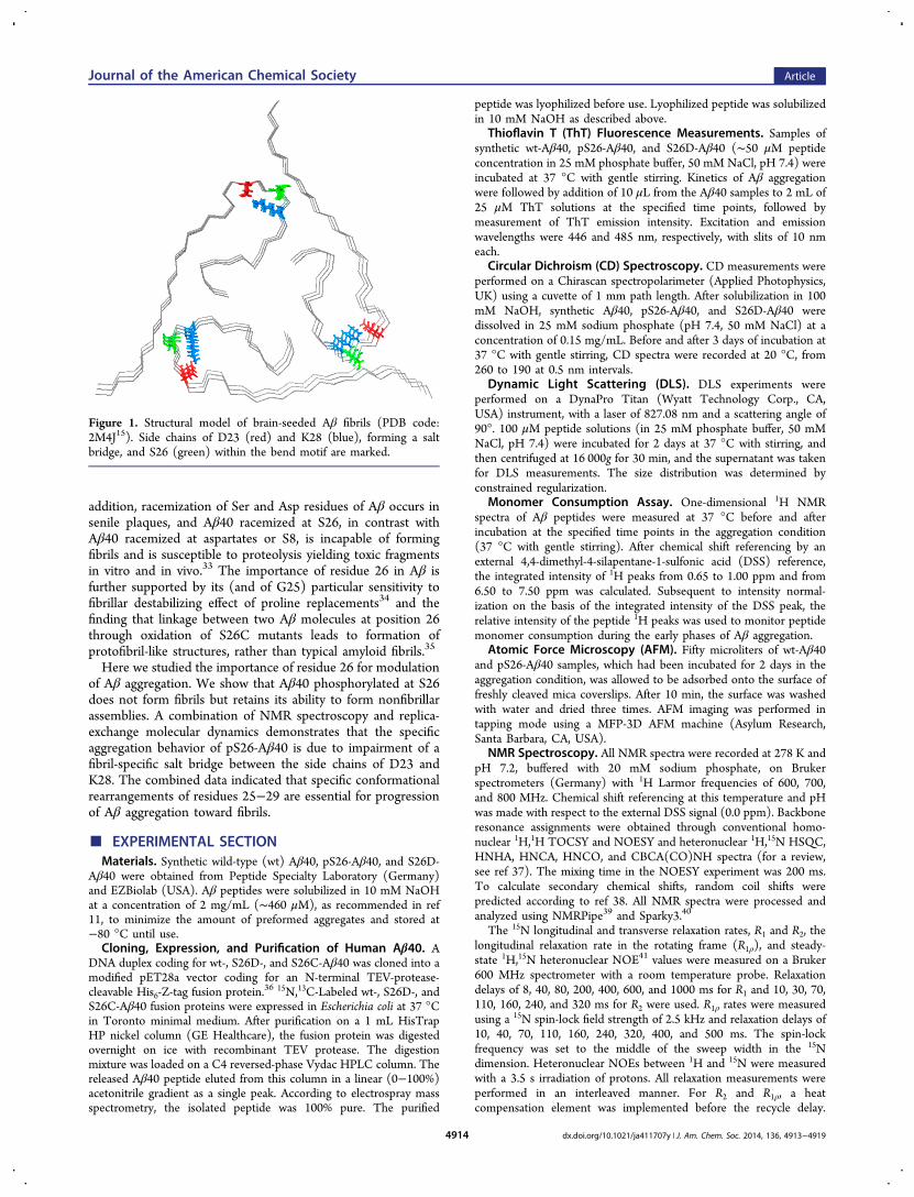
fibrillar Assemblies. Aβ can be phosphorylated at serine 26 invivo.31 To investigate its effect on misfolding of Aβ, weincubated wt-Aβ40 and pS26-Aβ40 peptide solutions underaggregation-promoting conditions. Following a lag phase of ∼4h, ThT emission intensity of wt-Aβ40 increased, in agreementwith a nucleation-dependent mechanism (Figure 2a). Incontrast, ThT emission intensity of pS26-Aβ40 remainednearly constant up until ∼12 h and showed only a slightincrease even after 30 h of incubation, in line with a previousobservation.32
According to atomic force microscopy, wt-Aβ40 formedfibrils of up to 10−12 nm in diameter, while for pS26-Aβ40,only globular species of various sizes were observed (Figure 2band Supporting Information Figure S1). Notably, we could notovercome the inability of pS26-Aβ40 to form β-sheet-richfibrillar aggregates at higher peptide concentrations and longerincubation times (Figure S2), suggesting that the lack of pS26-Aβ40 fibrils is not caused by a kinetic barrier against aggregateformation.Next, we monitored peptide monomer consumption using
NMR spectroscopy. Conversion of Aβ monomers to oligomersdecreases the observable NMR signal due to line broadening.Indeed, for pS26-Aβ40, a 10% loss of signal intensity was
observed during a 24 h incubation period at 37 °C in the NMRtube (Figure S3a). Under more aggregation-prone conditions(as used in CD and ThT experiments; see below), the amountof aggregated pS26-Aβ40 increased to 20% (Figure S3b). Inline with aggregation of pS26-Aβ40, light scattering intensityincreased during the first 2 h of incubation (Figure S4a), andsoluble nonfibrillar assemblies of pS26-Aβ40 with a hydro-dynamic radius of approximately 100 nm were found after 24 h(Figure S4b). At the end of a 48 h period of aggregation, thesupernatant of pS26-Aβ40 scattered light more strongly thaneither wt-Aβ40 or Aβ phosphorylated at S8 (pS8-Aβ40)(Figure S4c), a variant with a high propensity to formoligomers and fibrils.30 Far-UV circular dichroism furthermoreshowed thatafter 3 days of aggregation and in contrast to wt-Aβ40pS26-Aβ40 retained a CD spectrum that is character-istic for a largely disordered state (Figure 2c).To investigate whether the nonfibrillar assemblies of pS26-
Aβ40 are toxic, we performed MTT assays with primary cortical
Figure 2. Phosphorylation at S26 prohibits Aβ40 aggregation into β-sheet-rich fibrils but allows Aβ40 assembly into nonfibrillar aggregates.(a) Evolution of ThT fluorescence emission intensity over the courseof incubation of wt- and pS26-Aβ40 in an aggregation-promotingcondition. (b) AFM images of Aβ40 after 2 days of aggregation; pS26-Aβ40 (right) is unable to form fibrillar aggregates, in contrast to wt-Aβ40 (left). (c) Far-UV CD spectra of Aβ40 variants duringaggregation.
Journal of the American Chemical Society Article
dx.doi.org/10.1021/ja411707y | J. Am. Chem. Soc. 2014, 136, 4913−49194915

neurons. While aggregated wt-Aβ40 resulted in decreasedviability, no significant effect was observed after incubation ofneurons with pS26-Aβ40 aggregates or monomers, indicatingthat the soluble nonfibrillar aggregates of pS26-Aβ40 werenontoxic (Figure S5).Conformational Dynamics at Serine 26. To obtain
insight into the aggregation properties of pS26-Aβ, we used acombination of NMR spectroscopy and molecular dynamicssimulation. Due to the disordered nature of monomeric Aβ,NMR signal dispersion is low (Figure S6).49,50 Estimation ofthe exchange contribution to the 15N transverse relaxation raterevealed a distinct dynamic behavior of residues S26 to G29. Rexvalues of S26, N27, and G29 exceeded those of neighboringresidues (Figure S7 and Figure 3a), and the exchange rates of
their amide protons were very large (Figure 3b), demonstratingthat residues 26−29 experience strong conformationalexchange in the disordered monomeric peptide.NMR chemical shifts are highly sensitive probes for
formation of secondary and tertiary polypeptide structure.51
HA secondary chemical shifts of synthetic Aβ40 point to aslight but visible propensity for β-turn formation for residuesE22−G25 (Figure 4a). Nuclear Overhauser enhancement(NOE) cross-peaks between D23 and G25, as well as betweenN27 and G29 (Figure 4b), further supported the presence ofheterogeneous turn-like structures in the region that experi-ences strong conformational exchange (Figure 3).
Next, we asked how phosphorylation at S26 affects theseconformational propensities. Figure S8 demonstrates thatperturbations of NMR chemical shifts are restricted to theimmediate vicinity of residue 26. Notably, the dispersion of theHB but not HD resonances of N27 was increased (Figure S9),suggesting thatalthough a detailed analysis of NOEs washampered due to signal overlapphosphorylation at S26results in more rigid backbone conformations of N27. Adecrease in dynamics was further supported by the intensity ofthe HN−HA correlation peak of N27, which rose by more than2-fold in pS26-Aβ40; that is, the conformational exchange inthe region 26−29 was strongly reduced upon phosphorylationat S26.
Phosphorylation at Serine 26 Interferes with For-mation of the D23−K28 Salt Bridge. According to NMR,phosphorylation at S26 only influences the vicinity of the site ofphosphorylation. We therefore performed replica-exchangemolecular dynamic (REMD) simulations of the Aβ fragmentA21−A30 in its unphosphorylated and S26-phosphorylatedforms at 278 K. Each replica was sampled for 20 ns. In line withsampling of mostly disordered conformations, carbon chemicalshifts predicted from the simulation correlated with exper-imental values (Figure S10). Analysis of the conformationssampled during the simulation revealed that phosphorylationincreased the end-to-end distances of Aβ(21−30) (Figure 4c),in line with the population of more extended states. In addition,phosphorylation decreased the propensity for turn/bendformation of residues E22−S26 (Figures 4d and S11).Inspection of the most populated clusters of each peptideprovided a rationale for the observed changes. In the cluster ofAβ(21−30), a salt bridge between the side chains of D23 andK28 was present, the salt bridge that is a key feature of Aβfibrils,12,13 and stabilizes a turn-like structure (Figure 4e). Incontrast, the most abundant cluster of pS26-Aβ(21−30) doesnot contain this salt bridge. Instead, the positively charged sidechain of K28 forms a salt bridge with the nearby phosphategroup at residue 26. Due to the formation of this new saltbridge, the loop between residues 23−28 cannot be formed andthe end-to-end distance of the peptide is increased.
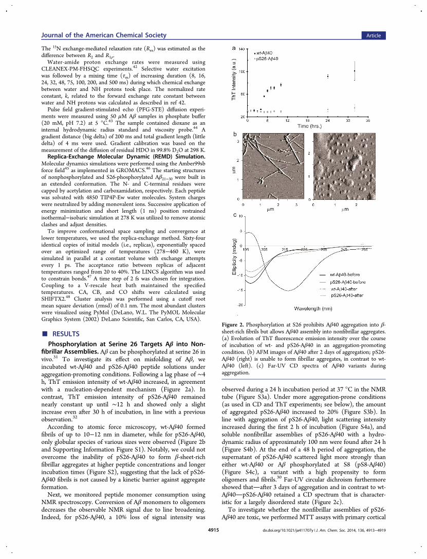
Phosphorylation Impairs Conformational Changes.To provide additional support for the structural and dynamicchanges induced by phosphorylation, we produced 15N,13C-labeled protein of wt-Aβ40 and a phosphomimetic variant ofAβ40, in which S26 was mutated to aspartic acid. Phospho-mimetic variants are particularly useful for in vivo studies, asthey allow investigation of a defined phosphorylation state. Thevalidity of S26D-Aβ40 is supported by REMD simulations:upon S26D mutation, the propensity of residues 22−26 to formbends/turns was diminished (Figure S11), and the salt bridgebetween the side chains of D23 and K28 was lost. Instead, theK28 side chain was located in proximity to the negativelycharged side chain of D26 (Figure S11c). In addition, the S26Dmutation prolonged the lag phase of Aβ fibrillar aggregation(Figure S12).Next we analyzed NMR chemical shifts in S26D-Aβ40. In
line with the findings for pS26-Aβ40, chemical shift changeswere restricted to residues around the site of mutation (FigureS13). According to the NMR analysis, residues D23−N27 havean increased propensity for extended conformations in S26D-Aβ40 (Figures 5a and S14).52 In line with the specificity of theobserved changes, an Aβ variant in which S26 was mutated tocysteine (S26C-Aβ40) did not showin reducing conditiona similar change (Figure S15). The NMR chemical shifts
Figure 3. Conformational plasticity of residues G25−G29 inmonomeric Aβ40. (a) Residue-specific exchange-mediated 15Ntransverse relaxation rates (Rex) of monomeric Aβ40. Residues 25−29 show larger Rex than the rest of Aβ40, suggesting their involvementin conformational exchange dynamics. The higher Rex values ofresidues around H6, H13, and H14 are probably caused by changes inthe protonation state of the histidine side chains. (b) Residue-specificwater-amide proton exchange rates of Aβ40.
Journal of the American Chemical Society Article
dx.doi.org/10.1021/ja411707y | J. Am. Chem. Soc. 2014, 136, 4913−49194916

further supported on the basis of the random coil index (RCI)order parameter that the mobility of residues D23−N27 wasdiminished in the S26D variant (Figure S16a).53 In addition,changes in exchange-mediated contributions to NMR relaxationrates were consistent with rigidification of the Aβ backbone:15N Rex and water-amide proton exchange rates of S26-G29,which had the highest values in wt-Aβ40 (Figure 3), werestrongly decreased in S26D-Aβ40 (Figures 5b and S16b). Onthe other hand, the S26C mutation did affect the backbonedynamics of Aβ much less (Figure S17). The data demonstratethat the S26D phosphomimetic mutation decreases thebackbone mobility of residues 25−29 on multiple time scales.
■ DISCUSSIONOur study demonstrates that conformational rearrangementsaround serine 26 are required for conversion of Aβ monomersto fibrils. Phosphorylation at S26, in the region that is mostprone to conformational changes in monomeric Aβ, interfereswith Aβ aggregation into β-sheet-rich fibrils but allows
formation of nonfibrillar assemblies. NMR spectroscopy andmolecular dynamics simulations demonstrated that impairedfibrillization is associated with a reduced plasticity of the residuestretch G25−G29. Formation of a salt bridge between K28 andthe phosphorylated residue S26 rigidifies the structure andinterferes with formation of the salt bridge between the sidechains of K28 and D23 and therefore β-hairpin formation. TheD23−K28 salt bridge may thus be essential for fibrillization butnot for oligomerization.Formation and stabilization of the β-hairpin plays an
important role in aggregation of Aβ.12−14,25 In agreementwith previous reports,11,50 our NMR data indicate thatmonomeric Aβ transiently adopts a β-hairpin conformation insolution (Figures 3, 4, 5, and S14). Stabilization of the β-hairpinconformation in the aggregated state shifts the equilibriumbetween β-hairpin and other monomer conformations towardthe β-hairpin conformation. Indeed, when D23 and K28 arechemically constrained by a lactam bridge, the aggregation rateof Aβ increases by 3 orders of magnitude.54 The strong increase
Figure 4. Phosphorylation of S26 impairs formation of the fibril-specific D23−K28 salt bridge. (a) HA secondary chemical shifts of wt- (black) andpS26-Aβ40 (gray). (b) Selected region of a 2D 1H,1H NOESY spectrum of wt-Aβ40. Cross-peaks between the HN resonance of G25 and HA ofD23, and between HA of N27 and HN of G29 point to a propensity of residues 22−25 and 26−29 for β-turn formation. (c) Histogram of end-to-end distances (distance between CA atoms of A21 and A30) obtained from MD simulations of Aβ(21−30) in the S26-phosphorylated andnonphosphorylated states. (d) Percentage of coil conformation observed in the MD ensemble of wt- and pS26-Aβ(21−30). (e) Representativestructures of the highest-population cluster in the MD ensembles of wt- (left) and pS26-Aβ(21−30) (right).
Journal of the American Chemical Society Article
dx.doi.org/10.1021/ja411707y | J. Am. Chem. Soc. 2014, 136, 4913−49194917
in the aggregation rate is attributed to a decrease in entropicpenalty of bend formation.55 Moreover, the conformationformed by residues 25−29 can constitute a template over whicha second Aβ molecule adopts the same structure, stabilizing theβ-hairpin and favoring Aβ oligomerization.23,25
S26 is located at the center of the turn motif formed byresidues G25−G29 (Figure 1). Introduction of a negativelycharged phosphate group at this position will causeintermolecular repulsive interactions that lead to destabilizationof the fibrillar conformation. Moreover, in some amyloid fibrils(including the recently reported brain-seeded Aβ40 fibrils15),the charged side chains of D23 and K28 would point to a lowdielectric environment, which is thermodynamically unfavor-able unless their charges are neutralized via a salt bridge. Theimpairment of the D23−K28 salt bridge by phosphorylation atS26 is therefore expected to result in thermodynamic instabilityof those Aβ fibrils. Indeed, we found that even at very highpeptide concentrations and long incubation periods amyloidfibrils were not formed (Figure S2). In contrast to amyloidfibrils, Aβ preglobulomers and globulomers can contain amixture of intramolecular antiparallel and intermolecularparallel β-sheets with G25−G29 constituting the connectingbend.26 In the currently available structural models of Aβoligomers, S26 is not located in a tightly packed region. Thus,phosphorylation at S26 is expected to have a smaller, if any,destabilizing impact on Aβ oligomers when compared to fibrils,in line with the observed nonfibrillar assembly of pS26-Aβ40(Figures 2, S1, and S4).
A certain amount of plasticity in the G25−G29 turn region isrequired for conversion of oligomers into ordered fibrils.2,56,57
In line with this hypothesis, enhanced aggregation of variants ofAβ causing early onset AD, such as E22K, E22G, E22Q, andD23N, was linked to destabilization of the turn-like structure.19
Moreover, overstabilization of the turn through chemical cross-linking of A21 and A30 in a double-cysteine mutant reducedfibril formation and stabilized neurotoxic Aβ oligomers.58 OurNMR data demonstrate that upon phosphorylation at S26(Figure S9), as well as mutation to the phosphomimetic variantS26D (Figures 5, S16, and S17), residues 23−28 become morerigid. Due to the decreased turn plasticity of pS26-Aβ,fibrillization is impaired, whereas nonfibrillar aggregation canstill occur. Consistent with this finding, constrained motion ofresidue 26 through introduction of an intermolecular disulfidebond blocked conversion of intermediate protofibril-like Aβ40aggregates into typical amyloid fibrils.35
Post-translational modifications of Aβ that enhance itscytotoxic aggregation are expected to contribute to thepathogenesis of late onset AD. Our data suggest thatphosphorylation at S26 suppresses the formation of toxicoligomers and fibrils while stabilizing monomers and nontoxicsoluble assemblies of nonfibrillar morphology. We have recentlyshown that phosphorylation of Aβ at S8 by extracellular proteinkinases promotes cytotoxic aggregation of Aβ.30 In contrast topS26-Aβ, Aβ phosphorylated at S8 showed a high propensity toform β-sheet-rich oligomers and fibrils,30 indicating thatphosphorylation of Aβ at distinct sites can lead to drasticallydifferent consequences on pathogenic aggregation. In addition,as phosphorylation at both S8 and S26 modulates Aβaggregation, targeting phosphorylation/dephosphorylation ofAβ might offer new ways for prevention of late onset sporadicAD.
■ CONCLUSIONWe demonstrated that phosphorylation of Aβ at S26 blocksconversion of Aβ monomers to β-sheet-rich fibrils. Our findingshighlight the importance of the plasticity of residues G25−G29in the control of Aβ aggregation and the role of the D23−K28salt bridge in Aβ fibrillization. Targeting the plasticity ofresidues G25−G29 by influencing phosphorylation of serine 26might provide a therapeutic route for late onset sporadic AD.
■ ASSOCIATED CONTENT*S Supporting InformationSupporting data as Figures S1−S17. This material is availablefree of charge via the Internet at http://pubs.acs.org.
■ AUTHOR INFORMATIONCorresponding [email protected] authors declare no competing financial interest.
■ ACKNOWLEDGMENTSWe thank Alice Soragni for acquisition of 2D homonuclearspectra. This work was supported by DFG (ZW 71/2-2 and 3-2) to M.Z. and DFG (Grant WA1477/6-2) to J.W.
■ REFERENCES(1) Hardy, J.; Selkoe, D. J. Science 2002, 297, 353.(2) Fandrich, M. J. Mol. Biol. 2012, 421, 427.
Figure 5. S26D phosphomimetic mutation decreases turn plasticity ofAβ40. (a) Probability score for β-strand conformation according toCA, CB, CO, N, HN, and HA chemical shifts52 for wt- (black) andS26D-Aβ40 (gray). (b) Changes in exchange-mediated transverserelaxation rates (Rex) upon S26D mutation. Residues 23−29 decreasein Rex after mutation, indicating that motions on the micro-to-millisecond time scale are diminished in this region.
Journal of the American Chemical Society Article
dx.doi.org/10.1021/ja411707y | J. Am. Chem. Soc. 2014, 136, 4913−49194918
(3) Shankar, G. M.; Li, S.; Mehta, T. H.; Garcia-Munoz, A.;Shepardson, N. E.; Smith, I.; Brett, F. M.; Farrell, M. A.; Rowan, M. J.;Lemere, C. A.; Regan, C. M.; Walsh, D. M.; Sabatini, B. L.; Selkoe, D.J. Nat. Med. 2008, 14, 837.(4) De Felice, F. G.; Velasco, P. T.; Lambert, M. P.; Viola, K.;Fernandez, S. J.; Ferreira, S. T.; Klein, W. L. J. Biol. Chem. 2007, 282,11590.(5) Gellermann, G. P.; Byrnes, H.; Striebinger, A.; Ullrich, K.;Mueller, R.; Hillen, H.; Barghorn, S. Neurobiol. Dis. 2008, 30, 212.(6) Lesne, S.; Koh, M. T.; Kotilinek, L.; Kayed, R.; Glabe, C. G.;Yang, A.; Gallagher, M.; Ashe, K. H. Nature 2006, 440, 352.(7) Lashuel, H. A.; Hartley, D.; Petre, B. M.; Walz, T.; Lansbury, P.T., Jr. Nature 2002, 418, 291.(8) Stefani, M. Prog. Neurobiol. 2012, 99, 226.(9) Glabe, C. G. J. Biol. Chem. 2008, 283, 29639.(10) Riek, R.; Guntert, P.; Dobeli, H.; Wipf, B.; Wuthrich, K. Eur. J.Biochem. 2001, 268, 5930.(11) Hou, L.; Shao, H.; Zhang, Y.; Li, H.; Menon, N. K.; Neuhaus, E.B.; Brewer, J. M.; Byeon, I. J.; Ray, D. G.; Vitek, M. P.; Iwashita, T.;Makula, R. A.; Przybyla, A. B.; Zagorski, M. G. J. Am. Chem. Soc. 2004,126, 1992.(12) Petkova, A. T.; Ishii, Y.; Balbach, J. J.; Antzutkin, O. N.;Leapman, R. D.; Delaglio, F.; Tycko, R. Proc. Natl. Acad. Sci. U.S.A.2002, 99, 16742.(13) Petkova, A. T.; Yau, W. M.; Tycko, R. Biochemistry 2006, 45,498.(14) Luhrs, T.; Ritter, C.; Adrian, M.; Riek-Loher, D.; Bohrmann, B.;Dobeli, H.; Schubert, D.; Riek, R. Proc. Natl. Acad. Sci. U.S.A. 2005,102, 17342.(15) Lu, J. X.; Qiang, W.; Yau, W. M.; Schwieters, C. D.; Meredith, S.C.; Tycko, R. Cell 2013, 154, 1257.(16) Lazo, N. D.; Grant, M. A.; Condron, M. C.; Rigby, A. C.;Teplow, D. B. Protein Sci. 2005, 14, 1581.(17) Borreguero, J. M.; Urbanc, B.; Lazo, N. D.; Buldyrev, S. V.;Teplow, D. B.; Stanley, H. E. Proc. Natl. Acad. Sci. U.S.A. 2005, 102,6015.(18) Baumketner, A.; Bernstein, S. L.; Wyttenbach, T.; Lazo, N. D.;Teplow, D. B.; Bowers, M. T.; Shea, J. E. Protein Sci. 2006, 15, 1239.(19) Grant, M. A.; Lazo, N. D.; Lomakin, A.; Condron, M. M.; Arai,H.; Yamin, G.; Rigby, A. C.; Teplow, D. B. Proc. Natl. Acad. Sci. U.S.A.2007, 104, 16522.(20) Krone, M. G.; Baumketner, A.; Bernstein, S. L.; Wyttenbach, T.;Lazo, N. D.; Teplow, D. B.; Bowers, M. T.; Shea, J. E. J. Mol. Biol.2008, 381, 221.(21) Fawzi, N. L.; Phillips, A. H.; Ruscio, J. Z.; Doucleff, M.;Wemmer, D. E.; Head-Gordon, T. J. Am. Chem. Soc. 2008, 130, 6145.(22) Murray, M. M.; Krone, M. G.; Bernstein, S. L.; Baumketner, A.;Condron, M. M.; Lazo, N. D.; Teplow, D. B.; Wyttenbach, T.; Shea, J.E.; Bowers, M. T. J. Phys. Chem. B 2009, 113, 6041.(23) Larini, L.; Shea, J. E. Biophys. J. 2012, 103, 576.(24) Tanzi, R. E. Cold Spring Harbor Perspect. Med. 2012, 2.(25) Hoyer, W.; Gronwall, C.; Jonsson, A.; Stahl, S.; Hard, T. Proc.Natl. Acad. Sci. U.S.A. 2008, 105, 5099.(26) Yu, L.; Edalji, R.; Harlan, J. E.; Holzman, T. F.; Lopez, A. P.;Labkovsky, B.; Hillen, H.; Barghorn, S.; Ebert, U.; Richardson, P. L.;Miesbauer, L.; Solomon, L.; Bartley, D.; Walter, K.; Johnson, R. W.;Hajduk, P. J.; Olejniczak, E. T. Biochemistry 2009, 48, 1870.(27) Jarrett, J. T.; Berger, E. P.; Lansbury, P. T., Jr. Biochemistry 1993,32, 4693.(28) Sullivan, C. P.; Berg, E. A.; Elliott-Bryant, R.; Fishman, J. B.;McKee, A. C.; Morin, P. J.; Shia, M. A.; Fine, R. E. Neurosci. Lett. 2011,505, 109.(29) Bouter, Y.; Dietrich, K.; Wittnam, J. L.; Rezaei-Ghaleh, N.;Pillot, T.; Papot-Couturier, S.; Lefebvre, T.; Sprenger, F.; Wirths, O.;Zweckstetter, M.; Bayer, T. A. Acta Neuropathol. 2013, 126, 189.(30) Kumar, S.; Rezaei-Ghaleh, N.; Terwel, D.; Thal, D. R.; Richard,M.; Hoch, M.; McDonald, J. M.; Wullner, U.; Glebov, K.; Heneka, M.T.; Walsh, D. M.; Zweckstetter, M.; Walter, J. EMBO J. 2011, 30, 2255.(31) Milton, N. G. Subcell. Biochem. 2005, 38, 381.
(32) Milton, N. G. Neuroreport 2001, 12, 3839.(33) Kaneko, I.; Morimoto, K.; Kubo, T. Neuroscience 2001, 104,1003.(34) Williams, A. D.; Shivaprasad, S.; Wetzel, R. J. Mol. Biol. 2006,357, 1283.(35) O’Nuallain, B.; Freir, D. B.; Nicoll, A. J.; Risse, E.; Ferguson, N.;Herron, C. E.; Collinge, J.; Walsh, D. M. J. Neurosci. 2010, 30, 14411.(36) Edlich, C.; Stier, G.; Simon, B.; Sattler, M.; Muhle-Goll, C.Structure 2005, 13, 277.(37) Whitehead, B.; Craven, C. J.; Waltho, J. P. Methods Mol. Biol.1997, 60, 29.(38) Tamiola, K.; Acar, B.; Mulder, F. A. J. Am. Chem. Soc. 2010, 132,18000.(39) Delaglio, F.; Grzesiek, S.; Vuister, G. W.; Zhu, G.; Pfeifer, J.;Bax, A. J. Biomol. NMR 1995, 6, 277.(40) Goddard, T. D.; Kneller, D. G. SPARKY 3; University ofCalifornia, San Francisco.(41) Palmer, A. G., III. Annu. Rev. Biophys. Biomol. Struct. 2001, 30,129.(42) Hwang, T. L.; van Zijl, P. C.; Mori, S. J. Biomol. NMR 1998, 11,221.(43) Johnson, C. S., Jr. Prog. Nucl. Magn. Reson. Spectrosc. 1999, 34,203.(44) Wilkins, D. K.; Grimshaw, S. B.; Receveur, V.; Dobson, C. M.;Jones, J. A.; Smith, L. J. Biochemistry 1999, 38, 16424.(45) Hornak, V.; Abel, R.; Okur, A.; Strockbine, B.; Roitberg, A.;Simmerling, C. Proteins 2006, 65, 712.(46) Berendsen, H. J. C.; Van Der Spoel, D.; Van Drunen, R. J.Comput. Phys. Commun. 1995, 91, 43.(47) Hess, B.; Bekker, H.; Berendsen, H. J. C.; Fraaije, J. G. E. M. J.Comput. Chem. 1997, 18, 1463.(48) Han, B.; Liu, Y.; Ginzinger, S. W.; Wishart, D. S. J. Biomol. NMR2011, 50, 43.(49) Yan, Y.; Wang, C. J. Mol. Biol. 2006, 364, 853.(50) Rezaei-Ghaleh, N.; Giller, K.; Becker, S.; Zweckstetter, M.Biophys. J. 2011, 101, 1202.(51) Wishart, D. S. Prog. Nucl. Magn. Reson. Spectrosc. 2011, 58, 62.(52) Shen, Y.; Bax, A. J. Biomol. NMR 2012, 52, 211.(53) Berjanskii, M. V.; Wishart, D. S. J. Am. Chem. Soc. 2005, 127,14970.(54) Sciarretta, K. L.; Gordon, D. J.; Petkova, A. T.; Tycko, R.;Meredith, S. C. Biochemistry 2005, 44, 6003.(55) Reddy, G.; Straub, J. E.; Thirumalai, D. J. Phys. Chem. B 2009,113, 1162.(56) Habicht, G.; Haupt, C.; Friedrich, R. P.; Hortschansky, P.;Sachse, C.; Meinhardt, J.; Wieligmann, K.; Gellermann, G. P.;Brodhun, M.; Gotz, J.; Halbhuber, K. J.; Rocken, C.; Horn, U.;Fandrich, M. Proc. Natl. Acad. Sci. U.S.A. 2007, 104, 19232.(57) Pellarin, R.; Guarnera, E.; Caflisch, A. J. Mol. Biol. 2007, 374,917.(58) Sandberg, A.; Luheshi, L. M.; Sollvander, S.; Pereira de Barros,T.; Macao, B.; Knowles, T. P.; Biverstal, H.; Lendel, C.; Ekholm-Petterson, F.; Dubnovitsky, A.; Lannfelt, L.; Dobson, C. M.; Hard, T.Proc. Natl. Acad. Sci. U.S.A. 2010, 107, 15595.
Journal of the American Chemical Society Article
dx.doi.org/10.1021/ja411707y | J. Am. Chem. Soc. 2014, 136, 4913−49194919