Embed Size (px)
Citation preview

Tutorial on Breast Pathology Part II: Invasive Carcinoma
Thomas J. Lawton MD, Director Seattle Breast Pathology Consultants, LLC
Seattle, WA

Invasive carcinoma, NST
• NST means “no special type”. • This is the most common type of invasive
carcinoma of the breast (around 65-75%). • This means the same as invasive ductal carcinoma
or invasive mammary carcinoma, NOS. • Invasive carcinomas are usually graded by the
pathologist using the Nottingham scheme.

The Nottingham Grading System • This grading system looks at three parts of your cancer
and gives a score of 1-3 for each part. • The scores for each of the 3 areas are added up and the
total score is translated into a Nottingham grade. The three parts your pathologist evaluates are: • how much the cancer resembles normal breast ducts (also
called tubule formation) • how aggressive the cells look (also called nuclear grade or
nuclear pleomorphism) • how fast the cancer is growing (also called mitotic rate)

How pathologists come up with a Nottingham grade
• Grade I: score 3-5 • Grade II: score 6-7 • Grade III: score 8-9
• We have posted images of different invasive carcinoma grades in the SBPC Image Gallery (under The SBPC Dialogue box)

Once a diagnosis of invasive carcinoma is made, the cancer should then be evaluated
for the following: • Your cancer should be tested for hormone
receptor status (ER and PR) and for HER2 over-expression or amplification.
• Your axillary lymph nodes should be evaluated, commonly by sentinel node procedure.
• At your definitive surgery, a size of your cancer should be reported as well as distance from the surgical margins.
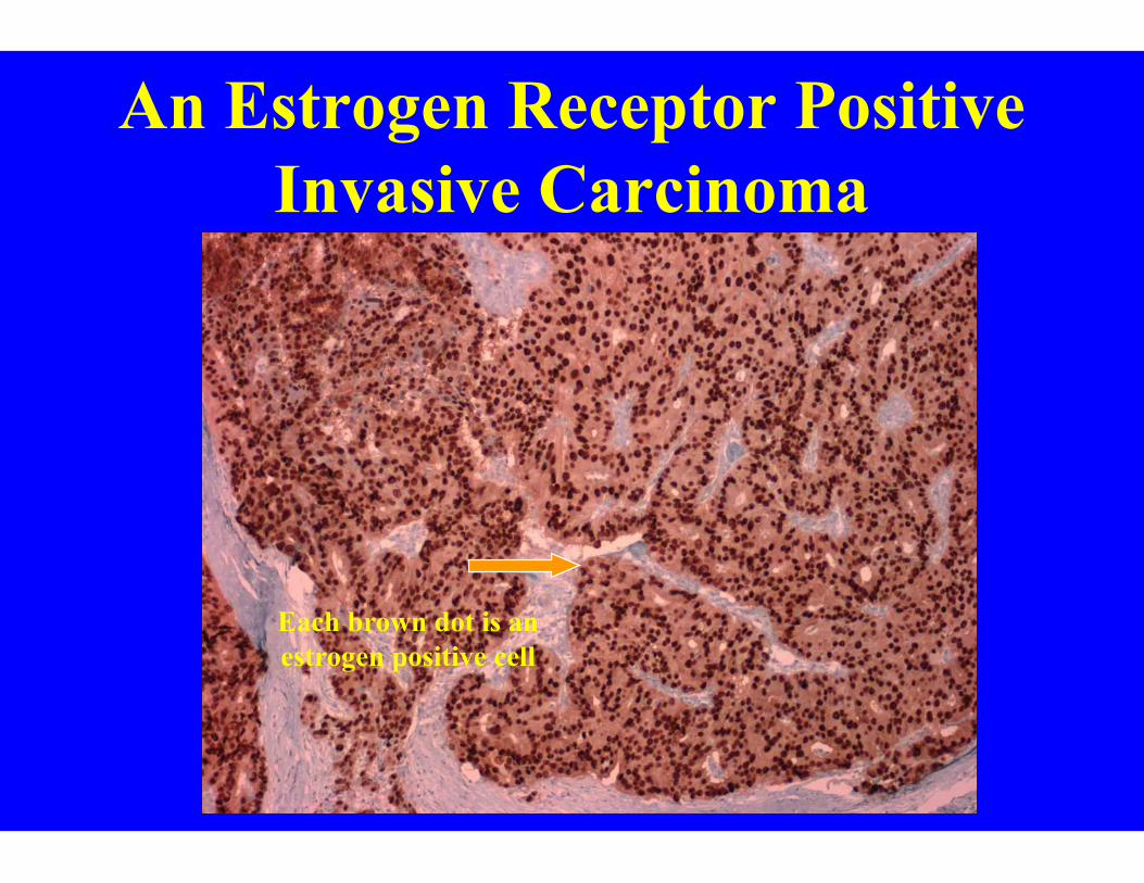
An Estrogen Receptor Positive Invasive Carcinoma
Each brown dot is an estrogen positive cell

HER2 • HER2 is a protein on the surface of normal breast
cells which can be over-expressed (too much protein) in approximately 20% of breast cancers.
• Two main ways to test: Immunohistochemistry and FISH (fluorescence in situ hybridization).
• New recommendations for testing for HER2 were proposed in 2007 (please see my article on changes in HER2 testing in the SBPC Library).

HER2 immunohistochemistry
The brown stain should be very dark and surround at least 30% of the cells to be
called 3+

Axillary lymph node evaluation
• The presence or absence of cancer cells in your axillary lymph nodes is important not only for staging but for your prognosis and treatment.
• A micrometastasis is defined as a focus of carcinoma in a lymph node measuring < 2 mm.
• The new staging system by AJCC talks about “isolated tumor cells” which are defined as a focus of cells in a node measuring < 0.2 mm (by routine pathology or by immunohistochemistry). In the new staging system, isolated tumor cells is still considered N0.

Isolated Tumor Cells (N0i) Detected by Immunohistochemistry
Cells identified by keratin
immunohistochemistry
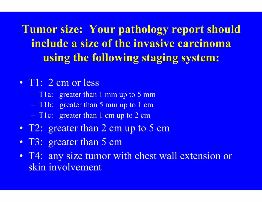
Tumor size: Your pathology report should include a size of the invasive carcinoma
using the following staging system: • T1: 2 cm or less
– T1a: greater than 1 mm up to 5 mm – T1b: greater than 5 mm up to 1 cm – T1c: greater than 1 cm up to 2 cm
• T2: greater than 2 cm up to 5 cm • T3: greater than 5 cm • T4: any size tumor with chest wall extension or
skin involvement

Tumor stage • Using the TNM system, your pathology report
should include a pathologic stage. • Usually this stage will appear only after your
definitive surgery, including evaluation of axillary lymph nodes.
• For more information on the AJCC staging system, please see my article in the SBPC library.

Stay tuned for further updates including special variants of
carcinoma and other tumors of the breast…..