Embed Size (px)
Citation preview
Instructions for use
Title Type II Natural Killer T Cells that Recognize Sterol Carrier Protein 2 Are Implicated in Vascular Inflammation in theRat Model of Systemic Connective Tissue Diseases
Author(s) Nishioka, Yusuke; Yamaguchi, Madoka; Kawakami, Ai; Munehiro, Maya; Masuda, Sakiko; Tomaru, Utano; Ishizu,Akihiro
Citation The American Journal of Pathology, 187(1), 176-186https://doi.org/10.1016/j.ajpath.2016.09.014
Issue Date 2017-01
Doc URL http://hdl.handle.net/2115/64475
Rights ©2017, Elsevier. Licensed under the Creative Commons Attribution-NonCommercial-NoDerivatives 4.0 Internationalhttp://creativecommons.org/licenses/by-nc-nd/4.0/
Rights(URL) http://creativecommons.org/licenses/by-nc-nd/4.0/
Type article
File Information Am. J. Pathol._187(1)_176-186.pdf
Hokkaido University Collection of Scholarly and Academic Papers : HUSCAP
IMMUNOPATHOLOGY AND INFECTIOUS DISEASES
Type II Natural Killer T Cells that Recognize SterolCarrier Protein 2 Are Implicated in VascularInflammation in the Rat Model of Systemic ConnectiveTissue DiseasesYusuke Nishioka,* Madoka Yamaguchi,* Ai Kawakami,* Maya Munehiro,y Sakiko Masuda,z Utano Tomaru,x and Akihiro Ishizuz
From the Graduate School of Health Sciences,* the Undergraduate School of Health Sciences,y and the Faculty of Health Sciences,z Hokkaido University,Sapporo; and the Department of Pathology,x Hokkaido University Graduate School of Medicine, Sapporo, Japan
Accepted for publicationSeptember 13, 2016.
Address correspondence toAkihiro Ishizu, M.D., Ph.D.,Faculty of Health Sciences,Hokkaido University, Kita-12,Nishi-5, Kita-ku, Sapporo0600812, Japan. E-mail:[email protected].
We previously generated a rat model that developed systemic connective tissue diseases, includingsynovitis, myositis, and small-vessel vasculitis (SVV), and established a vascular endothelial cellereactive T-cell clone, VASC-1, from the model. VASC-1 was determined to be a type II natural killer T-cellclone. In this study, we attempted to identify the antigen recognized by VASC-1. The monkey-derivedcell line COS-7 was used because VASC-1 does not bind naturally to COS-7, although the amino acidsequences are well conserved between monkey CD1d and rat CD1d. We generated 98 COS-7 clonestransfected with miscellaneous rat cDNA and screened them for VASC-1 binding. Consequently, we foundone clone, 4D2, which could bind to VASC-1. Sequencing identified the rat cDNA introduced into 4D2 assterol carrier protein 2 (SCP2). When VASC-1 was co-cultured with SCP2 knockdown rat vascularendothelial cells, VASC-1 binding was reduced significantly. Moreover, we designed a series of rat SCP2peptides and introduced them into COS-7 cells. On the basis of VASC-1 binding and proliferation, werevealed that the peptide rSCP2518-532 included the epitope recognized by VASC-1. Furthermore,immunization with rSCP2518-532 accelerated the development of SVV in the rat model. The collectivefindings suggest that type II natural killer T cells reactive with autologous SCP2 are implicated invascular inflammation in the rat model. (Am J Pathol 2017, 187: 176e186; http://dx.doi.org/10.1016/j.ajpath.2016.09.014)
We previously generated a rat model that developed sys-temic connective tissue diseases, including synovitis,myositis, and small-vessel vasculitis (SVV).1 In this model,a predominant infiltration of mononuclear cells wasobserved in the synovial and cardiac tissues and skeletalmuscles and around small vessels in the systemic organs.An autoimmune mechanism could be involved in thepathogenesis because several autoantibodies, such as anti-nuclear and anti-DNA antibodies, were detected in theserum. The presence of hyperreactivity of peripheral Tcells,2 disordered differentiation of T cells in the thymus,3
and functional impairment of regulatory T cells4 had beenfound in the model. In addition, we recently established aT-cell clone reactive with autologous vascular endothelialcells from the rat model and designated the clone as
VASC-1.5 VASC-1 was determined to be a clone of naturalkiller T (NKT) cells because this clone recognized a certainantigen presented by CD1d.NKT cells belong to a unique subset of T cells that share
surface markers and function with natural killer (NK) cellsand play important roles in the immune response.6 Thehallmark of NKT cells is their capacity to recognize antigens
Supported by grant-in-aid 26293082 from the Ministry of Education,Culture, Sports, Science and Technology of Japan (A.I.), a grant forResearch on Rare and Intractable Vasculitis from the Ministry of Health,Labor and Welfare of Japan (A.I.), and grant 15ek0109121 from the JapanAgency for Medical Research and Development (A.I.).Y.N., M.Y., and A.K. contributed equally to this work.Disclosures: None declared.
Copyright ª 2017 American Society for Investigative Pathology. Published by Elsevier Inc.
This is an open access article under the CC BY-NC-ND license (http://creativecommons.org/licenses/by-nc-nd/4.0).
http://dx.doi.org/10.1016/j.ajpath.2016.09.014
ajp.amjpathol.org
The American Journal of Pathology, Vol. 187, No. 1, January 2017
presented by class I major histocompatibility complexelikeCD1d.7,8 Among the NKT cells, two major subsets, namely,type I and type II, have been noted.8e10 Type I NKT cellsare characterized by the expression of a conserved T-cellreceptor (TCR) a-chain (Va24-Ja18 in humans andVa14-Ja18 in rodents) that pairs with a limited repertoire ofb-chains.11e13 This type of cell, also called invariant NKTcells, can bind to a marine spongeederived glycolipid,a-galactosylceramide (a-GalCer), presented by CD1d. Ingeneral, it is considered that type I NKT cells can recognizemicroorganism-derived glycolipids presented by CD1d andtrigger both the innate and acquired immune responsesagainst the microorganism. On the contrary, type II NKTcells express a more variable TCR repertoire and do notrespond to stimulation with a-GalCer. This type of cellcan recognize sulfatides and peptides presented byCD1d.7,8,14,15 Interestingly, NKT cells reactive with au-tologous peptides (suggestive of type II phenotype) exhibitan immunosuppressive property in healthy mice.16 It can beconsidered that the immunoregulatory mechanisms aremediated by type II NKT cells in healthy individuals asfollows. After, cells injured by an immune reaction expressautologous peptides on CD1d, type II NKT cells thatrecognize the autoantigens produce immunosuppressivecytokines to prevent an excessive spread of inflammation.
The NKT cell clone, VASC-1, established from the ratmodel of systemic connective tissue diseases, exhibited aTCR use other than the type I invariant TCR a-chain anddid not bind to a-GalCereloaded CD1d; therefore, it wasregarded as a type II NKT cell clone.5 Because VASC-1produced proinflammatory cytokines after an interactionwith autologous vascular endothelial cells, a functionalimpairment of the immunoregulatory autoreactive type IINKT cells could be present in the rat model. In this study,we attempted to identify the antigen recognized by VASC-1and discovered an intracellular lipid transfer molecule, sterolcarrier protein 2 (SCP2), as one of the autoantigens.
Materials and Methods
Rats
The rat model of systemic connective tissue diseases estab-lished in our laboratory1 and maintained under specificpathogen-free condition was used. These rats were transgenicfor the env-pX gene of human T-cell leukemia virus type I.The transgene was expressed ubiquitously in the systemicorgans, including hematopoietic cells. Because the transgenecoded the transcription factor p40tax, which could disturb theordinary transcription in the cells but did not code other viral
Table 1 Rat SCP2 Peptides
Name
Amino acid sequence* HydropathyindexM T G K M N P Q S A F F Q G K L K I A G N M G L A M K L Q S L Q L Q P D K A K L
rSCP2508-522 M T G K M N P Q S A F F Q G K �0.023rSCP2509-523 T G K M N P Q S A F F Q G K L 0.005rSCP2510-524 G K M N P Q S A F F Q G K L K �0.092rSCP2511-525 K M N P Q S A F F Q G K L K I �0.032rSCP2512-526 M N P Q S A F F Q G K L K I A 0.109rSCP2513-527 N P Q S A F F Q G K L K I A G 0.099rSCP2514-528 P Q S A F F Q G K L K I A G N 0.099rSCP2515-529 Q S A F F Q G K L K I A G N M 0.133rSCP2516-530 S A F F Q G K L K I A G N M G 0.222rSCP2517-531 A F F Q G K L K I A G N M G L 0.305rSCP2518-532 F F Q G K L K I A G N M G L A 0.305rSCP2519-533 F Q G K L K I A G N M G L A M 0.268rSCP2520-534 Q G K L K I A G N M G L A M K 0.089rSCP2521-535 G K L K I A G N M G L A M K L 0.216rSCP2522-536 K L K I A G N M G L A M K L Q 0.127rSCP2523-537 L K I A G N M G L A M K L Q S 0.215rSCP2524-538 K I A G N M G L A M K L Q S L 0.215rSCP2525-539 I A G N M G L A M K L Q S L Q 0.259rSCP2526-540 A G N M G L A M K L Q S L Q L 0.237rSCP2527-541 G N M G L A M K L Q S L Q L Q 0.139rSCP2528-542 N M G L A M K L Q S L Q L Q P 0.115rSCP2529-543 M G L A M K L Q S L Q L Q P D 0.107rSCP2530-544 G L A M K L Q S L Q L Q P D K �0.035rSCP2531-545 L A M K L Q S L Q L Q P D K A �0.026rSCP2532-546 A M K L Q S L Q L Q P D K A K �0.197rSCP2533-547 M K L Q S L Q L Q P D K A K L �0.167
The boldface peptides represent those with a hydropathy index >0.200.*The inserted region in 4D2.
NKT Cells in Vascular Inflammation
The American Journal of Pathology - ajp.amjpathol.org 177
constructive proteins, these rats were considered as modelswith abnormal gene transcription rather than simple modelsof human T-cell leukemia virus type I infection. Experimentswere performed in accordance with the Guidelines for theCare and Use of Laboratory Animals in Hokkaido University(permission No. 10-0029, 15-0034).
Cells
The rat inferior vena cavaederived vascular endothelialcells (RECs) and REC-reactive type II NKT cell clone,VASC-1, were established in our laboratory.5,17 RECs weremaintained in RPMI 1640 medium containing 10% fetalbovine serum (FBS) and 5 � 10�5 mol/L 2-mercaptoethanol(2-ME). VASC-1 was maintained in RPMI 1640 mediumcontaining 20% FBS, 5 � 10�5 mol/L 2-ME, and 0.01 mg/mL recombinant rat IL-2 (R&D Systems, Minneapolis,MN). The monkey kidneyederived fibroblastic cell line,COS-7, was maintained in Dulbecco’s modified Eagle’smedium (DMEM) containing 10% FBS.
Co-Culture of VASC-1 with RECs or COS-7
RECs or COS-7 was grown subconfluently in a well of six-well plates (1 � 105 per well) at 37�C. VASC-1 (2 � 105
per well) was co-cultured with the cells in RPMI 1640medium containing 20% FBS, 5 � 10�5 mol/L 2-ME, and0.01 mg/mL recombinant rat IL-2 overnight at 37�C. On thenext day, after the removal of the supernatants, the
remaining cells were washed with the co-culture mediumthree times and then observed under a phase-contrastmicroscope.
Knockdown of Rat Genes of RECs
RECs were seeded in 6-cm dishes (2 � 105/dish) and incu-bated overnight at 37�C. On the next day, the culture mediumwas replaced with serum-free 2-MEefree RPMI 1640 me-dium. Thereafter, siRNA for rat CD1d or rat SCP2 or nega-tive control siRNA (Life Technologies, Tokyo, Japan) wastransfected into RECs using siGENE (Life Technologies) inaccordance with the manufacturer’s instruction. Forty-eighthours after the siRNA transfection, total RNA was extrac-ted from RECs using an RNeasy Mini kit (Qiagen, Alameda,CA). RNA underwent reverse transcription to cDNA usingtranscriptase and oligo-dT primers (Promega, Tokyo, Japan).The primers for rat genes used in this study were CD1dsense: 50-TAGAAGCAGGGAAGCCAGACC-30, CD1dantisense: 50-TCCGCATTTGGCAGGATGTC-30, SCP2sense: 50-CTTCACGATTGCTTCTCTACC-30, SCP2 anti-sense: 50-CAGTGCTCCACCTTGTCCTTC-30, glyceralde-hyde-3-phosphate dehydrogenase (GAPDH) sense: 50-ATGGGAGTTGCTGTTGAAGTCA-30, and GAPDH anti-sense: 50-CCGAGGGCCCACTAAAGG-30. Real-time PCRwas run as follows: after denaturation at 94�C for 2 minutes,40 cycles of reaction at 94�C for 15 seconds and at 62�C for60 seconds were performed using the GoTaq 2-Step RT-qPCR System (Promega).
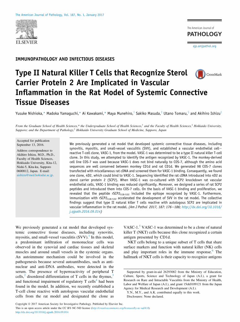
Figure 1 Involvement of CD1d on rat inferior vena cavaederived vascular endothelial cells (RECs) in vascular endothelial cellereactive T-cell clone(VASC-1) binding. A and B: Representative images of co-cultured VASC-1 with RECs. Knockdown of CD1d of RECs by siRNA at the levels of mRNA (C) and cellsurface protein (D). Experiments were performed in triplicate. Decrease in VASC-1 binding to RECs by the inhibition of CD1d using antibody and knockdown ofCD1d using siRNA (E and F). Data are expressed as means � SD (C and F). n Z 3 (F). *P < 0.05, **P < 0.01 (t-test). Scale bars: 10 mm (A); 50 mm (E). TCR,T-cell receptor.
Nishioka et al
178 ajp.amjpathol.org - The American Journal of Pathology
Flow Cytometry
Seventy-two hours after the siRNA transfection, RECs weredetached from the dishes and then allowed to react with theanti-mouse/rat CD1d antibody WTH-1 (Abcam, Cambridge,UK) or mouse IgG2a (BD Biosciences, San Jose, CA) wasthe isotype control on ice for 20 minutes (1 mg per 106 cells).After washing, the cells were then made to react with 1:2000diluted phycoerythrin-conjugated donkey anti-mouse IgGantibody (eBioscience, San Diego, CA) on ice for 30 mi-nutes. Flow cytometry was conducted using FACSCanto II(BD Biosciences), and the data were analyzed using Cell-Quest Pro software version 6.0 (BD Biosciences).
VASC-1 Binding to RECs or REC Knockdown of CD1d
Twenty-four hours before this experiment, the medium ofVASC-1 was replaced with a fresh one without IL-2. RECs orREC knockdown of CD1d was reseeded in two-well slidechambers (1 � 105 per well) and incubated overnight at37�C. On the next day, carboxyfluorescein succinimidylesterelabeled VASC-1 (2 � 105 per well) was co-culturedwith the cells in RPMI 1640 medium containing 20% FBS
and 5� 10�5 mol/L 2-ME for 2 hours at 37�C. For reference,a similar co-culture was conducted in the presence of 1mg/mL of the anti-CD1d antibody WTH-1. After the removalof the supernatants, the remaining cells were washed withPBS once and then fixed by acetone for 5 minutes at roomtemperature. The chambers were removed, and then the slideglasses were mounted with VECTASHIELD (Vector Labo-ratories, Burlingame, CA), which contains DAPI.
Comparison of Amino Acid Sequences of Monkey CD1dand Rat CD1d
The alignment analysis of amino acid sequences of monkeyCD1d (XP_007974824) and rat CD1d (NP_058775) wasconducted using Basic Local Alignment Search Tool(BLAST).
Transfection of Rat Genes into COS-7
COS-7 was grown subconfluently in wells of a six-wellplate (1 � 105 per well) at 37�C. Rat cDNA library derivedfrom the lungs of Sprague-Dawley strain (TKR9543, TakaraBio, Otsu, Japan) was transfected into COS-7 cells using
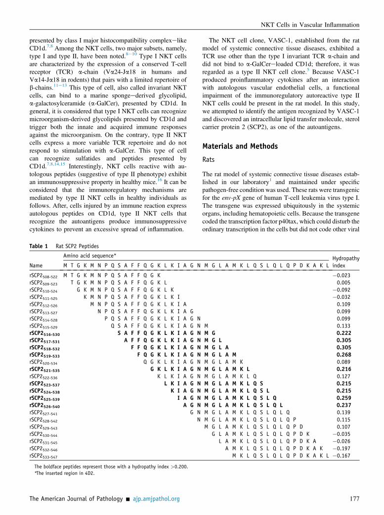
Figure 2 Vascular endothelial cellereactive T-cellclone (VASC-1) binding to COS-7 transfected withthe rat gene. A and B: Representative images ofco-cultured VASC-1 with COS-7. Comparison of aminoacid sequences of monkey CD1d (XP_007974824) andrat CD1d (NP_058775) (C). Arg78 and Asp79 in the a1helix (red box) and Asp152 and Thr155 in the a2 helix(green box), which are critical for antigen presenta-tion to type I natural killer T cells, are identical (blueletters). The plus sign indicates that the amino acidsbelong to the same amino acid family. Image ofco-cultured VASC-1 with COS-7 that has been trans-fected with rat gene (D). Experimental procedure thatidentified the COS-7 transfectant with VASC-1 binding,4D2 (E). IL-5 expression in VASC-1 co-cultured with ratinferior vena cavaederived vascular endothelial cells(RECs) or COS-7 transfectants (F). Total RNA wasextracted from VASC-1 that was co-cultured with orwithout RECs or COS-7 transfectants overnight. 4D2 isthe COS-7 transfectant that could bind to VASC-1.Another COS-7 transfectant, 6A4, was used as a con-trol for 4D2. RT-PCR for IL-5 was performed. Glycer-aldehyde-3-phosphate dehydrogenase (GAPDH) wasused as an internal reference for RNA extraction. Scalebar Z 10 mm (A and E). TCR, T-cell receptor.
NKT Cells in Vascular Inflammation
The American Journal of Pathology - ajp.amjpathol.org 179
FuGENE (Roche Diagnostics, Tokyo, Japan) in accordancewith the manufacturer’s instruction. Because the lungs werethe most susceptible organs of SVV induced by an i.v. in-jection of VASC-1,5 we used the cDNA library derivedfrom the lungs. This library contains more than 1 � 106
pAP3 neoplasmids carrying diverse rat cDNA fragments inits multiple cloning sites that lie between the T3 and T7promoters. Seventy-two hours after the transfection, thecells were exposed to 650 mg/mL of G418 in DMEM andcultured further for a week at 37�C. Live cells werecollected, reseeded sparsely (200 cells per 9-cm dish), andcultured in DMEM containing 325 mg/mL of G418 at 37�C.One week later, colony-forming live cells were picked undera phase-contrast microscope and transferred into wells of24-well plates. Consequently, 98 COS-7 transfectants wereestablished. These cells were stored at �150�C until use.
Verification of Rat Gene Insertion in COS-7Transfectants
DNA was extracted from the aforementioned 98 COS-7transfectants using a DNeasy Blood and Tissue Mini Kit(Qiagen). The insertion of rat genes was detected by PCRusing the T3 and T7 promoter primers, which lie at eitherend of the multiple cloning sites for rat genes. PCR was runusing AmpliTaq Gold 360 Master Mix (Applied Bio-systems, Yokohama, Kanagawa) as follows: after denatur-ation at 94�C for 2 minutes, 35 cycles of reaction at 94�C
for 30 seconds, at 55�C for 30 seconds, and at 72�C for 60seconds. Electrophoresis of the PCR products through 1%agarose gel revealed that a single rat gene fragment wasinserted in 45 COS-7 transfectants and more than twofragments were inserted in 42 COS-7 transfectants, althoughno insertion of the rat gene was evident in 11 COS-7transfectants.
Co-Culture of VASC-1 and COS-7 Transfectants
Twenty-four hours before this experiment, the medium ofVASC-1 was replaced with a fresh one without IL-2. EachCOS-7 transfectant that carried a single rat gene fragment(n Z 45) was grown subconfluently in a well of six-wellplates (1 � 105 per well) at 37�C. VASC-1 (2 � 105 perwell) was co-cultured with the cells in RPMI 1640 mediumcontaining 20% FBS and 5 � 10�5 mol/L 2-ME overnightat 37�C. On the next day, after the removal of the super-natants, the remaining cells were washed with the co-culturemedium three times and then observed under a phase-contrast microscope. A COS-7 transfectant, 4D2, wasfound to bind VASC-1.
Detection of Cytokine Expression in VASC-1Co-Cultured with COS-7 Transfectants
After an overnight co-culture with the COS-7 transfectant,4D2, according to the same protocol as above, total RNA
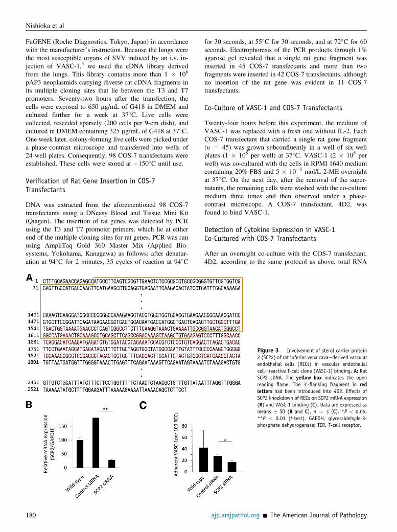
Figure 3 Involvement of sterol carrier protein2 (SCP2) of rat inferior vena cavaederived vascularendothelial cells (RECs) in vascular endothelialcellereactive T-cell clone (VASC-1) binding. A: RatSCP2 cDNA. The yellow box indicates the openreading flame. The 30-flanking fragment in redletters had been introduced into 4D2. Effects ofSCP2 knockdown of RECs on SCP2 mRNA expression(B) and VASC-1 binding (C). Data are expressed asmeans � SD (B and C). n Z 3 (C). *P < 0.05,**P < 0.01 (t-test). GAPDH, glyceraldehyde-3-phosphate dehydrogenase; TCR, T-cell receptor.
Nishioka et al
180 ajp.amjpathol.org - The American Journal of Pathology
was extracted from VASC-1 using an RNeasy Mini kit.RNA underwent reverse transcription to cDNA using tran-scriptase and oligo-dT primers. The primers for rat genesused in this study were IL-5 sense: 50-GACGAGCAAT-GAGACGATGAGGCT-30, IL-5 antisense: 50-ACAGTGC-CCCCTCGGACAGTT-30, and the former GAPDH senseand GAPDH antisense. PCR was run using AmpliTaq Gold360 Master Mix as follows: after denaturation at 94�C for 30seconds, 35 cycles of reaction at 58�C for 30 seconds and at72�C for 30 seconds. For the positive and negative controls,VASC-1 was co-cultured with RECs and with another COS-7 transfectant, 6A4, respectively.
Identification of the Rat Gene Transfected into 4D2
DNA was extracted from the COS-7 transfectant, 4D2,which could bind to VASC-1, using the DNeasy Blood andTissue Mini Kit (Qiagen). The transfected rat gene via thepAP3 neoplasmid was amplified by PCR using the T3 andT7 promoter primers, and then the PCR product wassequenced directly using GenomeLab Dye TerminatorCycle Sequencing with Quick Start Kit (Beckman Coulter,Tokyo, Japan). Consequently, the rat gene transfected into4D2 was identified to code SCP2.
VASC-1 Binding to RECs or REC Knockdown of SCP2
This assay was performed similarly to the assay for VASC-1binding to REC knockdown of CD1d.
Transduction of SCP2 Peptides into COS-7
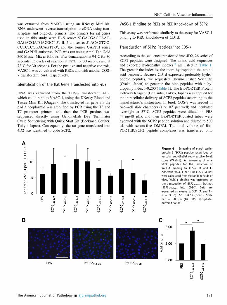
According to the sequence transfected into 4D2, 26 series ofSCP2 peptides were designed. The amino acid sequencesand expected hydropathy indexes18 are listed in Table 1.The greater the index is, the more hydrophobic the aminoacid becomes. Because CD1d expressed preferably hydro-phobic peptides, we requested Thermo Fisher Scientific(Osaka, Japan) to generate the nine peptides with a hy-dropathy index >0.200 (Table 1). The BioPORTER ProteinDelivery Reagent (Genlantis, Tokyo, Japan) was applied forthe intracellular delivery of SCP2 peptides according to themanufacturer’s instruction. In brief, COS-7 was seeded intwo-well slide chambers (1 � 105 per well) and incubatedovernight at 37�C. SCP2 peptides were diluted in PBS(4 mg/40 mL), and then BioPORTER-coated tubes werehydrated with the SCP2 peptide solution and diluted to 500mL with serum-free DMEM. The total volume of Bio-PORTER/SCP2 peptide complexes was transferred onto
Figure 4 Screening of sterol carrierprotein 2 (SCP2) peptide recognized byvascular endothelial cellereactive T-cellclone (VASC-1). A: Screening of nineSCP2 peptides for the induction ofVASC-1 binding to COS-7. B and C:Adherent VASC-1 per 100 COS-7 valueswere calculated from six random fields ofview. VASC-1 binding was increased bythe transduction of rSCP2518-532, but notrSCP2526-540, into COS-7. Data areexpressed as means � SEM (A and C).n Z 3 (C). *P < 0.05 (t-test). Scalebar Z 50 mm (B). PBS, phosphate-buffered saline.
NKT Cells in Vascular Inflammation
The American Journal of Pathology - ajp.amjpathol.org 181
COS-7 cells after being washed with serum-free DMEM,and then these cells were incubated in the culture mediumfor 5 hours at 37�C.
VASC-1 Binding to COS-7 or COS-7 Transduced SCP2Peptides
This assay was performed similarly to the assay for VASC-1binding to RECs.
VASC-1 Proliferation Assay
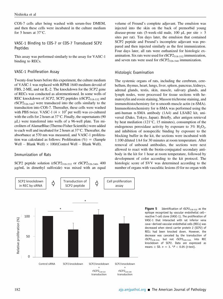
Twenty-four hours before this experiment, the culture mediumof VASC-1 was replaced with RPMI 1640 medium devoid ofFBS, 2-ME, and rat IL-2. The knockdown for the SCP2 geneof RECs was conducted as aforementioned. In some wells ofREC knockdown of SCP2, SCP2 peptides (rSCP2518-532 andrSCP2526-540) were transduced into the cells similarly to thetransduction into COS-7. Thereafter, these cells were washedwith PBS twice. VASC-1 (4 � 105 per well) was co-culturedwith the cells for 2 hours at 37�C. Finally, the supernatants (90mL) were transferred into wells of a 96-well plate. Ten mi-croliters of AlamarBlue (Thermo Fisher Scientific) were addedto each well and incubated for 2 hours at 37�C. Thereafter, theabsorbance at 570 nm was measured, and VASC-1 prolifera-tion was calculated as follows: Proliferation (%) Z (SampleWell � Blank Well) � 100/(Control Well � Blank Well).
Immunization of Rats
SCP2 peptide solution (rSCP2518-532 or rSCP2526-540, 400mg/mL in dimethyl sulfoxide) was mixed with an equal
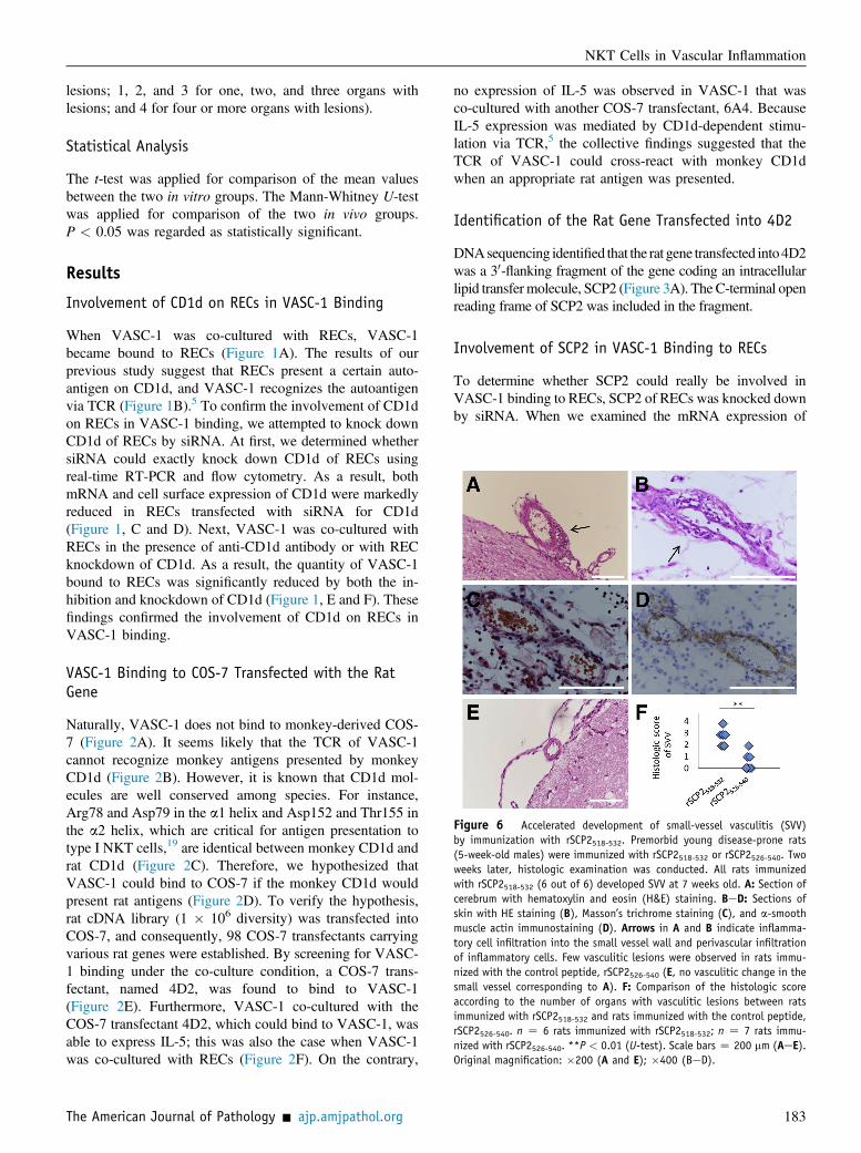
volume of Freund’s complete adjuvant. The emulsion wasinjected into the skin on the back of premorbid youngdisease-prone rats (5-week-old male, 100 mL per site � 5sites per rat). Ten days later, the emulsion that containedSCP2 peptide and Freund’s incomplete adjuvant was pre-pared and then injected similarly as the first immunization.Four days later, all rats were euthanized for histologic ex-amination. Six rats were used for rSCP2518-532 immunization,and seven rats were used for rSCP2526-540 immunization.
Histologic Examination
The systemic organs of rats, including the cerebrum, cere-bellum, thymus, heart, lungs, liver, spleen, pancreas, kidneys,adrenal glands, testis, skin, muscle, salivary glands, andlymph nodes, were processed for tissue sections with he-matoxylin and eosin staining,Masson trichrome staining, andimmunohistochemistry for a-smooth muscle actin (a-SMA).Immunohistochemistry for a-SMA was performed using theanti-human a-SMA antibody (1A4) and LSAB2 kit, uni-versal (Dako, Tokyo, Japan). Briefly, after antigen retrievalby heat mediation (121�C, 15 minutes), consumption of theendogenous peroxidase activity by exposure to 3% H2O2,and inhibition of nonspecific binding by exposure to theblocking buffer in the kit, the sections were incubated with1:100 diluted 1A4 for 30 minutes at room temperature. Afterremoval of unbound antibodies, the sections were nextallowed to react with the biotin-conjugated secondary anti-body in the kit for 1 hour at room temperature, followed bydevelopment of color according to the kit protocol. Thehistologic score of SVV was determined according to thenumber of organs with vasculitic lesions (0 for no organ with
SCP2 knockdownin REC by siRNA
Transduction of SCP2 peptide
Cell proliferationassay
0.00
0.50
1.00
)% ( noitarefilorp evitaleR
100
50
0
Control siRNA
*
*
SCP2 knockdown SCP2 knockdown+
rSCP2518-532transduction
SCP2 knockdown+
rSCP2526-540transduction
Figure 5 Identification of rSCP2518-532 as theepitope recognized by vascular endothelial cellereactive T-cell clone (VASC-1). The proliferation ofVASC-1 that interacted with rat inferior venacavaederived vascular endothelial cells (RECs) wasdecreased when sterol carrier protein 2 (SCP2) ofRECs had been knocked down. However, thedecrease was canceled by the transduction ofrSCP2518-532, but not rSCP2526-540, into RECknockdown of SCP2. Data are expressed asmeans � SD. n Z 3. *P < 0.05 (t-test).
Nishioka et al
182 ajp.amjpathol.org - The American Journal of Pathology
lesions; 1, 2, and 3 for one, two, and three organs withlesions; and 4 for four or more organs with lesions).
Statistical Analysis
The t-test was applied for comparison of the mean valuesbetween the two in vitro groups. The Mann-Whitney U-testwas applied for comparison of the two in vivo groups.P < 0.05 was regarded as statistically significant.
Results
Involvement of CD1d on RECs in VASC-1 Binding
When VASC-1 was co-cultured with RECs, VASC-1became bound to RECs (Figure 1A). The results of ourprevious study suggest that RECs present a certain auto-antigen on CD1d, and VASC-1 recognizes the autoantigenvia TCR (Figure 1B).5 To confirm the involvement of CD1don RECs in VASC-1 binding, we attempted to knock downCD1d of RECs by siRNA. At first, we determined whethersiRNA could exactly knock down CD1d of RECs usingreal-time RT-PCR and flow cytometry. As a result, bothmRNA and cell surface expression of CD1d were markedlyreduced in RECs transfected with siRNA for CD1d(Figure 1, C and D). Next, VASC-1 was co-cultured withRECs in the presence of anti-CD1d antibody or with RECknockdown of CD1d. As a result, the quantity of VASC-1bound to RECs was significantly reduced by both the in-hibition and knockdown of CD1d (Figure 1, E and F). Thesefindings confirmed the involvement of CD1d on RECs inVASC-1 binding.
VASC-1 Binding to COS-7 Transfected with the RatGene
Naturally, VASC-1 does not bind to monkey-derived COS-7 (Figure 2A). It seems likely that the TCR of VASC-1cannot recognize monkey antigens presented by monkeyCD1d (Figure 2B). However, it is known that CD1d mol-ecules are well conserved among species. For instance,Arg78 and Asp79 in the a1 helix and Asp152 and Thr155 inthe a2 helix, which are critical for antigen presentation totype I NKT cells,19 are identical between monkey CD1d andrat CD1d (Figure 2C). Therefore, we hypothesized thatVASC-1 could bind to COS-7 if the monkey CD1d wouldpresent rat antigens (Figure 2D). To verify the hypothesis,rat cDNA library (1 � 106 diversity) was transfected intoCOS-7, and consequently, 98 COS-7 transfectants carryingvarious rat genes were established. By screening for VASC-1 binding under the co-culture condition, a COS-7 trans-fectant, named 4D2, was found to bind to VASC-1(Figure 2E). Furthermore, VASC-1 co-cultured with theCOS-7 transfectant 4D2, which could bind to VASC-1, wasable to express IL-5; this was also the case when VASC-1was co-cultured with RECs (Figure 2F). On the contrary,
no expression of IL-5 was observed in VASC-1 that wasco-cultured with another COS-7 transfectant, 6A4. BecauseIL-5 expression was mediated by CD1d-dependent stimu-lation via TCR,5 the collective findings suggested that theTCR of VASC-1 could cross-react with monkey CD1dwhen an appropriate rat antigen was presented.
Identification of the Rat Gene Transfected into 4D2
DNAsequencing identified that the rat gene transfected into4D2was a 30-flanking fragment of the gene coding an intracellularlipid transfermolecule, SCP2 (Figure 3A). The C-terminal openreading frame of SCP2 was included in the fragment.
Involvement of SCP2 in VASC-1 Binding to RECs
To determine whether SCP2 could really be involved inVASC-1 binding to RECs, SCP2 of RECs was knocked downby siRNA. When we examined the mRNA expression of
Figure 6 Accelerated development of small-vessel vasculitis (SVV)by immunization with rSCP2518-532. Premorbid young disease-prone rats(5-week-old males) were immunized with rSCP2518-532 or rSCP2526-540. Twoweeks later, histologic examination was conducted. All rats immunizedwith rSCP2518-532 (6 out of 6) developed SVV at 7 weeks old. A: Section ofcerebrum with hematoxylin and eosin (H&E) staining. BeD: Sections ofskin with HE staining (B), Masson’s trichrome staining (C), and a-smoothmuscle actin immunostaining (D). Arrows in A and B indicate inflamma-tory cell infiltration into the small vessel wall and perivascular infiltrationof inflammatory cells. Few vasculitic lesions were observed in rats immu-nized with the control peptide, rSCP2526-540 (E, no vasculitic change in thesmall vessel corresponding to A). F: Comparison of the histologic scoreaccording to the number of organs with vasculitic lesions between ratsimmunized with rSCP2518-532 and rats immunized with the control peptide,rSCP2526-540. n Z 6 rats immunized with rSCP2518-532; n Z 7 rats immu-nized with rSCP2526-540. **P < 0.01 (U-test). Scale bars Z 200 mm (AeE).Original magnification: �200 (A and E); �400 (BeD).
NKT Cells in Vascular Inflammation
The American Journal of Pathology - ajp.amjpathol.org 183
SCP2 of RECs transfected with siRNA for SCP2, theexpression was markedly reduced (Figure 3B). Correspond-ingly, VASC-1 binding to RECs was decreased significantlyby the knockdown of SCP2 (Figure 3C). These findings sug-gest the involvement of SCP2 in VASC-1 binding to RECs.
Identification of SCP2 Epitope Recognized by VASC-1
On the basis of the SCP2 gene sequence that had beentransfected into 4D2, we designed 26 series of SCP2 pep-tides (Table 1). Because CD1d expressed preferably hy-drophobic peptides, the nine peptides with a hydropathyindex >0.200 (Table 1) were selected to be generated. Toidentify the epitope of SCP2 recognized by VASC-1, thenine peptides were transduced into COS-7, and then VASC-1 binding was screened. It is conceivable that cell surfaceCD1d molecules have been occupied by putative antigens.Thus, we used the transfection procedure to express theSCP2 peptides on CD1d. We also determined that theaddition of SCP2 peptides in the culture medium could notincrease the VASC-1 proliferation co-cultured with RECknockdown of SCP2 (data not shown). As a result,
transduction of rSCP2518-532 induced the highest binding ofVASC-1 to COS-7 (Figure 4A). On the contrary, trans-duction of rSCP2526-540 did not promote VASC-1 binding toCOS-7 at all. Repeated experiments using rSCP2518-532 andrSCP2526-540 reproduced similar results (Figure 4, B and C).Moreover, we found that REC-dependent proliferation of
VASC-1 was decreased significantly when SCP2 of RECshad been knocked down by siRNA and that the decreasecould be canceled by the transduction of rSCP2518-532 butnot rSCP2526-540 (Figure 5). The collective findings suggestthat rSCP2518-532 includes the epitope recognized byVASC-1.
Accelerated Development of SVV by Immunization withrSCP2518-532
Next, we determined whether immunization withrSCP2518-532 could promote the development of SVV. Forthis purpose, premorbid young disease-prone rats were used.Although SVV usually develops in rats >2 months old,1,20
all rats immunized with rSCP2518-532 (n Z 6/6) developedSVV at 7 weeks old (Figure 6, AeD). The histologic scores
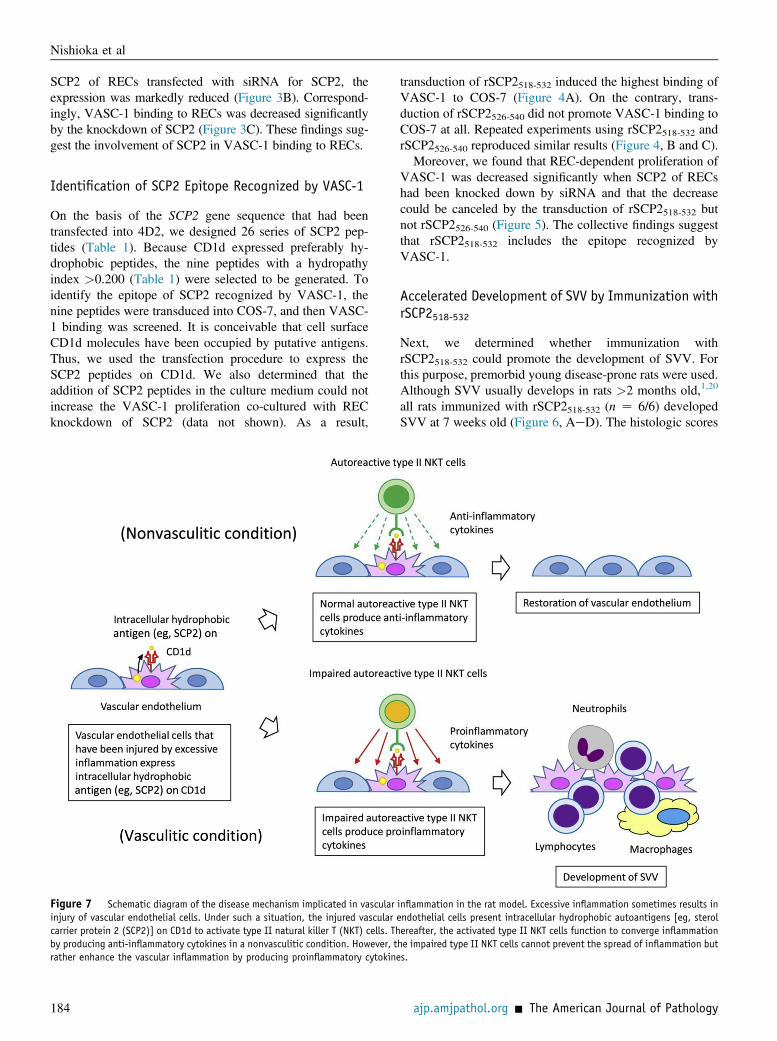
Figure 7 Schematic diagram of the disease mechanism implicated in vascular inflammation in the rat model. Excessive inflammation sometimes results ininjury of vascular endothelial cells. Under such a situation, the injured vascular endothelial cells present intracellular hydrophobic autoantigens [eg, sterolcarrier protein 2 (SCP2)] on CD1d to activate type II natural killer T (NKT) cells. Thereafter, the activated type II NKT cells function to converge inflammationby producing anti-inflammatory cytokines in a nonvasculitic condition. However, the impaired type II NKT cells cannot prevent the spread of inflammation butrather enhance the vascular inflammation by producing proinflammatory cytokines.
Nishioka et al
184 ajp.amjpathol.org - The American Journal of Pathology
according to the number of organs with vasculitic lesionswere significantly higher in rats immunized withrSCP2518-532 than in those immunized with the controlpeptide, rSCP2526-540 (Figure 6, E and F). These resultsindicate that rSCP2518-532-reactive cells, including type IINKT cells, are critically implicated in the development ofvascular inflammation in the rat model.
Discussion
In the present study, we found that the rat type II NKT cellclone, VASC-1, can recognize SCP2 peptides presented byCD1d. CD1d is known as a class I major histocompatibilitycomplexelike antigen-presenting molecule that prefers topresent hydrophobic antigens, including bacterial glyco-lipids.6 Although recent studies have revealed that sulfatidesand peptides can be also presented by CD1d,14,15,21 thehydrophobicity of CD1d is a primary characteristic based onthe molecular structure.
SCP2 is a 13-kDa protein that is implicated in intracel-lular lipid transfer.22 The N-terminal 32 amino acids ofSCP2 form an amphipathic a-helix domain, which functionsas a lipid carrier and a ligand-binding region simulta-neously. On the contrary, the hydrophobic surface of the ahelix constitutes a hollow structure with b-strands in theSCP2 molecule. This hollow structure binds tophospholipids.
The SCP2 gene fragment that had been transduced in theCOS-7 transfectant with VASC-1 binding, 4D2, lacked thetranslation initiation codon for the complete SCP2 molecule.However, it contained some in-frame codons codingmethionine, which could initiate the subsequent translationinto certain fragmented proteins, including a part of theb-strands of the SCP2 molecule. It is conceivable that theSCP2 fragments expressed in 4D2 are hydrophobic in termsof the expected binding to phospholipids. Therefore, it isreasonable to consider that the SCP2 peptides can be pre-sented by CD1d in 4D2. Moreover, this study has revealedthat the SCP2 epitope recognized by VASC-1 is present inrSCP2518-532. This peptide is the most hydrophobic amongthe 26 candidate peptides; hence, it is consistent with thehydrophobicity of CD1d.
More recently, Girardi et al23 reviewed that peptides,which included the [FW]-X-X-[ILM]-X-X-W motif, couldbind to mouse CD1d. Although rSCP2518-532 does notinclude the CD1d-binding motif, another CD1d-bindingpeptide, mCII707-721,
16 does not include the motif either.We considered that diverse antigens could be presented byCD1d.
We do not have any evidence to conclude that the SCP2peptide is the sole autologous antigen presented by CD1d.Because the CD1d molecule can present diverse kinds ofantigens, such as glycolipids, sulfatides, and peptides,regardless of the presence of the [FW]-X-X-[ILM]-X-X-Wmotif, it seems likely that several peptides derived from
intracellular proteins, if hydrophobic, can be presented byCD1d. We were probably able to isolate an NKT cell clonethat recognized the SCP2 peptide by chance.
The expression of SCP2 is distributed widely in the lipidmetabolismerelated organs and cells, including the liverand vascular endothelial cells.24 Although SCP2 is associ-ated with the pathogenesis of arteriosclerosis,25 the associ-ation between SCP2 and vasculitides remains unrevealed.Further studies are needed to clarify the role of SCP2 in thepathogenesis of vasculitides.
Accumulated studies have revealed the immunoregula-tory properties of type II NKT cells in the immune reaction.Liu et al16 found that NKT cells reactive with autologouspeptides (suggestive of type II phenotype) exhibited animmunosuppressive property in healthy mice. Correspond-ingly, the activation of type II NKT cells ameliorated themurine experimental hepatitis.26e28 Furthermore, Terabeet al29 found that type II NKT cells were essential for thedown-regulation of tumor immunosurveillance in wild-typemice. On the contrary, the association of the disorder of typeII NKT cells with inflammatory diseases is also reported.Liao et al30 found that the disordered regulation of type IINKT cells could cause a spontaneous development of colitisin CD1d transgenic mice. In summary, type II NKT cellsbelong to the immunoregulatory T cells, and the disorder ofthese cells can be implicated in immune-related inflamma-tory diseases. In our previous study, the type II NKT cellclone, VASC-1, had proinflammatory but not anti-inflammatory properties when made to react with autolo-gous vascular endothelial cells.5
In conclusion, the collective evidence suggests that adisordered immunoregulatory function of autoreactive typeII NKT cells can be implicated in the development ofvascular inflammation in the rat model. The impairment ofvascular endothelial cell-reactive type II NKT cells with animmunoregulatory property could be implicated in thepathogenic modification of diverse human vasculitides, inwhich vascular endothelial cells are injured by inflammation(Figure 7). Although a full comprehension of the patho-genesis of vasculitides remains far off, this study can be astream of light that illuminates the pathway.
References
1. Yamazaki H, Ikeda H, Ishizu A, Nakamaru Y, Sugaya T, Kikuchi K,Yamada S, Wakisaka A, Kasai N, Koike T, Hatanaka M, Yoshiki T: Awide spectrum of collagen vascular and autoimmune diseases intransgenic rats carrying the env-pX gene of human T lymphocyte virustype I. Int Immunol 1997, 9:339e346
2. Nakamaru Y, Ishizu A, Ikeda H, Sugaya T, Fugo K, Higuchi M,Yamazaki H, Yoshiki T: Immunological hyperresponsiveness inHTLV-I LTR-env-pX transgenic rats: a prototype animal model forcollagen vascular and HTLV-I-related inflammatory diseases. Patho-biology 2001, 69:11e18
3. Fugo K, Ishizu A, Ikeda H, Hayase H, Sugaya T, Higuchi M, Tsuji M,Abe A, Suzuki A, Shibata M, Takahashi T, Yoshiki T: The role of thethymus in development of necrotizing arteritis in transgenic rats
NKT Cells in Vascular Inflammation
The American Journal of Pathology - ajp.amjpathol.org 185
carrying the env-pX gene of human T-cell leukemia virus type-I. Am JPathol 2002, 161:755e761
4. Higuchi M, Ishizu A, Ikeda H, Hayase H, Fugo K, Tsuji M, Abe A,Sugaya T, Suzuki A, Takahashi T, Koike T, Yoshiki T: Functionalalteration of peripheral CD25þCD4þ immunoregulatory T cells in atransgenic rat model of autoimmune diseases. J Autoimmun 2003, 20:43e49
5. Iinuma C, Waki M, Kawakami A, Yamaguchi M, Tomaru U, Sasaki N,Masuda S, Matsui Y, Iwasaki S, Baba T, Kasahara M, Yoshiki T,Paletta D, Herrmann T, Ishizu A: Establishment of a vascular endo-thelial cell-reactive type II NKT cell clone from a rat model of auto-immune vasculitis. Int Immunol 2015, 27:105e114
6. Wu L, Gabriel CL, Parekh VV, Van Kaer L: Invariant natural killer Tcells: innate-like T cells with potent immunomodulatory activities.Tissue Antigens 2009, 73:535e545
7. Girardi E, Zajonc DM: Molecular basis of lipid antigen presentation byCD1d and recognition by natural killer T cells. Immunol Rev 2012,250:167e179
8. Rossjohn J, Pellicci DG, Patel O, Gapin L, Godfrey DI: Recognition ofCD1d-restricted antigens by natural killer T cells. Nat Rev Immunol2012, 12:845e857
9. Godfrey DI, MacDonald HR, Kronenberg M, Smyth MJ, VanKaer L: NKT cells: what’s in a name? Nat Rev Immunol 2004, 4:231e237
10. Macho-Fernandez E, Brigl M: The Extended Family of CD1d-Restricted NKT Cells: sifting through a Mixed Bag of TCRs, Anti-gens, and Functions. Front Immunol 2015, 6:362
11. Matsuura A, Kinebuchi M, Chen HZ, Katabami S, Shimizu T,Hashimoto Y, Kikuchi K, Sato N: NKT cells in the rat: organ-specificdistribution of NK T cells expressing distinct Va 14 chains. J Immunol2000, 164:3140e3148
12. Godfrey DI, Stankovic S, Baxter AG: Raising the NKT cell family. NatImmunol 2010, 11:197e206
13. Monzon-Casanova E, Paletta D, Starick L, Muller I, Sant’Angelo DB,Pyz E, Herrmann T: Direct identification of rat iNKT cells revealsremarkable similarities to human iNKT cells and a profound deficiencyin LEW rats. Eur J Immunol 2013, 43:404e415
14. Girardi E, Maricic I, Wang J, Mac TT, Iyer P, Kumar V, Zajonc DM:Type II natural killer T cells use features of both innate-like andconventional T cells to recognize sulfatide self antigens. Nat Immunol2012, 13:851e856
15. Patel O, Pellicci DG, Gras S, Sandoval-Romero ML, Uldrich AP,Mallevaey T, Clarke AJ, Le Nours J, Theodossis A, Cardell SL,Gapin L, Godfrey DI, Rossjohn J: Recognition of CD1d-sulfatidemediated by a type II natural killer T cell antigen receptor. NatImmunol 2012, 13:857e863
16. Liu Y, Teige A, Mondoc E, Ibrahim S, Holmdahl R, Issazadeh-Navikas S: Endogenous collagen peptide activation of CD1d-restricted
NKT cells ameliorates tissue-specific inflammation in mice. J ClinInvest 2011, 121:249e264
17. Ishizu A, Ishikura H, Nakamaru Y, Takeuchi E, Kimura C, Koike T,Yoshiki T: Thy-1 induced on rat endothelium regulates vascularpermeability at sites of inflammation. Int Immunol 1995, 7:1939e1947
18. Kyte J, Doolittle RF: A simple method for displaying the hydropathiccharacter of a protein. J Mol Biol 1982, 157:105e132
19. Zajonc DM, Kronenberg M: CD1 mediated T cell recognition ofglycolipids. Curr Opin Struct Biol 2007, 17:521e529
20. Ishizu A, Yoshiki T: Pathogenesis of vasculitis in env-pX Rats. AnnVasc Dis 2012, 5:296e299
21. Zeng Z, Castano AR, Segelke BW, Stura EA, Peterson PA, Wilson IA:Crystal structure of mouse CD1: an MHC-like fold with a large hy-drophobic binding groove. Science 1997, 277:339e345
22. Filipp FV, Sattler M: Conformational plasticity of the lipid transferprotein SCP2. Biochemistry 2007, 46:7980e7991
23. Girardi E, Wang J, Zajonc DM: Structure of an alpha-helical peptideand lipopeptide bound to the nonclassical major histocompatibilitycomplex (MHC) class I molecule CD1d. J Biol Chem 2016, 291:10677e10683
24. Schroeder F, Atshaves BP, McIntosh AL, Gallegos AM, Storey SM,Parr RD, Jefferson JR, Ball JM, Kier AB: Sterol carrier protein-2: newroles in regulating lipid rafts and signaling. Biochim Biophys Acta2007, 1771:700e718
25. Hirai A, Kino T, Tokinaga K, Tahara K, Tamura Y, Yoshida S:Regulation of sterol carrier protein 2 (SCP2) gene expression in ratperitoneal macrophages during foam cell formation: a key role for freecholesterol content. J Clin Invest 1994, 94:2215e2223
26. Halder RC, Aguilera C, Maricic I, Kumar V: Type II NKT cell-mediated anergy induction in type I NKT cells prevents inflamma-tory liver disease. J Clin Invest 2007, 117:2302e2312
27. Arrenberg P, Halder R, Kumar V: Cross-regulation between distinctnatural killer T cell subsets influences immune response to self andforeign antigens. J Cell Physiol 2009, 218:246e250
28. Maricic I, Sheng H, Marrero I, Seki E, Kisseleva T, Chaturvedi S,Molle N, Mathews SA, Gao B, Kumar V: Inhibition of type I naturalkiller T cells by retinoids or following sulfatide-mediated activation oftype II natural killer T cells attenuates alcoholic liver disease in mice.Hepatology 2015, 61:1357e1369
29. Terabe M, Swann J, Ambrosino E, Sinha P, Takaku S, Hayakawa Y,Godfrey DI, Ostrand-Rosenberg S, Smyth MJ, Berzofsky JA: Anonclassical non-Va14Ja18 CD1d-restricted (type II) NKT cell issufficient for down-regulation of tumor immunosurveillance. J ExpMed 2005, 202:1627e1633
30. Liao CM, Zimmer MI, Shanmuganad S, Yu HT, Cardell SL,Wang CR: Dysregulation of CD1d-restricted type II natural killer Tcells leads to spontaneous development of colitis in mice. Gastroen-terology 2012, 142:326e334
Nishioka et al
186 ajp.amjpathol.org - The American Journal of Pathology


![The Sterol Methyltransferases SMT1, SMT2, and … Sterol Methyltransferases SMT1, SMT2, and SMT3 Influence Arabidopsis Development through Nonbrassinosteroid Products1[W][OA] Francine](https://img.pdfslide.net/doc/110x75/5ce0f3ae88c993700d8b6654/the-sterol-methyltransferases-smt1-smt2-and-sterol-methyltransferases-smt1-smt2.jpg)

![Membrány a membránový transport - ulbld.lf1.cuni.cz · STEROL LIPIDS STEROL LIPIDS = lipid molecules with backbone derived from cyclopenta[a]phenanthrene (?) Division according](https://img.pdfslide.net/doc/110x75/5e14db8b3fcccd648c5ac62a/membrny-a-membrnov-transport-ulbldlf1cunicz-sterol-lipids-sterol-lipids.jpg)


![Structural basis of sterol recognition and nonvesicular ...myweb.chonnam.ac.kr/~stbiochm/Publications/PDF/[2018 PNAS] LAM.pdf · Structural basis of sterol recognition and nonvesicular](https://img.pdfslide.net/doc/110x75/5c95af6109d3f2de7d8d04e3/structural-basis-of-sterol-recognition-and-nonvesicular-myweb-stbiochmpublicationspdf2018.jpg)