Embed Size (px)
Citation preview

INFECTION AND IMMUNITY,0019-9567/01/$04.00�0 DOI: 10.1128/IAI.69.11.6785–6795.2001
Nov. 2001, p. 6785–6795 Vol. 69, No. 11
Copyright © 2001, American Society for Microbiology. All Rights Reserved.
Type III Secretion-Dependent Cell Cycle Block Caused in HeLa Cellsby Enteropathogenic Escherichia coli O103
JEAN-PHILIPPE NOUGAYREDE,* MICHELE BOURY, CHRISTIAN TASCA, OLIVIER MARCHES,ALAIN MILON, ERIC OSWALD, AND JEAN DE RYCKE
UMR 960 de Microbiologie Moleculaire, Institut National de la Recherche Agronomique-EcoleNationale Veterinaire de Toulouse, 31076 Toulouse Cedex, France
Received 21 February 2001/Returned for modification 30 May 2001/Accepted 3 August 2001
Rabbit enteropathogenic Escherichia coli (EPEC) O103 induces in HeLa cells an irreversible cytopathic effectcharacterized by the recruitment of focal adhesions, formation of stress fibers, and inhibition of cell prolif-eration. We have characterized the modalities of the proliferation arrest and investigated its underlyingmechanisms. We found that HeLa cells that were exposed to the rabbit EPEC O103 strain E22 progressivelyaccumulated at 4C DNA content and did not enter mitosis. A significant proportion of the cells were able toreinitiate DNA synthesis without division, leading to 8C DNA content. This cell cycle inhibition by E22 wasabrogated in mutants lacking EspA, -B, and -D and was restored by transcomplementation. In contrast, intiminand Tir mutants retained the antiproliferative effect. The cell cycle arrest was not a direct consequence of theformation of stress fibers, since their disruption by toxins during exposure to E22 did not reverse the cell cycleinhibition. Likewise, the cell cycle arrest was not dependent on the early tyrosine dephosphorylation eventstriggered by E22 in the cells. Two key partner effectors controlling entry into mitosis were also investigated:cyclin B1 and the associated cyclin-dependent kinase 1 (Cdk1). Whereas cyclin B1 was not detectably affectedin E22-exposed cells, Cdk1 was maintained in a tyrosine-phosphorylated inactive state and lost its affinity forp13suc1-agarose beads. This shows that Cdk1 is implicated in the G2/M arrest caused by EPEC strain E22.
Enteropathogenic Escherichia coli (EPEC) constitutes a ma-jor cause of severe diarrheal disease in infants of the develop-ing world (33). EPEC bacteria colonize the intestinal mucosaand produce specific attaching-and-effacing (A/E) lesions ongut enterocytes, characterized by intimate bacterial adhesion,formation of gross cytoskeletal structures beneath intimatelyattached bacteria, and destruction of the brush border mi-crovilli (32). The intimate adhesion is considered to play acentral role in EPEC-mediated disease, but the mechanisms bywhich EPEC causes diarrhea remains poorly characterized.
In the human reference EPEC strain E2348/69, the deter-minants of A/E lesions are encoded within a 35-kb chromo-somal pathogenicity island, the locus of enterocyte effacement(LEE) (30). Similar pathogenicity islands are present in rabbitEPEC O103, in rabbit EPEC O15 strain RDEC-1, in entero-hemorrhagic E. coli, and in other A/E pathogens (10, 14). TheLEE encodes a type III secretion system homologous to thosefound in other pathogens, dedicated to the secretion and trans-location of pathogenicity-associated proteins (20, 28). At leastfive proteins are secreted via this type III machinery, namely,EspA, -B, -D, and -F and Tir (21–23, 27, 31). EspA formsappendages that link the bacteria to target cells to allow thetranslocation of EspB and Tir into host cells (23, 25, 49).Recent experimental data suggest that EspB and EspD mightbe inserted in the target cell membrane to function as trans-locators for effector proteins (26, 46, 48, 49). In addition, EspBis found in the cytoplasm of exposed cells, and when transientlyexpressed in cultured cells it promotes actin rearrangements
(44, 49). Once inserted in the target cell membrane, Tir servesas a receptor for an EPEC outer membrane protein, intimin(23, 40). This interaction leads to the local rearrangement ofthe cytoskeleton together with intimate adhesion of the bacte-rium on the host cell.
In addition to intimate adhesion, EPEC bacteria also mod-ulate signaling pathways within cultured cells. EPEC bacteriaactivate protein kinase C and alter the phosphorylation state ofseveral host proteins (14). Tyrosine dephosphorylation of sev-eral unidentified host proteins was correlated with the inhibi-tion of phagocytosis by cultured macrophage cells (24, 15).Tyrosine phosphorylation of phospholipase C-�1 and induc-tion of inositol triphosphate and Ca2� fluxes have been de-scribed, but increased intracellular Ca2� is not required forA/E lesion formation (14, 24). EPEC increases paracellularpermeability and stimulates ion secretion (7, 41). The integra-tion of the A/E lesion and host cell responses in the inductionof diarrhea is not fully elucidated.
We have previously shown that rabbit EPEC O103, the rab-bit EPEC O15 strain RDEC-1, and certain human clinicalEPEC isolates, but not the human reference EPEC strainE2348/69, trigger in HeLa cells an irreversible cytopathic effect(CPE). The CPE is characterized by the progressive recruit-ment of focal adhesions and assembly of stress fibers (10, 35).The cytoskeletal alterations are associated with an arrest of cellproliferation, as assessed by the inhibition of protein synthesis(10). The CPE is not reproducible by using supernatants orconcentrated sonicates of CPE-positive EPEC strains, nor is itattributable to cytotoxic necrotizing factor, a toxin known toalso cause the irreversible formation of actin stress fibers (10,36). The triggering of the cytoskeletal alterations depends on afunctional EPEC type III secretion machinery and requiresEspA, -B, and -D but not Tir or intimin (10, 29, 35). No single
* Corresponding author. Present address: Division of Infectious Dis-eases, University of Maryland at Baltimore, 10 S. Pine St., Baltimore,MD 21201. Phone: (410) 706-7560. Fax: (410) 706-8700. E-mail:[email protected].
6785
on July 18, 2018 by guesthttp://iai.asm
.org/D
ownloaded from

espA, espB, or espD gene encodes the specific informationneeded to trigger the cytoskeletal rearrangements, since eachespA, espB, or espD mutant is fully complemented by the cor-responding esp gene cloned from CPE-negative E2348/69 (35).
In the present study, we have investigated in more detail thearrest of cell proliferation that is associated with the cytoskel-etal rearrangements. We found that cells exposed to rabbitEPEC strain E22 irreversibly accumulated at 4C and 8C DNAcontent without entering mitosis. This effect was not function-ally related to cytoskeletal rearrangement but was linked to themaintenance of the cyclin-dependent kinase Cdk1, a key effec-tor driving entry into mitosis, in a premitotic, tyrosine-phos-phorylated state.
MATERIALS AND METHODS
Bacteria and HeLa cell cultures. EPEC strains and their mutants are listed inTable 1. The E22 espA, espB, espD, tir, and eae mutants were shown to benonpolar and are described elsewhere (29, 35). Before interaction with cellcultures, bacteria were grown at 37°C in Penassay broth (Difco) supplementedwith appropriate antibiotics. HeLa cells (ATCC CCL2) were cultivated in Ea-gle’s minimum essential medium (MEM) supplemented with 10% fetal calfserum (FCS) (Gibco), L-glutamine (200 mM), and gentamicin (40 �g/ml) at 37°Cin a 5% CO2 atmosphere. Synchronization of HeLa cells at the G1/S border wascarried out with nonconfluent cell cultures (106 cells in a 10-cm-diameter culturedish) by the double thymidine block method, and synchronization in promet-aphase was achieved using nocodazole (100 nM for 16 h) (8). Type 1 cytolethaldistending toxin (CDT-I) was prepared and added to the cells as describedpreviously (8, 11). Labeling the cells with 5-bromo-2�-deoxyuridine (BrdU) (5�g/ml; Boehringer) was achieved for 30 min or 6 h.
Interaction between HeLa cells and bacteria. This assay was described previ-ously (10). Briefly, interactions were carried out in MEM buffered with 25 mMHEPES supplemented with 5% FCS and 1% mannose, with a starting inoculumof 103 bacteria per cell. At the end of the 4-h interaction period, the cells werewashed four to six times with Earle balanced saline solution and fixed, or theywere further incubated in MEM with 10% FCS and 200 �g of gentamicin/ml for24, 48, or 72 h.
For stress fiber inhibition experiments, cells were preincubated for 2 h in theinteraction medium in the presence of 1 �g of purified epidermal cell differen-tiation inhibitor (EDIN) (kindly provided by M. Sugai [43])/ml or a 1:100 dilutionof a filtered sonic lysate of BL21(pDC3B) (a gift from P. Boquet) containing theDC3B chimeric toxin (3). Bacteria were then added and left in contact with cellsfor 4 h as described above. Control cells were pretreated with a lysate of BL21without the plasmid.
For tyrosine phosphatase inhibition experiments, we used phenylarsine oxide(PAO) (1 �M; Calbiochem) or pervanadate (PV). PV was freshly made bymixing hydrogen peroxide (1 �l of 30% stock) with 25 �l of 500 mM sodiumvanadate (Na3VO4) (Sigma) in 424 �l of phosphate-buffered saline (pH 7.4). Theremaining hydrogen peroxide was removed by treatment with catalase (100�g/ml) (Sigma). PV was added to the cells at a 1:1,000 dilution after 2 h of EPECinfection. Control cells were pretreated with the solution lacking sodium vana-date.
The trypan blue uptake assay was performed as already described (9). Thelactate dehydrogenase (LDH) release assay was performed on HeLa cells in
96-well plates. After the interaction, the supernatant was collected and the LDHwas quantified with the cytotoxicity kit from Roche Molecular Biochemicals,according to the manufacturer’s recommendations.
Fluorescence microscopy. Microtubules were stained with rat anti-�-tubulin(clone YL1/2; Sera-lab) and fluorescein isothiocyanate (FITC)-conjugated rabbitanti-rat immunoglobulin G (IgG) antibodies (Vector) as already described (8).DNA was stained with diaminophenylindole (0.1 �g/ml; Molecular Probes).F-actin was labeled with rhodamine-phalloidin according to the manufacturer’sinstructions (Molecular Probes). Focal adhesions were demonstrated by labelingvinculin with anti-vinculin mouse monoclonal antibodies (clone VIN-11-5;Sigma) and FITC-conjugated anti-mouse IgG goat antiserum (Immunotech) aspreviously described (10). Slides were examined by fluorescence microscopy witha Leica microscope.
Flow cytometry analysis. The flow cytometry analysis of DNA and cyclin B1content of HeLa cells was performed as described previously (8). Briefly, cellswere trypsinized, fixed in 70% ethanol, permeabilized with 0.25% Triton X-100,and incubated with anti-cyclin B1 mouse monoclonal antibodies (clone GNS1;Pharmingen) followed by FITC-conjugated goat anti-mouse IgG (Sigma). Con-trol experiments were achieved using an irrelevant isotypic mouse IgG (Immu-notech). In the final step, cells were suspended in a DNA staining solutioncontaining propidium iodide (10 �g/ml; Sigma) and RNase (1 mg/ml; Sigma) inphosphate-buffered saline.
Incorporated BrdU was labeled according to a procedure previously described(12), including a thermal denaturation of DNA at 100°C followed by indirectimmunofluorescence using anti-BrdU mouse monoclonal antibodies (clone 3D4;Pharmingen) and FITC-conjugated goat anti-mouse IgG (Sigma). DNA stainingwas then performed as described above.
Flow cytometry analyses were performed on a FACScalibur flow cytometer(Becton Dickinson), using the red (630 nm) emission for DNA quantification andgreen (530 nm) for BrdU and cyclin B1 quantification. The data from at least 104
cells were collected and analyzed using CellQuest and ModFit softwares (BectonDickinson). Cell aggregates were identified and removed from analysis by gating.
Western blotting analysis of phosphotyrosine proteins. HeLa cells lysis andimmunoprecipitation of phosphotyrosine proteins were carried out using TritonX-100 as described elsewhere (24). Protein samples were resolved by sodiumdodecyl sulfate–7% polyacrylamide gel electrophoresis (SDS–7% PAGE) andblotted onto Immobilon-P membranes, and tyrosine-phosphorylated proteinswere revealed with antiphosphotyrosine monoclonal antibodies (clone 4G10;Upstate Biotechnology) followed by secondary goat anti-mouse IgG (Fab-spe-cific)-peroxidase conjugate (Sigma) and then developed with the ECL chemilu-minescence detection system (Amersham).
Demonstration of Cdk1 in E22-exposed cells. HeLa cells (5 � 105) were lysedin 100 �l of Laemmli buffer, and the samples (40 �g of proteins, from about 105
cells) were resolved by SDS–12% PAGE. Cdk1 was demonstrated by directWestern blotting using the PhosphoPlus cdc2(Tyr15) antibody kit (New EnglandBiolabs) following the recommendations of the manufacturer. In other experi-ments, Cdk1 (Cdc2) was affinity purified using yeast p13suc1 and revealed aspreviously described (8, 45). Briefly, 2 � 106 cells were lysed in 300 �l of modifiedRIPA buffer, and Cdk1 was purified from 200 �g of total proteins by affinity toyeast recombinant p13suc1 -agarose (Upstate Biotechnology). Samples were re-solved by SDS–12% PAGE and blotted onto Immobilon-P. The three isoformsof Cdk1 were demonstrated with rabbit anti-Cdk1 antibodies (Gibco) followedby secondary goat anti-rabbit IgG-peroxidase conjugate (Biosys). After stripping,the slow-migrating tyrosine-phosphorylated isoform of Cdk1 was revealed with4G10 monoclonal antibodies as before.
Raw data of the blots were scanned with an Astra 1220S scanner (Umax), and
TABLE 1. EPEC strains used in this study
Strain Description (reference)
E2348/69 ..........................................................................................Prototype O127:H6 human EPEC strainE22 ...................................................................................................Prototype rabbit EPEC O103:K-:H2, rhamnose-negative strain (10)E22�Eae..........................................................................................E22 eae::aphT, nonpolar Eae� mutant (35)E22�Tir ...........................................................................................E22 tir::aphT, nonpolar Tir� mutant (29)E22�EspA .......................................................................................E22 espA::aphT, nonpolar EspA� mutant (35)E22�EspB .......................................................................................E22 espB::aphT, nonpolar EspB� mutant (35)E22�EspD.......................................................................................E22 espD::aphT, nonpolar EspD� mutant (35)E22�EspA(pBRespAEPEC) ...........................................................E22�EspA transformed with espA from E2348/69 cloned into pBRSK vector (35)E22�EspB(pBRespBEPEC)............................................................E22�EspB transformed with espB from E2348/69 cloned into pBRSK vector (35)E22�EspD(pBRespDEPEC)...........................................................E22�EspD transformed with espD from E2348/69 cloned into pBRSK vector (35)
6786 NOUGAYREDE ET AL. INFECT. IMMUN.
on July 18, 2018 by guesthttp://iai.asm
.org/D
ownloaded from

the protein amount in each revealed band was estimated by densitometry, usingNational Institutes of Health image software (available at http://rsb.info.nih.gov/nih-image/).
RESULTS
EPEC strain E22 triggers an irreversible inhibition of mi-tosis. HeLa cells exposed to E22 for 4 h were irreversiblyimpaired in their proliferation, as assessed by cell counting(Fig. 1A). In contrast, cells exposed to E2348/69 were inhibitedfor about 24 h, after which their growth rate was similar to thatof control cells (Fig. 1A). Morphologically, cells exposed toE22 progressively swelled and flattened, whereas cells exposedto E2348/69 behaved like control cells (data not shown), inagreement with previous observations (10).
This irreversible arrest in cell proliferation indicated that thecell cycle could be altered in a specific manner in E22-exposedcells. We thus analyzed the DNA content of cells exposed toE22 or E2348/69 by flow cytometry. The cell cycle distributionof cells exposed to E2348/69 was similar to that of control cells24 h (Fig. 1B) or 48 to 72 h after the interaction (not shown).
In contrast, cells exposed to E22 progressively accumulated inG2/M (4C DNA content), while the number of cells in G0/G1
(2C DNA content) declined (Fig. 1B). In addition, a third peakat 8C DNA units was visible 72 h after the infection with E22(Fig. 1B).
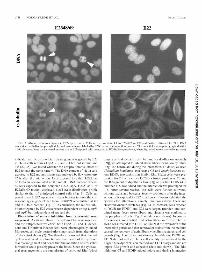
The foregoing results strongly suggested that the cells ex-posed to E22 were prevented from entering mitosis. In order tosubstantiate this result, DNA and �-tubulin were stained inE2348/69- or E22-exposed cells 24 h after the interaction. Asshown by fluorescence microscopy, no figure of chromatin con-densation (characteristic of prophase) or reorganization of mi-crotubules into mitotic spindle was observed in E22-exposedcells (Fig. 2). These cells appeared mononucleated, and theirnuclei were swollen (Fig. 2). In contrast, cells exposed toE2348/69 were actively dividing, showing typical mitotic spin-dle and figures of chromatin condensation (Fig. 2). For bothE22- and E2348/69-exposed cells, these observations were con-firmed 48 and 72 h after the interaction (not shown).
E22-induced mitosis inhibition is EspA, -B, and -D depen-dent but Tir and intimin independent. Our previous works
FIG. 1. Demonstration of the irreversible arrest in HeLa cell proliferation (A) and cell cycle perturbation (B) triggered by E22. Cells wereexposed for 4 h to E2348/69 or E22 or left uninfected, and they were further cultivated for the indicated times. (A) Cell proliferation afterinteraction. Cultures were fixed and stained with Giemsa, and cells within random microscope fields were counted (objective, �20). Each pointis the mean of four independent measures. (B) Cell distribution according to DNA content, analyzed by flow cytometry after staining of DNA withpropidium iodide. The percentages of cell populations are shown in each case.
VOL. 69, 2001 CELL CYCLE ARREST CAUSED BY EPEC 6787
on July 18, 2018 by guesthttp://iai.asm
.org/D
ownloaded from
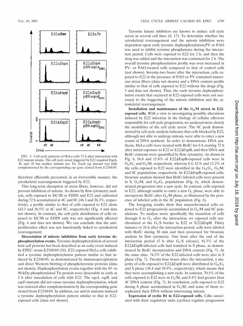
indicate that the cytoskeletal rearrangement triggered by E22in HeLa cells requires EspA, -B, and -D but not intimin andTir (29, 35). We tested whether the antiproliferative effect ofE22 follows the same pattern. The DNA content of HeLa cellsexposed to E22 mutant strains was analyzed by flow cytometry72 h after the interaction. Cells exposed to either E22�Eaeor E22�Tir accumulated at 4C and 8C DNA content, where-as cells exposed to the nonpolar E22�EspA, E22�EspB, orE22�EspD mutant displayed a cell cycle distribution profilesimilar to that of uninfected control cells (Fig. 3). Cells ex-posed to each E22 esp mutant strain bearing in trans the cor-responding esp gene cloned from E2348/69 accumulated at 4Cand 8C DNA content (Fig. 3). In conclusion, the mitosis inhi-bition triggered by E22 was a process dependent on espA, espB,and espD but independent of eae and tir.
Dissociation of mitosis inhibition from cytoskeletal rear-rangement. As shown above, the cytoskeletal rearrangementand the antiproliferative effect, both EspA, -B, and -D depen-dent and Tir/intimin independent, were phenotypically linked.Moreover, cell cycle perturbations may result from alterationsof the cytoskeleton (2). We therefore reasoned that the cellcycle arrest could be a functional consequence of the cytoskel-etal rearrangement and hence that the inhibition of stress fiberformation could possibly prevent the block. Since the cytoskel-etal rearrangements are reminiscent of activated Rho (which
plays a central role in stress fiber and focal adhesion assembly[39]), we attempted to inhibit stress fibers formation by inhib-iting Rho before and during the interaction. To do so, we usedClostridium botulinum exoenzyme C3 and Staphylococcus au-reus EDIN, two toxins that inhibit Rho. HeLa cells were pre-treated for 2 h with either DC3B (a fusion protein of C3 andthe B fragment of diphtheria toxin [3]) or purified EDIN (43),and then E22 was added and the interaction was prolonged for4 h. After several washes, the cells were further cultivatedwithout toxins and bacteria. Seventy-two hours after the inter-action, cells exposed to E22 in absence of toxins exhibited thecytoskeleton alterations, namely, numerous stress fibers andclustered vinculin stretches (Fig. 4). In contrast, cells exposedto DC3B (or EDIN) and E22 were larger, rounder, and con-tained many fewer stress fibers, and vinculin was confined tothe periphery of cells (Fig. 4 and data not shown). In controlexperiments, we verified that actin fibers were disrupted inHeLa cells treated with DC3B or EDIN at the expiration of theinteraction period and that removal of toxins from the mediumcaused the recovery of actin fibers, vinculin structures, and cellgrowth (Fig. 4 and data not shown). In addition, DC3B andEDIN did not reduce HeLa cell viability (as assessed by theTrypan blue dye exclusion method and LDH assay) and did notimpair E22 growth and adhesion (data not shown). The Rhoinhibitors C3 and EDIN added before and during interaction
FIG. 2. Absence of mitotic figures in E22-exposed cells. Cells were exposed for 4 h to E2348/69 or E22 and further cultivated for 24 h. DNAwas stained with diaminophenylindole, and �-tubulin was labeled by FITC indirect immunofluorescence. The same fields were photographed with a�100 objective. Note the increased nuclear size in E22-exposed cells, compared to E2348/69-exposed cells, where figures of mitosis are visible (arrows).
6788 NOUGAYREDE ET AL. INFECT. IMMUN.
on July 18, 2018 by guesthttp://iai.asm
.org/D
ownloaded from
therefore efficiently prevented, in an irreversible manner, thecytoskeletal rearrangement triggered by E22.
This long-term disruption of stress fibers, however, did notprevent inhibition of mitosis. As shown by flow cytometry anal-ysis, cells exposed to DC3B or EDIN and E22 and cultivatedduring 72 h accumulated at 4C and 8C (46.3 and 36.3%, respec-tively), a profile similar to that of cells exposed to E22 alone(43.3 and 36.5% at 4C and 8C, respectively) (Fig. 4 and datanot shown). In contrast, the cell cycle distribution of cells ex-posed to DC3B or EDIN only was not significantly affected(Fig. 4 and data not shown). We can conclude that the anti-proliferative effect was not functionally linked to cytoskeletalrearrangement.
Dissociation of mitosis inhibition from early tyrosine de-phosphorylation events. Tyrosine dephosphorylation of severalhost cell proteins has been described as an early event inducedby EPEC strain E2348/69 (24). E22-exposed HeLa cells exhib-ited a tyrosine dephosphorylation pattern similar to that in-duced by E2348/69, as demonstrated by immunoprecipitationand direct Western blotting of phosphotyrosine proteins (datanot shown). Dephosphorylation events together with the 85- to90-kDa phosphorylated Tir protein were detectable as early as2 h after inoculation of cells with E22. The espA, espB, andespD mutants did not cause tyrosine dephosphorylation, whichwas restored after complementation by the corresponding genecloned from E2348/69. In addition, eae and tir mutants induceda tyrosine dephosphorylation pattern similar to that in E22-exposed cells (data not shown).
Tyrosine kinase inhibitors are known to induce cell cyclearrest in several cell lines (6, 17). To determine whether thecytoskeletal rearrangement and the mitosis inhibition weredependent upon early tyrosine dephosphorylation,PV or PAOwas used to inhibit tyrosine phosphatases during the interac-tion period. Cells were exposed to E22 for 2 h, and then thedrug was added and the interaction was continued for 2 h. Theoverall tyrosine phosphorylation profile was even increased inPV- or PAO-treated cells compared to that of control cells(not shown). Seventy-two hours after the interaction, cells ex-posed to E22 in the presence of PAO or PV contained numer-ous stress fibers (data not shown) and a DNA content profilesimilar to that of cells exposed to E22 without the drugs (Fig.5 and data not shown). Thus, the early tyrosine dephosphory-lation events that occurred in E22-exposed cells were not nec-essary to the triggering of the mitosis inhibition and the cy-toskeletal rearrangement.
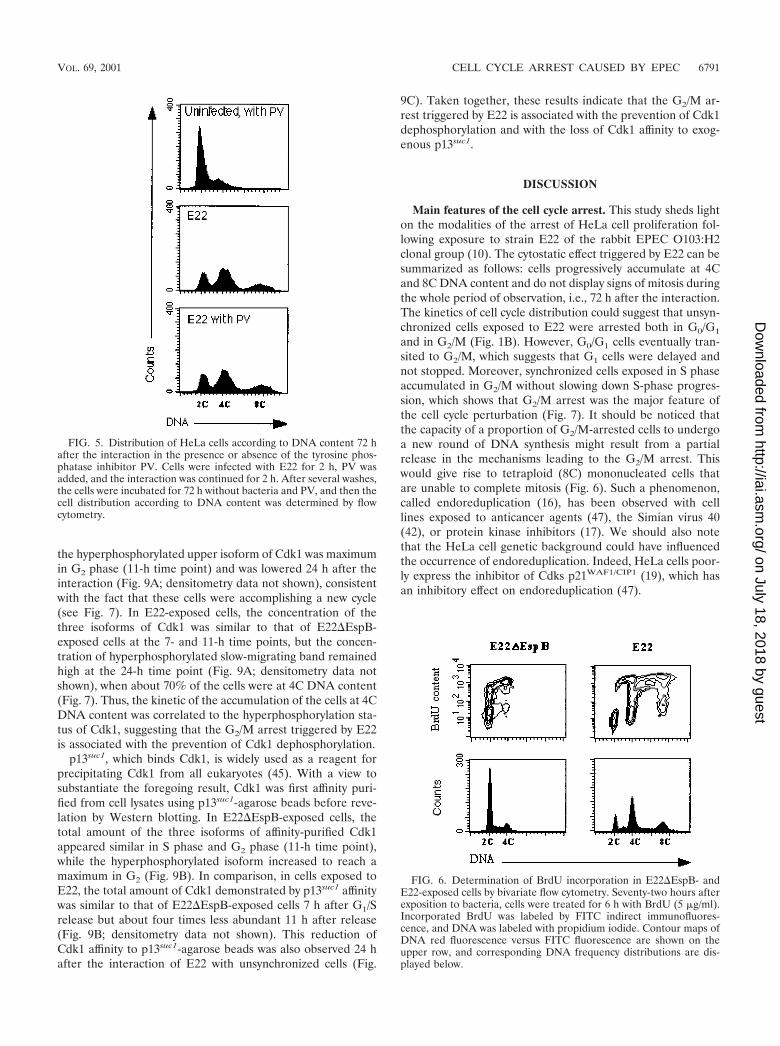
Installation and maintenance of the G2/M arrest in E22-exposed cells. With a view to investigating possible alterationsinduced by E22 infection in the biology of cellular effectorsresponsible for cell cycle progression, we analyzed more closelythe modalities of the cell cycle arrest. The 8C peak demon-strated by cell cycle analysis indicates that cells blocked by E22,although not able to undergo mitosis, were able to enter a newround of DNA synthesis. In order to demonstrate DNA syn-thesis, HeLa cells were treated with BrdU for 6 h starting 72 hafter initial exposure to E22 or E22�EspB, and then DNA andBrdU contents were quantified by flow cytometry. As shown inFig. 6, 56.8 and 15.8% of E22�EspB-exposed cells were inG0/G1 and G2/M, respectively, whereas 8.4, 42.9, and 15.2% ofthe cells exposed to E22 were identified in the G0/G1, G2/M,and 8C populations, respectively. In E22�EspB-exposed cells,bivariate analysis showed that BrdU-labeled cells were presentin S, G2/M, and G0/G1 populations (Fig. 6), which demon-strated progression into a new cycle. In contrast, cells exposedto E22, although unable to enter a new G1 phase, were able toincorporate BrdU after G2/M phase, as illustrated by the pres-ence of labeled cells in the 8C population (Fig. 6).
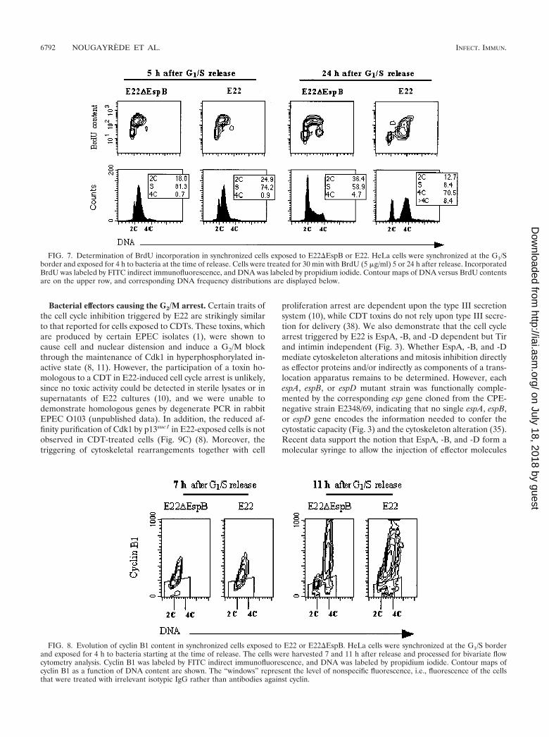
The foregoing results show that unsynchronized cells ex-posed to E22 progressively accumulated in G2/M and 8C pop-ulations. To analyze more specifically the transition of cellsthrough S to G2 after the interaction, we exposed cells syn-chronized at the G1/S border to E22 or E22�EspB. Thirtyminutes or 24 h after the interaction period, cells were labeledwith BrdU during 30 min and then processed for bivariateanalysis by flow cytometry. One hour after the end of theinteraction period (5 h after G1/S release), 81.3% of theE22�EspB-infected cells had transited in S phase, as demon-strated by BrdU incorporation and DNA content (Fig. 7). Atthe same time, 74.2% of the E22-infected cells were also in Sphase (Fig. 7). Twenty-four hours after the interaction, a ma-jority of cells exposed to E22�EspB were distributed in G0/G1
and S phase (36.4 and 58.9%, respectively), which means thatthey were accomplishing a new cycle. In contrast, 70.5% of thecells exposed to E22 were in G2/M, and 8.4% had greater than4C DNA content (Fig. 7). In conclusion, cells exposed to E22during S phase accumulated in G2/M, and some of them re-replicated their DNA without intervening mitosis.
Expression of cyclin B1 in E22-exposed cells. Cdks associ-ated with their regulatory units (cyclins) regulate progression
FIG. 3. Cell cycle patterns of HeLa cells 72 h after interaction withE22 mutant strains. The cell cycle arrest triggered by E22 required EspA,-B, and -D but neither intimin nor Tir. Each esp mutant was fullycomplemented by the corresponding esp gene cloned from E2348/69.
VOL. 69, 2001 CELL CYCLE ARREST CAUSED BY EPEC 6789
on July 18, 2018 by guesthttp://iai.asm
.org/D
ownloaded from

through the eukaryotic cell cycle. More specifically, cyclin B1and Cdk1 control entry into mitosis. The cellular concentrationof cyclin B1 is an immediate determinant of the transition fromG2 to mitosis (34). To test whether cyclin B1 expression wasaffected by exposition to E22, cells synchronized in G1/S wereinfected as before, and the cyclin B1 concentration as a func-tion of DNA content was determined by bivariate flow cytom-etry analysis. In cells exposed to E22�EspB, cyclin B1 accu-mulated during S phase (7 h after G1/S release) and reached amaximum 11 h after G1/S release (Fig. 8), when the majority ofcells were in G2 and about 5% were in mitosis (not shown).E22-exposed cells showed a similar cyclin B1 accumulation inS and G2/M (Fig. 8). This indicated that cyclin B1 synthesis wasnot affected by exposition to E22 and prompted us to investi-gate Cdk1.
Cdk1 modification in E22-exposed cells. Cdk1 is expressedat a constant level over the different phases of the cell cycle,but its phosphorylation level increases during interphase to
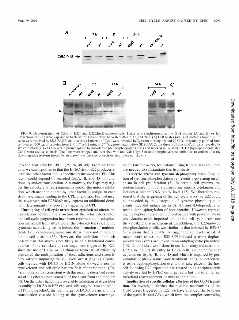
reach a maximum in G2. Initiation of mitosis is then triggeredby the activation of Cdk1, which results from dephosphoryla-tion of the Tyr-15 residue (34). The amount of Cdk1 and itstyrosine phosphorylation status were determined in cells syn-chronized in G1/S and exposed to bacteria as before. To vali-date the method, we used cells arrested in prometaphase bynocodazole (resulting in dephosphorylated active Cdk1) andcells blocked in G2/M by treatment with CDT-I (resulting inhyperphosphorylated inactive Cdk1 [8]). The three isoformsof Cdk1 were demonstrated by direct Western blotting usinganti-Cdk1 antibody (Fig. 9A and B), and after stripping ofthe membrane, antiphosphotyrosine antibodies were used toconfirm that the slow-migrating Cdk1 isoform was tyrosinephosphorylated (not shown). As expected, the fast-migratingdephosphorylated isoform of Cdk1 was dominant in nocoda-zole-treated cells, while in CDT-I-treated cells the slow-mi-grating hyperphosphorylated isoform of Cdk1 was prevalent(Fig. 9A). In E22�EspB-exposed cells, the concentration of
FIG. 4. Cytoskeletal rearrangement and cell distribution according to DNA content of HeLa cells 72 h after exposition to E22 in the presenceor absence of the Rho inhibitor DC3B. HeLa cells were pretreated for 2 h with DC3B, and then E22 was added and the interaction was continuedfor 4 h. Control cells were treated with DC3B but left uninfected. After several washes, the cells were incubated for 72 h without bacteria and toxin.F-actin was stained with rhodamine-phalloidin, and vinculin was labeled by FITC indirect immunofluorescence. Corresponding cell distributionaccording to DNA content was determined by flow cytometry.
6790 NOUGAYREDE ET AL. INFECT. IMMUN.
on July 18, 2018 by guesthttp://iai.asm
.org/D
ownloaded from
the hyperphosphorylated upper isoform of Cdk1 was maximumin G2 phase (11-h time point) and was lowered 24 h after theinteraction (Fig. 9A; densitometry data not shown), consistentwith the fact that these cells were accomplishing a new cycle(see Fig. 7). In E22-exposed cells, the concentration of thethree isoforms of Cdk1 was similar to that of E22�EspB-exposed cells at the 7- and 11-h time points, but the concen-tration of hyperphosphorylated slow-migrating band remainedhigh at the 24-h time point (Fig. 9A; densitometry data notshown), when about 70% of the cells were at 4C DNA content(Fig. 7). Thus, the kinetic of the accumulation of the cells at 4CDNA content was correlated to the hyperphosphorylation sta-tus of Cdk1, suggesting that the G2/M arrest triggered by E22is associated with the prevention of Cdk1 dephosphorylation.
p13suc1, which binds Cdk1, is widely used as a reagent forprecipitating Cdk1 from all eukaryotes (45). With a view tosubstantiate the foregoing result, Cdk1 was first affinity puri-fied from cell lysates using p13suc1-agarose beads before reve-lation by Western blotting. In E22�EspB-exposed cells, thetotal amount of the three isoforms of affinity-purified Cdk1appeared similar in S phase and G2 phase (11-h time point),while the hyperphosphorylated isoform increased to reach amaximum in G2 (Fig. 9B). In comparison, in cells exposed toE22, the total amount of Cdk1 demonstrated by p13suc1 affinitywas similar to that of E22�EspB-exposed cells 7 h after G1/Srelease but about four times less abundant 11 h after release(Fig. 9B; densitometry data not shown). This reduction ofCdk1 affinity to p13suc1-agarose beads was also observed 24 hafter the interaction of E22 with unsynchronized cells (Fig.
9C). Taken together, these results indicate that the G2/M ar-rest triggered by E22 is associated with the prevention of Cdk1dephosphorylation and with the loss of Cdk1 affinity to exog-enous p13suc1.
DISCUSSION
Main features of the cell cycle arrest. This study sheds lighton the modalities of the arrest of HeLa cell proliferation fol-lowing exposure to strain E22 of the rabbit EPEC O103:H2clonal group (10). The cytostatic effect triggered by E22 can besummarized as follows: cells progressively accumulate at 4Cand 8C DNA content and do not display signs of mitosis duringthe whole period of observation, i.e., 72 h after the interaction.The kinetics of cell cycle distribution could suggest that unsyn-chronized cells exposed to E22 were arrested both in G0/G1
and in G2/M (Fig. 1B). However, G0/G1 cells eventually tran-sited to G2/M, which suggests that G1 cells were delayed andnot stopped. Moreover, synchronized cells exposed in S phaseaccumulated in G2/M without slowing down S-phase progres-sion, which shows that G2/M arrest was the major feature ofthe cell cycle perturbation (Fig. 7). It should be noticed thatthe capacity of a proportion of G2/M-arrested cells to undergoa new round of DNA synthesis might result from a partialrelease in the mechanisms leading to the G2/M arrest. Thiswould give rise to tetraploid (8C) mononucleated cells thatare unable to complete mitosis (Fig. 6). Such a phenomenon,called endoreduplication (16), has been observed with celllines exposed to anticancer agents (47), the Simian virus 40(42), or protein kinase inhibitors (17). We should also notethat the HeLa cell genetic background could have influencedthe occurrence of endoreduplication. Indeed, HeLa cells poor-ly express the inhibitor of Cdks p21WAF1/CIP1 (19), which hasan inhibitory effect on endoreduplication (47).
FIG. 6. Determination of BrdU incorporation in E22�EspB- andE22-exposed cells by bivariate flow cytometry. Seventy-two hours afterexposition to bacteria, cells were treated for 6 h with BrdU (5 �g/ml).Incorporated BrdU was labeled by FITC indirect immunofluores-cence, and DNA was labeled with propidium iodide. Contour maps ofDNA red fluorescence versus FITC fluorescence are shown on theupper row, and corresponding DNA frequency distributions are dis-played below.
FIG. 5. Distribution of HeLa cells according to DNA content 72 hafter the interaction in the presence or absence of the tyrosine phos-phatase inhibitor PV. Cells were infected with E22 for 2 h, PV wasadded, and the interaction was continued for 2 h. After several washes,the cells were incubated for 72 h without bacteria and PV, and then thecell distribution according to DNA content was determined by flowcytometry.
VOL. 69, 2001 CELL CYCLE ARREST CAUSED BY EPEC 6791
on July 18, 2018 by guesthttp://iai.asm
.org/D
ownloaded from
Bacterial effectors causing the G2/M arrest. Certain traits ofthe cell cycle inhibition triggered by E22 are strikingly similarto that reported for cells exposed to CDTs. These toxins, whichare produced by certain EPEC isolates (1), were shown tocause cell and nuclear distension and induce a G2/M blockthrough the maintenance of Cdk1 in hyperphosphorylated in-active state (8, 11). However, the participation of a toxin ho-mologous to a CDT in E22-induced cell cycle arrest is unlikely,since no toxic activity could be detected in sterile lysates or insupernatants of E22 cultures (10), and we were unable todemonstrate homologous genes by degenerate PCR in rabbitEPEC O103 (unpublished data). In addition, the reduced af-finity purification of Cdk1 by p13suc1 in E22-exposed cells is notobserved in CDT-treated cells (Fig. 9C) (8). Moreover, thetriggering of cytoskeletal rearrangements together with cell
proliferation arrest are dependent upon the type III secretionsystem (10), while CDT toxins do not rely upon type III secre-tion for delivery (38). We also demonstrate that the cell cyclearrest triggered by E22 is EspA, -B, and -D dependent but Tirand intimin independent (Fig. 3). Whether EspA, -B, and -Dmediate cytoskeleton alterations and mitosis inhibition directlyas effector proteins and/or indirectly as components of a trans-location apparatus remains to be determined. However, eachespA, espB, or espD mutant strain was functionally comple-mented by the corresponding esp gene cloned from the CPE-negative strain E2348/69, indicating that no single espA, espB,or espD gene encodes the information needed to confer thecytostatic capacity (Fig. 3) and the cytoskeleton alteration (35).Recent data support the notion that EspA, -B, and -D form amolecular syringe to allow the injection of effector molecules
FIG. 7. Determination of BrdU incorporation in synchronized cells exposed to E22�EspB or E22. HeLa cells were synchronized at the G1/Sborder and exposed for 4 h to bacteria at the time of release. Cells were treated for 30 min with BrdU (5 �g/ml) 5 or 24 h after release. IncorporatedBrdU was labeled by FITC indirect immunofluorescence, and DNA was labeled by propidium iodide. Contour maps of DNA versus BrdU contentsare on the upper row, and corresponding DNA frequency distributions are displayed below.
FIG. 8. Evolution of cyclin B1 content in synchronized cells exposed to E22 or E22�EspB. HeLa cells were synchronized at the G1/S borderand exposed for 4 h to bacteria starting at the time of release. The cells were harvested 7 and 11 h after release and processed for bivariate flowcytometry analysis. Cyclin B1 was labeled by FITC indirect immunofluorescence, and DNA was labeled by propidium iodide. Contour maps ofcyclin B1 as a function of DNA content are shown. The “windows” represent the level of nonspecific fluorescence, i.e., fluorescence of the cellsthat were treated with irrelevant isotypic IgG rather than antibodies against cyclin.
6792 NOUGAYREDE ET AL. INFECT. IMMUN.
on July 18, 2018 by guesthttp://iai.asm
.org/D
ownloaded from
into the host cells by EPEC (25, 26, 48, 49). From all thesedata, we can hypothesize that the EPEC strain E22 produces atleast one other factor that is specifically involved in CPE. Thisfactor could depend on secreted EspA, -B, and -D for func-tionality and/or translocation. Alternatively, the Esps may trig-ger the cytoskeletal rearrangements and/or the mitosis inhibi-tion, which are then altered by other factor(s) unique to eachstrain, eventually leading to the CPE phenotype. For instance,the negative strain E2348/69 may express an additional domi-nant determinant that prevents triggering of CPE.
Uncoupling of cell cycle arrest from cytoskeletal alteration.Correlation between the structure of the actin cytoskeletonand cell cycle progression have been reported: endoreduplica-tion may result from alteration of the cytoskeleton (2), and thecytotoxic necrotizing toxins induce the formation of multinu-cleated cells containing numerous stress fibers and in parallelinhibit cell division (36). However, the inhibition of mitosisobserved in this study is not likely to be a functional conse-quence of the cytoskeletal rearrangement triggered by E22,since the use of EDIN or C3 chimeric toxin DC3B efficientlyprevented the multiplication of focal adhesions and stress fi-bers without impairing the cell cycle arrest (Fig. 4). Controlcells treated with DC3B or EDIN alone showed a normalcytoskeleton and cell cycle pattern 72 h after treatment (Fig.4), an observation consistent with the recently described rever-sal of C3 effects upon removal of the toxin from the medium(4). On the other hand, the irreversible inhibition of stress fiberassembly by DC3B in E22-exposed cells suggests that the smallGTP-binding RhoA, the main target of DC3B, is crucial in thetransduction cascade leading to the cytoskeleton rearrange-
ment. Further works, for instance using Rho mutant cell lines,are needed to substantiate this hypothesis.
Cell cycle arrest and tyrosine dephosphorylation. Regula-tion of tyrosine phosphorylation represents a governing mech-anism in cell proliferation (5). In certain cell systems, theprotein kinase inhibitor staurosporine impairs cytokinesis andinduces a higher DNA ploidy level (17). We therefore rea-soned that the triggering of the cell cycle arrest by E22 couldbe preceded by the disruption of tyrosine phosphorylationevents. E22 did induce an EspA, -B, and -D-dependent ty-rosine dephosphorylation of host proteins. However, repress-ing the dephosphorylation induced by E22 with pervanadate orphenylarsine oxide impaired neither the cell cycle arrest northe cytoskeletal rearrangement. In addition, the E22-inducedphosphorylation profile was similar to that induced by E2348/69, a strain that is unable to trigger the cell cycle arrest. Arecent work shows that E2348/69-induced tyrosine dephos-phorylation events are linked to an antiphagocytic phenotype(15). Unpublished work done in our laboratory indicates thatE22 also inhibits its entry in HeLa cells, an inhibition thatdepends on EspA, -B, and -D and which is impaired by per-vanadate or phenylarsine oxide treatment. Thus, the detectabletyrosine dephosphorylation events that take place in the hostcell following E22 exposition are related to an antiphagocyticactivity exerted by EPEC on target cells but not to either cy-toskeletal rearrangement or mitosis inhibition.
Implication of specific cellular effectors of the G2-M transi-tion. To investigate further the possible mechanisms of theG2/M arrest triggered by E22, we have analyzed the behaviorof the cyclin B1 and Cdk1, which form the complex controlling
FIG. 9. Demonstration of Cdk1 in E22- and E22�EspB-exposed cells. HeLa cells synchronized at the G1/S border (A and B) or leftunsynchronized (C) were exposed to bacteria for 4 h and were harvested after 7, 11, and 24 h. (A) Cell lysates (40 �g of proteins from 5 � 105
cells) were resolved by SDS-PAGE, and the three isoforms of Cdk1 were revealed by Western blotting. (B and C) Cdk1 was affinity-purified fromcell lysates (200 �g of proteins from 2 � 106 cells) using p13suc1-agarose beads. After SDS-PAGE, the three isoforms of Cdk1 were revealed byWestern blotting. Cells blocked in prometaphase by nocodazole (dephosphorylated Cdk1) and blocked in G2/M by CDT-I (hyperphosphorylatedCdk1) were used as controls. The blots were stripped and reprobed with anti-Cdk1-Tyr15 or anti-phosphotyrosine antibodies to confirm that theslow-migrating isoform (noted by an arrow) was tyrosine phosphorylated (data not shown).
VOL. 69, 2001 CELL CYCLE ARREST CAUSED BY EPEC 6793
on July 18, 2018 by guesthttp://iai.asm
.org/D
ownloaded from

the transition from G2 to mitosis. In normal cells, cyclin B1accumulates during S phase to reach a maximum in late G2,whereas Cdk1 is present at a constant level over the differentphases of the cell cycle. Cdk1 activity is downregulated byphosphorylation of Tyr-15 and Thr-14 residues, which remainphosphorylated during interphase until onset of M phase (34).The G2/M arrest triggered by E22 could not be accounted forby a lack of cyclin B1 expression, since cyclin B1 expression wasnot affected in synchronous cells exposed to E22 (Fig. 8). Onthe other hand, we observed in E22-exposed synchronous cellsan accumulation of inactive Cdk1 phosphorylated on Tyr-15(Fig. 9A), in association with the accumulation of the cells at4C DNA content (Fig. 7). Since Cdk1 dephosphorylation is aprerequisite for its activation and entry into mitosis, the lack ofCdk1 dephosphorylation may account for the G2/M arrest. Afurther clue to the determinism of the cell cycle arrest trig-gered by E22 is provided by our observation that the G2/Marrest was associated with a drastic reduction of the level ofCdk1 affinity purified with p13suc1-agarose beads (Fig. 9B andC). p13suc1 is the founding member of the cyclin-dependentkinase subunit (Cks) family of proteins that bind and regulateCdks, and it is widely used as a reagent for precipitating Cdk’sfrom all eukaryotes (45). We can hypothesize that the bindingof an endogenous Cks protein may have impaired binding ofCdk1 to p13suc1-agarose beads. Indeed, overexpression of Cksabolishes entry into mitosis and causes an accumulation ofinactive Cdk1 phosphorylated on Tyr-15 (13, 37). Alternative-ly, a putative Cdk1 alteration could alter its affinity to p13suc1
and participate in the cell cycle perturbation. The fact thatcertain cell cycle yeast mutants, carrying temperature-sensitiveCdk alleles, show a decrease in p13suc1-bound Cdk withoutchange in overall Cdk levels supports this idea (4a). The ki-netics of association of Cdk1 with cyclin B1 and endogenousCks together with their subcellular localization should now beassessed in order to explain the defect of exogenous p13suc1
affinity to Cdk1 and further unravel the alteration of cell cyclemachinery in E22-exposed cells. Elucidation of the abnormalbehavior of Cdk1 toward p13suc1 may provide a clue on up-stream signaling events triggered upon E22 interaction andeventually preventing cell entry into mitosis.
Concluding remarks. The cell cycle arrest triggered byE22 appears to be relevant to other CPE-positive strains ofthe rabbit EPEC O103:H2 clonal group, rabbit EPEC O15strain RDEC-1, and some human clinical EPEC isolates(10), since they had a similar effect on the HeLa cell cycle(data not shown). Is a modulation of the eukaryotic cellcycle relevant in EPEC pathogenesis? A cell cycle arrest ofstem cells in the crypts of Lieberkhun, which supply cells tointestinal villi, could reduce the shedding of epithelia andtherefore prolong the local existence of attached bacteria. Inaddition, the ability to inhibit proliferation could constitutea powerful weapon for immune evasion. There is emergingevidence that a growing family of pathogenic bacteria cansubvert the eukaryotic cell cycle (18). Future work should huntfor a putative mitosis-inhibiting factor translocated into host cellsby EPEC in an EspABD-dependent manner and should evalu-ate the impact of such a cell cycle modulation activity on thenatural history of disease.
ACKNOWLEDGMENTS
We are indebted to M. Sugai for the kind gift of the purified EDINand to P. Boquet for the gift of the plasmid encoding DC3B. We thankJ. R. Seavitt, S. Boullier, and S. Peres for useful advice, C. Watrin fortechnical assistance, and E. Blank for critical reading of the manu-script.
This work was supported by grants from the Region Midi-Pyrenees,from INRA (AIP Microbiologie), and from the DGER and by grant1335 from the European Community Program FAIR. J.-P.N. was arecipient of a scholarship from INRA and Biove Company, and O.M.was a recipient of a scholarship from ENVT.
REFERENCES
1. Albert, M. J., S. M. Faruque, A. S. Faruque, K. A. Bettelheim, P. K. Neogi,N. A. Bhuiyan, and J. B. Kaper. 1996. Controlled study of cytolethal dis-tending toxin-producing Escherichia coli infections in Bangladeshi children.J. Clin. Microbiol. 34:717–719.
2. Assoian, R. K., and X. Zhu. 1997. Cell anchorage and the cytoskeleton aspartners in growth factor dependent cell cycle progression. Curr. Opin. CellBiol. 9:93–98.
3. Aullo, P., M. Giry, S. Olsnes, M. R. Popoff, C. Kocks, and P. Boquet. 1993.A chimeric toxin to study the role of the 21 kDa GTP binding protein rho inthe control of actin microfilament assembly. EMBO J. 12:921–931.
4. Barth, H., C. Olenik, P. Sehr, G. Schmidt, K. Aktories, and D. K. Meyer.1999. Neosynthesis and activation of rho by Escherichia coli cytotoxic ne-crotizing factor (CNF1) reverse cytopathic effects of ADP-ribosylated Rho.J. Biol. Chem. 274:27407–27414.
4a. Brizuela, L., G. Draetta, and Beach D. 1987. p13suc1 acts in the fission yeastcell division cycle as a component of the p34cdc2 protein kinase. EMBO J.6:3507–3514.
5. Chernoff, J. 1999. Protein tyrosine phosphatases as negative regulators ofmitogenic signaling. J. Cell. Physiol. 180:173–181.
6. Choi, Y. H., L. Zhang, W. H. Lee, and K. Y. Park. 1998. Genistein-inducedG2/M arrest is associated with the inhibition of cyclin B1 and the inductionof p21 in human breast carcinoma cells. Int. J. Oncol. 13:391–396.
7. Collington, G. K., I. W. Booth, and S. Knutton. 1998. Rapid modulation ofelectrolyte transport in Caco-2 cell monolayers by enteropathogenic Esche-richia coli (EPEC) infection. Gut 42:200–207.
8. Comayras, C., C. Tasca, S. Y. Peres, B. Ducommun, E. Oswald, and J. DeRycke. 1997. Escherichia coli cytolethal distending toxin blocks the HeLa cellcycle at the G2/M transition by preventing cdc2 protein kinase dephosphor-ylation and activation. Infect. Immun. 65:5088–5095.
9. Crane, J. K., S. Majumdar, and D. F. Pickhardt III. 1999. Host cell deathdue to enteropathogenic Escherichia coli has features of apoptosis. Infect.Immun. 67:2575–2584.
10. De Rycke, J., E. Comtet, C. Chalareng, M. Boury, C. Tasca, and A. Milon.1997. Enteropathogenic Escherichia coli O103 from rabbit elicits actin stressfibers and focal adhesions in HeLa epithelial cells, cytopathic effects that arelinked to an analog of the locus of enterocyte effacement. Infect. Immun.65:2555–2563.
11. De Rycke, J., V. Sert, C. Comayras, and C. Tasca. 2000. Sequence of lethalevents in HeLa cells exposed to the G2 blocking cytolethal distending toxin.Eur. J. Cell Biol. 79:192–201.
12. Dolbeare, F., and J. R. Selden. 1994. Immunochemical quantitation of bro-modeoxyuridine: application to cell-cycle kinetics. Methods Cell Biol. 41:297–316.
13. Dunphy, W. G., and J. W. Newport. 1989. Fission yeast p13 blocks mitoticactivation and tyrosine dephosphorylation of the Xenopus cdc2 protein ki-nase. Cell 58:181–191.
14. Frankel, G., A. D. Phillips, I. Rosenshine, G. Dougan, J. B. Kaper, and S.Knutton. 1998. Enteropathogenic and enterohaemorrhagic Escherichia coli:more subversive elements. Mol. Microbiol. 30:911–921.
15. Goosney, D. L., J. Celli, B. Kenny, and B. B. Finlay. 1999. EnteropathogenicEscherichia coli inhibits phagocytosis. Infect. Immun. 67:490–495.
16. Grafi, G. 1998. Cell cycle regulation of DNA replication: the endoredupli-cation perspective. Exp. Cell Res. 244:372–378.
17. Hall, L. L., J. P. Th’ng, X. W. Guo, R. L. Teplitz, and E. M. Bradbury. 1996.A brief staurosporine treatment of mitotic cells triggers premature exit frommitosis and polyploid cell formation. Cancer Res. 56:3551–3559.
18. Henderson, B., M. Wilson, and J. Hyams. 1998. Cellular microbiology: cy-cling into the millennium. Trends Cell Biol. 8:384–387.
19. Hwang, E. S., L. K. Naeger, and D. DiMaio. 1996. Activation of the endog-enous p53 growth inhibitory pathway in HeLa cervical carcinoma cells byexpression of the bovine papillomavirus E2 gene. Oncogene 12:795–803.
20. Jarvis, K. G., J. A. Giron, A. E. Jerse, T. K. McDaniel, M. S. Donnenberg,and J. B. Kaper. 1995. Enteropathogenic Escherichia coli contains a putativetype III secretion system necessary for the export of proteins involved in attach-ing and effacing lesion formation. Proc. Natl. Acad. Sci. USA 92:7996–8000.
21. Kenny, B., and B. B. Finlay. 1995. Protein secretion by enteropathogenicEscherichia coli is essential for transducing signals to epithelial cells. Proc.
6794 NOUGAYREDE ET AL. INFECT. IMMUN.
on July 18, 2018 by guesthttp://iai.asm
.org/D
ownloaded from
Natl. Acad. Sci. USA 92:7991–7995.22. Kenny, B., L. C. Lai, B. B. Finlay, and M. S. Donnenberg. 1996. EspA, a
protein secreted by enteropathogenic Escherichia coli, is required to inducesignals in epithelial cells. Mol. Microbiol. 20:313–323.
23. Kenny, B., R. DeVinney, M. Stein, D. J. Reinscheid, E. A. Frey, and B. B.Finlay. 1997. Enteropathogenic E. coli (EPEC) transfers its receptor forintimate adherence into mammalian cells. Cell 91:511–520.
24. Kenny, B., and B. B. Finlay. 1997. Intimin-dependent binding of entero-pathogenic Escherichia coli to host cells triggers novel signaling events,including tyrosine phosphorylation of phospholipase C-gamma1. Infect. Im-mun. 65:2528–2536.
25. Knutton, S., I. Rosenshine, M. J. Pallen, I. Nisan, B. C. Neves, C. Bain, C.Wolff, G. Dougan, and G. Frankel. 1998. A novel EspA-associated surfaceorganelle of enteropathogenic Escherichia coli involved in protein translo-cation into epithelial cells. EMBO J. 17:2166–2176.
26. Kresse, A. U., M. Rohde, and C. A. Guzman. 1999. The EspD protein ofenterohemorrhagic Escherichia coli is required for the formation of bacterialsurface appendages and is incorporated in the cytoplasmic membranes oftarget cells. Infect. Immun. 67:4834–4842.
27. Lai, L. C., L. A. Wainwright, K. D. Stone, and M. S. Donnenberg. 1997. Athird secreted protein that is encoded by the enteropathogenic Escherichiacoli pathogenicity island is required for transduction of signals and forattaching and effacing activities in host cells. Infect. Immun. 65:2211–2217.
28. Lee, C. A. 1997. Type III secretion systems: machines to deliver bacterialproteins into eukaryotic cells? Trends Microbiol. 5:148–156.
29. Marches, O., J. P. Nougayrede, S. Boullier, J. Mainil, G. Charlier, I. Ray-mond, P. Pohl, M. Boury, J. De Rycke, A. Milon, and E. Oswald. 2000. Roleof Tir and intimin in the virulence of rabbit enteropathogenic Escherichiacoli (REPEC) of serotype O103:H2. Infect. Immun. 68:2171–2182.
30. McDaniel, T. K., and J. B. Kaper. 1997. A cloned pathogenicity island fromenteropathogenic Escherichia coli confers the attaching and effacing pheno-type on E. coli K-12. Mol. Microbiol. 23:399–407.
31. McNamara, B. P., and M. S. Donnenberg. 1998. A novel proline-rich protein,EspF, is secreted from enteropathogenic Escherichia coli via the type IIIexport pathway. FEMS Microbiol. Lett. 166:71–78.
32. Moon, H. W., S. C. Whipp, R. A. Argenzio, M. M. Levine, and R. A. Gian-nella. 1983. Attaching and effacing activities of rabbit and human entero-pathogenic Escherichia coli in pig and rabbit intestines. Infect. Immun. 41:1340–1351.
33. Nataro, J. P., and J. B. Kaper. 1998. Diarrheagenic Escherichia coli. Clin.Microbiol. Rev. 11:142–201.
34. Norbury, C., and P. Nurse. 1992. Animal cell cycles and their control. Annu.Rev. Biochem. 61:441–470.
35. Nougayrede, J. P., O. Marches, M. Boury, J. Mainil, G. Charlier, P. Pohl,J. De Rycke, A. Milon, and E. Oswald. 1999. The long-term cytoskeletal
rearrangement induced by rabbit enteropathogenic Escherichia coli is Espdependent but intimin independent. Mol. Microbiol. 31:19–30.
36. Oswald, E., M. Sugai, A. Labigne, H. C. Wu, C. Fiorentini, P. Boquet, andA. D. O’Brien. 1994. Cytotoxic necrotizing factor type 2 produced by virulentEscherichia coli modifies the small GTP-binding proteins Rho involved inassembly of actin stress fibers. Proc. Natl. Acad. Sci. USA 91:3814–3818.
37. Patra, D., and W. G. Dunphy. 1996. Xe-p9, a Xenopus Suc1/Cks homolog,has multiple essential roles in cell cycle control. Genes Dev. 10:1503–1515.
38. Pickett, C. L., and C. A. Whitehouse. 1999. The cytolethal distending toxinfamily. Trends Microbiol. 7:292–297.
39. Ridley, A. J., and A. Hall. 1992. The small GTP-binding protein rho regulatesthe assembly of focal adhesions and actin stress fibers in response to growthfactors. Cell 70:389–399.
40. Rosenshine, I., S. Ruschkowski, M. Stein, D. J. Reinscheid, S. D. Mills, andB. B. Finlay. 1996. A pathogenic bacterium triggers epithelial signals to forma functional bacterial receptor that mediates actin pseudopod formation.EMBO J. 15:2613–2624.
41. Savkovic, S. D., A. Koutsouris, and G. Hecht. 1997. Activation of NF-kappaBin intestinal epithelial cells by enteropathogenic Escherichia coli. Am. J.Physiol. 273:C1160–C1167.
42. Scarano, F. J., J. A. Laffin, J. M. Lehman, and T. D. Friedrich. 1994. Simianvirus 40 prevents activation of M-phase-promoting factor during lytic infec-tion. J. Virol. 68:2355–2361.
43. Sugai, M., K. Hashimoto, A. Kikuchi, S. Inoue, H. Okumura, K. Matsumoto,Y. Goto, H. Ohgai, K. Moriishi, B. Syuto, et al. 1992. Epidermal cell differ-entiation inhibitor ADP ribosylates small GTP-binding proteins and induceshyperplasia of epidermis. J. Biol. Chem. 267:2600–2604.
44. Taylor, K. A., P. W. Luther, and M. S. Donnenberg. 1999. Expression of theEspB protein of enteropathogenic Escherichia coli within HeLa cells affectsstress fibers and cellular morphology. Infect. Immun. 67:120–125.
45. Vogel, L., and B. Baratte. 1996. Suc1: cdc2 affinity reagent or essential cdkadaptor protein? Prog. Cell Cycle Res. 2:129–135.
46. Wachter, C., C. Beinke, M. Mattes, and M. A. Schmidt. 1999. Insertion ofEspD into epithelial target cell membranes by infecting enteropathogenicEscherichia coli. Mol. Microbiol. 31:1695–1707.
47. Waldman, T., C. Lengauer, K. W. Kinzler, and B. Vogelstein. 1996. Uncou-pling of S phase and mitosis induced by anticancer agents in cells lacking p21.Nature 381:713–716.
48. Warawa, J., B. B. Finlay, and B. Kenny. 1999. Type III secretion-dependenthemolytic activity of enteropathogenic Escherichia coli. Infect. Immun. 67:5538–5540.
49. Wolff, C., I. Nisan, E. Hanski, G. Frankel, and I. Rosenshine. 1998. Proteintranslocation into host epithelial cells by infecting enteropathogenic Esche-richia coli. Mol. Microbiol. 28:143–155.
Editor: J. T. Barbieri
VOL. 69, 2001 CELL CYCLE ARREST CAUSED BY EPEC 6795
on July 18, 2018 by guesthttp://iai.asm
.org/D
ownloaded from