Embed Size (px)
Citation preview
Investigation of the effect of freeze-dried human serum albumin on the biocompatibility of cancellous bone
allograft
Ph.D. Thesis
Miklós Weszl D.Pharm.
Semmelweis University PhD School of Basic Medicine
Supervisor: Zsombor Lacza, MD, DSc Official Reviewers: József Piffkó, DMD, MD, PhD Tibor Glasz, MD, PhD
Head of the Complex Examination Committee: István Antal, D.Pharm., PhD Members of the Complex Examination Committee: Zoltán Benyó, MD, DSc Emília Madarász, Professor emerita Éva Szökő, D.Pharm, DSc
Budapest 2017

2
1. Introduction The replacement of segmental bone defects is still a challenge for
orthopaedic surgeons, especially when the self-healing ability of the bone is
compromised. Under such circumstances bone grafts often fail to
incorporate into the host tissue leading to the development of nonunion.
Currently, autogeneic bone grafts are known as the best for the replacement
of bony defects because they are immunologically identical to the host, thus
their incorporation is more secure than that of materials of foreign origin.
Furthermore, the autogeneic bone grafts have innate biological and
biophysical cues that are supposed to facilitate their fast incorporation. In
spite of the beneficial properties the extensive clinical use of autografts is
not possible owing to their limited availability, therefore there is an
increasing need for donor bone (allograft) as a potent alternative of the
autografts. Chemical sterilization and antigen extraction followed by freeze-
drying of bone allografts is a beneficial preparation method to reduce the
risk of disease transmission and graft-versus-host reaction, thereby ensure
the safe clinical use of the allografts. However, the chemical treatment may
deteriorate the biological utility of the bone allografts reducing their
biocompatibility with host cells and tissues that compromises their
osseointegration, eventually.
2. Objectives The main objective of the present doctoral work was to investigate
and improve the in vitro and in vivo biocompatibility of chemically
sterilized, antigen extracted, freeze-dried human bone grafts.
Specific questions were:

3
2.1. Does the chemical treatment detrimentally affect the in vivo
biocompatibility of freeze-dried human bone grafts?
2.2. What coating substance and method would be sufficient to enhance the
biological value of chemically sterilized, antigen extracted freeze-dried
human bone grafts?
2.3. How would the coating influence the in vitro and in vivo
biocompatibility of the freeze-dried human bone grafts?
3. Materials and methods 3.1. Investigation of the in vivo biocompatibility of chemically sterilized, antigen extracted freeze-dried human bone graft The animal experiment had been approved by the Local Committee
of Animal Research Ethics. Male Wistar rats (Toxi-Coop, Hungary)
weighing 500–600g were anesthetized with 1.5 L/min oxygen, 200cm3/min
halothane (Sigma Aldrich, St Louis, MO). The tail was washed three times
with braunol (Braun Medical, Bethlehem PA) and ligatured at the tail root
for the prevention of bleeding. The tip of the tail was surgically removed
after which a standardized defect was created by drilling through the distal
side of the caudal vertebrae (C4-C5) by using a custom made drill with 2
mm diameter, and with a shoulder at 3.5 mm to ensure a standardized depth.
To prevent the self-regeneration of the vertebrae a stainless steel spacer was
implanted into the drill hole. The wound was sutured and the animals were
returned to their cages. After 12 weeks, the animals were anesthetized and
the spacer was replaced. The cavital defect was filled either with PMMA
(Heraeus Palacos R; n=5), premixed calcium phosphate cement (pCPC;
n=5), Sr-doped calcium phosphate composite spheres (SrCPS; n=5), with
impacted chemically sterilized, antigen extracted human freeze-dried bone
chips (West Hungarian Regional Tissue Bank; n=5) or left empty (n=7), and

4
the wound was closed. At this time point a third group of animals (n=5) was
added to serve as a positive control. Within this group, a defect was created
as previously described, but the defect was left to heal normally without any
spacer. The wound was closed by the same procedure as mentioned earlier.
Twelve weeks later, all animals were over-anesthetized and euthanized by
exsanguination. The last two vertebrae (last operated vertebra plus one
healthy vertebra) were fixed in 4% formaldehyde and analyzed with µCT
(Skyscan 1172 X-ray micro- tomography Skyscan, Kontich, Belgium) and
histology.
The µCT scans were carried out applying a 60 kV voltage and an
Al-filter. Reconstruction was done with a modified Feldkamp algorithm
using Skyscan Nrecon software. The µCT reconstruction was obtained by
rotating the view through 180 degrees (rotation step 0.5 degrees). SkyScan
CTvox (Kontich, Belgium) was used for the 3D visualization.
Formaldehyde fixed vertebrae were decalcified by immersing them
into Biodec-R solution for 1 week. Five micron longitudinal sections were
cut from the paraffin blocks and mounted on glass slides. Conventional
hematoxylin-eosin (Merck & Co) staining was used to confirm the results of
the µCT measurements.
3.2. In vitro experimental design
Three types of bone grafts, such as chemically sterilized, antigen
extracted human cancellous bone allograft (West Hungarian Regional
Tissue Bank), lyophilized bovine bone (BioOss, Geistlich Pharma AG) and
synthetic hydroxyapatite (META BIOMED) were divided into three main
experimental groups, i.e. control, Test A and Test B groups (Table 1).
Preparation of the chemically sterilized, antigen-extracted freeze-
dried allografts: the cadaveric bones were washed in methanol for 4 hours,

5
then they were digested in a solution of 0.1 M phosphate buffer saline,
10mM sodium-azide and 10mM monoiodineacetic acid for 24 hours. Next,
the bones were subjected to partial decalcification using 0.6 M HCl at room
temperature for 4 to 6 hours. The as-produced human bone grafts were
sterilized in ethylene-dioxide at 27°C, then they were freeze-dried
aseptically (primer drying: 32°C, 2Pa, 12h; second drying: 32°C, 0Pa, 12h).
Test A and Test B groups were further divided into subgroups
according to the method of coating, such as aqueous coating and freeze-
dried coating, respectively. As coating substances albumin of human serum
origin (200g/1000ml, BIOTEST), fibronectin of human serum origin
(20µg/ml, Sigma Aldrich), and 1,5% porcine type I collagen (Biom' up)
were used. The albumin was diluted in 1:2 using phosphate buffered saline.
As control, uncoated bone grafts were used in the experiments. In Test
group A, the bone grafts were soaked into the aqueous solution of either
human serum derived albumin or fibronectin or collagen and incubated at +
4°C overnight. Upon the elapse of the incubation period the bone grafts
were removed from the protein solutions and placed into cell culture dishes
where mesenchymal stem cells (MSCs) were seeded onto their surfaces
instantly. In Test group B, bone grafts were incubated overnight in aqueous
solutions of the same proteins, as it was detailed in the case of Test group A.
However, after overnight incubation the bone grafts were removed from the
aqueous protein solution and they were freeze-dried at 32 °C, at 1 Pa for 24
hours. After freeze-drying the bone grafts were placed into cell culture
dishes where cells were seeded onto their surfaces immediately.
Samples before cell seeding were taken from each batch of Test A
and Test B groups for mechanical and optical characterization. For
mechanical characterization the Vickers hardness test method was applied,
6
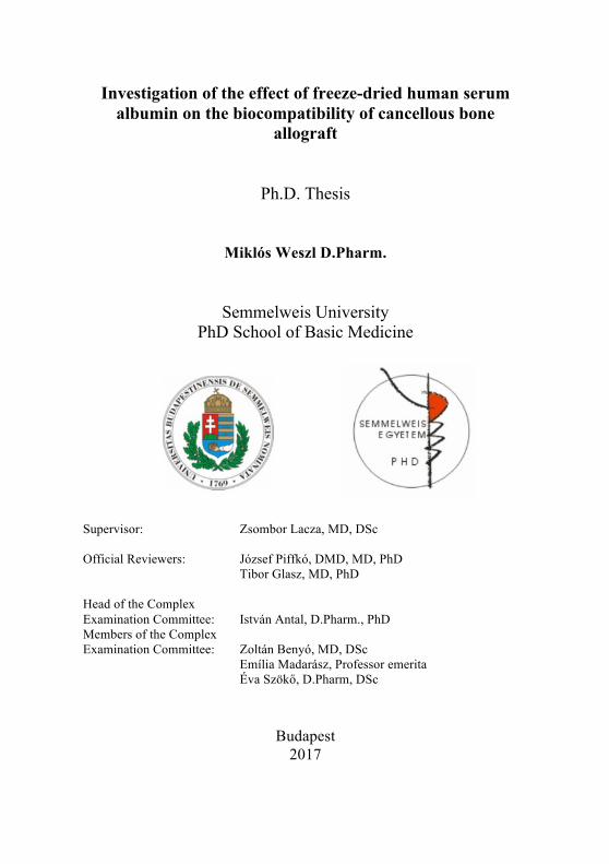
while scanning electron microscopic images were acquired for structural
characterization. The HV Vickers – hardness measurement is performed
with a 136o angle of the vertex and square based diamond-pyramid. Flat
surface areas were selected for micro-hardness measurement where the
diamond-pyramid was pressed into with 50 g load weight for a period of 5
seconds. On each sample at least five measurements were carried out, while
the two diagonals of the impression and the micro-hardness values were
measured and averaged. The numeric value (HV) of the Vickers – hardness
was determined based on the following formula: the load force (F) explicit
in Newton (N) was divided by the surface area (A) of the impression in
mm2, and then the result was multiplied with a constant (C = 0,102).
The surface characteristic of the bone grafts was investigated by
scanning electron microscopy (SEM) (Philips XL 30). An argentiferous
adhesive was applied on the bottom of the samples that were coated with an
electrically conductive gold layer using a vacuum-pulverisation method.
The procedure was then performed in vacuum. The photographs were taken
in the secunder electron (SE) mode with 15kV accelerating voltage. The
secunder electrons are able to emerge from the uppermost layers, having a
thickness of 5-50 nm, meaning that they are extremely sensitive to the
disproportionate surface. The full surface area of the samples was
investigated and representative microscopic images were acquired at 50x,
200x and 1000x magnifications.

7
Table 1. In vitro experimental groups. In Test group A, the bone grafts were immersed into the aqueous solution of either human serum derived albumin (A) or fibronectin (B) or collagen (C). In Test group B, the bone grafts were incubated overnight in the aqueous solutions of the same proteins, which then were freeze-dried onto the surface of the bone grafts (α, β, γ).
Allograft BioOss Hydroxyapatite
Control Uncoated Uncoated Uncoated Test A A) Aqueous albumin
B) Aqueous fibronectin C) Aqueous collagen
A) Aqueous albumin B) Aqueous fibronectin C) Aqueous collagen
A) Aqueous albumin B) Aqueous fibronectin C) Aqueous collagen
Test B α) Freeze-dried albumin β) Freeze-dried fibronectin γ) Freeze-dried collagen
α) Freeze-dried albumin β) Freeze-dried fibronectin γ) Freeze-dried collagen
α) Freeze-dried albumin β) Freeze-dried fibronectin γ) Freeze-dried collagen
Isolation of mesenchymal stem cells: Human bone marrow samples
were obtained from young patients (aged 2-20) during standard orthopaedic
surgical procedures, with the informed consent of the patients or their
parents under approved ethical guidelines set by the Ethical Committee of
the Hungarian Medical Research Council. Only such tissues were used that
otherwise would have been discarded. The bone marrow was taken into T75
flasks and diluted with Dulbecco’s Modified Eagle’s Medium culture
medium containing 10% foetal calf serum (FCS), 100 U/ml penicillin and
10 µg/ml streptomycin, 2mM L-glutamine and 1g/l glucose. The flasks were
incubated at 37 °C in a fully humidified atmosphere of 5% CO2 for 3 days.
After the incubation period the bone marrow derived mesenchymal stem
cells (BMSCs) adhered to the surface of the flasks, the remaining
components of bone marrow were eliminated by washing with phosphate-
buffered saline. The used BMSCs were between passages 1 and 5 in the
experiments. The BMSCs were labelled with the fluorescent membrane dye
Vybrant DiD (excitation/emission: 644/665 nm, Molecular Probes,
Invitrogen, USA) for 30 minutes at 37°C in monolayer.

8
The protocol that we followed for the isolation and culturing of
dental pulp derived mesenchymal stem cells (DPSCs) was based on the
procedure of Gronthos et al, however it was implemented with minor
modifications in our experiment. Human impacted third molars were
collected from adults (18–26 years of age). The tooth was cut around the
cemento-enamel junction by sterile dental fissure burs to expose the pulp
chamber. The pulp tissue had been removed from the crown and the roots
then digested in a solution of collagenase type I (3 mg/ml) and dispase (4
mg/ml) for 1 h at 37°C. Single-cell suspensions were obtained by passing
the cells through a 70 µm strainer and were seeded into 6-well plates with
alpha modification of Eagle’s medium (α-MEM) supplemented with 20%
FCS, 100 µM L-ascorbic acid 2-phosphate, 2 mM L-glutamine, 100
units/ml penicillin, 100 µg/ml streptomycin, then grown under standard cell
culture conditions. As opposed to BMSCs, the DPSCs did not take up the
Vybrant DiD dye to allow uniform staining, thus their proliferation was
followed up with UV-VIS spectrophotometer (BIOTEK Powerwave XS)
using Alamar Blue assay (Biosource, Invitrogen, USA).
The lineage specificity of cells was confirmed by the presence of
lineage-specific cell surface markers with flow cytometry (BD® FacsCalibur,
Becton Dickinson, NJ, USA). Haematopoietic lineage-specific surface
markers (CD34, CD45) and mesenchymal surface markers (CD73, CD90,
CD105 and CD166) were investigated.
The mesenchymal stem cells (MSCs) were seeded onto the surface
of the bone grafts by two methods, i.e. A) under standard cell culture
conditions; and B) under dynamic cell culture conditions:
A) The MSCs had been trypsinized and suspended in culture
medium then applied with pipette to the surface of the test and control bone
grafts (100.000 cells per graft). After seeding, the cells were expanded on

9
the bone grafts under standard cell culture conditions for 18 days and their
proliferation was investigated at the 3rd and 18th days.
B) First, 100.000 cells per scaffold were seeded on the surface of
bone grafts and stored under standard culture conditions for 24 hours.
Following the incubation period, the bone grafts were placed into a
bioreactor tube, which had been filled with 25 ml cell culture medium
comprising 1,5 million MSCs in suspension. The bone grafts had been
incubated in bioreactor under dynamic cell culture conditions for 24 hours
then the cells were further expanded on the surface of bone grafts under
standard culture conditions for 18 days. The viability and quantity of
attached MSCs on the surface was investigated after 3 and 18 days of
incubation.
The survival of the fluorescent dye labelled BMSCs was observed
with confocal microscopy (LSM 510 META, Zeiss) on the surface of
control and test groups. Three individual view fields were randomly
selected on the surface of the grafts where the quantity of pixels belonging
to the fluorescent BMSCs was measured.
The cell culture medium of DPSCs was supplemented with
10w/w% AlamarBlue containing medium, which other components were
identical to the normal culture medium of the cells. The bone grafts were
incubated in this medium for 4 hours in cell culture incubator under
standard cell culture condition. After incubation 200 µl of supernatant was
pipetted into 96-well plate and the absorbance was measured at 570 nm and
600 nm wavelengths.
Test A and Test B subgroups (Table 1) were investigated in an
observational study with longitudinal design where the mutual exposure was
cell seeding onto the surface of bone grafts and the survival (effect of the
exposure) of the seeded cells was measured at two time points during the

10
experiments, i.e. 3rd and 18th days. The possible outcomes of the exposure
were classified according to pre-set verification criteria that also constituted
the basis of the evaluation of the performance of the bone grafts in the
course of the in vitro study (Table 2). Only those grafts were further
investigated in vivo animal studies that facilitated the long-term survival and
proliferation of the MSCs in the in vitro experiments.
Repeated measures one-way ANOVA analysis was performed
(Tukey’s post hoc test) to compare the quantity of MSCs on the scaffolds. A
p value < 0.05 was considered significant.
Table 2. Verification criteria to evaluate the in vitro performance of coated bone grafts. Those bone grafts were progressively excluded from further investigations that did not facilitate the adherence or proliferation compared to their uncoated counterparts. Those bone grafts were excluded from further experiments that felt into at least one exclusion category.
Baseline Decrease Stagnate Increase
Adherence
Quantity of attached cells on uncoated bone grafts
Decrease in the quantity of adhered cells compared to baseline Exclude
Does not affect the quantity of adhered cells compared to baseline Exclude
Increase in the quantity of adhered cells compared to baseline Include
Proliferation
Proliferation rate of cells on uncoated bone grafts
Decrease in the proliferation rate compared to baseline Exclude
Does not affect the proliferation rate compared to baseline Exclude
Increase in the proliferation rate compared to baseline Include
3.3. In vivo biocompatibility study of coated human bone grafts
The Institutional Review Board of Semmelweis University
approved the surgical protocol of the animal study. The investigation of the
in vivo biocompatibility of the coated bone grafts was carried out in a
nonunion model, where the self-healing potential of the bone was
compromised. Adult male Wistar rats (n=39) weighing 496–692g were
housed and maintained at 12/12 day/night cycles and were provided with

11
water and lab chow ad libitum. The animals were anaesthetized with
halothane in a 1:1 mixture of N2O and O2. The surgical site on the thigh
around the femur was shaved and disinfected. The skin, the subcutaneous
layer and the fascia were incised, the tensor fascia latae, and the vastus
lateralis muscles were separated from the biceps femoris muscle. The femur
was exposed from the hip joint to the knee, with special care to preserve the
periosteum. A 5 hole steel plate (Mini plate; Sanatmetal, Eger, Hungary)
was fixed to the diaphysis of the femur by four 1.5 mm wide and 8 mm long
cortical screws (Sanatmetal, Eger, Hungary) using two proximal and two
distal holes, while leaving the middle hole empty. After fixing the plate by
the screws, an osteoperiosteal segment was removed at the level of the
middle hole using a reciprocating saw (Electric Pen Drive, Synthes GmbH,
Oberdorf, Switzerland). The bone was cut precisely through both cortical
layers together with the periosteum. The size of the defect was 2 mm, where
a preformed thick surgical-grade sterile PMMA (Heraeus Medical,
Wehrheim, Germany) bone cement spacer was interposed for four weeks.
The PMMA spacer was secured to the plate by 3-0 non-absorbable sutures
to avoid displacement. Then, the muscle lobes were stitched by 3-0
interrupted absorbable sutures over the femur, and the skin was closed by 3-
0 interrupted nylon sutures. After 4 weeks a second procedure was
performed to allow bone grafting. In the first step, the sutures were removed
and the femur was exposed as previously described. Then the PMMA spacer
was removed and the defect was either left empty, or filled with an uncoated
or a freeze-dried albumin coated bone human bone graft block. After graft
insertion the wound was closed in the same manner as detailed earlier.
Following 4 weeks the animals were sacrificed by exsanguination under
anaesthesia and the femora were harvested. The development of a

12
union/nonunion was evaluated by µCT (Skyscan 1172 X-Ray
microtomograph, Kontich, Belgium).
4. Results 4.1. In vivo biocompatibility of chemically sterilized, antigen extracted freeze-dried human bone graft Figure 1 (upper image on Panel a) shows that the defect was completely
filled with PMMA and no bone formation was observed. In the vertebrae
filled with human bone chips no or slow resorption was observed, and
limited bone healing (bottom image on Panel a). In case of pCPC, in the
vertebrae (middle image on Panel a) new bone formation was observed
(white arrow). Bone defects without any spacer or filling showed good bone
regeneration with new trabecular bone being formed in each sample
providing a defect union rate of 100% (Figure 1, Panel b). In the delayed
healing group, where the spacers were removed at 12th weeks and the defect
was left empty for an
additional 12 weeks, bone
formation was observed in
three defects while no or very
limited bone formation was
observed in the remaining four
defects, where the union rate
was 43% (Figure 1, Panel c).
The µCT images demonstrated
that trabecular bone was formed in the defects filled with SrCPS with a
union rate of 80% with four defects being completely filled with new bone
while in one specimen bone was formed, but it did not fill the entire defect
(Figure 1, Panel d).
Figure 1: shows the representative micro-CT images after 12 weeks of the operated vertebra.

13
The histological assessment
showed that there was some
bone formation in the bone
chip group; however the
bone chips are still
demarcated from the bone.
In the histology image bone
chips are seen to have direct contact with newly formed bone (Figure 2). In
contrast, the histological assessment confirmed that the SrCPS and pCPC
had good biocompatibility and at the time of observation there were no
visible signs of remaining SrCPS particles at light microscopy level
indicating that the material was resorbed and built into the newly formed
bone stock (Figure 2).
4.2. Physical characteristics of coated bone grafts and in vitro adherence and proliferation of MSCs thereon The SEM analysis revealed significant differences in the macro-,
and microstructure of freeze-dried human bone allograft, hydroxyapatite
and lyophilized bovine bone (BioOss) (Figure 3). Hydroxyapatite exhibited
the most compact structure with low number of micro-pores. In contrast, the
texture of lyophilized bovine bone was rich in large diameter channels. The
freeze-dried cancellous allogeneic bone graft exhibited segmented surface
with pores of various sizes. The coating of the surface of bone grafts with
freeze-dried albumin amorphous protein flakes masked the original
structural differences. The albumin flakes showed the most homogeneous
distribution on the surface of Bio-Oss, whereas the continuity of the
albumin flakes was disrupted with random uncoated areas on allografts.
The xenogeneic BioOss showed the lowest Vickers-hardness (14,9
HV ± 4,1). The allogeneic bone graft showed higher values (55,1 HV ± 7,7)
Figure 2: Representative hematoxylin and eosin stained histological section of the distal end of rat-tail vertebra after 12 weeks of normal healing and SrCPS, pCPC and human bone graft implantation.

14
than BioOss, whereas the synthetic hidroxiapatite was a magnitude harder
than the other two grafts (320,4 HV ± 44,6). The micro-hardness values of
freeze-dried albumin coated and uncoated allogeneic bone grafts were very
similar (55.1N ± 7.7 vs. 53.9N ± 7.9, respectively).
Figure 3. Macro- and microstructure of freeze-dried albumin coated and uncoated bone grafts. Scanning electron microscopy shows significant differences in the texture of allograft, hydroxyapatite (HAP), and lyophilized bovine bone graft (BioOss). Hydroxyapatite has the most compact structure with low porosity. On the other hand, high connectivity and thin wall-thickness typifies BioOss. The structure of human bone allograft is different from the other two. It is more compact than BioOss and its surface contains multiple micro-pores. By coating the surface of bone grafts with albumin amorphous protein chips mask the apparent differences in the micro- and nanostructure.
The coating of the surface of allografts with aqueous collagen or
fibronectin slightly increased the initial adherence of BMSCs compared to
the uncoated control, but the quantity of the cells was decreasing between
the 3rd and 18th days of the experiments (Figure 4). In contrast, the albumin

15
coating of the surface of allografts markedly improved the initial adherence
of BMSCs, however the cells disappeared from the surface by the 18th day
(mean of pixels at the 3rd day: 2373 ± 142; at the 18th day: 0)
Figure 4. Adherence and proliferation of BMSCs on the surface of allogeneic bone grafts coated with aqueous proteins. The bar diagram shows that coating of allografts with aqueous albumin resulted in high cell density at day 3 (p* < 0.05), whereas fibronectin and collagen slightly increased the initial cell adherence. Irrespective to the composition of aqueous coating proteins only a few cells were detectable on the surface of allografts at day 18. Panels A-B show representative confocal microscopic images of uncoated allografts (blue) with Vybrant-DiD labelled BMSCs (red). The few observed cells on the surface at day 3 diminished even more by the 18th day. Freeze-drying of serum albumin onto the surface of allografts
reversed the tendency, thus the adhered BMSCs remained on the surface
and showed moderate proliferation during the 18 day long experimental
period (mean of pixels at the 3rd day: 1658 ± 278; mean of pixels at the 18th
day: 2082, ± 110; p < 0.05). Interestingly, the freeze-drying of fibronectin
and collagen onto the surface of allografts did not affect positively either the
initial adherence or the proliferation BMSCs compared to allografts coated
with the aqueous form that of proteins (Figure 5).

16
Figure 5. Adherence and proliferation of BMSCs on the surface of bone allografts coated with freeze-dried proteins. The bar diagram shows that freeze-drying of albumin onto the surface of bone allografts significantly increased the quantity of initially adhered cells at day 3 compared to freeze-dried collagen or fibronectin coating. However, the multiplication of the adhered cells was moderate on the surface of freeze-dried albumin coated allografts during the experimental period. Panel A-B show representative images of freeze-dried albumin coated bone. The proliferation of the attached cells was observed on the day 18. Dynamic seeding significantly increased the quantity of initially
adhered BMSCs on the surface of the freeze-dried albumin coated allografts
(Figure 6) compared to the standard culture conditions and the adhered cells
showed intense proliferation (mean of pixels under standard condition at
day 1: 197±23, under dynamic conditions at day 1: 9825±1208; at the 7th
day: 15025±1704). The same tendency was observed when DPSCs were
seeded on the surface of freeze-dried albumin coated allografts under
dynamic and standard culture conditions (standard condition: mean of
reduced Alamar Blue (%) at 3rd day: 14.5±2.23; mean at 7th day: 33.7±0,06;
dynamic condition: mean of reduced Alamar Blue (%) at 1st day:
33.5±2.23; mean at 7th day: 60.9±1,09).
Figure 6. Attachment of BMSCs and DPSCs onto the surface of freeze-dried albumin coated allografts under dynamic conditions. The dynamic cell culture conditions markedly improved the initial attachment (p† < 0.05) of both BMSCs (A) and DPSCs (B) which retained their capability of proliferating (p*< 0.05).

17
4.3. Osseointegration of freeze-dried human serum albumin coated
human bone graft
Implantation of albumin coated bone grafts into a segmental bone
defect model of delayed bone healing resulted in better integration of the
freeze-dried albumin coated grafts than the uncoated ones. The implanted
grafts were located in the defect in each case, but only the freeze-dried
albumin-coated grafts triggered the ingrowth of new bone from the bone
ends resulting in the union of the defect (Figure 7).
Figure 7. Reconstructed 3-dimensional µCT images of osteotomized rat femurs after 4 weeks of the grafting procedure. The left column shows that without freeze-dried albumin coating the allogeneic human bone graft does not coalesce with the bone ends and there is no bony consolidation. In contrast, when the grafts were coated with albumin there was apparent bone coalescence and a bony callus was formed.
5. Discussion The results show that the in vivo biocompatibility of the chemically
sterilized, antigen extracted freeze-dried human bone grafts is inferior to the
comparator synthetic bone substitutes. The results supported that human
serum albumin is a suitable coating substance to enhance the biological
performance that of human bone grafts in in vitro and in vivo experimental
settings. The freeze-drying procedure supports the reproducible biological
performance of albumin coating, however, the homogeneity of albumin
flakes is poor between the trabeculae of the human bone graft. The albumin
coating does not influence the microhardness of the freeze-dried allografts;
collagen I and fibronectin was not appropriate tosupply the long-term attachment and proliferation ofMSCs. It is possible that the mineralized surface offreeze-dried human bone allograft does not contain ad-equate binding ligands which can permanently anchorcollagen I and fibronectin. Under physiologic bone for-mation, collagen, and other structure proteins firstbuild up the texture of the bone tissue followed bymineral deposition. Therefore it is not surprising thatworking in the opposite direction, that is, puttingstructure proteins on top of a mineralized scaffold doesnot yield optimal results.25
When the cells are seeded on the surface of freeze-dried albumin coated allograft the protein absorbs
water and creates a miroenvironment for MSCs withhigh local albumin content. Since serum is a well-known supporting agent for cell proliferation, thismicroenvironment probably increases the viability offreshly deposited cells. These cells then easily regaintheir metabolic activity and start to deposit their ownbiofilm which further supports proliferation. Thehuman bone structure and pore size play a crucial rolein this mechanism. As it was observed in our electronmicroscopic images, MSCs are not forming a monolay-er on the surface of bone but rather span the poresand establish minimal contact with the surface. This isin stark contrast to cell culture in traditional 2Dmonolayers on plastic surfaces and possibly explainswhy the proliferation-inducing properties of serumoutweigh attachment factors. The larger pore size oflyophilized bovine bone graft or the smaller pores onthe surface of synthetic hydroxyapatite do not provideoptimal spatial arrangements for the MSCs. The lackof cell proliferation on other graft materials furthersupports this explanation. This mechanism providesan opportunity to colonize the surface of freeze-driedallografts in a simple rotating bioreactor system,which is frequently used in clinical tissue-engineeringapplications. In addition to bone marrow, proliferationof dental pulp-derived derived MSCs was alsoincreased by albumin coating, making this technologysuitable for dental applications.
The preliminary in vivo testing of albumin coatedbone grafts showed an increased ingrowth on newbone compared to the uncoated ones. This observationhighlights that early colonization of the graft with
Figure 5. In vivo biocompatibility of a bone graft with or without albumin coating. Reconstructed 3-dimensional mCT images areshown of rat femora after 4 weeks of implantation of the graft. The left column shows that without albumin coating the bone graft doesnot integrate into the bone ends and there is no bony consolidation. In contrast, when the grafts were coated with albumin there isgood ingrowth from the rat femur and a bony callous is formed.
Figure 6. Attachment and proliferation of BMSCs on the sur-face of albumin coated and uncoated bone grafts. The coating ofhydroxyapatite (HAP) and lyophlized bovine bone (BioOss) withfreeze-dried human serum albumin does not improve theattachment and proliferation of BMSCs on the surface of graftsin contrast with mineralized bone allografts. !p < 0.05.
ADHERENCE AND PROLIFERATION OF MSCS 495
JOURNAL OF ORTHOPAEDIC RESEARCH MARCH 2012

18
the agitation under dynamic culture condition does not reduce its biological
value. Intriguingly, the albumin coating increases mainly the initial
adherence of MSCs on the surface of allografts, whereas significant
proliferation is only seen after seeding under dynamic culture conditions.
After implantation into a nonunion site, albumin coating improved the
ingrowth of new bone from the host and resulted in the union of the bone
ends. Interestingly, the albumin coating does not improve the in vitro
biocompatibility of either the lyophilized bovine bone or the synthetic
hydroxyapatite bone scaffolds.
There might be common reasons behind the low incorporation rate
and the low biocompatibility with MSCs of freeze-dried human bone
allograft. The chemical treatment of allografts kills not only pathogenic
microorganisms and reduce the quantity of antigens but destroys the
osteoinductive and osteogenic molecules turning bone grafts into
mineralized scaffolds with reduced biological value. Although the
mechanism of action of albumin coating is not known, but we hypothesize
that it may play a crucial role in the recruitment and activation of osteogenic
cells based on the volume expansion (colloidal suspension) theory.
After bone replacement surgery, alike after injury when bony
injury occurs, rapid and active inflammatory response floods the injury zone
with blood cells, platelets, monocytes, macrophages and other cells of the
inflammatory cascade. The result of this process is that the injury site gets
isolated from the rest of the body, becoming avascular to insure that the
local injury environment does not propagate to the rest of the body. These
segregation processes that occur at the bone fracture or injury sites
eventually result in the repair blastema and outer surrounding reparative
callus. The fracture hematoma has been proven to be a source of signalling
molecules, such as interleukins, tumour necrosis factor-α, fibroblast growth

19
factor, insulin-like growth factor, platelet-derived growth factor, vascular
endothelial growth factor, and the transforming growth factor β superfamily
members that are supposed to induce a cascade of cellular events that
initiate healing. These signal molecules might be adsorbed by the colloidal
suspension of albumin that increases the local concentration of such
biological cues, while it provides a natural delivery system that allows the
prolonged release of those signals. This assumption is based upon the high,
non-selective affinity of serum albumin to bind various biomolecules
creating a high capacity natural buffer (reservoir) for them. The increased
local concentration and sustained availability of those soluble cues may
enhance cellular events in the repair tissue allowing the union of the bone
ends in our animal model when the self-healing ability of the bone was
compromised.
The same volume expansion theory of albumin may explain the in
vitro performance of freeze-dried albumin coating. It seems to be a possible
explanation that freeze-dried albumin adsorbs water when the cells are
seeded onto the surface of allografts in aqueous media. The water
adsorption may trigger the volume expansion of albumin yielding colloidal
suspension that is temporally trapped in the channels of the allograft due to
its high viscosity. This colloidal suspension of albumin may embed MSCs
and keep them in the pores and channels of the allograft providing enough
time to produce extracellular matrix and establish focal adhesions with the
surface.

20
6. Conclusions 6.1. Chemically sterilized, antigen-extracted freeze-dried human bone grafts
showed reduced in vivo osseointegration in compromised bone defect model
compared to synthetic bone fillers.
6.2. Freeze-dried human serum albumin is a suitable substance for the
reproducible coating of cancellous human bone grafts.
6.3. The freeze-dried human serum albumin coating improved the in vitro
and in vivo biocompatibility of the chemically sterilized, antigen-extracted
human bone grafts.
7. Publications Publications related to the present thesis [1] Weszl M, Skaliczki G, Cselenyák A, Kiss L, Major T, Schandl K, Bognár E, Stadler G, Peterbauer A, Csönge L, Lacza Z. Freeze-dried human serum albumin improves the adherence and proliferation of mesenchymal stem cells on mineralized human bone allografts. J Orthop Res. 2012 Mar;30(3):489-96. [2] Hulsart-Billström G, Xia W, Pankotai E, Weszl M, Carlsson E, Forster-Horváth C, Larsson S, Engqvist H, Lacza Z. Osteogenic potential of Sr-doped calcium phosphate hollow spheres in vitro and in vivo. J Biomed Mater Res A. 2013 Aug;101(8):2322-31. [3] Aberg J, Pankotai E, Hulsart Billström G, Weszl M, Larsson S, Forster-Horváth C, Lacza Z, Engqvist H. In vivo evaluation of an injectable premixed radiopaque calcium phosphate cement. Int J Biomater. 2011;2011:232574. Publications not related to the present thesis [1] Horváthy DB, Vácz G, Cselenyák A, Weszl M, Kiss L, Lacza Z. Albumin-coated bioactive suture for cell transplantation. Surg Innov. 2013 Jun;20(3):249-55. [2] Skaliczki G, Weszl M, Schandl K, Major T, Kovács M, Skaliczki J, Redl H, Szendrői M, Szigeti K, Máté D, Dobó-Nagy C, Lacza Z. Compromised bone healing following spacer removal in a rat femoral defect model. Acta Physiol Hung. 2012 Jun;99(2):223-32. [3] Skaliczki G, Schandl K, Weszl M, Major T, Kovács M, Skaliczki J, Szendrői M, Dobó-Nagy C, Lacza Z. Serum albumin enhances bone healing in a nonunion femoral defect model in rats: a computer tomography micromorphometry study. Int Orthop. 2013 Apr;37(4):741-5. [4] Terdik A , Klára T , Csönge L , Lacza Z , Bognár E , Weszl M. Csontpótló anyagok összehasonlító mikrokeménység vizsgálata BIOMECHANICA HUNGARICA 6:(2) pp. 13-17. (2013)