Embed Size (px)
Citation preview

Ubiquitylation activates a peptidase thatpromotes cleavage and destabilization of itsactivating E3 ligases and diverse growthregulatory proteins to limit cellproliferation in ArabidopsisHui Dong,1,6 Jack Dumenil,1,6 Fu-Hao Lu,1,6 Li Na,2,6 Hannes Vanhaeren,3 Christin Naumann,4
Maria Klecker,4 Rachel Prior,1 Caroline Smith,1 Neil McKenzie,1 Gerhard Saalbach,1 Liangliang Chen,2
Tian Xia,2 Nathalie Gonzalez,3 Mathilde Seguela,1,5 Dirk Inze,3 Nico Dissmeyer,4 Yunhai Li,2
and Michael W. Bevan1
1John Innes Centre, NorwichNR4 7QA, United Kingdom; 2State Key Laboratory of Plant Cell and Chromosome Engineering, CASCentre of Excellence in Molecular Plant Biology, Institute of Genetics and Developmental Biology, Chinese Academy of Sciences,Beijing 100101, China; 3VIB-UGent Centre for Plant Systems Biology, Ghent University, 9052 Gent, Belgium; 4Leibniz Institute ofPlant Biochemistry (IPB), D-06120 Halle, Germany
The characteristic shapes and sizes of organs are established by cell proliferation patterns and final cell sizes, butthe underlying molecular mechanisms coordinating these are poorly understood. Here we characterize a ubiquitin-activated peptidase called DA1 that limits the duration of cell proliferation during organ growth in Arabidopsisthaliana. The peptidase is activated by two RING E3 ligases, Big Brother (BB) and DA2, which are subsequentlycleaved by the activated peptidase and destabilized. In the case of BB, cleavage leads to destabilization by the RINGE3 ligase PROTEOLYSIS 1 (PRT1) of theN-end rule pathway. DA1 peptidase activity also cleaves the deubiquitylaseUBP15, which promotes cell proliferation, and the transcription factors TEOSINTE BRANCED 1/CYCLOIDEA/PCF 15 (TCP15) and TCP22, which promote cell proliferation and repress endoreduplication. We propose thatDA1 peptidase activity regulates the duration of cell proliferation and the transition to endoreduplication anddifferentiation during organ formation in plants by coordinating the destabilization of regulatory proteins.
[Keywords: ubiquitylation; organ size; Arabidopsis; ubiquitin-activated peptidase; N-end rule-mediated degradation]
Supplemental material is available for this article.
Received October 17, 2016; revised version accepted January 11, 2017.
The shapes and sizes of organs are established by mecha-nisms that orient cell proliferation and determine the finalnumbers and sizes of cells forming the organ. Transplanta-tion experiments showed that some animal organs havean intrinsic mechanism that determines their final sizeby controlling the duration of cell proliferation (Barryand Camargo 2013), which is controlled in part by theHIPPO/YAP pathway that limits cell proliferation andpromotes apoptosis (Pan 2010). However, the mecha-nisms coordinating cell proliferation and cell size duringorgan growth remain poorly understood (Johnston and
Gallant 2002). Due to the simpler planar structures oftheir organs, such as leaves and petals, and the absenceof cell movement due to rigid cell walls, plants havesome experimental advantages for studying organ growth(Green et al. 2010).Leaf growth in plants is initiated at shootmeristems (for
review, see Sluis and Hake 2015). After specification ofboundaries and growth axes, the leaf lamina grows in aninitial period of cell division inwhich cell size is relativelyconstant, followed by a transition to endoreduplication as-sociated with cell expansion and differentiation (Breueret al. 2010; De Veylder et al. 2011). The transition fromcell proliferation to cell expansion is spatially and tempo-rarily regulated during leaf growth and appears to progressfrom the tip to the base of the leaf as a cell division arrest
5Present address: UMR1318, Institut Jean-Pierre Bourgin, 78000Versailles, France.6These authors contributed equally to this work.Corresponding authors: [email protected], [email protected] Published online ahead of print. Article and publication date areonline at http://www.genesdev.org/cgi/doi/10.1101/gad.292235.116. Free-ly available online through the Genes & Development Open Accessoption.
© 2017 Dong et al. This article, published in Genes & Development, isavailable under a Creative Commons License (Attribution 4.0 Internation-al), as described at http://creativecommons.org/licenses/by/4.0/.
GENES & DEVELOPMENT 31:1–12 Published by Cold Spring Harbor Laboratory Press; ISSN 0890-9369/17; www.genesdev.org 1
Cold Spring Harbor Laboratory Press on April 3, 2019 - Published by genesdev.cshlp.orgDownloaded from
front (Kazama et al. 2010) accompanied by shifts in geneexpression patterns (Efroni et al. 2008; Andriankaja et al.2012). A key question is how the transition from cell pro-liferation to cell expansion and differentiation is coordi-nated to generate a correctly sized organ.
The RING E3 ligases Big Brother (BB) (Disch et al. 2006)and DA2 (Xia et al. 2013) limit the duration of cell prolif-eration during organ growth. Members of the DA1 familyalso limit cell proliferation (Li et al. 2008), and loss-of-function mutations in BB and DA2 interact synergis-tically with the da1-1 allele of DA1 to increase organand seed size in Arabidopsis (Li et al. 2008; Xia et al.2013), suggesting that one of their growth-limiting activi-ties ismediated by enhancing the growth-repressive activ-ity ofDA1 familymembers. Genetic analyses showed thatDA1 reduced the stability of bothUBP15 (Du et al. 2014), adeubiquitylation enzyme promoting cell proliferation(Liu et al. 2008), and TEOSINTE BRANCED 1/CYCLOI-DEA/PCF 14 (TCP14) and TCP15 proteins (Peng et al.2015), which repress endoreduplication by transcriptionalcontrol of RETINOBLASTOMA-RELATED1 (RBR1) andCYCLIN A2;3 (CYCA2;3) gene expression (Li et al. 2012).
Here we show that DA1 is an endopeptidase activatedby multiple ubiquitylations mediated by the E3 ligasesBB and DA2. In a feedback mechanism, DA1 then cleavesBB andDA2, leading to their destabilization. DA1-mediat-ed cleavage of BB exposed a destabilizing N-terminal thatwas substrate for the N-end rule E3 ligase PROTEOLYSIS1 (PRT1). This mechanism is predicted to transiently acti-vate DA1 peptidase, which also cleaves UBP15, TCP15,and the related TCP22, leading to their predicted inactiva-tion and destabilization. DA1 peptidase may thereforecontribute to the concerted transition from cell prolifera-tion to endoreduplication and differentiation, limitingorgan size.
Results
Genetic and physical interactions of DA1, BB, and DA2
Wepreviously identified genetic interactions between theda1-1 allele ofDA1 and genes encoding theRINGE3 ligas-es BB (Li et al. 2008) and DA2 (Xia et al. 2013) that led tosynergistic increases in seed and organ sizes. In this study,we used the da1-1-enhancing allele of BB called eod1-2(Li et al. 2008) and refer to the mutant version as bb-eod1-2 and the wild-type version as BB. The da1-1 allele,an R358K change in a highly conserved region, had anegative influence on the functions of DA1 and the closefamily member DAR1, but the basis of this was notknown, which complicated interpretation of DA1 func-tion. We therefore assessed phenotypes of a loss-of func-tion T-DNA allele of DA1 (da1-ko1).
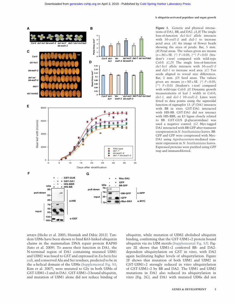
Measurements of petal and seed sizes using high-resolu-tion scanning showed that the da1-ko1 T-DNA allele ledto increased petal (Fig. 1A,B) and seed (Fig. 1C,D) sizes andthat it also interacted genetically with the loss-of-function allele bb-eod1-2 and da2-1 in both petal sizeand seed area. This showed thatDA1 can be studied inde-pendently of other DA1 family members. Both the da1-1
and bb-eod1-2mutations increased themaximum growthrate, while the double mutant da1-1 bb-eod1-2 showed afurther increased maximum growth rate and continuedto grow for ∼5 d longer than either single mutant (Fig.1E). The time at maximum growth rates was slightly ear-lier in bb-eod1-2 than in Columbia (Col-0), in contrast toda1-1 and da1-1 bb-eod1-2, which showed a 3-d retarda-tion of the time of maximum growth rate, and final leafsizes showed a more than additive increase in the doublemutant, as observed previously (Li et al. 2008). These dataindicated that BB may influence leaf final size at earlierstages of growth than DA1. We demonstrated previouslythat DA1 and DA2 physically interact (Xia et al. 2013).Pull-down experiments showed that GST-tagged DA1also interacted with HIS-tagged BB but not with HIS-tagged BBR (BB-related; At3g19910), a close homolog ofBB (Fig. 1F; Breuninger and Lenhard 2012). These in vitrointeractions were verified by Agrobacterium-mediatedcoexpression of BB-GFP and Myc-tagged DA1 in Nicotia-na benthamiana leaves. Myc-DA1 was detected only in acomplex with BB-GFP and not GFP (Fig. 1G).
DA1 is multiply ubiquitylated by BB and DA2
The interactions of DA1 with BB and DA2 suggested thatDA1 might be a substrate of these RING E3 ligases, so weconducted in vitro ubiquitylation reactions using BB,DA2, and BBR E3 ligases. Figure 2A shows that BB ubiqui-tylated DA1 in an E1- and E2-dependent reaction, as didDA2 (Fig. 2B), while BBR did not (Fig. 2C). SupplementalFigure S1 shows that DA2 also ubiquitylated DAR1 andDAR2 but not DAR3. The extent of DA1 ubiquitylationsuggested that DA2 was more efficient at ubiquitylationthan BB, and the sizes of ubiquitylated DA1 indicatedthat between four and seven ubiquitin molecules maybe conjugated to DA1. Mass spectrometric analyses ofubiquitylated DA1 prepared in vitro were used to identifypeptides containing the characteristic diglycine ubiquity-lation signature of a lysine residue (KGG). Analysis ofDA1 ubiquitylated by DA2 or BB identified seven ubiqui-tylated lysine residues in DA1, with four lysines in theC-terminal domain of DA1 (K381, K391, K475, andK591) consistently conjugated with ubiquitin (Supple-mental Fig. S2). This number of ubiquitylation sites con-curred with the patterns of ubiquitylation observed inFigure 2, A and B, suggesting that DA1molecules aremul-tiply ubiquitylated (Haglund et al. 2003; Komander andRape 2012). Mutation of the consistently ubiquitylatedlysines to arginine in DA1 [termed DA1(4K-4R)] did notreduce ubiquitylation by DA2 in vitro (Fig. 2D), andmass spectrometric analyses showed ectopic ubiquityla-tion of other lysines across DA1 (Supplemental Fig. S2B).Therefore, the DA1 ubiquitylation mechanism has apreference, but not specificity, for certain lysines. Thesepatterns of ubiquitylation are shown in Figure 2D.
DA1 and four other family members have multipleubiquitin interaction motifs (UIMs) that interact withubiquitin (Li et al. 2008; Peng et al. 2015). UIMs are partof a larger class of ubiquitin-binding domains (UBDs)formed from a single α helix that is often found inmultiple
Dong et al.
2 GENES & DEVELOPMENT
Cold Spring Harbor Laboratory Press on April 3, 2019 - Published by genesdev.cshlp.orgDownloaded from
arrays (Hicke et al. 2005; Husnjak and Dikic 2012). Tan-demUIMs have been shown to bind K63-linked ubiquitinchains in the mammalian DNA repair protein RAP80(Sato et al. 2009). To assess their function in DA1, theN-terminal region of DA1 containing mutated UIM1and UIM2 was fused to GST and expressed in Escherichiacoli, and conserved Ala and Ser residues, predicted to be inthe α-helical domain of the UIMs (Supplemental Fig. S3;Kim et al. 2007), were mutated to Gly in both UIMs ofGST-UIM1+2 and inDA1. GST-UIM1+2 bound ubiquitin,and mutation of UIM1 alone did not reduce binding of
ubiquitin, while mutation of UIM2 abolished ubiquitinbinding, confirming that the GST-UIM1+2 protein boundubiquitin via its UIM motifs (Supplemental Fig. S3). Fig-ure 2E shows that UIM1+2 conferred BB- and DA2-dependent ubiquitylation on GST in vitro, with DA2again facilitating higher levels of ubiquitylation. Figure2F shows that mutation of both UIM1 and UIM2 inGST-UIM1+2 strongly reduced in vitro ubiquitylationof GST-UIM1+2 by BB and DA2. The UIM1 and UIM2mutations in DA1 also reduced its ubiquitylation invitro (Fig. 2G), and DA1 with mutated UIMs did not
Figure 1. Genetic and physical interac-tions of DA1, BB, and DA2. (A,B) The singleloss-of-function da1-ko1 allele interactswith bb-eod1-2 and da2-1 to increasepetal area. (A) An image of flower headsshowing the sizes of petals. Bar, 5 mm.(B) Petal areas. The values given are means(n = 36) ± SE. (∗) P < 0.05; (∗∗) P < 0.01 (Stu-dent’s t-test) compared with wild-typeCol-0. (C,D) The single loss-of-functionda1-ko1 allele interacts with bb-eod1-2and da2-1 to increase seed area. (C ) Tenseeds aligned to reveal size differences.Bar, 2 mm. (D) Seed areas. The valuesgiven are means (n = 50) ± SE. (∗) P < 0.05;(∗∗) P < 0.01 (Student’s t-test) comparedwith wild-type Col-0. (E) Dynamic growthmeasurements of leaf 1 width in Col-0,da1-1, and da1-1 bb-eod1-2. Lines werefitted to data points using the sigmoidalfunction of sigmaplot 13. (F ) DA1 interactswith BB in vitro. GST-DA1 interactedwith HIS-BB. GST-DA1 did not interactwith HIS-BBR, an E3 ligase closely relatedto BB. GST-GUS (β-glucuronidase) wasused a negative control. (G) Myc-taggedDA1 interacted with BB-GFP after transientcoexpression inN. benthamiana leaves. BB-GFP and GFP were coexpressed with Myc-DA1 using Agrobacterium-mediated tran-sient expression in N. benthamiana leaves.Expressed proteins were purified using GFPtrap and immunoblotted.
A ubiquitin-activated peptidase and organ growth
GENES & DEVELOPMENT 3
Cold Spring Harbor Laboratory Press on April 3, 2019 - Published by genesdev.cshlp.orgDownloaded from

Figure 2. DA1 is multiply ubiquitylated by BB and DA2 in a ubiquitin interaction motif (UIM)-dependent reaction. (A–C ) In vitro ubiq-uitylation of DA1 by the RING E3 ligases BB (A) and DA2 (B), but not BBR (C ), in an E1-, E2-, and ubiquitin-dependent reaction. Anti-Flagantibodies detected Flag-ubiquitylated forms of Flag-DA1 ranging from >70 kDa to ∼130 kDa. Anti-HIS antibodies detected BB-HIS, DA2-HIS, or BBR-HIS fusion proteins. (D) Both Flag-DA1 and Flag-DA1(4K-4R) are ubiquitylated by DA2 in similar patterns in an in vitroubiquitylation reaction. In the bottom panel, Ub represents a ubiquitin moiety conjugated to a lysine at the approximate location inDA1 and DA1(4K-4R). Regions of protein similarity with known domains are shown: UIM1 andUIM2 are similar to UIMs, LIM is similarto canonical LIM domains, LIM-like is a related motif found in DA1 family members, and peptidase contains a predicted peptidase activesite. (E) An in vitro ubiquitylation reaction with DA2 and BB as E3 ligases and GST-UIM1+2. GST-UIM1+2 is ubiquitylated in a patternsimilar to that of DA1 by both DA2 and BB, with DA2 conferring higher levels of ubiquitylation than BB. (F ) An in vitro ubiquitylationreaction with DA2 and BB as E3 ligases and GST-UIM1+2 with mutations that reduce ubiquitin binding. Mutated versions of UIM1 andUIM2 strongly reduced DA2- and BB-mediated ubiquitylation of GST-UIM1+2. (G) A time course of Flag-DA1 and Flag-DA1(UIM1+2)with mutations in the UIMs as in E. These strongly reduced DA1 ubiquitylation. (H) DA1(UIM1+2) is not functional in vivo, as it doesnot complement the large petal phenotype of the da1-ko1 dar1-1 double mutant. Two independent homozygous T-DNA insertion lineswere scored for petal size and compared with wild-type Col-0 and da1-ko1 dar1-1. The values given aremeans (n = 120) ± SE, expressed asthe percentage of wild-type Col-0 petal areas. Student’s t-test showed no significant differences between the transformants and the paren-talda1-ko1 dar1-1 line. (I ) TransgenicArabidopsis plants expressing aGFP-DA1 fusion protein under the control of the 35S promoterwereused to detectDA1ubiquitylation in vivo. GFP ran as a dimer on the gel due to high protein concentrations. Protein extract input levels areshown using anti-tubulin antibody.
4 GENES & DEVELOPMENT
Cold Spring Harbor Laboratory Press on April 3, 2019 - Published by genesdev.cshlp.orgDownloaded from

complement the large petal size in the double mutantda1-ko dar1-1 (Fig. 2H). To detect ubiquitylation invivo, DA1 was expressed from the constitutive 35S pro-moter as an N-terminal GFP fusion protein and purifiedfrom seedling tissues using a GFP trap. Characteristic pat-terns of DA1 ubiquitylation were detected on purifiedGFP-DA1 (Fig. 2I, right panel). Therefore, DA1 is ubiqui-tylated by the E3 ligases BB and DA2 in vitro by aUIM1- and UIM2-dependent mechanism, DA1 is ubiqui-tylated in vivo, and UIMs are required for DA1 function.
DA1 cleaves BB and DA2 with a ubiquitin-dependentpeptidase activity
A time course of BB-HIS incubatedwith purified Flag-DA1that had been ubiquitylated by BB or incubated with non-ubiquitylated Flag-DA1 showed that, in the presenceof ubiquitylated DA1, a HIS-tagged BB fragment of ∼35kDawas produced after 4 h of incubation (Fig. 3A, arrows).When ubiquitylated Flag-DA1 was incubated with DA2-HIS, a 25-kDa HIS-tagged DA2 cleavage product wasalso detected after 4 h of incubation (Fig. 3A, arrows). Sim-ilar experiments using Flag-DA1 ubiquitylated by DA2showed identical patterns of BB-HIS and DA2-HIS cleav-age (Fig. 3B). BBR-HIS did not show a cleavage productin these conditions. Thus, DA1 ubiquitylated by eitherBB or DA2 generated cleavage products from both BBand DA2 in vitro.Examination of the conservedC-terminal region ofDA1
revealed an extended sequence motif, HEMMHX15EE(Supplemental Fig. S4), which is a zinc aminopeptidaseactive site found in clan MA endopeptidases (Rawlingset al. 2012). TheHEMMHmotif wasmutated to AEMMA,removing the putative zinc-coordinating histidine resi-dues, to form DA1(pep). Figure 3C shows that DA1(pep)and DA1 were ubiquitylated in vitro to an equal extentby both BB and DA2. In an in vitro time-course reaction,ubiquitylated DA1(pep) did not generate the 25-kDaHIS-taggedDA2 band seen after incubationwith ubiquity-lated DA1 (Fig. 3D). Coexpression of BB-Flag, DA2-Flag,or BBR-Flag with HA-DA1 or HA-DA1(pep) in da1-ko1dar1-1 mutant leaf protoplasts showed that HA-DA1,but not HA-DA1(pep), generated a similar-sized 35-kDaBB-Flag cleavage product (Fig. 3E, top panel, arrow) asseen in in vitro reactions (Fig. 3A,B). Longer exposure ofthe same Western blot (Fig. 3E, bottom panel) was re-quired to identify the 25-kDa DA2-Flag cleavage product,which was not generated by coexpression with DA1(pep).Figure 3F shows that the mutation in DA1 abolishingDA1 peptidase activity did not complement the da1-ko1dar1-1 large petal phenotype, establishing thatDA1 pepti-dase activity is required for in vivo function. To detectDA1 peptidase activity in vivo, transgenic plants express-ing BB::gsGreen-BB gene fusion and a RING domain mu-tant version that was predicted to be more stable in vivodue to reduced autopolyubiquitylation (Disch et al.2006) were generated. Analysis of GFP trap-purified pro-teins (Fig. 3G, left panel) showed a cleavage product ofthe expected size generated from RING mutant gsGreenprotein in two independent transformants. Full-length
wild-type gsGreen-BB was not detected, although lowlevels of an expected cleavage product were identified.For comparison, the same constructs, together with anoncleavable form (AY-GG) (see Fig. 5B, below), wereexpressed using the 35S promoter in protoplasts withDA1 (Fig. 3G, right panel). This showed the predictedDA1-mediated BB cleavage product, which was not gener-ated in the AY-GG version of BB.A Förster resonance energy transfer (FRET) DA1 pepti-
dase sensor was constructed using eGFP donor andmCherry acceptor pairs (van der Krogt et al. 2008) con-nected by BB to provide anothermeasure ofDA1 peptidaseactivity in vivo. Cleavage of the fluorophore pair by DA1would increase the fluorescence lifetime toward that ofeGFP-BB compared with that of the intact sensor proteinby impairing energy transfer between the fluorophores.The peptidase sensor and a control donor sensor weretransfected into da1-ko1 dar1-1 root protoplasts, andfluorescence lifetime imaging (FLIM) was performed. Fig-ure 4A shows that the fluorescence lifetime (τ) of the GFP-BB donor control was ∼2.48 nsec, while that of an intactdonor–acceptor pair was ∼2.25 nsec, demonstratingefficient FRET. When cotransfected with DA1, the fluo-rescent lifetime of the donor–acceptor pair increased to∼2.38 nsec. Lifetime imaging of typical transfected pro-toplasts showed a generalized cellular localization ofDA1-mediated cleavage. Figure 4B shows that the eGFP-BB-mCherry donor–acceptor pair was cleaved by DA1peptidase at the expected site in transfected root proto-plasts. Therefore, DA1 has a latent peptidase activitythat is activated by multiple ubiquitylation mediatedby its UIM1+2 domain and the RING E3 ligases BBand DA2, and activated DA1 peptidase then specificallycleaves these two E3 ligases.
Identification of a DA1 peptidase cleavage site in BB
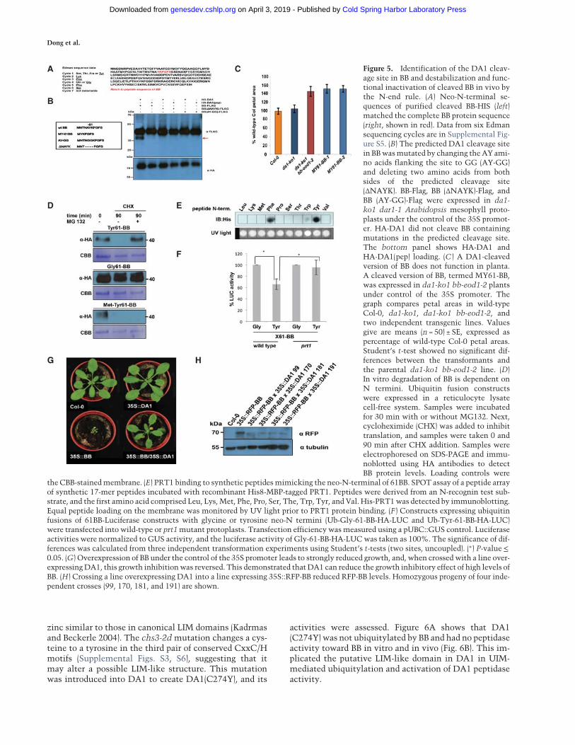
To define the potential functions of DA1-mediated cleav-age, the DA1 cleavage site in BB was identified usingEdman sequencing of purified cleaved BB-HIS. Supple-mental Figure S5 shows neo-N-terminal amino acid se-quences that had a unique match to six amino acids inBB (Fig. 5A). This indicated a potential DA1 cleavagesite within BB between A60 and Y61, consistent with thesizes of BB and its ∼35-kDa cleaved form (Fig. 3A). Twomutant forms of BB were made to assess this potentialDA1 cleavage site: a four-amino-acid deletion surround-ing the site (ΔNAYK) and AY changed to GG (AY-GG)(Fig. 5B). These proteins were coexpressed in Arabidopsisda1-ko1 dar1-1 mesophyll protoplasts as C-terminal Flagfusion proteins with HA-DA1 and HA-DA1(pep). Figure5B shows that the mutant BB-Flag proteins were notcleaved by DA1, establishing that DA1 peptidase activitycleaved BB between A60 and Y61. A cleaved form of BB,called MY61-BB, was also made with an initiator Metfollowed by Y61 (Fig. 5B). MY61-BB was expressed usingthe 35S promoter in da1-ko1 bb-eod1-2mutantArabidop-sis. Its lack of complementation of bb-eod1-2 (Fig. 5C)showed that DA1 peptidase-mediated cleavage reducedBB activity.
A ubiquitin-activated peptidase and organ growth
GENES & DEVELOPMENT 5
Cold Spring Harbor Laboratory Press on April 3, 2019 - Published by genesdev.cshlp.orgDownloaded from

Figure 3. DA1 is an endopeptidase activated by multiple ubiquitylations and cleaves the E3 ligases BB and DA2 that ubiquitylate it.(A,B) Time course of an in vitro reaction of Flag-DA1 or Flag-DA1 ubiquitylated by BB (A) or DA2 (B) with BB-HIS, DA2-HIS, and BBR-HIS. The bottom panels show loading of Flag-DA1 and Flag-DA1Ub-BB. After 4 h, cleavage products (shown by red arrows) of BB andDA2 had been produced by ubiquitylated Flag-DA1 but not Flag-DA1. BBR was not cleaved under these conditions. (C ) An in vitro ubiq-uitylation reaction of DA1 and DA1(pep) using BB-HIS (top panel) and DA2-HIS (bottom panel) as E3 ligases. Ubiquitin-dependentmultiplemonoubiquitylations of Flag-DA1 and Flag-DA1(pep) by both BB andDA2were detected. (D) A time course of an in vitro cleavagereaction usingDA2-HIS as a substrate (left panels) and Flag-DA1 or Flag-DA1(pep) ubiquitylated by either BB orDA2 (loading shown in thebottom panel). The red arrow in thebottom left panel indicates theDA2 cleavage product at 4 h thatwas produced only by Flag-DA1Ub andnot Flag-DA1(pep)Ub. (E) Arabidopsis da1-ko1 dar1-1 mesophyll protoplasts were cotransfected with plasmids expressing BB-Flag,DA2-Flag, BBR-Flag, HA-DA1, and HA-DA1(pep). The same-sized cleavage products (red arrows) from BB-Flag and DA2-Flag were detect-ed as seen inA andB above. (Middle panel) Longer exposure of the top immunoblot showed cleavedDA2. The bottom panel shows loadingof HA-DA1 and HA-DA1(pep). (F ) DA1(pep) is not functional in vivo, as it does not complement the large petal phenotype of the da1-ko1dar1-1 double mutant. Transformants expressing 35S::DA1(pep) were scored for petal size and compared with wild-type Col-0 andda1-ko1 dar1-1. The values given aremeans (n = 150) ± SE, expressed as percentage of wild-type Col-0 petal areas. Student’s t-test showedno significant differences between the transformants and the parental da1-kodar1-1 line. (G) Cleavage of gsGreen-BB is shown in planta inthe left panel and in transiently expressed protoplasts in the right panel for comparison. Large-scale protein extracts from transgenic 8-d-old seedlings expressingBB::gsGreen-BB andBB::gsGreen-BB (RING)were purified on aGFP trap. Loading controls used levels of freeGFP.The expected size cleavage products (arrows) were observed in plant extracts and protoplasts for comparison.
Dong et al.
6 GENES & DEVELOPMENT
Cold Spring Harbor Laboratory Press on April 3, 2019 - Published by genesdev.cshlp.orgDownloaded from

BB stability is dependent on it N terminusand N-end rule function
DA1 cleavage products of DA2 were unstable, indicatingthat one function of DA1-mediated cleavage may be todestabilize proteins (Fig. 3E). This was also observed forBB in cell-free degradation assays, in which MY61-BBwas unstable compared with wild-type BB (SupplementalFig. S6). To test the role of the neo-N terminus of BB onprotein stability, 61BB proteins with different N termini(Y, G, and MY) were expressed using the ubiquitin fu-sion technique (UFT) (Bachmair et al. 1986). HA-taggedconstructs were translationally coexpressed in a cell-freerabbit reticulocyte system with or without MG132 pro-teasome inhibitor, and translation was stopped by theaddition of cycloheximide. Y61-BB was highly unstable,whereas G61-BB was stable (Fig. 5D). Interestingly, theartificial MY61-BB was also highly unstable in a protea-some-independent mechanism. The neo-N-terminal se-quence of DA1-cleaved BB starts with YK, a potentiallydestabilizing sequence of a type II N-end rule degron (Var-shavsky 2011). The N-end rule E3 ligase PRT1 mediatesthe stability of model N-end rule substrates with sucharomatic N-terminal residues (Potuschak et al. 1998). Toassess the potential role of PRT1 in N-end rule-mediateddegradation of BB, we tested the binding of PRT1 to 17-mer peptides representing variants of the neo-N termini
of BB on a backbone sequence of an N-end rule testsubstrate in SPOT (synthetic peptide arrays onmembranesupport technique) assays. Purified recombinant HIS-MBP-PRT1 protein was incubated with the SPOT array,and bindingwas visualized byWestern blotting. Recombi-nant PRT1 had a preference for binding to the large aro-matic acids tyrosine and phenylalanine, consistent withpreviously suggested specificity (Fig. 5E; Potuschak et al.1998; Stary 2003; Faden et al. 2016). To assess whetherPRT1 had a role in DA1-mediated BB degradation, BBwas expressed with an N-terminal ubiquitin fusion anda C-terminal luciferase fusion to reveal neo-N termini inCol-0 or prt1 mutant mesophyll protoplasts. BB-LUC ac-tivity was reduced in wild-type protoplasts with a neo-N-terminal tyrosine, which was not seen in prt1 mutantprotoplasts (Fig. 5F). Neo-N-terminal glycine BB-LUC lev-els were not altered in either Col-0 or prt1 mutant proto-plasts. This indicated a strong dependence of Tyr-61BBstability on PRT1 activity. In planta evidence supportingthe role of DA1 in reducing the growth inhibitory role ofBB via N-end rule-mediated degradation was shown bythe suppression of growth reduction in a transgenic 35S::RFP-BB overexpression line by overexpression of DA1(Fig. 5G). Western blots (Fig. 5H) confirmed that 35S::DA1 reduced levels of RFP-BB.
Functional analyses of DA1
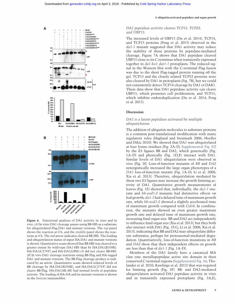
We showed previously that the da1-1 allele of DA1 has anegative interfering phenotype with respect to the closelyrelated family member DAR1 (Li et al. 2008). The pepti-dase activity of the protein encoded by the da1-1 allele,called DA1(R358K), which has an arginine to a lysineresidue altered in a highly conserved C-terminal region,(Supplemental Fig. S4) was assessed. This mutation didnot influence ubiquitylation of Flag-DA1(R358K) (Fig.6A) or create a site for ectopic ubiquitylation of Flag-DA1(R358K), as determined bymass spectrometric analy-sis (Supplemental Fig. S2C). The peptidase activity ofubiquitylated Flag-DA1(R358K) was qualitatively as-sessed in vitro and in vivo (using HA-DA1(R358K)) bycomparison with wild-type DA1 peptidase activity (Fig.6A,B). Both assays showed that DA1(R358K) had lowerpeptidase activity compared with DA1, suggesting thatregions of the conserved C-terminal region are requiredfor peptidase activity and that the da1-1 phenotype maybe due to reduced peptidase activity. Figure 6B also showsthat DA1(4K-4R), which is ubiquitylated (Fig. 2E), hadpeptidase activity toward BB. This suggested that precisepatterns of ubiquitylation are not required for activatingDA1 latent peptidase activity.DAR4, another DA1 family member (Li et al. 2008),
encodes a protein with an N-terminal TIR-NB-LRR andhas a gain-of-function chs3-2d allele in the conservedC-terminal region (Supplemental Fig. S3) that activatedconstitutive defense responses (Xu et al. 2015). Align-ments revealed high similarity to predicted protein se-quences from the photosynthetic bacteria Roseiflexus sp(Supplemental Fig. S7; Burroughs et al. 2011) that includedfour pairs of CxxC/H motifs with the potential to bind
Figure 4. Detection of DA1-mediated cleavage of BB in vivo us-ing FRET. (A) Root protoplasts of da1-ko1 dar1-1 plants weretransfected with the FRET construct eGFP-BB or a controleGFP-BB-mCherry construct together with DA1 to detect DA1-mediated cleavage of BB. Transfected 2protoplasts were imagedusing multiphoton microscopy, and fluorescence half-times ofprotoplasts (n = 13) were captured. The heat map shows fluores-cent lifetime values, and typical protoplasts are shown to illus-trate fluorescent half-lives imaged over the cell. The box plotsshow significantly increased fluorescence lifetime after DA1transfection. (∗∗) P≤ 0.001, Student’s t-test. (B) Cleavage ofeGFP-BB-mCherry by DA1 in the imaged protoplasts shown inA. The arrow shows the major cleavage product of ∼40 kDa ex-pected from DA1 cleavage near the N terminus of BB.
A ubiquitin-activated peptidase and organ growth
GENES & DEVELOPMENT 7
Cold Spring Harbor Laboratory Press on April 3, 2019 - Published by genesdev.cshlp.orgDownloaded from
zinc similar to those in canonical LIM domains (Kadrmasand Beckerle 2004). The chs3-2d mutation changes a cys-teine to a tyrosine in the third pair of conserved CxxC/Hmotifs (Supplemental Figs. S3, S6), suggesting that itmay alter a possible LIM-like structure. This mutationwas introduced into DA1 to create DA1(C274Y), and its
activities were assessed. Figure 6A shows that DA1(C274Y) was not ubiquitylated by BB and had no peptidaseactivity toward BB in vitro and in vivo (Fig. 6B). This im-plicated the putative LIM-like domain in DA1 in UIM-mediated ubiquitylation and activation of DA1 peptidaseactivity.
Figure 5. Identification of the DA1 cleav-age site in BB and destabilization and func-tional inactivation of cleaved BB in vivo bythe N-end rule. (A) Neo-N-terminal se-quences of purified cleaved BB-HIS (left)matched the complete BB protein sequence(right, shown in red). Data from six Edmansequencing cycles are in Supplemental Fig-ure S5. (B) The predicted DA1 cleavage sitein BBwasmutated by changing theAY ami-no acids flanking the site to GG (AY-GG)and deleting two amino acids from bothsides of the predicted cleavage site(ΔNAYK). BB-Flag, BB (ΔNAYK)-Flag, andBB (AY-GG)-Flag were expressed in da1-ko1 dar1-1 Arabidopsis mesophyll proto-plasts under the control of the 35S promot-er. HA-DA1 did not cleave BB containingmutations in the predicted cleavage site.The bottom panel shows HA-DA1 andHA-DA1(pep) loading. (C ) A DA1-cleavedversion of BB does not function in planta.A cleaved version of BB, termed MY61-BB,was expressed in da1-ko1 bb-eod1-2 plantsunder control of the 35S promoter. Thegraph compares petal areas in wild-typeCol-0, da1-ko1, da1-ko1 bb-eod1-2, andtwo independent transgenic lines. Valuesgive are means (n = 50) ± SE, expressed aspercentage of wild-type Col-0 petal areas.Student’s t-test showed no significant dif-ferences between the transformants andthe parental da1-ko1 bb-eod1-2 line. (D)In vitro degradation of BB is dependent onN termini. Ubiquitin fusion constructswere expressed in a reticulocyte lysatecell-free system. Samples were incubatedfor 30 min with or without MG132. Next,cycloheximide (CHX) was added to inhibittranslation, and samples were taken 0 and90 min after CHX addition. Samples wereelectrophoresed on SDS-PAGE and immu-noblotted using HA antibodies to detectBB protein levels. Loading controls were
the CBB-stainedmembrane. (E) PRT1 binding to synthetic peptidesmimicking the neo-N-terminal of 61BB. SPOT assay of a peptide arrayof synthetic 17-mer peptides incubated with recombinant His8-MBP-tagged PRT1. Peptides were derived from an N-recognin test sub-strate, and the first amino acid comprised Leu, Lys,Met, Phe, Pro, Ser, The, Trp, Tyr, and Val. His-PRT1was detected by immunoblotting.Equal peptide loading on the membrane was monitored by UV light prior to PRT1 protein binding. (F ) Constructs expressing ubiquitinfusions of 61BB-Luciferase constructs with glycine or tyrosine neo-N termini (Ub-Gly-61-BB-HA-LUC and Ub-Tyr-61-BB-HA-LUC)were transfected into wild-type or prt1mutant protoplasts. Transfection efficiency was measured using a pUBC::GUS control. Luciferaseactivities were normalized to GUS activity, and the luciferase activity of Gly-61-BB-HA-LUCwas taken as 100%. The significance of dif-ferences was calculated from three independent transformation experiments using Student’s t-tests (two sites, uncoupled). (∗) P-value≤0.05. (G) Overexpression of BB under the control of the 35S promoter leads to strongly reduced growth, and, when crossed with a line over-expressingDA1, this growth inhibitionwas reversed. This demonstrated thatDA1 can reduce the growth inhibitory effect of high levels ofBB. (H) Crossing a line overexpressing DA1 into a line expressing 35S::RFP-BB reduced RFP-BB levels. Homozygous progeny of four inde-pendent crosses (99, 170, 181, and 191) are shown.
Dong et al.
Cold Spring Harbor Laboratory Press on April 3, 2019 - Published by genesdev.cshlp.orgDownloaded from
DA1 peptidase activity cleaves TCP15, TCP22,and UBP15
The increased levels of UBP15 (Du et al. 2014), TCP14,and TCP15 proteins (Peng et al. 2015) observed in theda1-1 mutant suggested that DA1 activity may reducethe stability of these proteins by peptidase-mediatedcleavage. Figure 7A shows that DA1 peptidase cleavedUBP15 close to its C terminus when transiently expressedtogether in da1-ko1 dar1-1 protoplasts. The reduced sig-nal in the Western blot with the C-terminal Flag fusionwas due to the short Flag-tagged protein running off thegel. TCP15 and the closely related TCP22 proteins werealso cleaved by DA1 in protoplasts (Fig. 7B), but we couldnot consistently detect TCP14 cleavage by DA1 or DAR1.These data show that DA1 peptidase activity can cleaveUBP15, which promotes cell proliferation, and TCP15,which inhibits endoreduplication (Du et al. 2014; Penget al. 2015).
Discussion
DA1 is a latent peptidase activated by multipleubiquitylation
The addition of ubiquitin molecules to substrate proteinsis a common post-translational modification with manyregulatory roles (Haglund and Stenmark 2006; Hoellerand Dikic 2010). We showed that DA1 was ubiquitylatedat four lysine residues (Fig. 2A–D; Supplemental Fig. S2)by the E3 ligases BB and DA2, which genetically (Fig.1A–D) and physically (Fig. 1D,E) interact with DA1.Similar levels of DA1 ubiquitylation were observed invivo (Fig. 2I). Loss-of-function mutants of BB and DA2synergistically increased the large organ phenotypes of aDA1 loss-of-function mutant (Fig. 1A–D; Li et al. 2008;Xia et al. 2013). Therefore, ubiquitylation mediated bythese two E3 ligases may increase the growth-limiting ac-tivity of DA1. Quantitative growth measurements ofleaves (Fig. 1E) showed that, individually, the da1-1 mu-tant and bb-eod1-2 mutants had distinctive effects onleaf growth;da1-1 had a delayed time ofmaximumgrowthrate, while bb-eod1-2 showed a slightly accelerated timeof maximum growth compared with Col-0. In combina-tion, the mutants showed an even greater maximumgrowth rate and delayed time of maximum growth rate,increasing final organ size. BB and DA2 act independentlyto influence final organ size (Xia et al. 2013), but they bothalso interact with DA1 (Fig. 1F,G; Li et al. 2008; Xia et al.2013), indicating that BB andDA2may ubiquitylate differ-ent substrates, perhaps for proteasomal-mediated degra-dation. Quantitatively, loss-of-function mutations in BBand DA2 show that their independent effects on growthare less than that of da1-1 (Fig. 1A–D).Members of the DA1 family have a canonical MA
clan zinc metallopeptidase active site domain in theirconserved C-terminal regions (Supplemental Fig. S4; Tho-lander et al. 2010; Rawlings et al. 2012) that was requiredfor limiting growth (Fig. 3F). BB- and DA2-mediatedubiquitylation activated DA1 peptidase activity in vitroand in transiently expressed protoplasts (Fig. 3A,E),
Figure 6. Functional analyses of DA1 activity in vitro and invivo. (A) In vitro DA1 cleavage assays using BB-HIS as a substratefor ubiquitylated Flag-DA1 and mutant versions. The top panelshows the reaction at 0 h, and the middle panel shows the reac-tions at 4 h. The red arrow indicates cleaved BB-HIS. The loadingand ubiquitylation status of input HA-DA1 and mutant versionsis shown.Quantitative scans showed that BB-HISwas cleaved to agreater extent by wild-type DA1-HIS than by HA-DA1(R358K).HA-DA1(C274Y) and HA-DA1(UIM1+2) did not cleave BB-HIS.(B) In vivo DA1 cleavage reactions using BB-Flag and HA-taggedDA1 and mutant versions. The BB-Flag cleavage product is indi-cated by an arrow. Quantitative scans showed reduced levels ofBB cleavage by HA-DA1(R358K), and HA-DA1(C274Y) did notcleave BB-Flag. HA-DA1(4K-4R) had normal levels of peptidaseactivity. The loading of HA-DA and its mutant versions is shownin the bottom immunoblot.
A ubiquitin-activated peptidase and organ growth
GENES & DEVELOPMENT 9
Cold Spring Harbor Laboratory Press on April 3, 2019 - Published by genesdev.cshlp.orgDownloaded from

establishing a biochemical foundation for their joint activ-ities in growth control. Zinc metallopeptidases are main-tained in an inactive form by a “cysteine switch” (VanWart and Birkedal-Hansen 1990) that coordinates a cyste-ine residuewith the zinc atom at the active site to block it.Conformational changes release this and activate thepeptidase.
Ubiquitylation of DA1 has the potential to trigger aconformational change thatmay release inhibition of pep-
tidase activity. Hoeller et al. (2006) showed that UBD-and UIM-mediated monoubiquitylation of endocytoticproteins, including epsin, led to a conformational chan-ge mediated by intramolecular interactions betweenUBDs/UIMs and cis-ubiquitin, which regulated endocy-tosis. The binding of ubiquitin to DA1 UIMs was re-quired for DA1 function in vivo (Fig. 2H), and theUIMs conferred patterns of ubiquitylation on the heter-ologous protein GST similar to that seen for DA1 (Fig.2G [for DA1], E,F [for GST-UIM1+2]). Related observa-tions were seen in the monoubiquitylation of epsin(Oldham et al. 2002) through coupled monoubiquityla-tion (Woelk et al. 2006), where UIMs recruit the UIM-containing protein to the ubiquitylation machinery bydirect interaction with ubiquitin coupled to ubiquitindonor proteins (Haglund and Stenmark 2006). Mutationof Cys274 in the C-terminal zinc finger loop of the LIM-like domain of DA1 abrogated both ubiquitylationand peptidase activity (Fig. 6A,B), suggesting a function-al role for this ancient conserved LIM-like domain(Supplemental Fig. S7; Burroughs et al. 2011) in pepti-dase activation. Analyses of conformational changescaused by DA1 ubiquitylation and their influence onpeptidase activity are required to establish this potentialmechanism.
DA1 cleavage destabilizes its activating E3 ligases(BB and DA2), and cleavage of BB leads to targeting bythe N-recognin PRT1
The RING E3 ligases BB and DA2 activate DA1 peptidaseby ubiquitylation and are also cleaved by DA1 peptidase(Figs. 3A,B,E,G, 4). Once cleaved, DA2 appeared to be de-stabilized in transiently expressed protoplasts (Fig. 3E).Identification of the DA1 cleavage site in BB (Fig. 5A,B)revealed Y61-BB at the neo-N terminus of cleaved BB.This neo-N terminus conferred proteasome-mediateddegradation in a cell-free system (Fig. 5D). This degrada-tion depended on recognition of the neo-N terminus bythe Arabidopsis E3 ligase PRT1 (Fig. 5E,F; Potuschaket al. 1998; Stary 2003), an N-recognin catalyzing N-endrule-mediated degradation (Varshavsky 2011) with a sug-gested preference for aromatic amino acid N termini.Interestingly, the neo-N-terminal MY61-BB, which wasused to express a cleaved version of BB in planta, con-ferred strong proteasome-independent instability (Fig.5D) in a mechanism that is not yet clear. The lack ofMY61-BB function in vivo (Fig. 5C) supported the obser-vation that DA1-mediated cleavage of BB leads to itsloss of function in vivo. Overexpression of BB strongly re-duced growth, as expected from its inhibitory role ingrowth (Disch et al. 2006). The reversal of this inhibitionby overexpression of DA1, which reversed growth inhibi-tion (Fig. 5G) and reduced RFP-BB levels (Fig. 5H), is con-sistent with a mechanism involving DA1-mediatedreduction of BB activity via peptidase-mediated cleavageand subsequent degradation by the N-end rule pathway.Such an activation–destruction mechanism mediated byBB, DA2, and DA1 may provide a way of tightly control-ling peptidase activity. The physiological role of these
Figure 7. DA1 cleaves UBP15, TCP15, and TCP22 in vivo. (A,B)In vivo cleavage reactions of UBP15-3Flag and 3Flag-UBP15 (A) aswell as 3-Flag-TCP15, 3-Flag-TCP22, and 3-Flag TCP5 (a non-cleaved control) (B) usingHA-DA1 andHA-DA1(pep). Constructsexpressed from the 35S promoterwere cotransfected intoda1-ko1dar1-1 mesophyll protoplasts. UBP15, TCP14, TCP15, andTCP22 cleavage products are shown in the top immunoblots.The bottom immunoblots show HA-DA1 and HA-DA1(pep) pro-tein levels. The approximate locations of DA1 cleavage sites (ar-rows) are shown inUBP15, TCP15, andTCP22. (C ) Amodel of theproposed transient mechanism of DA1 peptidase activation andthe consequences of DA1-mediated cleavage of growth regulatorsduring organ growth. PRT1 activity is shown as degrading BB.
Dong et al.
10 GENES & DEVELOPMENT
Cold Spring Harbor Laboratory Press on April 3, 2019 - Published by genesdev.cshlp.orgDownloaded from

mechanisms, which often involve ubiquitylation and pro-teolytic degradation, is to drive unidirectional cellularprocesses; for example, in cell cycle progression (Reed2003). The factors that trigger DA1 ubiquitylation byBB and coordinate the activities of BB and DA2 remainunknown.
DA1 peptidase activity also cleaves diversegrowth regulators
We showed previously that TCP14 and TCP15 functiondownstream fromDA1 and other family members in con-trolling organ size in Arabidopsis, and reduced functionof DA1 family members led to increased TCP14 andTCP15 protein levels (Peng et al. 2015). Similarly, levelsof UBP15 protein, which promotes cell proliferation (Liuet al. 2008) and also functions downstream from DA1,were increased in the da1-1 reduced-function mutant(Du et al. 2014). We showed that TCP15 and the relatedTCP22 as well as UBP15 were cleaved by DA1 peptidaseactivity (Fig. 7A,B) but could not reliably detect TCP14cleavage by DA1 or DAR1. DA1-mediated cleavage ofTCP15 and UBP15 is a plausible mechanism that ac-counts for these observed reduced protein levels, similarto DA1-mediated inactivation and destabilization of BBby peptidase cleavage. Taken together, these observationssuggest a mechanism (Fig. 7C) in which DA1 peptidase,activated transiently by BB or DA2, coordinates a “one-way” cessation of cell proliferation and the initiation ofendoreduplication through the cleavage and potential in-activation of proteins that promote cell proliferation andinhibit endoreduplication.
Materials and methods
Plant materials, growth conditions, and organ sizemeasurements
A. thaliana Col-0 was the wild-type plant used. Plants weregrown in growth rooms at 20°C with 16-h day/8-h dark cycles us-ing either soil or MS medium supplemented with 0.5% glucose.Petal and seed areas were imaged by high-resolution scanning(3600 dpi; Hewlett Packard Scanjet 4370) and analyzed usingImageJ software (http://rsbweb.nih.gov/ij).
In vitro DA1-mediated cleavage assays
Flag-DA1 was ubiquitylated in vitro using either DA2-HIS orBB-HIS as E3 ligases, purified using Flag magnetic beads, andquantified, and 100 ng was added to 100 ng of BB-HIS, DA2-HIS, or BBR-HIS in a 30-µL reaction in 50 mM Tris HCl (pH7.4) and 5 mM MgCl2. Reactions were carried out for 4 h at 30°C and terminated by the addition of SDS sample buffer.
Mass spectrometry analysis
DA1 ubiquitylation patterns were determined from trypsinizedproteins purified on SDS-PAGE gels. For liquid chromatogra-phy-tandem mass spectrometry analysis, peptides were appliedto an LTQ-Orbitrab (Thermo-Fischer) using a nanoAcquityultraperformance liquid chromatography system (Waters Ltd.).Further details are in the Supplemental Material.
Acknowledgments
We thank Dr. Paul Thomas (Henry Wellcome Laboratory forCell Imaging, University of East Anglia) for advice and operatingthe multiphoton microscope, and Dr. Cristoph Bücherl (TheSainsbury Laboratory, Norwich) for advice on FRET. We thankShimadzu Europa GMBH for carrying out Edman sequencing.We thank Andreas Bachmair for the prt1 EMS allele, and YukikoYoshida for the TR-TUBE construct. This work was supported byBiological and Biotechnological Sciences Research Council(BBSRC) grant BB/K017225 and Strategic Programme grant BB/J004588 to M.W.B., and European Commission contract 037704(AGROnomics) to M.W.B. and D.I. J.D. was supported by a Bio-technology and Biological Sciences Research Council (BBSRC)CASE Studentship, C.N. was supported by a PhD Fellowshipfrom the Landesgraduiertenförderung Sachsen-Anhalt, and N.D.was supported by an Independent Junior Research Group grantfrom the ScienceCampus Halle–Plant-based Bioeconomy, theDeutsche Forschungsgemeinschaft (DFG; grant DI 1794/3-1),the DFG Graduate Training Centre (GRK1026), and the LeibnizInstitute of Plant Biochemistry. Y.L. was supported by the Na-tional Natural Science Foundation of China (grants 91417304,31425004, 91017014, 31221063, and 31100865), the National Ba-sic Research Program of China (grant 2009CB941503), and theMinistry of Agriculture of China (grant 2016ZX08009-003). M.W.B. and Y.L. are in the Chinese Academy of Sciences (CAS)-John Innes Centre (JIC) Centre of Excellence in Plant and Micro-bial Sciences (CEPAMS). M.W.B., J.D., H.D., F.-H.L, H.V., N.D.,Y.L., and D.I. designed the research; H.D., F.-H.L., J.D., R.P., H.V., C.N., M.K., C.S., N.M., L.N., H.V., T.X., L.C., G.S., N.G.,and M.S. performed the research and analyzed the data; and M.W.B. wrote the paper.
References
Andriankaja M, Dhondt S, De Bodt S, Vanhaeren H, Coppens F,DeMilde L, Mühlenbock P, Skirycz A, Gonzalez N, BeemsterGTS, et al. 2012. Exit from proliferation during leaf develop-ment in Arabidopsis thaliana: a not-so-gradual process.Dev Cell 22: 64–78.
Bachmair A, Finley D, Varshavsky A. 1986. In vivo half-life of aprotein is a function of its amino-terminal residue. Science234: 179–186.
Barry ER, Camargo FD. 2013. The Hippo superhighway: signalingcrossroads converging on the Hippo/Yap pathway in stemcells and development. Curr Opin Cell Biol 25: 247–253.
Breuer C, Ishida T, Sugimoto K. 2010. Developmental control ofendocycles and cell growth in plants. Curr Opin Plant Biol13: 654–660.
Breuninger H, LenhardM. 2012. Expression of the central growthregulator BIG BROTHER is regulated by multiple cis-ele-ments. BMC Plant Biol 12: 41.
Burroughs AM, Iyer LM, Aravind L. 2011. Functional diversifi-cation of the RING finger and other binuclear treble clefdomains in prokaryotes and the early evolution of the ubiqui-tin system. Mol Biosyst 7: 2261.
De Veylder L, Larkin JC, Schnittger A. 2011. Molecular controland function of endoreplication in development and physiolo-gy. Trends Plant Sci 16: 624–634.
Disch S, Anastasiou E, Sharma VK, Laux T, Fletcher JC, LenhardM. 2006. The E3 ubiquitin ligase BIG BROTHER controlsArabidopsis organ size in a dosage-dependent manner. CurrBiol 16: 272–279.
Du L, Li N, Chen L, Xu Y, Li Y, Zhang Y, Li C, Li Y. 2014. Theubiquitin receptor DA1 regulates seed and organ size by
A ubiquitin-activated peptidase and organ growth
GENES & DEVELOPMENT 11
Cold Spring Harbor Laboratory Press on April 3, 2019 - Published by genesdev.cshlp.orgDownloaded from
modulating the stability of the ubiquitin-specific proteaseUBP15/SOD2 in Arabidopsis. Plant Cell 26: 665–677.
Efroni I, Blum E, Goldshmidt A, Eshed Y. 2008. A protracted anddynamic maturation schedule underlies Arabidopsis leafdevelopment. Plant Cell 20: 2293–2306.
Faden F, Ramezani T,Mielke S, Almudi I, Nairz K, FroehlichMS,Höckendorff J, Brandt W, Hoehenwarter W, Dohmen RJ, et al.2016. Phenotypes on demand via switchable target proteindegradation in multicellular organisms. Nat Commun 7:12202.
Green AA, Kennaway JR, Hanna AI, Bangham JA, Coen E. 2010.Genetic control of organ shape and tissue polarity. PLoS Biol8: e1000537.
Haglund K, Stenmark H. 2006. Working out coupled monoubi-quitination. Nat Cell Biol 8: 1218–1219.
Haglund K, Di Fiore PP, Dikic I. 2003. Distinct monoubiquitinsignals in receptor endocytosis. Trends Biochem Sci 28:598–603.
Hicke L, SchubertHL, Hill CP. 2005. Ubiquitin-binding domains.Nat Rev Mol Cell Biol 6: 610–621.
Hoeller D, Dikic I. 2010. Regulation of ubiquitin receptors bycoupled monoubiquitination. Subcell Biochem 54: 31–40.
HoellerD,CrosettoN, BlagoevB, RaiborgC, TikkanenR,WagnerS, Kowanetz K, Breitling R, MannM, Stenmark H, et al. 2006.Regulation of ubiquitin-binding proteins by monoubiquitina-tion. Nat Cell Biol 8: 163–169.
Husnjak K, Dikic I. 2012. Ubiquitin-binding proteins: decodersof ubiquitin-mediated cellular functions. Annu Rev Biochem81: 291–322.
Johnston LA, Gallant P. 2002. Control of growth and organ size inDrosophila. Bioessays 24: 54–64.
Kadrmas JL, BeckerleMC. 2004. The LIM domain: from the cyto-skeleton to the nucleus. Nat Rev Mol Cell Biol 5: 920–931.
Kazama T, Ichihashi Y, Murata S, Tsukaya H. 2010. The mecha-nism of cell cycle arrest front progression explained by aKLUH/CYP78A5-dependentmobile growth factor in develop-ing leaves of Arabidopsis thaliana. Plant Cell Physiol 51:1046–1054.
Kim H, Chen J, Yu X. 2007. Ubiquitin-binding protein RAP80mediates BRCA1-dependent DNA damage response. Science316: 1202–1205.
Komander D, Rape M. 2012. The ubiquitin code. Annu RevBiochem 81: 203–229.
Li Y, Zheng L, Corke F, Smith C, Bevan MW. 2008. Control of fi-nal seed and organ size by theDA1 gene family inArabidopsisthaliana. Genes Dev 22: 1331–1336.
Li Z-Y, Li B, Dong A-W. 2012. The Arabidopsis transcription fac-tor AtTCP15 regulates endoreduplication by modulating ex-pression of key cell-cycle genes. Mol Plant 5: 270–280.
Liu Y, Wang F, Zhang H, He H,Ma L, Deng XW. 2008. Functionalcharacterization of the Arabidopsis ubiquitin-specific prote-ase gene family reveals specific role and redundancy of indi-vidual members in development. Plant J 55: 844–856.
Oldham CE, Mohney RP, Miller SLH, Hanes RN, O’Bryan JP.2002. The ubiquitin-interacting motifs target the endocyticadaptor protein epsin for ubiquitination. Curr Biol 12:1112–1116.
Pan D. 2010. The hippo signaling pathway in development andcancer. Dev Cell 19: 491–505.
Peng Y, Chen L, Lu Y, Wu Y, Dumenil J, Zhu Z, BevanMW, Li Y.2015. The ubiquitin receptors DA1, DAR1, and DAR2 redun-dantly regulate endoreduplication bymodulating the stabilityof TCP14/15 in Arabidopsis. Plant Cell 27: 649–662.
Potuschak T, Stary S, Schlögelhofer P, Becker F, Nejinskaia V,Bachmair A. 1998. PRT1 of Arabidopsis thaliana encodes acomponent of the plant N-end rule pathway. Proc Natl AcadSci 95: 7904–7908.
Rawlings ND, Barrett AJ, Bateman A. 2012. MEROPS: the data-base of proteolytic enzymes, their substrates and inhibitors.Nucleic Acids Res 40: D343–D350.
Reed SI. 2003. Ratchets and clocks: the cell cycle, ubiquitylationand protein turnover. Nat Rev Mol Cell Biol 4: 855–864.
Sato Y, Yoshikawa A, Mimura H, Yamashita M, Yamagata A,Fukai S. 2009. Structural basis for specific recognition of Lys63-linked polyubiquitin chains by tandem UIMs of RAP80.EMBO J 28: 2461–2468.
Sluis A, Hake S. 2015. Organogenesis in plants: initiationandelaboration of leaves. Trends Genet 31: 300–306.
Stary S. 2003. PRT1 ofArabidopsis is a ubiquitin protein ligase ofthe plant N-end rule pathway with specificity for aromaticamino-terminal residues. Plant Physiol 133: 1360–1366.
Tholander F, Roques B-P, Fournié-Zaluski M-C, ThunnissenMMGM, Haeggström JZ. 2010. Crystal structure of leukotri-ene A4 hydrolase in complex with kelatorphan, implicationsfor design of zinc metallopeptidase inhibitors. FEBS Lett584: 3446–3451.
van der Krogt GNM, Ogink J, Ponsioen B, Jalink K. 2008. A com-parison of donor-acceptor pairs for genetically encoded FRETsensors: application to the Epac cAMP sensor as an exampleed. K.-W. Koch. PLoS One 3: e1916.
Van Wart HE, Birkedal-Hansen H. 1990. The cysteine switch: aprinciple of regulation of metalloproteinase activity with po-tential applicability to the entire matrix metalloproteinasegene family. Proc Natl Acad Sci 87: 5578–5582.
Varshavsky A. 2011. The N-end rule pathway and regulation byproteolysis. Protein Sci 20: 1298–1345.
Woelk T, Oldrini B, Maspero E, Confalonieri S, Cavallaro E, DiFiore PP, Polo S. 2006. Molecular mechanisms of coupledmonoubiquitination. Nat Cell Biol 8: 1246–1254.
Xia T, Li N, Dumenil J, Li J, Kamenski A, BevanMW, Gao F, Li Y.2013. The ubiquitin receptor DA1 interacts with the E3 ubiq-uitin ligase DA2 to regulate seed and organ size in Arabidop-sis. Plant Cell 25: 3347–3359.
Xu F, Zhu C, Çevik V, Johnson K, Liu Y, Sohn K, Jones JD, HolubEB, Li X. 2015. Autoimmunity conferred by chs3-2D relies onCSA1, its adjacent TNL-encoding neighbour. Sci Rep 5: 8792.
Dong et al.
12 GENES & DEVELOPMENT
Cold Spring Harbor Laboratory Press on April 3, 2019 - Published by genesdev.cshlp.orgDownloaded from

10.1101/gad.292235.116Access the most recent version at doi: published online February 6, 2017Genes Dev.
Hui Dong, Jack Dumenil, Fu-Hao Lu, et al.
Arabidopsisregulatory proteins to limit cell proliferation in destabilization of its activating E3 ligases and diverse growth Ubiquitylation activates a peptidase that promotes cleavage and
Material
Supplemental
http://genesdev.cshlp.org/content/suppl/2017/02/06/gad.292235.116.DC1
Published online February 6, 2017 in advance of the full issue.
License
Commons Creative
.http://creativecommons.org/licenses/by/4.0/License (Attribution 4.0 International), as described at
, is available under a Creative CommonsGenes & DevelopmentThis article, published in
ServiceEmail Alerting
click here.right corner of the article or
Receive free email alerts when new articles cite this article - sign up in the box at the top
Published by © 2017 Dong et al.; Published by Cold Spring Harbor Laboratory Press
Cold Spring Harbor Laboratory Press on April 3, 2019 - Published by genesdev.cshlp.orgDownloaded from