Embed Size (px)
DESCRIPTION
Won Ju Hong · Yu Na Kang · Koo Jeong Kang1Departments of Pathology and 1Surgery, Keimyung University School of Medicine, Daegu, Korea
Citation preview
ACG CASE REPORTS JOURNAL
acgcasereports.gi.org ACG Case Reports Journal | Volume 1 | Issue 2 | January 201474
IMAGE | PEDIATRICS
A Rare Case of Childhood Undifferentiated Embryonal Sarcoma of the Liver Managed SuccessfullyGangadhar Rao Gondu, DNB1, Vimalakar Reddy Eppa, DNB2, Avinash Sowdepalli, DNB1, Mohan Narasimhan, DNB, FACS2, and Ramesh Ardhanari, MCh, FRCS2
1Department of General Surgery, Meenakshi Mission Hospital and Research Centre, Madurai, Tamil Nadu, India 2Department of Surgical Gastroenterology, Meenakshi Mission Hospital and Research Centre, Madurai, Tamil Nadu, India
Case ReportA 13-year-old boy presented with low-grade fever, upper abdominal pain, loss of appetite, and weight loss for 2 months, with a history of swelling in his upper abdomen for 15 days. On examination, a 17 x 20-cm intraab-dominal, smooth, firm, non-tender swelling was noted occupying epigastric and left hypochondrium. The mass moved with respiration, and finger insinuation between mass and right coastal margin was not possible. There was no ascites and further examination was unremarkable. Liver function tests and α-fetoprotein were normal.
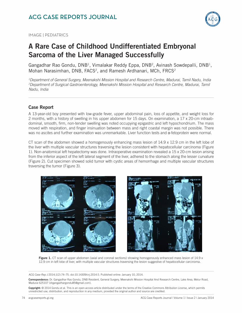
CT scan of the abdomen showed a homogenously enhancing mass lesion of 14.9 x 12.9 cm in the left lobe of the liver with multiple vascular structures traversing the lesion consistent with hepatocellular carcinoma (Figure 1). Non-anatomical left hepatectomy was done. Intraoperative examination revealed a 15 x 20-cm lesion arising from the inferior aspect of the left lateral segment of the liver, adhered to the stomach along the lesser curvature (Figure 2). Cut specimen showed solid tumor with cystic areas of hemorrhage and multiple vascular structures traversing the tumor (Figure 3).
ACG Case Rep J 2014;1(2):74–75. doi:10.14309/crj.2014.5. Published online: January 10, 2014.
Correspondence: Dr. Gangadhar Rao Gondu, DNB Resident, General Surgery, Meenakshi Mission Hospital And Research Centre, Lake Area, Melur Road, Madurai 625107 ([email protected]).
Copyright: © 2014 Gondu et al. This is an open-access article distributed under the terms of the Creative Commons Attribution License, which permits unrestricted use, distribution, and reproduction in any medium, provided the original author and source are credited.
Figure 1. CT scan of upper abdomen (axial and coronal sections) showing homogenously enhanced mass lesion of 14.9 x 12.9 cm in left lobe of liver, with multiple vascular structures traversing the lesion suggestive of hepatocellular carcinoma.

Publish your work in ACG Case Reports JournalACG Case Reports Journal is a peer-reviewed, open-access publication that provides GI fellows, private practice clinicians, and other members of the health care team an opportunity to share interesting case reports with their peers and with leaders in the field. Visit http://acgcasereports.gi.org for submission guidelines. Submit your manuscript online at http://mc.manuscriptcentral.com/acgcr.
Gondu et al
acgcasereports.gi.org
Childhood Undifferentiated Embryonal Sarcoma of the Liver
75 ACG Case Reports Journal | Volume 1 | Issue 2 | January 2014
Disclosures
Author contributions: All the authors contributed to evaluating and managing the case and to writing the manuscript. GR Gondu is the article guarantor.
Financial disclosure: No conflicts of interest or funding sources to disclose.
Informed consent was obtained for this case report.
Received: September 19, 2013; Accepted: December 17, 2013
Figure 2. Intraoperative picture showing tumor arising from inferior surface of left lobe of liver, adhered to stomach along lesser curvature.
Figure 3. Excised specimen surrounded by capsule. Cut section showing myxoid stroma with areas of hemorrhagic necrosis.
Figure 4. (A) 100x picture showing normal liver in right upper part and tumor in the lower part. (B) 100x tumor showing mixture of highly atypical spindle and giant cells with sarcomatous appearance. (C) 400x large tumor giant cells with sarcoma-like appearance. (D) 400x vimentin positivity in tumor cells.
Postoperatively, 1 unit of packed red blood cell transfusion was given and the patient was discharged on postoperative day 8. Histopathological examination reported a tumor pre-dominantly showing spindle cells arranged in sheets em-bedded in myxoid stroma. Vimentin was strongly positive. Desmin, carcinoembryonic antigen, and α-fetoprotein were negative. This suggested undifferntiated embryonal sarcoma of liver (USEL; Figure 4), a rare, highly malignant neoplasm that is more common in children. Bone scan was normal. Adjuvant chemotherapy was given. The patient is now as-ymptomatic on regular follow-up. We stress the role of his-topathology and IHC in diagnosing UESL and of timely en bloc resection and postoperative chemotherapy in improving survival rates.
D
A B
C