Embed Size (px)
Citation preview
570 IEEE TRANSACTIONS ON ULTRASONICS, FERROELECTRICS, AND FREQUENCY CONTROL, VOL. 68, NO. 3, MARCH 2021
Ultrasonic Monitoring of Dentin DemineralizationJosep Rodríguez-Sendra , Inés Torres, Noé Jiménez , Salvatore Sauro, and Francisco Camarena
Abstract— Demineralization is a process of loss of min-erals in the dental hard tissue that affects seriously thehealth of the patients, as it diminishes the tooth resis-tance, generating chewing problems by altering the occlusalstructure, hypersensitivity, and pulpal problems. Deminer-alization can be produced by pathological processes aserosion or caries, or by surgical processes as etching. Dueto the complexity of natural demineralization processes,it is mandatory to provide quantitative and standardizedtests to allow their study in controlled laboratory conditions.Ultrasonic techniques are suitable for this purpose as theyare nondestructive,quick, and provide localized mechanicalinformation about the tissue, which is related with its degreeof demineralization. In the present work, we evaluate thecomplete process of demineralization of the human dentinunder controlled laboratory conditions using a pulse-echoultrasonic technique.Up to 15 human dentin teeth have beendemineralized with phosphoric acid at 10%. The time-of-flight measurements using the pulse-echo system allows toobtain the speedof sound in healthy(3415 m/s) and deminer-alized dentin tissue (1710 m/s), as well as to characterize thedynamical process of the acid penetration, which generateswell-defined boundaries between two media (demineralizedand mineralized dentin), showing very different mechanicalproperties. These boundaries advance in depth at an initialrate of 2.9 µm/min, decelerating at −9.3 nm/min2 until thewhole demineralization of the sample is achieved. In addi-tion, the technique allows to measure the relevance of thedemineralization produced by the acid residues inside thetooth once it has been removed from the acidic solution.Beyond the assessmentof artificialdemineralization lesionsunder laboratory conditions, as demonstrated in this arti-cle, the proposed technique opens new approaches to theassessment of demineralization caused by natural cariesin vivo.
Index Terms— Demineralization, denistry, pulse-echo,ultrasonic.
Manuscript received July 24, 2020; accepted August 6, 2020. Dateof publication August 10, 2020; date of current version February 24,2021. This work was supported in part by the Spanish Ministry ofScience, Innovation and Universities (MICINN) under Grant “Juan dela Cierva - Incorporación” (IJC2018-037897-I) and Grant “ProyectosI + D + i 2019” (PID2019-111436RB-C22), in part by the AgènciaValenciana de la Innovació under Grant INNCON/2020/009, and in partby the European Union through the Programa Operativo del FondoEuropeo de Desarrollo Regional (FEDER) of the Comunitat Valenciana2014–2020 under Grant IDIFEDER/2018/022. (Corresponding author:Josep Rodríguez-Sendra.)
Josep Rodríguez-Sendra, Noé Jiménez, and Francisco Camarenaare with the Instituto de Instrumentación para Imagen Molecular,Universitat Politècnica de València, 46022 Valencia, Spain (e-mail:[email protected]).
Inés Torres is with the Departamento de Odontología, CEU UniversidadCardenal Herrera, 46113 Moncada, Spain, and also with the InstitutoValenciano de Investigaciones Odontológicas, 46701 Gandia, Spain.
Salvatore Sauro is with the Departamento de Odontología, CEUUniversidad Cardenal Herrera, 46113 Moncada, Spain.
Digital Object Identifier 10.1109/TUFFC.2020.3015668
I. INTRODUCTION
DENTAL caries is one of the most prevalent diseasesworldwide. According to the World Health Organization,
between 60% and 90% of school-age children and almost100% of adults have or have had caries injuries [1]. Dentalcaries is a multifactorial disease that appears when, in a firststage, enamel and, then, dentin lose their minerals due to acidsproduced from bacterial metabolism when break down sugarsor food debris [2], [3]. Knowledge of the demineralizationprocess in dentin is crucial in cavity preparation, as the dentistmust remove part of the dentin to place a filling and thecriterion for deciding when to stop the removal of dentinmaterial has to do with its degree of mineralization. Objectivecriteria should be established at this point. On the other hand,the use of bioactive materials has recently been proposedto facilitate the remineralization of damaged dentin, makingit possible to preserve a greater amount of patient’s dentin[4]–[6]. Thus, it is important to be able to assess the degreeof mineralization of the dentin to make these decisions andto be able to quantify the degree of improvement of the newproducts.
Dentin consists of about 70% inorganic material (hydrox-yapatite), 20% organic base (highly mineralized type I col-lagen fibers), and about 10% water [7]. Its demineralizationprocess involves at least two stages: the dissolution of bio-minerals by organic acids and the subsequent degradationof dentin matrix by proteases [8]. Natural dentin deminer-alization processes are geometrically complex, with varia-tions in width, depth, severity, and color, making it difficultto find many similar lesions for in vitro studies [8]. Forthis reason, standardized artificial protocols using acids havebeen proposed to reproduce real lesions and more meticulousinvestigations with the combination of new technologies arerequired [8].
Artificial standardized demineralization lesions offer manyadvantages when compared with the use of natural lesionsfor research because demineralization protocols can bereproducible and lesions with different size can be read-ily made based on demineralization kinetics of a givenacid [9]. Some studies have shown that these acids, mainlyethylenediaminetetra-acetic acid (EDTA) or phosphoric acid(PA), can selectively demineralize extrafibrillar minerals andkeep intrafibrillar minerals at least partially, if not completelyintact, both of them demineralizing with minimum collagendegradation [10]. The acidic agents remove the smear layerand open the dentin tubules, demineralize the dentin surface,and increase the porosity of the intertubular dentin [11].
Different techniques have been proposed to study the dem-ineralization process of the tooth, such as scanning electronmicroscopy (SEM) [12], atomic force microscopy (AFM) [13],
1525-8955 © 2020 IEEE. Personal use is permitted, but republication/redistribution requires IEEE permission.See ht.tps://ww.w.ieee.org/publications/rights/index.html for more information.
Authorized licensed use limited to: UNIVERSIDAD POLITECNICA DE VALENCIA. Downloaded on February 26,2021 at 11:02:46 UTC from IEEE Xplore. Restrictions apply.
RODRÍGUEZ-SENDRA et al.: ULTRASONIC MONITORING OF DENTIN DEMINERALIZATION 571
X-ray tomographic microscopy (XTM) [14], optical coherencetomography (OCT) [15], or nanoindentation [16]. Attin andWegehaupt [17] described in 2014 different techniques andmethods for assessment the dental erosion.
Ultrasonics techniques have also been widely used for toothcharacterization during last decades. In 1963 Baum et al.[18] used ultrasound to image the internal structures of thetooth. In 2008, Ghorayeb et al. [19] presented a review whichexamined the diagnostic applications of dental ultrasound,starting with the pioneering work up to the current work.In addition, Marotti et al. [20] presented in 2013 a reviewwhere it was described and discussed the use of ultrasound inseveral areas as dental scanning, caries detection, and implantdentinstry among others.
The acoustic properties of dental tissues have been evaluatedusing noninvasive ultrasonic techniques by different authors.Watanabe et al. [21] measured the longitudinal and shearsound velocity to determine the elastic modulus in mineralized(17.4 GPa) and demineralized (1.46 GPa) human dentin,finding big differences. A similar study was performed byYasuda et al. [22] for bovine tooth, obtaining an elasticmodulus of 16.9 GPa (mineralized) and 2.1 GPa (demineral-ized) and a velocity of 3675 m/s (mineralized) and 1597 m/s(demineralized). These studies quantitatively show that dentinbehaves close to a soft tissue after complete demineralization.Ng et al. [23], [24], Maev et al. [25], Miyazaki et al. [26],and John [27], [28] measured longitudinal wave velocity inhuman molar dentin obtaining values from 3317 to 4050 m/sas a function of the type of teeth. Löst et al. [29] andJohn et al. [30] described a method which allows measure-ments on coplanar ground sections of human teeth with alateral resolution of 0.2 mm using a water tank using a20-MHz transducer. Miyazaki et al. [26] showed that themechanical properties of tissue do not vary as long as thesamples are taken with the same section of the same tooth.Lees et al. [31] used an ultrasonic system to continuouslymonitor the development of a demineralized layer due toetching by hydrochloric acid. Yamaguchi et al. [32], [33]conducted an experiment in bovine tooth to demonstrate theprotection capabilities against demineralization of the caseinphosphopeptide-amorphous calcium phosphate paste and theresults were monitored comparing changes in the longitudinalwave velocity. A similar study was performed by Endo et al.[34] to evaluate the apparently good protection capabilitiesof calcium phosphate desensitizer to prevent demineraliza-tion. All these studies have demonstrated the hypothesisthat the volumetric concentration of minerals in dental hardtissue is related with the propagation velocity of ultrasonicwaves.
In the present work, we study the complete process ofdemineralization of the human dentin under controlled labo-ratory conditions using a nondestructive pulse-echo ultrasonictechnique. The main objective is to demonstrate the capabilityof ultrasound to monitor an artificial demineralization processof the dentin with acid that is a widespread practice in dentistryto prepare samples for subsequent studies. The measurement ofthe time-of-flight of the acoustic pulses allows us to obtain theeffective sound velocity in demineralized and healthy dentintissue, as well as to demonstrate that the acid-penetration
process defines a clear boundary between two media withvery different mechanical properties. The present ultrasonictechnique, which incorporates an adapter to reduce the contactarea between the emitter and the dentin, is suitable to be usedin in vivo studies.
This article is organized as follows. Section II shows thesample preparation procedure, the demineralization protocoland the ultrasonic monitoring methods. Section III presentsthe results obtained for two demineralization studies. Finally,Section IV shows the discussion and conclusions.
II. MATERIALS AND METHODS
A. Sample Preparation
A set of 18 healthy permanent human molars without cariesor fillings extracted for orthodontic or periodontal reasonswere obtained with informed consent. The teeth were storedin distilled water. The occlusal surfaces were polished usingsequentially finer grades of polishing paper SI-C, grit 320, 500,and 1000 (Versocit, Struers A/S, Copenhagen, Denmark) untilthe occlusal enamel were completely removed. A polishinghead that applies a constant force of 10 N (Vector LCPower Head, Buehler, Lake Bluff, IL, USA) in conjunctionwith a water-cooled rotating polishing machine (Meta-Serv3000 Grinder-Polisher, Buehler, Lake Bluff) was used at arotating speed of 250 rotations per minute. The molars withoutocclusal surfaces were placed in a water-cooled diamond saw(Micromet evolution, Remet, Bologna, Italy) and were crosssectioned to approximately 1.1-mm-thick samples, as shownin Fig. 1(a). The surfaces were polished with abrasive paperto obtain a standardized thickness of 1.09 ± 0.05 mm. Allsamples were immersed in distilled water and cleaned usingan ultrasonic bath to remove the debris. After that, the sampleswere stored in distilled water.
B. Demineralization Procedure
A total of 14 samples were located together with PA(Panreac Química, Barcelona, Spain) at 10% concentration ina Pyrex recipient and agitated with a magnetic shaker duringthe whole demineralization process.
A set of six samples were used for the SEM and Fouriertransform infrared (FTIR) spectroscopy evaluation. Half of thespecimens, N = 3, were completely demineralized for 5 h andhalf of the specimens, N = 3, were maintained in distilledwater and used as control samples.
For the first ultrasonic experiment (E1), five samples werecompletely demineralized for 290 min and one sample wasmaintained in distilled water to be used as the baselinecontrol. Every 10 min, all samples were removed from theacidic solution and cleaned with distilled water to perform theultrasonic measurements, which delayed 3 min approximately.After that, the samples were returned to the acidic solutionto continue with the demineralization process. The controlsample was monitored following the same procedure but itwas conserved in distilled water all the time.
For the second ultrasonic experiment (E2), six sampleswere demineralized to a different degree. The six sampleswere located in the Pyrex recipient with the PA at 10%concentration. Each hour, one of the samples was extracted,
Authorized licensed use limited to: UNIVERSIDAD POLITECNICA DE VALENCIA. Downloaded on February 26,2021 at 11:02:46 UTC from IEEE Xplore. Restrictions apply.
572 IEEE TRANSACTIONS ON ULTRASONICS, FERROELECTRICS, AND FREQUENCY CONTROL, VOL. 68, NO. 3, MARCH 2021
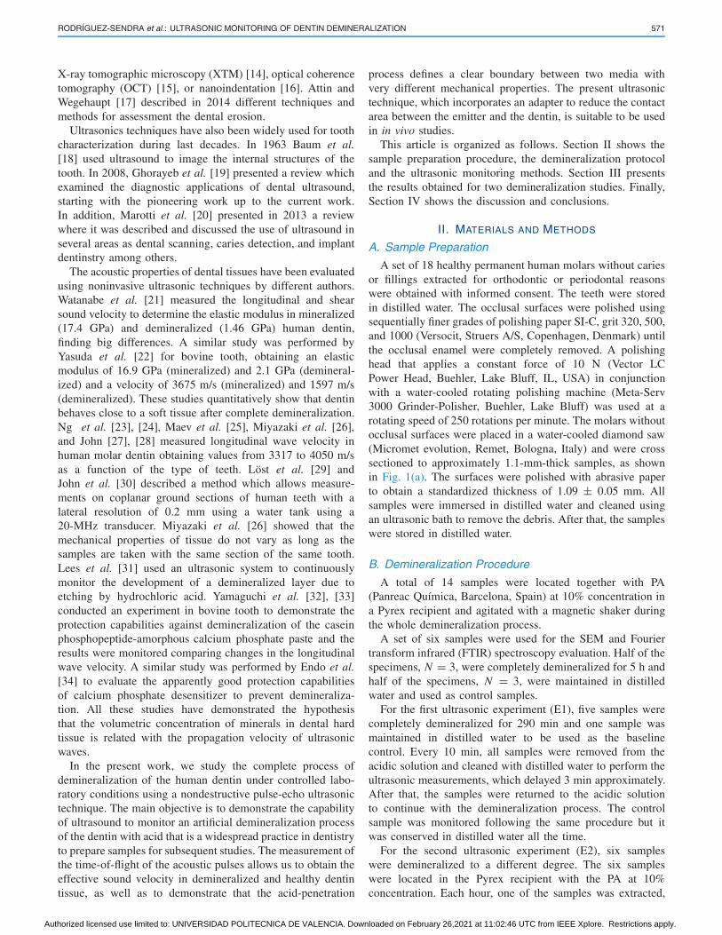
Fig. 1. (a) Photograph of a sample after preparation. (b) Photograph of the experimental set-up. (c) Example of an RF-signal obtained for a partiallydemineralized sample, showing several echoes.
cleaned, and ultrasonically monitored. Then, the sample wasconserved in distilled water and monitored each 60 min.
C. SEM and FTIR Measurement
First, three further sound samples and three demineral-ized samples were analyzed using ATR/FTIR Spectrometer(Perkin-Elmer, Beaconsfield, U.K.) with a spectral resolutionof 4 cm−1 to characterize the chemical composition of thedentin before and after acid treatment. After the FTIR analysis,the samples were analyzed by SEM. The specimens weredehydrated in ascending concentration of alcohol, mounted onaluminum stubs, and sputter coated. The morphology of thespecimens was analyzed using a Hitachi S3500 SEM (HitachiHigh Technologies, Maidenhead, U.K.).
D. Ultrasonic Measurement
An Olympus SONOPEN V260-45 transducer with a nom-inal central frequency of 15 MHz and a bandwidth from5.98 to 17.68 MHz (−6 dB) was used as emitter and receiverusing a pulse-echo technique to obtain the time of flight. Thistransducer presents a delay line with a diameter of D = 2 mmand a ratio between the diameter and the wavelength ofD/λ = 20 wavelengths per aperture in water. The choice of thesource is a compromise between a small physical dimensionmandatory for dental applications and a high D/λ ratio toenhance the acoustic energy collected by the transducer.
Fig. 1(b) shows a photograph of the experimental setup.The transducer was placed perpendicularly to the top surfaceof the dentin sample to record the echo signals. At theopposite side, the sample was in contact with a rigid supportto enhance the reflection of the ultrasonic waves at theboundary. An ultrasonic pulser-receiver (US-Wave, LecoeurElectronique, Chuelles, France) was used to emit and receiveultrasonic pulses. The received signal was digitized within thepulser-receiver at a sampling frequency of 125 MHz. An exam-ple of the RF-signal registered for a partially demineralizedsample is shown in Fig. 1(c). For each experiment, a totalof 500 acquisitions were performed in five different points onthe top surface of the dentin [100 acquisitions per point, eachpoint randomly distributed in the dentin area of each sample,see Fig. 1(b)]. The approximate duration of each ultrasonicmeasurement was 3 min. After that, the sample was returned tothe acidic solution (ultrasonic experiment E1) or to the distilledwater tank (ultrasonic experiment E2). All experiments wereperformed at a controlled temperature of 23 ± 0.2 ◦C.
E. Simulations
Finite element method (FEM) simulations of the pulsepropagation through layered and homogeneous media wereperformed using the Acoustics Module of COMSOL Multi-physics software in one dimension and in time domain. Theexperimental data measured by ultrasound methods beforedemineralization and after complete demineralization wereused to define the mechanical properties of the demineralizedand healthy dentin in the simulation. The input waveformin the simulations at the air-dentin boundary was the firstderivative of a Gaussian pulse (bipolar pulse) with spectralcomponents from 5.98 to 17.68 MHz, that is similar to theexperiments.
III. RESULTS
A. SEM and FTIR Experiments
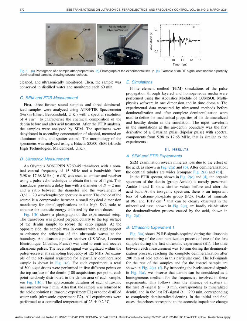
SEM examination reveals minerals loss due to the effect ofthe acid, as shown in Fig. 2(a) and (b). After demineralization,the dentinal tubules are wider [compare Fig. 2(a) and (b)].
In the FTIR spectra, shown in Fig. 2(c) and (d), the organicspectrum of the dentin (group Amide) is mostly preserved.Amide I and II show similar values before and after theacid bath. At the inorganic spectrum, there is an importantloss of calcium-phosphate group (PO). Peaks of mineralsat 961 and 1019 cm−1 that can be clearly observed in themineralized case, shown in Fig. 2(c), are hardly visible afterthe demineralization process caused by the acid, shown inFig. 2(d).
B. Ultrasonic Experiment 1
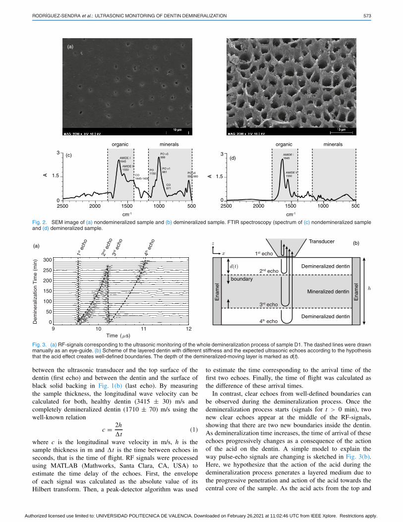
Fig. 3(a) shows 29 RF-signals acquired during the ultrasonicmonitoring of the demineralization process of one of the fivesamples during the first ultrasonic experiment (E1). The timebetween each measurement was 10 min during the demineral-ization process, reaching the complete demineralization after280 min of acid action in this particular case. The RF-signalsfor the rest of the samples and for the control sample areshown in Fig. 4(a)–(f). By inspecting the backscattered signalsin Fig. 3(a), we observe that dentin can be considered as ahomogeneous medium for the frequencies involved in theseexperiments. This follows from the absence of scatters inthe first RF-signal (t = 0 min, corresponding to mineralizeddentin) and in the last RF-signal (t = 290 min, correspondingto completely demineralized dentin). In the initial and finalcases, the echoes correspond to the acoustic impedance change
Authorized licensed use limited to: UNIVERSIDAD POLITECNICA DE VALENCIA. Downloaded on February 26,2021 at 11:02:46 UTC from IEEE Xplore. Restrictions apply.
RODRÍGUEZ-SENDRA et al.: ULTRASONIC MONITORING OF DENTIN DEMINERALIZATION 573
Fig. 2. SEM image of (a) nondemineralized sample and (b) demineralized sample. FTIR spectroscopy (spectrum of (c) nondemineralized sampleand (d) demineralized sample.
Fig. 3. (a) RF-signals corresponding to the ultrasonic monitoring of the whole demineralization process of sample D1. The dashed lines were drawnmanually as an eye-guide. (b) Scheme of the layered dentin with different stiffness and the expected ultrasonic echoes according to the hypothesisthat the acid effect creates well-defined boundaries. The depth of the demineralized-moving layer is marked as d(t).
between the ultrasonic transducer and the top surface of thedentin (first echo) and between the dentin and the surface ofblack solid backing in Fig. 1(b) (last echo). By measuringthe sample thickness, the longitudinal wave velocity can becalculated for both, healthy dentin (3415 ± 30) m/s andcompletely demineralized dentin (1710 ± 70) m/s using thewell-known relation
c = 2h
�t(1)
where c is the longitudinal wave velocity in m/s, h is thesample thickness in m and �t is the time between echoes inseconds, that is the time of flight. RF signals were processedusing MATLAB (Mathworks, Santa Clara, CA, USA) toestimate the time delay of the echoes. First, the envelopeof each signal was calculated as the absolute value of itsHilbert transform. Then, a peak-detector algorithm was used
to estimate the time corresponding to the arrival time of thefirst two echoes. Finally, the time of flight was calculated asthe difference of these arrival times.
In contrast, clear echoes from well-defined boundaries canbe observed during the demineralization process. Once thedemineralization process starts (signals for t > 0 min), twonew clear echoes appear at the middle of the RF-signals,showing that there are two new boundaries inside the dentin.As demineralization time increases, the time of arrival of theseechoes progressively changes as a consequence of the actionof the acid on the dentin. A simple model to explain theway pulse-echo signals are changing is sketched in Fig. 3(b).Here, we hypothesize that the action of the acid during thedemineralization process generates a layered medium due tothe progressive penetration and action of the acid towards thecentral core of the sample. As the acid acts from the top and
Authorized licensed use limited to: UNIVERSIDAD POLITECNICA DE VALENCIA. Downloaded on February 26,2021 at 11:02:46 UTC from IEEE Xplore. Restrictions apply.
574 IEEE TRANSACTIONS ON ULTRASONICS, FERROELECTRICS, AND FREQUENCY CONTROL, VOL. 68, NO. 3, MARCH 2021
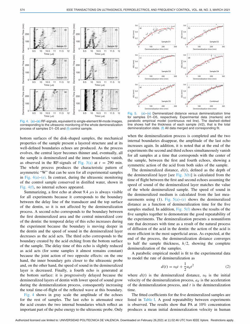
Fig. 4. (a)–(e) RF-signals, equivalent to single-element M-mode images,corresponding to the ultrasonic monitoring of the whole demineralizationprocess of samples D1–D5 and (f) control sample.
bottom surfaces of the disk-shaped samples, the mechanicalproperties of the sample present a layered structure and at itswell-defined boundaries echoes are produced. As the processevolves, the central layer becomes thinner and, eventually, allthe sample is demineralized and the inner boundaries vanish,as observed in the RF-signals of Fig. 3(a) at t = 290 min.The whole process produces the characteristic pattern ofasymmetric “W” that can be seen for all experimental samplesin Fig. 4(a)–(e). In contrast, during the ultrasonic monitoringof the control sample conserved in distilled water, shown inFig. 4(f), no internal echoes appeared.
Summarizing, a first echo at about 9.4 μs is always visiblefor all experiments because it corresponds to the boundarybetween the delay line of the transducer and the top surfaceof the dentin, so it is not affected by the demineralizationprocess. A second echo corresponds to the boundary betweenthe first demineralized area and the central mineralized coreof the dentin: the temporal delay of this echo increases duringthe experiment because the boundary is moving deeper inthe dentin and the speed of sound in the demineralized layerdecreases as the acid acts. The third echo corresponds to theboundary created by the acid etching from the bottom surfaceof the sample. The delay time of this echo is slightly reducedas acid acts (for some samples it almost remains constant)because the joint action of two opposite effects: on the onehand, the inner boundary gets closer to the ultrasonic probeand, on the other hand, the speed of sound in the demineralizedlayer is decreased. Finally, a fourth echo is generated atthe bottom surface: it is progressively delayed because thedemineralized layers of dentin with lower speed of sound growduring the demineralization process, consequently increasingthe total time-of-flight of the reflected wave at this boundary.
Fig. 4 shows in gray scale the amplitude of the echoesfor the rest of samples. The last echo is attenuated oncethe acid creates the two internal boundaries which reflect animportant part of the pulse energy to the ultrasonic probe. Only
Fig. 5. (a)–(e) Demineralized distance versus demineralization timefor samples D1–D5, respectively. Experimental data (markers) andparabolic empirical model (continuous red line). The dashed–dottedline shows half the thickness of each sample (h/2), that is the totaldemineralization state. (f) All data merged and corresponding fit.
when the demineralization process is completed and the twointernal boundaries disappear, the amplitude of the last echoincreases again. In addition, it is noted that at the end of theexperiments the second and third echoes simultaneously vanishfor all samples at a time that corresponds with the center ofthe sample, between the first and fourth echoes, showing asymmetric action of the acid from both sides of the sample.
The demineralized distance, d(t), defined as the depth ofthe demineralized layer [see Fig. 3(b)] is calculated from thetime of flight between the first and second echoes assuming thespeed of sound of the demineralized layer matches the valueof the whole demineralized sample. The speed of sound inthe demineralized medium is calculated from the last mea-surements using (1). Fig. 5(a)–(e) shows the demineralizeddistance as a function of demineralization time for the fivesamples studied. In addition, Fig. 5(f) shows the results of thefive samples together to demonstrate the good repeatability ofthe experiments. The demineralization presents a nonuniformrate that decreases with time as a result of the natural processof diffusion of the acid in the dentin: the action of the acid ismore efficient in the most superficial areas. As expected, at theend of the process, the demineralization distance convergesto half the sample thickness, h/2, showing the completedemineralization of the samples.
A parabolic empirical model is fit to the experimental datato model the rate of demineralization as
d(t) = v0t + 1
2adt2 (2)
where d(t) is the demineralized distance, v0 is the initialvelocity of the demineralization process, ad is the accelerationof the demineralization process, and t is the demineralizationtime.
The fitted coefficients for the five demineralized samples arelisted in Table I. A good repeatability between experimentsis observed. The results show that PA at 10% concentrationproduces a mean initial demineralization velocity in human
Authorized licensed use limited to: UNIVERSIDAD POLITECNICA DE VALENCIA. Downloaded on February 26,2021 at 11:02:46 UTC from IEEE Xplore. Restrictions apply.
RODRÍGUEZ-SENDRA et al.: ULTRASONIC MONITORING OF DENTIN DEMINERALIZATION 575
TABLE ISAMPLE THICKNESS AND FITTED PARAMETERS
dentin of v0 = 3.5 ± 0.4 μm/min, with a mean decelerationof ad = −11.0 ± 3.5 nm/min2.
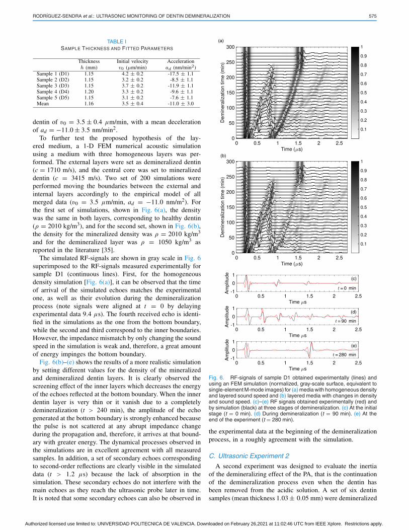
To further test the proposed hypothesis of the lay-ered medium, a 1-D FEM numerical acoustic simulationusing a medium with three homogeneous layers was per-formed. The external layers were set as demineralized dentin(c = 1710 m/s), and the central core was set to mineralizeddentin (c = 3415 m/s). Two set of 200 simulations wereperformed moving the boundaries between the external andinternal layers accordingly to the empirical model of allmerged data (v0 = 3.5 μm/min, ad = −11.0 nm/m2). Forthe first set of simulations, shown in Fig. 6(a), the densitywas the same in both layers, corresponding to healthy dentin(ρ = 2010 kg/m3), and for the second set, shown in Fig. 6(b),the density for the mineralized density was ρ = 2010 kg/m3
and for the demineralized layer was ρ = 1050 kg/m3 asreported in the literature [35].
The simulated RF-signals are shown in gray scale in Fig. 6superimposed to the RF-signals measured experimentally forsample D1 (continuous lines). First, for the homogeneousdensity simulation [Fig. 6(a)], it can be observed that the timeof arrival of the simulated echoes matches the experimentalone, as well as their evolution during the demineralizationprocess (note signals were aligned at t = 0 by delayingexperimental data 9.4 μs). The fourth received echo is identi-fied in the simulations as the one from the bottom boundary,while the second and third correspond to the inner boundaries.However, the impedance mismatch by only changing the soundspeed in the simulation is weak and, therefore, a great amountof energy impinges the bottom boundary.
Fig. 6(b)–(e) shows the results of a more realistic simulationby setting different values for the density of the mineralizedand demineralized dentin layers. It is clearly observed thescreening effect of the inner layers which decreases the energyof the echoes reflected at the bottom boundary. When the innerdentin layer is very thin or it vanish due to a completelydemineralization (t > 240 min), the amplitude of the echogenerated at the bottom boundary is strongly enhanced becausethe pulse is not scattered at any abrupt impedance changeduring the propagation and, therefore, it arrives at that bound-ary with greater energy. The dynamical processes observed inthe simulations are in excellent agreement with all measuredsamples. In addition, a set of secondary echoes correspondingto second-order reflections are clearly visible in the simulateddata (t > 1.2 μs) because the lack of absorption in thesimulation. These secondary echoes do not interfere with themain echoes as they reach the ultrasonic probe later in time.It is noted that some secondary echoes can also be observed in
Fig. 6. RF-signals of sample D1 obtained experimentally (lines) andusing an FEM simulation (normalized, gray-scale surface, equivalent tosingle-element M-mode images) for (a) media with homogeneous densityand layered sound speed and (b) layered media with changes in densityand sound speed. (c)–(e) RF signals obtained experimentally (red) andby simulation (black) at three stages of demineralization. (c) At the initialstage (t = 0 min). (d) During demineralization (t = 90 min). (e) At theend of the experiment (t = 280 min).
the experimental data at the beginning of the demineralizationprocess, in a roughly agreement with the simulation.
C. Ultrasonic Experiment 2
A second experiment was designed to evaluate the inertiaof the demineralizing effect of the PA, that is the continuationof the demineralization process even when the dentin hasbeen removed from the acidic solution. A set of six dentinsamples (mean thickness 1.03 ± 0.05 mm) were demineralized
Authorized licensed use limited to: UNIVERSIDAD POLITECNICA DE VALENCIA. Downloaded on February 26,2021 at 11:02:46 UTC from IEEE Xplore. Restrictions apply.
576 IEEE TRANSACTIONS ON ULTRASONICS, FERROELECTRICS, AND FREQUENCY CONTROL, VOL. 68, NO. 3, MARCH 2021
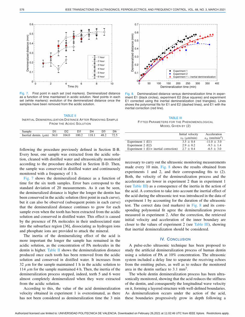
Fig. 7. First point in each set (red markers). Demineralized distanceas a function of time maintained in acidic solution. Next points in eachset (white markers): evolution of the demineralized distance once thesamples have been removed from the acidic solution.
TABLE IIINERTIAL DEMINERALIZATION DISTANCE AFTER REMOVING SAMPLE
FROM THE ACIDIC SOLUTION
following the procedure previously defined in Section II-B.Every hour, one sample was extracted from the acidic solu-tion, cleaned with distilled water and ultrasonically monitoredaccording to the procedure described in Section II-D. Then,the sample was conserved in distilled water and continuouslymonitored with a frequency of 1 h.
Fig. 7 shows the demineralized distance as a function oftime for the six teeth samples. Error bars correspond to thestandard deviation of 20 measurements. As it can be seen,the demineralized distance is higher the longer the dentin hasbeen conserved in the acidic solution (first point in each curve),but it can also be observed (subsequent points in each curve)that the demineralized distance continues to grow for eachsample even when the tooth has been extracted from the acidicsolution and conserved in distilled water. This effect is causedby the presence of PA molecules in their undissociated stateinto the subsurface region [36], dissociating as hydrogen ionsand phosphate ions are provided to attack the mineral.
The inertia of the demineralizing effect of the acid ismore important the longer the sample has remained in theacidic solution, as the concentration of PA molecules in thedentin is higher. Table II shows the demineralization distanceproduced once each tooth has been removed from the acidicsolution and conserved in distilled water. It increases from32 μm for the sample maintained 1 h in the acidic solution to114 μm for the sample maintained 4 h. Then, the inertia of thedemineralization process stopped, indeed, teeth 5 and 6 werealmost completely demineralized when they were extractedfrom the acidic solution.
According to this, the value of the acid demineralizationvelocity obtained in experiment 1 is overestimated, as therehas not been considered as demineralization time the 3 min
Fig. 8. Demineralized distance versus demineralization time in exper-iment E1 (black circles), experiment E2 (blue squares) and experimentE1 corrected using the inertial demineralization (red triangles). Linesshows the polynomial fits for E1 and E2 (dashed lines), and E1 with theinertial correction (red line).
TABLE IIIFITTED PARAMETERS FOR THE PHENOMENOLOGICAL
MODEL GIVEN BY (2)
necessary to carry out the ultrasonic monitoring measurementsmade every 10 min. Fig. 8 shows the results obtained fromexperiments 1 and 2, and their corresponding fits to (2).Both, the velocity of the demineralization process and theacceleration are lower in experiment 2 than in experiment 1(see Table III) as a consequence of the inertia in the action ofthe acid. A correction to take into account the inertial effect ofthe acid during the ultrasonic test was introduced in the data ofexperiment 1 by accounting for the duration of the ultrasonictest. The correct data (red markers) in Fig. 8 and its corre-sponding polynomial fit agree the demineralization processmeasured in experiment 2. After the correction, the retrievedinitial velocity and acceleration of the inner boundary arecloser to the values of experiment 2 (see Table III), showingthat inertial demineralization should be considered.
IV. CONCLUSION
A pulse-echo ultrasonic technique has been proposed tostudy the artificial demineralization process of human dentinusing a solution of PA at 10% concentration. The ultrasonicsystem included a delay line to separate the receiving echoesfrom the emitting pulses, as well as to reduce the monitoredarea in the dentin surface to 3.1 mm2.
The whole dentin demineralization process has been ultra-sonically monitored, showing that the acid reduces the stiffnessof the dentin, and consequently the longitudinal wave velocityon it, forming a layered structure with well-defined boundaries.As demineralization occurs under the action of the acid,these boundaries progressively grow in depth following a
Authorized licensed use limited to: UNIVERSIDAD POLITECNICA DE VALENCIA. Downloaded on February 26,2021 at 11:02:46 UTC from IEEE Xplore. Restrictions apply.
RODRÍGUEZ-SENDRA et al.: ULTRASONIC MONITORING OF DENTIN DEMINERALIZATION 577
decelerated pattern. This model, which fits the experimentaldata, has been corroborated through simulations. We observedthat the demineralization process is not constant, it was esti-mated that the velocity of penetration of the demineralizedlayer starts at 3.5 μm/min, that is when the acid attacksthe surface of the dentin, but as the acid penetrates thisvelocity is decelerated by −11 nm/min2. In addition, it hasbeen demonstrated the fact that the demineralization processdoes not stop once the sample is removed from the acidicsolution. Instead, several extra tens of micrometers of dentinare demineralized depending on the concentration of cations inthe dentin. The correction of the effect of the demineralizationinertia changes the dynamic of the demineralization processto 2.9 μm/min for the initial demineralization velocity and−9.3 nm/min2 for the deceleration.
The technique provides an indeterminacy of 40 μm in thecalculation of the demineralized distance, including uncertain-ties due to irregularities in the preparation of the samples,in the estimation of the time of flight and in the measure-ment of the thickness of the sample during the ultrasonicmeasurement. It should be said here that the sample can beslightly compressed during the contact between the delay lineof the probe, especially when demineralization is importantand, therefore, the stiffness of the dentin is reduced.
The proposed technique can be used to monitor artificialstandardized demineralization lesions at laboratory conditions,but also, because of the reduced dimension of the probe,to evaluate demineralization produced by natural caries invivo. It is worth to mention here that the measurement ofthe thickness of the sample is not required to evaluate therelevance of a demineralization process, as the change in thestiffness between the demineralized and mineralized tissueprovides a clear echo whose time of flight informs aboutthe demineralization depth if a previous database of soundvelocities have been obtained.
ACKNOWLEDGMENT
This work has been developed within the framework of theIVIO-UPV Chair. The authors want to thank the support andadvice of Dr. Joan Faus.
REFERENCES
[1] D. P. Catherine Le Galès-Camus and P. E. Petersen, WHO ReleasesNew Report on Global Problem of Oral Diseases. Geneva, Switzerland:World Health Organization, 2004.
[2] C. Longbottom, M.-C. Huysmans, N. Pitts, and M. Fontana,“Glossary of key terms,” in Monographs in Oral Science. Basel,Switzerland: Karger, 2009, pp. 209–216. [Online]. Available:ht.tps://ww.w.ncbi.nlm.nih.gov/pubmed/19494688
[3] R. H. Selwitz, A. I. Ismail, and N. B. Pitts, “Dental caries,”Lancet, vol. 369, no. 9555, pp. 51–59, Jan. 2007. [Online]. Available:ht.tps://ww.w.ncbi.nlm.nih.gov/pubmed/17208642
[4] M. Torabinejad and N. Chivian, “Clinical applications of mineral triox-ide aggregate,” J. Endodontics, vol. 25, no. 3, pp. 197–205, Mar. 1999.[Online]. Available: ht.tps://ww.w.ncbi.nlm.nih.gov/pubmed/10321187
[5] L. Han and T. Okiji, “Bioactivity evaluation of three calcium silicate-based endodontic materials,” Int. Endodontic J., vol. 46, no. 9,pp. 808–814, Feb. 2013.
[6] M. G. Gandolfi, F. Siboni, T. Botero, M. Bossù, F. Riccitiello, andC. Prati, “Calcium silicate and calcium hydroxide materials for pulpcapping: Biointeractivity, porosity, solubility and bioactivity of currentformulations,” J. Appl. Biomaterials Funct. Mater., vol. 13, no. 1,pp. 0–0, Sep. 2014.
[7] I. A. Mjör, “Human coronal dentine: Structure and reactions,”Oral Surg., Oral Med., Oral Pathol., vol. 33, no. 5, pp. 810–823,May 1972. [Online]. Available: ht.tps://ww.w.sciencedirect.com/science/article/abs/pii/0030422072904513
[8] Y.-C. Chien et al., “Distinct decalcification process of dentinby different cariogenic organic acids: Kinetics, ultrastructure andmechanical properties,” Arch. Oral Biol., vol. 63, pp. 93–105,Mar. 2016. [Online]. Available: ht.tps://ww.w.sciencedirect.com/science/article/pii/S0003996915300509
[9] K. Saeki et al., “Recovery after PILP remineralization of dentinlesions created with two cariogenic acids,” Arch. Oral Biol., vol. 82,pp. 194–202, Oct. 2017. [Online]. Available: htt.ps://ww.w.sciencedirect.com/science/article/pii/S0003996917301826
[10] B. Li et al., “Selective demineralisation of dentine extrafibrillarminerals—A potential method to eliminate water-wet bonding inthe etch-and-rinse technique,” J. Dentistry, vol. 52, pp. 55–62,Sep. 2016. [Online]. Available: ht.tps://w.ww.sciencedirect.com/science/article/pii/S030057121630135X
[11] J. Perdigão, “Dentin bonding as a function of dentin structure,”Dental Clinics North Amer., vol. 46, no. 2, pp. 277–301,Apr. 2002. [Online]. Available: htt.ps://ww.w.sciencedirect.com/science/article/abs/pii/S0011853201000088?via%3Dihub
[12] J. Perdigão, P. Lambrechts, B. Van Meerbeek, Â. R. Tomé, G. Vanherle,and A. B. Lopes, “Morphological field emission-SEM study of theeffect of six phosphoric acid etching agents on human dentin,” DentalMater., vol. 12, no. 4, pp. 262–271, Jul. 1996. [Online]. Available:ht.tps://ww.w.sciencedirect.com/science/article/pii/S0109564196800339
[13] K. D. Jandt, “Atomic force microscopy of biomaterials sur-faces and interfaces,” Surf. Sci., vol. 491, no. 3, pp. 303–332,Oct. 2001.
[14] J. H. Kinney, M. Balooch, D. L. Haupt, S. J. Marshall, andG. W. Marshall, “Mineral distribution and dimensional changesin human dentin during demineralization,” J. Dental Res.,vol. 74, no. 5, pp. 1179–1184, May 1995. [Online]. Available:ht.tps://journals.sagepub.com/doi/abs/10.1177/00220345950740050601
[15] C. H. Wilder-Smith et al., “Quantification of dental erosionsin patients with gerd using optical coherence tomography beforeand after double-blind, randomized treatment with esomeprazoleor placebo,” Amer. J. Gastroenterol., vol. 104, no. 11, p. 2788,2009.
[16] F. M. Herkströter, M. Witjes, J. Ruben, and J. Arends, “Time dependencyof microhardness indentations in human and bovine dentine comparedwith human enamel (Short Communication),” Caries Res., vol. 23, no. 5,pp. 342–344, 1989.
[17] T. Attin and F. J. Wegehaupt, “Methods for assessment of den-tal erosion,” in Monographs in Oral Science. Basel, Switzer-land: S. KARGER AG, 2014, pp. 123–142. [Online]. Available:ht.tps://ww.w.karger.com/Article/Abstract/360355
[18] G. Baum, I. Greenwood, S. Slawski, and R. Smirnow, “Observa-tion of internal structures of teeth by ultrasonography,” Science,vol. 139, no. 3554, pp. 495–496, Feb. 1963. [Online]. Available:ht.tps://science.sciencemag.org/content/139/3554/495
[19] S. R. Ghorayeb, C. A. Bertoncini, and M. K. Hinders, “Ultrasonog-raphy in dentistry,” IEEE Trans. Ultrason., Ferroelectr., Freq. Con-trol, vol. 55, no. 6, pp. 1256–1266, Jun. 2008. [Online]. Available:ht.tps://ieeexplore.ieee.org/abstract/document/4536920
[20] J. Marotti et al., “Recent advances of ultrasound imaging in dentistry–a review of the literature,” Oral Surg., Oral Med., Oral Pathol. OralRadiol., vol. 115, no. 6, pp. 819–832, Jun. 2013. [Online]. Available:ht.tps://ww.w.sciencedirect.com/science/article/pii/S2212440313001727
[21] T. Watanabe, M. Miyazaki, H. Inage, and H. Kurokawa,“Determination of elastic modulus of the components at dentin-resin interface using the ultrasonic device,” Dental Mater.J., vol. 23, no. 3, pp. 361–367, 2004. [Online]. Available:ht.tps://ww.w.jstage.jst.go.jp/article/dmj1982/23/3/23_3_361/_article/-char/ja/
[22] G. Yasuda, H. Inage, T. Takamizawa, H. Kurokawa, A. Rikuta,and M. Miyazaki, “Determination of elastic modulus of deminer-alized resin-infiltrated dentin by self-etch adhesives,” Eur. J. OralSci., vol. 115, no. 1, pp. 87–91, Feb. 2007. [Online]. Available:h.ttps://ww.w.ncbi.nlm.nih.gov/pubmed/17305722
[23] S. Y. Ng, M. W. J. Ferguson, P. A. Payne, and P. Slater, “Ultra-sonic studies of unblemished and artificially demineralized enamel inextracted human teeth: A new method for detecting early caries,”J. Dentistry, vol. 16, no. 5, pp. 201–209, Oct. 1988. [Online]. Available:ht.tps://ww.w.sciencedirect.com/science/article/pii/030057128890070X
Authorized licensed use limited to: UNIVERSIDAD POLITECNICA DE VALENCIA. Downloaded on February 26,2021 at 11:02:46 UTC from IEEE Xplore. Restrictions apply.
578 IEEE TRANSACTIONS ON ULTRASONICS, FERROELECTRICS, AND FREQUENCY CONTROL, VOL. 68, NO. 3, MARCH 2021
[24] S. Y. Ng, P. A. Payne, N. A. Cartledge, and M. W. J. Ferguson,“Determination of ultrasonic velocity in human enamel and dentine,”Arch. Oral Biol., vol. 34, no. 5, pp. 341–345, 1989. [Online]. Available:ht.tps://ww.w.sciencedirect.com/science/article/pii/0003996989901076
[25] R. G. Maev, L. A. Denisova, E. Y. Maeva, and A. A. Denissov,“New data on histology and physico-mechanical properties ofhuman tooth tissue obtained with acoustic microscopy,” UltrasoundMed. Biol., vol. 28, no. 1, pp. 131–136, Jan. 2002. [Online].Available: ht.tps://ww.w.sciencedirect.com/science/article/abs/pii/S030156290100480X
[26] M. Miyazaki, H. Inage, and H. Onose, “Use of an ultrasonicdevice for the determination of elastic modulus of dentin,”J. Oral Sci., vol. 44, no. 1, pp. 19–26, 2002. [Online]. Available:ht.tps://ww.w.jstage.jst.go.jp/article/josnusd1998/44/1/44_1_19/_article/-char/ja/
[27] C. John, “The corono-apically varying ultrasonic velocity in human harddental tissues,” J. Acoust. Soc. Amer., vol. 116, no. 1, pp. 545–556,Jul. 2004.
[28] C. John, “The laterally varying ultrasonic velocity in the dentin of humanteeth,” J. Biomech., vol. 39, no. 13, pp. 2388–2396, Jan. 2006.
[29] C. Lost, K.-M. Irion, C. John, and W. Nussle, “Two-dimensionaldistribution of sound velocity in ground sections of dentin,” DentalTraumatol., vol. 8, no. 5, pp. 215–218, Oct. 1992. [Online].Available: ht.tps://onlinelibrary.wiley.com/doi/abs/10.1111/j.1600-9657.1992.tb00246.x
[30] C. John, K. Irion, W. Nüssle, and C. Löst, “The resolution of a2-dimensional ultrasonic velocity profile of human tooth sections,”Schweizer Monatsschrift fur Zahnmedizin= Revue mensuelle suissed’odonto-stomatologie= Rivista mensile svizzera di odontologia estomatologia, vol. 104, no. 1, pp. 25–30, 1994. [Online]. Available:ht.tps://europepmc.org/article/med/8108688
[31] S. Lees, F. B. Gerhard, and F. G. Oppenheim, “Ultrasonicmeasurement of dental enamel demineralization,” Ultra-sonics, vol. 11, no. 6, pp. 269–273, Nov. 1973. [Online].Available: htt.ps://w.ww.sciencedirect.com/science/article/abs/pii/0041624X73901042
[32] K. Yamaguchi, M. Miyazaki, T. Takamizawa, H. Inage, and B.K. Moore, “Effect of CPP–ACP paste on mechanical properties ofbovine enamel as determined by an ultrasonic device,” J. Den-tistry, vol. 34, no. 3, pp. 230–236, Mar. 2006. [Online]. Available:ht.tps://ww.w.ncbi.nlm.nih.gov/pubmed/16112336
[33] K. Yamaguchi, M. Miyazaki, T. Takamizawa, H. Inage, andH. Kurokawa, “Ultrasonic determination of the effect of caseinphosphopeptide-amorphous calcium phosphate paste on the demineral-ization of bovine dentin,” Caries Res., vol. 41, no. 3, pp. 204–207, 2007.[Online]. Available: ht.tps://w.ww.karger.com/Article/Abstract/99319
[34] H. Endo et al., “Evaluation of a calcium phosphate desensitizer using anultrasonic device,” Dental Mater. J., vol. 32, no. 3, pp. 456–461, 2013.
[35] R. M. Carvalho, M. Yoshiyama, E. L. Pashley, and D. H. Pashley, “Invitro study on the dimensional changes of human dentine after deminer-alization,” Arch. Oral Biol., vol. 41, no. 4, pp. 369–377, Apr. 1996.
[36] J. Featherstone and A. Lussi, “Understanding the chemistry of dentalerosion,” in Monographs in Oral Science. Basel, Switzerland: Karger,2006, pp. 66–76.
Josep Rodríguez-Sendra received the B.Sc.degree in telecommunication and the M.Sc.degree in acoustics from the Universitat Politèc-nica de València (UPV), Valencia, Spain,in 2014 and 2015, respectively, where he iscurrently pursuing the Ph.D. degree with the Insti-tuto de Instrumentación para Imagen Molecular(i3M).
In 2016, he enrolled the team at UltrasoundMedical and Industrial Laboratory (UMIL), i3M,to research on ultrasonic technology applied to
odontology. His research interest concerns new technologies for bio-medical ultrasound applications.
Inés Torres received D.D.S. degree fromCardenal Herrera CEU University, Moncada,Spain, in 2008, and post-graduate programs inendodontics in 2009, orthodontics in 2011, anddental prosthetics in 2013. She is currently pur-suing the Ph.D. degree in dental biomaterials withCEU Universidad Cardenal Herrera.
She has been an Associate Professor of den-tal pathology and therapy at Cardenal HerreraCEU University since 2009 and a Co-Director ofEndodontic Post-Graduate Program at the Insti-
tuto Valenciano de Investigaciones Odontológicas (IVIO), PolytechnicUniversity of Valencia, Valencia, Spain, since 2016. She also collab-orates in other post-graduate dental programs, dental private practiceexclusively to endodontics.
Ms. Torres is a member of the “Asociación Española de Endodoncia.”
Noé Jiménez received the B.Sc. degree intelecommunication and the M.Sc. and Ph.D.degrees in acoustics from the Universitat Politèc-nica de València, Valencia, Spain, in 2007,2010, 2015, respectively.
In 2014 and 2019, he worked for the Euro-pean Space Agency, Valencia, for noise con-trol at the launch pad using periodic structures.In 2015, he joined the French CNRS (UMR6613),Le Mans, France, for a postdoctoral position toresearch on deep-subwavelength metamateri-
als. In 2017, he enrolled the Spanish National Research Council (CSIC)to research on biomedical ultrasound applications at the Instituto deInstrumentación para Imagen Molecular (i3M). He has been a VisitingResearcher at Columbia University, New York City, NY, USA, and at theUniversity of Salford, Manchester, U.K. Since 2013, he has published38 journal papers and participated in more than 100 conferences.His research interest concerns from fundamental research in waves incomplex and structured media to biomedical ultrasound applications.
Salvatore Sauro is currently an Adjunct Pro-fessor (Senior Lecturer full-time) with DentalBiomaterials and Minimally Invasive Dentistry,University CEU-Cardenal Herrera, Moncada,Spain. He was a Honorary Senior Lecturer in bio-materials, biophotonics, and tissue engineeringwith the King’s College London Dental Institute(KCLDI), Guy’s Hospital, London, U.K. He wasalso a Visiting Professor with the Federal Uni-versity of Moscow, School of Dentistry, Moscow,Russia, and with The University of Hong Kong,
Dental School, China. He has been working in dental biomaterials,preventive and minimally invasive dentistry research for 15 years(JCR—H-Index: 31), and he has published, in collaboration with inter-nationally renowned researchers, more than 120 articles in internationalpeer-reviewed journals with high impact on the dental and biomaterials’field.
Francisco Camarena received the Ph.D. degreein physics from the Universitat de Valéncia,Valencia, Spain, in 2003.
He is a Founding Member and the Head ofthe Ultrasound Medical and Industrial Labora-tory (UMIL). He is a Permanent Researcher atthe Instituto de Instrumentación para ImagenMolecular (i3M) and an Associate Professor withthe Department of Applied Physics, UPV, Valen-cia. He is the Head of the Instituto Valencianode Investigaciones Odontológicas (IVIO) Chair
(IVIO-UPV), dedicated to the promotion and development of trainingactivities, research, dissemination, and technology transfer in the fieldof odontology. He is the Director of the Scientific Unit of BusinessInnovation (UCIE, i3M). He has participated in more than 80 national andinternational conferences related to acoustics and ultrasonics. He hasauthored or coauthored around 60 articles in national and internationalpeer-reviewed journals.
Authorized licensed use limited to: UNIVERSIDAD POLITECNICA DE VALENCIA. Downloaded on February 26,2021 at 11:02:46 UTC from IEEE Xplore. Restrictions apply.