Embed Size (px)
Citation preview

Pictorial essay Med Ultrason 2014, Vol. 16, no. 1, 48-59DOI: 10.11152/mu.2014.2066.161.lc1mz2
AbstractContrast-enhanced ultrasound is widely indicated in the study of splenic diseases, especially due to its good specificity in
the differentiation of benign from malignant splenic lesions. The purpose of this pictorial essay is to offer a review of the most common splenic pathologies, while illustrating them with sonographic images.
Keywords: spleen, ultrasonography, contrast-enhanced ultrasound, colour-Doppler, elastography
Ultrasonography of the spleen. Pictorial essay.
Liliana Chiorean1, Mihnea Zdrenghea2, Radu Badea3
1Department of Radiology and Computed Tomography, “Octavian Fodor” Institute of Gastroenterology and Hepatology, 2Hematology Department, Institute of Oncology, 3Department of Ultrasonography, “Octavian Fodor” Institute of Gastroenterology and Hepatology, “Iuliu Haţieganu” University of Medicine and Pharmacy, Cluj-Napoca, Romania
Received 14.12.2013 Accepted 30.12.2013 Med Ultrason 2014, Vol. 16, No 1, 48-59 Corresponding author: Radu Badea, Institute of Gastroenterology and Hepatology, Department of Ultrasound “Iuliu Hatieganu” University of Medicine and Pharmacy Cluj Napoca, 19-21 Croitorilor Str, 400163, Cluj Napoca, RO Email: [email protected]
Introduction
Ultrasonography (US) is extensively used in imaging the abdomen, being frequently the first method of choice for an abdominal survey. As part of the abdominal US examination, US of the spleen still plays an important role in diagnosis, even though other imagistic modalities are becoming increasingly competitive [1].
Even if it has been named ‘the forgotten organ’, care-ful examination of the spleen can bring important diag-nostic clues in pathologies such as oncologic, hematolog-ic, infectious, and metabolic, abdominal trauma, portal hypertension, and many other focal or diffuse splenic changes of different etiologies.
Examination Technique
The spleen should be examined with the patient on their back or right side, using a 3-5 MHz curved linear transducer
while the patient exhales in order to avoid spleen cover-age by the left lung tissue. The best window to view the spleen is at the level of 10th or 11th intercostal spaces, on the left midaxillary line. Colour Doppler imaging may some-times assist in an accurate diagnosis, but it can only as-sess the macrocirculation. Contrast-enhanced US (CEUS) can assess both macro- and microcirculation in real-time. It requires a skilled user, being highly operator dependent. The examination should be made using a high-frequency transducer, and tissue harmonic imaging in the presence of a low mechanical index (MI) [1-3]. In our institution, we usually inject intravenously 2.4 ml of contrast-agent Sono-Vue (Bracco, Italy), followed by 10 cc of saline solution. In-jection of this contrast agent can be immediately repeated, even if rarely necessary. Elastography is a new investigation technique used to assess the elasticity of tissues, being the imagistic equivalent of palpation used from ancient times in medicine. The technique is different for every US machine producer with software for elastography available [4].
Normal Appearance
On US, the spleen is crescent shaped, with a smooth outer convexity, and an inner margin intended or nodu-lous [1]. Its normal size is less than 12 cm in length, and it may decrease with age [5]. The normal appearance of the parenchyma is very homogeneous and uniform, with an echogenicity slightly greater than that of normal he-

49Med Ultrason 2014; 16(1): 48-59
patic parenchyma [6]. At CEUS, the phases of contrast enhancement are broadly divided into arterial (10–25 s), portal venous (30–120 s), and late phases (over 120 s) [7]. During the arterial time, the arterial splenic vessels are displayed (fig 1a), while in the portal venous phase, the splenic parenchyma becomes homogenously en-hanced (also called the parenchymal phase) (fig 1b).
Abnormal Findings
Focal lesions in the spleen are rare in comparison with those located in other solid viscera. The accurate analysis of internal architecture of the lesion can give us impor-tant diagnostic clues, due to the US’s ability in depicting the calcifications (long term process) or gas within a le-sion (usually originating from bacterial infection), those being most frequently related to benign findings. The ma-lignant lesions are more frequently multifocal (especially
metastasis), and tend to be diffuse and ill-defined due to rapid growth [8]. Because the US appearance of disease in the spleen is rather nonspecific, a thorough knowledge of the patient’s history and associated symptoms may be very helpful in the differential diagnosis [6].
Different patterns of enhancement for focal splenic le-sions have been described at CEUS examination: benign lesions that have constantly nonenhancing or isoenhanc-ing pattern and malignant lesions with enhancement in ar-terial time, followed by rapid wash-out, or progressively hypoenhacing with heterogenous aspect. This last aspect was described in some studies as having 100% sensitivity, and 83% specificity for malignancy [2,3,9-12].
Diffuse Splenic DiseaseThe basic finding in many diseases of the spleen is the
acute or chronic increase in the size of the organ. Spleno-megaly can be caused by infections, liver or blood diseases, problems with the lymph system, or other conditions [5].
Fig 1. Gray-scale and CEUS of the spleen (dual examination): a) arterial time (at 21 seconds after contrast agent administration) – the arterial splenic vessels are seen; b) portal time (42 seconds), the splenic paren-chyma becomes homogenously enhanced (also called the parenchymal time).
Fig 2. Splenomegaly. a) 2-D ultrasound – enlarged spleen; b) CEUS – inhommogenous enhancement during the arterial time, starting at 7 seconds after contrast administration, and reaching the peack at 17 seconds; c) strain elastogram, showing inhommogenous pattern, stiffer subcapsulary.
50 Liliana Chiorean et al Ultrasonography of the spleen
Presence of an enlarged spleen may need fusion of multiple images or the use of panoramic images in order to view the entire organ (fig 2a). The aspect of enhancement in CEUS is nonspecific and inconstant (fig 2b). In marked spleno-megaly, a delay in contrast up-taken and a less intense opacification of splenic parenchyma may be found [2]. The elastogram may present with an inhomogeneous pattern, with stiffer areas located subcapsular, due to enlarged pa-renchyma compressed under the splenic capsule (fig 2c).
Focal Splenic LesionsCystsCysts are the most common benign lesions of the
spleen, and usually present no diagnostic problems. They may be primary or acquired. The primary ones are true cysts, with epithelium, endothelium, and membrane lin-ing, and may be congenital, parasitic, or neoplastic. Most often primary cysts are simple, uniloculated, but they may also present incomplete septations, or punctate cal-
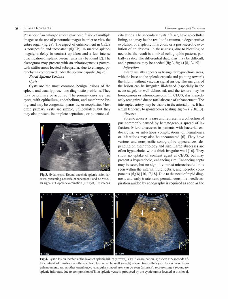
Fig 3. Hydatic cyst. Round, anechoic splenic lesion (ar-row), presenting acoustic enhancement, and no vascu-lar signal at Doppler examination (C = cyst, S = spleen).
Fig 4. Cystic lesion located at the level of splenic hilum (arrows), CEUS examination. a) aspect at 5 seconds af-ter contrast administration – the anechoic lesion can be well seen; b) arterial time – the cystic lesion presents no enhancement, and another unenhanced triangular shaped area can be seen (asterisk), representing a secondary splenic infarctus, due to compression of hilar splenic vessels, produced by the cystic tumor located at this level.
cifications. The secondary cysts, ‘false’, have no cellular lining, and may be the result of a trauma, a degenerative evolution of a splenic infarction, or a post-necrotic evo-lution of an abscess. In these cases, due to bleeding or necrosis, the result is a mixed echographic pattern, par-tially cystic. The differential diagnosis may be difficult, and a puncture may be needed (fig 3, fig 4) [8,13-15].
InfarctionInfarct usually appears as triangular hypoechoic areas,
with the base on the splenic capsule and pointing towards the hilum, without vascular signal inside. The margins of the lesion can be irregular, ill-defined (especially in the acute stage), or well delineated, and the texture may be homogenous or inhomogeneous. On CEUS, it is immedi-ately recognized due to total absence of enhancement. The interrupted artery may be visible in the arterial time. It has a high tendency to spontaneous healing (fig 5-7) [2,10,13].
AbscessSplenic abscess is rare and represents a collection of
pus commonly caused by hematogenous spread of in-fection. Micro-abscesses in patients with bacterial en-docarditis, or infectious complications of hematomas or infarctions may also be encountered [6]. They have various and nonspecific sonographic appearances, de-pending on their etiology and size. Large abscesses are often hypoechoic, with a thick irregular wall [16]. They show no uptake of contrast agent at CEUS, but may present a hyperechoic, enhancing rim. Enhancing septa may be seen, but no sign of contrast microcirculation is seen within the internal fluid, debris, and necrotic com-ponents (fig 8) [10,17,18]. Due to the need of rapid diag-nosis and early treatement, percutaneous fine-needle as-piration guided by sonography is required as soon as the

51Med Ultrason 2014; 16(1): 48-59
suspicion has been raised, especially because the clinical diagnostic triad is present in only 44% of cases [13,19].
HemangiomasHemangioma is the most common benign solid lesion of
the spleen characterized by a proliferation of blood-filled spac-es lined and separated by the endothelium. It is usually a soli-tary, well-marginated lesion, having less than 2 cm in size. The
Fig 5. Splenic infarct. a) sonogram shows a triangular hypoechoic lesion (asterisk); b) CEUS, arterial time – the splenic lesion is unenhanced; c) CEUS, prenchymal phase – the lesion presents no enhancement (S = spleen; R = left kidney).
Fig 6. Splenic infarcts (asterisk) in a patient with splenic and portal vein thrombosis. a) Doppler examina-tion – showing the absence of the vascular signal inside the hypoechoic, wedge-shaped triangular lesions; b,c) CEUS of the spleen – the lessions shows no contrast agent uptake, nor in the arterial, neither in the parenchymal phase; d) Doppler examinantion showing the absence of the vascular signal at the level of the splenic vein; e) CEUS reveals absence of contrast agent inside the splenic vein in the late venous phase; f) CEUS late phase – no enhancement inside the portal vein (S = spleen, SV = splenic vein, PV = portal vein).
sonographical appearance is different for the two histological types of hemangiomas: the cavernous hemangioma appears as a mixed echogenic or hypoechoic structure, with possible calcifications or cystic component; a capillary hemangioma is hyperechoic, with well-defined margins. The most frequent complications are rupture and bleeding [1,6,13]. At CEUS, capillary hemangiomas are usually isoenhancing with the ad-

52 Liliana Chiorean et al Ultrasonography of the spleen
jacent splenic parenchyma. A hyperechoic splenic lesion on gray-scale, undetectable after contrast-agent administration is diagnosed as a hemangioma [10]. Cavernous hemangiomas can present a diffuse or centripetal filling-in, rapid or slow, but with a greater enhancement degree (fig 9, fig 10) [2].
TraumaComputed-tomography is the reference standard in
detecting and monitoring splenic trauma [1]. Fissures
Fig 7. Splenic infarcts through microembolisations. a) Sonogram showing an inhomogeneous spleen, with a large hypoechoic area, wedge-shaped (asterisk), and others smaller (arrow); b,c) no enhancement inside the splenic lesions.
Fig 8. Splenic abscess. a) Sonogram shows a large, hypoechoic splenic lesion, having inhomogenous content, with debris (asterisk); b) absence of vascular signal inside the lesion; c,d) arterial and portal time of CEUS examination – the splenic lesion is unenhanced (S = spleen).
may be hyperechoic or hypoechoic compared to splenic parenchyma, contusions appear as slightly hypoechoic, ill-defined areas, and lacerations may be seen as clearly hypoechoic band, linear or branched, perpendicular to the splenic surface [2,20]. A fresh hematoma imposes a hyperechoic structural change within the splenic paren-chyma, while an organized hematoma shows a varied echogenic structure, and a decreased or a lack of con-

53Med Ultrason 2014; 16(1): 48-59
trast agent uptake at CEUS, better evident during the late phase of enhancement [1,21].
More severe lesions can also cause a subcapsular he-matoma. Free fluid in the perisplenic region of the ab-dominal cavity is an indirect sign of a ruptured spleen [22].
Color Doppler and CEUS are helpful in detecting hemorrhage and posttraumatic splenic pseudoaneurysms.
With CEUS the acute hemorrhages and pseudoaneurysms are indicated by an early-phase hyperechoic pool or jet within the splenic parenchyma or perisplenic hematomas (fig 11-14) [21].
LymphomaLymphoma is one of the most common primary ma-
lignancy of the spleen, being more common than splenic metastases. The spleen usually is larger, but a normal
Fig 9. Splenic hemangioma (asterisk). a) Splenic sonography shows a hypoechoic splenic lesion in an asymptomatic patient; b,c) the splenic lesion is isoenhancing comparing to the adjacent splenic paren-chyma.
Fig 10. Splenic hemangioma (arrows). a) 2-D splenic US reveals a round, hypoechoic, well-delineated splenic lesion; b) the lesion present a centripetal fill-in pattern of enhancement; c) during the portal, parenchymal phase, the lesion could not be distinguished, being isoenhanced with the rest of splenic pa-renchyma.

54 Liliana Chiorean et al Ultrasonography of the spleen
Fig 12. Subcapsular splenic hematoma. a) peripheral crescent-shaped hematoma; b) no contrast-agent ex-travasation inside the hematoma was seen, which represents absence of active bleeding at the time the exami-nation has been performed (S = spleen, H = hematoma).
Fig 11. Intraparenchymal splenic hematoma in a young patient who recently suffered an abdominal trauma. a) 2-D US – subcapsular splenic mass, well delineated, with mixed content, partially liquid, partially solid; b) Doppler-US – partial vascular signal inside the lesion, possibly representing active bleeding; c) Strain-elastography – the splenic focal lesion is less stiff compared to the adjacent splenic parenchyma; d) ARFI – velocities inside the lesion up till 1.07 m/sec, confirming the strain result of a soft content; e,f) CEUS reevaluation after 4 days – there’s no contrast agent inside the lesion, which sustains the absence of an ac-tive bleeding at the time the examination was performed; g,h) contrast-curves – no enhancement inside the splenic lesion, and normal contrast-agent uptake of the adjacent parenchyma.

55Med Ultrason 2014; 16(1): 48-59
Fig 13. Large subcapsular hematoma. a) Absence of vascular Doppler signal inside hematoma; b) absence of contrast-agent extravasation inside hematoma, excluding active bleeding at examination time; c) surgically resected spleen – the large subcapsular hematoma is evident; d) abdominal contrast-enhanced CT scan, arte-rial time – subcapsular splenic hematoma, with inhomogeneous content, and perihepatic ascites (S = spleen, H = hematoma).
Fig 14. Old hematoma at CEUS. a) early arterial time – the hematoma has a clear content, without contrast-agent inside; b) late arterial time – no extravasation of contrast inside the large hematoma (H = hematoma).
56 Liliana Chiorean et al Ultrasonography of the spleen
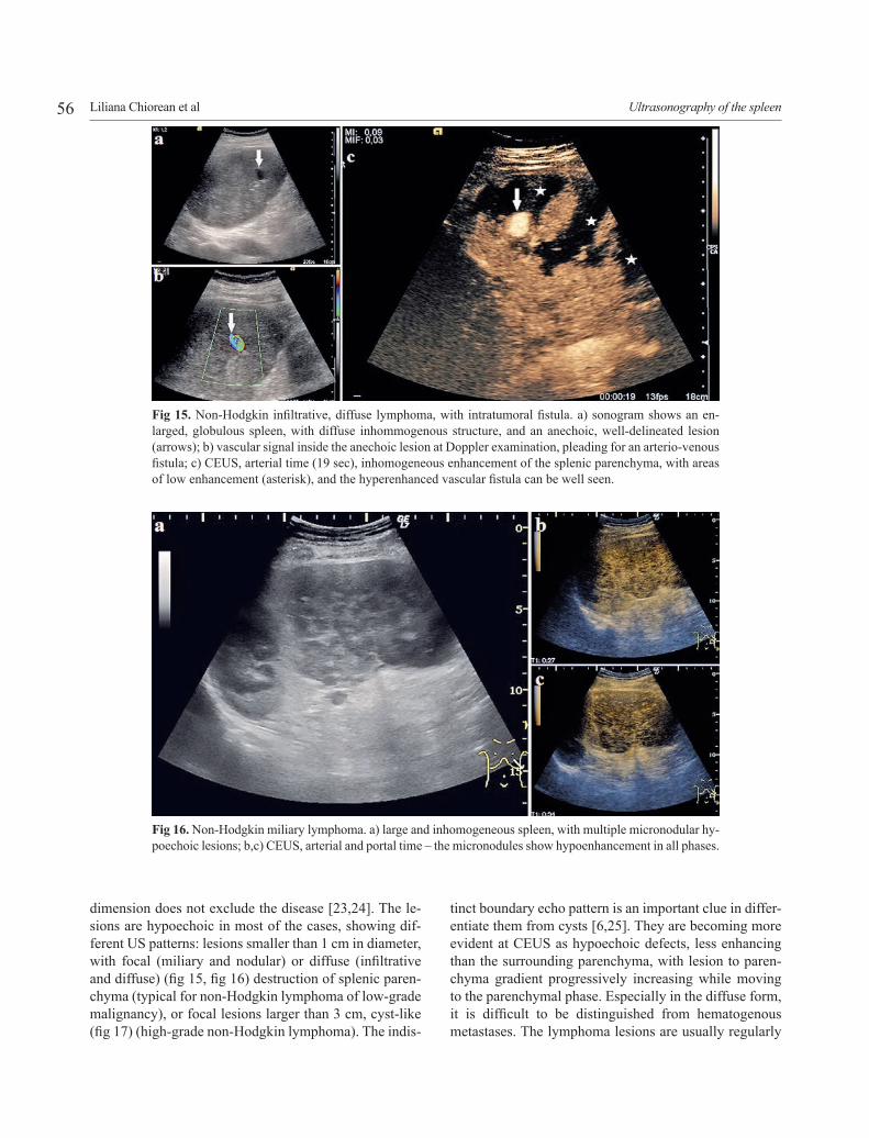
dimension does not exclude the disease [23,24]. The le-sions are hypoechoic in most of the cases, showing dif-ferent US patterns: lesions smaller than 1 cm in diameter, with focal (miliary and nodular) or diffuse (infiltrative and diffuse) (fig 15, fig 16) destruction of splenic paren-chyma (typical for non-Hodgkin lymphoma of low-grade malignancy), or focal lesions larger than 3 cm, cyst-like (fig 17) (high-grade non-Hodgkin lymphoma). The indis-
Fig 15. Non-Hodgkin infiltrative, diffuse lymphoma, with intratumoral fistula. a) sonogram shows an en-larged, globulous spleen, with diffuse inhommogenous structure, and an anechoic, well-delineated lesion (arrows); b) vascular signal inside the anechoic lesion at Doppler examination, pleading for an arterio-venous fistula; c) CEUS, arterial time (19 sec), inhomogeneous enhancement of the splenic parenchyma, with areas of low enhancement (asterisk), and the hyperenhanced vascular fistula can be well seen.
Fig 16. Non-Hodgkin miliary lymphoma. a) large and inhomogeneous spleen, with multiple micronodular hy-poechoic lesions; b,c) CEUS, arterial and portal time – the micronodules show hypoenhancement in all phases.
tinct boundary echo pattern is an important clue in differ-entiate them from cysts [6,25]. They are becoming more evident at CEUS as hypoechoic defects, less enhancing than the surrounding parenchyma, with lesion to paren-chyma gradient progressively increasing while moving to the parenchymal phase. Especially in the diffuse form, it is difficult to be distinguished from hematogenous metastases. The lymphoma lesions are usually regularly

57Med Ultrason 2014; 16(1): 48-59
deposited, while metastases are usually anarchically dis-posed [2,13,26]. Lesions with increased echogenicity are unusual in patients with lymphoma, and require histo-logical confirmation [2,13].
MetastasisSplenic metastases are rare lesions, usually seen in
patients with advanced malignant disease, having a poor prognosis. The most frequent metastases are from lym-
phoma and melanoma, followed by carcinoma of the ovary, breast, lung, stomach, prostate, colon, liver, and pancreas [5,8]. As in malignant lymphoma, splenic me-tastases are predominantly hypoechoic, target lesions with a hypoechoic halo being suggestive for metastasis (sign of aggressive behavior) [13]. Sometimes hyper-echoic (eg, carcinoma of the colon) or inhomogeneous lesions with a necrotic center can be found (rapid growth
Fig 17. Non-Hodgkin cyst-like lymphoma. a) sonogram shows a splenic round, hypoechoic mass with indis-tinct boundaries; b,c) the mass is hypoenhanced in all phases, being highly suggestive for malignancy (S = spleen, L = lymphoma).
Fig 18. Multiple splenic metastasis from a colonic cancer. a) multiple hyperechoic splenic masses, anarchi-cally disposed, some with hypoechoic rim (arrow); b,c) the lesions present an hypoenhancement pattern in all phases at CEUS.

58 Liliana Chiorean et al Ultrasonography of the spleen
Fig 19. Hypoechoic, round splenic mass, highly suggestive for metastasis. a) 2D – large mass occupying the inferior pole of the spleen, with indistinct boundaries; b,c,d) CEUS showing a mass with early rapid and intense enhancement, and with wash-out (M = mass, S = spleen).
Fig 20. Splenic metastasis with unknown origin. a) gray-scale US showing a large, hyperechoic round splen-ic mass; b,c,d,e) CEUS – the lesion is hypoenhanced in all phases compared to the surrounding parenchyma (M = mass).
59Med Ultrason 2014; 16(1): 48-59
or mucinous origin – eg, ovarian carcinoma). With the exception of metastases of mucinous primary tumors, calcifications are rarely seen (fig 18-20) [27-29].
Conclusion
Sonography is a widely available, noninvasive, use-ful and valuable tool for diagnosing and follow-ups of splenic abnormalities, both benign and malignant. The additional use of CEUS can improve its diagnostic valid-ity, especially when interpreted in the clinical context of the case. In many cases, pathologic confirmation is nec-essary to make a definitive diagnosis.
References:
1. Benter T, Klühs L, Teichgräber U. Sonography of the spleen. J Ultrasound Med 2011; 30: 1281-1293.
2. Catalano O, Sandomenico F, Matarazzo I, Siani A. Con-trast-enhanced sonography of the spleen. AJR Am J Roent-genol 2005; 184: 1150-1156.
3. Von Herbay A, Barreiros AP, Ignee A, et al. Contrast-en-hanced ultrasonography with SonoVue: differentiation be-tween benign and malignant lesions of the spleen. J Ultra-sound Med 2009; 28: 421-434.
4. Bamber J, Cosgrove D, Dietrich CF, et al. EFSUMB guide-lines and recommendations on the clinical use of ultrasound elastography. Part 1: Basic principles and technology. Ul-traschall Med 2013; 34: 169-184.
5. Andrews MW. Ultrasound of the spleen. World J Surg 2000; 24: 183-187.
6. Chen MJ, Huang MJ, Chang WH, et al. Ultrasonography of splenic abnormalities. World J Gastroenterol 2005; 11: 4061-4066.
7. Bolondi L, Correas JM, Lencioni R, Weskott HP, Piscaglia F. New perspectives for the use of contrast-enhanced liver ultrasound in clinical practice. Dig Liver Dis 2007; 39: 187-195.
8. Wan YL, Cheung YC, Lui KW, Tseng JH, Lee TY. Ul-trasonographic findings and differentiation of benign and malignant focal splenic lesions. Postgrad Med J 2000; 76: 488-493.
9. Stang A, Keles H, Hentschke S, et al. Differentiation of be-nign from malignant focal splenic lesions using sulphur hex-afluoride-filled microbubble contrast-enhanced pulseinver-sion sonography. AJR Am J Roengenol 2009; 193: 709-721.
10. Catalano O, Lobianco R, Sandomenico F, D’Elia G, Siani A. Real-time contrast-enhanced ultrasound of the spleen: examination technique and preliminary clinical experience. Radiol Med 2003; 106: 338-356.
11. Catalano O, Sandomenico F, Vallone P, D’Errico A, Siani A. Contrast-enhanced sonography of the spleen. Semin Ul-trasound CT MR 2006; 27: 426-433.
12. Tafuto S, Catalano O, Barba G, et al. Real-time contrast-enhanced specific ultrasound in staging and follow-up of splenic lymphomas. Front Biosci 2006; 11: 2224-2229.
13. Goerg C, Schwerk WB, Goerg K. Sonography of focal le-sions of the spleen. AJR Am J Roentgenol 1991; 156: 949-953.
14. Balzan SM, Riedner CE, Santos LM, Pazzinatto MC, Fontes PR. Posttraumatic splenic cysts and partial splenec-tomy: report of a case. Surg Today 2001; 31: 262-265.
15. Giovagnoni A, Giorgi C, Goteri C. Tumors of the spleen. Cancer Imaging 2005; 5: 73-77.
16. Caremani M, Occhini U, Caremani A, et al. Focal splenic lesions: US findings. J Ultrasound 2013; 16: 65-74.
17. Lim AK, Patel N, Eckersley RJ, Taylor-Rosbinson SD, Cosgrove DO, Blomley MJ. Evidence for spleen-specific uptake of a microbubble contrast agent: a quantitative study in healthy volunteers. Radiology 2004; 231: 785-788.
18. Gorg C, Bert T. Second-generation sonographic contrast agent for differential diagnosis of perisplenic lesions. AJR Am J Roentgenol 2007; 186: 621-626.
19. Changchien CS, Tsai TL, Hu TH, Chiou SS, Kuo CH. So-nographic patterns of splenic abscess: an analysis of 34 proven cases. Abdom Imaging 2002; 27: 739-745.
20. McKenney KL, Nuñez DB Jr, McKenney MG, Asher J, Zelnick K, Shipshak D. Sonography as the primary screen-ing technique for blunt abdominal trauma: experience with 899 patients. AJR Am J Roentgenol 1998; 170: 979-985.
21. Catalano O, Lobianco R, Sandomenico F, Siani A. Splenic trauma: evaluation with contrast-specific sonography and a second-generation contrast medium - preliminary experi-ence. J Ultrasound Med 2003; 22: 467-477.
22. Goerg C, Colle J, Goerg K, Prinz H, Zugmaier G. Sponta-neous rupture of the spleen: ultrasound patterns, diagnosis and follow-up. Br J Radiol 2003; 76: 704-711.
23. Goerg C, Schwerk WB, Goerg K, Havemann K. Sono-graphic patterns of the affected spleen in malignant lym-phoma. J Clin Ultrasound 1990; 18: 569-574.
24. Abbott RM, Levy AD, Aguilera NS, Gorospe L, Thompson WM. From the archives of the AFIP: primary vascular neo-plasms of the spleen — radiologic-pathologic correlation. Radiographics 2004; 24: 1137-1163.
25. Urrutia M, Mergo PJ, Ros LH, Torres GM, Ros PR. Cystic masses of the spleen: radiologic-pathologic correlation. Ra-diographics 1996; 16: 107-129.
26. Robertson F, Leander P, Ekberg O. Radiology of the spleen. Eur Radiol 2001; 11: 80-95.
27. Alloni R, Garberini A, Caputo D, Coppola R. Solitary splenic metastasis of ovarian carcinoma: report of two cas-es. Surg Today 2008; 38: 1144-1147.
28. Compérat E, Bardier-Dupas A, Camparo P, Capron F, Char-lotte F. Splenic metastases: clinicopathologic presentation, differential diagnosis, and pathogenesis. Arch Pathol Lab Med 2007; 131: 965-969.
29. Ushijima K, Nishida T, Okura N, et al. Solitary splenic re-currence of ovarian cancer: case report and review of the literature. Arch Gynecol Obstet 1999; 263: 79-81.