Embed Size (px)
Citation preview

2/14/2019
1
ULTRASOUND OF THE FETAL HEART
Cameron A. Manbeian, MD
Disclosure Statement
• Today’s faculty: Cameron Manbeian, MD does not have any relevant financial relationships with commercial interests or affiliations to disclose.
CONGENITAL HEART DISEASE
•A leading cause of infant mortality (4-13/1000 live births)• From 1950-1994, 42% of infant deaths reported to
the World Health Organization were attributable to cardiac defects!• Structural cardiac anomalies were among the most
frequently missed by prenatal ultrasonography.•Cardiac screening exam of the fetus is designed to
maximize the detection of heart anomalies during the 2nd trimester scan.

2/14/2019
2
ULTRASOUND SCREENING- Technical Factors
• Transducer- higher frequency probes improve likelihood of detection of subtle defects at the expense of reduced acoustic penetration.
•Highest possible transducer frequency should be used for all exams, recognizing the trade-off between resolution and penetration.
•A single acoustic focal zone and a relatively narrow image field should be used.
ULTRASOUND SCREENING- Technical Factors
• Images should be magnified so the heart fills at least 1/3-1/2 the screen. Cine loop assists in evaluation such as normal movement of heart valve leaflets.
• Should be performed between 18-22 weeks menstrual age.
• Limitations: maternal obesity, prone fetal position, abdominal scars and late gestation.
INTERNATIONAL SOCIETY OF ULTRASOUND IN OBSTETRICS AND GYNECOLOGY GUIDELINES
• Cardiac screening exam should now include both the four-chamber view and outflow tract views.
2/14/2019
3
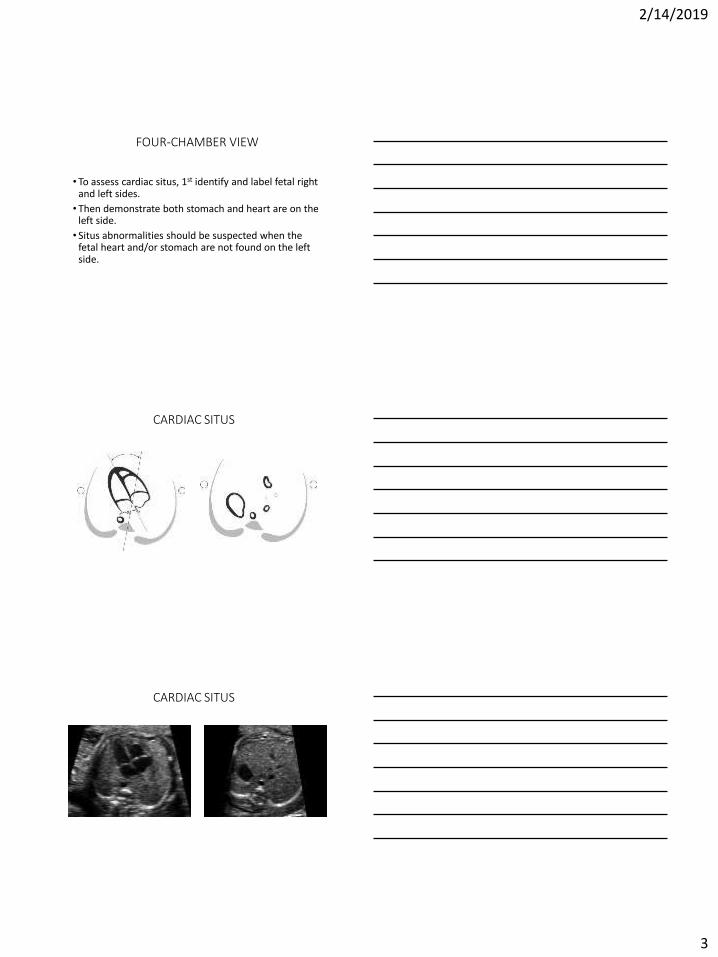
FOUR-CHAMBER VIEW
• To assess cardiac situs, 1st identify and label fetal right and left sides.
• Then demonstrate both stomach and heart are on the left side.
• Situs abnormalities should be suspected when the fetal heart and/or stomach are not found on the left side.
CARDIAC SITUS
CARDIAC SITUS

2/14/2019
4
FOUR CHAMBER VIEW
•The heart is mainly situated on the left side of the chest and the long axis normally points to the left by about 45 +/- 20 degrees.•Careful attention should be paid to the cardiac axis and position.•Abnormal axis increases the risk of a cardiac malformation especially involving the outflow tracts. A shift of the axis to the left may also occur with gastroschisis and omphalocele.
FOUR CHAMBER VIEW
•Abnormal displacement of the heart from its normal anterior left position can be caused by a diaphragmatic hernia, space occupying lesion such as a cystic adenomatoid malformation or CCAM, or fetal lung hypoplasia/agenesis.•A normal heart is usually no larger than 1/3 the area
of the chest.• Some views may show a small hypoechoic rim around
the fetal heart which can be mistaken for a pericardial effusion.
FOUR CHAMBER VIEW
•Both atria normally appear similar in size. The foramen ovale flap should open into the left atrium.
• The lower rim of atrial septal tissue, septum primum, should be present. This forms part of the crux, the point where the lower part of the atrial septum meets the upper part of the ventricular septum and where the atrioventricular valves insert.
• Pulmonary veins can often be seen entering the left atrium, and when feasible, 2 veins should be visualized.

2/14/2019
5
FOUR CHAMBER VIEW
FOUR CHAMBER VIEW
•Moderator band – a distinct muscle bundle that crosses the right ventricular cavity near the apex and helps identify the morphological RV.• The LV apex is smooth.•Both ventricles should be similar in size and have no
evidence of thickened walls. (Mild ventricular disproportion can occur as a normal variant in the 3rd
trimester).•RV and LV length should measure equal.
2/14/2019
6
FOUR CHAMBER VIEW
• The interventricular septum should be examined carefully for cardiac wall defects from the apex to the crux.
• Septum is best seen when the angle of insonation is perpendicular to it.
•When the angle is parallel a defect may be falsely suspected because of acoustic drop-out artifact.
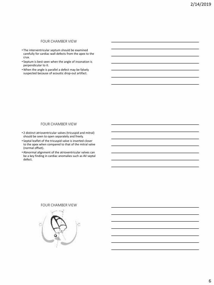
FOUR CHAMBER VIEW
• 2 distinct atrioventricular valves (tricuspid and mitral) should be seen to open separately and freely.
• Septal leaflet of the tricuspid valve is inserted closer to the apex when compared to that of the mitral valve (normal offset).
•Abnormal alignment of the atrioventricular valves can be a key finding in cardiac anomalies such as AV septal defect.
FOUR CHAMBER VIEW

2/14/2019
7
OUTFLOW TRACT VIEWS
•Great vessels should be approximately equal in size and cross each other at right angles from their origins.
• Images should include connection to the appropriate ventricle and adequate opening of the arterial valves.
OUTFLOW TRACT VIEWS
• Evaluation of the outflow tracts increases the detection rates for major cardiac malformations above those achievable by the four-chamber view alone.
•More likely to identify conotruncal abnormalities such as: tetralogy of Fallot, transposition of the great arteries, double outlet right ventricle and truncus arteriosus.
LVOT VIEW
•Confirms the presence of a great vessel originating from the morphological left ventricle.
• Should demonstrate continuity of the ventricular septum and the anterior wall of this vessel, the aorta.
•Aortic valves should move freely and should not be thickened.
• LVOT view helps to identify outlet ventricular septal defects and conotruncal abnormalities.

2/14/2019
8
LVOT VIEW
LVOT VIEW
RVOT VIEW•Confirms the presence of a great vessel originating
from the morphological RV.
• It is usually slightly larger than the aortic root.
• PV moves freely and should not be thickened.
•Vessel originating from the RVOT can only be confirmed as the pulmonary artery if it branches after a short course (right first). The normal PA continues distally towards the left side and into the ductus arteriosus that connects to the descending aorta.

2/14/2019
9
RVOT VIEW
RVOT VIEW
THREE VESSEL(3V) VIEW AND 3VT (TRACHEA)
• 3V view and 3VT view is desirable and should be attempted as part of the routine cardiac screening exam.
• From left to right: PA, Aorta and SVC.
• The PA is the most anterior and SVC is the most posterior.
• Their relative diameters decrease from left to right with the PA larger than the aorta and the aorta larger than the SVC.

2/14/2019
10
3V VIEW
THREE VESSEL(3V) VIEW AND 3VT (TRACHEA)
• Take note of correct position, alignment and size.
•Complete transposition of the great arteries, tetralogy of Fallot, and pulmonary atresia with a VSD are likely to have an abnormal 3V view.
3VT VIEW
•Ductal & aortic arches are positioned to the left of the trachea.• They form a V as they both join the descending aorta.•Aortic arch is the most cranial of the two arches.• The trachea is identified as a hyperechoic ring
surrounding a small fluid-filled space.• 3VT view is likely to enable detection of lesions such
as: coarctation, right aortic arch, double aortic arch and vascular rings.

2/14/2019
11
3VT VIEW
HEART RATE AND RHYTHM
•Normal rate ranges from 120-160 bpm.
• Fixed bradycardia, heart rates that remain below 110bpm OR
• Fixed tachycardia, heart rates that remain above 180bpm require further evaluation.
•Occasional skipped beats are typically not significant.
5 AXIAL VIEWS OF THE HEART

2/14/2019
12
Standard transverse scanning planes
Sagittal Views
Short Axis views of the Heart

2/14/2019
13
A FEW INTERESTING CASES
CASE 1

2/14/2019
14
ABNORMAL AXIS- PULMONARY ATRESIA
CASE 2

2/14/2019
15
DIFFERENTIAL DIAGNOSIS
HYPOPLASTIC LEFT HEART SYNDROMELV is underdeveloped
MV is very small
AV is very small
Ascending aorta is underdeveloped
SHONE’S COMPLEXNeed 3 of 8 described anomalies effecting the left side of the
heart, such as abnormal MV, coarctation of the aorta, small LV.

2/14/2019
16
CASE 3

2/14/2019
17
PULMONARY ATRESIA
CASE 4

2/14/2019
18

2/14/2019
19
D-TRANSPOSITION OF THE GREAT VESSELS
Transposition of the Great Vessels
Outflow vessels are side by side.
CASE 5

2/14/2019
20
TRICUSPID ATRESIA
• Plate like Tricuspid valve with no movement. Blood cannot flow through it.
•Associated with a large VSD which is how blood gets to the RVOT.
• LV is normal to large in size.
Echogenic Cardiac Focus• Small bright dot (<3mm) in ventricle of heart• Should be bright as bone to be a true finding• Turn down gain until all that is seen is ECF and bone to
confirm true finding.• ECF is most often an isolated finding in low-risk patient
(incidental).• Isolated ECF in a low-risk patient is considered a normal
finding.• Multiple and bilateral ECFs have an increased risk of
aneuploidy (abnormal number of chromosomes)• It is not a cardiac defect, probably microcalcification within
papillary muscle

2/14/2019
21
REFERENCES
• ISUOG Practice Guidelines (updated): sonographic screening examination of the fetal heart, Ultrasound Obstet Gynecol 2013; 41: 348-359
• STATdx, Echogenic Cardiac Focus, Roya Sohaey M.D.
• STATdx, Abnormal Outflow Tracts, Michael Puchalski