Embed Size (px)
Citation preview
U L T R A S T R U C T U R A L A N D B I O C H E M I C A L S T U D I E S O N
T H E P R E C U R S O R R I B O S O M A L
P A R T I C L E S I S O L A T E D F R O M R A T L I V E R N U C L E O L I
S. MATSUURA, T. MORIMOTO, Y. TASHIRO, T. HIGASHINAKAGAWA,
and M. M U R A M A T S U
From the Department of Physiology, Kansai Medical University, Moriguchi, Osaka, the Mitsubishi- Kasei Institute of Life Sciences, Machida, Tokyo, and the Department of Biochemistry, Tokushima University School of Medicine, Tokushima, Japan
ABSTRACT
Sucrose density gradient analyses of pH 5.5 and pH 7.4 extracts from rat liver nucleoli revealed the presence of two broad peaks of approximately 60S and 80S, and 60S and 80-100S, respectively. Ribonucleoprotein (RNP) particles containing precursor ribosomal R N A in these peaks have been characterized by electron microscopy and R N A analyses. Spherical particles only were found in the 60S peak of the pH 5.5 extract, from which 28S R N A and smaller R N A (23S and 18S RNA) exclusively were extracted. In the broad 80S peak of the pH 5.5 extract, about 60% of the particles were spherical while 30% were rodlike. In the R N A species present there were 28S plus smaller R N A (80%) and 35S R N A (20%). The 60Speak of the pH 7.4 extract contained mainly spherical particles (84%), and the R N A species present was mostly 28S plus smaller R N A (89%). In addition to spherical particles (43%), a number of rodlike (31%) and filamentous molecules (26%) were observed in the heavier side of the 80-100S peak of the pH 7.4 extract, from which 45S (14%), 35S (26%), and 28S and smaller R N A (60%) were extracted. Thus the precursor ribosomal particles containing 45S R N A and 35S R N A appear to be filamentous and rodlike molecules, respectively. Folding of loose ribonucleoprotein filaments into compact, spherical, large subparticles may be part of the maturation process of ribosomal large subparticles, in addition to the so-called sequential cleavage of RNA.
It has been well established that eukaryotic ribo- somal RNA is synthesized in the form of a large common precursor RNA (45S RNA) in the nu- cleolus and then, through a number of steps, is processed to mature 28S and 18S RNA (see reviews by Darnell (1), Maden (2), and Perry (3, 4). These precursor rRNAs are present as ribonu- cleoprotein (RNP) complexes, such as have been isolated from HeLa cells (5, 6, 7), L cells (8, 9, 10),
ascites tumor cells (11), and liver cells (12, 13, 14). The buoyant density of the RNP particles was definitely less than that of mature ribosomes, and it has been proposed that during the maturation of ribosomes in the nucleoli the reduction in RNA size is accompanied by a progressive diminution in this protein-to-RNA ratio (10). Recently Kumar and Warner (6) have shown that nucleolar precur- sor ribosomal particles have extra proteins in
THE JOURNAL OF CELL BIOLOGY • VOLUME 63, 1974 pages 629-640 629

addit ion to the major i ty of the r ibosomal proteins fround on the cytoplasmic large r ibosomal subpar- ticles.
Ul t ras t ruc tura l and autoradiographic studies of nucleoli in situ or of isolated nucleoli have shown tha t nucleoli are composed of a fibrillar compo- nent, a g ranular component , and nucleolus- associated chromat in (15, 16, 17), and that the fibril lar componen t is a precursor of the granular component ; [3H]uridine is first incorporated into
the fibrillar componen t and then t ransferred to the granular componen t (18-21).
Na r ayan et al. (12) and Koshiba et al. (13) isolated R N P particles f rom rat liver nucleoli and observed them, Their R N P particles, however, conta ined mainly 30-32S and 28S R N A .
In this report, r ibosomal precursor particles conta ining 45S and 35S R N A were extracted from rat liver nucleoli in an a t t empt to clarify the biosynthesis and matu ra t ion of r ibosomes in nu-
cleoli.
M A T E R I A L S A N D M E T H O D S
Isolation of Nuclei and Nucleoli
Male albino rats (Donryu strain) weighing 200 300 g were fed laboratory chow ad libitum, and fasted over- night before sacrifice. Nuclei and nucleoli were isolated from the perfused livers in the presence of magnesium ion, according to the procedure of Higashinakagawa et al. (22). The recovery of nuclei from the liver homoge- nate was about 60%, whereas recovery from the nuclear suspension was 30%.
Extraction of RNP Particles from Isolated Nucleoli
RNP particles were extracted from nucleoli according to the procedures of Muramatsu et al. (14). The nucleolar pellet obtained from 40 g of liver was mixed with 50 pl of 0.1% DNase solution (Worthington Biochemical Corp., Freehold, N.J., electrophoretically purified, RNase- free), and then homogenized by 10 strokes of a motor- driven Potter-Elvehjem homogenizer with a Teflon pes- tle, in 2.0 ml of pH 5.5 extraction medium (50 mM sodium acetate buffer (pH 5.5), 25 mM KCI, 0.1% polyvinylsulfate (PVS), and 1% Brij 98). After being left standing at 0°C for 10 min, the suspension was cen- trifuged at 12,000 g for 10 min and the supernate was stored as the pH 5.5 extract. The pellet was resuspended in 2.0 ml of pH 7.4 extraction medium (50 mM Tris-HCl buffer [pH 7.4], 25 mM KC1, 0.1% PVS, and 1% Bcij 98), and after 10 min in an ice-bath the suspension was centrifuged at 12,000 g for 10 rain, and the supernate was stored as the pH 7.4 extract for further use.
Fractionation of the Nucleolar Extracts by Sucrose Density Gradient Centrifugation
A 2-ml portion from each extract was layered onto 16 ml, 10-30% (wt/wt) sucrose gradients in 50 mM sodium acetate buffer (pH 5.5), 25 mM KCI, and 5 mM MgCI2. After centrifugation at 23,000 rpm for 12 15 h in a Hitachi RPS 25-3 rotor, the gradients were monitored at 260 nm from the top to the bottom and fractionated into about 30 tubes by an Auto Densiflow (Buchler In- struments, Fort Lee, N. J.) coupled to a Hitachi 124 spectrophotometer and a fraction collector.
Preparation of Rat Liver Ribosomal Large Subparticles and Subsequent Treatment with pH 5.5 Extraction Medium
Cytoplasmic large ribosomal subparticles were pre- pared from rat liver free polyribosomes according to the methods of Blobel and Sabatini (23), except that 750 mM KCI was used instead of 500 mM as control for electron microscope studies of the precursor particles.
In this experiment, the nucleolar RNP particles were prepared in the presence of 0.1% PVS and 1% Brij 98. As it is feasible that exposure to these surface-active agents may induce conformational change in the spherical large subparticles to rodlike or filamentous molecules, an aliquot of the large subparticle solution was treated with the pH 5.5 extraction medium. After several hours the medium was replaced by the conventional TKM buffer (50 mM Tris-HC1 buffer, pH 7.4, 25 mM KCI, 5 mM MgCI~), either by dialysis or by centrifugation, after which the particles were observed under the electron microscope.
Analyses of RNA by Sucrose Density Gradient Centrifugation and by Polyacrylamide Gel Electrophoresis
RNA was extracted from nucleolar RNP particles by the hot phenol-SDS method (24) and analyzed either by sucrose density gradient centrifugation or by polyacryl- amide gel electrophoresis, with a composite gel of 2.25% acrylamide-0.5% agarose (25). The molecular weight was estimated from the relationship between electrophoretic mobility and molecular weight, with 28S (1.7 x 10⁶ daltons) and 18S rRNA (0.7 5< l0 s daltons) as a standard.
Chemical Determination and Counting of Nuclei and Nucleoli
Nucleic acid and protein were extracted from precur- sor particles or from nucleoli by Siekevitz's procedure (26). RNA was dete'rmined by the orcinol reaction with rat liver RNA as a standard (27), DNA by Burton's modification of the diphenylamine method (28), and
630 THE JOURNAL OF CELL BIOLOGY • VOLUME 63, 1974
protein by Lowry's method (29), with bovine serum albumin as a standard.
Isolated nuclei and nucleoli were stained with 0.1% (wt/vol) azur C in 0.25 M sucrose and 0.03% azur C in 7% sodium citrate containing 0.25 M sucrose, respec- tively, and were counted in a hemocytometer.
Electron Microscopy
Specimens for electron microscopy were first dialyzed against a buffer solution to remove sucrose and then were mounted directly onto a carbon-coated collodion film with a micropipette, or were sprayed from a low-pressure nebulizer.
Shadow casting with platinum-carbon, and negative staining with uranyl acetate solution were done as described previously (30). Specimens were observed in a Hitachi HU-12 electron microscope. The photographic plates were printed with the emulsion side of the plate facing the surface of the bromide paper.
R E S U L T S
Biochemical S tudies
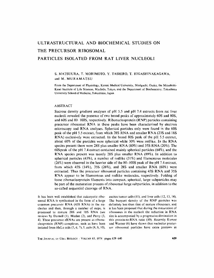
CHEMICAL CO*MPOSITION OF NUCLEOLI: Chemical composition of the nucleolar fraction (Table I) paralleled that reported by other authors (31-34), except for a higher D N A content. This is presumably due to the fact that repeated washing of the nucleolar fraction with the extraction me- dium was not carried out.
Nucleolar R N A was effectively extracted at pH 5.5 and pH 7.4, and the total recovery of R N A was approximately 90%. Such being the case, it is considered that the R N P particles in the extracts were indeed nucleolar R N P particles.
SUCROSE DENSITY GRADIENT ANALYSES OF RNA EXTRACTED FROM THE NUCLEOLAR
EXTRACTS: R N A was extracted by the hot phe- nol-SDS method and then was analyzed by sucrose density gradient centrifugation in order to charac- terize the species in the nucleolar extracts. Al- though both extracts contained 45S, 35S, 28S, 23S, and 18S R N A , the relative amounts were quite different. In the pH 5.5 extract, 28S and 35S R N A were the major RNAs, comprising 38% and 37% of the total R N A in the extract, respectively. In the pH 7.4 extract, 45S and 35S R N A were found in a larger amount (22 and 29%) in addition to 28S, 23S, and 18S R N A (t8, 21, and 6%).
R N A from the nucleolar extracts was also analyzed by polyacrylamide gel electrophoresis, and bands corresponding to 45S, 35S, 32S, 28S, 23S, and 18S were identified. The molecular weights of 45S, 35S, 32S, and 23S R N A were estimated to be 4.1, 3.0, 2.1, and 0.97 × l06 daltons, respectively. These values are in good agreement with the molecular weights of precursor R N A s extracted from HeLa cell nucleoli (35). Thus, the numbers of 45S and 35S R N A molecules per nucleolus were calculated to be 1.37 x l04 and 4 x l0 ~ molecules, respectively. As these values paralleled the corresponding data of l x 104 and 4 x l0 ~ molecules for the HeLa cell nucleolus reported previously (36), serious degradation of the extracted precursor particles was ruled out.
SUCROSE DENSITY GRADIENT ANALYSES OF THE NUCLEOLAR EXTRACT" The above results show that although partial fractionation of the nucleolar R N P particles is possible by succes- sive extraction at pH 5.5 and 7.4, these extracts were still quite heterogeneous with respect to the presence of the R N A species. Fractionation of the
TABLE l
Chemical Composition ofNuc&olar and Subnucleolar Fractions
Fraction Protein RNA DNA
N ucleolar fraction 0.123 (100) 0.0349 (100) 0.0640 (100)
pH 5.5 extract 0.0615 (50) 0.0168 (48) 0.0603* (94)
pH 7.4 extract 0.0425 (35) 0.0142 (41 ) 0.00455 (7.1)
Residue 0.0072 (5.9) 0.00278 (8) 0.00203 (3.2)
Fractions are expressed by yield in milligrams of protein, RNA, and DNA per gram of liver. The values in parentheses indicate distribution of protein, RNA, and DNA in the various subnuclear fractions (nucleolar fraction 100%). * Although nucleoli were extracted in the presence of DNase, most DNA was recovered as acid-insoluble DNA in the pH 5.5 extract. This is attributed to the incomplete digestion of DNA. Incubation with DNase was carried out in the absence of MgCI2 and in the presence of PVS, a potent inhibitor of DNase.
MATSUURA ET AL. Precursor Ribosomal Particles 631
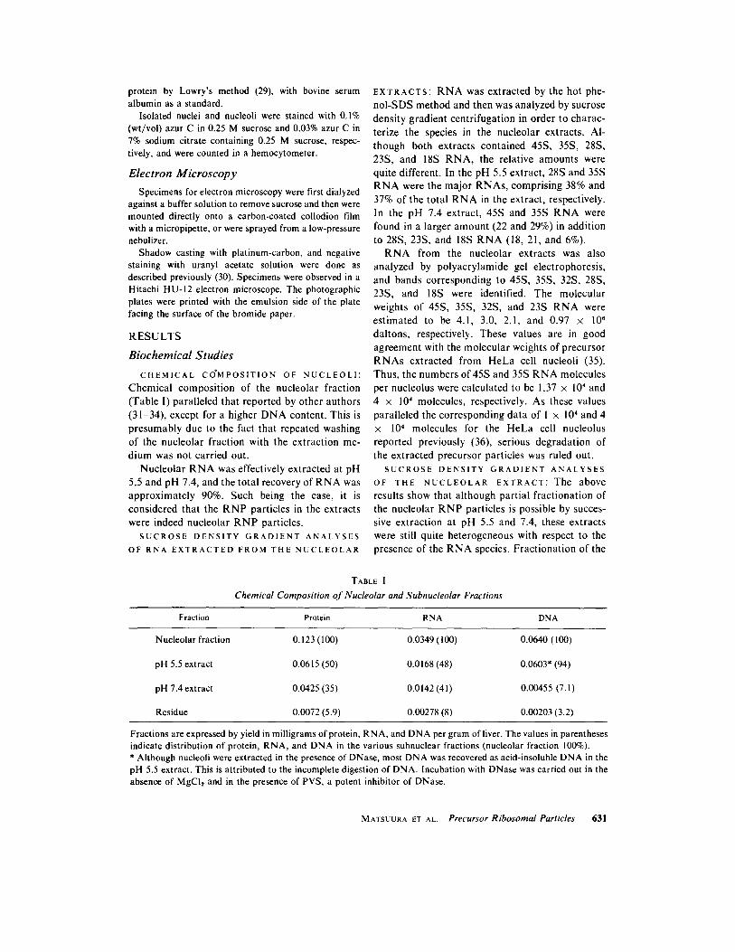
R N P particles from both extracts by sucrose density gradient centrifugation was thus at- tempted.
Sedimentation profiles of the pH 5.5 and pH 7.4 extracts are shown in Fig. 1 A and B, respectively. In Fig. 1 A, two broad components can be seen approximately at the 60S and 80S regions, respec- tively. Similarly, two broad components are pres- ent approximately at the 60S region and the 80 100S region as shown in Fig. I B.
0 4
0.2
E c
O tO c~ 0
"6
0.4 o u~
x~
0.2
B'
I I I I J L I I
o ' ~ ~ ' ;2 ' ,'6 '
ml from the top
FIGURE 1 Sucrose density gradient profile of the nu- cleolar RNP particles extracted using pH 5.5 (A) and pH 7.4 (B) medium. Small letters above the profiles indicate the RNP particle fractions used for further analyses.
C H A R A C T E R I Z A T I O N O F T H E R N P P A R -
T I C L E F R A C T I O N S BY S U C R O S E D E N S I T Y
GRADIENT CENTRIFUGATION: In order to characterize the RNP particles in the gradients, the 60 100S region was divided into four fractions as shown in Fig. 1: fraction a contained the 60S component; fraction b the heavier side of the 60S component and the lighter side of the 80S (pH 5.5 extract) or the 80 100S component (pH 7.4 ex- tract); fraction c the broad peak of the 80S or the 80-100S component; and fraction d the heavier side of the 80S or the 80-100S component, respec- tively. The homogeneity of each fraction and the sensitivity of the RNP particles to RNase treat- ment were re-examined by sucrose density gradient centrifugation.
The sedimentation patterns showed fraction 5 a to be rather homogeneous and composed mainly of the 60S component, whereas the other fractions were heterogeneous and composed of at least two of the 60S and 80S or the 80-100S components. The heavier component was more dominating in the faster-sedimenting fractions. The incubation of these fractions at 0°C for 15 min with 2 gg of RNase markedly reduced the area of the 80S or 80-100S component. The effect of RNase diges- tion was, therefore, more clearly demonstrated in fraction d than in fraction a.
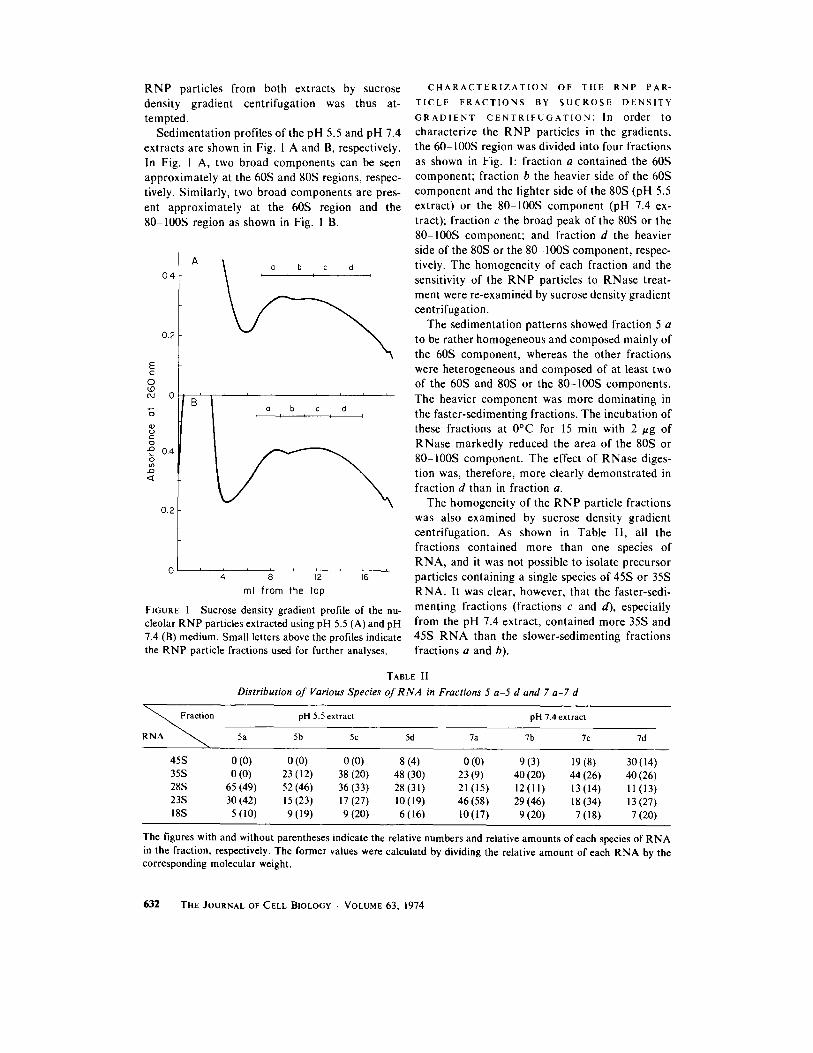
The homogeneity of the RNP particle fractions was also examined by sucrose density gradient centrifugation. As shown in Table II, all the fractions contained more than one species of RNA, and it was not possible to isolate precursor particles containing a single species of 45S or 35S RNA. It was clear, however, that the faster-sedi- menting fractions (fractions c and d), especially from the pH 7.4 extract, contained more 35S and 45S R N A than the slower-sedimenting fractions fractions a and b).
TABLE l I
Distribution of Various Species of RNA in Fractions 5 a-5 d and 7 a-7 d
t'°n pH 5.5 extract pH 7.4 extract
5a 5b 5c 5d 7a 7b 7c 7d
45S 0 (0) 0 (0) 0 (0) 8 (4) 0 (0) 9 (3) 19 (8) 30 (14) 35S 0 (0) 23 (12) 38 (20) 48 (30) 23 (9) 40 (20) 44 (26) 40 (26) 28S 65(49) 52(46) 36(33) 28(31) 21(15) 12(11) 13(14) 11(13) 23S 30 (42) 15 (23) 17 (27) I0 (19) 46 (58) 29 (46) 18 (34) 13 (27) 18S 5(10) 9(19) 9(20) 6(16) 10(17) 9(20) 7(18) 7(20)
The figures with and without parentheses indicate the relative numbers and relative amounts of each species of RNA in the fraction, respectively. The former values were caiculatd by dividing the relative amount of each RNA by the corresponding molecular weight.
632 THE JOURNAL OF CELL BIOLOGY . VOLUME 63, 1974
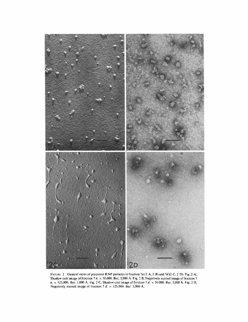
FIGURE 2 Generalviews of precursor RNP particles in fraction 7a(2 A, 2 B) and7d(2 C, 2 D). Fig. 2 A, Shadow-cast image of fraction 7 a. × 50,000. Bar, 2,000 A. Fig. 2 B, Negatively stained image of fraction 7 a. × 125,000. Bar, 1,000 A. Fig. 2 C, Shadow-cast image of fraction 7 d. x 50,000. Bar, 2,000 A. Fig. 2 D, Negatively stained image of fraction 7 d. z 125,000. Bar, 1,000 A.
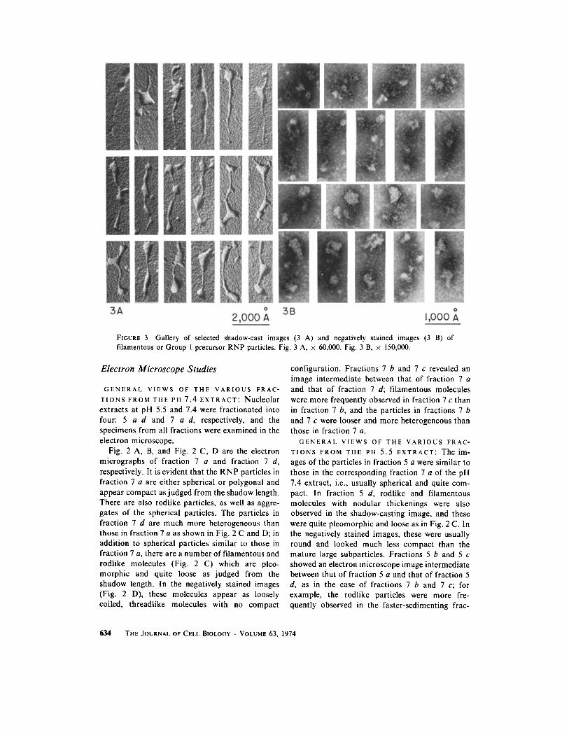
FIGURE 3 Gallery of selected shadow-cast images (3 A) and negatively stained images (3 B) of filamentous or Group 1 precursor RNP particles. Fig. 3 A, × 60,000. Fig. 3 B, × 150,000.
Electron Microscope Studies
G E N E R A L V I E W S O F T H E V A R I O U S F R A C -
T I O N S F R O M T H E P H 7.4 E X T R A C T : Nacleolar extracts at pH 5.5 and 7.4 were fractionated into four: 5 a-d and 7 a-d, respectively, and the specimens from all fractions were examined in the electron microscope.
Fig. 2 A, B, and Fig. 2 C, D are the electron micrographs of fraction 7 a and fraction 7 d, respectively. It is evident that the R N P particles in fraction 7 a are either spherical or polygonal and appear compact as judged from the shadow length. There are also rodlike particles, as well as aggre- gates of the spherical particles. The particles in fraction 7 d are much more heterogeneous than those in fraction 7 a as shown in Fig. 2 C and D; in addition to spherical particles similar to those in fraction 7 a, there are a number of filamentous and rodlike molecules (Fig. 2 C) which are pleo- morphic and quite loose as judged from the shadow length. In the negatively stained images (Fig. 2 D), these molecules appear as loosely coiled, threadlike molecules with no compact
configuration. Fractions 7 b and 7 c revealed an image intermediate between that of fraction 7 a and that of fraction 7 d; filamentous molecules were more frequently observed in fraction 7 c than in fraction 7 b, and the particles in fractions 7 b and 7 c were looser and more heterogeneous than those in fraction 7 a.
GENERAL VIEWS OF THE VARIOUS FRAC- TIONS FROM THE PH 5.5 EXTRACT: The im- ages of the particles in fraction 5 a were similar to those in the corresponding fraction 7 a of the pH 7.4 extract, i.e., usually spherical and quite com- pact. In fraction 5 d, rodlike and filamentous molecules with nodular thickenings were also observed in the shadow-casting image, and these were quite pleomorphic and loose as in Fig. 2 C. In the negatively stained images, these were usually round and looked much less compact than the mature large subparticles. Fractions 5 b and 5 c showed an electron microscope image intermediate between that of fraction 5 a and that of fraction 5 d, as in the case of fractions 7 b and 7 c; for example, the rodlike particles were more fre- quently observed in the faster-sedimenting frac-
634 THE JOURNAL OF CELL BIOLOGY - VOLUME 63, 1974
tions (7 c, 7 d) than in the slower-sedimenting fractions (7 a, 7 b).
C Y T O P L A S M I C M A T U R E L A R G E S U B P A R -
T I C L E S A N D T H E E F F E C T OF PVS T R E A T -
M E N T : The shadow-cast and negatively stained images of mature large ribosomal subparticles were compact and spherical and it is possible to identify rounded and skiff-shaped configurations of the 60S ribosomal subparticles in the negatively stained images, such as were observed and re- ported earlier (37).
Electron microscope observations of large sub- particles treated with PVS and Brij 98 revealed
that such treatment induces little or no detectable change in the images of the large subparticles. Thus it is concluded that rodlike or filamentous molecules are not artifacts produced by unfolding of the mature large subparticles in vitrO.
C L A S S I F I E D I M A G E S OF T H E R N P P A R -
T I C L E S F R O M T H E PH 7.4 E X T R A C T S : A s
the RNP particles in the various fractions from the pH 7.4 and 5.5 extracts were usually hetero- geneous in shape and size, we attempted to classify these particles according to axial ratio, assuming them to be prolate ellipsoids.
Group 1 (Fig. 3 A) consists of filamentous RNP
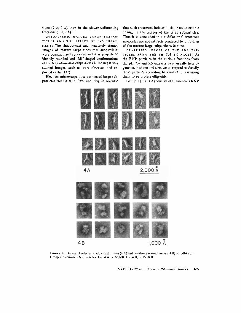
FIGURE 4 Gallery of selected shadow-cast images (4 A) and negatively stained images (4 B) of rodlike or Group 2 precursor RNP particles. Fig. 4 A, x 60,000. Fig. 4 B, x 150,000.
MATSUURA ET AL. Precursor Ribosomal Particles 635
molecules with an axial ratio of about 10 or more. These molecules are sometimes several hundred nm in length (about 500 nm on the average), and their width is variable. Their appearance is, there- fore, partly filamentous and partly nodular and quite irregular. Negatively stained profiles of RNP particles resembling the filamentous, loosely coiled, threadlike molecules are shown in Fig. 3 B.
Group 2 has rodlike RNP particles of which the axial ratio was about five, as shown in Fig. 4 A and B. The thickness of the molecules was not always uniform and the configuration of the particles was quite irregular. Occasionally observed were several filamentous processes resembling carrot roots. Fig. 4 B shows negatively stained images of particles in Group 2. These individual particles were loose in structure and irregular in shape, but were more compact and spherical, however, than those in Group 1.
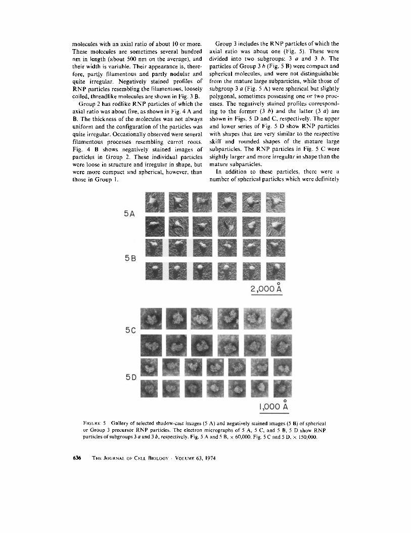
Group 3 includes the RNP particles of which the axial ratio was about one (Fig. 5). These were divided into two subgroups: 3 a and 3 b. The particles of Group 3 b (Fig. 5 B) were compact and spherical molecules, and were not distinguishable from the mature large subparticles, while those of subgroup 3 a (Fig. 5 A) were spherical but slightly polygonal, sometimes possessing one or two proc- esses. The negatively stained profiles correspond- ing to the former (3 b) and the latter (3 a) are shown in Figs. 5 D and C, respectively. The upper and lower series of Fig. 5 D show RNP particles with shapes that are very similar to the respective skiff and rounded shapes of the mature large subparticles. The RNP particles in Fig. 5 C were slightly larger and more irregular in shape than the mature subparticles.
In addition to these particles, there were a number of spherical particles which were definitely
FIGURE 5 Gallery of selected shadow-cast images (5 A) and negatively stained images (5 B) of spherical or Group 3 precursor RNP particles. The electron micrographs of 5 A, 5 C, and 5 B, 5 D show RNP particles of subgroups 3 a and 3 b, respectively. Fig. 5 A and 5 B, x 60,000. Fig. 5 C and 5 D, x 150,000.
636 THE JOURNAL OF CELL BIOLOGY • VOLUME 63, 1974
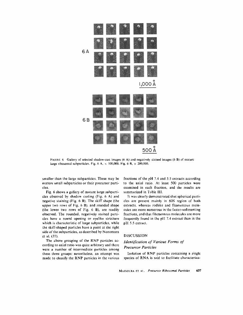
FIGURE 6 Gallery of selected shadow-cast images (6 A) and negatively stained images (6 B) of mature large ribosomal subparticles. Fig. 6 A, x 100,000. Fig. 6 B, x 200,000.
smaller than the large subparticles. These may be mature small subparticles or their precursor parti- cles.
Fig. 6 shows a gallery of mature large subparti- cles observed by shadow casting (Fig. 6 A) and negative staining (Fig. 6 B). The skiff shape (the upper two rows of Fig. 6 B), and rounded shape (the lower two rows of Fig. 6 B), are readily observed. The rounded, negatively stained parti- cles have a round opening or eyelike structure which is characteristic of large subparticles, while the skiff-shaped particles have a point at the right side of the subparticles, as described by Nonomura et al. (37).
The above grouping of the RNP particles ac- cording to axial ratio was quite arbitrary and there were a number of intermediate particles among these three groups; nevertheless, an attempt was made to classify the RNP particles in the various
fractions of the pH 7.4 and 5.5 extracts according to the axial ratio. At least 500 particles were examined in each fraction, and the results are summarized in Table IlI.
It was clearly demonstrated that spherical parti- cles are present mainly in 60S region of both extracts, whereas rodlike and filamentous mole- cules are more numerous in the faster-sedimenting fractions, and that filamentous molecules are more frequently found in the pH 7.4 extract than in the pH 5.5 extract.
DISCUSSION
Identification o f Various Forms o f
Precursor Particles
Isolation of RNP particles containing a single species of RNA is said to facilitate characteriza-
MATSUURA ET AL. Precursor Ribosomal Particles 637
tion of ribosomal precursor particles. However, despite several trials, including CsCI density gradi- ent centrifugation, we were unsuccessful.
An attempt was then made to correlate the distribution of the various species of ribosomal RNA (Table !I) with the distribution of the classified images of the RNP particles (Table III) in the various fractions of the pH 5.5 and pH 7.4 extracts. For this purpose, specimens from each fraction were utilized for simultaneous electron microscopy and RNA analyses, after which the results were compared.
The relative numbers of various species of RNA in each fraction were calculated by dividing the relative amount of each RNA species by the corresponding molecular weight (bracketed in Table II).
Fraction 5 a contained 28S RNA (about 50%) and smaller RNA (about 50%). Approximately 50% of the particles found in this fraction were spherical and similar in appearance and size to mature large subparticles. Since Group 3 b parti- cles are probably mature large subparticles (see Results), Group 3 a particles are considered nu- cleolar 60S particles containing 30S RNA, a direct precursor of mature 28S RNA (38, 39). The other 50% of the particles were smaller in size and flatter in shape than mature large subparticles. These may be either mature small subparticles or their precursor particles which contain 23S RNA, as proposed by Egawa et al. (40).
In fractions 5 c and 5 d, another type of particle appeared. These were rodlike or Group 2 particles which constituted about 30% of the population. More than 20% of RNA in these fractions was 35S RNA, and it would appear that these rodlike particles are the precursor particles containing 35S RNA,
In fraction 7 a, spherical particles were predomi- nant (84%), and the RNA species observed was mainly 28S plus smaller RNA (91%).
In fraction 7 d, numerous filamentous (26%) and rodlike (31%) molecules were observed. The rela- tive amounts of 45S, 35S, and 28S plus smaller RNA found in this fraction were 14%, 26%, and 60%, respectively. From these results it is proposed that the filamentous molecules are the precursor particles containing 45S RNA. This suggestion is supported by the results of RNase digestion studies which revealed that the precursor particles con- taining 45S and 35S RNA were quite sensitive to RNase digestion (7, 41).
Possible Ultrastructural Change o f
Precursor Particles in the Maturation
Process o f Mature Large Subparticles
It has been demonstrated that ribosomal precur- sor RNA is synthesized in the nucleolus and is readily taken up by protein synthesized in the cytoplasm (see reviews by Darnell (1) and Perry (3, 4)). The ribosomal precursor particles undergo all subsequent maturation in the form of nucleo- protein particles, and it is evident that the precur- sor particles possess a relatively higher proportion of protein to RNA and that ribosome maturation involves, in addition to a decrease in the size of the RNA molecules, a sequential decrease in the proportion of associated proteins.
Filamentous molecules and rodlike molecules appear to be precursor particles containing 45S and 35S RNA, respectively, and if such is the case, the ribosome maturation process may also involve gradual conformational changes from loose fila- mentous structure to compact spherical molecules as schematized in Fig. 7.
This postulation is also supported by uitrastruc- tural and electron microscope autoradiographic studies of mammalian nucleoli in situ (15-21). The authors of those studies have reported that there are two basic components in nucleoli, one fibrillar and the other granular, and that SH-labeled uridine
TABLE III Distribution of Various Precursor RNP Particles in the Various Fractions of the pH 5.5 and pH 7.4 Nucleolar
Extracts
Fraction pH 5.5 extract
Particles ~ 5a 5b 5c 5d
pH 7.4 extract
7a 7b 7c 7d
Filamentous 0 2 8 15 0 5 11 26 Rodlike 0 27 29 31 16 32 38 31 Spherical 100 71 63 54 84 63 51 43
The figures indicate the relative numbers of various RNP particles in each fraction.
638 THE JOURNAL OF CELL BIOLOGY - VOLUME 63, 1974
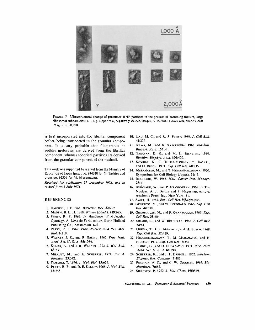
FIGURE 7 Ultrastructural change of precursor RNP particles in the process of becoming mature, large ribosomal subparticles (L ~ R). Upper row, negatively stained images, x 150,000. Lower row, shadow-cast images, × 60,000.
is first incorporated into the fibrillar component
before being transported to the granular compo- nent. It is very probable that filamentous or
rodlike molecules are derived from the fibrillar component , whereas spherical particles are derived
from the granular component of the nucleoli.
This work was supported by a grant from the Ministry of Education of Japan (grant no. 844020 for Y. Tashiro and grant no. 92336 for M. Muramatsu).
Received for publication 27 December 1973, and in revised form 3 July 1974.
R E F E R E N C E S
1. DARNELL, J. F. 1968. Bacteriol. Rev. 32:262. 2. MADEN, B. E. H. 1968. Nature (Lond.). 219:685. 3. PERRY, R. P. 1969. In Handbook of Molecular
Cytology. A. Lima de Faria, editor. North Holland Publishing Co., Amsterdam. 620.
4. PERRY, R. P. 1967. Prog. Nucleic Acid Res. Mol. Biol. 6:219.
5. WARNER, J. R., and R. SOEIRO. 1967. Proc. Natl. Acad. Sci. U. S. A. 58:1964.
6. KUMAR, A., and J. R. WARNER. 1972. J. Mol. Biol. 63:233.
7. MIRAULT, M., and K. SCHERRER. 1971. Eur. J. Biochem. 23:372.
8. TAMAOKI, T. 1966. J. Mol. Biol. 15:624. 9. PERRY, R. P., and D. E. KELLEY. 1966. J. Mol. Biol.
16:255.
10. LIAU, M. C., and R. P. PERRY. 1969. J. Cell Biol. 42:272.
I I. IZAWA, M., and K. KAWASHIMA. 1968. Biochim. Biophys. Acta. 155:51.
12. NARAYAN, K. S., and M. L. BIRNSTIEL. 1969. Biochim. Biophys. Acta. 190:470.
13. KOSHIBA, K., C. THIRUMALCHARY, Y. DASKAL, and H. BUSCH. 1971. Exp. Cell Res. 68:235.
14. MURAMATSU, M., and T. HIGASHINAKAGAWA. 1970. Symposium for Cell Biology (Japan). 21:15.
15. BERNHARD, W. 1966. Natl. Cancer Inst. Monogr. 23:13.
16. BERNHARD, W., and P. GRANBOULAN. 1968. In The Nucleus. A. J. Dalton and F. Haguenau, editors. Academic Press, Inc., New York. 81.
17. SWIFT, H. 1963. Exp. Cell Res. 9(Suppl.):54. 18. GEUSKENS, M., and W. BERNHARD. 1966. Exp. Cell
Res. 44:579. 19. GRANBOULAN, N., and P. GRANBOULAN. 1965. Exp.
Cell Res. 38:604. 20. SIMARD, R., and W. BERNHARD. 1967. J. Cell Biol.
34:61. 21. UNUMA, T., J. e. ARENDELL, and H. BUSCH. 1968.
Exp. Cell Res. 52:429. 22. HIGASHINAKAGAWA, T., i . MURAMATSU, and H.
SUGANO. 1972. Exp. Cell Res. 71:65. 23. BLOBEL, G., and D. D. SABATINI. 1971. Proc. Natl.
Acad. Sci. U . S . A . 68:390. 24. SCHERRER, K., and J. F. DARNELL. 1962. Biochem.
Biophys. Res. Commun. 7:486. 25. PEACOCK, A. C., and C. W. DINGMAN. 1967. Bio-
chemistry. 7:668. 26. SIEKEVITZ, P. 1952. J. Biol. Chem. 195:549.
MATSUURA ET AL. Precursor Ribosomal Particles 639
27. MEJBAUM, W. 1939. Z. Physiol. Chem. 258:117. 28. BURTON, K. 1956. Biochem. J. 62:315. 29. LOWRY, O. H., N. J. ROSEaROUGH, A. L. FARR, and
P. J. RANDALL. 1951. J. Biol. Chem. 193:365. 30. MATSUURA, S., Y. TASHIRO, S. OSAWA, and E.
OTAKA. J. Mol. Biol. 47:383. 31. LAIRD, A. K. 1954. Exp. CellRes. 6:30. 32. MURAMATSU, M., K. SMETANA, and H. BUSCH.
1963. Cancer Res. 23:510. 33. HIGASHI, K., S. NARAYAN, H. P. ADAMS, and H.
BUSCH. 1966. Cancer Res. 26:1582. 34. DESJARDINS, P., K. SMETANA, and H. BUSCH. 1965.
Exp. Cell Res. 40:127. 35. McCONKEY, F. H., and J. W. HOPKINS. 1969. J.
Mol. Biol. 39:545. 36. WEINBERG, P. A., and S. PENMAN. 1968. J. Mol,
Biol. 38:289. 37. NONOMURA, Y., G. BLOBEL, and D. D. SABATINI.
1971. J. Mol. Biol. 60:303. 38. FUJISAWA, T., S. ABE, T. KAWADA, M. SATAKE, and
K. OGATA. 1973. Biochim. Biophys. Acta. 324:226. 39. FUJISAWA, T., S. ABE, M. SATAKE, and K. OGATA.
1973. Biochim. Biophys. Acta. 324:241. 40. EGAWA, K., Y. C. CHOI, and H. BUSCH. 1971. J.
Mol. Biol. 56:565. 41. LIAU, M. C., N. C. CRAIG, and R. P. PERRY. 1968.
Biochim. Biophys. Acta. 169:196.
640 THE JOURNAL OF CELL BIOLOGY . VOLUME 63, 1974