Embed Size (px)
Citation preview
JOURNAL OF BACTERIOLOGY, Feb., 1966Copyright @ 1966 American Society for Microbiology
Ultrastructure of Escherichia coli Cells Infectedwith Bacteriophage R17
RICHARD M. FRANKLIN AND NICOLE GRANBOULAN
Laboratoire de Microscopie Electronique, Institut de Recherches sur le Cancer, Villejdif, Seine, France,and Department of Pathology, University of Colorado Medical Center, Denver, Colorado
Received for publication 14 October 1965
ABSTRACT
FRANKLIN, RICHARD M. (Institut de Recherches sur le Cancer, Villejuif, Seine,France), AND NICOLE GRANBOULAN. Ultrastructure of Escherichia coli cells infectedwith bacteriophage R17. J. Bacteriol. 91:834-848. 1966-Ultrastructural changes inEscherichia coli cells infected with ribonucleic acid (RNA) bacteriophage R17 were
studied under conditions of one-step growth. No morphological alterations were seen
during the latent period. During the period of rapid viral synthesis, a fibrillar lesionsurrounded by ribonucleoprotein particles was observed in a polar region. Late ininfection, paracrystalline arrays of virions were found in over 90% of the cells. Whenprotein synthesis was blocked by chloramphenicol at 20 min postinfection, allowingcontinued viral RNA synthesis without production of coat protein, a dense fibrillararea appeared in a paranuclear region. Cytochemical studies were done on cells em-bedded in hydroxypropyl methacrylate, a water-miscible embedding agent. Theparacrystalline arrays of virions were digested after extensive treatment with eitherpepsin or ribonuclease. Shorter digestion with pepsin resulted in better definition ofthe crystal regions. The fibrillar area found in chloramphenicol-treated cells was
digested by ribonuclease but not by pepsin, and was also resistant to lead extraction.This region probably represents a pool of virus-specific RNA.
The following two papers deal with the morpho-logical changes associated with the multiplicationof the ribonucleic acid (RNA) bacteriophage R17in its host cell Escherichia coli. Bacteriophage R17multiplies rapidly in male strains of E. coli andreaches high intracellular phage titers (15). Thepresence of paracrystalline arrays of virions hasbeen reported for several related RNA phages(6, 21). The present report includes a descriptionof paracrystalline arrays of R17 in the later stagesof infection but emphasizes the morphologicalalterations in the earlier stages.
Chloramphenicol, an inhibitor of protein syn-thesis, prevents viral RNA synthesis when addedprior to 8 min postinfection (14). But, whenchloramphenicol is added 15 to 20 min after in-fection, there is an accumulation (3, 14) and con-
tinued synthesis of viral RNA (3, 5). These factsare utilized in this study in a search for morpho-logical evidence for a pool of accumulated viralRNA.
In morphological studies of this type, identifi-cation of structures in terms of their macro-molecular components is tenuous. Therefore, useis made of differential enzyme digestions accord-
ing to principles enunciated by Bernhard andTournier (2). This is possible through the use ofa new hydrophilic embedding agent, hydroxy-propyl methacrylate (11). Studies utiliing differ-ential extraction or differential enzyme digestionare further corroborated by means of specificlabeling of macromolecular components by triti-ated precursors, followed by high-resolutionautoradiography. This technique is discussed inthe accompanying paper (8a).
MATERIALS AND METHODS
Media. MS broth was prepared according to theformula of Davis and Sinsheimer (4), without theaddition of thiamine hydrochloride. MS broth wasused for the growth of the host cell and in phagegrowth studies.
For plaque assays, both bottom and top agar con-tained the same ingredients as MS broth (no thiaminehydrochloride). Bottom agar contained 1% agar
(Difco) and top agar contained 0.8% agar.Sources ofmaterials. Chloramphenicol was obtained
from Parke, Davis & Co., Detroit, Mich. Pepsin (twotimes crystallized) and ribonuclease (pancreatic, frac-tion A, phosphate-free, lyophilized) were obtainedfrom Worthington Biochemical Corp., Freehold, N.J.
834
Vol. 91, No. 2Printed in U.S.A.
on August 15, 2019 by guest
http://jb.asm.org/
Dow
nloaded from
ULTRASTRUCTURE OF PHAGE-INFECTED E. COLI
2-Hydroxypropyl methacrylate (HPMA) was obtainedfrom Rohm and Haas Co., Philadelphia, Pa.; a,a'-azobisisobutyronitrile, from Fluka A. G., Buchs,Switzerland; and Carbowax 1500, from Gurr Limited,London, England.
Anitiserum. Antiserum to R17 phage was preparedby injecting rabbits subcutaneously with highly puri-fied virus preparations (see Granboulan and Franklin,nmaniuscript in preparationz) mixed with an equal volumeof a Freund-type adjuvant (9 parts of Drakeol 6VR to1 part of Arlacel A). Each rabbit received between 45and 90 mg of virus or 30 to 60 mg of virus protein.Only one injection was needed, and the antibody titerremained constant between 1 and 9 months after theinitial injection.
Cells anid viruls. The original strain of E. coli K-12strain Hfri (X) meth- was kindly provided by E. B.Ellis and A. F. Graham. A nonlysogenic strain wasderived from this by picking survivors of a heavy doseof ultraviolet radiation. This strain is designated asHfri (X-). The original strain and the derived straingrow, to a limited extent, in TPG (22) without Casa-mino Acids (Difco) but supplemented only withL-methionine at 100 ,g/ml. Phage X was assayed onE. coli strain W1327 (nonlysogenic) kindly providedby L. M. Morse. No X phage could be detected in thesupematant fluid of a culture of Hfri (X-) or by platingHfri directly on W1327.
The otiginal strain of bacteriophage R17 was kindlyprovided by E. B. Ellis and A. F. Graham. Grown onHfri (X), the stock of R17 contained 1 X plaque-form-ing unit (PFU) per 4.8 X 10¹¹ R17 PFU (assayed onthe F- strain W1321). Phage X-free R17 was isolatedby four successive plaque passages on E. coli Hfr1(X-). Neither growth of the RNA phage nor metabo-lism of the infected cells was influenced by the presenceor absence of phage X (Franklin, utiipublished data).
Growth of virus. For virus growth curves, the cellswere grown in the appropriate medium (MS or TCG,see 7) and harvested during logarithmic growth (2 X10⁷ to 5 X 10⁸ cells per milliliter). The centrifugedpellet of cells was mixed with a small volume of virusat an appropriate multiplicity. After allowing virusadsorption to take place (5 min at room temperature),the complexes were diluted in cold TPG salts andcentrifuged. In some experiments, the pellet of in-fected cells was incubated for 5 min at room tempera-ture with a 1 :100 dilution of R17 antibody (K = 8,900min-l) and then washed three times in cold TPG salts.Dilution of the cells into the appropriate warm me-dium was considered the "zero time" of virus multi-plication. At given time intervals, samples were diluted1:10 into MS broth containing 10%lO (v/v) chloroform.Phage was titered by the usual plaque technique.
For electron microscopy, cells were infected at amultiplicity of approximately 10 to 20 PFU per cell,again in a small volume of medium at room tempera-ture. After allowing 5 min of adsorption, the cells werediluted into MS broth at 37 C to start viral multipli-cation. Samples were collected and fixed at appropri-ate times.
Preparationi of specimenis for electroln microscopy.For standard ultrastructural studies, bacteria werefixed according to the procedure of Ryter and Kellen-
berger (18). A 15-ml amount of culture was mixedwith 1.5 ml of fixative (1%; OSO4) in Veronal-acetatebuffer and was immediately centrifuged for 8 min at5,000 rev/min. The pellet was suspended in 1 ml offixative with 0.1 ml of tryptone broth added and wasleft at room temperature overnight. This suspensionwas then diluted in 8 ml of Veronal-acetate buffer andcentrifuged, and the pellet was suspended in 3 dropsof Agarose (1.5%) at 45 C. Upon solidification, theAgarose was cut into approximately 1-mm cubes,which were treated for 2 hr at room temperature inuranyl acetate solution (0.5%,t7 in Veronal-acetate buf-fer).
For ultrastructural cytochemistry, bacteria werefixed as just described, but were embedded in hydroxy-propyl methacrylate (11) without prior uranyl acetatetreatment.
For embedding in Epon, the blocks were dehy-drated in acetone (25, 50, 75%'- for 15 min; 90 and100% for 30 min) and embedded as follows: 20%/OEpon-80%yc acetone for 30 min, 50%' Epon-50% ace-tone for 30 min, 70%o Epon-30% acetone for 30 min,Epon overnight, and final embedding in Epon the nextday with polymerization at 60 C.
For HPMA embedding, the blocks, stained or un-stained with uranyl acetate, were partially dehydratedin aqueous solutions of Carbowax 1500 (1) accordingto the following schedule: 70 and 85%c for 15 min at37 C, embedding in the final embedding solution ofHPMA prepolymerized with the catalyst a,a'-bisiso-butyronitrile (0.4') followed by ultraviolet polymer-ization at 4 C.
Enzymatic digestions were done with sections ofunstained specimens embedded in HPMA. ReducedOS04 was removed by floating the sections on 6%H202 in phosphate buffer for 10 min at room tempera-ture (12, 13). Enzymatic digestions (10, 12) consistedof treatment with pepsin, 0.01% in 0.1 N HCI, pH 1,for 30 min at 37 C and with ribonuclease A, 0.1% indistilled water, pH 6.8, for 30, 45, or 120 min at 37 C.Control sections were floated on H202 as describedabove, followed by floating on distilled water for thesame time and at the same temperature as the sectionstreated with enzymes.
Unstained sections embedded in HPMA werestained by one of the following treatments: (i) 5%uranyl acetate in Veronal-acetate buffer (pH 4.8) for5 or 10 min, (ii) lead citrate for 10 min (17), or (iii)uranyl acetate for 5 min followed by lead citrate for5 to 10 sec. All specimens were examined witha Siemens Elmiskop I electron microscope operatingat 80 kv with an objective aperature of 50 ,u.
RESULTS
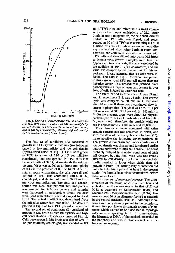
Growth of bacteriophage R17 in E. coli Hfr(X-). Many factors influenced the growth of RNAbacteriophages (see 15). These included the nu-trient medium, age of cells, multiplicity of infec-tion, cell density, and state of cells (preirradiatedwith ultraviolet light, pretreated with actinomycin,etc.). Two growth curves, shown in Fig. 1, illus-trate the conditions most applicable to this andthe following study.
835VOL. 91, 1966
on August 15, 2019 by guest
http://jb.asm.org/
Dow
nloaded from
FRANKLIN AND GRANBOULAN.re t
40 60 80 100
TIME IN MINUTESFIG. 1. Growth of bacteriophage R17 in Escherichia
coli Hfr1 (X-) under conditions of (A) low multiplicity,low cell density, in TCG synthetic medium (open circles)and of (B) high multiplicity, relatively high cell density,in MS nutrient broth (closed circles).
The first set of conditions (A) consisted ofgrowth in TCG synthetic medium (see followingpaper) at low multiplicity and low cell density(open-circled curve of Fig. 1). Cells were grownin TCG to a titer of 2.95 x 10⁸ per milliliter,centrifuged, and resuspended in TPG salts (thebalanced salts of TCG) at one-tenth the originalvolume. Virus was added at an input multiplicityof 0.15 in the presence of 0.01 M KCN. After 10min at room temperature, the cells were diluted10-fold in TPG salts containing 0.01 M KCN,centrifuged, and diluted into warm TCG to initi-ate virus multiplication. The final cell concen-tration was 1,000 cells per milliliter. One portionwas assayed for infective centers and sampleswere harvested at appropriate times; the cellswere lysed with chloroform and assayed for totalPFU. The actual multiplicity, determined fromthe infective center data, was 0.046. The data areplotted in Fig. 1 as total PFU per infective center.The second set of conditions (B) consisted of
growth in MS broth at high multiplicity and highcell concentration (closed-circle curve of Fig. 1).Cells were grown in MS broth to a titer of 2.40 x101 per milliliter, centrifuged, resuspended in 0.9
ml of TPG salts, and mixed with a small volumeof virus at an input multiplicity of 21.7. After5 min at room temperature, the cells were diluted10-fold in TPG salts, centrifuged, and resus-pended in 10 ml of TPG salts containing a 1:100dilution of anti-R17 rabbit serum to neutralizeany unadsorbed virus. After 5 min at room tem-perature, the cells were washed three times withTPG salts and then diluted into warm MS brothto initiate virus growth. Samples were taken atappropriate time intervals, the cells were lysed bythe addition of 10% (v/v) chloroform, and thevirus was assayed by the plaque test. In this ex-periment, it was assumed that all cells were in-fected. The data in Fig. 1, therefore, are plottedin this case as total PFU per cell rather than perinfective center. This procedure is justified, sinceparacrystalline arrays of virus can be seen in over90% of cells infected as described here.The latent period in experiment A was 29 min
and in experiment B it was 35 min. The growthcycle was complete by 60 min in A, but evenafter 80 min in B there was a continued slow in-crease in phage titer. The yield was 675 PFU percell in A and 4,500 PFU per cell (at 140 min) inB. On the average, there were about 5.5 physicalparticles per PFU (see Granboulan and Franklin,in preparation); therefore, the particle yield percell is approximately 3,700 in A and 25,000 in B.
These two experiments compared with othergrowth experiments not presented in detail, andwith the data of Paranchych and Graham (15),make possible the following generalizations. (i)The growth curve examined under conditions oflow cell density was sharper and terminated earlierthat that performed at high cell density. There wasprobably delayed lysis under conditions of highcell density, but the final yield was not greatlyaffected by cell density. (ii) Growth in syntheticmedia resulted in lower virus yields than didgrowth in broth. (iii) Multiplicity of infection didnot affect the latent period, at least in the presentstudy. (iv) Intracellular virus accumulated beforethere was release.
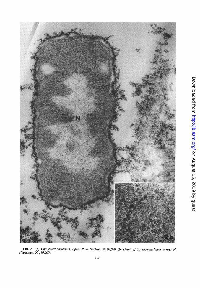
Ultrastructure of uninfected bacteria. The ultra-structure of the strain of E. coli used here andembedded in Epon was similar to that of E. coliK-12 as described by Kellenberger, Ryter, andSechaud (9). Deoxyribonucleic acid (DNA) fila-ments about 30 A in diameter formed a networkin the central nucleoid (Fig. 2a). Although ribo-somes were very densely packed in the cytoplasm,it was often possible to distinguish groups of ribo-somes which seemed to be associated in topologi-cally linear arrays (Fig. 2a, b). In some sections,the filamentous DNA of the nucleoid extended tothe periphery and was in close contact with thebacterial membrane.
-J-Jw
wOwIL.z
LL.a.
836 J. BAc-rERIOL.
on August 15, 2019 by guest
http://jb.asm.org/
Dow
nloaded from
4
; t' ,; 4~~~~~:RZ
4T
FIG. 2. (a) Uninfected bacterium. Epon. N =ribosomes. X 180,000.
Nucleus. X 80,000. (b) Detail of (a) showing linear arrays of
837
.:. IIL. .1 "I.,,. ':
.%; 4
A
I.tI
Al...:.
Ip " ''.,.
40': 7"-
.otE
on August 15, 2019 by guest
http://jb.asm.org/
Dow
nloaded from
FRANKLIN AND GRANBOULAN
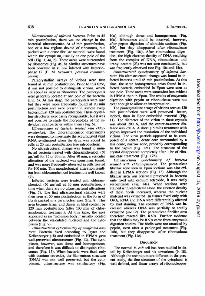
Ultrastructure of infected bacteria. Prior to 45min postinfection, there was no change in thebacterial ultrastructure. At 45 min postinfection,one or a few regions devoid of ribosomes, butpacked with a dense fibrillar material, were foundwithin the cytoplasm, usually at one pole of thecell (Fig. 3; 4a, b). These areas were surroundedby ribosomes (Fig. 4a, b). Similar structures havebeen observed in E. coli infected with bacterio-phage f2 (F. M. Schwartz, personal communi-cation).
Paracrystalline arrays of virions were firstfound at 70 min postinfection. Prior to this time,it was not possible to distinguish virions, whichare about as large as ribosomes. The paracrystalswere generally located at one pole of the bacteria(Fig. 5). At this stage, the paracrystals were rarebut they were more frequently found at 90 minpostinfection and were present in almost everybacterium at 120 min postinfection. These crystal-line structures were easily recognizable, but it wasnot possible to study the morphology of the in-dividual viral particles within them (Fig. 6).
Ultrastructure of bacteria treated with chlor-amphenicol. The chloramphenicol experimentswere designed to investigate the accumulated viralRNA synthesized when the drug was added tocells at 20 min postinfection (see introduction).No ultrastructural change was found in unin-
fected bacteria treated with chloramphenicol (50,ug/ml) for 15 or 30 min. After 80 min, a vesicularalteration of the nucleoid was sometimes found,and was more frequently observed in cells treatedfor 100 min. This morphological alteration result-ing from chloramphenicol treatment is well known(9).
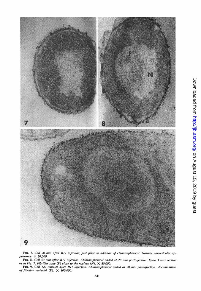
Infected bacteria were treated with chloram-phenicol (50 ,ug/ml) at 20 min postinfection, atime when there are no ultrastructural alterations(Fig. 7). The first ultrastructural changes werethen seen at 50 min postinfection in the form offibrils packed in a paranuclear area (Fig. 8). Thisarea became larger and denser in fibril content by120 min postinfection (after 100 min of chlor-amphenicol treatment). At this time, the areaappeared as an "inclusion body," usually locatedbetween the vesiculated nucleoid and the cyto-plasm (Fig. 9).
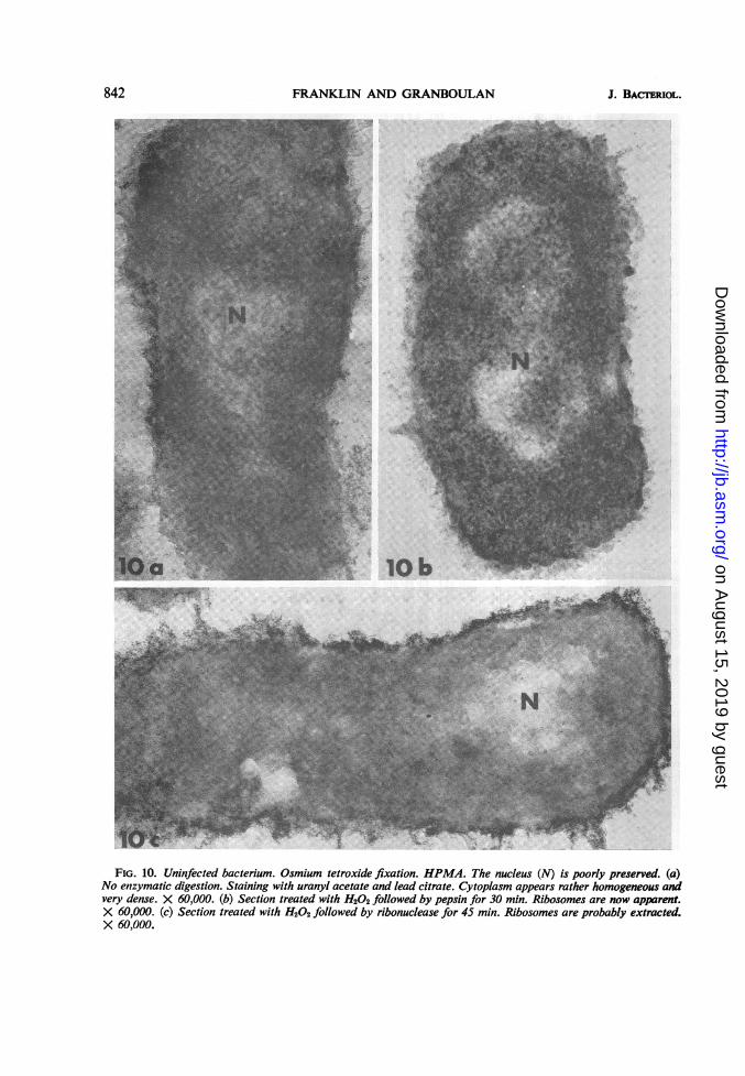
Ultrastructural cytochemistry of uninfected bac-teria. Bacteria fixed according to Ryter andKellenberger (18) and embedded in HPMA gavewell-preserved ultrastructure (Fig. 13). The cyto-plasm, however, was dense and homogeneous.and therefore it was difficult to distinguish ribo-somes (Fig. 13). When bacteria were fixed onlywith osmium tetroxide, the filamentous structure(DNA) was not well preserved, but the cyto-plasmic ultrastructure was satisfactory (Fig.
IOa), although dense and homogeneous (Fig.lOa). Ribosomes could be observed, however,after pepsin digestion of ultrathin sections (Fig.lOb), but they disappeared after ribonucleasetreatment (Fig. lOc). After ribonuclease diges-tion, the high electron density of DNA resultingfrom the complex of DNA, ribonuclease, anduranyl acetate (23) was not seen consistently, butwas frequently observed (see Fig. lOc and 15c).
Ultrastructural cytochemistry of infected bac-teria. No ultrastructural change was found in in-fected bacteria until 45 min postinfection. At thistime, the same homogeneous areas found in in-fected bacteria embedded in Epon were seen atone pole. These areas were somewhat less evidentin HPMA than in Epon. The results of enzymaticdigestion with pepsin or ribonuclease were notclear enough to allow an interpretation.The paracrystalline arrays of virions seen at 120
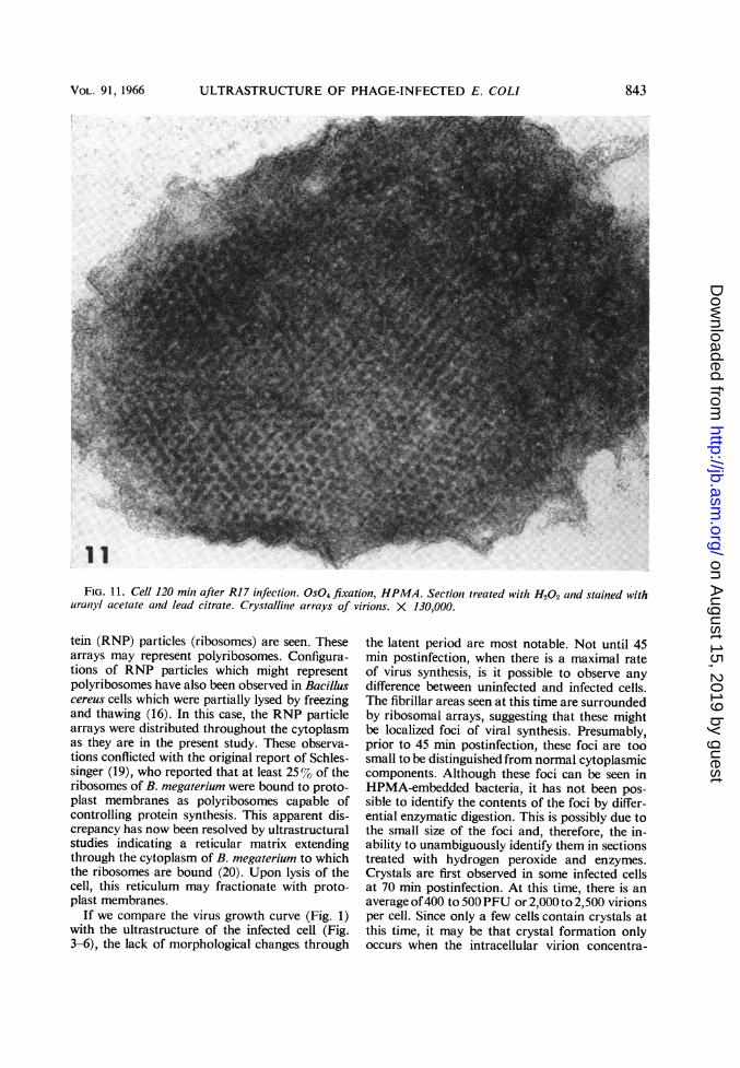
min postinfection were well preserved-better,indeed, than in Epon-embedded material (Fig.11). The diameter of the virion in these crystalswas about 200 A, and the center-to-center dis-tance was 230 A. A short (15 min) treatment withpepsin improved the resolution of the individualvirions. The virus particle appeared to be com-posed of a central dense core surrounded by aless dense, narrow zone, probably correspondingto the capsid (Fig. 12a). The structure of thecrystal disappeared completely after 1 hr of ribo-nuclease treatment (Fig. 12b).
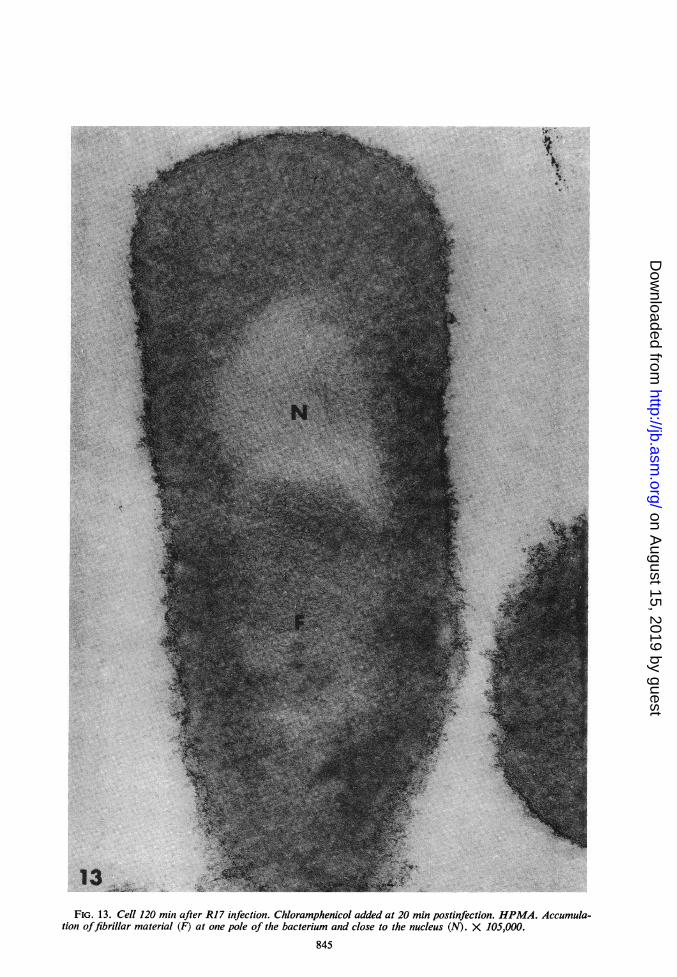
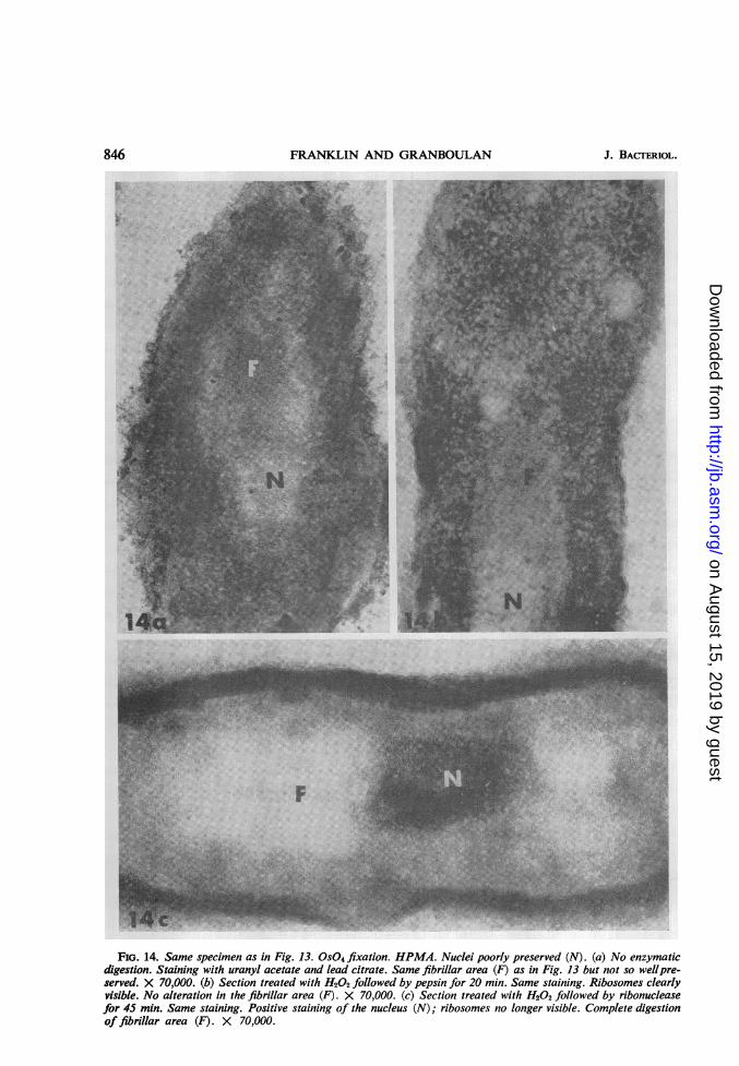
Ultrastructural cytochemistry of bacteriatreated with chloramphenicol. The paranuclearfibrillar zone seen in Epon sections was also evi-dent in HPMA sections (Fig. 13). Although thefibrillar area was lesswell preserved in bacteriaonly fixed with osmium tetroxide, it was easilyrecognizable (Fig. 14a). When sections werestained with lead citrate alone, the electron densityof these fibrils increased, whereas the nuclearmaterial was extracted. In tissues fixed only withOS04, RNA and DNA were differentially affectedby lead staining. The contrast of RNA was in-creased whereas DNA was partially or totallyextracted (see 12). The paranuclear fibrillar areatherefore reacted like RNA. Further evidencethat the fibrils may be RNA came from enzymaticdigestion studies. The fibrils were not attacked bypepsin, even after a prolonged treatment (Fig.14b), but they disappeared after ribonucleasetreatment (Fig. 14c).
DIscussIoNThe normal E. coli cell has been studied in de-
tail by Kellenberger and his coworkers (9, 18).Although the techniques are different in the pres-ent study, the fine structure of the cytoplasm iswell defined, and linear arrays of ribonucleopro-
838 J. BACTrERIOL.
on August 15, 2019 by guest
http://jb.asm.org/
Dow
nloaded from
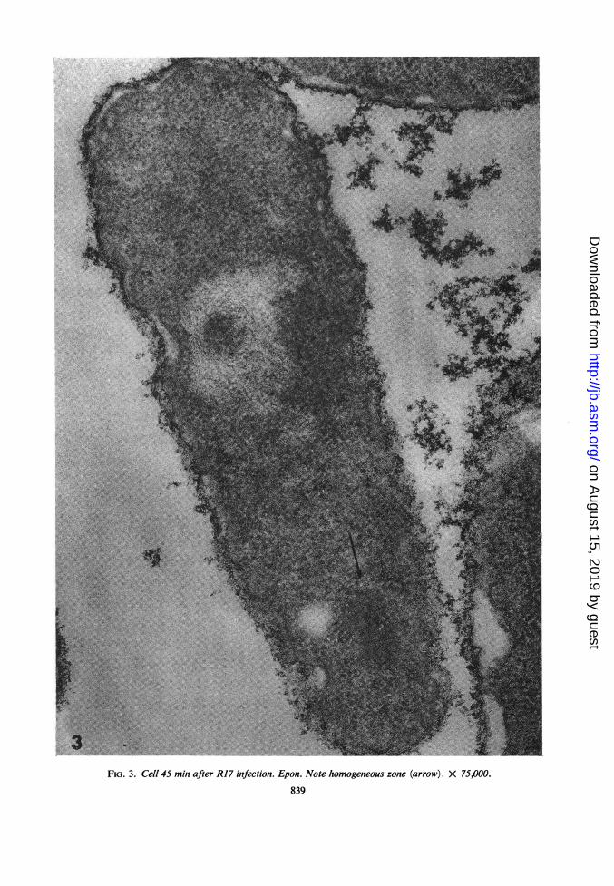
FIG. 3. Cell 45 min after R17 infection. Epon. Note homogeneous zone (arrow). X 75,000.
839
.....
on August 15, 2019 by guest
http://jb.asm.org/
Dow
nloaded from
FIG. 4. (a) High magnification of the lower portion of Fig. 3a, showing the area containing closely intertwinedfibrils but no ribosomes. Ribosomes surround the area. X 240,000. (b) Ribosomes around and penetrating into asimilar area (arrow). X 180,000.
FIG. 5. Cell 70 min after R17 infection. Epon. A paracrystal region (c) may be seen at one pole of the bac-terium, adjacent to the nucleus (N). X 75,000.
FIG. 6. Cell 90 min after R17 infection. Epon. Section of a paracrystal. The individual viral particles cannotbe distinguished. X 100,000.
840
on August 15, 2019 by guest
http://jb.asm.org/
Dow
nloaded from
7.teO ltt
- >';85
9FIG. 7. Cell 20 min after R17 infection, just prior to addition of chloramphenicol. Normal nonvesicular ap-
pearance. X 80,000.FIG. 8. Cell 50 min after R17 infection. Chloramphenicol added at 20 min postinfection. Epon. Cross section
as in Fig. 7. Fibrillar zone (F) close to the nucleus (N). X 80,000.FIG. 9. Cell 120 minutes after R17 infection. Chloramphenicol added at 20 min postinfection. Accumulation
of fibrillar material (F). X 100,000.
841
on August 15, 2019 by guest
http://jb.asm.org/
Dow
nloaded from
FRANKLIN AND GRANBOULAN
10b
FIG. 10. Uninfected bacterium. Osmium tetroxide fixation. HPMA. The nucleus (N) is poorly preserved. (a)No enzymatic digestion. Staining with uranyl acetate and lead citrate. Cytoplasm appears rather homogeneous andvery dense. X 60,000. (b) Section treated with H202 followed by pepsin for 30 min. Ribosomes are now apparent.X 60,000. (c) Section treated with H202 followed by ribonuclease for 45 min. Ribosomes are probably extracted.X 60,000.
842 J. BACrERioL.
on August 15, 2019 by guest
http://jb.asm.org/
Dow
nloaded from
ULTRASTRUCTURE OF PHAGE-INFECTED E. COLI
FIG. 11. Cell 120 min after RI 7 infection. 0S04 fixation, HPMA. Sectioni treated with H202 and stained withuran1yl acetate and lead citrate. Crystalline arrays of virions. X 130,000.
tein (RNP) particles (ribosomes) are seen. Thesearrays may represent polyribosomes. Configura-tions of RNP particles which might representpolyribosomes have also been observed in Bacilluscereus cells which were partially lysed by freezingand thawing (16). In this case, the RNP particlearrays were distributed throughout the cytoplasmas they are in the present study. These observa-tions conflicted with the original report of Schles-singer (19), who reported that at least 25% of theribosomes of B. megaterium were bound to proto-plast membranes as polyribosomes capable ofcontrolling protein synthesis. This apparent dis-crepancy has now been resolved by ultrastructuralstudies indicating a reticular matrix extendingthrough the cytoplasm of B. megaterium to whichthe ribosomes are bound (20). Upon lysis of thecell, this reticulum may fractionate with proto-plast membranes.
If we compare the virus growth curve (Fig. 1)with the ultrastructure of the infected cell (Fig.3-6), the lack of morphological changes through
the latent period are most notable. Not until 45min postinfection, when there is a maximal rateof virus synthesis, is it possible to observe anydifference between uninfected and infected cells.The fibrillar areas seen at this time are surroundedby ribosomal arrays, suggesting that these mightbe localized foci of viral synthesis. Presumably,prior to 45 min postinfection, these foci are toosmall to be distinguished from normal cytoplasmiccomponents. Although these foci can be seen inHPMA-embedded bacteria, it has not been pos-sible to identify the contents of the foci by differ-ential enzymatic digestion. This is possibly due tothe small size of the foci and, therefore, the in-ability to unambiguously identify them in sectionstreated with hydrogen peroxide and enzymes.Crystals are first observed in some infected cellsat 70 min postinfection. At this time, there is anaverage of400 to 500PFU or 2,000 to 2,500 virionsper cell. Since only a few cells contain crystals atthis time, it may be that crystal formation onlyoccurs when the intracellular virion concentra-
843VOL. 91, 1966
on August 15, 2019 by guest
http://jb.asm.org/
Dow
nloaded from
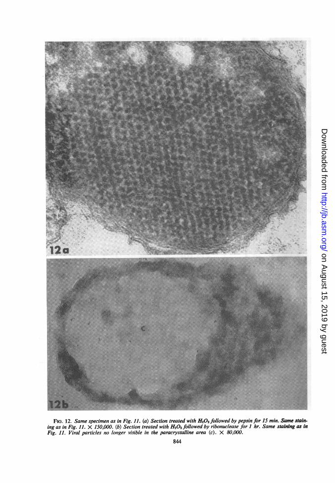
FIG. 12. Same specimen as in Fig. 11. (a) Section treated with H202 followed by pepsin for 15 min. Same stain-ing as in Fig. 11. X 150,000. (b) Section treated with H202 followed by ribonuclease for I hr. Same staining as inFig. 11. Viral particles no longer visible in the paracrystalline area (c). X 80,000.
844
4
on August 15, 2019 by guest
http://jb.asm.org/
Dow
nloaded from
ai ^ -
a.^.Si
(e.. *T
.e f* e..
Ililf
':. .;; '.Ev..
!H :0 L ..iS tS+<"' " :.. ', ..
2'' ', .,',,.X:
,'." '.':
t:.:\ :,, 74 3:
'''',, .. ..' z... r ...S;J_
... . ... S i.
:: *.. . . * ._*':... .... ... :: r.'_.. . .,* +; . l, -v ', . r
,,',"N''' '"_
ds'.. !0''|^.fS . ,..' X.:i. X
,.S.'.',,^' ,''' {.W
S ,.o
,> P
:->:s @, }:
.. .s .:'r ::;
e b :.^i .: ..
'-..':;' A:i
... .. a>j
': 1''* t.'
.:. asx
, ': ,'.l... : :';': ..,
:5._
..... *SY''. n, G.. s.. : : .*. 1.'. fl
:, .'. d N.S
.,.,. .. ws
::
13 * >,,*,,Sp$
FIG. 13. Cell 120 min after R17 infection. Chloramphenicol added at 20 min postinfection. HPMA. Accumula-tion offibrillar material (F) at one pole of the bacterium and close to the nucleus (N). X 105,000.
845
on August 15, 2019 by guest
http://jb.asm.org/
Dow
nloaded from
FRANKLIN AND GRANBOULAN J. BACTERIOL.
FIG. 14. Same specimen as in Fig. 13. OS04 fixation. HPMA. Nuclei poorly preserved (N). (a) No enzymaticdigestion. Staining with uranyl acetate and lead citrate. Same fibrillar area (F) as in Fig. 13 but not so wellpre-served. X 70,000. (b) Section treated with H202 followed by pepsin for 20 min. Same staining. Ribosomes clearlyvisible. No alteration in the fibrillar area (F). X 70,000. (c) Section treated with H202 followed by ribonucleasefor 45 min. Same staining. Positive staining of the nucleus (N); ribosomes no longer visible. Complete digestionof fibrillar area (F). X 70,000.
846
on August 15, 2019 by guest
http://jb.asm.org/
Dow
nloaded from
ULTRASTRUCTURE OF PHAGE-INFECTED E. COLI
tion is somewhat higher. Most cells contain crys-tals at 90 min when there are about 10,000 virionsper cell. It was not possible to identify virionsoutside of the paracrystalline areas, since they are
of approximately the same size and density as
RNP particles. In the study of De Petris andNava (6) on M2, another RNA phage, the numberof virions per cell was estimated at 10,000 fromcounts of virions in paracrystals in sectionedbacteria. Crystalline arrays of virions which filledover half of a bacterial section were reported bySchwartz and Zinder (21). Possibly all of theintracellular virions coalesce to form one or sev-
eral intracellular crystals if the bacteria do notlyse before the virion density reaches some criticalconcentration of 2,500 to 10,000 per cell. Sincethe volume of an E. coli cell is approximately2 X 10-12 cm3, this would be a concentration of1015 to 5 X 10¹⁵ virions per cm3. It is possible toobtain such concentrations of purified virus andto obtain crystal formation from such solutions.Thus, in vivo crystal formation may be a physicalprocess similar to in vitro crystallization.The virus in the paracrystalline regions can be
seen more clearly after "etching" with pepsin.More extensive pepsin or ribonuclease digestionremoves these crystalline areas. This suggests thatthere may be a protein and RNA matrix by whichthe crystalline areas are firmly bound in the cyto-plasm. In well-defined areas of "etched" crystalsthere is hexagonal close-packing of virions. Thepacking is not uniform throughout an entire sec-tion, however, thus emphasizing the paracrystal-linity.From the biological and biochemical studies on
the synthesis and possible accumulation of bac-teriophage RNA in the presence of chlorampheni-col (3, 5, 14), it appears likely that the paranu-
clear fibrillar area found in infected cells treatedwith chloramphenicol at 20 min postinfection is apool of viral RNA. To support this contention,we may note the absence of this morphologicalfeature in uninfected cells treated with chloram-phenicol, its digestion by ribonuclease but not bypepsin, and its resistance to prolonged extractionwith lead salts. All of these properties suggestthat the fibrillar structures in the paranucleararea represent RNA of a type found only in thebacteriophage-infected cell. Unfortunately, it wasnot possible to distinguish single- and double-stranded RNA or the replicative intermediate(7, 8) in this sectioned material.
ACKNOWLEDGMENTSWe acknowledge the advice of W. Bernhard and the
devoted technical assistance of Rita Ladwig.This investigation was supported by grant B-14646
from the National Science Foundation and Public
Health Service grant AI-05320-VR from the NationalInstitute of Allergy and Infectious Diseases.
LITERATURE CITED
1. BERNHARD, W. 1964. Inclusion de tissus en pre-sence d'eau. European Regional Conf. Elec-tron Microscopy, 3rd, vol. B, p. 9-10. Publish-ing House of the Czechoslovak Academy ofSciences, Prague.
2. BERNHARD, W., AND P. TOURNIER, 1962. Ultra-structural cytochemistry applied to the study ofvirus infection. Cold Spring Harbor Symp.Quant. Biol. 27:67-82.
3. COOPER, S., AND N. D. ZINDER. 1963. The growthof an RNA bacteriophage: the role of proteinsynthesis. Virology 20:605-612.
4. DAVIS, J. E., AND R. L. SINSHEIMER. 1963. Thereplication of bacteriophage MS2. 1. Transferof parental nucleic acid to progeny phage. J.Mol. Biol. 6:203-207.
5. DELIus, H., AND P. H. HOFSCHNEIDER. 1964. Twoeffects of inhibition of protein synthesis on thereplication of M12 bacteriophage RNA. J.Mol. Biol. 10:554-556.
6. DE PETRIS, S., AND G. NAVA. 1963. Sex specificbacteriophages of E. coli K12. II. Electron mi-croscope observations on the structure andintracellular multiplication of bacteriophage ,u2.Giom. Microbiol. 11:1-7.
7. ERIKSON, R. L., M. L. FENWICK, AND R. M.FRANKLIN. 1964. Replication of bacteriophageRNA: studies on the fate of parental RNA. J.Mol. Biol. 10:519-529.
8. FENWICK, M. L., R. L. ERIKSON, AND R. M.FRANKLIN. 1964. Replication of the RNA ofbacteriophage R17. Science 146:527-530.
8a. GRANBOULAN, N., AND R. M. FRANKLIN. 1966.High-resolution autoradiography ofEscherichiacoil cells infected with bacteriophage R17. J.Bacteriol. 91:849-857.
9. KELLENBERGER, E., A. RYTER, AND J. SECHAUD.1958. Electron microscope study of DNA-con-taining plasms. II. Vegetative and mature phageDNA as compared with normal bacterial nu-cleoids in different physiological states. J.Biophys. Biochem. Cytol. 4:671-678.
10. LEDUC, E. H., AND W. BERNHARD. 1962. Watersoluble embedding media for ultrastructuralcytochemistry. Digestion with nucleases andproteinases, p. 21-45. In R. J. C. Harris [ed.],Interpretation of Ultrastructure Symp. Intern.Soc. Cell Biol., vol. 1. Academic Press, Inc.,New York.
11. LEDUC, E. H., AND S. J. HOLT. 1965. Hydroxy-propyl methacrylate, a new water-miscible em-bedding medium for electron microscopy. J.Cell Biol. 26:137-155.
12. MARINOZZI, V. 1964. Cytochimie ultrastructuraledu nucleole-RNA et proteines intranucleo-laires. J. Ultrastruct. Res. 10:433-456.
13. MERRIAM, R. W. 1958. The contribution of loweroxides of osmium to the density of biologicalspecimens in electron microscopy. J. Biophys.Biochem. Cytol. 4:579-582.
847VOL. 91,y 1966
on August 15, 2019 by guest
http://jb.asm.org/
Dow
nloaded from
FRANKLIN AND GRANBOULAN
14. PARANHYCH, W., AND D. B. ELLIS. 1964. In vivosynthesis of phage R17 RNA in the presence ofchloramphenicol. Virology 24:635-44.
15. PARANCHYCH, W., AND A. F. GRAHAM. 1962.Isolation and properties of an RNA-containingbacteriophage. J. Cellular Comp. Physiol.60:199-208.
16. PFER, R. M., AND D. G. LUNDGREN. 1964.Electron microscopy of polyribosomes withinBacillus cereus. J. Bacteriol. 88:1119-1129.
17. REYNOLDS, E. S. 1963. The use of lead citrate athigh pH as an electron-opaque stain in electronmicroscopy. J. Cell Biol. 17:208-212.
18. RYTER, A., AND E. KELLENBERGER. 1958. eftudeau microscope electronique de plasmas c6nte-nant de l'acide desoxyribonucleique. I. Lesnucleoides des bacteries en croissance active. Z.Naturforsch. 13b:597-605.
19. SCHLESSINGER, D. 1963. Protein synthesis bypolyribosomes on protoplast membranes of B.megaterium. J. Mol. Biol. 7:569-582.
20. SCHLESSINGER, D., V. T. MARCHESE, AND B. C.K. KWAN. 1965. Binding of ribosomes to cyto-plasmic reticulum of Bacillus megaterium. J.Bacteriol. 90:456-466.
21. SCHWARTZ, F. M., AND N. D. ZIsDER. 1963.Crystalline aggregates in bacterial cells infectedwith the RNA bacteriophage f2. Virology21:276-278.
22. SINSHEIMER, R. L., B. STARMAN, C. NAGLER, ANDS. GUTHRIE. 1962. The process of infection withbacteriophage 0 X174. I. Evidence for a "iep-licative form." J. Mol. Biol. 4:142-160.
23. YOTSUYANAGI, Y. 1960. Mise en evidence au mic-roscope electronique des chromosomes de lalevure par une coloration specifique. Compt.Rend. 250:1522-1524.
848 J. BACTERIOL.
on August 15, 2019 by guest
http://jb.asm.org/
Dow
nloaded from