Embed Size (px)
Citation preview

I
Comparison between rapid and conventional
methods for diagnosis of bacterial meningitis.
M. Phil (Microbiology) Thesis
Dr. Umme Habiba MBBS
Department of Microbiology Mymensingh Medical College
Mymensingh, Bangladesh January 2012
id4988937 pdfMachine by Broadgun Software - a great PDF writer! - a great PDF creator! - http://www.pdfmachine.com http://www.broadgun.com

II
III
Declaration
I hereby declare that the whole work submitted as a thesis entitled �Comparison
between rapid and conventional methods for diagnosis of bacterial meningitis� in
the department of the microbiology, Mymensingh Medical College, Dhaka
University, for the Degree of Master of philosophy is the result of my own
investigation and was carried out under the supervision of Professor Dr. Md. Akram
Hossain.
I, further declare this thesis or part thereof has not been concurrently submitted for the
award of any Degree or Diploma anywhere.
Dr. Umme Habiba Signature of the Candidate

IV
ACKNOWLEDGEMENT
All praises belongs to Almighty Allah, the most merciful, the most beneficent and the
most kind for giving me the opportunity, courage and enough energy to carry out and
complete the entire thesis work.
I am very grateful and deeply indebted to my honourable teacher and guide Professor
Dr. Md. Akram Hossain, Head of the Department of Microbiology, Mymensingh
Medical College. It is my great pleasure to express my deepest regards and whole
hearted indebtedness to him for his inspiring encouragement, continuous guidance,
active cooperation, constant supervision, valuable suggestions, constructive criticism
and help in carrying out this work successfully.
I am grateful and express thanks to the honorable members of the Ethical Review
Committee for giving kind approval to my research protocol. I am obliged to
Professor Dr. Md. Aminul Haque Principal of Mymensingh Medical College,
Mymensingh for his kind permission to conduct the thesis.
I would like to express my deepest regards and gratitude to my respected teacher Dr.
Md. Chand Mahmud, Assistant Professor, Department of Microbiology, Mymensingh
Medical College, Mymensingh, for his constructive criticism in correcting this thesis
with his wise advice and active cooperation in correcting the manuscript.
I am cordially expressing my respect and complements to Dr. Shyamal Kumar Paul,
Assistant Professor, Department of Microbiology, Mymensingh Medical College,
Mymensingh, for his cordial cooperation, and thoughtful suggestions in my thesis
work.
V
I owe my gratitude to respected teacher DR. Salma Ahmed, Assistant Professor,
Department of Microbiology, Mymensingh Medical College, Mymensingh for
valuable advice and cordial cooperation.
I also grateful to Professor Nobumichi Kobayashi, Sapporo Medical University of
Japan for providing with us primers of PCR and technical assistance for this study and
also grateful to Professor Jalaluddin Ashraful Hoque, Professor of BIRDEM for
valuable advice and cordial cooperation.
I like to express gratitude to all other teachers, M.Phil students, laboratory
technologist and other staffs of Microbiology department, Mymensingh Medical
College, Mymensingh for their constant help and sincere cooperation during the entire
study period.
I express my gratitude to my parents Late Md. Abdul Muttalib Mian and Late Effat
Ara Begum, who always used to inspire me for higher studies and for being my
constant source of inspiration and encouragement in every step of my life.
I remained incomplete if I do not express whole hearted thanks and gratitude to my
husband Dr. Md. Sirajul Islam Bhuiyan for sparing me so much time in this job from
all sorts of social and family responsibilities and sharing my pain and pleasure. I
would like to express my heartiest affection for my younger sister Rifat Habiba and
my sons, Mustahsin and Ahnaf who had been deprived of my care and attention
during the thesis work.
I remained incomplete if I do not express wholehearted thanks and gratitude to my
beloved in laws Md. Afazuddin Bhuiyan and Ms. Rahima Begum for their blessings,

VI
affectionate support and spearing me so much time in this job from all sorts of social
and familial responsibilities.
Lastly I am indebted to all those persons from whom I have collected samples. May
Allah give them better rewards. Thanks again.
Mymensingh, July, 2012 Dr. Umme Habiba
VII
CONTENTS
Page No.
LIST OF TABLES VI
LIST OF FIGURES VII
LIST OF ABBREVIATIONS VIII
SUMMARY X
INTRODUCTION 1
CHAPTER 1: HYPOTHESIS 5
OBJECTIVES 6
CHAPTER 2: REVIEW OF LITERATURE 7
CHAPTER 3: MATERIALS AND METHODS 54
CHAPTER 4: RESULTS 68
CHAPTER 5: DISCUSSION 90
CHAPTER 6: CONCLUSION AND RECOMMENDATIONS 98
CHAPTER 7: LIMITATION 99
CHAPTER 8: LIST OF REFERENCES 100
CHAPTER 9: PHOTOGRAPH I
VIII
CHAPTER 10: APPENDICES Xii
LIST OF TABLES
Table No. Title Page No.
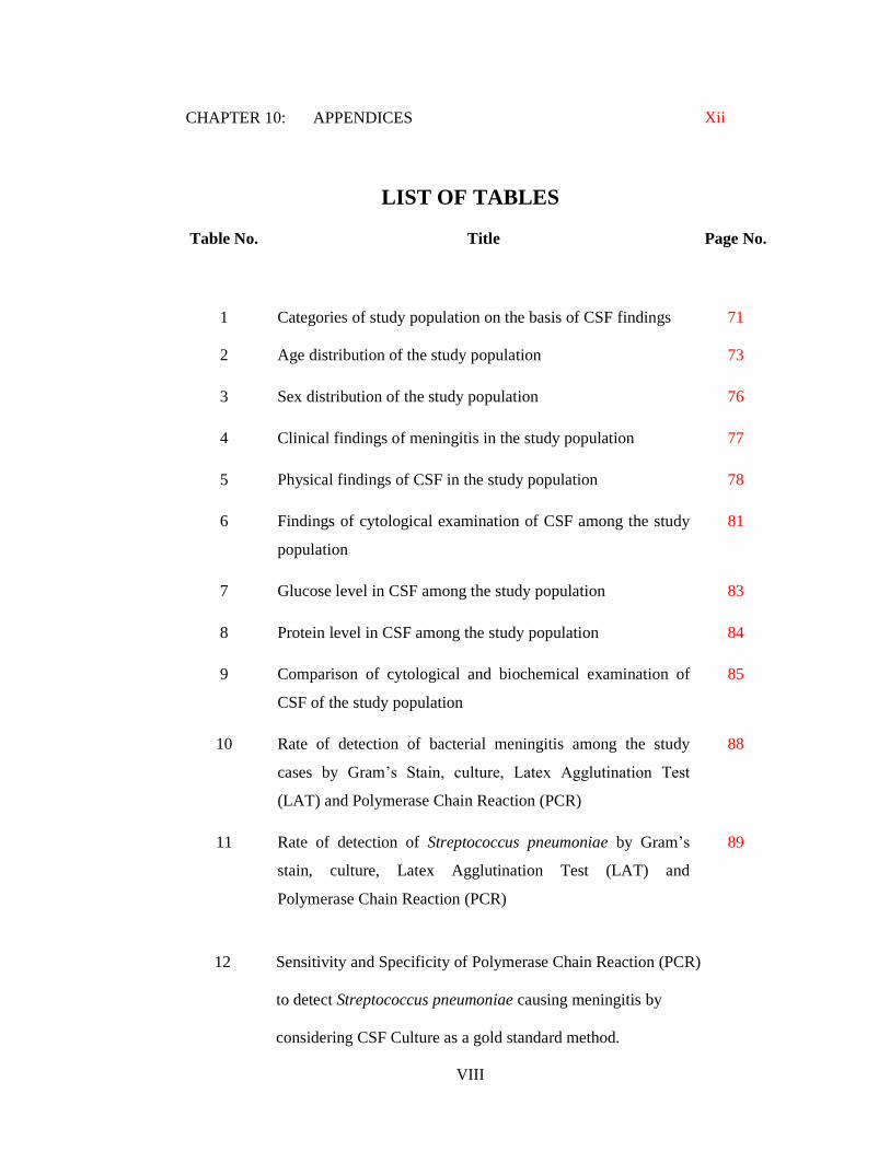
1 Categories of study population on the basis of CSF findings 71
2 Age distribution of the study population 73
3 Sex distribution of the study population 76
4 Clinical findings of meningitis in the study population 77
5 Physical findings of CSF in the study population 78
6 Findings of cytological examination of CSF among the study
population
81
7 Glucose level in CSF among the study population 83
8 Protein level in CSF among the study population 84
9 Comparison of cytological and biochemical examination of
CSF of the study population
85
10 Rate of detection of bacterial meningitis among the study
cases by Gram�s Stain, culture, Latex Agglutination Test
(LAT) and Polymerase Chain Reaction (PCR)
88
11 Rate of detection of Streptococcus pneumoniae by Gram�s
stain, culture, Latex Agglutination Test (LAT) and
Polymerase Chain Reaction (PCR)
89
12 Sensitivity and Specificity of Polymerase Chain Reaction (PCR)
to detect Streptococcus pneumoniae causing meningitis by
considering CSF Culture as a gold standard method.
IX
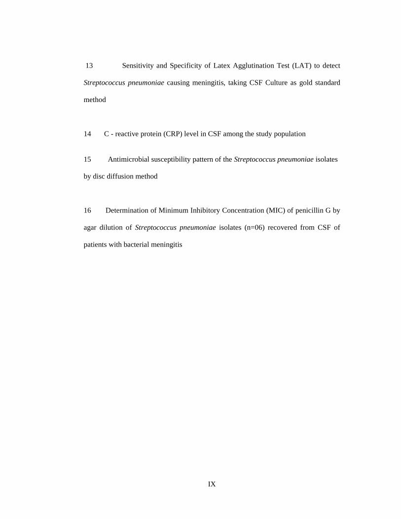
13 Sensitivity and Specificity of Latex Agglutination Test (LAT) to detect
Streptococcus pneumoniae causing meningitis, taking CSF Culture as gold standard
method
14 C - reactive protein (CRP) level in CSF among the study population
15 Antimicrobial susceptibility pattern of the Streptococcus pneumoniae isolates
by disc diffusion method
16 Determination of Minimum Inhibitory Concentration (MIC) of penicillin G by
agar dilution of Streptococcus pneumoniae isolates (n=06) recovered from CSF of
patients with bacterial meningitis

X
LIST OF FIGURES
Figure No. Title Page No.
1 Pie diagram showing the distribution of bacterial isolates
among the CSF culture positive cases
69
2 Age group distribution of study population 70
3 Sex pattern of study population. 72
4 Different test performed for diagnosis of typhoid fever. 75
5 Antimicrobial susceptibility pattern of the Salmonella
typhi isolates
79
6 Antibody detection among suspected cases by ICT
method.
82
7 Comparison of positivity of Blood culture and PCR for
diagnosis of typhoid fever.
86
XI
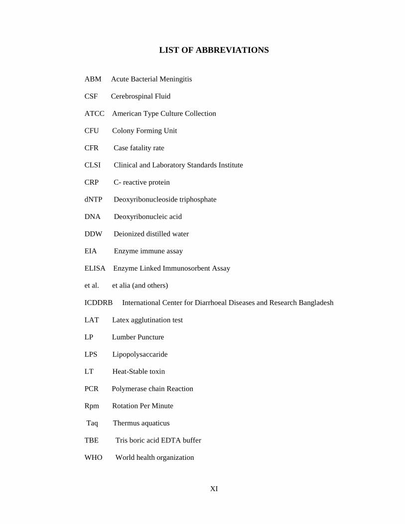
LIST OF ABBREVIATIONS
ABM Acute Bacterial Meningitis
CSF Cerebrospinal Fluid
ATCC American Type Culture Collection
CFU Colony Forming Unit
CFR Case fatality rate
CLSI Clinical and Laboratory Standards Institute
CRP C- reactive protein
dNTP Deoxyribonucleoside triphosphate
DNA Deoxyribonucleic acid
DDW Deionized distilled water
EIA Enzyme immune assay
ELISA Enzyme Linked Immunosorbent Assay
et al. et alia (and others)
ICDDRB International Center for Diarrhoeal Diseases and Research Bangladesh
LAT Latex agglutination test
LP Lumber Puncture
LPS Lipopolysaccaride
LT Heat-Stable toxin
PCR Polymerase chain Reaction
Rpm Rotation Per Minute
Taq Thermus aquaticus
TBE Tris boric acid EDTA buffer
WHO World health organization

XII
WBC White Blood Cell
XIII
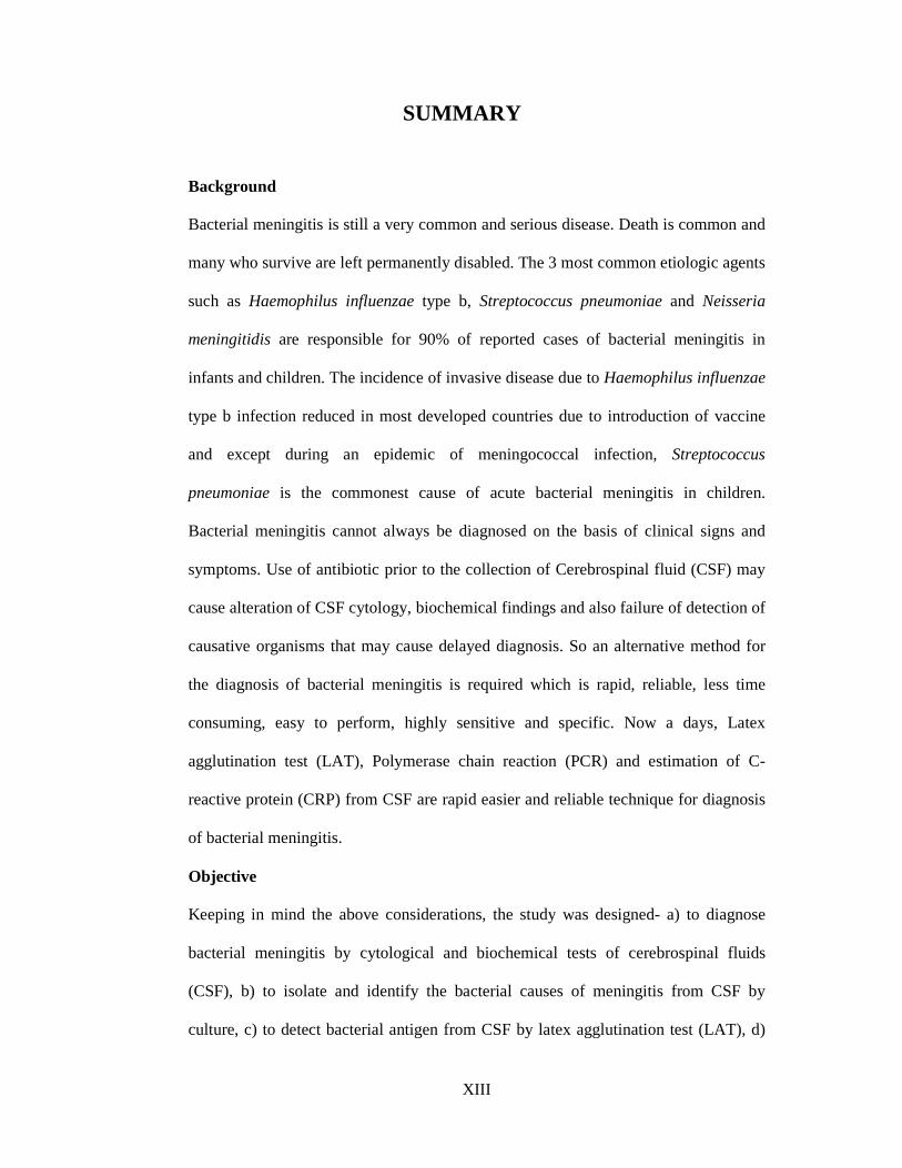
SUMMARY Background
Bacterial meningitis is still a very common and serious disease. Death is common and
many who survive are left permanently disabled. The 3 most common etiologic agents
such as Haemophilus influenzae type b, Streptococcus pneumoniae and Neisseria
meningitidis are responsible for 90% of reported cases of bacterial meningitis in
infants and children. The incidence of invasive disease due to Haemophilus influenzae
type b infection reduced in most developed countries due to introduction of vaccine
and except during an epidemic of meningococcal infection, Streptococcus
pneumoniae is the commonest cause of acute bacterial meningitis in children.
Bacterial meningitis cannot always be diagnosed on the basis of clinical signs and
symptoms. Use of antibiotic prior to the collection of Cerebrospinal fluid (CSF) may
cause alteration of CSF cytology, biochemical findings and also failure of detection of
causative organisms that may cause delayed diagnosis. So an alternative method for
the diagnosis of bacterial meningitis is required which is rapid, reliable, less time
consuming, easy to perform, highly sensitive and specific. Now a days, Latex
agglutination test (LAT), Polymerase chain reaction (PCR) and estimation of C-
reactive protein (CRP) from CSF are rapid easier and reliable technique for diagnosis
of bacterial meningitis.
Objective
Keeping in mind the above considerations, the study was designed- a) to diagnose
bacterial meningitis by cytological and biochemical tests of cerebrospinal fluids
(CSF), b) to isolate and identify the bacterial causes of meningitis from CSF by
culture, c) to detect bacterial antigen from CSF by latex agglutination test (LAT), d)
XIV
to amplify LytA gene of Streptococcus pneumoniae by Polymerase chain reaction
(PCR) from cerebrospinal fluids (CSF), e) to estimate C- reactive protein (CRP) level
from cerebrospinal fluids (CSF) for diagnosis of bacterial meningitis, f) to compare
the results of LAT, PCR and CRP in CSF as diagnostic tools with that of conventional
methods for diagnosis of bacterial meningitis, g) to determine antimicrobial
susceptibility of bacterial isolates by disc diffusion method and minimum inhibitory
concentration (MIC) of penicillin by agar dilution method.
Methodology
This cross sectional study was carried out in the Department of Microbiology,
Mymensingh Medical College, Mymensingh during the period of July 2010 to
January 2012. A total of 115 clinically suspected meningitis patients age ranging from
0 day to 18 years were included in this study. Specimens of CSF was collected from
each of the cases aseptically and were tested by cytological examination, biochemical
tests, Gram�s stain, culture, level of CRP, LAT and PCR for LytA gene and then
results were analyzed using appropriate statistical methods.
Results
On the basis of cytological and biochemical examination of CSF the study population
was categorized into three groups, bacterial meningitis 35 (30.4%) cases, viral
meningitis 68 (59.1%) cases and normal CSF 12 (10.4%) cases. Among the 35 cases
of bacterial meningitis culture were positive in 7 (20.0%) and Gram�s stains were
positive in 6 (17.1%) of cases. Streptococcus pneumoniae were detected by isolation
in 6 (17.1%), by LAT in 15 (42.8%) and by PCR in 22 (62.8%) of cases among 35
bacterial meningitis cases. All the culture and Gram�s stain positive cases for
Streptococcus pneumoniae were also positive by LAT and PCR. The level of CRP of
CSF was found normal (<0.2 mg/L) in all (100%) cases of viral meningitis and
XV
normal CSF cases and higher (>0.2 mg/L) in 33 (94.2%) cases of bacterial meningitis.
The sensitivity, specificity of PCR were (100%, 100%) and LAT were (100%, 100%)
respectively, by using CSF culture as gold standard.
Conclusion
Analyzing the findings of the present study, it is concluded that PCR and LAT both
were highly sensitive and specific but PCR was found superior to other available
methods for detection of bacterial meningitis.
Recommendation
On the basis of the result of this study, it may be recommended to take necessary
steps regarding setup and start of PCR analysis of CSF at least in the tertiary care
hospitals as referral centre.
Secondly, efforts should be made to introduce LAT from CSF at Upazilla level or at
least in district level of Bangladesh.
XVI
Comparison between rapid and conventional methods
for diagnosis of bacterial meningitis.

XVII
Chapter-1
Introduction and Objectives

XVIII
INTRODUCTION
Meningitis is the inflammation of leptomeninges (pia and arachnoidmatter) covering
the brain and spinal cord. This inflammatory process is not confined to meninges
rather spread to adjacent brain tissue (Khan 2003). Meningitis presents with the
characteristic combination of pyrexia, headache and stiffness of the neck and
irritability of the meninges with positive Kernigs�s and Brudzinski�s signs (Haslett et
al. 2002).
Meningitis may be acute, sub acute and chronic, which may have infectious or
noninfectious causes (Jacewicz 2009). Infectious causes of meningitis include
bacteria, viruses, fungi, Mycobacterium tuberculosis, Lyme disease, actinomyces,
Treponema pallidum and occasionally protozoa or other parasites (Prober 2007; Khan
and Rahman 2011; Jacewicz 2009). Noninfectious causes of meningitis may be due to
sarcoidosis, systemic lupus erythematosus with CNS involvement, tumor and
leukemia (Prober 2007).
Bacterial meningitis is still a very common and serious disease (Abro et al. 2008).
Globally 1.2 million cases of bacterial meningitis are estimated to occur every year
with 135,000 deaths (Alam et al. 2007). World Health Organization (WHO) reported
that the deaths from bacterial meningitis in Europe, America, Africa and South East
Asia was, 15,000, 18,000, 20,000 and 73,000 respectively in 2002 (WHO 2004).
Each year in USA the incidence of bacterial meningitis is 0.01% (Schueler, Beckett
and Gettings 2010). The incidence of bacterial meningitis varies from 0.022% to
0.266% in newborns, more common in developing countries (Silva et al. 2007).
Muangchana et al. (2009) reported that in Thailand the incidence of bacterial
meningitis is 0.024%.
XIX
Aetiology of bacterial meningitis varies with age and immune status. Common causes
of neonatal bacterial meningitis are Group B streptococci, Escherichia coli and
Listeria monocytogenes (Brooks, Butel and Morse 2004; Cheesbrough 2000; Jawetz,
Melnick, and Adelberg�s 2004; Llorens and McCracken 2003; Prober 2007). Group D
Streptcocci (enterococcus) and gram negative Flavobacterium meningosepticum may
also cause neonatal meningitis (Cheesbrough 2000). Neisseria meningitidis,
Streptococcus pneumoniae and Haemophilus influenzae type b are the predominant
causes of acute bacterial meningitis in the first year of life. Gram negative meningitis
e.g. Escherichia coli, Klebsiella spp. Enterobacter spp. Pseudomonas spp. and
Haemophilus influenzae type b can cause infection in immunocompromised patients
(Murray 2007; Jacewicz 2009). Ceyhan et al. (2008) mentioned in their study that
90% of reported cases of bacterial meningitis in infants and children more than 1
month of age were caused by the 3 most common etiologic agents Haemophilus
influenzae type b, Streptococcus pneumoniae and Neisseria meningitidis.
Cherian et al. (1998) had mentioned in their study that the incidence of invasive
disease due to Haemophilus influenzae type b infection reduced in most developed
countries and except during an epidemic of meningococcal infection, Streptococcus
pneumoniae is the commonest cause of acute bacterial meningitis in children. A
combined vaccine for Haemophilus influenzae type b was introduced on 15 January
2009 by the government of Bangladesh through expanded programme on
immunization (EPI) (ICDDR.B 2011).
Abro et al. (2008) from Pakistan, Taskin et al. (2004) from Turkey, Alamgir et al.
(2008) from Bangladesh, Das et al. (2003) and Mani et al. (2007) from India revealed
Streptococcus pneumoniae in 25.58%, 36.36%, 37.5%, 61.0%, 61.8% of the total
cases of bacterial meningitis respectively.
XX
The case fatality rates (CFRs) in bacterial meningitis was 26% in developed countries
even with antimicrobial therapy and availability of advanced intensive care, which
were higher ranging from 16-32% in developing countries (Afifi et al. 2007; Mani et
al. 2007). Permanent neurological sequelae such as hearing loss, mental retardation,
seizures and behavioral changes may occur in up to 50% of survivors even having
antimicrobial therapy (Welinder-Olsson et al. 2007). Gurley et al. (2009) from
Bangladesh reported that among all meningitis cases bacterial meningitis constitutes
25% and case fatality rate was 14%.
Because of the high mortality and morbidity resulting from bacterial meningitis, rapid
and accurate diagnosis is needed to increase the survival rate and decrease
complications. Therefore delay in diagnosis and initiation of proper antimicrobial
therapy can result a poor outcome. Bacterial meningitis cannot always be diagnosed
on the basis of clinical sign and symptoms. So laboratory support is essential for the
rapid diagnosis of meningitis (Mani et al. 2007).
Conventional methods for diagnosis of bacterial meningitis is based on examination
of CSF including physical, biochemical, cytological, Gram�s staining and culture (Das
et al. 2003).
Though Gram�s stain is simple, rapid and less expensive method for detecting bacteria
but it has some limitations. Its sensitivity and specificity depends on skill hands with
appropriate techniques. Gray and Fedorko (1992) mentioned that in Gram�s stains,
concentrations <103 CFU/ml of CSF are associated with positive findings in 25%
whereas concentrations of bacteria >105 CFU/ml of CSF lead to positive results in up
to 97% of cases. The �gold standard� for diagnosis of any infection including
meningitis is the isolation and identification of the causative agent (Trampuz et al.
2007). But it requires a day or more for growth and can also give false result if not
XXI
properly transported and stored as they are fastidious organisms (Das et al. 2003).
Alamgir et al. (2008) from Bangladesh mentioned that positivity of Gram�s stain and
culture may decrease to 40%-60% and <50% respectively in patients who have
received prior anti-microbial therapy. Das et al. (2003) reported that the sensitivity of
Gram�s stain and CSF culture were 18% and 0% respectively in patients who have
received antimicrobial therapy. Antibiotic therapy may cause alteration of CSF
cytology, biochemical findings and also failure of detection of causative organism, so
the diagnosis may be delayed (Alamgir et al. 2008). Moreover, in primary level
hospital culture facility is not available.
So an alternative method for the diagnosis of bacterial meningitis is required which is
rapid, reliable, less time consuming, easy to perform, sensitive and specific. Latex
agglutination test (LAT) may be an important diagnostic tool which fulfills the above
criteria. LAT is sensitive and specific for Streptococcus pneumoniae, Haemophilus
influenzae type b, group B Streptococcus, Neisseria meningitidis group A, C, Y,
W135, Neisseria meningitidis B DQG Escherichia coli k1 (Begum et al. 2007; Das et
al. 2003). LAT has been proven to be useful but are not entirely satisfactory because
of inadequate sensitivity and specificity. Reliable results are obtained only for
samples containing more than 105 CFU per ml, but approximately 45% of patients
with meningitis have less than 105 CFU per ml (Plessis, Smith and Klugman 1998).
Polymerase chain reaction (PCR) is also highly sensitive and specific technique for
diagnosis of bacterial meningitis (Saha et al. 2005; Poppert et al. 2005). Werno and
Murdoch (2008) from New Zealand found sensitivity and specificity of PCR to detect
pneumococcal meningitis 92%-100% and 100% respectively in their study. PCR now
can detect low number of pathogens in clinical specimens which does not require the
presence of viable organisms (Plessis, Smith and Klugman 1998). Detection of LytA
XXII
gene by PCR may be the alternate method to diagnose pneumococcal meningitis
(Saha et al. 2005).
The resistance of Streptococcus pneumoniae to penicillin and other antimicrobial
agents is increasing in many parts of the world (Alam et al. 2007). So the
antimicrobial susceptibility of the isolates recovered from CSF of patients with
meningitis by disk diffusion method and minimum inhibitory concentration (MIC) of
penicillin by routine agar dilution method will be done.
The concentration of C-reactive protein (CRP) in blood is increased in patients with
inflammatory diseases as acute phase proteins. Shimetani et al. (2001) revealed that
the CRP in CSF was extremely high in 23.3% of bacterial meningitis and in 10% of
viral meningitis patients. Shameem, Kumar & Neelagund (2008) found among 236
cases of untreated bacterial meningitis 129 (54%) cases were found to have elevated
CRP in CSF.
In developing country like Bangladesh it is important to search a simple, rapid as well
as reliable technique for diagnosis of bacterial meningitis that will provide appropriate
therapy to reduce the morbidity and mortality especially in children. So far it was
known that study on bacterial meningitis was very limited in Bangladesh and this was
the first study on bacterial meningitis in Mymensingh Medical College and Hospital.
Having described the above background, this study was undertaken to evaluate LAT,
PCR and estimation of CRP in CSF for rapid and reliable diagnosis of bacterial
meningitis
XXIII
OBJECTIVES
General objective:
To compare between rapid and conventional methods for diagnosis of bacterial
meningitis.
Specific objectives:
a) To diagnose bacterial meningitis by cytological and biochemical tests of
cerebrospinal fluids (CSF).
b) To isolate and identify the bacterial causes of meningitis from CSF by culture.
c) To detect bacterial antigen from CSF by latex agglutination test (LAT).
d) To amplify LytA gene of Streptococcus pneumoniae by Polymerase chain reaction
(PCR) from cerebrospinal fluids (CSF).
e) To estimate C- reactive protein (CRP) level from cerebrospinal fluids (CSF) for
diagnosis of bacterial meningitis.
f) To compare the results of LAT, PCR and CRP in CSF as diagnostic tools with that
of conventional methods for diagnosis of bacterial meningitis.
g) To determine antimicrobial susceptibility of bacterial isolates by disc diffusion
method and minimum inhibitory concentration (MIC) of penicillin by agar dilution
method.

XXIV
Chapter-2
Review of literature
XXV
Review of literature Meningitis
Definition and classification
Meningitis refers to an inflammatory process of the leptomeninges and CSF within
the subarachnoid space of the brain and Spinal cord and the ventricular system
(Begum et al.2007; Kumar, Abbas and Fausto 2004). Meningoencephalitis refers to
inflammation of the meninges and brain parenchyma (Kumar, Abbas and Fausto
2004).
Infection of the central nervous system (CNS) may be diffuse or focal. Meningitis and
encephalitis are examples of diffuse infection. Meningitis implies primary
involvement of meninges, whereas encephalitis indicates brain parenchymal
involvement. Because these anatomic boundaries are often not distinct, many patients
have evidence of both meningeal and parenchymal involvement and should be
considered to have meningoencephalitis (Probe 2007).
Meningitis may be acute bacterial meningitis, sub acute meningitis and chronic
meningitis (Jacewicz 2009). Meningitis is also classified as acute bacterial or
pyogenic meningitis, viral meningitis, Tubercular meningitis, parasitic meningitis and
fungal meningitis on the basis of infectious agent (Prober 2007; Khan and Rahman
2011; Haslett 2002). Sub acute meningitis is a meningeal inflammation that lasts >2
weeks and chronic meningitis last >1 month, may have infectious or noninfectious
causes. Infectious causes include fungi, Mycobacterium tuberculosis, Lyme disease,
AIDS, actinomyces, Treponema pallidum and viruses (Jacewicz 2009).
Epidemiology

XXVI
Bacterial meningitis is a major cause of death and disability in children worldwide.
More than one million cases with 200,000 deaths have been estimated to each year
due to bacterial meningitis (Begum et al. 2007). Acute bacterial meningitis is one of
the most severe infectious diseases, causing neurologic sequelae and accounting for
an estimated 171,000 deaths worldwide per year (Ceyhan et al. 2008). Permanent
neurological sequelae such as hearing loss, mental retardation, seizures and behavioral
changes may occur in up to 50% of survivors even having antimicrobial therapy
(Welinder-Olsson et al. 2007). Mani et al. (2007) mentioned in their study that the
mortality rate due to bacterial meningitis remains significantly high in developing
countries, ranging from 16-32%. The mortality reported by WHO in 2002 was 15,000
deaths in Europe, 18,000 deaths in Americans, and 20,000 deaths in Africa from
meningitis. The numbers of deaths are more in developing countries which are about
73,000 deaths from meningitis in South East Asia 2002 (WHO, 2004).
Globally 1.2 million cases of meningitis are estimated to occur every year with
135,000 deaths (Alam et al. 2007). In USA the incidence of meningitis is 26,990
(0.009%) among 293,655,405, in United Kingdom is 5,539 (0.009%) among
60,270,708 and in Australia it is 1,830 (0.009%) among 19,913,144 estimated
populations. In South East Asia, the incidence of meningitis in India is 97,892
(0.009%) among 1,065,070,607, in Pakistan is 14,632 (0.009%) among 159,196,336
and in Sri Lanka it is 1,829 (0.009%) among 19,905,165 estimated populations (US
Census Bureau, International Data Base 2004). Fuller et al. (2003) mentioned that
bacterial meningitis was the cause of 125,000 deaths each year in infants and young
children and 96% of these occurred in less developed countries where up to 50% of
children with this disease died and 25-50% of survivors had neurological sequelae.
XXVII
The annual attack rate in the United States is about 3 cases per 100,000 populations.
The major pathogens for the causation of meningitis (per 100,000) were Streptooccus
pneumoniae, 1.1; Neisseria meningitidis, 0.6; group B Streptococcus, 0.3; Listeria
monocytogenes, 0.2; and Haemophilus influenzae, 0.2 (Sadighian and pourmand
2009). Muangchana et al. (2009) have been found the incidence rate of bacterial
meningitis is 24.6 per 100,000 among the Thai children under five years. The
incidence of meningitis in Bangladesh is 12,990 (0.009%) among 141,340,476
estimated populations of people (US Census Bureau, International Data Base 2004).
Kornelisse et al. (1995) reported in their study, epidemiologic studies in the
Netherlands have estimated that the annual incidence of pneumococcal meningitis is
0.001% (1.5 cases per 100.000 populations) and the incidence of pneumococcal
meningitis in children younger than 5 years of age is substantially higher that is
0.007% (7 cases per 100,000 populations).
Etiology
Acute bacterial meningitis is causes by Group B streptococci during the first two
months of life (Jacewicz 2009; Prober 2007; Cheesbrough 2000). Escherichia coli,
Group B streptococci and Listeria monocytogenes are the most common cause of
neonatal meningitis. Group D Streptcocci (enterococcus) and Klebsiella may also
causes neonatal bacterial meningitis (Brooks, Butel and Morse 2004; Cheesbrough
2000; Jawetz, Melnick, and Adelberg�s 2004; Llorens and McCracken 2003; Prober
2007). Gram negative Flavobacterium meningosepticum may also cause neonatal
meningitis (Cheesbrough 2000). There after Neisseria meningitidis and Streptococcus
pneumoniae are the predominant causes of acute bacterial meningitis. Meningococcal
meningitis occurs most often in the first year of life. Gram negative meningitis e.g.
Escherichia coli, Klebsiella spp. Enterobacter spp. Pseudomonas spp. and
XXVIII
Haemophilus influenzae type B can occur in immunocompromised patients (Jacewicz
2009).
Ceyhan et al. (2008) already mentioned in their study 90% of reported cases of acute
bacterial meningitis in infants and children more than 1 month of age were caused by
the 3 most common etiologic agents are Haemophilus influenzae type B,
Streptococcus pneumoniae, and Neisseria meningitidis.
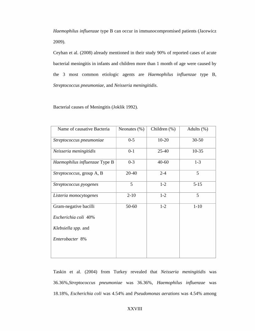
Bacterial causes of Meningitis (Joklik 1992).
Name of causative Bacteria Neonates (%) Children (%) Adults (%)
Streptococcus pneumoniae 0-5 10-20 30-50
Neisseria meningitidis 0-1 25-40 10-35
Haemophilus influenzae Type B 0-3 40-60 1-3
Streptococcus, group A, B 20-40 2-4 5
Streptococcus pyogenes 5 1-2 5-15
Listeria monocytogenes 2-10 1-2 5
Gram-negative bacilli
Escherichia coli 40%
Klebsiella spp. and
Enterobacter 8%
50-60 1-2 1-10
Taskin et al. (2004) from Turkey revealed that Neisseria meningitidis was
36.36%,Streptococcus pneumoniae was 36.36%, Haemophilus influenzae was
18.18%, Escherichia coli was 4.54% and Pseudomonas aerations was 4.54% among

XXIX
total 22 cases by bacterial culture. Currently, the organisms most commonly
responsible for community-acquired bacterial meningitis are Streptococcus
pneumoniae (22.5%), Neisseria meningitidis (56.5%), and Haemophilus influenzae
(20.5%). Haemophilus influenzae was once the most common cause of bacterial
meningitis in the United States (USA). Now a day�s Streptococcus pneumoniae is a
major cause of childhood bacterial meningitis where the incidence of Haemophilus
influenzae meningitis declined dramatically following the introduction of the
Haemophilus influenzae type B (Hib) vaccine (Ceyhan et al. 2008).
Now a day, the organisms most commonly responsible for community-acquired
bacterial meningitis are Streptococcus pneumoniae (61.8%), Haemophilus influenzae
(1.8%), Neisseria meningitidis (1%), other gram negative bacilli, Streptococcus spp.
and Staphylococcus aureus were isolated from 4.9%, 2.3% and 1.8%cases
respectively among 73.8% of total bacterial meningitis (Mani et al. 2007). In their
study, Abro et al. (2008) from Pakistan found Neisseria meningitidis were 41.86%,
Streptococcus pneumoniae were 25.58% and other bacteria were 4.47%. Brouwer,
Tunkel and Beek (2010) mentioned that Streptococcus pneumoniae is now the most
common etiological agent of bacterial meningitis in the United States and Europe,
accounting for 61% of total cases in the United States.
Begum et al. (2007) from Bangladesh revealed as bacterial meningitis 28.89% among
which 44.44% were Haemophilus influenzae, 33.33% were Streptococcus
pneumoniae, 16.67% were Neisseria meningitidis and 5.56% were Neisseria
meningitidis B/ E. coli. On another study of Bangladesh Gurley et al. (2009) reported
that 25% meningitis was caused by bacteria among which the case fatality rate was
14%. She also reported that Neisseria meningitidis any serogroup were 8%,
Streptococcus pneumoniae were 4% and Haemophilus influenzae were1%. In Dhaka
XXX
Shishu Hospital from 2001 to 2004, 346 cases of bacterial meningitis, 236 was culture
positive out of which Haemophilus influenzae were 52.96%, Streptococcus
pneumoniae were 36.86%, Neisseria meningitidis were 2.54% and other
miscellaneous bacteria were 7.62% (Saha et al. 2005).
Prior to the availability of Haemophilus influenzae type b conjugate vaccines in the
United States, it was accounted for 45 to 48% of all the cases of bacterial meningitis.
Now it accounts for only 7% of cases. Previously most cases in the United States were
infants and children under 6 years of age (peak incidence, 6 to 12 months of age),
with the majority of cases being caused by capsular type b strains. Haemophilus
influenzae type b conjugate vaccines have led to a profound reduction in the incidence
of Haemophilus influenzae type b meningitis (Brouwer, Tunkel and Beek 2010).
Schuchat et al. (1997) of USA found Haemophilus influenzae type b meningitis is
now becoming predominantly a disease of adults rather than infants & young children
because of vaccine related reduction of the disease. In this Study they also found the
incidence rate of Haemophilus influenzae type b was 0.2 per 100,000 populations and
the case fatality rates were 6%, based on cases with known outcomes. For the first
time in Bangladesh a combination vaccine for Haemophilus influenzae type b was
introduced on 15 January 2009 in expanded programme on immunization (EPI) of the
government of Bangladesh. The new pentavalent (5-in-1) combination vaccine
includes Hib, diphtheria, tetanus, pertussis and hepatitis B. This vaccine is now
included in the routine government immunization programme of Bangladesh that
immunizes about four million children each year (icddr,b 2011).
Pathogenesis and pathology
Bacterial meningitis most commonly results from bacteraemia through
haematogenous dissemination of microorganisms from a distant site of infection.

XXXI
Rarely meningitis may follow bacterial invasion from a contiguous focus of infection
such as paranasal sinusitis, otitis media, mastoiditis, orbital cellulitis, or cranial or
vertebral osteomyelitis or may occur after introduction of bacteria via penetrating
cranial injuries, dermal sinus tracts, or meningomyeloceles (Prober 2007).
Bacterial colonization of nasopharynx with microorganism is the usual source of the
bateraemia (Prober 2007). Each of the three main pathogens that are N.meningitidis,
H.influenzae and S. pneumoniae is able to colonize the nasopharynx (Hart 2009). The
risk of colonization is greatest in the period immediately after colonization (Prober
2007 and Hart 2009). But there may be prolonged carriage of the colonizing organism
without disease (Prober 2007).
After attachment to epithelial cells bacteria in the nasopharynx enter the circulation by
breaching the mucosa. N. meningitidis may be transported across the mucosal surface
within a phagocytic vacuole after ingestion by the epithelial cell. Bacterial survival in
the bloodstream is enhanced by large bacterial capsules that interfere with opsonic
phagocytosis and are associated with increased virulence. Host-related developmental
defects in bacterial opsonic phagocytosis also contribute to the bacteraemia. In young,
nonimmune hosts, the defect may be due to an absence of preformed IgM or IgG
anticapsular antibodies, whereas in immunodeficient patients, various deficiencies of
components of the complement or properdin system may interfere with effective
opsonic phagocytosis. Splenic dysfunction may also reduce opsonic phagocytosis by
the reticuloendothelial system (Prober 2007). There is evidence of association
between respiratory tract infection with viruses or mycoplasma and meningococcal
meningitis (Hart 2009).
A relatively small number of microbial pathogens have been shown to account for
most cases of meningitis in infants and children, but how those pathogens cross the
XXXII
blood�brain barrier and cause meningitis is incompletely understood. The blood-brain
barrier is a structural and functional barrier that is formed by brain microvascular
endothelial cells, which protects the brain from any microbes and toxins circulating in
the blood. However, meningitis-causing pathogens, including E. coli, group B
streptococcus, S. pneumoniae, and N. meningitidis, have been shown to cross the
blood-brain barrier as live bacteria. Meningitis-causing pathogens cross the blood�
brain barrier transcellularly, paracellularly or by means of infected phagocytes (so-
called Trojan horse mechanism). Transcellular traversal of the blood�brain barrier has
been shown for most meningitis-causing pathogens in infants and children, including
E coli, group B streptococcus, and S .pneumoniae (Kim 2010).
Recent studies have shown that microbial traversal of the blood�brain barrier happens
via microbial interactions with host receptors. For example E. coli penetration into the
brain involves its binding to and invasion of the human brain microvascular
endothelial cells (HBMEC) that constitute the blood�brain barrier. The E. coli
proteins that contribute to HBMEC binding (FimH and OmpA) do so through
interactions with their respective HBMEC receptors, CD48 and endoplasmin
(formerly gp96). Other meningitis-causing pathogens, such as group B Streptococcus
and L. monocytogenes, possess several microbial structures that allow their binding to
and invasion of HBMEC. Group B streptococcal binding to HBMEC happens via
Lmb (laminin-binding protein), FbsA (fibrinogen-binding protein), pile, and IgA (via
lipoteichoic acid anchoring). S. pneumoniae crosses the blood�brain barrier partly
through interaction between cell-wall phosphorylcholine and the platelet-activating
factor receptor (PAFR). PAFR has also been shown to interact with Hib 40 but its
contribution to Hib traversal of the blood�brain barrier is unclear. N. meningitidis
invasion of HBMEC is mediated by the outer membrane protein Opc binding to
XXXIII
fibronectin, thereby anchoring the bacteria to the integrin á5â1 receptor on the cell
surface.29 In addition, pili of N. meningitidis bind to CD46 on HBMEC, and lipo-
oligosaccharides have been shown to contribute to a high-degree of bacteraemia and
subsequent penetration into the CNS (Kim 2010).
Bacteria gain entry to the CSF through the choroid plexus of the lateral ventricles and
the meninges and then circulate to the extracerebral CSF and subarachnoid space.
Bacteria rapidly multiply because the CSF concentrations of complement and
antibody are inadequate to inhibit bacterial proliferation. Chemotactic factors then
incite a local inflammatory response characterized by polymorphonuclear cell
infiltration. The presence of bacterial cell wall lipopolysaccharide (endotoxin) of
gram-negative bacteria (H. influenzae type b and N. meningitidis), pneumococcal cell
wall components (teichoic acid) and of peptidoglycan in all of them stimulates a
marked inflammatory response, with local production of tumor necrosis factor,
interleukin 1, prostaglandin E, and other inflammatory mediators. The subsequent
inflammatory response is characterized by neutrophilic infiltration, increased vascular
permeability, alterations of the blood-brain barrier, and vascular thrombosis.
Meningitis-associated brain injury is not caused simply by viable bacteria. It occurs as
a consequence of the host reaction to the inflammatory cascade initiated by bacterial
components (Prober 2007).
The pia-arachnoid is congested and infiltrated with inflammatory cells. A thin layer of
pus forms and this may later organize to form adhesions. These may cause obstruction
to the free flow of CSF leading to hydrocephalus, or they may damage the cranial
nerves at the base of the brain. The CSF pressure rises rapidly and the protein content
increases with acellular reaction that varies in type and severity according to the
nature of the inflammation and the causative organism. An obliterative endarteritis of
XXXIV
the leptomeningeal arteries passing through the meningeal exudates may produce
secondary cerebral infarction. Pneumococcal meningitis is often associated with a
very purulent CSF and a high mortality, especially in older adults (Haslett 2004).
Cerebrospinal Fluid (CSF) findings in different causes of meningitis
Bacterial Meningitis: Meningitis is probably bacterial if the leucocytes count in CSF
is ≥100 cells/mm3 with > 50% neutrophils and or growth of the organism in culture
(Saha et al. 2005). The ratio of glucose in CSF to glucose in blood is <0.4 and the
level of CSF protein >200mg/dl (Abro et al. 2008). Alamgir et al. (2008) from
Bangladesh mentioned in their study that bacterial meningitis was diagnosed by
positive culture or Gram�s stain or WBC count of CSF (100 to 10,000x106/L) with
mostly polymorphs and with decreased glucose level (<40 mg/dl) and elevated protein
(>45 mg/dl). When the CSF contains mainly polymorphonuclear neutrophils, it is
also referred as bacterial meningitis. In bacterial meningitis the CSF is typically turbid
due to presence of large numbers of leucocytes e.g. from 100 to several thousand/mm3
most of which are polymorphs (Prober 2007).
Viral meningitis: Viral meningitis is the meningitis without the evidence of
pathogenic bacteria in the CSF of patients who showed symptoms of meningitis.
Aseptic meningitis can be assigned as viral, tuberculous, fungal, syphilitic or
protozoal (Prober 2007). In case of viral meningitis CSF presents with negative CSF
culture and Gram�s stain with cell count 06-99 / mm3 with lymphocytosis, normal
protein and glucose level (Saha et al. 2005). Alamgir et al. (2008) from Bangladesh
mentioned in their study, in case of viral meningitis WBC count of CSF (> 10 �
500x106/L) with mostly lymphocytes and with elevated protein >40 mg/dl, normal
glucose concentration with negative CSF culture and Gram�s stain. The CSF is clear
or only slightly turbid and contains moderate numbers of leucocytes rarely >1,000

XXXV
cells/ mm3, most of which are lymphocytes. Protein is elevated 50-200 mg/dl & sugar
is normal (Prober 2007).
In case of Tuberculous meningitis the CSF shows moderate rise in cell count (10 �
500 cells/ mm3) and most are lymphocytes. There is moderate rise of protein 100-
3,000 mg/dl and sugar is slightly reduced. Fungal meningitis presents with moderately
increased (5 � 500 cells/ mm3) cell count with predominantly mononuclear cells.
Protein content is increased (25 � 500 mg/dl) and sugar is decreased. Syphilitic
meningitis is the meningitis with increased cells count predominantly lymphocytes.
Protein is elevated (50 � 200 mg/dl) and sugar is normal. Protozoal meningitis
presents with the elevated cell count (1000 � 10,000 cells/ mm3) and mainly
polymorphonuclear cells. Protein is elevated (50 � 500 mg/dl) and sugar is normal or
slightly decreased (Prober 2007).
Clinical features:
The clinical features of bacterial meningitis in infants and children can be non-
specific, variable, or even absent. In infants, they may be present with fever,
hypothermia, lethargy, distress, seizures or bulging frontanelles. In older children,
clinical features may be also present with fever, headaches, photophobia, nausea,
vomiting, confusion, lethargy, or irritability. Other signs of bacterial meningitis on
physical examination include Kernig�s sign, Brudzinski�s sign, focal neurological
findings, and increased intracranial pressure. Signs of meningeal irritation are present
in 75% of children with bacterial meningitis at the time of presentation. Absence of
meningeal irritation in children with bacterial meningitis was substantially more
common in those younger than 12 months (Ceyhan et al. 2008; Kim 2010; Prober
2007).

XXXVI
Hart (2009) stated that 17% of neonates with meningitis present with a bulging
frontanelle, 33% with opisthotonos, 23% with neck stiffness and 12% with
convulsions. Focal neurological signs, such as cranial neuropathies of the ocular,
oculomotor, abducens, facial or auditory may be present in children (10-20%) with
bacterial meningitis except pneumococcal meningitis, where a focal neurological sign
is more than 30%. Headache, emesis, bulging frontanele, oculomotor or abducens
nerve palsy, stupor or coma may suggest increased intracranial pressure (ICP)
(Prober, 2007). Kiwanuka and Mwanga (2001) found that the outcome of bacterial
meningitis after antimicrobial treatment, 19.7% improved without any sequelae and
28.9% improved with sequelae, 36.8% died and absconded 14.5%.
Review of diagnostic procedure
Diagnosis of meningitis is done conventionally by Gram�s stain, culture, cytological
and biochemical test such as protein and sugar content of CSF (Das et al. 2003).
Recent advances in immuno-chemisry have provided new approaches. Latex
agglutination test (LAT), Coagglutination test (COAG) and Counter
Immunoelectrophoresis (CIEP) for rapid detection of soluble bacterial antigens.
Gram�s stain and culture are routinely used for diagnosis of meningitis.
Demonstration of microorganism by Gram�s stain is not always certain, lack of
specificity. To detect the bacteria in Gram�s stain, require ≥ 105 bacteria per ml of
CSF. Culture is time consuming, requires facilities and is likely to be negative results
patients with the prior antibiotic therapy. Other methods for detection of bacteria and
component of bacteria in CSF by Enzyme immunoassay (EIA), Gas liquid
chromatography (GLC) and Polymerase Chain Reaction (PCR) (Gray and Fedorko
1992).
Cell count
XXXVII
Normal CSF contains 0-5 lecukocytes/cu mm, mainly lymphocytes, though in
neonates cell count is up to 30/mm3 (Collee et al., 1996). In case of bacterial
meningitis, white blood cell (WBC) counts from CSF are 100-10,000 or more, usually
300�2000 cells/mm3. Conversely, in case of aseptic meningitis the WBC count from
CSF is less than 200 cells/ mm3 (Cheesbrough 2000; Prober 2008; Graham 2003).
Predominate polymorphonuclear (PMN) cells in CSF ≥50% indicate bacterial
Meningitis (Negrini et al. 2011; Saha et al. 2005).
Neutrophilic pleocytosis may be present in patients during the early stages of acute
viral meningitis, the shift to lymphocytic predominance within 12-24 hours of the
initial LP (Prober, 2007). A CSF WBC count >2000/cu mm and CSF neutrophil count
>1180/mm3 is highly significant for bacterial meningitis (Scheld 1998).
Level of Protein
Normal CSF protein level is 15-45 mg/dl. Protein level of 100-500 mg/dl is highly
significant for bacterial meningitis. In case of aseptic Meningitis protein level of CSF
may be 50-200mg/dl (Cheesbrough 200; Prober 2008). Elevated CSF protein is seen
in infections, intracranial hemorrhages, multiple sclerosis, Guillain Barri syndrome,
malignancies, some endocrine abnormalities, certain medication use and a variety of
inflammatory conditions (Dean et al. 2003)
Level of Glucose
Normal glucose level of CSF is >50mg/dl (usually 45-72mg/dl). CSF glucose is about
two thirds (75%) of the serum glucose (Prober 2008; Dean et al. 2003). In case of
bacterial meningitis the glucose level is decreased, usually <40 mg/dl. On the other
hand in case of aseptic meningitis the glucose level of CSF is usually normal
(Cheesbrough 2000; Prober 2008).
Gram�s Stain
XXXVIII
Gram�s stain examination of CSF is a simple, rapid, accurate and less expensive
method for detecting bacteria and inflammatory cells. Detecting the organism by
Gram�s stain are correlates with the concentration of bacteria in CSF. Gray and
Fedorko (1992) mentioned in their study that the Gram�s stain is generally accepted to
be most reliable at detecting ≥105 bacteria/ml of CSF. Concentrations of 103 or fewer
colony forming units per ml are associated with positive Gram�s stain about 25% of
the time, whereas CSF concentrations of bacteria of 105or above lead to positive
microscopy results in up to 97% of cases (Gray and Fedorko 1992; Leonard et al.
1984; Feigin and Pearlman 1998). The clinical utility of the Gram�s stain also
depends on the bacterial pathogen. Bacteria have been observed in 90% of cases of
meningitis caused by S. pneumoniae, 86% by H influenzae, 75% by N. meningitidis
and 50% by Gram�s negative bacilli. The CSF Gram�s stain is positive in less than
50% of patients with Listeria monocytogenes meningitis (Greenlee 1990; Gray and
Fedorko 1992). In addition, the probability of identifying the organism may decrease
patients who have received prior antimicrobial therapy to 40-60% (Gray and Fedorko
1992).
Culture
CSF bacterial cultures are positive in >80% of untreated patients of bacterial
meningitis (Ross and Tyler 2001). The CSF culture positively is reduced to <50% in
patients with previous antimicrobial therapy (Tunkel and Scheld 2001). In Brazil,
etiological agent identification by culture is between (50-60%) of the cases of
bacterial meningitis (Camargos et al. 1995). The �gold standard� for diagnosis of any
infection including meningitis is the demonstration or isolation of the causative agent.
Cerebrospinal fluid culture is the gold standard for diagnosis of bacterial meningitis
(Trampuz et al. 2007). But it requires a day or more for growth and can also give false
XXXIX
result if not properly transported, stored and if antibiotic is initiated before the
specimen is taken (Das et al. 2003).
Blood Culture
Blood cultures should be performed in all patients with suspected meningitis. Blood
Cultures may reveal the bacteria in 80-90% of cases of childhood meningitis (Prober.
2007).
Methods of detecting bacterial Antigens
Latex Agglutination Test (LAT)
In the mid 70s, an immunochemical technique was developed to identify the
etiological agents in purulent meningitis, such as latex agglutination test. LAT assays
for antigen detection utilize latex polystyrene beads with immunoglobulin molecules
non specifically adsorbed into their surface. In the presence of homologous antigen,
grossly visible agglutination of the antibody coated latex beads occurs (Gray and
Fedroko 1992). LAT is simple and quick methods, which have high sensitivity and
specificity, were 93.0% and 100% respectively. It is simple in execution &
interpretation of results, can be performed by non-specialized laboratory technicians.
It also needs short period of time to get the results available to the attending
physicians (Camargos et al. 1995). Moreover, administration of antibiotics for short
interval before the test will not alter the results because this test does not depends on
viable organisms (Plessis, Smith and Klugman 1998). It is more sensitive than CIEP
(Leinonen and Kayhty 1978).
An alternative method for the diagnosis of bacterial meningitis is required which is
reliable, less time consuming, easy to perform, sensitive and specific. Latex
agglutination test (LAT) may be an important diagnostic tool which fulfills the above
criteria. LAT is sensitive and specific for Streptococcus pneumoniae, Haemophilus
influenzae type B, group B Streptococcus, Neisseria meningitidis group A, C, Y,
XL
W135 and Neisseria meningitidis B � Escherichia coli k1 (Das et al. 2003; Tunkel
and Scheld 2001). LAT has been proven to be useful but are not entirely satisfactory
because of inadequate sensitivity. Reliable results are obtained only for samples
containing more than 105 CFU per ml. But approximately 45% of patients with
meningitis have less than 105 CFU per ml (Plessis, Smith and Klugman 1998).
Quellung Procedure:
This procedure can be used to confirm the presence of organisms with morphology
typical of Streptococcus pneumoniae, N. meningitidis, or H. influenzae, type b. In the
Quellung procedure antisera specific for the capsular polysaccharides of each of these
three bacteria are mixed with separate portions of clinical specimens. A drop of CSF,
a loopful of specific antisera and saturated methylene blue can be mixed on a
microscope slide, covered and examined under oil objective. The formation of antigen
antibody complexes on the surfaces of these bacteria induces changes in their capsules
and the capsule appears to be clear & swollen when viewed microscopically (Gray
and Fedorko et al. 1992; Joklik et al. 1992).
Co agglutination Test (COAG)
COAG reagents are composed of suspensions of Staphylococcus aureus that contain
the cell surface component protein A, a 12,000- to 43,000 molecular-weight protein
that is covalently linked to the peptidoglycan of the bacterium. Immunoglobulin G
molecules adhere to protein A by the Fc end of the immunoglobulin G molecule; the
immunoreactive Fab end remains free to react with specific antigen. In the presence of
specific antigen, grossly visible agglutination of the staphylococci takes place (Gray
and Fedorko 1992). COAG test is simple, specific, low cost, rapid and sensitive
method for detecting soluble antigens in body fluids of various infections (Gray and
Fedorko 1992; Suksanong and Dajani 1977). No false-positive reactions were noted
XLI
by either test in body fluids from controls. The coagglutination test is more sensitive
than the countercurrent immunoelectrophoresis method and could be a valuable tool
for detecting antigens in body fluids of patients with various infections (Suksanong
and Dajani 1977).
Countercurrentimmunoelectrophoresis (CIEP)
CIEP has been shown to be a rapid technique for diagnosis (with in 1 hour) of
bacterial meningitis caused by H. influenzae type b; S. pneumoniae; N. meningitidis
group A. C. W 135 and D; and group B Streptococcus. It also is possible to detect
antigens from Kl strains of Escherichia coli, L-monocytogenes; Klebsiella
pneumoniae, and Ps. aeruginosa. The methodology employed is sensitive and can
detect non-viable bacteria, thus permitting the detection of bacterial antigen, even in
patients who have been pre-treated with appropriate antibiotic. The sensitivity and
specificity of CIEP is high (Gray and Fedorko 1992).
Limulus amoebocyte Lysate (endotoxin) (LAL) assay:
LAL assay is a very sensitive and specific assay for the detection of endotoxin in
CSF. A correctly performed LAL assay can detect approximately 103 gram-negative
bacteria per ml of specimen. LAL assays have a sensitivity of 93% and a specificity of
99.4%. However this test does not distinguish between specific gram negative
organisms and a negative test does not rule out the diagnosis of gram- positive
meningitis (Gray and Fedorko 1992).
Gas liquid chromatography (GLC):
This technique facilitates the separation, quantitation and identification of several
constituents of physiological fluids. Amines, alcohols, carbohydrates, and short-chain
fatty acids are examples of microbial metabolites that are produced in body tissues
and fluids and that can be detected by GLC (Gray and Fedorko 1992). Brice et al.
XLII
used GLC techniques to establish chromatography patterns for the following five
common bacterial agents of meningitis S. pneumoniae. H. influenzae, N. meningtidis,
S. aureus and Escherichia coli. Lipid, carbohydrate, and lipopolysaccharide
components served as characteristic markers for identification of these organism
GLC, might be a useful assay for rapid diagnosis of bacterial meningitis (Brice et al.
1979; Gray and Fedorko 1992). GLC has not been widely used for the diagnosis of
bacterial meningitis because, the technique requires equipment that is expensive and
the methodology is technically demanding than the antigen detection assays (Gray
and Fedorko 1992).
C � Reactive Protein (CRP):
CRP is a reactive protein of the acute inflammatory process or infectious process.
Macrophages located at the inflammatory site, such as the meninges will release
cytokines that are Interleukin (IL) -1, IL-6 and Tumor necrotizing factor (TNF). IL-6
is the major factor that stimulates hepatocytes to produce CRP (Sirijaichingkul et al.
2005). CRP rises rapidly in the first 24 � 48 hours of occurrence of bacterial
meningitis (Prasad, Nair and Kalghalgi 2005). The CRP increases within 6 to 18 hours
after the first clinical symptoms are noted and can reach 1000 times its normal values,
achieving a maximum at 2 to 5 days after onset of a significant infection and
decreasing quickly after successful therapy because its short half-life of 19 hours . So,
detection of CRP is the reliable, cost-effective, rapid screening tests which can be
performed in any standard pathology laboratory to help in the early differential
diagnosis and management of meningitis (Belagavi and Shalini 2011). CRP
synthesized by the liver in response to several diseases, including trauma, infections,
neoplasms and collagen-vascular diseases. Some reports suggest that CRP may be
XLIII
synthesized in the CNS, however intrathecal synthesis appears to be minimal and the
majority of CSF CRP is indeed derived from serum (Watson and Scott 1995). The
presence of CRP in CSF depend on the permeability of the blood-brain barrier as well
the availability of CRP in serum. So concentration of CRP in serum is much higher
than in CSF (Bowers 1985; Gershom, Briggeman-Mol and Zegher 1986). Bowers
(1985) found that the levels of CRP in CSF were <0.2 mg/L incase of 49.0% of
pneumococcal meningitis and 0.2 mg/L or more in 51.0% of cases of pneumococcal
meningitis. Philip (2003) mentioned that the CRP level above 0.4 mg/L as substantial
for bacterial meningitis. Coric et al. (2012) also reported in their study that the
minimum concentration for CRP in CSF were 0.40 mg/L in patients with bacterial
meningitis and incase of uninfected cases CRP in CSF were between 0.10 mg/L �
0.55 mg/L. Shameem, Kumar and Neelagund (2008) were found in their study, in
cases of pretreated bacterial meningitis 29.0% were with elevated level of CRP in
CSF and in untreated cases 54.66% were found with elevated level of CRP in CSF.
Polymerase chain reaction (PCR):
This technique is a primer mediated temperature dependent technique for the
enzymatic amplification of a specific DNA sequence. The technique is self contained
and easily automated because all reactions take place in a single vessel (Gray and
Fedorko 1992). Adequate treatment requires rapid detection and identification of the
bacteria, because permanent neurological sequelae may occur in up to 50% of
survivors (Welinder-Olsson et al. 2007).
PCR has been used to amplify of bacterial DNA from patients for rapid detection of
bacterial meningitis. After the initiation of antimicrobial therapy reduces the isolation
from culture decreases from 50% to <5%, but it probably does not affect PCR results
(Saravolatz et al. 2003). PCR now can detect low number of pathogens in clinical

XLIV
specimens which does not require the presence of viable organisms (Plessis, Smith
and Klugman 1998).
LytA Gene:
The lytA-encoded major autolysin (N-acetylmuramoyl-L-alanine amidase) of
Streptococcus pneumoniae is a member of a widely distributed group of cell wall-
degrading enzymes located in the cell envelope and postulated to play roles in a
variety of physiological functions associated with cell wall growth, wall turnover and
cell separation in microorganisms. The pneumococcal autolysin has a modular
organization. The catalytic function is located in the N-terminal domain and the C-
terminal domain, composed of six repeat units and a short tail which acts as a binding
arm attaching the enzyme to the choline residues of pneumococcal cell walls
(Whatmore and Dowson 1999). Detection of LytA gene by PCR is the alternate
method to diagnose pneumococcal meningitis (Saha et al. 2005). This is also more
sensitive and rapid method for diagnosis of pneumococcal meningitis by PCR now a
day because it can detect low number of pathogens in clinical specimens and does not
require the presence of viable organisms (Plessis, Smith and Klugman 1998).
Autolysin may play a direct role in virulence by mediating the release of cell wall
components shown to be highly inflammatory in animal models. In addition, it has
been suggested that autolysin plays an indirect role in pathogenesis by mediating cell
lysis and the subsequent release of virulence factors such as pneumolysin, not actively
exported from the cell. In support of a role for lytA in virulence, isogenic lytA mutants
have been found to be significantly less virulent than the parent strain in some animal
models and when inoculated into the mouse lung in a model of pneumonia, lytA
XLV
mutants are cleared rapidly and do not invade the bloodstream (Whatmore and
Dowson 1999).
Treatment of bacterial meningitis
Bacterial meningitis is a life threatening illness that is prevalent worldwide. Prior to
the introduction of antibiotics in the 1940s, case fatality rates for epidemic and
endemic bacterial meningitis exceeded 70%. Since then, antibiotic use has reduced
case fatality rates of bacterial meningitis to 25% or less (Khan et al. 2011).
On the basis of causative agents and according to the age group the choice of
antimicrobial agents for treatment of bacterial meningitis is determined. For the
causative agents (S. agalactiae, Esch. coli and L. monocytogenes) of neonatal
meningitis the drugs of choice are ampicillin, gentamicin, cefotaxime or
aminoglycoside. Incase of infants and children the common organisms are S.
pneumoniae, H. influenzae and N. meningitidis and the choice of drugs are penicillin
G, ampicillin, cephalosporin or vancomycin. For the treatment of
immunocompromised patients the choice of antimicrobial agents is ampicillin,
cephalosporin or vancomycin (Brouwer, Tunkel and Beek 2010).
Minimum inhibitory concentration (MIC)
The progressive emergence and rapid dissemination of antibiotic resistance in the
pneumococcus require a rapid and accurate identification of Streptococcus
pneumoniae to provide the appropriate antimicrobial therapy (Llull, Lopez and Garcia
2006). The resistance of Streptococcus pneumoniae to penicillin and other
antimicrobial agents is increasing in many parts of the world (Alam et al. 2007). So
the antimicrobials susceptibility of the isolates recovered from CSF of patients with
XLVI
meningitis by disk diffusion method and minimum inhibitory concentration (MIC) of
penicillin by routine agar dilution method should detect. The ranges of susceptibility
of penicillin G for Streptococcus pneumoniae are sensitive to ≤0.06 µgm/ml,
intermediate between the ranges from 0.10 to 1.0 µgm/ml and resistant to ≥2.0
µgm/ml (Kelly, Jacobs and Appelbaum 1999; Alam et al. 2007). The test was
performed on Mueller-Hinton agar supplemented with 5% defibrinated sheep blood.
Inoculum was prepared by direct suspension of pneumococcal colonies grown
overnight on sheep blood agar and matched to a 0.5 McFarland opacity standard tube
(Alam et al. 2007). The plates were incubated under 5% to 10% CO2 at 350 C for 24
hours (Kelly, Jacobs and Appelbaum 1999).
Bacteria causing meningitis
Streptococcus pneumoniae
History
Streptococcus pneumoniae has a long and fascinating history. It was isolated from
human saliva in 1881 in independent studies by Sterberg and Pasteur
(Alonsodevelasco et al. 1995; Joklik et al. 1992). The following year Friedlander
demonstrated its association with acute lobar pneumonia and within the next 10 years
the range of pneumococcal infection was elucidated with remarkable speed (Joklik et
al. 1992). In 1928, Griffith observed that when heat-killed encapsulated pneumococci
and live strains constitutively lacking any capsule were concomitantly injected into
mice, the non-capsulated could be converted into encapsulated pneumococci with the
same capsular type as the heat killed strain. Years later, the nature of this
XLVII
��transforming principle,�� or carrier of genetic information, was shown to be DNA
(Alonsodevelasco et al. 1995).
Taxonomy
Species Streptococcus pneumoniae belongs to the family Streptococcaceac and genus
Streptococcus (Joklik et al. 1992).
Morphology
Gram positive, lancet shaped, encapsulated diplococcic (0.5 to 1.25) µm in diameter.
They can occur in pairs, singly or in short chain (Alonsodevelasco et al. 1995; Joklik
et al. 1992; Brooks et al. 2004; Werno and Murdoch 2008). Capsule may be
demonstrated by "Quellung reaction" (Llull, Lopez and Garcia 2006).
Cultural Characters
On blood agar media, young culture of encapsulated pneumococci produce
circular, small, glistening, smooth dome shaped colonies 1 mm in diameter. When
pneumococcal colonies become older on blood agar media, it produces
draughtsman colonies (collapse of the center of the colony) due to autolytic changes.
Colonies incubated aerobically are surrounded by a zone of á-haemolysis, similar to
greenish discoloration as observed with viridans streptococci. Optimum PH 7.4 to 7.8
and required an increased CO2 concentration (5-10%). Under aerobic condition, a
significant amount of H2O2 is formed along with acetic and formic acids.
Streptococcus pneumoniae does not produce catalase or peroxidase, the accumulation
of H2O2 kills organism, unless catalase is provided by the addition of red blood cells
to the culture media (Joklik et al. 1992; Werno and Murdoch 2008).
Biochemical reaction and Serodiagnosis:
Streptococcus pneumoniae is catalase negative (Cheesbrough, 2000; Joklik et al.
1992). Classically, differentiation of Streptococcus pneumoniae from other alpha-
XLVIII
hemolytic that is viridans group streptococci depends on the optochin (Opt)
susceptibility test, bile or deoxycholate (Doc) solubility, and immunological reaction
with type-specific antisera (the capsular reaction or the Quellung test) (Llull, Lopez
and Garcia 2006; Werno and Murdoch 2008).
Antigenic structure
1. Capsular antigens: Streptococcus pneumoniae expresses at least 91 different
polysaccharide (PS) capsules (Yu et al. 2008). These polysaccharides are antigenic
and form the basis for the separation of Streptococcus pneumoniae into different
serotypes by capsular "swelling reaction". Although these Streptococcus pneumoniae
capsular serotypes are identified, serotypes 8, 4, 3, 14, 7, 12, 9, 1, 18, 19, 6 and 23 are
the 12 most frequent isolates (Joklik et al. 1992). Serotypes affecting infants and
children (60%) are mostly 6A, 14, 19F and 23F and in adults, serotypes 3, 19F and 6A
accounted for only 31% of the isolations (Alonsodevelasco et al. 1995). The
polysaccharide capsule is essential for the pathogenicity of the Streptococcus
pneumoniae and stimulates the production of antibodies that are protective against
subsequent infection with Streptococcus pneumoniae of the homologous types (Joklik
et al. 1992).
2. Somatic antigen: (a) M protein: M protein is the type specific protein antigens
analogous to the M protein of Streptococcus pyogenes but inmmunologically distinct
are present in Streptococcus pneumoniae. Antibodies to pneumococcal M protein are
not protective (Joklik et al. 1992).
(b) C polysaccharide: The species specific carbohydrate is a major structural
component of the cell wall of all Streptococcus pneumoniae is uniformly distributed
on both the inside and the outside of the wall. It is a teichoic acid polymer containing
phosphocholine as a major antigenic determinant. Phosphocholine of C

XLIX
polysaccharide is responsible for the interaction with C-reactive protein (CRP), which
is elevated in patients with acute inflammatory diseases. The binding of CRP to
polysaccharide C can activate complement and mediate phagocytosis (Joklik et al.
1992). C polysaccharide is responsible for inflammatory effects that�s are activation of
the alternative complement pathway, resulting in anaphylatoxin production,
enhancement of vascular permeability, mast cell degranulation, PMN activation, IL-1
production increased, cytopathic for endothelium, mediator of attachment to
endothelial cells (Alonsodevelasco et al. 1995).
(c) F antigen: Another major antigenic component of the Streptococcus pneumoniae is
the F or Forssman antigen, a determinant that cross-reacts with Forssmann series of
mammalian cell surface antigens (Joklik et al. 1992).
Determinants of pathogenicity
a) Polysaccharide capsule: Protection against phagocytosis is provided by the
polysaccharide capsule, which exerts an antiphagocytic effect (Alonsodevelasco et al.
1995; Joklik et al. 1992). Polysaccharide capsule of Streptococcus pneumoniae is
responsible for lack of activation of alternative complement pathway, deposition of
opsonically inactive complement components and no or low immunogenicity of some
serotypes (Alonsodevelasco et al. 1995).
b) Adherence: Attachment to the mucosal surface is an initial event in colonization
and infection. Streptococcus pneumoniae attaches by interacting with the N-
acetylglucosamine-galactose moiety of cell surface glycolipids. The capsular
polysaccharide appears not to determine the adhesive capacity (Joklik et al. 1992).
Enzymes
a) Neuraminidase: It may be used as exposure of receptors for Streptococcus
pneumoniae (Alonsodevelasco et al. 1995). A number of organisms that colonize the

L
respiratory tract produce the glycosidic enzyme neuraminidase. This enzyme is only
one of the factors contributing to the invasiveness of the organism (Joklik et al. 1992).
b) Proteases and IgA protease: Streptococcus pneumoniae produce immunoglobulin-
degrading extracellular proteases. Proteases that degrade secretory IgA (S-IgA), IgA,
IgG and IgM have been found in a large number of isolates from ill patients as well as
in symptomless carriers. These enzymes have an important role facilitating bacterial
colonization on mucosal surfaces (Joklik et al. 1992). IgA1 protease is responsible for
counteracts mucosal defense mechanisms (Alonsodevelasco et al. 1995).
Toxins
a) Haemolysin (Pneumolysin O): Streptococcus pneumoniae produce a Haemolysin or
pneumolysin O, with the properties similar to those of oxygen-labile Haemolysin
streptolysin O of Streptococcus pyogenes (Joklik et al. 1992).
b) Purpura producing substance: This substance produces purpura and dermal
hemorrhage in experimental animals (Joklik et al. 1992).
c) Autolysin: Autolysin of Streptococcus pneumoniae cell wall plays a role in cell
division and in causing lysis of Streptococcus pneumoniae in the presence of surface-
active agents and antimicrobial agents that inhibit cell wall synthesis. It also
contributes to the organism�s virulence (Joklik et al. 1992). Autolysin is responsible
for release of pneumolysin and cell wall products (Alonsodevelasco et al. 1995).
Epidemiology and clinical manifestations
Kornelisse et al. (1995) reported in their study, epidemiologic studies in the
Netherlands have estimated that the annual incidence of pneumococcal meningitis is
`1.5 cases per 100,000 populations and the incidence of pneumococcal meningitis in
children younger than 5 years of age is substantially higher that is 7 cases per 100,000
populations. Streptococcus pneumoniae is the most common cause of bacterial
LI
meningitis community-acquired pneumonia and bacteraemia in children and adults
and the most common cause of acute otitis media in children (Cobo, Cabezas-
Fernández and Cabeza-Barrera 2012; Kornelisse et al 1995: O�Brien et al. 2009).
Brouwer, Tunkel and Beek (2010) mentioned that Streptococcus pneumoniae is now
the most common etiological agent of bacterial meningitis in the United States and
Europe, accounting for 61% of total cases in the United States.
Although treatment regimens for Streptococcus pneumoniae meningitis are still
improving such as adjunctive therapy with corticosteroids, the mortality rate has not
changed over half a century and remains as high as 25% with neurological sequelae in
up to half of survivors. Pneumococcal meningitis is secondary to a primary infection
focus (e.g. ear focus in 30%, lung focus in 25%, sinusitic focus in 10%), and
bacteraemia is present in up to 3/4 of all cases. Consequently, the causes of death
from pneumococcal meningitis may be multifactoral and due to both neurological
complications such as brain herniation, seizures and systemic complications such as
septic shock, multiorgan dysfunction (Østergaard, Konradsen and Samuelsson 2005).
Immune response to pneumococcal antigens:
Clearance of Streptococcus pneumoniae in the absence of specific Ab may be
facilitated by CRP-mediated complement activation. However, the anti-pneumococcal
effects of CRP are presumably not mediated by the ability to induce phagocytosis,
except for serotypes that possess phosphorylcholine in their capsular polysaccharide.
Moreover, it has been suggested that pneumolysin may counteract the protective
effects of CRP. In the presence of anti-capsular Ab, Streptococcus pneumoniae are
rapidly cleared from the blood, mainly by the liver and to a lesser extent by the
spleen; however, complement is necessary to achieve effective clearance. Whether
activated by the classical or the alternative pathway by CRP, Ab, or the

LII
polysaccharide itself, complement deposition on the capsule (but not on the cell wall)
is essential for pneumococcal phagocytosis and clearance. Detectable levels of IgM
against PC are observed in infants after infection with Streptococcus pneumoniae,
frequently even in the first year of life. Carriage also sometimes results in the
development of anti-PC IgM in infants, although the induction of (cross-reactive)
anti- phosphorylcholine antibodies by other organisms cannot be excluded. The
amount of anti- phosphorylcholine Ab is age dependent: Ab are present in nearly all
older children and adults, but their levels decrease after the age of 50 to 60 years. It is
likely that infants can elicit anti-capsular polysaccharide Ab of the IgG class
following pneumococcal infection, although they rarely do so in the case of
asymptomatic carriage with poorly immunogenic pediatric serotypes 23F and 19F.
Anti-capsular IgG responses have been observed in infants under 2 years of age
following Haemophilus influenzae type b meningitis (Alonsodevelasco et al. 1995).
Antimicrobial susceptibility
Now a day progressive emergence and rapid dissemination of antibiotic resistance in
the Streptococcus pneumoniae require a rapid and accurate identification of
Streptococcus pneumoniae to provide the appropriate antimicrobial therapy
(Alonsodevelasco et al 1995; Llull, Lopez and Garcia 2006). Depending on the
clinical situation treatment options include extended-spectrum cephalosporins,
macrolides, fluoroquinolones and vancomycin. Penicillin is considered a preferred
antimicrobial agent only for Streptococcus pneumoniae but now a days penicillin
become resistance to Streptococcus pneumoniae and caused by altered penicillin-
binding proteins (Murray et al. 2007). Alam et al. (2007) from Bangladesh found 90%
of Streptococcus pneumoniae was sensitive to penicillin, 95% was sensitive to
erythromycin, 80% was resistant to gentamicin and 60% was resistant to

LIII
cotrimoxazole. Streptococcus pneumoniae was susceptible to macrolides till last
1980s in USA but now 25% of strains of these organisms are resistant and caused by
ErmB-mediated methylation of 23S rRNA (causing a high level resistance phenotype)
or MefA-mediated efflux of macrolides (resulting in a low level resistance
phenotype). In USA two-third of macrolide resistance is mediated by MefA and in
Europe by ermB gene. The prevalence of fluoroquinolone resistance is below than 1%
and is due to mutations in DNA topoisomerase IV or subunit of DNA gyrase (Murray
et al.2007).
Prevention
Because of the emergence of antibiotic-resistant Streptococcus pneumoniae, renewed
efforts were undertaken in the 1970s to develop better pneumococcal polysaccharide
vaccines. In 1978, a 14-valent vaccine was licensed in the United States and in 1983 a
23- valent vaccine became available and latter included new serotypes based on the
most current knowledge of serotype distribution and cross-reactivities between
various serotypes (Alonsodevelasco et al 1995).
Since introduction of the 7-valent pneumococcal conjugate vaccine (PCV7), the
estimated annual incidence of invasive disease (including bacteraemia) in the USA
has decreased (Cobo, Cabezas-Fernández and Cabeza-Barrera 2012). Attempts at the
beginning of this century to induce protective immunity against Streptococcus
pneumoniae in humans by vaccination with whole killed pneumococci were not very
successful because of the adverse side effects caused by the large amounts of inocula
used. Pneumococcal polysaccharide elicits long-lasting Ab and protection in healthy
adults. However, revaccination does not result in anamnestic responses and the
vaccine is poorly immunogenic in population groups that are at high risk for
pneumococcal disease (Alonsodevelasco et al 1995).
LIV
Streptococcus agalactiae
Streptococcus agalactiae is a ß-haemolytic streptococcus, falls under Lancefield
group B on the basis of cell wall carbohydrate antigens (Joklik et al. 1992)
Taxonomy
Species Streptococcus agalactiae belongs to the Family Streptococcaceac and Genus
Streptococcus (Joklik et al. 1992).
Morphology and Physiology
Streptococcus agalactiae or Group B streptococcus (GBS) is Gram positive, catalase
negative coccus (Burham and Tyrrell 2003). They are arranged in short chain or
moderately long chains. In liquid media, however, they tend to grow as diplococci or
in short chains (Joklik et al. 1992).
Cultural Characters
Colonies on 5% sheep blood agar media are usually large and mucoid (1to 2 mm)
with a relative small zone of haemolysis. 5% to 15% of isolates may be
nonhaemolytic. Most of the strains produce yellow, red or orange pigment when
incubated anaerobically on appropriate media. The â- haemolysis is attributed to a
Haemolysin distinct from those of the group A streptococcus (Joklik et al. 1992).
Antigenic Structure
Virulence factors of GBS are the capsule, lipoteichoic acid, CAMP (Christie, Atkins,
Munch, Peterson) factor, beta-haemolysin, superoxide dismutase, C5a peptidase,
extracellular matrix protein interactions, hyluronate lyase, C-protein, two-component
regulatory systems, and induction of apoptosis (Burham and Tyrrell 2003).
The group-specific carbohydrate of Streptococcus agalactiae is composed of D-
glusamine, D-galactose, glycerol and L-rhamnose where rhamnose is the major
LV
antigenic determinant. There are at least six capsular serotypes (Ia. Ib/c. Ia/c, II, III &
IV). In some of these serotypes, surface proteins serve as antigenic markers in
addition to the capsular type-specific polysaccharide. The frequency of isolation of
various serotypes varies from local to local, with type II & III usually being more
common (Joklik et al. 1992).
Epidemiology and clinical manifestations
Group B streptococci are commonly found as flora of the pharynx, GIT and vagina.
Approximately 15-20% of pregnant women are vaginal carriers (Joklik et al. 1992).
GBS is an opportunistic human pathogen that can cause disease in newborns,
pregnant women and non-pregnant adults. Illness due to GBS can manifest as
meningitis, pneumonia, septicaemia, and bone and soft tissue infections (Burham and
Tyrrell 2003). It is the major cause of neonatal meningitis (Brouwer, Tunkel and Beek
2010; Tazi et al. 2010). Group B streptococcus is responsible for 66% of all cases of
bacterial meningitis in the United States have been reported to occur during the first 3
months of life. Risk factors for S. agalactiae meningitis in neonates consist of
premature rupture of membranes, maternal fever, positive vaginal group B
streptococcus culture, prematurity, clinical asphyxia in the neonate and an Apgar
score of less than 3 at 1 min. Infection occurs after perinatal vertical transmission or
horizontal transmission from caregivers in the first weeks (Brouwer, Tunkel and Beek
2010).
The pathobiology of GBS infection indicates that the organism must be able to
traverse a number of diverse cell types, including epithelial and endothelial cells, to
cause disease in the host (Burham and Tyrrell 2003). A single clone, GBS ST-17 is
strongly associated with a deadly form of the infection called late-onset disease
(LOD), which is characterized by meningitis in infants after the first week of life. The
LVI
pathophysiology of LOD remains poorly understood, but our epidemiological and
histopathological results point to an oral route of infection (Tazi et al. 2010).
Treatment
Penicillin G is the antibiotic of choice for group B streptococcal infections. A
majority of strains are also sensitive to erythromycin, chloramphenicol,
cephalosporins, vancomycin, imipenem and clindamycin (Joklik et al. 1992).
Prevention
Several studies demonstrated that the intravenous or intramuscular injection of
antimicrobial agents in colonized women is highly effective in reducing neonatal
colonization with group B streptococcus. One meta-analysis of seven trials (including
studies of carriers with and without risk factors) estimated a 30-fold reduction of
early-onset neonatal group B streptococcal disease with intrapartum antimicrobial
chemoprophylaxis. Gynecologists have established guidelines for the prevention of
early-onset disease that recommend the universal screening of all pregnant women for
rectovaginal colonization at 35 to 37 weeks of gestation and the administration of
antimicrobial prophylaxis to carriers (Burham and Tyrrell 2003).
Listeria monocytogenes
Listeria monocytogenes is a facultative intracellular Gram positive bacillus that causes
serious infections mainly in immunocompromised patient and newborn infants.
Meningitis and bacteraemia are the frequent manifestations of Listeriosis. The most
unique of its many clinical forms, is infection of the genital tract of gravid female and
infection of the offspring either before birth, or during delivery (Joklik et al. 1992).
Taxonomy
LVII
Species Listeria monocytogenes belongs to the Family Lactobacillaceae and Genus
Listeria (Murray et al. 2007)
Morphology and Physiology
Listeria are Gram positive, non spore forming coccobacilli that have a tendency to
occur in short chains of three to five organisms. In stained preparations, they often
assume a typical diphtheriod palisade arrangement, a property that was responsible
for their previous incorrect classification with the corynebacteria. Listeria
monocytogenes is 0.4 to 0.5 µm by 0.5 to 2.0 µm in size (Joklik et al. 1992).
Cultural Characteristics
Listeria grows on media such as tryptose agar and sheep blood agar media. On the
clear colorless tryptose agar colonies are translucent and are easily recognized by their
characteristic blue-green colour when viewed with oblique light. On Sheep agar,
colonies resemble Streptococcus colonies and are 0.5 to 1.5 mm in diameter. In 5%
sheep blood agar media, a characteristic small zone of haemolysis can be observed.
Listeria monocytogenes is aerobic to microaerophilic, but growth is improved when
cultures are incubated under reduced oxygen and a 5% to 10% concentration of CO2
at 370c (Joklik et al. 1992).
Biochemical Characters
The organism is facultative anaerobe, catalase positive and motile. Listeria
monocytogenes are actively motile at 200 C to 250 C but not at 37°C. The motility test
rapidly differentiates Listeria from diptheroids. They produces acid from glucose but
not gas (Joklik et al. 1992).
Antigenic Structure
Listeria monocytogenes have been separated into 13 serotypes on the basis of their
somatic (O) & flagellar (H) antigens. It is important for epidemiological purposes. In

LVIII
the USA & Canada, Serotype 4b is the predominant strain. At present time, three
strains. ½a, ½b & 4b) cause at least 90% of all clinical Listeria infection throughout
the world (Joklik et al. 1992).
Determinants of pathogenicity
Listeria monocytogenes is a facultative intracellular organism that invades and grows
in a variety of mammalian cells, including macrophages, epithelial cells and fibroblast
(Drevets 1998). The capacity of the organism to enter the cytoplasm of the cells, to
grow and to spread to adjacent cells is essential for the full expression of its
pathogenic potential. Subsequent to internalization the organism escapes from a host
vacuole and undergoes rapid division in the cytoplasm of the host cell before
becoming encapsulated by short actin filaments. These filaments recognize into a long
tail extending from only one end of the bacterium. The tails mediates movement of
the host cell. At the periphery, protrusions are formed that can then penetrate
neighboring cells and allow the bacterium to enter. This method of spread from cell to
cell explains the requirement for a cell-mediated immunity. Since the organisms are
never extracellular, host humoral antibody would not be effective ((Joklik et al. 1992).
The cholesterol dependent pore-forming haemolysin listeriolysin O (LLO) plays a
major role in Listeria monocytogenes virulence for mammals (Pushkareva and
Ermolaeva 2010). Products with roles in phagosomal lysis and escape into the
cytoplasm include LLO, a pore-forming toxin encoded by hly, and two C-type
phospholipases, a phosphoinositol-specific phospholipase C encoded by plcA and a
broad-spectrum phospholipase C encoded by plcB that cleaves phosphatidylcholine
(PC-PLC). These enzymes act with LLO to facilitate phagosomal escape and cell-to-
cell spread and also may be involved in stimulating intracellular signaling in the
eukaryotic target. The mpl gene encodes an enzyme that processes the immature form
LIX
of PC-PLC into a mature form. Intracellular motility and subsequent cell-to-cell
spread is dependent upon the ActA protein, which is essential for polymerization of
host F-actin. The recently described inl family of genes encode internalin A and
internalin B proteins that are involved in binding and invasion of eukaryotic cells
(Drevets 1998).
Lipopolysaccharide (LPS) of Listeria monocytogenes is believed to be responsible for
the transient cold-agglutinin syndrome observed in some patients with septicaemic
Listeria infections (Joklik et al. 1992).
Epidemiology and clinical manifestations
Listeria monocytogenes is a causative agent of listeriosis, a food-borne disease
associated with such severe manifestations as meningitis, meningoencephalitis and
miscarriages in pregnant women. High mortality rates make listeriosis one of the most
important issues among food-borne Infections (Pushkareva and Ermolaeva 2010).
Although Listeria monocytogenes is an uncommon adult pathogen, it is the third most
common cause of bacterial meningitis in neonates. Furthermore, prenatal Listeria
infections cause abortion, stillbirth, and a devastating septic illness, granulomatosis
infantisepticum (McKAYt and LU 1999).
Listeria meningitis in children presents predominantly in the first month of life and
symptoms consist of fever, irritability and meningeal signs in almost all patients. L.
monocytogenes causes about 2% of cases of bacterial meningitis in the United States.
Serotypes 1/2b and 4b have been implicated in up to 80% of meningitis cases.
Overall, neurological sequelae have been described for 25% of patients surviving
Listeria meningitis. In recent years, the incidence of invasive disease caused by L.
monocytogenes has been decreasing, likely as a result of a decrease in organism

LX
contamination in ready-to-eat food and is associated with a decrease in nonperinatal
Listeria-associated deaths (Brouwer, Tunkel and Beek 2010).
Treatment
Ampicillin and penicillin are highly effective against L. monocytogenes and one of
these antibiotics should therefore be included in therapy for immunocompromised and
elderly patients with suspected or proven bacterial meningitis. Expanded-spectrum
cephalosporins are not effective against this organism. Although aminoglycosides
have proven enhanced killing in vitro, retrospective clinical data on its use showed no
benefit (Brouwer, Tunkel and Beek 2010).
Prevention
There is no vaccine for the prevention of listeriosis. In new born infants, listeriosis is
preventable by early recognition and prompt treatment of the mother ((Joklik et al.
1992).
Haemophilus influenzae
Haemophilus influenzae is pleomorphic, small Gram�s negative rods with fastidious
growth requirements. The name of genus comes from the requirement by these
organisms for accessory growth factors found in blood, that is haemo (Greek for
blood) and philos (Greek for loving) (Joklik et al. 1992).
Taxonomy
Species Haemophilus influenzae belongs to the Family Pasteurellaceae and Genus
Haemophilus (Joklik et al. 1992; Todar 2012).
History
The organism was first isolated by Pfeiffer during the 1892 influenza pandemic. The
frequency of its presence in the nasopharynx of patients with influenza and in
postmortem lung cultures led to the erroneous assumption that it was the etiologic

LXI
agent of influenza-thus the designation the influenza bacillus. As was later shown,
influenza is caused by virus. The role of Haemophilus influenzae during the
pandemics of 1890 and 1918 was apparently that of secondary invader (Joklik et al.
1992).
Morphology:
Haemophilus influenzae is small, gram negative coccobacillus (Gilsdorf, Marrs and
Foxman 2004; Murray 2007). On primary culture the organisms are predominantly
coccobacillary and uniform in shape, size of 0.2 to 0.3 by 0.5 to 0.8 µm. It is gram
negative, but may appear gram-variable unless staining is done very carefully (Joklik
et al. 1992).
Cultural Characters:
Chocolate agar media are the most generally used media for the isolation of
Haemophilus species. It also grows on levinthal and Field enriched media maximum
growth occurs at 37°C. PH 7.4 to 7.8 under aerobic condition. For primary isolation,
incubation in the presence of 10% CO2 is recommended. Colonies on chocolate agar
are smooth, low convex, grayish, translucent and attains a diameter of 0.5 to 1.5 mm
in 24 hours. It has distinct mousy odour. Capsulated strains usually produce larger and
mucoid colonies, may appear 3-4 mm in diameter. In the ordinary blood agar media,
mild heat releases heat stable X factor (precursor of haemin) and heat labile V factors
(nicotinamide adenine dinucleotide) from the blood cells, and also inactivates V factor
splitting enzymes, without destroying the V factor itself (Joklik et al. 1992).
Biochemical reaction and Serodiagnosis:
H. influenzae can be divided into 8 biotypes (I, II, III, IV, V, VI, VII and VIII) on the
basis of three biochemical test. Biotypes I, II, V and VII are Indole positive, biotype
LXII
I, II, III and IV are Urease positive and biotype I, IV, V and VI are Ornithine
decarboxylase (Collee et al. 1996).
Serodiagnosis: The H. influenzae colonies can also be diagnosed by agglutination test
with corresponding antiscra (Joklik et al. 1992).
Classification
There are six structurally and antigenically distinct capsular serotypes designated
from a to f, and nonencapsulated (nontypeable) strains (Gilsdorf, Marrs and Foxman
2004; Silva and Marin 2001).
Antigenic structure
1. Capsular antigen: Encapsulated H. influenzae strains express one of six
chemically and antigenically distinct capsules, designated types are a to f. Among
these the type b capsule is the most important as virulence factor for invasive H.
influenzae disease. Capsules are responsible for antiphagocytic function (Gilsdorf
1998). The phophoribosylribitol phosphate (PRRP) capsule of H. influenzae plays a
critical role in the pathogenesis of invasive disease caused by this organism (Gilsdore
1998; Joklik et al. 1992).
2. Lipooligosaccharide (LOS): LOS of H. influenzae differs from
lipopolysaccharide (LPS) of gram-negative enteric organisms in that it possesses
considerably shorter oligosaccharide side chains. Although H. influenzae LOS lacks
the highly variable O-side-chain-repeating units that are characteristic of LPS, it still
displays strain-to-strain structural heterogeneity. At least 10 serotypes of H.
influenzae LOS have been identified. LOS is responsible for membrane integrity of H.
influenzae (Gilsdorf 1998).
3. Protein: A number of H. influenzae surface proteins have been identified and
characterized. They are HMW1, HMW2, P1, P2, P4, P5, P6, HifA, HifD, HifE, Hia

LXIII
(Hsf), IgA protease, D15, Lipoprotein D, Phosphodiesterase, HxuA and Haemopexin
binding protein. Although the functions of some of these proteins have been defined,
for many others their roles in pathogenesis or microbial physiology remain unknown
(Gilsdorf 1998).
IgA Proteases: IgA protease is the enzymes that have the unique ability to hydrolyze
the human IgA1 heavy chain as their only known substrate. The IgA proteases are
neutral endopeptidases, distinguishable from other microbial enzymes in that their
cleavage fragments, Fab-á and Fc-á, do not undergo a secondary degradation. When
H. influenzae primarily infects human mucosal surfaces where host defense is
mediated by secretory IgA, cleavage of IgA may contribute to the organism�s
virulence potential (Joklik et al. 1992).
4. Pilus: Pilus is responsible for adherence of H. influenzae to human mucosal cells
(Gilsdorf 1998).
5. Somatic antigen: Consists of an outer and an inner membrane containing
protein and lipooligosaccharide (LOS) antigens (Joklik et al. 1992).
Epidemiology and clinical manifestations
H. influenzae colonize in the nasopharynges of healthy humans, may causes
respiratory infections such as acute otitis media, sinusitis, bronchitis and pneumonia,
as well as invasive blood-borne infections such as meningitis, septic arthritis, and
cellulitis (Gilsdorf, Marrs and Foxman 2004). Haemophilus influenzae is an important
cause of meningitis and severe cases of lower respiratory infection (LRI) in children
in developing countries. In children with meningitis, H. influenzae type b organisms
are the most frequently encountered serotype, but in some countries type a strains are
also implicated. In children with LRI, type b organisms are also important, but the
proportion of organisms with other serotypes and non-typeable strains is greater than

LXIV
that associated with cases of meningitis. In developing countries, nearly all cases of
H. influenzae meningitis and a substantial fraction of cases of LRI occur in children
younger than one year of age. This age distribution is younger than that seen in the
continental United States, where more than one-half of the cases of invasive H.
influenzae disease are in children older than one year of age (Munson et al. 1989).
Haemophilus influenzae type b is a major cause of several infectious syndromes in
young children, the most important of which is meningitis. The mortality rate for H.
influenzae meningitis in the United States is less than 10% and 10%-15% of survivors
are left with neurologic sequelae (Daum et al. 1986).
H. influenzae was most common in children 1 month to 4 years of age. Recently, the
frequency of meningitis due to H. influenzae in children has declined dramatically
because of widespread vaccination against H. influenzae type b. Specifically, from
1985 to 1991 there was an 82 percent reduction in the incidence of H. influenzae
meningitis in children under five years old (Quagliarello and Scheld 1997).
Immune response to H. influenzae type b antigens:
Antibodies to the H. influenzae type b capsular polysaccharide can be generated by
infection with bacteria that possess cross-reacting surface antigens. These antibodies
are bactericidal in vitro and protective experimental disease. Antibodies to the H.
influenzae outer membrane antigens also appear to play a role in immunity. They are
bactericidal, promote complement-mediated phagocytosis and are protective in animal
model systems. In adults and older children resistance to clinical infection has
correlated with antibody titers to somatic antigens in the absence of anticapsular type
b activity. Natural resistance to H. influenzae is based on a multifactorial antibody
response to H. influenzae itself as well as heterologous antigens cross-reactive the
type b capsular polysaccharide (Joklik et al. 1992).

LXV
Antimicrobial susceptibility:
Recommendations for the treatment of H. influenzae causing meningitis are affected
by the widespread prevalence of b-lactamase�producing strains. These strains
accounted for 32 percent of the 1304 isolates tested in the survey published most
recently by the CDC. Although chloramphenicol is often effective, because resistance
to it is rare in the United States (appearing in 0.1 percent of 1025 isolates tested),
broad-spectrum cephalosporins came into wide use when they were found to be as
effective as ampicillin and chloramphenicol in the treatment of H. influenzae
meningitis. Currently cefotaxime or ceftriaxone is recommend for patients with H.
influenzae meningitis, because of the evidence of superior sterilization of
cerebrospinal fluid and the lower incidence of hearing loss as compared with that
following treatment with other cephalosporins such as cefuroxime (Quagliarello and
Scheld 1997).
Prevention:
Prior to the availability of H. influenzae type b conjugate vaccines in the United
States, H. influenzae accounted for 45 to 48% of all cases of bacterial meningitis.
Now it accounts for only 7% of cases. Previously most cases in the United States were
infants and children under 6 years of age (peak incidence, 6 to 12 months of age),
with the majority of cases being caused by capsular type b strains. H. influenzae type
b conjugate vaccines have led to a profound reduction in the incidence of H.
influenzae type b meningitis. Each vaccine consists of a carrier protein covalently
conjugated to the polyribosylribitol phosphate (PRP) or parts of the PRP of the
outermost layer of the microorganism. The vaccine is recommended for
administration to all infants beginning at 2 months of age with a series of three
inoculations, followed by a booster dose at 12 to 15 months of age; if the PRP outer

LXVI
membrane protein (OMP) (PedVaxHIB) is administered at 2 and 4 months, a dose at
6 months is not required (Brouwer, Tunkel and Beek 2010).
Schuchat et al. (1997) and Brouwer, Tunkel and Beek (2010) mentioned in their
study in USA and Europe H. influenzae type b meningitis is now becoming
predominantly a disease of adults rather than infants & young children because of
vaccine related reduction of the disease.
Escherichia coli
History
Escherichia coli originally were known as Bacterium coli and were identified in 1885
by the German pediatrician, Theodor Escherich. E. coli is widely distributed in the
intestine of humans and warm-blooded animals and is the predominant facultative
anaerobe in the bowel and part of the essential intestinal flora that maintains the
physiology of the healthy host (Feng, Weagant and Grant 2007).
Taxonomy
Sepsis Escherichia coli belongs to Family Enterobacteriaceae and Genus Escherichia
(Feng, Weagant and Grant 2007; Joklik et al. 1992).
Biochemical and cultural character
Morphology
E. coli is Gram-negative, facultative anaerobic and non-sporulating. Cells are
typically rod-shaped and are about 2 micrometres (ìm) long and 0.5 ìm in diameter,
with a cell volume of 0.6 - 0.7 (ìm)3 (Feng , Weagant and Grant 2007).
Cultural and biochemical characters
Esch.coli grows well on commonly used media such as Blood agar media,
MacConkey agar media and CLED agar media. The optimum temperature for growth
LXVII
is 36-370 C. After overnight incubation E. coli produce 1-4 mm diameter colonies. On
MacConkey agar media it ferments lactose producing pink colonies and on CLED
agar media they produce yellow colonies (Cheesbrough 2000). They are aerobic or
facultative anaerobes (Feng 2007; Obata-Yasuoka et al. 2002). They produce gas
within 48 h at 35°C (Fenge 2007). On enteric isolation most strains appear as lactose
fermenting colonies, some are â- haemolytic on blood agar, majority of isolates are
non pigmented and motile (Joklik et al. 1992). E. coli strains were identified by
biochemical assays, including oxidase and indole, in some suspicious colonies the
confirmation was realized with hydrogen sulphide, citrate and urease. The presence of
haemolytic or mucoid colonies was taken as evidence of E. coli pathogenicity
(Herrera-Luna et al. 2009).
Antigenic structure
Serologic typing of Esch.coli is based primarily on the determination of the O
(somatic) antigen type, H (flagellar) antigen type and when applicable, the K
(capsular) antigen type. There are more than 164 O antigens, 100 K antigens and 50 H
antigens described for E .coli. The H antigens can be further subdivided into the L, A,
and B subgroups. Determination of the antigenic profile of the various strains is useful
in epidemiologic studies e.g. serotype O157:H7 produces a Shiga like toxin that is
responsible for hemorrhagic colitis (Joklik et al. 1992).
Virulence Factors
Virulence factors include:
1. Surface factors
a) Kl-polysialic acid capsules: It enables the organism to resist killing by both human
neutrophils and normal serum. This capsule aid in the survival of the organism in the
LXVIII
blood and CSF of neonates, because of its similarity to the embryonic form of the
neural cell adhesion molecule (N. CAM) (Joklik et al. 1992).
b) Adhesin or colonization factors (specific adherence factors that allow colonization
of atypical sites for E coli): They are motile with peritrichous flagella and fimbriae or
pili are present with which the organism attached to the host cells. (Joklik et al. 1992).
2. Toxins (secreted toxins and other effectors proteins)
a) Heat-labile enterotoxin: Heat-labile enterotoxin (LT) activates adenylate cyclase
which eventually leads to chloride secretion and diarrhea (Joklik et al. 1992).
b) Heat-stable enterotoxin: Heat-stable enterotoxin (STa) activates guanylate cyclase
that provokes ion secretion, which leads to diarrhea (Joklik et al. 1992).
c) Cytotoxins: Escherichia coli and their subtypes (O26, O111, O118 and O157) are
firmly associated with emergent food-borne diseases especially Shiga toxin-producing
Escherichia coli (STEC). The pathogenicity of STEC O157:H7 is associated with a
number of virulence factors, including Shiga toxin 1 (encoded by the stx1 gene),
Shiga toxin 2 (encoded by the stx2 gene), intimin (encoded by the eaeA gene) and
enterohaemolysin (encoded by the Ehly gene). In humans enterohaemorrhagic E. coli
(EHEC), a subset of STEC, is associated with severe systemic disease as
haemorrhagic colitis (HC), haemolytic-ureic syndrome (HUS) and thrombosis
thrombocytopenic purpura (TTP), especially in infants, young children and in the
elderly. The production of Shiga toxin appears to be essential but not solely
responsible for the pathogenic effect. However, Shiga toxins has major role in the
pathogenesis of haemorrhagic colitis and haemolytic-uraemic syndrome by inducing
microvascular and cytotoxic changes (Herrera-Luna et al. 2009).
3. Plasmids:
LXIX
Plasmids are self-replicating, extrachromosomal replicons that that can be transmitted
between bacteria and agents of change in microbial populations. They can encode
genes for a variety of factors that contribute to pathogenesis, including antimicrobial
resistance, virulence and the metabolism of rare substances. Transmission of plasmids
plays a large role in the growing problem of antibiotic resistance (Johnson and Nolan
2009).
Epidemiology and clinical manifestations
Escherichia coli is one of the commensals in the human intestinal tract. As a
commensal, it contributes to the maintenance of health of a person (Sharma, Bhat and
Shenoy 2007). Escherichia coli are also one of the common organisms in the
microflora of pregnant as well as nonpregnant women. E. coli have been reportedly
identified in 9-28% of non-pregnant women and 24-31% of pregnant women. Vaginal
E. coli has also been reported to be sexually transmissible to a male partner. The E.
coli implicated in various human infections can be broadly classified as intestinal and
extraintestinal. Among the extraintestinal E. coli, uropathogenic E. coli (UPEC) and
neonatal meningitis E. coli (NMEC) have been identified (Obata-Yasuoka et al.
2002).
Vaginal colonization with E. coli is associated with various genitourinary, obstetric
and neonatal complications such as the severe form of pelvic inflammatory disease,
urinary tract infections, wound infections and other soft tissue infections, very-low-
birth-weight infants, early-onset neonatal septicaemia and meningitis(Obata-Yasuoka
et al. 2002; Sharma, Bhat and Shenoy 2007).
Treatment
Drugs of choice for treatment of E. coli are ampicillin, gentamicin and cefotaxime
(Brouwer, Tunkel and Beek 2010). Prophylactic treatment with trimethoprim-
sulfamethoxazole reduces the incidence of traveler�s diarrhea (Joklik et al. 1992).
LXX
Neisseria meningitidis
History
Meningococcal disease was described by Vieusseux in 1805 during an outbreak with
33 deaths in the vicinity of Geneva, Switzerland. The Italian pathologists Marchiafava
and Celli first described intracellular oval micrococci in a sample of CSF in 1887.
Anton Weichselbaum in 1887 first identified bacterium causing meningococcal
disease in the CSF of six of eight patients of bacterial meningitis and the bacterium
was named Neisseria intracellularies (Manchanda, Gupta and Bhalla 2006).
Taxonomy:
Species Neisseria meningitidis belongs to the Family Neisseriaceae and Genus
Neisseria (Joklik et al. 1992)
Morphology
Neisseria meningitidis are Gram negative, bean shaped, capsulated, non-motile
aerobic or facultative anaerobes diplococcic. They are oxidase positive and also
catalase positive (Manchanda, Gupta and Bhalla 2006). The diameter of cocci of
Neisseria meningitidis is 0.6 - 1.0 µm, occurring singly but often in pairs with
adjacent sides flattened (Joklik et al. 1992). Cultural characteristics
N. meningitidis species are fastidious organisms with complex nutritional growth
requirements (Joklik et al. 1992). N. meningitidis are aerobic but 5-10% CO2 is
essential for their growth (Manchanda, Gupta and Bhalla 2006). Their optimum
temperature for growth is 35-37° C. In chocolate agar media, the colonies are usually
grayish transparent, smooth disc, 1-2 mm in diameter. After 48 hours of incubation,
colonies are larger with opaque raised center and thin transparent margins, which may
LXXI
be crenated, no haemolysis on blood agar. Thayer-Martins selective medium permits
recognition of N. meningitidis from other bacterial contaminants (Joklik et al. 1992).
Biochemical reaction and serodiagnosis
N. meningtidis is oxidase positive and catalase positive, produce acid from glucose
and maltose, but not form lactose and sucrose (Manchanda, Gupta and Bhalla 2006).
N. meningitidis can be diagnosed by agglutination of the suspected colonies with
corresponding antisera (Joklik et al. 1992).
Antigenic structure
Thirteen serogroups of N. meningitidis designated as A, B, C, H, I, K, L, M, X, Y, Z,
W135 and 29E (serogroup D is no longer recognized) have been identified on the
basis of immunologic specificity of capsular polysaccharide. Serogroups A, B, C, Y
and W-135 are responsible for the great majority of clinically significant disease
(Manchanda, Gupta and Bhalla 2006).
Virulence Factors
1. Capsular polysaccharide: The capsular polysaccharide is a major virulence factor
and is a homopolymer or heteropolymer consisting of monosaccharide, disaccharide
or trisaccharide repeating units (Manchanda, Gupta and Bhalla 2006). The capsular
polysaccharide contributes to the invasive properties of the meningococci by
inhibiting phagocytosis (Joklik et al. 1992).
2. Lipopolysaccharide (LPS): Outer membrane composed of lipids, outer membrane
proteins (OMPs) and lipopolysaccharides (Manchanda, Gupta and Bhalla 2006). The
LPS from meningococci is a more potent inducer of the dermal Shwartzman
phenomenon through that the organisms invade and multiply, they release large
quantities of LPS as do the neutrophil-ingesting organisms. These materials released

LXXII
in the cells of the vascular endothelium cause vascular necrosis and induce an
inflammatory response. The endotoxins of N. meningitidis are responsible for vascular
damage, especially which visualized in the characteristic skin lesions (Joklik et al.
1992).
3. IgA1 proteases (Manchanda, Gupta and Bhalla 2006): These enzymes protease
cleave the heavy chain of IgA1 in the hinge region. Two enzymes type 1 and type 2
from N. meningitidis cleave a proline-serine or prolyl-threonyl bond, producing intact
Fc and Fab fragments, respectively (Joklik et al. 1992).
4. Pili (Manchanda, Gupta and Bhalla 2006).
Epidemiology and clinical manifestations
Neisseria meningitidis is a strictly human bacterium encountered in the pharynx in
about 10% of the general population as asymptomatic carriage. This bacterium can
also cause severe infections such as septicemia and meningitis when it crosses the
epithelial barrier to invade the bloodstream and the meninges. Close contacts of a
patient with meningococcal disease have a highly elevated risk of contracting the
disease (Taha et al. 2010). Meningococcal infections occur worldwide as endemic
disease. Serogroups A, B, C, Y and W135 are responsible for about 90% of infections
caused by Neisseria meningitidis. Serogroups A, B and C account for most cases of
meningococcal disease throughout the world, with serogroups A and C predominating
throughout Asia and Africa and serogroups B and C responsible for the majority cases
in Europe and the Americas (Manchanda, Gupta and Bhalla 2006).
Epidemic rates of meningococcal disease varies from <1-3/100,000 in many
developed nations to 10-25/100,000 in some developing countries (Manchanda, Gupta
and Bhalla 2006).

LXXIII
Treatment
In 1937 sulfonamides decreased mortality up to 10% in case of meningococcal
meningitis. In 1950s and 1960s sulfonamide resistance N. meningitidis were identified
and penicillin and chloramphenicol were started for the treatment of meningococcal
meningitis. Since the 1980s, decreased penicillin susceptibility (MIC >0.25 mg/L) has
been reported in several countries. Decreased sensitivity to penicillin is caused by a
reduced affinity to penicillin binding protein type 2 (PBP2) and PBP3 (Manchanda,
Gupta and Bhalla 2006). Now a day cephalosporin, ampicillin and vancomycin are the
drugs of choice for the treatment of N. meningitidis causing meningitis (Brouwer et al.
2010). In many European countries, rifampicn is a first-line agent for
Chemoprophylaxis of meningococcal meningitis (Taha et al. 2010).
Prevention
Out of five common serogroups responsible for >90% of meningococcal disease,
vaccines are available for group A, C, Y and, W-135(Manchanda, Gupta and Bhalla
2006; Taha et al. 2010). Two types of meningococcal vaccines are licensed,
Meningococcal polysaccharide vaccines (bivalent and quadrivalent) and
meningococcal conjugated polysaccharide vaccine (Manchanda, Gupta and Bhalla
2006).
Group D Streptococcus (Enterococcus faecalis)
Enterococcus is a Greek word, which means intestinal coccus. Enteron means
intestine and coccus means a grain or berry (Schleifer and Kilpper-Balz 1984). They
are the normal commensal of the vagina and intestinal tract (Cheesbrough 2000).
Taxonomy
Species Enterococcus faecalis belongs to the Family Enterococcaceae and Genus
Enterococcus (Schleifer and Kilpper-Balz 1984).

LXXIV
Morphology
Enterococci are ovoid shaped Gram positive cocci. They are arranged singly or in
pairs or in short chain or moderately long chains (Schleifer and Kilpper-Balz 1984).
Cultural and biochemical character
Isolates of Group D Streptococcus (Enterococcus) was identified on the basis of
small, cream or white, smooth colonies with non haemolysis on blood agar media as
well as on chocolate agar media (Forbes, Sahm and Weissfeld 2002). On MacConkey
agar media it produced dark-red magenta colonies. Biochemically the isolates were
Catalase negative (Cheesbrough 2000). They are facultatively anaerobic. Optimum
growth temperature is 35°C. Most strains survive heating at 60°C for 30 min. Grow in
6.5% NaCl and at pH 9.6 (Schleifer and Kilpper-Balz 1984).
Antigenic Structure
In contrast to most of the other groups of Streptococcus, the group D antigen is not a
cell wall carbohydrate but a lipoteichoic acid associated with the cytoplasmic
membrane. It is a polyglycerol phosphate in which some of the glycerol is substituted
with D-alanine and glucose. The cell wall polysaccharides in the species serve as
type-specific antigens. E. faecalis contains only glutamic acid, lysine and alanine
(Joklik et al. 1992).
Clinical manifestations
They are frequently associated with urinary and biliary tract infections, bacteraemia
and septicemia, endocarditis, wound infection (Joklik et al. 1992). They are also
responsible for bacterial meningitis (Cheesbrough 2000).
Treatment

LXXV
Generally group D Streptococcus are resistant to penicillin G, ampicillin and
penicillinase- resistant to penicillins. Now a day, they are treated with gentamicin or
streptomycin, vancomycin and erythromycin (Joklik et al. 1992).
LXXVI
Chapter-3
Materials and methods
Materials and methods
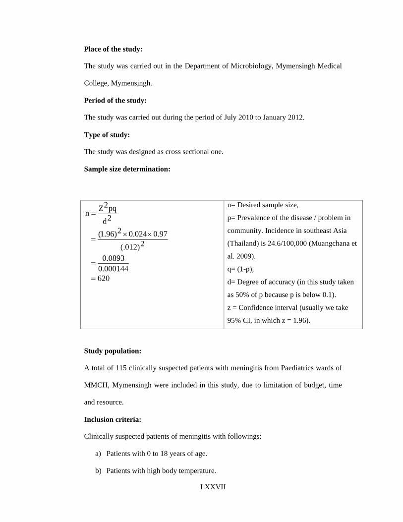
LXXVII
Place of the study:
The study was carried out in the Department of Microbiology, Mymensingh Medical
College, Mymensingh.
Period of the study:
The study was carried out during the period of July 2010 to January 2012.
Type of study:
The study was designed as cross sectional one.
Sample size determination:
620 000144.0
0893.0
2)012(.
97.0024.02)96.1(
2d
pq2Zn
n= Desired sample size,
p= Prevalence of the disease / problem in
community. Incidence in southeast Asia
(Thailand) is 24.6/100,000 (Muangchana et
al. 2009).
q= (1-p),
d= Degree of accuracy (in this study taken
as 50% of p because p is below 0.1).
z = Confidence interval (usually we take
95% CI, in which z = 1.96).
Study population:
A total of 115 clinically suspected patients with meningitis from Paediatrics wards of
MMCH, Mymensingh were included in this study, due to limitation of budget, time
and resource.
Inclusion criteria:
Clinically suspected patients of meningitis with followings:
a) Patients with 0 to 18 years of age.
b) Patients with high body temperature.

LXXVIII
c) Feeding problems.
d) Vomiting.
e) Irritability.
f) Seizures or sluggishness.
g) High pitched crying.
Exclusion criteria:
a) Patients treated with antibiotics after admission.
b) Patients above 18 years of age.
c) Patient with brain hypoxia and brain trauma
Study population will be divided into categories according to the following
criteria of CSF:
On the basis of cytological tests and biochemical tests of CSF the study subjects were
categorized into three groups, which were identified as bacterial meningitis, viral
meningitis and normal CSF.
A. Bacterial meningitis:
1. Appearance: Purulent or cloudy
2. Total count of WBC (mm3): 100�10,000 or more;
Predominant cells are PMNs.
Usually many pus cells present.
3. Glucose (mg/dl): Decreased, usually <40
4. Protein (mg/dl): High usually 100�500.
5. Organisms usually seen on Gram stain and/ recovered by culture.
6. Antigen may be detected by agglutination test (Cheesbrough 2000; Prober 2007).
B. Viral meningitis:
1. Appearance: Clear or slightly turbid

LXXIX
2. Total count of WBC (mm3): Raised but rarely >1,000 cells.
Predominant cells are mononuclear cells.
3. Glucose (mg/dl): Generally normal.
2. Protein (mg/dl): Normal or increased (Cheesbrough 2000; Prober 2007).
C. Normal CSF:
1. Appearance: Clear, Colorless.
2. Total count of WBC (mm3) : <5 cell/mm3, ≥75% lymphocytes
3. Glucose (mg/dl): 45-72
4. Protein (mg/dl): 15�40 (Cheesbrough 2000; Prober 2007).
Data collection and analysis:
All the relevant history, clinical findings and laboratory records of every case were
systematically recorded in a pre-designed data sheet (Appendix-I) for each patient and
analyzed using computer based program statistical package for social science (SPSS)
version 12.0 programs.
Ethical implication:
The protocol was approved by ethical review committee of Mymensingh Medical
College Hospital. Informed written consent (Appendix-II) was taken from each
patient or from their parents before his or her entry into the study after counseling the
whole procedure in Bengali.
Specimen:
Cerebrospinal fluid (CSF)
Collection of CSF:
CSF was collected by a well-trained, experienced medical officer. It was collected
aseptically to prevent organisms being introduced into the central nervous system.
The fluid was collected from the arachnoid�s space. A sterile wide-bore needle was
LXXX
inserted between the fourth and fifth lumber vertebrae and the CSF was allowed to
drip into a dry sterile container.
a) CSF was collected into two sterile, dry, screw-capped containers one for culture (1
ml) and another one for other investigations (2-3 ml).
b) Sample was immediately delivered to the laboratory after proper labeling with
request form (Cheesbrough 2000).
Laboratory procedure:
CSF
Physical examination or macroscopic examination: CSF was observed for pressure during collection and appearance (clear or slightly
turbid or cloudy or definitely purulent), colour (contain blood or not) and clots was
observed after collection.
Normal CSF appears clear and colorless.
Culture of CSF specimen:
CSF was inoculated at bed side immediately after Lumber puncture, in Blood agar
medium, Chocolate agar medium and MacConkey agar medium. Then these plates
were incubated in candle jar at 370 C for 48 hrs (Cheesbrough 2000).
Isolation and identification of bacteria:
The Isolates from the collected specimens were identified on the basis of colony
morphology, Gram�s stain and appropriate biochemical tests.
Streptococcus pneumoniae:
LXXXI
Isolates of Streptococcus pneumoniae were identified on the basis of translucent or
mucoid colonies of 1-2 mm in diameter with alpha haemolysis on blood agar media.
On gram stained smear from colonies were appeared as Gram positive elongated
(lance late) diplococcus and in short chain. Biochemically the isolates were catalase
negative and sensitive to optochin (ethylhydrocupreine hydrochloride) (Cheesbrough
2000).
Group D Streptococcus (Enterococcus):
Isolates of Group D Streptococcus (Enterococcus) was identified on the basis of
small, cream or white, smooth colonies with non haemolysis on blood agar media as
well as on chocolate agar media (Forbes, Sahm and Weissfeld 2002). On MacConkey
agar media it produced dark-red magenta colonies. On gram stained smear from
colonies were appeared as Gram positive cocci in pairs or in short chain.
Biochemically the isolates were Catalase negative (Cheesbrough 2000).
Microscopic Examination:
Wet preparation:
A drop of uncentrifuged CSF was transferred to a slide and was covered with a cover
glass. This preparation was examined by using the 10X and 40X objectives, with the
condenser closed sufficiently to give good contrast and the findings were recorded
accordingly.
Cell count:
Total count of WBC was done by improved Neubauer counting chamber. A clean dry
improved Neubauer counting chamber with a clean dry cover slip was carefully

LXXXII
charged (poured) with an uncentrifuged CSF by micropipette and was waited for 2-3
minutes.
Counting was done by using 10x objectives and 40 x objectives. The number of white
cells that was found in 4 large squares corner was counted. If N number of cells was
found in 4 large squares, it was then make average which would be4
N. Then it was
multiplied by depth of the fluid. So the count was 4
N10 cells/ mm3 (1/10 is depth of
the fluid) (Ghai 2005).
Differential count of WBC in CSF:
The smear was prepared from the centrifuged deposits of CSF and then the smear was
stained with Leishman stain. After air dry of the smear it was examined under
microscope using high power and oil immersion lens (Chessbrough 2000).
Leishman staining technique:
A drop of centrifuged CSF was transferred to a slide and a smear was made. The
smear of CSF was covered with Leishman stain. Twice of the volume of PH 6.8
buffered water was added. The diluted stain should not overflow. It should be ensured
that the water is well mixed with the stain by blowing on the diluted stain (through a
straw) or mixing the stain and waited for 10 minutes. It was allowed to stain for 10
minutes. The stain was washed off with tap water. Wiped the back of the slide clean
and stranded it in a draining rack for the smear to dry. The blood was appeared neither
too pink nor too blue (Chessbrough 2000).
Gram staining technique:
A drop of centrifuged CSF was transferred to a slide and a smear was made. The
smear of CSF was fixed by heat. The fixed smear was covered with crystal violet stain

LXXXIII
for 30-60 seconds. Then the stain was washed off rapidly with clear water tip off all
the water. The smear was covered with Lugol�s iodine for 30-60 seconds. Then it was
washed off the iodine with clean water. The smear was decolorized rapidly with
acetone-alcohol and washed immediately with clean water. Then the smear was
covered with dilute carbol fuchsin stain (1 in 10) for 2 minutes. Then the smear again
was washed off the stain with clean water. Wiped the back of the slide clean, and
placed it in a draining rack for the smear to air � dry. A positive control smear and a
negative control smear were also prepared to compare with the test smear. The smear
was examined microscopically, first with the 40X objective to check the staining and
to see the distribution of material, and then with the oil immersion objective to report
the bacteria and cells (Chessbrough 2000).
Ziehl-Neelsen technique for Mycobacterium tuberculosis:
A drop of centrifuged CSF was transferred to a slide and a smear was made. The
smear of CSF was fixed by heat. Then smear was covered with carbol fuchsin stain
and was heated until vapor just begins to rise but not overheated. The stain was
washed off with clean water after 5 minutes and covered the smear with 3% v/v acid
alcohol for 5 minutes or until the smear was sufficiently decolorized (pale pink). Then
the stain was washed off well with clean water. And the smear was covered with
methylene blue stain for 1-2 minutes, using the longer time when the smear was thin.
The stain was washed off with clean water and placed it in a draining rack for air-dry.
The smear was examined microscopically with the 100X oil immersion object
(Chessbrough 2000).
Biochemical tests of the CSF specimens:
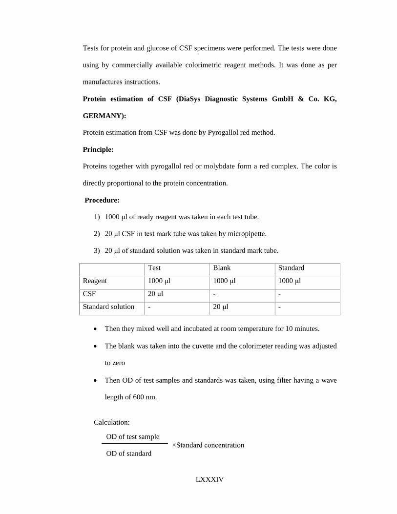
LXXXIV
OD of test sample ×Standard concentration OD of standard
Tests for protein and glucose of CSF specimens were performed. The tests were done
using by commercially available colorimetric reagent methods. It was done as per
manufactures instructions.
Protein estimation of CSF (DiaSys Diagnostic Systems GmbH & Co. KG,
GERMANY):
Protein estimation from CSF was done by Pyrogallol red method.
Principle:
Proteins together with pyrogallol red or molybdate form a red complex. The color is
directly proportional to the protein concentration.
Procedure:
1) 1000 ìl of ready reagent was taken in each test tube.
2) 20 ìl CSF in test mark tube was taken by micropipette.
3) 20 ìl of standard solution was taken in standard mark tube.
Test Blank Standard
Reagent 1000 ìl 1000 ìl 1000 ìl
CSF 20 ìl - -
Standard solution - 20 ìl -
Then they mixed well and incubated at room temperature for 10 minutes.
The blank was taken into the cuvette and the colorimeter reading was adjusted
to zero
Then OD of test samples and standards was taken, using filter having a wave
length of 600 nm.
Calculation:

LXXXV
= mg/dl.
Glucose estimation of CSF: (SPINREACT, SPAIN)
The glucose estimation from CSF was done by Enzymatic colorimetric method.
Principle:
Glucose oxidase (GOD) catalyses the Oxidation of glucose to gluconic acid. The
formed Hydrogen peroxide (H2O2), is detected by a chromogenic oxygen acceptor,
Phenol- amino-phenazone in the presence of peroxidase (POD). The intensity of the
colour is proportional to the glucose concentration is the sample.
D-Glucose + O2 + H2O Gluconic acid + H2O2
H2O2+ Phenol + Amino-phenazone Quinone + H2O
Procedure:
I) 1 ml of glucose reagent was taken in three test tubes.
II) 10 ìl CSF in test mark tube was taken by micropipette.
III) 10 ìl standard solution was taken in a tube labeled as standard ..
Test Blank Standard
Reagent 1ml 1ml 1ml
CSF 10 ìl - -
Standard solution - 10 ìl -
They were mixed well and incubated at room temperature for 10 minutes.
The reagent blank was taken into the cuvette of the colorimeter and the
colorimeter reading was adjusted to zero.
The absorbance of test sample and standards was measured against reagent blank
within 30 minutes using 530 nm filters.
LXXXVI
Calculation:
= mmol/L
= mmol/L
(mmol/L x 18 = mg/dl)
C-reactive protein (High sensitivity C-reactive protein Enzyme Immunoassay
Test Kit: (LumiQuick Diagnostics, Inc. U.S.A)
Principle of the Assay:
Detection of CRP from CSF by the hs CRP ELISA was based on the principle of a
solid phase enzyme-linked immunosorbent assay. The assay system utilizes a unique
monoclonal antibody directed against a distinct antigenic determinant on the CRP
molecule. This mouse monoclonal anti-CRP antibody is used for solid phase
immobilization (on the micro titer wells). A goat anti-CRP antibody is in the
antibody-enzyme (horseradish peroxidase) conjugate solution. The test sample is
allowed to react simultaneously with the two antibodies, resulting in the CRP
molecules being sandwiched between the solid phase and enzyme-linked antibodies.
After 45-minutes incubation at room temperature, the wells are washed with water to
remove unbound labeled antibodies. Substrate for HRP, tetramethylebenzidine (TMB)
reagent is added and incubated for 20 minutes, resulting in the development of blue
colour. The colour development is stopped with the addition of stop solution sulphuric
acid and the colour is changed to yellow. The concentration of CRP is directly
proportional the colour intensity of the test sample. Absorbance is measured
spectrophotometrically at 450 nm (LumiQuick Diagnostics, Inc. U.S.A).
Procedure:
OD of test sample ×Standard concentration OD of standard
LXXXVII
The samples of CSF were diluted 100 fold prior to use. The coated wells were secured
in the holder. Dispensed 50 µl of the undiluted CRP standards and diluted specimens
into appropriate wells. Again 50 µl of sample diluents were dispensed into each well.
Than mixed it thoroughly for 30 seconds and incubated at room temperature (18-
220C) for 30 minutes. The incubated mixtures were removed by flicking plate
contents into a waste container. The micro titer wells were rinsed and flicked 5 times
with deionized or distilled water. The wells were striked sharply onto absorbent paper
or paper towels to remove all residual water droplets. Dispensed 100 µl of CRP
Enzyme Conjugate Reagent into each well. That was mixed for 30 seconds and
incubated at room temperature for 30 minutes. Again the incubated mixture was
removed by flicking plate contents into a waste container. The micro titer wells were
rinsed and flicked 5 times with deionized or distilled water. The wells were striked
sharply onto absorbent paper or paper towels to remove all residual water droplets.
Then 100 µl of TMB reagent was dispensed into each well and mixed for 5 seconds
and again incubated at room temperature for 30 minutes. After 30 minutes 100 µl of
Stop Solution was added to each well to stop the reaction. That was mixed gently for
30 seconds. It was important to make sure that all the blue colour changed to yellow
colour completely. The absorbance was read at 450 nm with a micro titer well reader
within 15 minutes (LumiQuick Diagnostics, Inc. U.S.A).
Detection of bacterial antigen by latex agglutination test (LAT): (Remel Europe
Ltd. UK)
Principle:
The reagents consists of polystyrene latex particles which have been coated with
antibodies to the bacterial antigens except for the N. meningitides group B/E. coli K1

LXXXVIII
reagents where latex is coated with murine monoclonal antibody. These latex particles
agglutinate in the presence of sufficient homologous antigen and form clumps which
is visible to naked eye.
Latex agglutination test kit contains following reagents:
1. Strep B test latex (Pink cap)
2. H. influenzae test latex (Pale blue cap)
3. S. pneumoniae test latex (Yellow cap)
4. N. meningitidis ACY W135 test latex
5. N. meningitidis B/E coli K1 test latex (Brown cap)
6. Control latex for each organism (Dark blue cap)
7. Polyvalent positive control (Red cap)
Accessories:
1. Disposable reaction cards
2. Disposable mixing sticks
3. Disposable droppers
4. Black rubber teats
Procedures:
The CSF was heated at 1000 C for 5 minutes at water bath to minimize non-specific
reactions. Than the sample was centrifuged for 5 minutes at 3000 rpm. Latex reagents

LXXXIX
were shacked. One drop of each test latex or control was placed in to a separate circle
on a reaction card. One drop (approx. 40 µL) of supernatant CSF was dispensed by
using a disposable dropper next to each drop of latex. The contents of each circle were
mixed using a mixing stick, and was spread to cover the complete area of circle. The
card was rocked slowly and observed for agglutination for 3 minutes. The results were
read by naked eye under a good light.
Interpretation of results:
Positive reaction:
A positive reaction was indicated by the development of agglutination within 3
minutes of mixing the latex with the test sample, showing clearly visible sample of
the latex particle.
Negative reaction:
If the suspension did not clump and be retained it slightly milky appearance.
Quality control:
To verify that the latex will not auto agglutinated, a test was carried out, using the
diluent solution and the latex concerned. No agglutination will be occurred (Remel
Europe Ltd. UK)
Procedure of polymerase chain reaction for detection of LytA gene for diagnosis
of pneumococcal meningitis directly from CSF sample:
Processing of CSF samples and PCR amplification:
Three major steps:
1. Extraction of DNA from CSF samples.
XC
2. Amplification of DNA in thermal cycler.
3. Visualization/documentation under UV light
CSF samples:
One milliliter CSF sample that was collected from the patients of meningitis was used
for DNA extraction.
DNA extraction from CSF:
Extraction of DNA from CSF (1ml) was carried out by modified lysis buffer method.
One ml CSF was centrifuged at 13000 rpm for 5 minutes. Supernatant was discarded.
One ml 0.2% Triton X-100 was added to the pellet. The mixture was vortexed,
incubated at room temperature for 10 minutes. Again the samples were centrifuged at
13000 rpm for 10 minutes. Supernatant was discarded. One ml 0.2% Triton X-100
was added to the pellet again, vortexed and centrifuged at 13000 rpm for 10 minutes.
Washed with 1 ml nuclease free water, centrifuged for 3 minutes and supernatant was
discarded. Pellet was re-suspended in 30 µl nuclease free water. Boiled for 10 minutes
at 99° C then centrifuged for 3 minutes. Supernatant was used as template for PCR
(Jimenez and castro 2004; Nagarajan et al. 2009) with some modification.
Master mixture of detection of LytA gene of Streptococcus pneumoniae:
Primer-1 - 20 µl
Primer-2 - 20 µl
Nuclease free water - 80 µl
Preparation of master mixture for each reaction (50 µl) contained:
10x buffer - 5µl
200 micromole deoxy nucleoside triphosphate -4µl
Primer mixture - 1µl
2units of Taq DNA polymerase - 0.5µl
XCI
DDW/PCR water - 38.5µl
DNA sample -1µl
Protocol of Thermal cycles:
Initial denaturation at 94°C for 2 minutes followed by
Denaturation at 940C for 30 seconds.
Primer annealing at 550C for 30 seconds.
Extension at 72 0C for 30 seconds.
Final extension at 720C for 10 minutes.
Above steps was repeated for 30 cycles in an automated DNA thermal cycler
(Eppendorf, Germany). Final extension was done at 72°C for 10 minutes. The PCR
products were run in 1.5% agarose gels stained with ethidium bromide. Then that
were visualized under the UV transilluminator. Molecular size markers 1 kb will be
run concurrently (Saha et al. 2005).
Primer used for PCR:
The sequences from 5' to 3' ends of these primers were as follows:
Primer -1 (5'-TGAAGCGGATTATCACTGGC-3')
Primer -2 (5'-GCTAAACTCCCTG TATCAAGCG-3').
Above primers was used for amplify 273- bp sequence of LytA gene (Saha et al 2005).
Agarose gel electrophoresis:
The PCR product was analyzed by 1.5% agarose gel electrophoresis to detect specific
band which was 273-bp.
Preparation of agarose gel:
One percent agarose gel was prepared by melting 3 gm agarose in 200 ml of diluted
TBE buffer (1:4) using microwave oven or gas burner. The agarose was allowed to
cool at about 50°C and was poured into gel electrophoresis unit with spacers and
30 cycles
XCII
comb. After solidification of the gel, the comb was removed and wells were formed.
During electrophoresis, the gel was placed in a solution containing Tris- EDTA-
borate buffer (TBE buffer) and ethidium bromide (0.5 µg/ml) in the horizontal
electrophoresis chamber.
Loading and Running of the sample:
Ten micro liter of amplified product was mixed with 2.0 µl gel loading dye. The
mixture was slowly loaded into the well using disposable micropipette tips. Marker
DNA of known size (1000 bp ladder) was loaded in one well to determine the size of
amplified PCR products. Electrophoresis will carried out at 100 volts for 35 minutes.
Staining and visualization of the gel:
The band (273- bp) of the sample of the study was visualized by transilluminator. And
the gel was photographed by a digital camera and transferred the data to computer for
further documentation.
Determination of MIC of penicillin by Agar plate dilution method
Agar plate dilution test was used to determine the minimal inhibitory concentration
(MIC) of penicillin required for inhibiting or killing a microorganism.
Preparation of antimicrobial agents
One vial of penicillin was mixed with 5 ml distilled water. After that the
concentration of antimicrobial agent was 1,20,000 µgm/ml. Than 1 ml penicillin was
dissolved in 999 ml double autoclaved distilled water and added to the melted Muller-
Hinton agar with 5% sheep blood in different concentration (l/ml) used in the MIC.
The concentration of stock solution of antibiotics was 120 µgm/ml and 2 µgm/ml.
MIC range were 2 µgm/ml, 0.5 µgm/ml, 0.12 µgm/ml, 0.06 µgm/ml and 0.03 µgm/ml
(Kelly, Jacobs and Appelbaum 1999).
XCIII
Preparation of plates
500 ml Muller-Hinton medium of flask was autoclaved and allowed to cool at 500C in
water bath and 25 ml (5%) sheep blood was mixed. Then different concentration of
antibiotic solution was added to each flask, mixed thoroughly and antibiotic-
containing media was poured immediately on the plates. Two fold (Log2) serial
dilutions of the antimicrobials were added with agar medium. The concentrations of
the penicillin in different plates were 2 g/ml, 0.5 g/ml, 0.12 g/ml, 0.06 µgm/ml
and 0.03 µgm/ml was used in different plates. Plates were stored at 40C in plastic bag
and use within one week of preparation. Before inoculation, plates were dried.
Control plates of Muller-Hinton agar with 5% sheep blood containing no
antimicrobial agent was also prepared to ensure the effectiveness of the medium in
sustaining the growth of organism.
Turbidity Standard for MIC inoculum preparation
To standardize the inoculum density for a susceptibility test, a 0.5 McFarland standard
was prepared as described in CLSI 2010.
Inoculation and Incubation of the medium
Agar surface of the plates containing different concentration of penicillin and the
control plate contained no antimicrobial agents were spot inoculated with a digital
micropipette. A volume of 2 l suspension of the 104 CFU/ml organisms was spot
inoculated. Inoculation was done from the plate containing lowest concentration of
antimicrobial and the control plate was inoculated lastly. All the 6 different strains of
Streptococcus pneumoniae were tested at a time in each plate. Inoculated agar plates
was allowed to stand until the inoculums spot was completely absorbed and
afterwards it was incubated at 350C for overnight.
XCIV
Interpretation of results
The MIC represents the concentration of antimicrobial at which there is complete
inhibition of growth. In reading the end points, a barely visible haze of growth or a
single colony is disregarded. The results were interpreted according to the
recommendation of the study done by Kelly, Jacobs and Appelbaum (1999) and the
strains were referred to as resistant (R), sensitive (S) and intermediate in relation to
the MIC of antimicrobial agent.
Antimicrobial susceptibility testing by disc diffusion method
All the isolates were put into antibiotic susceptibility test by Kirby-Bauer disc
diffusion technique as per recommendation of CLSI 2010 (Furtado and Medeiros
1980). Panel of antibiotics were used. All tests were performed on Muller-Hinton
agar with 5% sheep blood. The surface was lightly and uniformly inoculated by cotton
swab. Prior to inoculation, the swab stick was dipped into bacterial suspension having
visually equivalent turbidity to 0.5 McFarland standards (Appendix-). The swab stick
was then took out and squeezed on the wall of the test tube to discard extra
suspension. Inoculated plates were incubated under 5% to 10% CO2 at 370 C for 24
hours. On the next day, plates were read by taking measurement of zone of inhibition.
Results were recorded and graded as Resistant (R), Intermediate (I) and Sensitive (S)
according to the reference zone of inhibition of particular antibiotic (CLSI 2010).
Antimicrobial agents used
A total of 12 antimicrobial agents were used for determining antibiogram of isolated
organisms according to Gram positive panel recommended by CLSI 2010. Antibiotics
were Penicillin G, Ampicillin, Ceftazidim, Ciprofloxacin, Chloramphenicol, Co-
trimoxazole, Erythromycin and Gentamicin.

XCV
Chapter � 4
Results
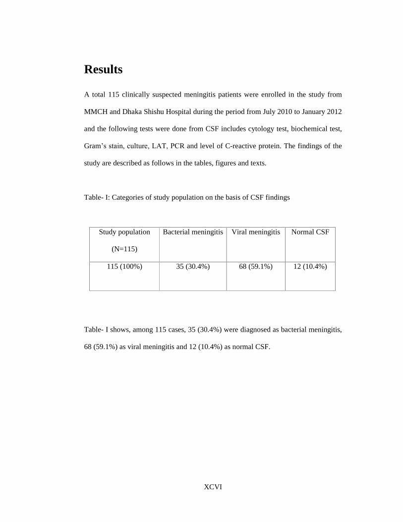
XCVI
Results
A total 115 clinically suspected meningitis patients were enrolled in the study from
MMCH and Dhaka Shishu Hospital during the period from July 2010 to January 2012
and the following tests were done from CSF includes cytology test, biochemical test,
Gram�s stain, culture, LAT, PCR and level of C-reactive protein. The findings of the
study are described as follows in the tables, figures and texts.
Table- I: Categories of study population on the basis of CSF findings
Study population
(N=115)
Bacterial meningitis Viral meningitis Normal CSF
115 (100%) 35 (30.4%) 68 (59.1%) 12 (10.4%)
Table- I shows, among 115 cases, 35 (30.4%) were diagnosed as bacterial meningitis,
68 (59.1%) as viral meningitis and 12 (10.4%) as normal CSF.
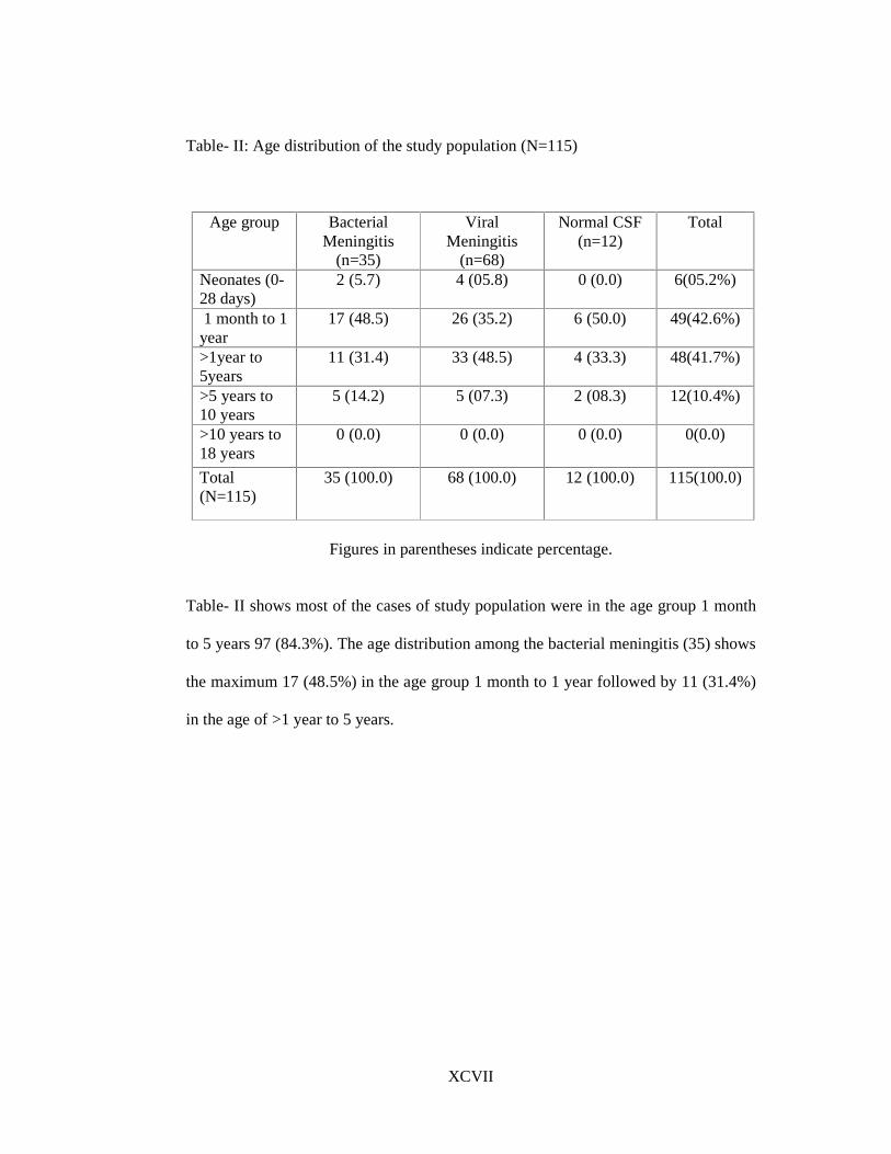
XCVII
Table- II: Age distribution of the study population (N=115)
Age group Bacterial Meningitis
(n=35)
Viral Meningitis
(n=68)
Normal CSF (n=12)
Total
Neonates (0-28 days)
2 (5.7) 4 (05.8) 0 (0.0) 6(05.2%)
1 month to 1 year
17 (48.5) 26 (35.2) 6 (50.0) 49(42.6%)
>1year to 5years
11 (31.4) 33 (48.5) 4 (33.3) 48(41.7%)
>5 years to 10 years
5 (14.2) 5 (07.3) 2 (08.3) 12(10.4%)
>10 years to 18 years
0 (0.0) 0 (0.0) 0 (0.0) 0(0.0)
Total (N=115)
35 (100.0) 68 (100.0) 12 (100.0) 115(100.0)
Figures in parentheses indicate percentage.
Table- II shows most of the cases of study population were in the age group 1 month
to 5 years 97 (84.3%). The age distribution among the bacterial meningitis (35) shows
the maximum 17 (48.5%) in the age group 1 month to 1 year followed by 11 (31.4%)
in the age of >1 year to 5 years.
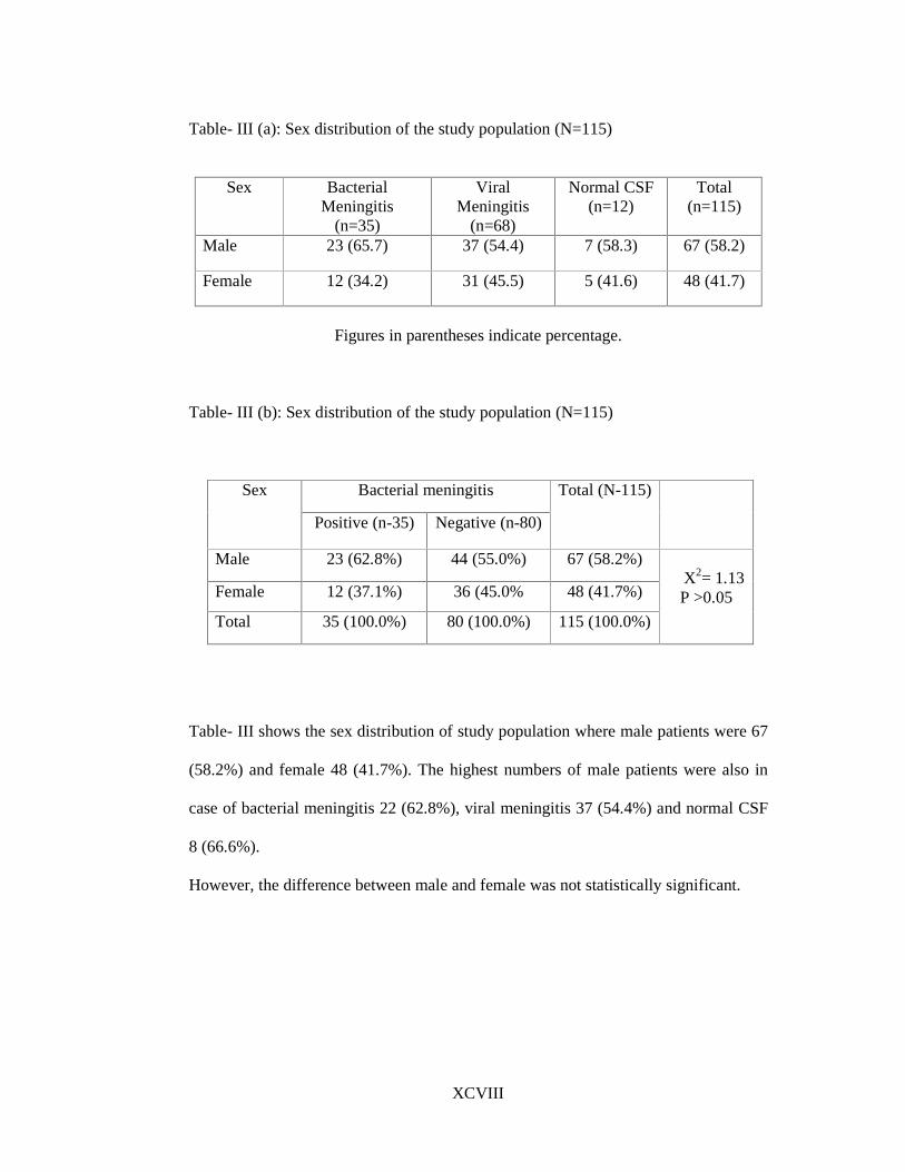
XCVIII
Table- III (a): Sex distribution of the study population (N=115)
Sex Bacterial Meningitis
(n=35)
Viral Meningitis
(n=68)
Normal CSF (n=12)
Total (n=115)
Male 23 (65.7) 37 (54.4) 7 (58.3) 67 (58.2)
Female 12 (34.2) 31 (45.5) 5 (41.6) 48 (41.7)
Figures in parentheses indicate percentage.
Table- III (b): Sex distribution of the study population (N=115)
Bacterial meningitis Sex
Positive (n-35) Negative (n-80)
Total (N-115)
Male 23 (62.8%) 44 (55.0%) 67 (58.2%)
Female 12 (37.1%) 36 (45.0% 48 (41.7%)
Total 35 (100.0%) 80 (100.0%) 115 (100.0%)
X2= 1.13
P >0.05
Table- III shows the sex distribution of study population where male patients were 67
(58.2%) and female 48 (41.7%). The highest numbers of male patients were also in
case of bacterial meningitis 22 (62.8%), viral meningitis 37 (54.4%) and normal CSF
8 (66.6%).
However, the difference between male and female was not statistically significant.

XCIX
Table- IV: Clinical findings of meningitis in the study population (N=115) Clinical findings of study subjects
Bacterial Meningitis
(n=35)
Viral Meningitis
(n=68)
Normal CSF (n=12)
Total (N=115)
Fever 35 (100.0%) 68 (100.0%) 12 (100.0%) 115 (100.0%)
Nausea and vomiting
16 (45.7%) 48 (70.5%) 4 (33.3%) 70 (60.8%)
Convulsion 21 (60.0%) 44 (64.7%) 9 (75.0%) 74 (64.3%)
Table- IV showing the clinical findings of meningitis where all of the study
population (115) were suffering from fever (100.0%) followed by nausea or vomiting
(60.8%) and convulsion (64.3%). Among the bacterial meningitis (35) cases 60.0%
and 45.7% were suffering from convulsion and vomiting or nausea respectively.

C
Table- V: Physical findings of CSF in the study population (N=115)
Findings of physical appearance
of CSF
Bacterial Meningitis (n=35)
Viral Meningitis (n=68)
Normal CSF (n=12)
Purulent 21 (60.0%) 0 (0.0%) 0 (0.0%)
Slightly turbid 14 (40.0%) 11 (16.1%) 0 (0.0%)
Clear 0 (0.0%) 57 (83.8%) 12 (100.0%)
Table- V shows the physical findings of CSF, in bacterial meningitis 21 (60.0%) were
purulent and 14 (40.0%) were slightly turbid. Clear CSF were 57 (83.8%) in case of
viral meningitis and 12 (100.0%) in cases of normal CSF.
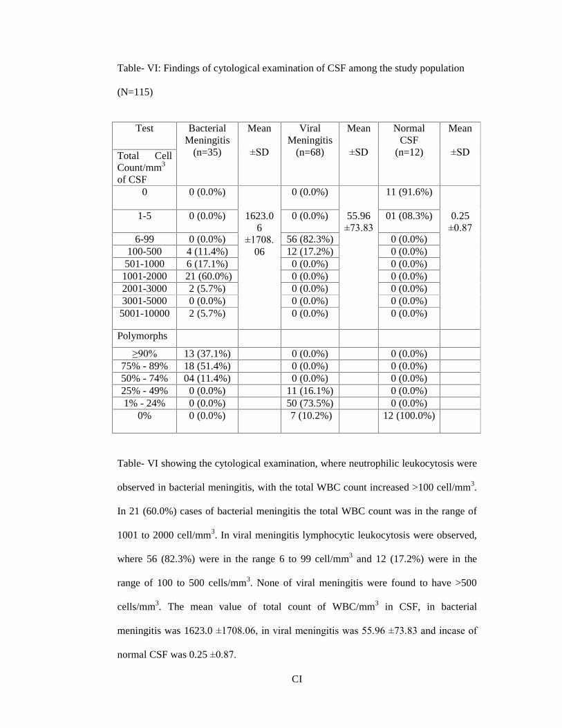
CI
Table- VI: Findings of cytological examination of CSF among the study population
(N=115)
Test
Total Cell Count/mm3 of CSF
Bacterial Meningitis
(n=35)
Mean
±SD
Viral Meningitis
(n=68)
Mean
±SD
Normal CSF
(n=12)
Mean
±SD
0 0 (0.0%) 0 (0.0%) 11 (91.6%)
1-5 0 (0.0%) 0 (0.0%) 01 (08.3%)
6-99 0 (0.0%) 56 (82.3%) 0 (0.0%) 100-500 4 (11.4%) 12 (17.2%) 0 (0.0%)
501-1000 6 (17.1%) 0 (0.0%) 0 (0.0%) 1001-2000 21 (60.0%) 0 (0.0%) 0 (0.0%) 2001-3000 2 (5.7%) 0 (0.0%) 0 (0.0%) 3001-5000 0 (0.0%) 0 (0.0%) 0 (0.0%)
5001-10000 2 (5.7%)
1623.06
±1708.
06
0 (0.0%)
55.96 ±73.83
0 (0.0%)
0.25 ±0.87
Polymorphs
≥90% 13 (37.1%) 0 (0.0%) 0 (0.0%) 75% - 89% 18 (51.4%) 0 (0.0%) 0 (0.0%) 50% - 74% 04 (11.4%) 0 (0.0%) 0 (0.0%) 25% - 49% 0 (0.0%) 11 (16.1%) 0 (0.0%) 1% - 24% 0 (0.0%) 50 (73.5%) 0 (0.0%)
0% 0 (0.0%) 7 (10.2%) 12 (100.0%)
Table- VI showing the cytological examination, where neutrophilic leukocytosis were
observed in bacterial meningitis, with the total WBC count increased >100 cell/mm3.
In 21 (60.0%) cases of bacterial meningitis the total WBC count was in the range of
1001 to 2000 cell/mm3. In viral meningitis lymphocytic leukocytosis were observed,
where 56 (82.3%) were in the range 6 to 99 cell/mm3 and 12 (17.2%) were in the
range of 100 to 500 cells/mm3. None of viral meningitis were found to have >500
cells/mm3. The mean value of total count of WBC/mm3 in CSF, in bacterial
meningitis was 1623.0 ±1708.06, in viral meningitis was 55.96 ±73.83 and incase of
normal CSF was 0.25 ±0.87.
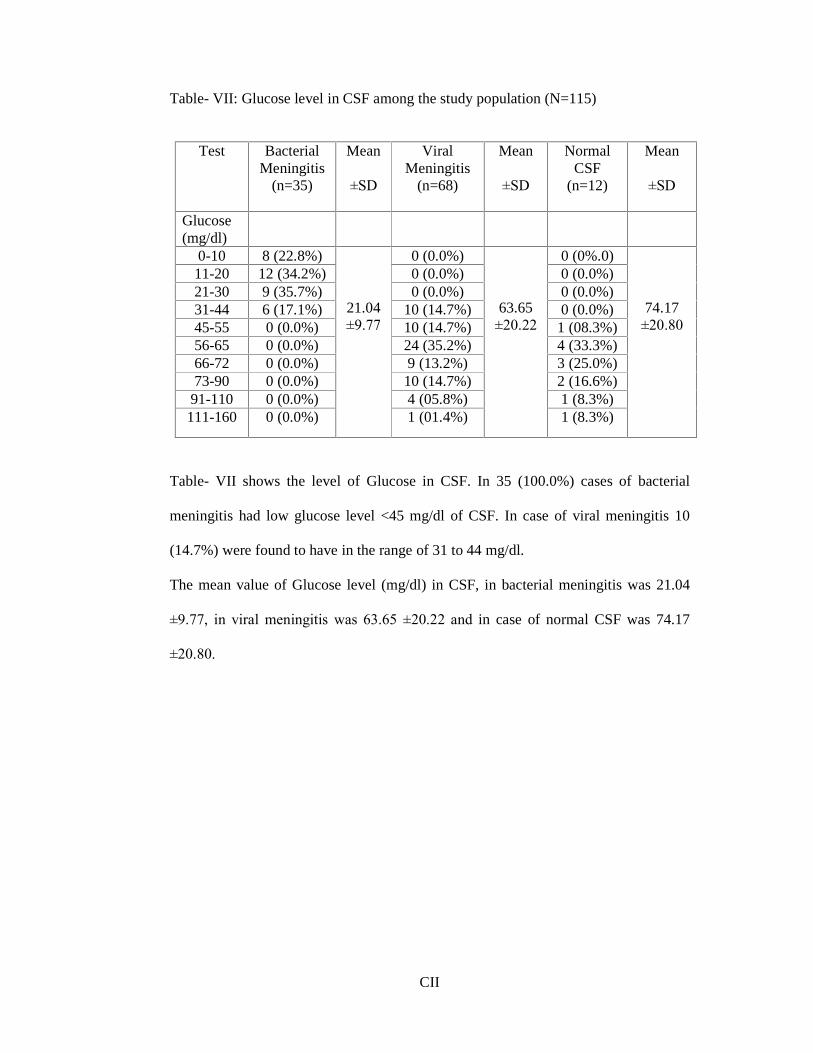
CII
Table- VII: Glucose level in CSF among the study population (N=115)
Test Bacterial
Meningitis (n=35)
Mean
±SD
Viral Meningitis
(n=68)
Mean
±SD
Normal CSF
(n=12)
Mean
±SD
Glucose (mg/dl)
0-10 8 (22.8%) 0 (0.0%) 0 (0%.0) 11-20 12 (34.2%) 0 (0.0%) 0 (0.0%) 21-30 9 (35.7%) 0 (0.0%) 0 (0.0%) 31-44 6 (17.1%) 10 (14.7%) 0 (0.0%) 45-55 0 (0.0%) 10 (14.7%) 1 (08.3%) 56-65 0 (0.0%) 24 (35.2%) 4 (33.3%) 66-72 0 (0.0%) 9 (13.2%) 3 (25.0%) 73-90 0 (0.0%) 10 (14.7%) 2 (16.6%)
91-110 0 (0.0%) 4 (05.8%) 1 (8.3%) 111-160 0 (0.0%)
21.04 ±9.77
1 (01.4%)
63.65 ±20.22
1 (8.3%)
74.17 ±20.80
Table- VII shows the level of Glucose in CSF. In 35 (100.0%) cases of bacterial
meningitis had low glucose level <45 mg/dl of CSF. In case of viral meningitis 10
(14.7%) were found to have in the range of 31 to 44 mg/dl.
The mean value of Glucose level (mg/dl) in CSF, in bacterial meningitis was 21.04
±9.77, in viral meningitis was 63.65 ±20.22 and in case of normal CSF was 74.17
±20.80.
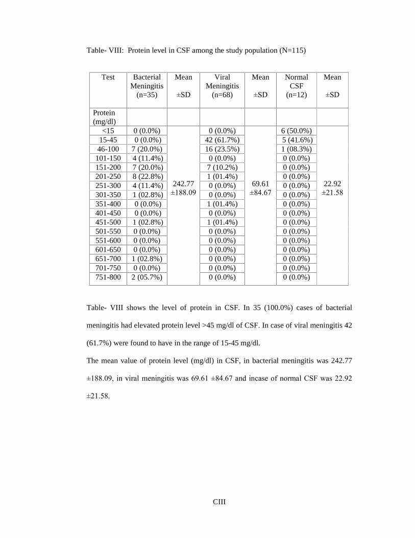
CIII
Table- VIII: Protein level in CSF among the study population (N=115)
Test Bacterial Meningitis
(n=35)
Mean
±SD
Viral Meningitis
(n=68)
Mean
±SD
Normal CSF
(n=12)
Mean
±SD
Protein (mg/dl)
<15 0 (0.0%) 0 (0.0%) 6 (50.0%) 15-45 0 (0.0%) 42 (61.7%) 5 (41.6%)
46-100 7 (20.0%) 16 (23.5%) 1 (08.3%) 101-150 4 (11.4%) 0 (0.0%) 0 (0.0%) 151-200 7 (20.0%) 7 (10.2%) 0 (0.0%) 201-250 8 (22.8%) 1 (01.4%) 0 (0.0%) 251-300 4 (11.4%) 0 (0.0%) 0 (0.0%) 301-350 1 (02.8%) 0 (0.0%) 0 (0.0%) 351-400 0 (0.0%) 1 (01.4%) 0 (0.0%) 401-450 0 (0.0%) 0 (0.0%) 0 (0.0%) 451-500 1 (02.8%) 1 (01.4%) 0 (0.0%) 501-550 0 (0.0%) 0 (0.0%) 0 (0.0%) 551-600 0 (0.0%) 0 (0.0%) 0 (0.0%) 601-650 0 (0.0%) 0 (0.0%) 0 (0.0%) 651-700 1 (02.8%) 0 (0.0%) 0 (0.0%) 701-750 0 (0.0%) 0 (0.0%) 0 (0.0%) 751-800 2 (05.7%)
242.77 ±188.09
0 (0.0%)
69.61 ±84.67
0 (0.0%)
22.92 ±21.58
Table- VIII shows the level of protein in CSF. In 35 (100.0%) cases of bacterial
meningitis had elevated protein level >45 mg/dl of CSF. In case of viral meningitis 42
(61.7%) were found to have in the range of 15-45 mg/dl.
The mean value of protein level (mg/dl) in CSF, in bacterial meningitis was 242.77
±188.09, in viral meningitis was 69.61 ±84.67 and incase of normal CSF was 22.92
±21.58.
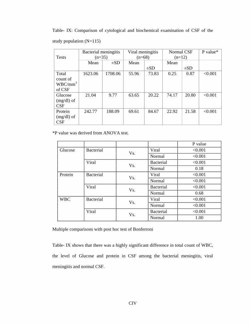
CIV
Table- IX: Comparison of cytological and biochemical examination of CSF of the
study population (N=115)
Bacterial meningitis (n=35)
Viral meningitis (n=68)
Normal CSF (n=12)
P value* Tests
Mean ±SD Mean ±SD
Mean ±SD
Total count of WBC/mm3 of CSF
1623.06 1708.06 55.96 73.83 0.25 0.87 <0.001
Glucose (mg/dl) of CSF
21.04 9.77 63.65 20.22 74.17 20.80 <0.001
Protein (mg/dl) of CSF
242.77 188.09 69.61 84.67 22.92 21.58 <0.001
*P value was derived from ANOVA test.
P value Viral <0.001 Bacterial
Vs. Normal <0.001 Bacterial <0.001
Glucose
Viral Vs.
Normal 0.18 Viral <0.001 Bacterial
Vs. Normal <0.001 Bacterial <0.001
Protein
Viral Vs.
Normal 0.68 Viral <0.001 Bacterial
Vs. Normal <0.001 Bacterial <0.001
WBC
Viral Vs.
Normal 1.00 Multiple comparisons with post hoc test of Bonferroni
Table- IX shows that there was a highly significant difference in total count of WBC,
the level of Glucose and protein in CSF among the bacterial meningitis, viral
meningitis and normal CSF.

CV
Table- X: Rate of detection of bacterial meningitis among the study cases by Gram�s
stain, culture, Latex Agglutination Test (LAT) and Polymerase Chain Reaction (PCR)
(n=35)
Test Positive Negative Total
Gram�s stain 6 (17.1%) 29 (82.8%) 35
Culture 7 (20.0%) 28 (80.0%) 35
Latex Agglutination Test
15 (42.8%) 20 (57.1%) 35
Polymerase Chain Reaction
22 (62.8%) 13 (37.1%) 35
Table- X shows comparison among the four investigations (Gram�s stain, culture,
LAT and PCR) performed for diagnosis of bacterial meningitis. Where 20.0% were
isolated by culture and 17.1% were Gram�s stain positive, 42.8% were LAT positive
and 62.8% were PCR positive among the study population (n=35).
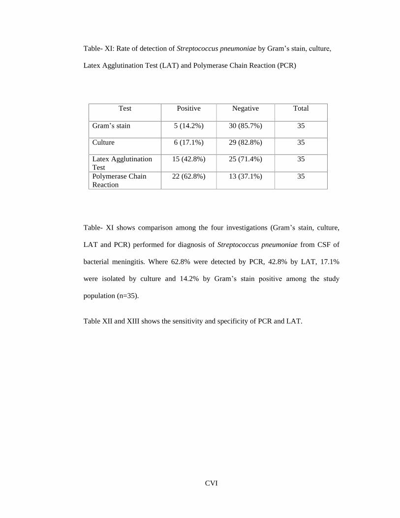
CVI
Table- XI: Rate of detection of Streptococcus pneumoniae by Gram�s stain, culture,
Latex Agglutination Test (LAT) and Polymerase Chain Reaction (PCR)
Test Positive Negative Total
Gram�s stain 5 (14.2%) 30 (85.7%) 35
Culture 6 (17.1%) 29 (82.8%) 35
Latex Agglutination Test
15 (42.8%) 25 (71.4%) 35
Polymerase Chain Reaction
22 (62.8%) 13 (37.1%) 35
Table- XI shows comparison among the four investigations (Gram�s stain, culture,
LAT and PCR) performed for diagnosis of Streptococcus pneumoniae from CSF of
bacterial meningitis. Where 62.8% were detected by PCR, 42.8% by LAT, 17.1%
were isolated by culture and 14.2% by Gram�s stain positive among the study
population (n=35).
Table XII and XIII shows the sensitivity and specificity of PCR and LAT.
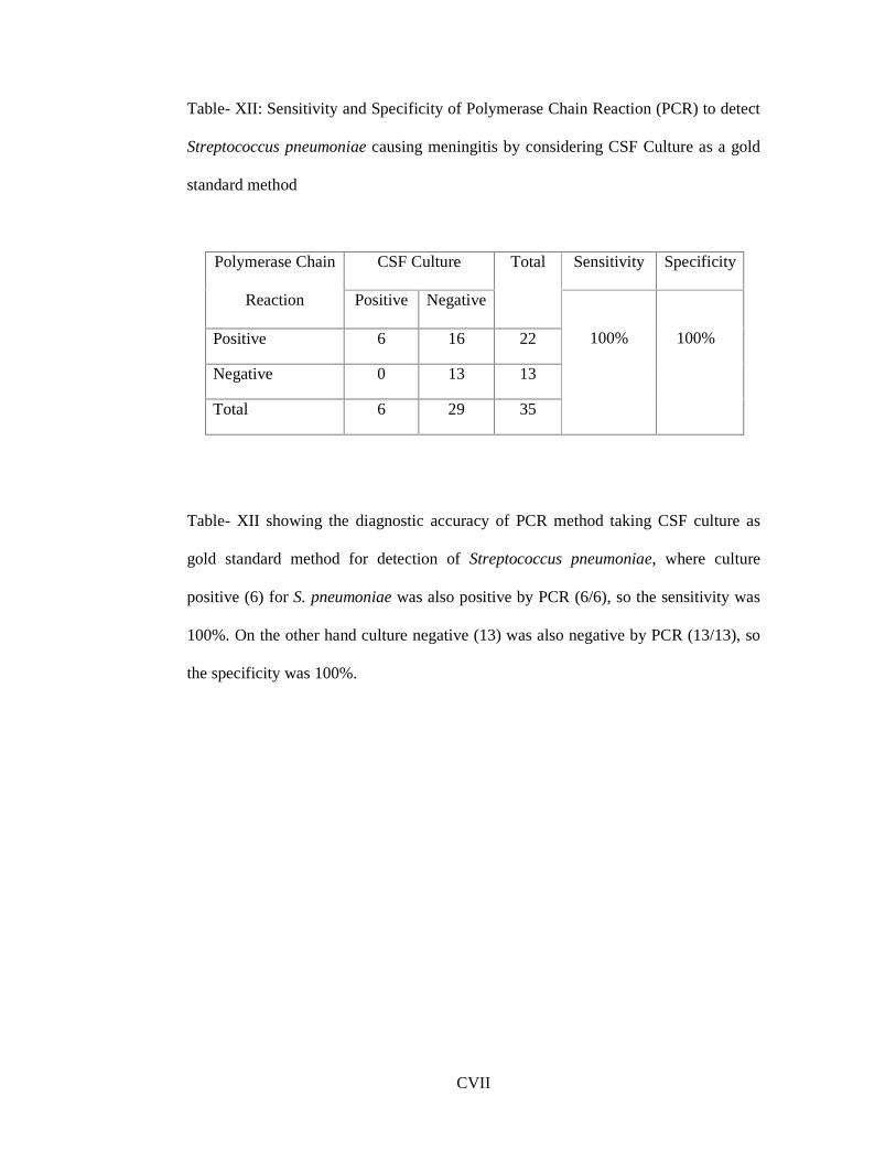
CVII
Table- XII: Sensitivity and Specificity of Polymerase Chain Reaction (PCR) to detect
Streptococcus pneumoniae causing meningitis by considering CSF Culture as a gold
standard method
CSF Culture Sensitivity Specificity Polymerase Chain
Reaction Positive Negative
Total
Positive 6 16 22
Negative 0 13 13
Total 6 29 35
100%
100%
Table- XII showing the diagnostic accuracy of PCR method taking CSF culture as
gold standard method for detection of Streptococcus pneumoniae, where culture
positive (6) for S. pneumoniae was also positive by PCR (6/6), so the sensitivity was
100%. On the other hand culture negative (13) was also negative by PCR (13/13), so
the specificity was 100%.

CVIII
Table- XIII: Sensitivity and Specificity of Latex Agglutination Test (LAT) to detect
Streptococcus pneumoniae causing meningitis, taking CSF Culture as gold standard
method
CSF Culture Sensitivity Specificity Latex
Agglutination Test Positive Negative
Total
Positive 6 9 15
Negative 0 20 20
Total 6 29 35
100%
100%
Table- XIII shows the diagnostic accuracy of LAT method taking CSF culture as gold
standard method for detection of Streptococcus pneumoniae, where culture positive
(6) for S. pneumoniae was also positive by LAT (6/6), so the sensitivity was 100%.
On the other hand culture negative (20) was also negative by LAT (20/20), so the
specificity was 100%.
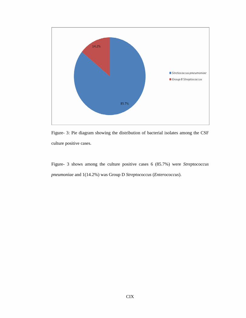
CIX
Figure- 3: Pie diagram showing the distribution of bacterial isolates among the CSF
culture positive cases.
Figure- 3 shows among the culture positive cases 6 (85.7%) were Streptococcus
pneumoniae and 1(14.2%) was Group D Streptococcus (Enterococcus).
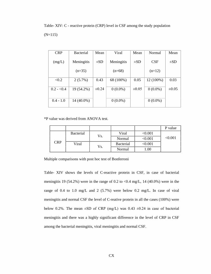
CX
Table- XIV: C - reactive protein (CRP) level in CSF among the study population
(N=115)
CRP
(mg/L)
Bacterial
Meningitis
(n=35)
Mean
±SD
Viral
Meningitis
(n=68)
Mean
±SD
Normal
CSF
(n=12)
Mean
±SD
<0.2 2 (5.7%) 68 (100%) 12 (100%)
0.2 - <0.4 19 (54.2%) 0 (0.0%) 0 (0.0%)
0.4 - 1.0 14 (40.0%)
0.43
±0.24
0 (0.0%)
0.05
±0.05
0 (0.0%)
0.03
±0.05
*P value was derived from ANOVA test.
P value Viral <0.001 Bacterial
Vs. Normal <0.001
Bacterial <0.001
CRP Viral Vs.
Normal 1.00
<0.001
Multiple comparisons with post hoc test of Bonferroni
Table- XIV shows the levels of C-reactive protein in CSF, in case of bacterial
meningitis 19 (54.2%) were in the range of 0.2 to <0.4 mg/L, 14 (40.0%) were in the
range of 0.4 to 1.0 mg/L and 2 (5.7%) were below 0.2 mg/L. In case of viral
meningitis and normal CSF the level of C-reative protein in all the cases (100%) were
below 0.2%. The mean ±SD of CRP (mg/L) was 0.43 ±0.24 in case of bacterial
meningitis and there was a highly significant difference in the level of CRP in CSF
among the bacterial meningitis, viral meningitis and normal CSF.

CXI
Table- XV: Antimicrobial susceptibility pattern of the Streptococcus pneumoniae
isolates (n=06) by disc diffusion method
Name of the Antimicrobial agents
Sensitive (S) Intermediate (I) Resistant (R)
Chloramphenicol 6(100.0%) 0(0.0%) 0(0.0%)
Ciprofloxacin 3(50.0%) 3(50.0%) 0(0.0%)
Penicillin G 2(33.3%) 4(66.6%) 0(0.0%)
Ampicillin 2(33.3%) 4(66.6%) 0(0.0%)
Co-trimoxazole 2(33.3%) 3(50.0%) 1(16.6%)
Gentamicin 2(33.3%) 3(50.0%) 1(16.6%)
Erythromycin 1(16.6%) 4(66.6%) 1(16.6%) Ceftazidim 0(0.0%) 0(0.0%) 6(100.0%)
Table- XV shows the antibiotic susceptibility of six different strains of Streptococcus
pneumoniae. All the stains (100%) were sensitive to chloramphenicol. Sensitivity to
each of penicillin G, ampicillin, gentamicine and co-trimozazole was 33.3%, whereas
that to ciprofloxacin and erythromycin was 50.0% and 16.6% respectively. All the
strains (100%) were resistant to caftazidim.
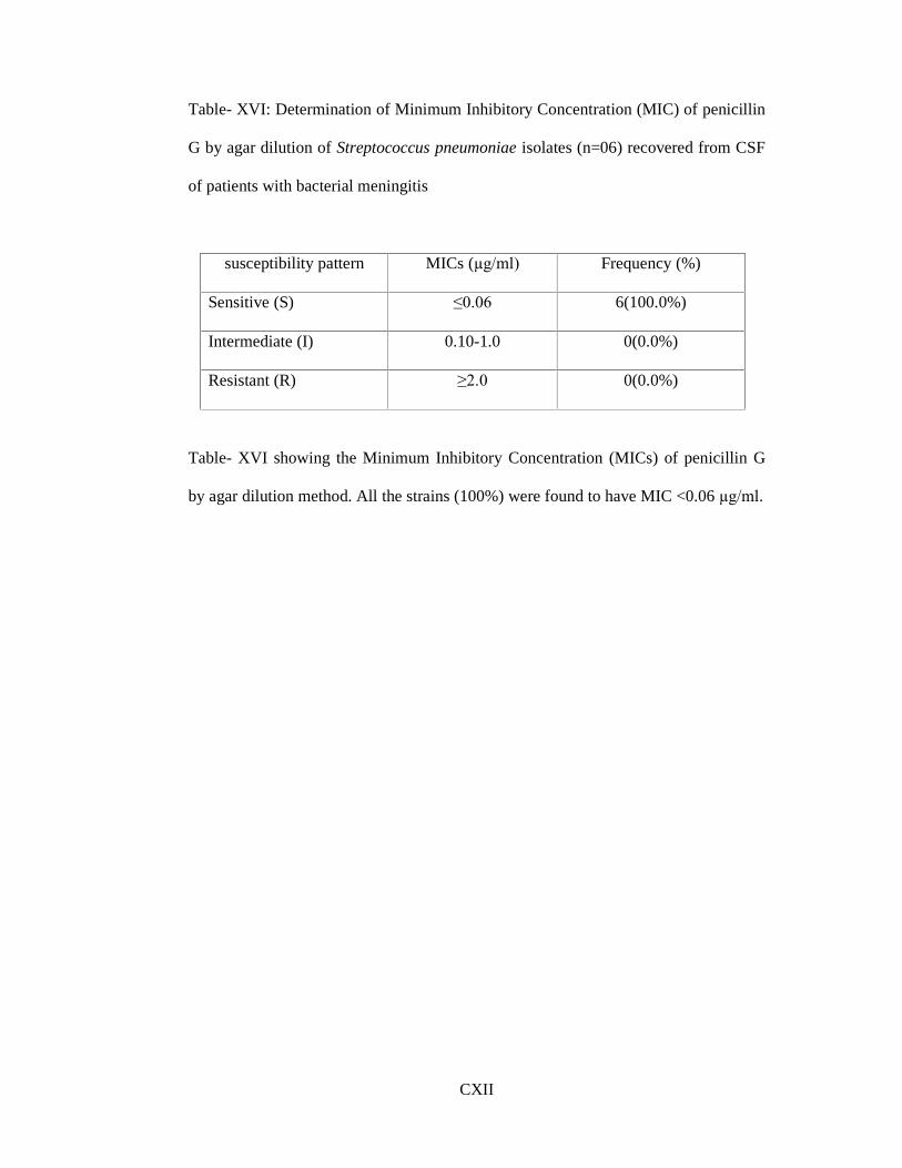
CXII
Table- XVI: Determination of Minimum Inhibitory Concentration (MIC) of penicillin
G by agar dilution of Streptococcus pneumoniae isolates (n=06) recovered from CSF
of patients with bacterial meningitis
susceptibility pattern MICs (µg/ml) Frequency (%)
Sensitive (S) ≤0.06 6(100.0%)
Intermediate (I) 0.10-1.0 0(0.0%)
Resistant (R) ≥2.0 0(0.0%)
Table- XVI showing the Minimum Inhibitory Concentration (MICs) of penicillin G
by agar dilution method. All the strains (100%) were found to have MIC <0.06 µg/ml.

CXIII
Chapter � 5
Discussion
CXIV
DISCUSSION
Bacterial meningitis is still a very common and serious disease (Abro et al. 2008).
Globally 1.2 million cases of bacterial meningitis are estimated to occur every year
with 135,000 deaths (Alam et al. 2007). The incidence of bacterial meningitis varies
from 0.022% to 0.266% in newborns, more common in developing countries (Silva et
al. 2007). The case fatality rates (CFRs) in bacterial meningitis was 26% in developed
countries even with antimicrobial therapy and availability of advanced intensive care,
which were higher ranging from 16-32% in developing countries (Afifi et al. 2007;
Mani et al. 2007). Permanent neurological complications may occur in up to 50% of
survivors even having antimicrobial therapy (Welinder-Olsson et al. 2007). Gurley et
al. (2009) from Bangladesh reported that among all meningitis cases bacterial
meningitis was 25% and was also responsible for 14% case fatality rate.
This cross sectional study was carried out with an aim to detect bacterial meningitis in
children by Latex agglutination test (LAT) for bacterial antigen and estimation of C -
reactive protein (CRP) level in CSF and to compare with the conventional methods.
Amplification of LytA gene of Streptococcus pneumoniae by Polymerase chain
reaction (PCR) from cerebrospinal fluids (CSF) were also carried out in this study.
LAT, PCR and estimation of CRP from CSF are rapid easier and reliable technique.
In developing countries like Bangladesh where culture facilities are not available in
all the centers, these parameters may play an important role in the diagnosis of
bacterial meningitis. A total of 115 clinically suspected meningitis patients age
ranging from 0 day to 18 years were included in the study, CSF samples were

CXV
collected and analyzed by the above mentioned tests for detection of bacterial
meningitis during the period from July 2010 to January 2012.
In our study, on the basis of cytological and biochemical examination of CSF, the
study population was categorized into three groups. We found bacterial meningitis 35
(30.4%) cases, viral meningitis 68 (59.1%) cases and normal CSF 12 (10.4%) cases
(Table- I). Alamgir et al. (2008) from Bangladesh and Nussinovitch et al. (2009) from
Israel also observed similar categories of the study populations in their study. Alamgir
et al. (2008) in Bangladesh had observed identical categories of the patients having
clinically suspected meningitis where they had observed bacterial meningitis 38
(25.34%), aseptic meningitis 94 (62.66%) and non meningitis 18 (12.0%). This
finding is consistent with the present study. Similarly, Narchi in Saudi Arabia (1997)
observed in his study that 35 (35.7%) were bacterial meningitis and 63 (64.3%) were
aseptic meningitis, which are comparable with the present study. Similar findings
were also reported by Nussinovitch et al. (2009) from Israel, Gurley et al. (2009) and
Chowdhury et al. (1992) from Bangladesh, where they found 19.74%, 24.0% and
20.0% of bacterial meningitis cases respectively.
In this study, the age of the study population ranges from 20 days to 10 years in case
of bacterial meningitis, 14 days to 10 years in case of viral meningitis and 3 months to
8 years in case of normal CSF findings (Table- II). Das et al. (2003) also observed
parallel age range among the total study population, where they found the age ranges
from 3 months to 13 years of age in their study. In the present study, majority of study
population 42.6% were in the age range from 1 month to 1 year and 41.7% were in
the age range from 1 year to 5 years. Chowdhury et al. (1992) from Bangladesh also

CXVI
found 49.6% cases in the age ranges from 1 month to 1year and 26.2% cases in the
age ranges from 1 month to 5 years which are almost similar to the present study.
In this present study, among the bacterial meningitis, the male and female ratio was
65.7:34.2, in viral meningitis 54.4:45.5 and in normal CSF 58.3:41.6 (Table- IIIa). As
regards to the incidence of meningitis in male and female, Taskin et al. (2004)
observed in their study, male and female ratio was 59.1:40.9 in bacterial meningitis,
72.7:27.3 in viral meningitis and 60:40 in normal CSF findings which are comparable
with the present study. Similar findings were reported by Das et al. (2003) in India.
The results of the present study closely resemble with the above mentioned study. No
significant difference was found among the three groups regarding the sex incident in
this study. The higher rate of positivity of bacterial meningitis in male subjects may
be due to inclusion of higher number of male patients in this study population.
In the present study, all the cases of study population were presented with fever
(100%). In case of bacterial meningitis 45.7% and 60.0% were presented with nausea
or vomiting and convulsion respectively. In case of viral meningitis 70.5% and 64.7%
were presented with nausea or vomiting and convulsion respectively and in case of
normal CSF findings nausea or vomiting and convulsion were presented 33.3% and
75.0% of cases respectively (Table- IV). This findings of the present study is very
similar with the study of Taskin et al. (2004) where they also found fever in 100%
cases, 27.2% and 31.8% were presented with nausea or vomiting and convulsion
respectively among the bacterial meningitis and in case of viral meningitis 54.5% and
31.8% were presented with nausea or vomiting and convulsion respectively. They
also found in their study, among the non-meningitis group 90.0% of cases having
convulsion and 10.0% of cases having nausea or vomiting.
CXVII
In the present study, 7(20.0%) cases yielded positive by culture, 6(17.1%) cases
positive in Gram�s stain, increased WBC count in 35(100.0%) cases with neutrophilia,
with low glucose contents and elevated protein level were in all cases (100%) of the
bacterial meningitis. These findings of culture, Gram�s stain and biochemical test in
bacterial meningitis patients are almost similar to a study by Chowdhury et al (1992)
in Bangladesh. Ceyhan et al (2008) in Turkey had observed similar findings in their
study where cultures were positive in 10.0% of cases. Begum et al. (2007) in
Bangladesh also found Gram�s stain positive 13.3% in their study which were almost
similar with this study. Alam et al. (2007) in Bangladesh and Das et al. (2003) in
India reported in their research 13.7% and 06.0% bacterial isolation among bacterial
meningitis cases respectively which are similar with this study.
Among the 68 viral meningitis cases it was observed that WBC count were >5 / mm3
in all cases with predominant lymphocyte in all the cases 68(100.0%), protein level
were <45 mg/dl in 42(61.7%) cases and glucose contents were <45 mg/dl in
10(14.7%) cases. It was observed that culture, Gram�s stain, cytology and
biochemical test were comparable with Saha et al. (2005) from Bangladesh, where
they found negative CSF culture and Gram�s stain with cell count 06-99 / mm3 with
lymphocytosis, normal protein and glucose level.
Among the 12 normal CSF cases it was observed that WBC count was ≤5 / mm3 in all
cases, where all the cells are lymphocytes 12 (100.0%), protein level were <45 mg/dl
in 9 (75.0%) cases and glucose contents were >45 mg/dl in 11 (91.6%) cases. It was
observed that culture, Gram�s stain, cytology and biochemical test were analogous
with Alamgir et al. (2008) in Bangladesh, where they found culture and Gram�s stain
CXVIII
negative with normal CSF cytology and normal protein and glucose level. In USA
similar findings was observed by Dulkerian et al (1995).
In this study, the Table- IX showed that the total count of WBC (per cubic mm) was
increased (1623.06 ±1708.06), the level of glucose (mg/dl) was reduced (21.04 ±9.77)
and the level of protein (mg/dl) was elevated (242.77 ±188.08) in bacterial meningitis.
So, these findings proved that the conventional methods may also be a useful tool for
categorizing the meningitis as bacterial meningitis, even in the absence of facilities
for doing other methods.
In this study it was observed that among the 35 bacterial meningitis cases Gram�s
stain, culture and LAT positive were in 15 (42.8%). Gram�s stain negative, culture
negative and LAT positive were in 9 (25.7%) cases. Gram�s stain�s negative, culture
positive and LAT positive were in 1(02.8%) cases. The rate of detection of 35
bacterial meningitis cases by different methods showed that culture positive were in 7
(20.0%) cases, Gram�s stain positive 6 (17.1%) cases and LAT positive were in 15
(42.8%) cases (Table- X). In this study culture positive cases were more than gram�s
stain cases it may be due to less bacterial load in this CSF specimen. The results of the
present study resemble with the Abro et al. (2008) and Alamgir et al. (2008) where
they also found gram�s stain positivity was less than culture in their study. Alamgir et
al. (2008) from Bangladesh also mentioned that Gram�s stain and culture may
decrease to 40%-60% and <50% respectively in patients who have received prior anti-
microbial therapy. Gray and Fedorko (1992) mentioned that in Gram�s stains,
concentrations <103 CFU/ml of CSF are associated with positive findings in 25%
whereas concentrations of bacteria >105 CFU/ml of CSF lead to positive results in up
to 97% of cases. The low frequency of Gram�s staining positive and culture positive
CXIX
in this study may be due to antibiotic intake in higher number of cases prior to CSF
collection.
In the present study CSF culture had yielded growth of Streptococcus pneumoniae in
6 (85.7%) cases and Group D Streptococcus in 1(14.2%) case among the 7 CSF
culture positive cases (Figure- 1). In this study among 35 bacterial meningitis cases 22
(62.8%) as Streptococcus pneumoniae was diagnosed either by LAT or by detection
of LytA gene by PCR (Table- XI). In case of 12 (34.3%) cases no organism could be
detected. Das et al. (2003) from India found Streptococcus pneumoniae in (61.0%),
Haemophilus influenzae type b in (19.0%), N. meningitidis A in (3%) and no
organisms could be detected in 17.0% cases, which are comparable with present
study. Mani et al. (2007) from India also reported Streptococcus pneumoniae as
predominant pathogen (61.8%) in their study which are almost consistent with the
present study. Cherian et al. (1998) had mentioned in their study the incidence of
invasive disease due to Haemophilus influenzae type B infection reduced in most
developed countries and except during an epidemic of meningococcal infection,
Streptococcus pneumoniae is the commonest cause of acute bacterial meningitis in
children. ICDDR.B (2011) reported that, in Bangladesh a combined vaccine for
Haemophilus influenzae type b was introduced on 15 January 2009 and it was
included in expanded programme on immunization (EPI) of the government of
Bangladesh. Haemophilus influenzae type b and Neisseria meningitidis were not
detected in this present study may be due to this reason. The undetected (34.3%)
cases of bacterial meningitis in this present study may be is due to LAT would only be
positive only in the presence of specific polysaccharide surface antigens for
Streptococcus pneumoniae, Haemophilus influenzae type b, group B Streptococcus,
Neisseria meningitidis group A, C, Y, W135, Neisseria meningitidis B and
CXX
Escherichia coli k1 and the primer was used for PCR in this study was specific for
detection of LytA gene of Streptococcus pneumoniae. Begum et al. (2007) in
Bangladesh and Das et al. (2003) in India were also found this limitation of LAT in
their studies which are similar with the present study.
Breton et al. (2002) mentioned in their study that bacterial meningitis may be caused
by Enterococcus in 0.3-4.0% cases which is very similar to the present study where
Enterococcus was found in 2.7% of all 35 cases of bacterial meningitis. Aletayeb,
Ahmad and Masood et al. (2010) in Iran found in their study 9.6% isolation rate of
Enterococcus. Khan et al. (2011) in India found 4.5% Enterococcus faecalis which is
comparable with the present study.
In this present study LAT were positive in 42.8% of cases of bacterial meningitis.
Mirdha, Gupta and Bhujwala (1991) from India and Begum et al (2007) from
Bangladesh also found LAT positive cases in 42.0% and 40.0 % cases of bacterial
meningitis respectively which are consistent with the present study. The sensitivity
and specificity of LAT method was determined by taking CSF culture as gold
standard method for detection of Streptococcus pneumoniae. In this study, culture
positive (6) for Streptococcus pneumoniae were also positive by LAT (6/6), so the
sensitivity was 100%. On the other hand culture negative (20) were also negative by
LAT (20/20), so the specificity was also found 100% (Table- XIII). Camargos et al.
(1995) and Das et al. (2003) found in their study the sensitivity and the specificity of
LAT was 86.5% and 100.0% and 83% and 100% respectively for detection of
Streptococcus pneumoniae.
The sensitivity and specificity of PCR method was determined by taking CSF culture
as gold standard method for Streptococcus pneumoniae. In this study, culture positive
CXXI
(6) cases for Streptococcus pneumoniae were also positive by PCR (6/6), so the
sensitivity was 100%. On the other hand culture negative (13) cases were also
negative by PCR (13/13), so the specificity was also found 100%. So in this present
study the sensitivity and specificity of PCR for detection of LytA gene of
Streptococcus pneumoniae were 100% and 100% respectively (Table- XII). Alam et
al. (2007) in Bangladesh were also reported in their study the specificity and
sensitivity of PCR for detection of LytA gene of Streptococcus pneumoniae were
100% and 100% respectively, which are very similar with this present study.
In this study, CRP of CSF was found normal (<0.2 mg/L) in all (100%) cases of viral
meningitis and normal CSF cases and higher (>0.2 mg/L) in 33 (94.2%) cases of
bacterial meningitis (Table- XV). Among the 7 culture positive cases the levels of
CRP in CSF were within the range of 0.59 mg/L to 0.97 mg/L. Coric et al. (2012)
mentioned that the mean CRP for patients with bacterial meningitis was 21.4 mg/L
(range 0.4 to 100 mg/L) and in uninfected control group 0.25 mg/L (range 0.1 to 0.5
mg/L). Bowers (1985) reported in his study, the levels of CRP in CSF of
pneumococcal meningitis were <0.2 mg/L in 49%, 0.2 to 1 mg/L in 11.76%, 1 to 5
mg/L in 27.45% and >5 mg/L in 11.76% of cases. Philip (2003) mentioned that CRP
level above 0.40 mg/L was substantial for bacterial meningitis. Shameem, Kumar and
Neelagund (2008) found in their study that, the elevated level of CRP in CSF was
29% of pretreated bacterial meningitis and 54.66% cases of untreated bacterial
meningitis. However Coric et al. (2012) and Bowers (1985) found higher level of
CRP in CSF in bacterial meningitis in their study and we found lower level, which
can be explained by antibiotic therapy prior to collection of CSF.
CXXII
In this study, the mean ±SD of CRP (mg/L) was 0.43 ±0.24 in bacterial meningitis,
0.05 ±0.05 in viral meningitis and 0.03 ±0.05 in normal CSF cases. So, there was a
highly significant difference in level of CRP in CSF between the bacterial meningitis,
viral meningitis and normal CSF.
In this study, the antibiotic susceptibility of six different strains of Streptococcus
pneumoniae was carried out by disc diffusion method. In this method, all the stains
(100%) were sensitive to chloramphenicol. Sensitivity to each of penicillin G,
ampicillin, gentamicin and co-trimoxazole was 33.3%, whereas that to ciprofloxacin
and erythromycin was 50.0% and 16.6% respectively. All the strains (100%) were
resistant to caftazidim (Table- XV). Alamgir et al. (2008) from Bangladesh found
81.8%, 81.8%, 72.7%, 90.0% and 81.8% sensitivity to penicillin, ampicillin,
gentamicin, erythromycin and chloramphenicol respectively, whereas Alam et al.
(2007) from the same country found 100% sensitivity to penicillin, ampicillin,
ciprofloxacin and chloramphenicol and 95.0%, 60.0% and 5.0% resistance to
gentamicin, co-trimoxazole and erythromycin respectively.
In the present study the minimum inhibitory concentration (MIC) of penicillin G was
determined by Agar dilution method, all isolates (06) of Streptococcus pneumoniae
recovered from CSF of patients with bacterial meningitis were sensitive (100%)
(Table- XVI). Though Llull, Lopez and Garcia (2006) and Alam et al. (2007) reported
in their study, the resistance of Streptococcus pneumoniae to penicillin is increasing
in many parts of the world. Alam et al. (2007) in Bangladesh found 90% isolates of
Streptococcus pneumoniae from CSF were sensitive to penicillin G in MIC, which is
comparable with the present study.
CXXIII
Chapter � 6
Conclusion and recommendations
CXXIV
Conclusion and recommendations
Analyzing the findings of the present study, it can be concluded that cytological
examination and biochemical test is still helpful for diagnosis of bacterial meningitis,
but Gram�s stain and culture have some limitations. Among the rapid diagnostic
procedures detection of antigen from CSF by LAT is easier, rapid and highly sensitive
and specific method and suitable to perform at all the levels including Upazilla health
complex (UHC). So, efforts should be made to introduce LAT from CSF at Upazilla
level or at least at district level of Bangladesh.
Though the CRP in CSF is increased in all bacterial meningitis cases but the level was
not highly increased, which may be due to use of prior antibiotic therapy.
In contrast, the PCR method was much superior to other methods yielding very high
sensitivity and specificity. Additionally, this method does not need presence of any
viable organism and can detect even when the pathogenic burden is very low. Thus,
despite the requirement of extensive infrastructure and specialized skilled personnel
for PCR method, which restricts its availability everywhere, especially in developing
countries, it can be made available in the reference centers for utilizations by other
healthcare facilities through sample referral system. So it is strongly recommended to
take necessary steps to setup and start PCR at least in the tertiary care hospitals.

CXXV
Chapter � 7
Limitation
CXXVI
Limitation
1. In this study the sample size was small than the estimated sample size due to
limitation of budget, time and resource.
2. We used only one primer (LytA gene) from a number of primers, specific for
pneumococcus and primers for other causative organisms were not included.

CXXVII
Chapter-8
List of references
CXXVIII
List of references
Abro, AH, Abdou, AH, Ali, H, Ustadi, AM and Hasab, AAH 2008, �Cerebrospinal
fluid analysis acute bacterial versus viral meningitis�, Pakistan Journal of Medical
Sciences, vol. 24, no.5, pp. 645- 650.
Alam, MR, Saha, SK, Nasreen, T, Latif, F, Rahman, SR and Gomes, DJ 2007,
�Detection, Antimicrobial Susceptibility and Serotyping of Streptococcus pneumoniae
from Cerebrospinal Fluid Specimens from Suspected Meningitis Patients� ,
Bangladesh j Microbiol, vol. 24, no.1, pp. 24-29.
Alamgir, F, Miah, RA, Saleh, AA 2008,�Bacterial antigen detection and CRP
estimation�, Bangladesh Med J, vol. 13, no. 1, pp. 17-20.
Aletayeb, MH, Ahmad, FS and Masood, D 2010, �Eleven-year study of causes of
neonatal bacterial meningitis in Ahvaz, Iran�, Pediatrics International, vol. 52, no. 3,
pp 463�466.
Alonsodevelasco, E, Verheul, AFM, Verhoef, J and Snippe, H 1995, �Streptococcus
pneumoniae: Virulence Factors, Pathogenesis, and Vaccines�, MICROBIOLOGICAL
REVIEWS, vol. 59, no. 4, pp. 591-603.
Afifi, S, Wasfy, MO, Azab, MA, Youssef, FG, Pimentel, G, Graham, TW, Mansour,
H, Elsayed, N, Earhart, K, Hajjeh, R and Mahoney, F 2007, �Laboratory based
surveillance of patients with bacterial meningitis in Egypt (1998-2004)�, Eur J Clin
Microbiol Infect Dis, vol. 26, pp. 331-340.
CXXIX
Balagavi, AC and Shalini, M 2011, �Cerebrospinal Fluid C Reactive Protein and
Adenosine Deaminase in Meningitis in Adults�, JAPI, vol. 59, pp. 557-560.
Begum, N, Ahmed, I, Salam, Md.A, Begum, S and Alam, KMF 2007, �Role of Latex
Particle Agglutination Test in the Diagnosis of Meningitis�, Bangladesh J Med
Microbiol, vol. 1, no. 1, pp. 10-12.
Bowers, GN 1985, �Significance of C-Reactive Protein in Spinal Fluid�, CLINICAL
CHEMISTRY, vol. 31, no. 2, pp. 345.
Breton, JR, Peset, V, Morcillo, F, Cano, J, Sarrion, A, Perez-Belles, C and
Gobernado, M 2002, �Neonatal meningitis due to Enterococcus spp: presentation of
four cases�, Enferm Infecc Microbiol Clin, vol. 20, no. 9, pp. 443-447.
Brooks, GF, Butel, JS and Morse, SA 2004, �The Streptococci�, in Jawetz, Melnick, &
Adelberg�s Medical Microbiology, 23rd edition, The McGraw-Hill Companies,
International edition, pp. 231-247.
Brouwer, MC, Tunkel, AR and Beek, DVD 2010, �Epidemiology, Diagnosis, and
Antimicrobial Treatment of Acute Bacterial Meningitis�, CLINICAL
MICROBIOLOGY REVIEWS, vol.23, no.3, pp. 467�492.
Brice, JL, Tornabene, TG and LaForce, FM 1979, �Diagnosis of bacterial meningitis
by gas-liquid chromatography�, J. Infect. Dis, vol. 140, pp. 443-452.
Burnham, CAD and Tyrrell, GJ 2003, �Virulence factors of group B streptococci�,
RMM, vol. 14, no. 4, pp. 109-118.

CXXX
Camargos, PAM, Almedia, MS, Cardoso, I, Filho, GL, Filho, DM, Martins, JI,
Batista, KWB, Silva, RCO and Antunes, CF 1995, �Latex particle agglutination test in
the diagnosis of Haemophilus influenzae type b, streptococcus pneumoniae and
Neisseria meningitidis A and C meningitis in infants and children�, J Clin Epidemiol,
vol. 45, no.10, pp. 1245-1250.
Ceyhan, M, Yildirim, I, Balmer, P, Borrow, R, Dikici, B, Turgut, M, Kurt, N and
Aydogan, A 2008, �A prospective study of etiology of childhood acute bacterial
meningitis, Turkey�, Emerging Infectious Diseases, vol. 14, no. 7, pp. 1089-1096.
Cherian, T, Lalitha, MK, Manoharan, A, Thomas, K, Yolken, RH and Steinhoff, MC
1998, � PCR-Enzyme Immunoassay for Detection of Streptococcus pneumoniae DNA
in Cerebrospinal Fluid Samples from Patients with Culture � Negative Meningitis�, J.
Clin. Micobiol., vol. 36, no. 12, pp. 3605-3608.
Cheesbrough, M 2000, Medical Laboratory Manual for tropical countries, vol.2,
ELBS cambridgeshire, Engl.
Chowdhury, MZU, Rahman, KM, Miah, RA, Satter, H and Hussain, T 1992,
�Bacterial meningitis in children�, Bangladesh Medical Journal, vol. 21, pp. 3-7.
Cobo, F, Cabezas-Fernández, MT and Cabeza-Barrera, MI 2012, �Streptococcus
pneumonia bacteremia: clinical and microbiological epidemiology in a health area of
Southern Spain�, Infectious Disease Reports, vol. 4, pp. 116-119.
Collee, JG, David, JP, Fraser, AG, Marnion, BP and Simmon, SA 1996, �Laboratory
strategy in the diagnosis of infective Syndromes�, in: Mackie and Mc Cartney
CXXXI
Practical Medical Microbiology, 14th ed, Churchill-Livingstone, New York, pp. 77-
80.
Coric, J, Pasic, A, Panjeta, M and Mujic, J 2012, � Evaluation of high sensitivity C-
reactive protein assay in cerebrospinal fluid on the Dimension RxL analyzer�, Journal
of Health Sciences, vol. 2, no. 1, pp. 13-16.
Das, BK, Gurbacharya, Mohapatra, TK and Misra, OP 2003, �Bacterial antigen
detection test in meningitis�, Indian Journal of Pediatrics, vol. 70, pp. 799-801.
Daum, RS, Granoff, DM, Gilsdorf, J, Murphy, T and Osterholm, MT 1986,
�Haemophilus influenzae type b infections in day care attendees: implications for
management�, Rev Infect Dis, vol. 8, no. 4, pp. 558-567.
Dean, A, Seehusen, MD, Mark, M, Reeves, MD, Demitri, A and Fomin, MD 2003,
�Cerebrospinal Fluid Analysis�, Am Fam Physician, vol. 68, no. 6, pp. 1103-1109.
Deivanayagan, N, Ashok, TP, Nedunchelian, K, Ahmed, SS and Mala, N 1993,
�Bacterial Meningitis. Diagnosis by Latex agglutination test and clinical features�,
Indian Pediatr, vol. 30, pp. 495-500.
Drevets, DA 1998, �Listeria monocytogenes Virulence Factors That Stimulate
Endothelial Cells�, INFECTION AND IMMUNITY, vol. 66, no. 1, pp. 232-238.
Dulkerian, SJ, Kilpatrick, L, Costarino, AT, McCawley, L, Fein, J, Corcoran, L, Zirin,
S and Harric, MC 1995, �Cytokine elevations in infants with bacterial and aseptic
meningitis�, J Pediatr, vol. 126, pp. 872-6.
CXXXII
Feigin, RD and Pearlman, E 1998, �Bacterial Meningitis beyond the neonatal period�,
in Text Book of pediatrics infectious diseases, Feigin, RD and Cherry, JD, 4th edition,
W.B. Saunders Company, Philadelphia.
Feng, P, Weagant, SD and Grant, MA 2007, �Enumeration of Escherichia coli and the
Coliform Bacteria�, in Bacteriological Analytical Manual, 8th ed., FDA/Center for
Food Safety and Applied Nutrition.
http://www.fda.gov/Food/ScienceResearch/LaboratoryMethods/BacteriologicalAnalyt
icalManualBAM/ucm064948.htm
Fuller, DG, Duke, T, Shann, F and Curtis, N 2003, �Antibiotic treatment for bacterial
meningitis in children in developing countries�, Ann Trop Paediatr, vol. 23, no. 4, pp.
233-53.
Furtado, GL and Medeiros, AA 1980, �Single-Disk Diffusion Testing (Kirby-Bauer)
of Susceptibility of Proteus mirabilis to Chloramphenicol: Significance of the
Intermediate Category�, JOURNAL OF CLINICAL MICROBIOLOGY, vol. 12, no.
4, pp. 550-553.
Forbes, BA, Sahm, DF and Weissfeld, AS 2002, Streptococcus, Enterococcus,
and Similar Organisms in Bailey & Scott�s, Diagnostic microbiology. 11th ed.
Mosby, Inc. St. Louis, Missouri, USA, pp. 298-314.
Gershom, E, Briggeman, GJJ and Zegher, F 1986, �Cerebrospinal fluid C-reactive
protein in meningitis: diagnostic value and pathophysiology�, EUROPEAN JOURNAL
OF PEDIATRICS, vol. 145, no. 04, pp. 246-249.
Ghai, CL 2005, A Text book of PRACTICAL PHYSIOLOGY, 6th edition, Jaypee
Brothers Medical Publishers (P) Ltd, New Delhi, India, pp. 52-53.

CXXXIII
Gilsdorf, JR, Marrs, CF and Foxman, B 2004, �Haemophilus influenzae: Genetic
Variability and Natural Selection To Identify Virulence Factors�, INFECTION AND
IMMUNITY, vol. 72, no. 5, pp. 2457-2461.
Gilsdorf, JR 1998, �Antigenic Diversity and Gene Polymorphisms in Haemophilus
influenzae�, INFECTION AND IMMUNITY, vol. 66, no. 11, pp. 5053-5059.
Graham, TP 2003, �Myth: Cerebrospinal fluid analysis can differentiate bacterial
meningitis from aseptic meningitis�, Can J Emerg Med, vol. 5, no. 5, pp. 348-349.
Gray, LD and Fedorko, DP 1992, �Laboratory Diagnosis of Bacterial Meningitis�,
CLIN. MICROBIOL. REV , vol. 5, no. 2, pp. 130-145.
Greenlee, J. E 1990, �Approach to diagnosis of meningitis.Cerebrospinal fluid
evaluation�, Infect. Dis. Clin. N. Am, vol. 4, pp.583-597.
Gurley, ES, Hossain, MJ, Mountgomery, SP, Petersen, LR, Sejvar, JJ, Mayer, LW,
Anne, W, Dull P, Nahar, N, Uddin, AKMR, Rahman, ME, Ekram, ARMS, Luby, SP
and Breiman, RF 2009, � Etiologies of Bacterial Meningitis in Bangladesh: Results
from a Hospital Based Study�, The American Society of Tropical Medicine and
Hygiene, vol. 81, pp. 475- 483.
Hart, CA 2009, �Bacterial Meningitis�, in MANSON�S Tropical Diseases. Medicine,
22nd edition, Saunders Elsevier, pp. 873-885.
Haslett, C, Chilvers, ER, Boon, NR and Colledge, NR 2002, Davidson�s, Principles
and Practice of Medicine, 19th edition, Churchill Livingstone, Edinburgh, pp. 1193-
1199.
CXXXIV
Herrera-Luna, C, Klein, D, Lapan, G, Revilla-Fernandez, S, Haschek, B, Sommerfeld-
Stur, I, Moestl, K, Baumgartner, W 2009, �Characterization of virulence factors in
Escherichia coli isolated from diarrheic and healthy calves in Austria shedding
various enteropathogenic agents�, Veterinarni Medicina, vol. 54, no. 1, pp. 1-11.
ICDDR,B 2011, �Hib vaccine introduced into routine immunization in Bangladesh�,
<http://www.supportforlife.org/media-centre/news/2016-hib-vaccine-introduced-into-
routine-immunization-in-bangladesh>
Jane, D 2011, �Cerebrospinal Fluid� , Nursingcrib.
< http://nursingcrib.com/anatomy-and-physiology/cerebrospinal-fluid/>
Jacewicz, M 2009, �Neurological Disorders�, in Merck Online Manual, Home edition,
Merck and Co., USA.
Jimenez, SMM and Castro, CN 2004, �Validation of a PCR for diagnosis of typhoid
fever and salmonellosis by amplification of the hilA gene in clinical samples from
Colombian patients�, Journal of Medical microbiology, vol. 53, pp. 875-878.
Johnson, KS and Sexton, DJ 2012, �Cerebrospinal fluid: Physiology and utility of an
examination in disease states�, Wolters Kluwer Health UpTo Date.
Johnson, TJ and Nolan, LK 2009, �Pathogenomics of the Virulence Plasmids of
Escherichia coli�, MICROBIOLOGY AND MOLECULAR BIOLOGY REVIEWS, vol.
73, no. 4, pp. 750�774.
CXXXV
<http://www.uptodate.com/contents/cerebrospinal-fluid-physiology-and-utility-of-an-
examination-in-disease-states>
Joklik, WK, Willett, HP, Amos, DB and Wilfert, CM 1992, Zinsser Microbiology,
20th edition, Prentice-Hall international Inc.
Kelly, ML, Jacobs, MR and Appelbaum, PC 1999, �Comparison of Agar Dilution,
Microdilution, E-Test, and Disk Diffusion Methods for Testing Activity of Cefditoren
against Streptococcus pneumonia� , Journal Of Clinical Microbiology, vol. 37, no.
10, pp. 3296-3299.
Khan, MR and Rahman, ME As 2003, �Meningis� , in Essence of pediatrics, 3rd
edition, Anwara Khan, Dhaka, pp. 463-468.
Khan, F, Rizvi, M, Fatima, N, Shukla, I, Malik, A and Khatoon, R 2011, �Bacterial
meningitis in North India: Trends over a period of eight years�, Neurology Asia, vol.
16, no.1, pp. 47�56.
Kim, KS 2010, �Acute bacterial meningitis in infants and children�, Lancet Infect Dis,
vol. 10, pp. 32-42.
Kiwanuka, JP and Mwanga, J 2001, �Childhood bacterial meningitis in Mbarara
Hospital, Uganda: antimicrobial susceptibility and outcome of treatment�, African
Health Sci, vol. 1, no. 1, pp. 9-11.
Kornelisse, RF, Westerbeek, CML, Spoor, AB, Heijde, BVD, Spanjaard, L, Neijens,
HJ and Groot, RD 1995, �Pneumococcal Meningitis in Children: Prognostic Indicators
and Outcome�, Clinical Infectious Diseases, vol. 21, pp. 1390-1397.
CXXXVI
Leinonen, M and Kayhty, H 1978,�Comparison of counter-current
immunoelectrophoresis, latex agglutination, and radioimmunoassay in detection of
soluble capsular polysaccharide antigens of Haemophilus influenzae type b and
Neisseria meningitidis of groups A or C�, Journal of CLINICAL PATHOLOGY, vol.
31, no. 12, pp. 1172-1176.
Leonard, J, Scolea, LAJR and Dryja, D 1984, �Quantitation of Bacteria in
Cerebrospinal Fluid and Blood of Children with Meningitis and its Diagnostic
Significance�, J of clin Microbiol, vol. 19, no. 2, pp. 187-190.
Llorens, XS and McCracken, GH 2003, �Bacterial meningitis in children�, The
Lancet, vol. 361, pp. 2139-2148.
Llull, D, Lopez, R and Garcia 2006, �Characteristic Signatures of the lytA Gene
Provide a Basis for Rapid and Reliable Diagnosis of Streptococcus pneumoniae
Infections�, JOURNAL OF CLINICAL MICROBIOLOGY, vol. 44, no. 4, pp. 1250�
1256.
Mani, R, Pradhan, S, Nagarathna, S, Wasiulla, R and Chandramuki, A 2007,
Bacteriological profile of community acquired acute bacterial meningitis: A ten-year
retrospective study in a tertiary neurocare centre in South India�, vol. 25, no. 2, pp.
108-114.
CXXXVII
Manchanda, V, Gupta, S and Bhalla, P 2006, �MENINGICOCCAL DISEASE:
HISTORY, EPIDEMIOLOGY, PATHOGENESIS, CLINICAL MANIFESTATIONS,
DIAGNOSIS, ANTIMICROBIAL SUSCEPTIBILITY AND PREVENTION�, Indian
Journal of Medical Microbiology, vol. 24, no. 1, p. 7-19.
McKAYt, DB and LU, CY 1991, �Listeriolysin as a Virulence Factor in Listeria
monocytogenes Infection of Neonatal Mice and Murine Decidual Tissue�,
INFECTION AND IMMUNITY, vol. 59, no. 11, pp. 4286-4290.
Mirdha, BR, Gupta, U and Bhujwala, RA 1991, �Latex Agglutination Test: An
Adjunct to the Laboratory Diagnosis of Pyogenic Bacterial Meningitis�, Indian J
Pediatr, vol. 58, pp. 521-524.
Muangchana, C, Chunsuttiwat, S, Rerks-Ngarm, S and Kunasol, P 2009, �Bacterial
meningitis incidence in Thai children estimated by a rapid assessment tool
(RAT)�, Southeast Asian J Trop Med Public Health, vol. 40(3), pp.553-62.
Munson, RS, Kabeer, MH, Lenoir, AA and Granoff, DM 1989, �Epidemiology and
prospects for prevention of disease due to Haemophilus influenzae in developing
countries�, Ref Infect Dis, vol. 3, pp. 588-597.
Narchi, H 1997, �CSF Bacterial Antigen Detecton Testing in the dlagnosis of
meningitis�, Annals of Saudi Medicine, vol. 17, no. 1, pp. 101-103.
Negrini, B, Kelleher, KJ and Wald, ER 2000, �Cerebrospinal Fluid findings in Aseptic
Versus Bacterial Meningitis�, Pediatrics, vol. 105, pp. 316-319.
Nagarajan, AG, Karnam, G, Lahiri, A, Allam, US, Chakravortty, D et al 2009,
�Reliable means of diagnosis and serovar determination of blood borne salmonella
CXXXVIII
strains. Quick PCR Amplification of Unique Genomic Loci by Novel primer sets�,
Journal of Clinical Microbiology, vol. 47, no. 8, pp. 2435-2441.
Nussinoovitch, M, Finkelstein, Y, Elishkevitz, KP 2009, �Cerebrospinal fluid lactate
dehydrogenase isoenzymes in children with bacterial and aseptic meningitis�,
Translational Reseach; the journal of laboratory and clinical medicine (Transl Res),
vol. 154, no. 4, pp. 214-218.
O�Brien, KL, Wolfson, LJ, Watt, JP, Henkle, E, Deloria-Knoll, M, McCall, N, Lee, E,
Mulholland, K, Levine, OS and Cherian, T 2009, �Burden of disease caused by
Streptococcus pneumoniae in children younger than 5 years: global estimates�,
www.thelancet.com, vol. 374, pp. 893-902.
Obata-Yasuoka, M, Ba-Thein W, Tsukamoto, T, Yoshikawa, H and Hayashi, H 2002,
�Vaginal Escherichia coli share common virulence factor profiles, serotypes and
phylogeny with other extraintestinal E. coli�, Microbiology, vol. 148, pp. 2745�2752.
Østergaard, C, Konradsen, HB and Samuelsson, S 2005, �Clinical presentation and
prognostic factors of Streptococcus pneumoniae meningitis according to the focus of
infection�, BMC Infectious Diseases, vol. 5, pp. 93.
Philip, AGS 2003, �Neonatal Meningitis in the New Millennium�, NeoReviews, vol. 4,
no. 3, pp. 73-80. http://neoreviews.aappublications.org/content/4/3/e73.full.pdf+html
Plessis, DM, Smith, AM and Klugman, KP, 1998 �Rapid Detection of Penicillin-
Resistant Streptococcus pneumoniae in Cerebrospinal Fluid by a Seminested-PCR
Stratwgy� , Journal Of Clinical Microbiology, vol. 36, no. 2, pp. 453-457.
Poppert, S, Essig, A, Stoehr, B, Steingruber, A, Wirths, B, Juretschko, S, Reischl, U
and Wellinghausen, N 2005 �Rapid Diagnosis of Bacterial Meningitis by Real-Time
CXXXIX
PCR and Fluorescence In Situ Hybridization�, Journal Of Clinical Microbiology, vol.
43, no. 7, pp. 3390-3397.
Prasad, PL, Nair, MNG and Kalghatgi, AT 2005, �Childhood Bacterial Meningitis and
Usefulness of C-reactive Protein�, MJAFI, vol. 61, no. 1, pp. 13-15.
Prober, CG 2007, �Central nerve system infections�, in Behraman RE, Kliegman RM,
Jenson HB eds, Nelson Textbook of pediatrics, 18th ed W.B Saunders, Philadelphia,
pp. 751-760.
Pushkareva, VI and Ermolaeva, SA 2010, �Listeria monocytoenes virulence factor
Listeriolysin O favors bacterial growth in co-culture with the ciliate Tetrahymena
pyriformis, causes protozoan encystment and promotes bacterial survival inside
cysts�, BMC Microbiology, vol. 10, no. 26, pp. 01-11.
Quagliarello, VJ and Scheld, WM 1997, �TREATMENT OF BACTERIAL
MENINGITIS�, The New England Journal of Medicine, vol. 336, no. 10, pp. 708-716.
Ross, KL and Tylor, KL 2001, �Bacterial Meningitis and other Suppurative Infection�,
in Harrison�s Principles of internal medicine, Braunwald E, Fauci As, Kasper DL,
Hauser SL, Longo DL and Jameson JL, eds.15th ed, vol. I, Mc Graw-Hill, New York,
pp. 2462-2471.
Ross, PW 1996, �Streptococcus Pneumoniae�, in Collee JG, Fraser AG, Marmion BP
and Simmon SA, editor, Mackie and McCartney Practical Medical Microbiology, 14th
ed, Churchill-Livingstone New York, pp. 275-281.

CXL
Saha, SK, Darmstadt, GL, Yamanaka, N, Billal, DS, Nasreen, T, Islam, M and
Hamer, DH 2005,�Rapid Diagnosis of Pneumococcal Meningitis Implications for
Treatment and Measuring Disease Burden� , The Pediatric Infectious Disease
Journal, vol. 24, no. 12, pp. 1093-1098.
Saravolatz, LD, Manzor, O, Vander, NV, Pawlak, J and Belian, B 2003, �Broad-
Range Bacterial Polymerase Chain Reaction for Early Detection of Bacterial
Meningitis�, Clinical Infectious Deseases, vol. 36, pp. 40-45.
Scheld, WM 1998, � Bacterial Meningits�, in Harrison�s Principles of Internal
Medicine, Fauci, AS, Braunwald, E, Esselbacher, KJ, Wilson, JD, Martin, JB, Kasper,
DL, Hauser, SL and Longo, DL, 14th edition, Mc Graw-Hill, New York, pp. 2419-
2426.
Schueler, SJ, Beckett, JH and Gettings, DS 2010, �Bacterial Meningitis Incidence�,
FreeM, <http://www.freemd.com/bacterial-meningitis/incidence.htm>
Schleifer, KH and Kilpper-Balz, R 1984, �Transfer of Streptococcus faecalis and
Streptococcus faecium to the Genus Enterococcus nom. Rev. as Enterococcus faecalis
comb. Nov. and Enterococcus faecium comb. Nov.�, International Journal of
Systematic Bacteriology, vol. 34, no. 1, pp. 31-34.
Schuhmann, MU, Ostrowski, KR, Draper, EJ, Chu, J, Ham, SD, Sood, S and
Mcallister, JP 2005, �The value of C-reactive protein in the management of shunt
infections�, J Neurosurg (Pediatrics 3), vol. 103, pp. 223�230.

CXLI
Seehusen, DA, Reeves, MM and Fomin, DA 2003, �Cerebrospinal Fluid Analysis�,
American Family Physician, vol. 68, no. 6, pp. 1103-1109.
Shameem, S, Kumar, VCS, Neelagund, YF 2008, �Bacterial meningitis: rapid
diagnosis and microbial profile: a multicentered study�, J Commun Dis, vol. 40, no.
2, pp. 111-120.
Sharma, S, Bhat, GK and Shenoy, S 2007, �Virulence factors and drug resistance in
Escherichia coli isolated from extraintestinal infections�, Indian J Med Microbiol,
vol. 25, pp. 369-373.
Shimetani, N, Shimetani, K and Mori, M 2001,�Levels of three inflammation
markers, C-reactive protein, serum amyloid A protein and procalcitonin, in the
serum and cerebrospinal fluid of patients with meningitis�, Scandinavian journal
of clinical and laboratory investigation, vol. 61(7), pp. 567-74.
Silvia, LPAD, Cavalheiro, LG, Queiros, F, Nova, CV and Lucena, R 2007, �Prevalence
of Newborn Bacterial Meningitis and Sepsis During the Pregnancy Period for
Public Health Care System Participants in Salvador, Bahia, Brazil�, BJID, vol. 11,
no. 2, pp. 272-276.
Silva, ME and Marin, JM 2001, �An epidemiological study of Haemophilus
influenzae at a Brazilian day care center�, Braz J Infect Dis, vol.5, no.5.
http://www.scielo.br/scielo.php?script=sci_arttext&pid=S1413-86702001000500004
Sirijaichingkul, S, Tiamkao, S, Sawanyawisuth, K and Chotmongkol, V 2005, �C
Reactive Protein for Differentiating Bacterial from Aseptic meningitis in Thai
Patients� J Med Assoc Thai, vol. 88, no. 9, pp. 1251-1256.
CXLII
Snell, RS 2006, �The ventricular system and the formation and fate of the
cerebrospinal fluids�, in Clinical Neuroanatomy, 7th edition, Little, Brown and
Company, Boston, pp. 344-349.
Suksanong, M and Dajani, AS 1977, �Detection of Haemophilus influenzae Type b
Antigens in Body Fluids, Using specifi Antibody-coated Staphlococci� , Jurnal of
Clinical Microbiology, vol. 5, no.1, pp. 81-85.
Taha, MK, Hedberg, ST, Szatanik, M, Hong, E, Ruckly, C, Abad, R, Bertrand, S,
Carion, F, Claus, H, Corso, A, Enriquez, R, Heuberger, S, Hryniewicz, w, Jolley, KA,
Kriz, P, Mollerach, M, Musilek, M, Neri, A, Oleen, P, Pana, M, Skoezynska, A,
Pereira, CS, Stefanclli, P, Tzanakaki, G, Unemo, M, Vazquez, JA, Vogel, U and
Wasko, I 2010, �Multicenter Study for Defining the Breakpoint for Rifampin
Resistance in Neisseria meningitidis by rpo B sequencing�, ANTIMICROBIAL
AGENTS AND CHEMOTHERAPY, vol. 54, no. 9, pp. 3651-3658.
Tazi, A, Disson, O, Bellais, S, Bouaboud, A, Dmytruk, N, Dramsi, S, Mistou, MY,
Khun. H, Mechler, C, Tardieux, I, Trieu-Cuot, P, Lecuit, M and Poyart, C 2010, �The
surface protein HvgA mediates group B streptococcus hypervirulence and meningeal
tropism in neonates�, J Exp Med, vol. 207, no. 11, pp. 2313-2322.
Tunkel, AR, Scheld, WN 2001, Acute Meningitis, In: Nandell GL, Bennett JE, Bolin
R eds, Mandell, Douglas and Bennett�s Principles and Practice of Infections Diseases,
5th ed, Churchill Livingstone, Philadelphia, pp. 959-996.
Taskin, E, Turgut, M, Kilic, M, Akbulut, H and Aygun, AD 2004, �Serum
procalcitonin and cerebrospinal fluid cytokines level in children with meningitis�,
Mediators of Inflammation, vol. 13, no. 4, pp. 269- 273.

CXLIII
The World Health Report, WHO 2004, Deaths from Meningitis, Health Grades Inc.
2010,
<http://www.wrongdiagnosis.com/m/meningitis/deaths.htm>
Trampuz, A, Steinhuber, A, Wittwer, M and Leib, SL 2007, �Rapid Diagnosis
experimental meningitis by bacterial heat production in cerebrospinal fluid�, BMC
infectious disease, vol. 7, pp. 116. http://www.biomedcentral.com/content/pdf/1471-
2334-7-116.pdf
US Census Bureau, International Data Base 2004, Statistics by Country for
meningitis, US Census Bureau, International Data Base.
<www.wrongdiagnosis.com/m/meningitis/stats-country.htm>
Watson, MA and Scott, MG 1995, �Clinical Utility of Biochemical Analysis of
Cerebrospinal Fluid�, CLIN. CHEM., vol. 41, no. 3, pp. 343-360.
Werno, AM and Murdoch, DR 2008, �Laboratory Diagnosis of Invasive
Pneumococcal Disease�, CID, vol. 46, pp. 926-932.
Welinder-Olsson, C, Dotevall, L, Hogevik, H, Jungnelius, R, Trollfors, B, Wahl, M
and Larsson, P 2007, �Comparison of broad-range bacterial PCR and culture of
cerebrospinal fluid for diagnosis of community-acquired bacterial meningitis�,
Clinical Microbiology and Infection, vol. 13, no. 9, pp. 879-886.
CXLIV
Whatmore, AM and Dowson, CG 1999, �The Autolysin-Encoding Gene (lytA) of
Streptococcus pneumonia Displays Restricted Allelic Variation despite Localized
Recombination Events with Genes of Pneumococcal Bacteriophage Encoding Cell
Wall Lytic Enzymes�, INFECTION AND IMMUNITY�, vol. 67, no. 9, pp. 4551-4556.
Yu, J, Carvalho, MdGS, Beall, B and Nahm, MH 2008, �A rapid pneumococcal
serotyping system based on monoclonal antibodies and PCR�, Journal of Medical
Microbiology, vol. 57, pp. 171�178.