Embed Size (px)
Citation preview

SummaryThe bulbourethral gland (or Cowper’s gland) plays an important role for fertility. It is, therefore, essential to have quantitative data
about the morphological and histomorphological structure of this gland in nonpathological conditions. In the present study, Holstein bull’s bulbourethral glands were collected, and the volume of the glands, total epithelial cell number and epithelial cell’s nuclear volume was estimated for the first time by stereological methods. The smooth fractionator technique for epithelial cell counting was used. Epithelial cell’s nuclear volume was estimated on vertical sections. The mean number of the epithelial cells (coefficient of error; CE) and the mean epithelial cell’s nuclear volume were found 322x107 (0.1) and 59.1 µm3 (SD=3.7) respectively. Finally, the present studies stereological findings are in the acceptable range.
Keywords: Bulbourethral gland, Cell number, Nuclear volume, Smooth fractionator, Stereology
Holştayn Boğalarda Bulbourethral (Cowper) Bezin Epitel Hücre Sayısı ve Epitel Hücre Çekirdeği Hacminin
Hesaplanması - Stereolojik Çalışma
ÖzetGlandula bulbourethralis’in fertilitedeki önemli rolünden dolayı bu eklenti bezinin sağlıklı hayvanlarda morfolojik ve histomorfolojik
yapısının sayısal değerleri önemlidir. Bu çalışmada, Holştayn ırkı boğalardan alınan gl. bulbourethralis’lerin hacim, toplam epitel hücre sayısı ve epitel hücre çekirdeklerinin hacimleri ilk kez stereolojik metotlar kullanılarak hesaplandı. Epitel hücre sayımı için smooth fractionator tekniği kullanıldı. Epitel hücrelerin çekirdek hacimleri vertikal kesitler ile saptandı. Epitel hücrelerinin sayısı 322x107,(CE:0,1) ve epitel hücre çekirdek hacmi 59.1 µm3 (SD=3.7) olarak hesaplandı. Elde edilen bulguların CE ve SD değerleri normal sınırlar içinde olduğu görüldü.
Anahtar sözcükler: Çekirdek hacmi, Gl bulbourethralis, Hücre sayısı, Smooth fractionator, Stereoloji
Unbiased Estimate of the Epithelial Cell Number and Epithelial Cell Nuclear Volume in the Bulbourethral (Cowper) Glands
of Holstein Bulls - A Stereological Study
MuratSırrıAKOSMAN*VuralÖZDEMİR*HasanHüseyinDEMİREL** JohnnieBremholmANDERSEN***
*
**
***
Department of Anatomy, Faculty of Veterinary, University of Afyon Kocatepe, ANS Kampüsü, TR-03200 Afyonkarahisar - TURKEYDepartment of Pathology, Faculty of Veterinary, University of Afyon Kocatepe, ANS Kampüsü, TR-03200 Afyonkarahisar - TURKEYStereology and Electron Microscopy Laboratory and Centre for Stochastic Geometry and Advance Bioimaging, Aarhus University Hospital, Aarhus - DENMARK
Makale Kodu (Article Code): KVFD-2012-7031
Sex accessory tissues play a crucial role in the reproductive process 1. The reproduction process of the ruminants is of considerable economic and biological interest 2. Therefore, a better understanding of the morphology and function
of the reproductive organs in ruminant species is highly desirable. The bulbourethral (cowper’s) glands are male accessory sex glands which are present in most mammals but absent in aquatic mammals and a few carnivores 3.
INTRODUCTION
İletişim (Correspondence) +90 272 2281312/165 [email protected]
Journal Home-Page: http://vetdergi.kafkas.edu.tronline SubmiSSion: http://vetdergikafkas.org RESEARCH ARTICLE
Kafkas Univ Vet Fak Derg19 (1): 13-18, 2013DOI: 10.9775/kvfd.2012.7031

14Unbiased Estimate of ...
The two main bulbourethral glands are situated in the bulbospongial tissue which’s found at a dorsolateral location at the caudal end of the urethra masculine 4,5. The gland has a mucoid secretion that produces by the epithelial cells in the gland and this secretion lubricates the urethra for the semen consistence 4-6. There is another physiological importance of the cowper’s gland which is promoting the semen coagulation. In the absence of the coagulant, spermatozoa are not transported through the cervix to the site of fertilization 3,4,7.
Most of studies on bulbourethral glands have dealt with ultrastructural or histological aspects; only a few provided relative or total glandular cell numbers which must be correlated with the amount of secretion the tissue can produce. Thus, it seems important not only to establish methodological guidelines for accessing the total number and nuclear volume of glandular cells, but also to obtain baseline values in non-pathological conditions. In order to reach these values, stereological methods must be preferred for the researchers. Stereological tools are unbiased, precise and in this case independent of shrinkage and can estimate the total number of cells in an organ 8.
Even though the prominent changes in cell number tumours of the bulbourethral gland rarely occur as opposed to the prostate gland. This is hard to explain because both glands are from the same developmental anlagen, the urogenital sinus, and are driven by dihydrotestosterone 1. It is unknown whether the difference in the pathology between these two glands resides in intrinsic factors within the gland or with extrinsic environmental or pathological factors. The answers to these questions may provide important insight into the carcinogenesis and/or prevention of prostate cancer 1.
The purpose of the present study was to determine the number and the nuclear volume of the glandular epithelial cells in the bulbourethral glands by means of design-based stereological methods.
MATERIAL and METHODS
Animals
This study was performed using 7 healthy Holstein bull’s paired (2.5 - 3.0 years, 650-700 kg) bulbourethral glands. All glands were removed immediately from their carcass after slaughter in abattoir.
Tissue Preparation and Sampling Protocol
The organs total volume measurement was performed on seven paired bulbourethral glands, their epithelial cell’s number estimated on the five (3 left and 2 right) bulbo- urethral glands secretory epithelium cells and their epithelial cell’s nuclear volume estimation performed on the five (2 left and 3 right) bulbourethral glands secretory epithelium cells. Beside these measurements, glands residual parts and
two paired remained glands were embedded in paraffin and cut into 4 µm thicknesses by rotary microtome (Leica RM 2155) and stained with hematoxylin-eosin for pathological evolution.
All glands were weighed (Kern, Balingen-Germany) and the lengths and the diameters measured by vernier caliper (Labomar, 304B-01- Turkey). The volumes of the glands were estimated by water displacement method. After this, all glands were immersion fixed in neutral buffered 10% formalin for two weeks.
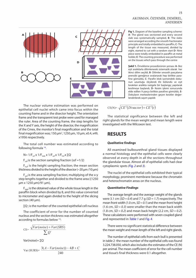
For cell counting; 3 left and 2 right glands were sliced into 3 mm intervals (the first cut at a random distance between the edge and 3 mm) and every second slab was chosen giving rise to a fraction of ½ Fig. 1 A-B. These slabs were placed according to the smooth fractionator design 9, smaller slabs were placed peripherally; larger ones were placed centrally and they were totally embedded in paraffin. The whole length of the tissue in the paraffin block was measured and was divided into 9 equal pieces by cutting in 8 equidistant intervals. First cut on the paraffin block was performed randomly into first interval length and then continue cutting same intervals to obtain 9 equal pieces. Every piece was turned 90° to the same side and all pieces were re-embedded paraffin in a metal block holder. This paraffin block was exhaustively sliced in 40 µm sections by rotary microtome (Leica RM2155) and slices were taken onto glass slide which pass through the center of the tissues in the paraffin block and were stained by Giemsa (Fig. 1).
To estimate the epithelial cell’s nuclear volume on vertical sections, 2 left and 3 right glands were sliced into 3 mm intervals (again the first cut at a random distance between the edge and 3 mm) and every second slab was chosen with a random start 10. The sampled slabs were cut into 3 mm bars with a random start and every 5th bar was chosen. The vertical axis of the bars was determined, and then the bars were rotated freely around this axis and embedded in paraffin. A 40 µm thick through the center of the tissue in the paraffin block was held for the volume estimation of the nuclei 11.
Stereological Analysis
The counting was performed with epithelial cell nucleus as counting units. The number of the epithelial cells were estimated using the computer software loaded Shtereom I 12, Olympus BH2 light microscope with motorized stage (Lang MS 316) (for the step lengths on the X, Y axis) and 3.2 MP Cmex camera (Euromex, Holland), under x100 oil-immersion lens objective. The thickness of the tissue measured and the movements in the Z axis was controlled using a microcator (Heidenhain, Germany). For counting the nearly 100-200 nucleus per gland according to disector principle 13, area of the counting frame was 91 µm2 and the step lengths for the X and Y axis was 1250 µm. The height of the disector was 10 µm.
15
AKOSMAN, ÖZDEMİR, DEMİREL, ANDERSEN
The nuclear volume estimation was performed on epithelial cell nuclei which came into focus within the counting frame and in the disector height. The orientation frame and the transparent test probe were used for managed the ruler. Area of the counting frame, the step lengths for the X and Y axis, the height of the disector, the magnification of the Cmex, the monitor’s final magnification and the total final magnification was; 150 µm2, 1250 µm, 10 µm, x0.4, x49, x1956 respectively.
The total cell number was estimated according to following formula 14.
N= 1/Fssf x 1/Fslab x 1/Fasf x 1/Fhsf x ∑Q-
Fssf; is the section sampling fraction (ssf =1/2)
Fhsf; is the height sampling fraction; the mean section thickness divided to the height of the disector (~20 µm /10 µm)
Fasf; is the area sampling fraction; multiplying of the x-y step lengths together and divided to the frame area ((1250 µm x 1250 µm)/91 µm),
Fslab; is the obtained value of the whole tissue length in the paraffin block when divided by 8, and this value converted to micrometer and again divided to the height of the slicing section (40 µm).
∑Q-; is the number of the counted epithelial cell nucleus
The coefficient of error for the number of counted nucleus and the section thickness was estimated altogether according to formulas below 13:
Var(noise)= ∑Q-
The statistical significance between the left and right glands for the mean weight and mean length were investigated with the Wilcoxon test.
RESULTS
Qualitative findings
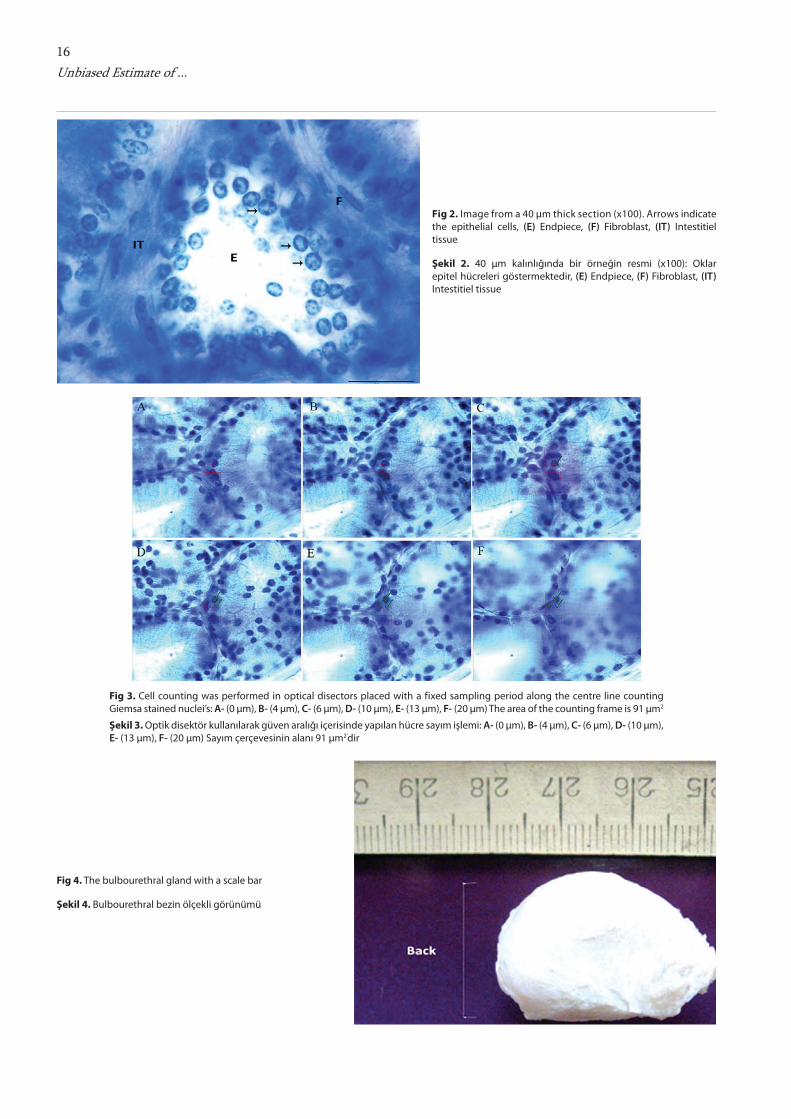
All examined bulbourethral gland tissues displayed a normal histology and the epithelial cells were clearly observed at every depth in all the sections throughout the glandular tissue. Almost all of epithelial cells had clear cytoplasmic spots (Fig. 2 and 3).
The nuclei of the epithelial cells exhibited their typical morphology, prominent membrane because the chromatin and heterochromatin areas on the nucleus.
Quantitative Findings
The average length and the average weight of the glands were 3.1 cm (SD = 0.4) and 7.7 g (SD = 1.7) respectively. The mean front width (1.0 cm, SD = 0.1) and the mean front height (1.6 cm, SD = 0.3) were smaller than the mean back width (1.8 cm, SD = 0.2) and mean back height (2.2 cm, SD = 0.2). These calculations were performed with seven coupled gland and represented in Table 1 and Fig. 4.
There were no significant statistical difference between the mean weight and mean length of the left and right glands.
The number of epithelial cells from each bull is presented in table 2 -the mean number of the epithelial cells was found 3.224.738.050, which also includes the estimates of the CE (N) per animal. The mean coefficient of error for the cell number and tissue’s final thickness were 0.1 altogether.
Fig 1. Diagram of the baseline sampling scheme: A- The gland was sectioned and every second slab was systematically sampled, B- The slabs were arranged according the smooth fractionator principle and totally embedded in paraffin, C- The length of the tissue was measured, divided by eight, started to cut with a random start D- Nine piece were totally embedded in paraffin at metal holder, E- The counting procedure was performed on the tissues which pass through the centre
Şekil 1. Örnekleme prosedürünün şeması: A- Bez eşit aralıklarla dilimlenerek sistematik olarak her ikinci dilim seçildi, B- Dilimler smooth parçalama prensibi gereğince sıralanarak hep birlikte para- fine gömüldü, C- Parafin blok içerisindeki doku-nun uzunluğu ölçülerek 8’e bölündü ve eşit bırakılan aralıkta rastgele bir başlangıç yapılarak kesilmeye başlandı, D- Kesim işlemi sonucunda elde edilen 9 parça birlikte parafine gömüldü, E- Dokuların merkezlerinden geçen kesitler değer-lendirilerek sayım yapıldı
CE=
Q
SRSVarnoiseVar )()( Var (noise)= ∑Q-
Var (SURS)= 240
4))((3 CBnoiseVarA CE(N)= tCEtorDiCE 22 sec
CE=
Q
SRSVarnoiseVar )()( Var (noise)= ∑Q-
Var (SURS)= 240
4))((3 CBnoiseVarA CE(N)= tCEtorDiCE 22 sec
CE=
Q
SRSVarnoiseVar )()( Var (noise)= ∑Q-
Var (SURS)= 240
4))((3 CBnoiseVarA CE(N)= tCEtorDiCE 22 sec
16Unbiased Estimate of ...
Fig 2. Image from a 40 µm thick section (x100). Arrows indicate the epithelial cells, (E) Endpiece, (F) Fibroblast, (IT) Intestitiel tissue
Şekil 2. 40 µm kalınlığında bir örneğin resmi (x100): Oklar epitel hücreleri göstermektedir, (E) Endpiece, (F) Fibroblast, (IT) Intestitiel tissue
Fig 4. The bulbourethral gland with a scale bar
Şekil 4. Bulbourethral bezin ölçekli görünümü
Fig 3. Cell counting was performed in optical disectors placed with a fixed sampling period along the centre line counting Giemsa stained nuclei’s: A- (0 µm), B- (4 µm), C- (6 µm), D- (10 µm), E- (13 µm), F- (20 µm) The area of the counting frame is 91 µm2
Şekil 3. Optik disektör kullanılarak güven aralığı içerisinde yapılan hücre sayım işlemi: A- (0 µm), B- (4 µm), C- (6 µm), D- (10 µm), E- (13 µm), F- (20 µm) Sayım çerçevesinin alanı 91 µm2’dir

17
AKOSMAN, ÖZDEMİR, DEMİREL, ANDERSEN
Total volumes, measured by water displacement method, of the glands were 7.12 cm3 (SD=1.5).
The mean volume of the epithelial cell nuclei was 59.1 µm3 (SD=3.7).
The total shrinkage in the z axis was calculated with the microcator for the thickness of the sliced tissue on the slide by making depth measurements on the counting areas and was found 44.5 % and the mean final thickness of the sliced tissue’s was found 22.2 µm (SD=0.97).
DISCUSSION
At the histological level in goats, boars and buffalo, Type I and Type II cells are observed in the entire glandular complex, though Type I cells dominate within the cranial disseminate glands, and Type II cells dominate within the bulbourethral glands 15-17. However, this study is not related with the types of the cells, just performed on the secretory portion of the bulbourethral gland. The number or abundance of the cells in the tissue is the important fundamental information in non-pathological conditions to recognize tumours.
From the morphometrical aspect, Abdel-Razek and Ali 18
were measured the length of the 2 years old bull’s bulbo- urethral glands and they found 1.2-2.1 cm long, however we found 3.1 cm long on the 2.5-3 years old bulls bulbo-
urethral glands.
In this study, we showed the epithelial cell number and the epithelial cell’s nuclear volume in the bull’s bulbo-urethral gland. However we detect that there is a lack of any other studies releated with the number and nuclear volume on this gland for to compare our results. We applied design based stereological methods for the cell counting and nuclear volume estimation. The optical fractionator is a precise, unbiased and modern stereological method
which combines the optical disector with the fractionator principle 14. The optical disector is a 3D probe for particle counting within a thick section 13,14,19,20. The fractionator is a sampling design that samples the organ systematic and in known fractions randomly 14,21. The smooth fractionator technique 9, which we performed on certain parts of the present study, is even more efficient than the original method 22. In order to increase efficiency, the items arrange (slab, sections, etc.) in a symmetric design with one peak and minimal jumps 9. In the present study, we applied general and smooth fractionator techniques modification. The application performed as sliced the whole gland into known distance and arranged these slices side by side 9 and totally embedded them into the paraffin and sliced that paraffin block into 9 equal distance pieces and these pieces embedded in a metal block holder and at the final step slices hold on the glass slide which pass through these pieces center. Thus, in order to save time there is no need to slice all selected tissues exhaustively.
Modern design based stereological methods produced unbiased estimates of the mean nuclear volume of arbitrarily shaped particles 11. Evaluation of the mean cell nuclear volume of an organ is especially important for objective histo- pathologic malignancy grading. Prognostic significance of the nuclear volume has been reported by Sørensen 23. The method produces efficient and precise results with highly statistical efficiency 11,23. This method was here performed with the combination of the vertical sectioning procedure
Table 1. The morphometric information of bulbourethral glands.
Tablo 1. Bulbourethral bezlerin morfometrik değerleri
No Side Weight(g)
Length(cm)
Frontwidth
Backwidth
Frontheight
Backheight Side Weight
(g)Length
(cm)Frontwidth
Backwidth
Frontheight
Backheight
1 left 6.6 3.0 0.8 1.6 1.7 2.1 right 6.2 2.8 0.9 1.6 2.0 2.2
2 left 9.8 2.6 1.0 2.0 1.5 2.5 right 10.0 2.8 1.0 2.0 1.6 2.4
3 left 9.6 3.4 1.0 1.9 1.4 2.5 right 9.3 3.4 1.1 2.1 1.4 2.2
4 left 6.5 3.2 1.0 1.7 1.5 2.1 right 6.1 3.1 1.1 1.6 1.7 2.1
5 left 5.9 2.8 0.9 1.7 1.6 2.2 right 5.1 2.7 0.8 1.6 1.1 1.8
6 left 8.3 3.5 1.2 1.7 2.0 2.3 right 8.7 3.6 1.1 1.7 1.5 2.3
7 left 7.1 3.2 1.1 1.7 1.6 2.0 right 7.5 3.5 0.8 1.8 1.3 2.2
mean 7.7 3.1 1.0 1.8 1.6 2.2 7.6 3.1 1.0 1.8 1.5 2.2
SD 1.6 0.3 0.1 0.1 0.2 0.2 1.8 0.4 0.1 0.2 0.3 0.2
Table 2. The mean epithelial cell number of the bulbourethral glands.
Tablo 2. Bulbourethral bezlerin epitel hücre sayıları
Gland No Epithelial Cell Number CE CV
1 3.86.109 0.095
2 2.39.109 0.105
3 3.49.109 0.102
4 2.80.109 0.102
5 3.55.109 0.098
Mean 3.22.109 0.100 0.19

18Unbiased Estimate of ...
which is simple and consumed less time than isotropic uniform sectioning 11,24 and enabled us to perform unbiased and very efficient estimations 25 of the cell nucleus. However, the paraffin embedding makes almost 50% shrinkage in the volume 26. In our study, we estimate that the mean shrinkage for the tissue on the slide was 44.5%.
In general, the error estimation of the method squared divided by the coefficient of variance squared should follow the following relationship: 0.25 < CE2/CV2 < 0.5. If it is below 0.25, you might be working too much, whereas if it is above 0.5 you might be working too little. In our study, we obtained a ratio of 0.29, which is in the suitable range 14,27.
Further studies, currently in progress, are oriented towards supplying a detailed functional interpretation of the secretory epithelium as well as the secretion of the gland.
REFERENCES
1. Chughtai B, Sawas A, O’Malley RL, Naik RR, Khan SA, Pentyala S: A Neglected gland: A review of the cowper’s gland. Int J Androl, 28, 74-77, 2005.
2. Abou-Elmagd A, Wrobel KH: The periurethral glandular complex in the Water buffalo: An ultrastructural, histological and lectin-histochemical study. Arch Histol Cytol, 52 (5): 501-512, 1989.
3. Dyce KM, Sack WO, Wensing CJG: Anatomia Veterinaria. 2nd ed., pp. 193, 567, McGraw-Hill Interamericana, Mexico, 1999.
4. Badia E, Briz MD, Pinart E, Sancho S, Garcia N, Bassols J, Pruneda A, Bussalleu E, Yeste M, Casas I, Bonet S: Structural and ultrastructural features of boar bulbourethral gland. Tissue Cell, 38 (1): 7-18, 2006.
5. Campero CM, Ladds PW, Thomas AD: Pathological findings in the bulbourethral glands of bulls. Aust Vet J, 65 (8): 241-244, 1988
6. Dursun N: Veteriner Anatomi II. 6th Baskı, s. 152, Medisan Yayınevi, Ankara, Türkiye, 1998.
7. Hart RG, Greenstein JS: A newly discovered role for cowper’s gland secretion in rodent semen coagulation. J Reprod Fertil, 17 (1): 87-94, 1968.
8. Santos M, Marcos R, Santos N, Malhao F, Monteiro RA, Rocha E: An unbiased stereological study on subpopulations of rat liver macrophages and on their numerical relation with the hepatocytes and stellate cells. J Anat, 214 (5): 744-751, 2009.
9. Gundersen HJG: The smooth fractionator. J Microsc, 207 (3): 191-210, 2002.
10. Møller A, Strange P, Gundersen HJG: Efficient estimation of cell volume and number using the nucleator and the disector. J Microsc, 159 (1): 61-71, 1990.
11. Sørensen FB: Stereological estimation of the mean and variance of nuclear volume from vertical sections. J microsc, 162(2): 203-229, 1991.
12. Oguz EO, Conkur ES, Sarı M: Shtereom I simple windows based software for stereology. volume and number estimations. Image Anal Stereol, 26, 45-50, 2007.
13. Gundersen HJG, Jensen EBV, Kieu K, Nielsen J: The efficiency of systematic sampling in stereology- reconsidered. J Microsc, 193 (3): 199- 211, 1999.
14. West MJ, Slomianka L, Gundersen HJG: Unbiased stereological estimation of the total Number of neurons in the subdivisions of the rat hippocampus using the Optical fractionator. Anat Rec, 231 (4): 482-97, 1991.
15. Wrobel KH: Untersuchungen zur feinstruktur und histochemie der glandula bulbourethralis der Ziege. Cell Tıssue Res, 108 (4): 582-596, 1970.
16. Wrobel KH: Histochemische untersuchungen am prostataparanchym der Ziege. Anat Histol Embryol, 1 (1): 64-72, 1972.
17. Wrobel KH, Sinowatz F: Vergieichende studien an den anhangsdrüsen der mannlichen Urethra. Acta Histochem Suppl, 31, 193-200, 1985.
18. Abdel-Razek KH, Ali A: Developmental changes of bull (Bos taurus) Genitalia as evaluated by caliper and ultrasonography. Reprod Domest Anim, 40, 23-27, 2005.
19. Braendgaard H, Evans SM, Howard CV, Gundersen HJG: The total number of neurons in the human neocortex unbiasedly estimated using optical disectors. J Microsc, 157(3): 285-304, 1990.
20. Evans S, Janson AM, Nyengaard JR: Quantitative Methods in Neuroscience: A Neuroanatomical Approach. 1st ed., 82, Oxford University Press, USA 2004.
21. Gundersen HJG, Bendtsen TF, Korbo L, Marcussen N, Møller A, Nielsen K, Nyengaard JR, Pakkenberg B, Sørensen FB, Vesterby A, West MJ: Some new, simple and efficient stereological methods and their use in pathological research and diagnosis. Apmis, 96, 379-394, 1988.
22. Gundersen HJG: Stereology of arbitrary particles. A review of unbiased number and size estimators and the presentation of some new ones, in memory of William R. Thompson. J. Microsc, 143, 3-45, 1986.
23. Sørensen FB: Objective histopathologic grading of cutaneous malignant melanomas by stereologic estimation of nuclear volume. Cancer, 63, 1784-1798, 1989.
24. Baddeley AJ, Gundersen HJG, Cruz-Orive LM: Estimation of surface area from vertical sections. J Microsc, 142 (3): 259-276, 1986.
25. Michel RP, Cruz-Orive LM: Application of the cavalieri principle and vertical sections method to lung: Estimation of volume and pleural surface area. J Microsc, 150 (2): 117-136, 1988.
26. Dorph-Petersen KA, Nyengaard JR, Gundersen HJG: Tissue shrinkage and unbiased stereological estimation of particle number and size. J Microsc, 204 (3): 232-246, 2001.
27. Lodrup AB, Karstoft K, Dissing TH, Pedersen M, Nyengaard JR: Kidney biopsies can be used for estimations of glomerular number and volume: A pig study. Virchows Arch, 452, 393-403, 2008.


![Closed-Form Unbiased Frequency Estimation of a Noisy ...€¦ · [18] I. M. Cherevko, “An estimate for the fundamental matrix of singularly perturbed differential-functionalequations](https://img.pdfslide.net/doc/110x75/5f93958dad3c26182565e9b5/closed-form-unbiased-frequency-estimation-of-a-noisy-18-i-m-cherevko-aoean.jpg)


![Message in a Bottle.1cs229.stanford.edu/proj2017/final-posters/5145028.pdf · binning estimate with a first order correction [2] is unbiased but suffers from large variance. Center:](https://img.pdfslide.net/doc/110x75/5e687851f08186014d5bd90e/message-in-a-bottle-binning-estimate-with-a-first-order-correction-2-is-unbiased.jpg)