Embed Size (px)
Citation preview
Unclassified ENV/JM/MONO(2015)18 Organisation de Coopération et de Développement Économiques Organisation for Economic Co-operation and Development 22-May-2015
___________________________________________________________________________________________
_____________ English - Or. English ENVIRONMENT DIRECTORATE
JOINT MEETING OF THE CHEMICALS COMMITTEE AND
THE WORKING PARTY ON CHEMICALS, PESTICIDES AND BIOTECHNOLOGY
GUIDANCE DOCUMENT ON THE IN VITRO SYRIAN HAMSTER EMBRYO (SHE) CELL
TRANSFORMATION ASSAY
Series on Testing & Assessement
No. 214
JT03376959
Complete document available on OLIS in its original format
This document and any map included herein are without prejudice to the status of or sovereignty over any territory, to the delimitation of
international frontiers and boundaries and to the name of any territory, city or area.
EN
V/JM
/MO
NO
(20
15
)18
Un
classified
En
glish
- Or. E
ng
lish

ENV/JM/MONO(2015)18
2

ENV/JM/MONO(2015)18
3
OECD Environment, Health and Safety Publications
Series on Testing and Assessment
No. 214
GUIDANCE DOCUMENT ON THE IN VITRO SYRIAN HAMSTER EMBRYO (SHE) CELL
TRANSFORMATION ASSAY
Environment Directorate
ORGANISATION FOR ECONOMIC CO-OPERATION AND DEVELOPMENT
Paris 2015

ENV/JM/MONO(2015)18
4
About the OECD
The Organisation for Economic Co-operation and Development (OECD) is an intergovernmental
organisation in which representatives of 34 industrialised countries in North and South America, Europe
and the Asia and Pacific region, as well as the European Commission, meet to co-ordinate and harmonise
policies, discuss issues of mutual concern, and work together to respond to international problems. Most of
the OECD’s work is carried out by more than 200 specialised committees and working groups composed
of member country delegates. Observers from several countries with special status at the OECD, and from
interested international organisations, attend many of the OECD’s workshops and other meetings.
Committees and working groups are served by the OECD Secretariat, located in Paris, France, which is
organised into directorates and divisions.
The Environment, Health and Safety Division publishes free-of-charge documents in eleven different
series: Testing and Assessment; Good Laboratory Practice and Compliance Monitoring; Pesticides;
Biocides; Risk Management; Harmonisation of Regulatory Oversight in Biotechnology; Safety of
Novel Foods and Feeds; Chemical Accidents; Pollutant Release and Transfer Registers; Emission
Scenario Documents; and Safety of Manufactured Nanomaterials. More information about the
Environment, Health and Safety Programme and EHS publications is available on the OECD’s World
Wide Web site (http://www.oecd.org/chemicalsafety/).
This publication was developed in the IOMC context. The contents do not necessarily reflect the views or
stated policies of individual IOMC Participating Organisations.
The Inter-Organisation Programme for the Sound Management of Chemicals (IOMC) was established in
1995 following recommendations made by the 1992 UN Conference on Environment and Development to
strengthen co-operation and increase international co-ordination in the field of chemical safety. The
Participating Organisations are FAO, ILO, UNDP, UNEP, UNIDO, UNITAR, WHO, World Bank and
OECD. The purpose of the IOMC is to promote co-ordination of the policies and activities pursued by the
Participating Organisations, jointly or separately, to achieve the sound management of chemicals in
relation to human health and the environment.
ENV/JM/MONO(2015)18
5
This publication is available electronically, at no charge.
Also published in the Series on Testing and Assessment link
For this and many other Environment,
Health and Safety publications, consult the OECD’s
World Wide Web site (www.oecd.org/chemicalsafety/)
or contact:
OECD Environment Directorate,
Environment, Health and Safety Division
2 rue André-Pascal
75775 Paris Cedex 16
France
Fax: (33-1) 44 30 61 80
E-mail: [email protected]
© OECD 2015
Applications for permission to reproduce or translate all or part of this material should
be made to: Head of Publications Service, [email protected], OECD, 2 rue André-
Pascal, 75775 Paris Cedex 16, France

ENV/JM/MONO(2015)18
6
FOREWORD
This document presents guidance for conducting the Syrian Hamster Embryo Cells Transformation
Assay (SHE CTA).
This document was preceded by the development of the Detailed Review Paper (DRP) 31 on “Cell
Transformation Assays for Detection of Chemical Carcinogens” (OECD, 2007), pre-validation study led
by ECVAM, ESAC peer review, and then from 2011 by work aimed at the development of a Test
Guideline. Despite support from some countries, concerns were expressed by others regarding the SHE
CTA and the approval of the draft TG at the April 2013 WNT meeting was considered premature. Efforts
were undertaken to try to address remaining scientific, technical issues, but nevertheless the draft Test
Guideline did not reach the stage of regulatory acceptance.
In November 2014, the Joint Meeting discussed options for moving forward in the area of non-
genotoxic carcinogenicity under the Test Guidelines Programme. The Joint Meeting advised 1) to proceed
with the development of guidance documents on the SHE CTA and Bhas-42 CTA, mainly to describe the
test procedures, and 2) to develop a guidance document at the OECD level outlining a conceptual
framework for the identification of non-genotoxic carcinogens for priority setting.
The draft document has been through two WNT commenting rounds in July and in December 2014.
Since all countries who commented either indicated approval or indicated that their comments did not
impede approval of the document and were only of editorial nature, this document was approved by written
procedure.
This document is published under the responsibility of the Joint Meeting of the Chemicals Committee
and the Working Party on Chemicals, Pesticides and Biotechnology.
ENV/JM/MONO(2015)18
7
GUIDANCE DOCUMENT ON THE
IN VITRO SYRIAN HAMSTER EMBRYO (SHE) CELL TRANSFORMATION ASSAY
PURPOSE
1. The purpose of this Guidance document is to allow the regulatory community to use the
described method as part of a weight of evidence approach in the testing of substances for carcinogenic
potential. There are a number of issues which have impeded consensus on the approval of the Test
Guideline; these issues mainly include the subjective nature of evaluating transformed phenotypic
morphology, the limited understanding of causal molecular mechanisms leading to the transformed SHE
colonies, the relatively small number of bona fide non-genotoxic carcinogens, as compared to genotoxic
carcinogens, that have been tested in the SHE cell transformation assay, and the way the assay might be
used in a regulatory framework.
Background
2. In vitro cell transformation refers to the induction of phenotypic alterations in cultured cells. Cell
transformation is an event in the multi-step process of tumour induction (1) (2). Transformed cells are
phenotypically different from normal cells and have the ability to induce tumours in susceptible animals
(3) (4) (5). It has been shown that SHE cells can be morphologically transformed by treatment with
genotoxic and non genotoxic carcinogens (6) (7) (8). Exposure results in an increase of morphologically
transformed (MT) colonies, which are characterised by disorganised growth patterns and mimicking an
early stage in the carcinogenic process.
3. The performance of the Syrian Hamster Embryo (SHE) cell transformation assay conducted at a
variety of pHs to detect transforming activity has been established on a large set of substances and has
been reviewed in a database summarized in the OECD Detailed Review Paper (DRP) 31 (6) (9). In
addition, the European Reference Laboratory for Alternatives to Animal Testing (EURL ECVAM) study
(10) (11) addressed the availability of standardized protocols, their transferability, within- and between-
laboratories reproducibility. The EURL ECVAM work also included the analysis of the degree of
similarity between protocols. It concluded that there are no elements suggesting that the EURL ECVAM
experiments differ notably from the experiments reported in the OECD DRP 31, thus making these data
acceptable for use in a retrospective evaluation.
4. When SHE CTA results are used as part of a testing strategy (not as results from a stand-alone
assay) and/or in a weight of evidence approach, they may contribute to the assessment of carcinogenic
potential of test chemicals (12). While the available data (see paragraph 14) would suggest that the SHE
CTA has greater sensitivity for carcinogens acting via genotoxic mechanisms, for non-genotoxic
carcinogens the added value of the SHE CTA is still debated and more information is required.
5. This Guidance Document (GD) provides an in vitro procedure of the SHE cell transformation
assay, as specified in Maire et al. (13) or in the EURL ECVAM DB-ALM protocol on SHE CTA (14),
conducted at pH 6.7 and 7.0. The assay can be performed at either pH 6.7 or 7.0 (see paragraphs 14)
provided proficiency has been demonstrated at the chosen pH (see paragraph 54-55). The morphology of
the normal colonies differs slightly at physiological pH compared to acidic pH, however, the conduct of
the assay at either pH has been shown to give similar results. Other than the difference in the pH, the
experimental protocol for both versions of the assay is the same.

ENV/JM/MONO(2015)18
8
Current knowledge and understanding about mechanisms involved in cell transformation/
6. The exact molecular mechanisms involved in cell transformations are only partially understood
(15) (16) 17). Although there are uncertainties regarding the causal mechanisms leading to the
transformed SHE colonies, the following paragraphs review current knowledge and understanding based
on the literature.
7. Evidence indicates that cell transformation results from alterations and changes in the expression
of genes involved in cell cycle control, genomic stability, proliferation and differentiation. Genetic changes
affecting these processes may result from direct genotoxic mechanisms. Also, disturbances of gene
expression and genomic stability through hyper- or hypomethylation of DNA, histone modifications and
nucleosomal remodelling are epigenetic mechanisms considered as fundamental in triggering a
carcinogenic process (18). Consistent with these diverse mechanisms, some SHE cell transformants have
been shown to harbour biallelic, inactivated p53 tumour suppressor genes (19). Carcinogens such as DES
can suppress DNA methylation in short-term treatments (20). The initial transformants induced by
polycyclic aromatic hydrocarbons frequently display DNA methylation-associated suppression of gene
expression known to be associated with embryonic differentiation and engineered re-expression suppresses
the transformed phenotype (21). SHE cell transformation by diethanolamine is driven by altered choline
metabolism, an important methyl donor in one-carbon metabolism leading to DNA methylation (22).
Introduction of an activated oncogene (v-Ha-ras), by transfection, will morphologically transform normal
SHE cells (23). Increased frequency of akinetochoric chromosome disjunction occurs during the growth of
the initial transformants (24) which could contribute to the aneuploid characteristic of immortalized clones
arising from such populations (reviewed in Ahmadzai et al., 2012 (25)).
8. Among later stage immortal and malignantly transformed descendents, global DNA
hypomethylation and site specific hypomethylation in ras and myc oncogenes have been observed (20)
(26). Also, methylation-associated suppression of cell cycle checkpoint gene expression, or mono- or
biallelic losses of these genes (ink4a, ink4b), as well as mutations in p53 have been found in immortal
Syrian hamster dermal cells (27). In morphologically transformed SHE cell lines, cell cycle checkpoint
control (G2) is often compromised (28). An activated proto-oncogene (cph) capable of transforming other
cells has been isolated from malignantly transformed SHE cells (29).
9. Non-genotoxic carcinogens have been postulated to act via a number of mechanisms such as
inhibition of gap junction intercellular communication oxidative stress, increased mitogenesis, decreased
apoptosis, interference with tubulin polymerization, inhibition of senescence through activation of
telomerase, interference with signal transduction pathways, and binding to receptors involved in hormone-
mediated processes, and in peroxisome proliferation. Instances of several of these mechanisms have been
demonstrated in SHE cell transformation. Oxidative stress was shown to be causally involved in
morphological transformation (30) (31). Imbalance of cell proliferation via an inhibition of apoptosis has
been related to cell transforming effects of some hepatic peroxisome proliferators and other transforming
agents in SHE cells (32) (33). Growth factor treatments of SHE cells, presumably acting through signal
transduction pathways, can also drive transformation (34) and gap junctional cell-to-cell communication is
frequently impaired by non-genotoxic carcinogens (35).
INITIAL CONSIDERATIONS AND LIMITATIONS
10. The SHE cells are normal diploid, metabolically and p53-competent primary cells, which retain
the ability to biotransform a wide range of xenobiotics as evidenced by studies with substances requiring
metabolic activation (6) (9) (36) (37) (38). From a 3Rs perspective, the use of primary cells means that a

ENV/JM/MONO(2015)18
9
small number of pregnant hamsters are euthanized; one hamster provides sufficient cells to perform at least
50 to 100 CTAs providing the cells are adequately preserved for future use. The metabolic capability of the
cells should be considered and discussed in the light of interpretation of test results. This is particularly
important when the test chemical requires metabolic activation. Exposure to test chemicals with
transforming capacity results in an increased number of morphologically transformed (MT) colonies,
which are characterised by disorganised growth patterns.
11. Transformation of primary, diploid SHE cells appears to follow a staged process. The
transformation assay in the SHE cells is based upon identifiable colonies of morphologically transformed
cells with irregular growth patterns. The transformants are thought to be stem cells with blockages in their
differentiation pathways (39). Upon further passages in vitro, transformed colonies clonally isolated from
treated cultures frequently generate cells with an infinite cellular lifespan or an ability to form tumours in
syngenic hosts. Untransformed clones become senescent (40) (41) (42). High frequencies of progression
to immortality and anchorage independence were also observed in bulk cultures of SHE cells (4).
12. Although conducted blindly, identification of morphologically transformed colonies by
microscopic scoring is subjective, as for any cytohistochemical endpoint. This subjectivity may, to some
extent, be overcome with appropriate training, and the use of photo catalogues (43) (44). However, to
improve the reliability of the scoring a second opinion or duplicate independent scoring is highly
recommended, especially for ambiguous colonies/or borderline pictures.
13. The assay would be improved by the development of objective measures for scoring
transformation, when these are validated. Some examples include biospectroscopy, which is being
explored to provide an objective determination of transformed colonies (45). In addition, molecular tools
such as gene expression changes promise to provide useful molecular markers for morphological
transformation, like those associated with cytoskeleton effects in the SHE cells (38).
14. To date comparative sensitivity and specificity of the pH 6.7 and 7.0 versions of the assay are
limited to a small chemical database (see annex to DRP 31(7)). An analysis of the sensitivity and
specificity of the assay for genotoxic and non-genotoxic carcinogens that were fully tested in in vitro and
in vivo genotoxicity assays was performed in 2014 and is available in an annex to the DRP.
15. When planning the experiment, careful choice of the optimum pH needs to be taken into account.
Parameters might include ionisable nature of the compounds as affecting the differences in reactivity or
bioavailability. The historical experience of the laboratory with the scoring at either pH should also be
considered. This needs to be taken into account until a wider chemical database has been generated.
16. At this time, the assay conducted as described in this Guidance Document does not provide
information on in vivo potency, or species-specificity or tissue-specificity of the cell transformations.
17. It should be noted that this method has been validated for mono-constituent substances only and
not multi-constituent substances, UVCBs (substances of unknown or variable composition, complex
reaction products or biological materials) or mixtures. Before use of the assay for the testing of a mixture
intended for a regulatory purpose, it should be considered whether, and if so why, it may provide adequate
results for that purpose. Such considerations are not needed, when there is a regulatory requirement for
testing of the mixture.
PRINCIPLE OF THE TEST METHOD
18. SHE cells are obtained from primary cultures of Syrian hamster embryos at 13 days of gestation.
After enzymatic tissue digestion, cells are collected, grown for 24 to 48 hours and then cryopreserved, and
stored in liquid nitrogen. One part of cryopreserved SHE cells is used as feeder cells, the other part as
ENV/JM/MONO(2015)18
10
target cells. The feeder cells are x-ray irradiated to inactivate their capability to replicate, and seeded as
nutrient base and support of metabolic activity. The target cells are used to assess morphological
transformation of colonies.
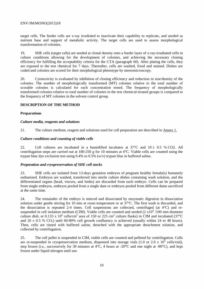
19. SHE cells (target cells) are seeded at clonal density onto a feeder layer of x-ray-irradiated cells in
culture conditions allowing for the development of colonies, and achieving the necessary cloning
efficiency for fulfilling the acceptability criteria for the CTA (paragraph 60). After plating the cells, they
are exposed to the test chemical for 7 days. Thereafter, cells are washed, fixed and stained. Dishes are
coded and colonies are scored for their morphological phenotype by stereomicroscopy.
20. Cytotoxicity is evaluated by inhibition of cloning efficiency and reduction in size/density of the
colonies. The number of morphologically transformed (MT) colonies relative to the total number of
scorable colonies is calculated for each concentration tested. The frequency of morphologically
transformed colonies relative to total number of colonies in the test chemical-treated groups is compared to
the frequency of MT colonies in the solvent control group.
DESCRIPTION OF THE METHOD
Preparations
Culture media, reagents and solutions
21. The culture medium, reagent and solutions used for cell preparation are described in Annex 1.
Culture conditions and counting of viable cells
22. Cell cultures are incubated in a humidified incubator at 37°C and 10 0.5 % CO2. All
centrifugation steps are carried out at 180-250 g for 10 minutes at 4°C. Viable cells are counted using the
trypan blue dye exclusion test using 0.4% to 0.5% (w/v) trypan blue in buffered saline.
Preparation and cryopreservation of SHE cell stocks
23. SHE cells are isolated from 13-days gestation embryos of pregnant healthy female(s) humanely
euthanized. Embryos are washed, transferred into sterile culture dishes containing wash solution, and the
differentiated organs (head, viscera, and limbs) are discarded from each embryo. Cells can be prepared
from single embryos, embryos pooled from a single dam or embryos pooled from different dams sacrificed
at the same time.
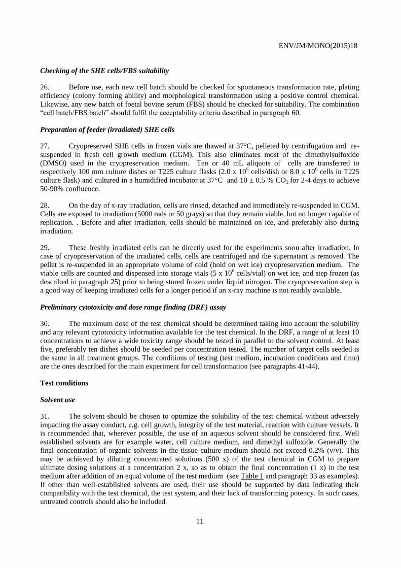
24. The remainder of the embryo is minced and dissociated by enzymatic digestion in dissociation
solution under gentle stirring for 10 min at room temperature or at 37°C. The first wash is discarded, and
the dissociation is repeated 2-4 times. Cell suspensions are collected, centrifuged (at 4°C) and re-
suspended in cell isolation medium (CIM). Viable cells are counted and seeded (2 x10⁶ /100 mm diameter
culture dish, or 0.133 x 10⁶ cells/cm
2 area of 150 or 225 cm
2 culture flasks) in CIM and incubated (37°C
and 10 ± 0.5 % CO2) until 60-80% cell growth confluency is achieved (usually within 24 to 48 hours).
Then, cells are rinsed with buffered saline, detached with the appropriate detachment solution, and
collected by centrifugation.
25. The cell pellet is suspended in CIM, viable cells are counted and pelleted by centrifugation. Cells
are re-suspended in cryopreservation medium, dispensed into storage vials (1.0 or 2.0 x 10⁶ cells/vial),
step frozen (i.e., successively for 30 minutes at 4°C, 4 hours at -20°C and one night at -80°C), and kept
frozen under liquid nitrogen until use.
ENV/JM/MONO(2015)18
11
Checking of the SHE cells/FBS suitability
26. Before use, each new cell batch should be checked for spontaneous transformation rate, plating
efficiency (colony forming ability) and morphological transformation using a positive control chemical.
Likewise, any new batch of foetal bovine serum (FBS) should be checked for suitability. The combination
“cell batch/FBS batch” should fulfil the acceptability criteria described in paragraph 60.
Preparation of feeder (irradiated) SHE cells
27. Cryopreserved SHE cells in frozen vials are thawed at 37°C, pelleted by centrifugation and re-
suspended in fresh cell growth medium (CGM). This also eliminates most of the dimethylsulfoxide
(DMSO) used in the cryopreservation medium. Ten or 40 mL aliquots of cells are transferred to
respectively 100 mm culture dishes or T225 culture flasks (2.0 x 10⁶ cells/dish or 8.0 x 10
6 cells in T225
culture flask) and cultured in a humidified incubator at 37°C and 10 ± 0.5 % CO2 for 2-4 days to achieve
50-90% confluence.
28. On the day of x-ray irradiation, cells are rinsed, detached and immediately re-suspended in CGM.
Cells are exposed to irradiation (5000 rads or 50 grays) so that they remain viable, but no longer capable of
replication. . Before and after irradiation, cells should be maintained on ice, and preferably also during
irradiation.
29. These freshly irradiated cells can be directly used for the experiments soon after irradiation. In
case of cryopreservation of the irradiated cells, cells are centrifuged and the supernatant is removed. The
pellet is re-suspended in an appropriate volume of cold (hold on wet ice) cryopreservation medium. The
viable cells are counted and dispensed into storage vials (5 x 10⁶ cells/vial) on wet ice, and step frozen (as
described in paragraph 25) prior to being stored frozen under liquid nitrogen. The cryopreservation step is
a good way of keeping irradiated cells for a longer period if an x-ray machine is not readily available.
Preliminary cytotoxicity and dose range finding (DRF) assay
30. The maximum dose of the test chemical should be determined taking into account the solubility
and any relevant cytotoxicity information available for the test chemical. In the DRF, a range of at least 10
concentrations to achieve a wide toxicity range should be tested in parallel to the solvent control. At least
five, preferably ten dishes should be seeded per concentration tested. The number of target cells seeded is
the same in all treatment groups. The conditions of testing (test medium, incubation conditions and time)
are the ones described for the main experiment for cell transformation (see paragraphs 41-44).
Test conditions
Solvent use
31. The solvent should be chosen to optimize the solubility of the test chemical without adversely
impacting the assay conduct, e.g. cell growth, integrity of the test material, reaction with culture vessels. It
is recommended that, wherever possible, the use of an aqueous solvent should be considered first. Well
established solvents are for example water, cell culture medium, and dimethyl sulfoxide. Generally the
final concentration of organic solvents in the tissue culture medium should not exceed 0.2% (v/v). This
may be achieved by diluting concentrated solutions (500 x) of the test chemical in CGM to prepare
ultimate dosing solutions at a concentration 2 x, so as to obtain the final concentration (1 x) in the test
medium after addition of an equal volume of the test medium (see Table 1 and paragraph 33 as examples).
If other than well-established solvents are used, their use should be supported by data indicating their
compatibility with the test chemical, the test system, and their lack of transforming potency. In such cases,
untreated controls should also be included.

ENV/JM/MONO(2015)18
12
Selection of test concentrations
32. The maximum concentrations to be tested in cell transformation assay depend on test chemical
solubility and cytotoxicity. For test chemicals of defined composition the highest dose level should be 0.01
M, 2 mg/mL or 2 µL/mL, whichever is the lowest. For test chemical of non-defined composition, e.g.
complex mixtures (plant extracts, tars, environmental extracts etc.), the top concentration should be at least
5 mg/mL. Poorly soluble chemicals should be tested up to the first concentration producing a visible
opacity in the final test medium observable by the unaided eye.
33. In addition to the controls, at least 5 test chemical concentrations should be used in the main
experiment. These are deducted from the range finding study and should include:
A high test concentration inducing no more than 50% cytotoxicity expressed by decrease in
relative plating efficiency and/or reduction in relative colonies density/size (by visual
appearance). If the test chemical does not show a cytotoxic effect, the highest dose is selected as
indicated in paragraph 32 above for soluble test chemicals, or as the visible solubility limit in the
final test medium for insoluble test chemicals;
At least one concentration which has no apparent effect on plating efficiency;
3 or 4 intermediate concentrations.
Table 1: Recommended concentrations of the test chemical in the intermediate solutions and in the final
test medium
Solvent*
Intermediate solution
(4 mL test medium)
Final test medium
(8 mL= 4mL
intermediate
solution+4mL GCM)
Concentration of the test
chemical
500x
2x
1x
Concentration of the
solvent
100%
0.4%
0.2%
*For water insoluble test chemicals, concentrated solutions (500x) may be prepared in Dimethyl
sulfoxide (DMSO). For water soluble test chemicals, an aqueous solvent is recommended.

ENV/JM/MONO(2015)18
13
Day 0 1 2 9
---------------------------------------------- //---------------------------------
Feeder Target Treatment Fixing
cells cells with test chemical staining
(2 mL) (2 mL) (4 mL)
Figure 1: Timeline of the SHE CTA assay (the volumes are per each 60 mm culture dish)
Preparation of test cultures
Feeder layer
34. On day 0 (feeder cells day), the irradiated SHE cells are seeded. The cell concentration is
adjusted to 20,000 – 30,000 cells/mL in CGM, and 2 mL of the cell suspension are added into each 60 mm
culture dish (4 to 6 x 10⁴ feeder cells/dish). In case of cryopreservation of irradiated cells, cryopreserved
cells are thawed at 37°C, and pelleted by centrifugation. The cell pellet is re-suspended in fresh CGM and
the viable cells are counted. Freshly irradiated cells can also be used for seeding of the feeder layer.
35. The culture dishes are incubated in a humified incubator at 37°C and 10 0.5% CO2 for 24 hours
before adding the target cells. For each test, at least 5 dishes filled with feeder cells only will be used
concurrently as controls for the inability of the feeder cells to replicate and to form colonies. No colony
should form in these dishes.
Target cells
36. Cryopreserved SHE cells are thawed at 37°C and seeded for growth in culture flasks. After an
incubation period (usually of 24 hours although shorter duration e.g. 5 hours can be used), the target cells
are detached, counted and the cell concentration is adjusted with CGM to a concentration where
approximately 25 - 45 colonies/dish can be obtained at the end of the test (see paragraph 60). Two mL of
the target cell suspension will be added to each culture dish containing feeder cells. Dishes will be
incubated in a humidified incubator at 37°C and 10 0.5% CO2 for 24 hours prior to treatment with the
test and control chemicals.
37. At cytotoxic dose levels, as determined in the Dose Range Finding (DRF) experiment, the target
cell number should be adjusted in order to yield the recommended number of 25 to 45 colonies per dish at
the end of the test to fulfil acceptance criteria (see paragraph 60). The adjustment of target cell number is
explained in paragraph 37.
Treatment of cultures
38. For practicality, one way of preparing dosing solution is to start with a concentration representing
twice (2x) the final concentration (Table 1). Each dosing solution (4 mL) will be transferred to individual
culture dishes (60 mm) already containing the CGM (4 mL) with feeder and target cells (final volume
8 mL) (Table 1). The cultures will be incubated in a humidified incubator at 37°C and 10 0.5% CO2 for 7
days without disturbance.
ENV/JM/MONO(2015)18
14
PROCEDURE
Preparation of test solutions
39. The test chemical solutions are prepared on the day of treatment. Solid test chemicals should be
dissolved in appropriate solvents and diluted, if appropriate, prior to treatment of the cells. Liquid test
chemicals may be added directly to the final test medium and/or diluted prior to treatment. Gaseous or
volatile chemicals should be tested by appropriate methods, determined on a case by case basis. Fresh
preparations of the test chemical should be used unless stability data demonstrate the acceptability of
storage. A series of solutions at different concentrations of the test chemical should be prepared under UV
filtered lights or protected from light.
Description of the cell transformation assay
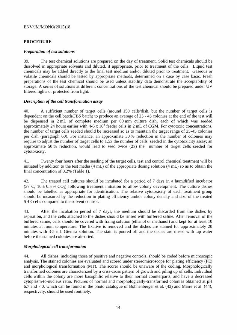
40. A sufficient number of target cells (around 150 cells/dish, but the number of target cells is
dependent on the cell batch/FBS batch) to produce an average of 25 - 45 colonies at the end of the test will
be dispensed in 2 mL of complete medium per 60 mm culture dish, each of which was seeded
approximately 24 hours earlier with 4-6 x 10⁴ feeder cells in 2 mL of CGM. For cytotoxic concentrations,
the number of target cells seeded should be increased so as to maintain the target range of 25-45 colonies
per dish (paragraph 60). For instance, an approximate 30 % reduction in the number of colonies may
require to adjust the number of target cells to 1.5x the number of cells seeded in the cytotoxicity assay; an
approximate 50 % reduction, would lead to seed twice (2x) the number of target cells seeded for
cytotoxicity.
41. Twenty four hours after the seeding of the target cells, test and control chemical treatment will be
initiated by addition to the test media (4 mL) of the appropriate dosing solution (4 mL) so as to obtain the
final concentration of 0.2% (Table 1).
42. The treated cell cultures should be incubated for a period of 7 days in a humidified incubator
(37°C, 10 0.5 % CO2) following treatment initiation to allow colony development. The culture dishes
should be labelled as appropriate for identification. The relative cytotoxicity of each treatment group
should be measured by the reduction in plating efficiency and/or colony density and size of the treated
SHE cells compared to the solvent control.
43. After the incubation period of 7 days, the medium should be discarded from the dishes by
aspiration, and the cells attached to the dishes should be rinsed with buffered saline. After removal of the
buffered saline, cells should be covered with fixing solution (ethanol or methanol) and kept for at least 10
minutes at room temperature. The fixative is removed and the dishes are stained for approximately 20
minutes with 3-5 mL Giemsa solution. The stain is poured off and the dishes are rinsed with tap water
before the stained colonies are air-dried.
Morphological cell transformation
44. All dishes, including those of positive and negative controls, should be coded before microscopic
analysis. The stained colonies are evaluated and scored under stereomicroscope for plating efficiency (PE)
and morphological transformation (MT). The scorer should be unaware of the coding. Morphologically
transformed colonies are characterized by a criss-cross pattern of growth and piling up of cells. Individual
cells within the colony are more basophilic relative to their normal counterparts, and have a decreased
cytoplasm-to-nucleus ratio. Pictures of normal and morphologically-transformed colonies obtained at pH
6.7 and 7.0, which can be found in the photo catalogue of Bohnenberger et al. (43) and Maire et al. (44),
respectively, should be used routinely.
ENV/JM/MONO(2015)18
15
45. Sparse colonies are not scored for MT (i.e. if a colony contains less than 50 cells, it is not
counted); however, they are included in the total number of colonies for plating efficiency determination..
Colonies at the edge of the dishes should be scored for MT if clearly morphologically transformed.
Generally, for each treatment group 1000 colonies should be evaluated for morphological cell
transformation (MT).
46. For each treatment group, normal (non-transformed) colonies and transformed colonies will be
enumerated to evaluate the plating efficiency (PE), the relative plating efficiency (RPE) and the
morphological transformation frequency (MTF) criteria detailed in paragraphs 56-58.
Controls
Solvent control
47. In case the test chemical is not water soluble, an appropriate solvent control should be used. If
DMSO or other organic solvents are selected, they should be used at a final concentration that does not
exceed 0.2% (see paragraph 31). The final concentration of any solvent should be the same in all solvent
control and treated dishes.
Positive control
48. Because of the high amount of data on Benzo[a]pyrene (B[a]P), it should be used as positive
control at the recommended concentrations of 1.0 to 5.0 µg/mL in dimethyl sulfoxide (DMSO) to
demonstrate the sensitivity of the assay . However, if justified, other chemicals can also be considered as
positive controls. Each laboratory should establish the performance of the positive control under their own
laboratory conditions (see paragraph 49).
Feeder cells control
49. For each test, at least 5 dishes with feeder cells only should be used concurrently to confirm the
inability of these cells to replicate and to form colonies. For a valid test, no colony should be formed in
these dishes at the end of the test period.
Solubility, pH, and osmolality
50. The solubility/precipitation of the test chemical in the solvent and in the test culture (medium)
should be visually assessed and documented in the test report.
51. The pH of the test chemical dosing solutions should be measured at the time of preparation of the
treatment medium and after at least four hours of undisturbed incubation in an incubator, in humidified
atmosphere at 37°C and 10 0.5 % CO2. In case of deviation from the expected pH at any time point, the
pH of the medium should be adjusted to the selected pH. Any deviation should be reported and considered
in the interpretation of the results.
52. The osmolality of the treatment medium should be measured prior to or at the time of performing
the preliminary cytotoxicity determination or the main experiment. Osmolality of the treatment medium
should be compared to the control medium, and any change used in the interpretation of results.
Proficiency of the laboratory
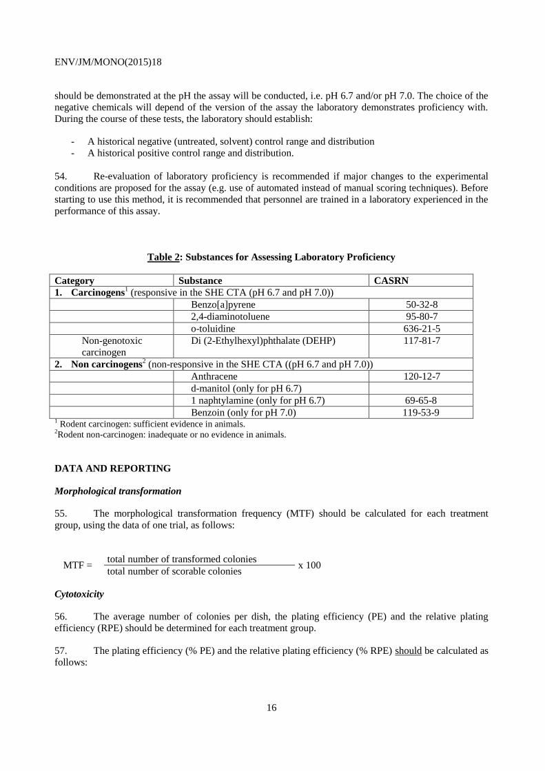
53. In order to demonstrate proficiency, a laboratory should perform tests with the four positive
chemicals acting via different mechanisms, and two negative chemicals, included in Table 2. Proficiency
ENV/JM/MONO(2015)18
16
should be demonstrated at the pH the assay will be conducted, i.e. pH 6.7 and/or pH 7.0. The choice of the
negative chemicals will depend of the version of the assay the laboratory demonstrates proficiency with.
During the course of these tests, the laboratory should establish:
- A historical negative (untreated, solvent) control range and distribution
- A historical positive control range and distribution.
54. Re-evaluation of laboratory proficiency is recommended if major changes to the experimental
conditions are proposed for the assay (e.g. use of automated instead of manual scoring techniques). Before
starting to use this method, it is recommended that personnel are trained in a laboratory experienced in the
performance of this assay.
Table 2: Substances for Assessing Laboratory Proficiency
Category Substance CASRN
1. Carcinogens1 (responsive in the SHE CTA (pH 6.7 and pH 7.0))
Benzo[a]pyrene 50-32-8
2,4-diaminotoluene 95-80-7
o-toluidine 636-21-5
Non-genotoxic
carcinogen
Di (2-Ethylhexyl)phthalate (DEHP) 117-81-7
2. Non carcinogens2 (non-responsive in the SHE CTA ((pH 6.7 and pH 7.0))
Anthracene 120-12-7
d-manitol (only for pH 6.7)
1 naphtylamine (only for pH 6.7) 69-65-8
Benzoin (only for pH 7.0) 119-53-9 1 Rodent carcinogen: sufficient evidence in animals.
2Rodent non-carcinogen: inadequate or no evidence in animals.
DATA AND REPORTING
Morphological transformation
55. The morphological transformation frequency (MTF) should be calculated for each treatment
group, using the data of one trial, as follows:
MTF = total number of transformed colonies
x 100 total number of scorable colonies
Cytotoxicity
56. The average number of colonies per dish, the plating efficiency (PE) and the relative plating
efficiency (RPE) should be determined for each treatment group.
57. The plating efficiency (% PE) and the relative plating efficiency (% RPE) should be calculated as
follows:

ENV/JM/MONO(2015)18
17
PE = total number of colonies per dish
x 100 total number of target cells seeded per dish
RPE = PE of treatment group
x 100 PE of the solvent control group
58. In addition to the RPE, the colony size and density (number of cells per colony) should be
recorded as parameters of cytotoxicity. The size and density is observed and recorded as three categories:
- Normal (+)
- Slightly reduced (++ ; 20 – 39 % reduction)
- Greatly reduced (+++; 40 – 60 % reduction)
Acceptability Criteria and historical controls
59. The following criteria have to be fulfilled for the validity of the assay:
At least 1000 colonies per experimental group should be available for morphological
transformation scoring. Occasionally, in case of a significant increase in morphological
transformation rate, less than 1000 colonies are acceptable. However the average number of
colonies per dish should normally not be less than 25.
An average of 25-45 colonies per dish should be available (46). Occasionally, in case of a negative
result, dishes with less than 25 colonies per dish are acceptable. Likewise, in case of a positive
result, dishes with more than 45 colonies are acceptable.
Cloning efficiency of the negative/solvent control is 20%.
No colony formation should be observed in the feeder cell dishes. Feeder cells should be visible in
the chemical treatment groups except if they are affected selectively by the test chemical. If the
feeder cells are affected by the test chemical, then this observation should be recorded, reported,
and considered in the interpretation of the results.
Transformation frequency in the negative controls (untreated and solvent) is within the
distribution of historical control data of the laboratory (e.g. 95% confidence interval). Based on
historical data from experienced laboratories and data from the EURL ECVAM validation study,
the upper limit of transformation frequency in the negative controls (untreated and solvent) is
0.6%.
The positive control chemical should induce a biologically relevant and statistically significant
increase in morphological cell transformation compared to the solvent control.
Data interpretation criteria
60. Although biological relevance of the results should be considered first, both statistical
significance and biological relevance of data are considered in the interpretation of the negative and
positive results. The level of concentration(s) increasing the MTF is carefully considered, taking into
account the range of cytotoxic/non-cytotoxic concentrations.
61. Providing that all acceptability criteria are fulfilled, the following criteria are considered for the
evaluation of results:

ENV/JM/MONO(2015)18
18
(1) the increase in MT colonies is concentration-related,
(2) at least one of the test concentration exhibits a statistically significant increase compared
to the concurrent negative control,
(3) the statistically significant result is outside the distribution range of the historical negative
control data (e.g. 95% confidence interval).
62. A result can be considered clearly biologically relevant and a test chemical is considered a clear
positive if all the above criteria are met.
63. A test chemical is considered as a clear negative if none of the criteria above (paragraph 62) are
met.
64. Results are statistically analysed using the one-sided Fisher’s exact test to determine if an
increase in morphological transformation occurred at each concentration level compared to the concurrent
solvent control. A p<0.05 level of significance indicates a treatment related effect on MTF. The Cochran-
Armitage trend test can be used to contribute to the evaluation of positive concentration-related response.
65. When results do not meet the criteria for a clear positive or a clear negative call, the test chemical
should be evaluated by expert judgement and/or the experiment should be repeated. Modification of study
parameters over an extended or narrowed range of concentrations, as appropriate, should be considered in
follow-up experiments. In rare cases, even after further investigations, the data set will preclude making a
conclusion of positive or negative results, and will therefore be concluded as equivocal.
Test report
66. The test report should include the following information:
Test chemical
Mono-constituent substance:
- physical appearance, water solubility, and additional relevant physicochemical properties;
- chemical identification, such as IUPAC or CAS name, CAS number, SMILES or InChI code,
structural formula, purity, chemical identity of impurities as appropriate and practically feasible,
etc.
Multi-constituent substance, UVBCs and mixtures:
- characterised as far as possible by chemical identity (see above), quantitative occurrence and
relevant physicochemical properties of the constituents.
Solvent (if appropriate)
- justification for choice of solvent
- concentrations tested and preparation of the dosing solutions
- signs of precipitation (absence or presence)
Cells
- source of cells
- number of cell subcultures
- maintenance of cell cultures
- absence of mycoplasms

ENV/JM/MONO(2015)18
19
- identification of serum (provider and batch number)
Test conditions
- rationale for selection of concentrations, including cytotoxicity data and solubility limitations
- composition of the media, CO2, pH
- serum concentration, origin and quality
- concentrations of test chemicals
- volume of solvent and test chemical added
- duration of treatment
- incubation temperature
- number of cells plated
- positive and negative controls
- criteria for scoring MT colonies
Results
- cytotoxicity results
- signs of precipitation
- pH, osmolality of culture media after addition of the test chemical
- number of total scorable colonies
- relative cloning efficiency
- concurrent feeder cell control
- dose-response relationship, where possible
- statistical analyses
- concurrent negative (solvent) and positive control data
- historical negative (solvent) and positive control data, with ranges, means, standard deviation,
and confidence interval (e.g. 95%)
67. Data should be presented in tabular form. The following values should be presented for each
group (treated and untreated groups, solvent and positive controls):
i. total number, and average number per dish of scorable colonies for each group
ii. plating and relative plating efficiency %
iii. cloning efficiency
iv. colony size/density
v. number of transformed colonies
vi. morphological transformation frequency (MTF %)
vii. Fisher’s exact test p-value (one-sided)
Discussion of the results
Conclusion

ENV/JM/MONO(2015)18
20
REFERENCES
1. Barrett J.C. and Ts’o P.O.P. (1978). Evidence for the progressive nature of neoplastic transformation
in vitro. Proc. Natl. Acad. Sci., USA, 75: 3761-3765.
2. Kakunaga T.H. and Kamasaki H. (1985). Transformation Assay of Established Cell Lines:
Mechanisms and Application. IARC Scientific Publications No 67. International Agency for
Research on Cancer, Lyon, 225p.
3. Berwald Y. and Sachs L. (1963). In vitro cell transformation with chemical carcinogens. Nature,
200, 1182-1184.
4. Newbold R.F., Overell R.W., Connell J.R. (1982). Induction of immortality is an early event in
malignant transformation of mammalian cells by carcinogens. Nature 299: 633-635.
5. Elias Z. et al. (1989). Cytotoxic and neoplastic effects of industrial hexavalent chromium pigments
in Syrian hamster embryo cells. Carcinogenesis 10 (11): 2043-2052.
6. OECD (2007). Detailed Review Paper on Cell transformation assays for detection of chemical
carcinogens, OECD Environment, Health and Safety Publications, Series on Testing and
Assessment, No. 31. OECD, Paris
7. Jacquet N. et al (2012). Carcinogenic potency of perfluorooctane sulfonate (PFOS) on Syrian
hamster embryo (SHE) cells. Arch. Toxicol. 86, 2, 305 – 314.
8. Jacquet N. et al (2012). Perfluorooctanoic acid (PFOA) acts as a tumor promoter on Syrian hamster
embryo (SHE) cells. Environ Sci Pollut Res 19, 2537–2549.
9. Vasseur P. and Lasne C. (2012). OECD Detailed Review Paper (DRP) number 31 on “Cell
Transformation Assays for Detection of Chemical Carcinogens”: main results and conclusions.
Mutat. Res. 744, 8-11.
10. Corvi R. et al (2012). ECVAM prevalidation study on in vitro cell transformation assays: general
outline and conclusions of the study. Mutat. Res.744, 12–19.
11. ECVAM (2011). Recommendation concerning the cell transformation assays using Syrian hamster
embryo cells (SHE) and the BALB/c 3T3 mouse fibroblast cell line for in vitro carcinogenicity
testing. Annex I: ESAC opinion on the ESAC peer review of an ECVAM-coordinated prevalidation
study concerning three protocols of the cell transformation assay (CTA) for in vitro carcinogenicity
testing. http://ihcp.jrc.ec.europa.eu/our activities/alt-animal-testing/.
12. Creton S. et al (2012). Cell transformation assays for prediction of carcinogenic potential: state of
the science and future research needs, Mutagenesis, 27,. 93–101.
13. Maire M.-A. et al. (2012). Recommended protocol for the Syrian hamster embryo (SHE) Cell
Transformation Assay, Mutat. Res. 744, 76– 81.
14. EURL ECVAM DataBase service on Alternative Methods to Animal Experimentation (DB-ALM)
protocol No. 136 on in vitro Syrian embryo hamster cell transformation assay (http://ecvam-
dbalm.jrc.ec.europa.eu/).
ENV/JM/MONO(2015)18
21
15. Combes R. et al. (1999). Cell Transformation Assays as Predictors of Human Carcinogenicity – The
Report and Recommendations of ECVAM Workshop 39, ATLA 27, 745-767.
16. LeBoeuf R.A. et al. (1999). Use of Syrian hamster embryo and BALB/c 3T3 cell transformation for
assessing the carcinogenic potential of chemicals, in The Use of Short and Medium-term Tests for
Carcinogens and Data on Genetic Effects in Carcinogenic Hazard Evaluation. D.B. McGregor, J.M.
Rice and S. Venitt, eds. IARC Scientific Publications No. 146, International Agency for Research on
Cancer, Lyon, 409-425.
17. Adler S. et al. (2011). Alternative (non-animal) methods for cosmetics testing: current status and
future prospects-2010, Arch. Toxicol. 85, 367–485.
18. Baylin S.B. and Ohm J.E. (2006), Epigenetic gene silencing in cancer: a mechanism for early
oncogenic pathway addiction? Nat Rev Cancer 6:107–116.
19. Albor, A. et al. (1994). 3-Methylcholanthrene inactivates the p53 gene in syrian hamster embryo
fibroblasts by inducing a specific intronic point mutation, Cancer Research, 54 (16), pp. 4502-4507.
20. Schiffmann, D. et al. (1996). Diethylstilbestrol induces stable, inheritable DNA-hypomethylation in
Syrian hamster embro cells throughout multistage neoplastic transformation. In Vitro Toxicol., 9 (2)
167-172.
21. Isfort, R.J. et al. (1997). Role of the H19 gene in Syrian hamster embryo cell tumorigenicity,
Molecular Carcinogenesis, 20 (2), pp. 189-193.
22. Lehman-McKeeman LD and Gamsky EA (2000). Choline supplementation inhibits diethanolamine-
induced morphological transformation in syrian hamster embryo cells: evidence for a carcinogenic
mechanism. Toxicological Science; 55(2), pp.303-10.
23. Thomassen, D.G. et al. (1985). Evidence for multiple steps in neoplastic transformation of normal
and preneoplastic Syrian hamster embryo cells following transfection with Harvey murine sarcoma
virus oncogene (v-Ha-ras), Cancer Research, 45 (2), pp. 726-732.
24. Kirchner, S. et al. (1993). Cytogenetic changes in primary, immortalized and malignant mammalian
cells, Toxicology Letters, 67 (1-3), pp. 283-295.
25. Ahmadzai, A.A. et al. (2012). The Syrian hamster embryo (SHE) assay (pH 6.7): Mechanisms of
cell transformation and application of vibrational spectroscopy to objectively score endpoint
alterations Mutagenesis, 27 (3), pp. 257-266.
26. Takahashi, M., Barrett, J.C., Tsutsui, T. (2002). Transformation by inorganic arsenic compounds of
normal Syrian hamster embryo cells into a neoplastic state in which they become anchorage-
independent and cause tumors in newborn hamsters, International Journal of Cancer, 99 (5), pp. 629-
634.
27. Yasaei, H. et al. (2013). Carcinogen-specific mutational and epigenetic alterations in INK4A,
INK4B and p53 tumour-suppressor genes drive induced senescence bypass in normal diploid
mammalian cells Oncogene, 32 (2), pp. 171-179.
28. Ashra, H., and Rao, K.V.K. (2006). Elevated phosphorylation of Chk1 and decreased
phosphorylation of Chk2 are associated with abrogation of G2/M checkpoint control during

ENV/JM/MONO(2015)18
22
transformation of Syrian hamster embryo (SHE) cells by Malachite green, Cancer Letters, 237 (2),
pp. 188-198.
29. Notario, V. et al. (1990). Frequent activation of non-ras transforming sequences in neoplastic Syrian
hamster cells initiated with chemical carcinogens, Oncogene, 5 (9), pp. 1425-1430.
30. Zhang H. et al. (2000a). Acrylonitrile-induced morphological transformation in Syrian hamster
embryo cells. Carcinogenesis 21, 727-733.
31. Zhang H. et al (2000b). Morphological transformation by 8-hydroxy-2’-deoxyguanosine in Syrian
hamster embryo (SHE) cells. Toxicol. Sci., 56, 303-31.
32. Maire, M.A., Rast, C., Vasseur, P. (2005). Di-(2-ethylhexyl)phthalate (DEHP) increases Bcl-2/Bax
ratio and modifies c-myc expression in Syrian hamster embryo (SHE) cells. Toxicology Letters, 158
(3), pp. 237-245.
33. Alexandre, S. et al. (2003). ZnCl2 induces Syrian hamster embryo (SHE) cell transformation,
Toxicology Letters, 142 (1-2), pp. 77-87.
34. Isfort, R.J. (2000). Mechanisms of cell transformation in the Syrian hamster embryo (SHE) cell
transformation system. Annals of the New York Academy of Sciences, 919, pp. 86-96.
35. Rivedal E., Yamasaki H., and Sanner T. (1994). Inhibition of gap junctional intercellular
communication in Syrian hamster embryo cells by TPA, retinoic acid and DDT. Carcinogenesis, 15:
689-694.
36. Pienta R.J., Poiley J.A. and W.B. Lebherz III (1977). Morphological transformation of early passage
golden Syrian hamster embryo cells derived from cryopreserved primary cultures as a reliable in
vitro bioassay for identifying diverse carcinogens. Int. J. Cancer, 19, 642-655
37. Schechtman L.M. (1985). Metabolic activation of procarcinogens by subcellular enzyme fractions in
the C3H 10T1/2 and BALB/c 3T3 cell transformation systems. In Transformation Assay of
Established Cell Lines: Mechanisms and Application. T. Kakunaga & H. Yamasaki, eds. IARC
Scientific Publications No 67, International Agency for Research on Cancer, Lyon, p 137-162
38. Landkocz Y. et al. (2011). Transcriptomic effects of di-(2-ethylhexyl)-phthalate in Syrian hamster
embryo cells: an important role of early cytoskeleton disturbances in carcinogenesis? BMC
Genomics 12:524.
39. LeBoeuf R.A. et al. (1999). Use of Syrian hamster embryo and BALB/c 3T3 cell transformation for
assessing the carcinogenic potential of chemicals. IARC Scientific Publications No. 146, Lyon, 409-
425.
40. LeBoeuf, R.A. et al. (1990). Multistage neoplastic transformation of Syrian hamster embryo cells
cultured at pH 6.70. Cancer Research, 50 (12), pp. 3722-3729.
41. Pienta, R.J., Poiley, J.A., Lebherz III., W.B. (1977). Morphological transformation of early passage
golden Syrian hamster embryo cells derived from cryopreserved primary cultures as a reliable in
vitro bioassay for identifying diverse carcinogens, International Journal of Cancer, 19 (5), pp. 642-
655.

ENV/JM/MONO(2015)18
23
42. Watanabe, M. and Suzuki, K. (1991). Expression dynamics of transforming phenotypes in X-
irradiated Syrian golden hamster embryo cells, Mutation Research - Fundamental and Molecular
Mechanisms of Mutagenesis, 249 (1), pp. 71-80.
43. Bohnenberger S. et al (2012). Photo catalogue for the classification of cell colonies in the Syrian
hamster embryo (SHE) Cell Transformation Assay at pH 6.7, Mutat. Res. 744, 82– 96.
44. Maire M.-A., C. Rast C., P. Vasseur P. (2012). Photo catalogue for the classification of cell colonies
in the Syrian hamster embryo (SHE) Cell Transformation Assay at pH 7.0, Mutation Research 744
97– 110.
45. Ahmadzai A.A. et al. (2012). Classification of test agent-specific effects in the Syrian hamster
embryo assay (pH 6.7) using infrared spectroscopy with computational analysis. Mutagenesis
27(3):375-382.
46. Kerckaert, G.A. et al. (1996). A comprehensive protocol for conducting the Syrian hamster embryo
cell transformation assay at pH 6.70 (1996), Mutat. Res. – Fund. Mol. Mech. Mutagen 356, 1, 65-
84.
ENV/JM/MONO(2015)18
24
ANNEX 1:
Culture medium, reagent and solutions used for cell preparation
The culture medium is DMEM: Dulbecco’s Modified Eagle’s Medium containing 1g/L glucose, 4 mM
glutamine and 110 mg/L sodium pyruvate, with or without phenol red. The media can be purchased
readymade from the vendors and should be stored according to the parameters (time, temperature)
provided with the batch media.
If powder media is used, depending on the pH selected, the DMEM medium is adjusted to pH 7.0 with 1.5
g/L NaHCO3 or to pH 6.7 with 0.75 g/L NaHCO3 and sterilized by membrane filtration (0.1 µm porosity).
The culture medium can be stored at 4° C during a period not exceeding 2 weeks.
This culture medium serves to prepare the following media:
- Complete growth medium (CGM)
The complete culture medium is prepared with addition of fetal bovine serum (FBS) at a concentration of
15% or 20% (v/v) for the SHE pH 7.0 and the SHE pH 6.7 CTAs, respectively.
- Cryopreservation medium
The cryopreservation medium is the pH-adjusted DMEM, added with 10% FBS and 10% DMSO or
with 20% FBS and 7.5% DMSO (recommended if the test is carried out at pH 6.7).
- Cell isolation medium (CIM)
The cell isolation medium is the pH-adjusted DMEM added with 15% FBS and antibiotics penicillin 100
U/mL and streptomycin 100 µg/mL).
The solutions used for cell preparation and assay protocol are as follows:
- Buffered saline (e.g. calcium- and magnesium-free Hank’s balanced solution (CMF- HBSS) or calcium-
and magnesium-free phosphate buffered saline (CMF-PBS))
- Colony staining solution: 10% (v/v) Giemsa in aqueous buffer
- Cell staining solution (e.g. 0.4% to 0.5% (w/v) trypan blue in buffered saline)
- Fixing solution: ethanol or methanol
- Detachment solution (e.g. 0.25% (w/v) trypsin in buffered saline or [0.05% (w/v) trypsin + 0.02% (w/v)
Na2EDTA-H2O] in buffered saline)
- Dissociation solution (e.g. dispase 2 U/mL in buffered saline or [1.25% (v/v) Enzar-T, 2.5% (v/v)
pancreatin with 200 U/mL of penicillin and 200 μg/mL streptomycin] in buffered saline)
- Wash solution: buffered saline with 200 U/mL of penicillin and 200 µg/mL streptomycin