Embed Size (px)
Citation preview

UNDERSTANDING HYDROCEPHALUSOffering solutions for patients.

UNDERSTANDING HYDROCEPHALUS
1 2
Dear Reader:
Your doctor has either recommended the CODMAN® HAKIM® Precision Fixed Pressure Valve, the CODMAN® HAKIM® Programmable Valve (CHPV) or the CODMAN CERTAS® Plus Programmable Valve for use in treating hydrocephalus. This handbook is designed to provide basic information regarding the use of
Codman Neuro valves. It is not a substitute for a thorough discussion with your doctor regarding the use of a particular Codman Neuro valve for treating your specific medical condition. If you have any questions concerning any of the Codman Neuro valves contact your doctor.
All Codman Neuro valves are used for the treatment of hydrocephalus. The CODMAN CERTAS Plus Programmable Valve and CODMAN HAKIM Programmable Valve (CHPV) are designed with a programmable feature that allows the doctor to adjust the setting of the valve if needed. The CODMAN HAKIM Precision Fixed Pressure Valves are not adjustable, but are available at five different fixed pressure settings.
As with all types of hydrocephalus shunts, both adjustable and non-adjustable, it is important for patients to be aware of information critical to successful management of their condition.
Please read the following information carefully and discuss any questions you may have with your doctor.

3
UNDERSTANDING HYDROCEPHALUS
43
What is hydrocephalus? 5
What are the causes and types? 7
What are some symptoms of uncontrolled hydrocephalus? 9
What are some of the diagnostic tools? 10
How is hydrocephalus managed? 11
What shunt systems are available? 12
How is the shunt surgically implanted? 13
What are the complications? 15
What kind of follow-up is required? 16
What is the difference between fixed and programmable valves? 18
How does the programming change the pressure setting? 21
Glossary 23
Important information 26
Resources Back Cover
CODMAN® HAKIM®
Programmable Valve
CODMAN® HAKIM® Precision Valve
CODMAN CERTAS® Plus
Programmable Valve
UNDERSTANDING HYDROCEPHALUS
655
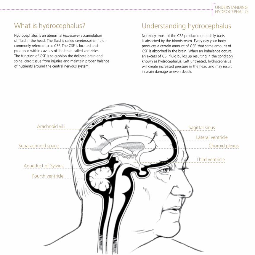
What is hydrocephalus?Hydrocephalus is an abnormal (excessive) accumulation of fluid in the head. The fluid is called cerebrospinal fluid, commonly referred to as CSF. The CSF is located and produced within cavities of the brain called ventricles. The function of CSF is to cushion the delicate brain and spinal cord tissue from injuries and maintain proper balance of nutrients around the central nervous system.
Arachnoid villi
Subarachnoid space
Aqueduct of Sylvius
Fourth ventricle
Understanding hydrocephalusNormally, most of the CSF produced on a daily basis is absorbed by the bloodstream. Every day your body produces a certain amount of CSF, that same amount of CSF is absorbed in the brain. When an imbalance occurs, an excess of CSF fluid builds up resulting in the condition known as hydrocephalus. Left untreated, hydrocephalus will create increased pressure in the head and may result in brain damage or even death.
Sagittal sinus
Lateral ventricle
Choroid plexus
Third ventricle
7
UNDERSTANDING HYDROCEPHALUS
8
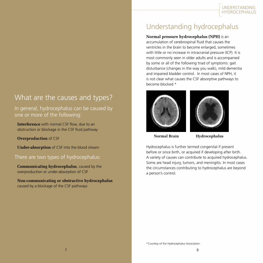
What are the causes and types?In general, hydrocephalus can be caused by one or more of the following:
Interference with normal CSF flow, due to an obstruction or blockage in the CSF fluid pathway
Overproduction of CSF
Under-absorption of CSF into the blood stream
There are two types of hydrocephalus:
Communicating hydrocephalus, caused by the overproduction or under-absorption of CSF
Non-communicating or obstructive hydrocephalus caused by a blockage of the CSF pathways
Understanding hydrocephalusNormal pressure hydrocephalus (NPH) is an accumulation of cerebrospinal fluid that causes the ventricles in the brain to become enlarged, sometimes with little or no increase in intracranial pressure (ICP). It is most commonly seen in older adults and is accompanied by some or all of the following triad of symptoms: gait disturbance (changes in the way you walk), mild dementia and impaired bladder control. In most cases of NPH, it is not clear what causes the CSF absorptive pathways to become blocked.*
*Courtesy of the Hydrocephalus Association.
Hydrocephalus is further termed congenital if present before or since birth, or acquired if developing after birth. A variety of causes can contribute to acquired hydrocephalus. Some are head injury, tumors, and meningitis. In most cases the circumstances contributing to hydrocephalus are beyond a person’s control.
Normal Brain Hydrocephalus

9
UNDERSTANDING HYDROCEPHALUS
109
What are some symptoms of uncontrolled hydrocephalus?When too much CSF exists within the brain the pressure within the skull may increase (except in case of Normal Pressure Hydrocephalus), causing symptoms such as headaches, nausea, vomiting, sleepiness, failing mental function, blurred vision, and loss of coordination. In infants, the skull bones are not completely formed and sutures (or joints between the bones of the skull) are not closed, so the increased amount of fluid may cause the skull to increase in size. This is a visual sign of hydrocephalus, but is only noticeable in infants and newborns.
Usually, hydrocephalus causes the ventricles of the brain to enlarge due to increased CSF within the skull. If a person exhibits symptoms of hydrocephalus, a doctor may perform several tests to confirm if hydrocephalus exists.
What are some of the diagnostic tools?Ultrasound, a device that uses sound to outline the structures within the skull. This is similar to scans that pregnant women have to check that their babies are healthy.
CT Scan (Computerized Tomography), a technique that uses x-rays to image and outline the size of the ventricles.
MRI (Magnetic Resonance Imaging), a technique that uses radio signals and a magnet to form images of the brain so that the ventricles can be visualized, so that the size and shape of the ventricles can be determined.
CSF Flow Studies, using dyes or other materials to trace the flow patterns of CSF.
Neuropsychological Test, a series of questions and answers used to determine if there is a loss of brain function due to hydrocephalus.
CSF tests to predict shunt responsiveness and/or determine shunt pressure include lumbar puncture, external lumbar drainage, measurement of CSF outflow resistance, intracranial pressure (ICP) monitoring and isotopic cisternography. Though there is no way to accurately predict any particular patient’s responsiveness to any particular shunt, many doctors find that these tests are helpful in determining the likelihood of a positive response to shunting. People who have abnormal bleeding tendencies or take medications that affect bleeding should talk with their medical team about any special precautions before invasive procedures.*
*Courtesy of the Hydrocephalus Association.

11
UNDERSTANDING HYDROCEPHALUS
12
How is hydrocephalus managed?A surgical procedure may be performed to divert the CSF from the ventricles to either the abdominal cavity or to a chamber in the heart known as the right atrium. Another technique exists, to channelize the CSF from the lumbar into the peritoneal cavity. By removing the CSF from the brain, the pressure in the skull may return to normal. To remove the CSF, the surgeon implants a flexible tube with a valve mechanism called a shunt system. A shunt may help to control the hydrocephalus, but is not a cure.
What shunt systems are available?Shunt systems come in a variety of models, but always have two similar parts: catheters, the tubing which transports and diverts the CSF from the ventricles to either the abdominal cavity or right atrium of the heart; and a valve that regulates the flow of CSF from the ventricles. Adjustable valves allow the surgeon to choose a pressure setting for the valve based on experience and the needs of the patient.
Many shunt systems also have a flexible flushing chamber called a reservoir. The reservoir serves several important functions. It allows the doctor to remove samples of CSF for testing, using a needle and syringe. The doctor also may inject fluid into the shunt system to test for flow, to be sure the system is working properly.
The parts of a shunt are named according to where they are implanted (placed) in the body. The portion of the tube which is inserted into the ventricles is called the ventricular catheter. The peritoneal catheter is the portion of the tube that drains CSF into the abdominal or peritoneal cavity. If a drainage tube is placed into the right atrium of the heart, it is called the atrial catheter.
To get a better understanding of what a shunt system looks like, ask your doctor or nurse to show you samples of the shunts they use.
All of the components of a shunt system are made from materials which are well-known to be tolerated by the body. For this reason, the entire shunt system is implanted under the skin. There are no external parts.
13
UNDERSTANDING HYDROCEPHALUS
14
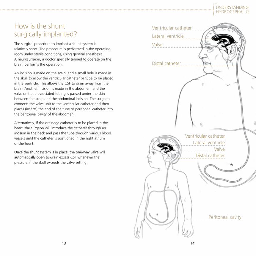
How is the shunt surgically implanted?The surgical procedure to implant a shunt system is relatively short. The procedure is performed in the operating room under sterile conditions, using general anesthesia. A neurosurgeon, a doctor specially trained to operate on the brain, performs the operation.
An incision is made on the scalp, and a small hole is made in the skull to allow the ventricular catheter or tube to be placed in the ventricle. This allows the CSF to drain away from the brain. Another incision is made in the abdomen, and the valve unit and associated tubing is passed under the skin between the scalp and the abdominal incision. The surgeon connects the valve unit to the ventricular catheter and then places (inserts) the end of the tube or peritoneal catheter into the peritoneal cavity of the abdomen.
Alternatively, if the drainage catheter is to be placed in the heart, the surgeon will introduce the catheter through an incision in the neck and pass the tube through various blood vessels until the catheter is positioned in the right atrium of the heart.
Once the shunt system is in place, the one-way valve will automatically open to drain excess CSF whenever the pressure in the skull exceeds the valve setting.
Ventricular catheter
Lateral ventricle
Valve
Distal catheter
Ventricular catheterLateral ventricle
ValveDistal catheter
Peritoneal cavity

15
UNDERSTANDING HYDROCEPHALUS
16
What are the complications?Patients and/or their carergivers also must be alert for the signs and symptoms resulting from shunt complications. The major complications of shunting are obstruction, infection, and overdrainage.
Obstruction
When a shunt malfunction occurs it is usually due to a partial or complete blockage of the system. The blockage can occur anywhere in the tubing or the valve and prevent the CSF from draining properly. If not corrected, this will cause the original hydrocephalus symptoms to return.
Infection
A shunt infection usually is caused by the patient’s own bacterial organisms, and is not acquired from exposure to other people. Infection should be suspected if there is any unusual redness or swelling of the wounds, or along the shunt system.
Overdrainage
This is caused by too much CSF being removed from the ventricles. This will cause the ventricles to decrease in size to a point where the brain may pull away from the skull. This may cause bleeding and require further surgery.
Other complications which can lead to the return of the hydrocephalus symptoms include underdrainage, disconnection of the tubing, and mechanical failure of the valve. If any symptoms occur or you suspect any complications, contact your doctor immediately.
16
What kind of follow-up is required?Generally, patients with an implanted shunt system are not restricted in their daily activities, except those involving great physical exertion. Your doctor will discuss with you any restrictions that may be advisable.
Because hydrocephalus is an ongoing condition, patients do require long term follow-up care by a doctor. Having regular medical checkups at intervals recommended by the neurosurgeon is advisable. Occasionally, patients with shunt systems require revisions. A revision is a surgical procedure to modify, repair or replace a shunt system due to a complication or changing patient conditions.
Regular follow-up visits will help the neurosurgeon to identify any subtle changes that may be indicative of a shunt problem. Patients and their carergivers should become familiar with the signs and symptoms of shunt complications as described on page 15 and shunt malfunction as described on page 17. Occasionally, the doctor can adjust the settings to treat these symptoms.

17
UNDERSTANDING HYDROCEPHALUS
18
Some Symptoms* of Shunt MalfunctionInfants Enlargement of the baby’s head
Fontanel is full and tense when the infant is upright and quiet
Swelling or redness along the shunt track
Fever
Unexplained irritability
Increasing muscle stiffness
Difficulty reaching developmental milestones
Vomiting
Sleepiness
Eyes pushed downwards
Seizures
Toddlers/Children Head enlargement
Fever
Vomiting
Headache
Irritability and/or sleepiness
Swelling or redness along the shunt track
A loss of previous abilities (sensory motor function)
Seizures (very rare)
Increasing muscle stiffness
Difficulty reaching developmental milestones
Personality changes
Worsening of school/academic performance
Adults Incontinence
Dementia
Headache
Vision Problems
Irritability and/or tiredness
Personality change
Loss of coordination or balance and/or difficulty walking
Seizures (very rare)
Difficulty in waking up or staying awake
Swelling or redness along the shunt track (infrequent)
This list of symptoms is for your reference only, and is not a diagnostic aid. If you are in doubt about your child’s or your own medical condition, consult your doctor immediately.
*List of symptoms was adapted from www.hydroassoc.org
What is the difference between fixed and programmable valves?Fixed Pressure Valves
To understand how a fixed pressure valve operates, a brief description of fixed pressure, or non programmable valves, is necessary. Hydrocephalus valves typically open at a specific pressure setting. The pressure setting is usually expressed in terms of millimeters of water (mmH
20). The pressure setting is
expressed as a number, within a specified range. For example: A medium pressure valve may have an opening pressure of 70 mmH
20 +/- 10. This means that the valve is manufactured
to open at one point between 60 and 80. Valves generally do not have an on/off switch, but open when pressure from the CSF is greater than the pressure exerted by the valve mechanism. This is called the “opening pressure.” The valve’s function is to open to allow CSF to drain and to close when the pressure in the skull is less than the valve setting.
Surgeons choose to use valves of a particular design and pressure setting based on experience and the patient’s condition. Nevertheless, after surgery the patient may experience symptoms or complications such as overdrainage or under drainage of CSF. In such cases, the surgeon may perform a shunt revision in order to replace the valve for one with a different pressure range. A revision is a surgical procedure to replace the complete shunt system or a component of a shunt system such as a valve.
The CODMAN HAKIM Precision Fixed Pressure Valve is an example of a non-programmable valve. It is available in 5 different fixed pressure settings.
19
UNDERSTANDING HYDROCEPHALUS
20
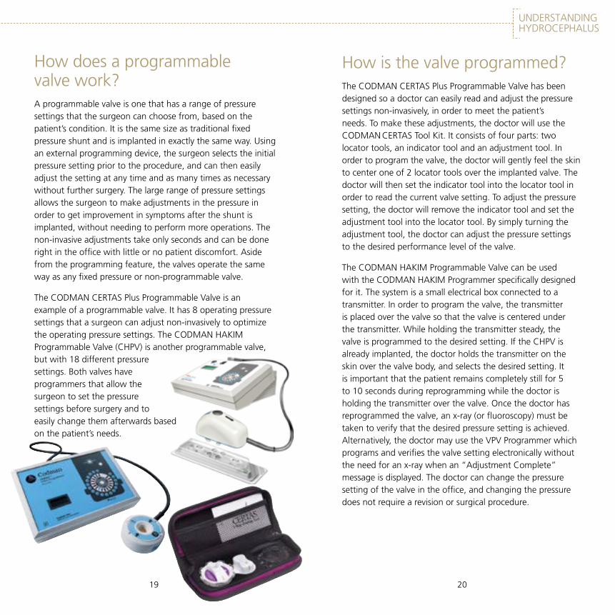
How does a programmable valve work?A programmable valve is one that has a range of pressure settings that the surgeon can choose from, based on the patient’s condition. It is the same size as traditional fixed pressure shunt and is implanted in exactly the same way. Using an external programming device, the surgeon selects the initial pressure setting prior to the procedure, and can then easily adjust the setting at any time and as many times as necessary without further surgery. The large range of pressure settings allows the surgeon to make adjustments in the pressure in order to get improvement in symptoms after the shunt is implanted, without needing to perform more operations. The non-invasive adjustments take only seconds and can be done right in the office with little or no patient discomfort. Aside from the programming feature, the valves operate the same way as any fixed pressure or non-programmable valve.
The CODMAN CERTAS Plus Programmable Valve is an example of a programmable valve. It has 8 operating pressure settings that a surgeon can adjust non-invasively to optimize the operating pressure settings. The CODMAN HAKIM Programmable Valve (CHPV) is another programmable valve, but with 18 different pressure settings. Both valves have programmers that allow the surgeon to set the pressure settings before surgery and to easily change them afterwards based on the patient’s needs.
How is the valve programmed?The CODMAN CERTAS Plus Programmable Valve has been designed so a doctor can easily read and adjust the pressure settings non-invasively, in order to meet the patient’s needs. To make these adjustments, the doctor will use the CODMAN CERTAS Tool Kit. It consists of four parts: two locator tools, an indicator tool and an adjustment tool. In order to program the valve, the doctor will gently feel the skin to center one of 2 locator tools over the implanted valve. The doctor will then set the indicator tool into the locator tool in order to read the current valve setting. To adjust the pressure setting, the doctor will remove the indicator tool and set the adjustment tool into the locator tool. By simply turning the adjustment tool, the doctor can adjust the pressure settings to the desired performance level of the valve.
The CODMAN HAKIM Programmable Valve can be used with the CODMAN HAKIM Programmer specifically designed for it. The system is a small electrical box connected to a transmitter. In order to program the valve, the transmitter is placed over the valve so that the valve is centered under the transmitter. While holding the transmitter steady, the valve is programmed to the desired setting. If the CHPV is already implanted, the doctor holds the transmitter on the skin over the valve body, and selects the desired setting. It is important that the patient remains completely still for 5 to 10 seconds during reprogramming while the doctor is holding the transmitter over the valve. Once the doctor has reprogrammed the valve, an x-ray (or fluoroscopy) must be taken to verify that the desired pressure setting is achieved. Alternatively, the doctor may use the VPV Programmer which programs and verifies the valve setting electronically without the need for an x-ray when an “Adjustment Complete” message is displayed. The doctor can change the pressure setting of the valve in the office, and changing the pressure does not require a revision or surgical procedure.

UNDERSTANDING HYDROCEPHALUS
2221
How does the programming change the pressure setting?The CODMAN CERTAS Plus Programmable Valve and the CODMAN HAKIM Programmable Valve both use a unique spring, ball, and cone mechanism to establish the pressure setting. To change the opening pressure of the valve, the tension is adjusted (changed) on the spring. A small magnetic device is activated by the programmer and rotates a cam or stepped mechanism, which turns to a new position and creates more or less tension on the spring. More tension on the spring increases the opening pressure, less tension on the spring lowers the opening pressure.
The CODMAN CERTAS Plus Programmable Valve can only be programmed with the CODMAN CERTAS Tool Kit. The CODMAN HAKIM Programmable Valve can only be programmed using the CODMAN HAKIM Programmer or VPV system.
Shunting is only a method for treating hydrocephalus, and is not a cure. Therefore, regular visits to your doctor are necessary for the continuing care of this condition. Since the CODMAN CERTAS Plus Programmable Valve and the CODMAN HAKIM Programmable Valve are both adjustable, your doctor can adjust the pressure setting of the valve and check the current pressure setting as part of your treatment plan.
If you have any questions concerning the CODMAN CERTAS Plus Programmable Valve or the CODMAN HAKIM Programmable Valve contact your doctor.

UNDERSTANDING HYDROCEPHALUS
23
Normal Pressure Hydrocephalus (NPH): an accumulation of cerebrospinal fluid that causes the ventricles in the brain to become enlarged, sometimes with little or no increase in intracranial pressure (ICP). It is most commonly seen in older adults and is accompanied by some or all of the following triad of symptoms: gait disturbance (changes in the way you walk), mild dementia and impaired bladder control. In most cases of NPH, it is not clear what causes the CSF absorptive pathways to become blocked
Non-Communicating Hydrocephalus: hydrocephalus caused by an obstruction in the ventricles or along the CSF pathway, causing a backup of fluid into the brain
Peritoneal Cavity: the cavity containing the abdominal organs; the belly
Shunt (n): an implanted system used to direct fluid from one part of the body to another. A shunt usually contains: catheters, valve and a reservoir
Skull: the bony structure surrounding the brain
To shunt (v): to divert fluid from one part of the body to another
Valve: a one-way, pressure or flow resistance device used to control the drainage of excess fluid from the brain
Ventricle: one of four cavities found within the brain where CSF can be accessed
24
Glossary Acquired Hydrocephalus: hydrocephalus occurring after birth
Atrium: one of the two upper chambers of the heart
Catheter: a silicone tube used to divert and drain CSF
Cerebrospinal Fluid (CSF): the watery fluid bathing the brain and the spinal cord
Communicating Hydrocephalus: hydrocephalus caused by an overproduction and/or reduced absorption of CSF in the presence of unobstructed ventricular pathways
Congenital Hydrocephalus: hydrocephalus caused by conditions existing at birth
CSF: see Cerebrospinal fluid
Hydrocephalus: a condition in which an increased amount of CSF exists in the ventricles and along the CSF pathways. This condition may occur when the rate of CSF production exceeds the rate of absorption, or when pathways of CSF flow are blocked. The result is excess fluid and pressure in the skull
Intra-cranial Pressure: pressure within the skull
Meningitis: an infection of the protective membranes covering the spinal cord and brain

25
UNDERSTANDING HYDROCEPHALUS
26
Important informationMagnetic fields generated from microwaves, wireless telephones, hightension wires, electric motors and transformers do not affect the valve setting.
If you sustain excessive force resulting in pain or direct trauma of the area over the valve, see your doctor to make sure there is no damage to the valve.
See your doctor for increasing headaches, blurred vision, nausea, vomiting or lethargy, as these may be a sign of valve dysfunction.
CODMAN CERTAS PLUS PROGRAMMABLE VALVE:
Testing shows that the valve mechanism is resistant to unintended changes in the setting in a 3 Tesla MRI. However, the clinician should confirm the valve setting after a magnetic resonance imaging (MRI) procedure.
Only a doctor should adjust the pressure setting using the CODMAN CERTAS Tool Kit.
CODMAN HAKIM PROGRAMMABLE VALVE: The use of MRI systems (up to 3 tesla) will not damage the valve mechanism, but may change the operating pressure of the valve. Following any MRI procedure, be sure to confirm the valve pressure setting and reprogram the valve if necessary. Alternatively, the VPV Programmer can be used to reprogram and confirm the setting.
High-powered magnets may affect the valve adjustment when placed close to the valve. Please contact your doctor if you suspect close exposure to strong magnets.
Only your doctor should adjust the pressure setting using the CODMAN HAKIM Programmer or the VPV.
This booklet provides a summary of the information regarding the appropriate use of the CODMAN HAKIM Precision Fixed Pressure Valve, the CODMAN CERTAS Plus Programmable Valve and the CODMAN HAKIM Programmable Valve. It is not a substitute for a thorough discussion with your doctor regarding the use of any Codman Neuro valve for your specific medical condition. If you have any questions or want more information, be sure to discuss your questions with your doctor.

Resources
Hydrocephalus Association, San Francisco
Email: [email protected]
www.depuysynthes.com www.LifeNPH.com www.hydro-kids.comwww.hydroassoc.org
DSUS/COD/1214/0223 01/15
© Codman Neuro, a division of DOI 2015. All rights reserved.
Codman & Shurtleff, Inc.325 Paramount DriveRaynham, MA 02767
For Customer Service in the USAcall: 1-800-255-2500
www.depuysynthes.com
For product information, please contact your local Sales Representative.