Embed Size (px)
Citation preview
Original Article
Understanding the Early Presentationof Mucopolysaccharidoses Disorders:Results of a Systematic LiteratureReview and Physician Survey
Lorne Clarke, MD1, Carolyn Ellaway, MBBS, PhD2,Helen E. Foster, MD, MBBS3, Roberto Giugliani, MD, PhD4,Cyril Goizet, MD, PhD5, Sarah Goring, MSc6, Sara Hawley, MSc7,Elaina Jurecki, MS, RD7, Zaeem Khan, MPH, BSc6,Christina Lampe, MD8 , Ken Martin, MD9,Suzanne McMullen, MHA, BSc6, John J. Mitchell, MD10,Fathima Mubarack, MSc, MHA7, H. Serap Sivri, MD11,Martha Solano Villarreal, MD, PhD12, Fiona J. Stewart, MB, BS13,Anna Tylki-Szymanska, MD, PhD14, Klane White, MD, MSc15,and Frits Wijburg, MD, PhD16
AbstractAs therapies are developed for rare disorders, challenges of early diagnosis become particularly relevant. This articlefocuses on clinical recognition of mucopolysaccharidoses (MPS), a group of rare genetic diseases related to abnormalitiesin lysosomal function. As quality of outcomes with current therapies is impacted by timing of intervention, minimizingtime to diagnosis is critical. The objective of this study was to characterize how, when, and to whom patients with MPSfirst present and develop tools to stimulate earlier recognition of MPS. A tripartite approach was used, including asystematic literature review yielding 194 studies, an online physician survey completed by 209 physicians who described
1 British Columbia Children’s Hospital Research Institute, University of British Columbia, Vancouver, British Columbia, Canada2 Sydney Children’s Hospital Network, Sydney University, Sydney, Australia3 Great North Children’s Hospital and Newcastle University, Newcastle upon Tyne, United Kingdom4 Medical Genetics Service HCPA, Dep Genet UFRGS & INAGEMP, Porto Alegre, Brazil5 Service de Genetique, CHU Bordeaux, Laboratoire MRGM, INSERM U 1211, University of Bordeaux, Bordeaux, France6 ICON plc, Vancouver, British Columbia, Canada7 BioMarin Pharmaceutical Inc., Novato, CA, USA8 Centre for Rare Diseases, Clinic for Children and Adolescents, Helios Dr. Horst Schmidt Kliniken, Wiesbaden, Germany9 UCSF Benioff Children’s Hospital Oakland, Oakland, CA, USA10 Montreal Children’s Hospital, Montreal, Quebec, Canada11 Hacettepe University Children’s Hospital, Ankara, Turkey12 Fundacion Cardioinfantil, Bogota, Colombia13 Belfast City Hospital, Belfast, United Kingdom14 Children’s Memorial Health Institute, Warsaw, Poland15 Seattle Children’s Hospital, Seattle, WA, USA16 Academic Medical Center, University Hospital of Amsterdam, Amsterdam, the Netherlands
Received June 7, 2018, and in revised form August 8, 2018. Accepted for publication August 8, 2018.
Corresponding Author:
Lorne Clarke, MD, British Columbia Children’s Hospital Research Institute, University of British Columbia, Vancouver, British Columbia, Canada.
Email: [email protected]
Journal of Inborn Errors of Metabolism& Screening2018, Volume 6: 1–12ª The Author(s) 2018DOI: 10.1177/2326409818800346journals.sagepub.com/home/iem
This article is distributed under the terms of the Creative Commons Attribution 4.0 License (http://www.creativecommons.org/licenses/by/4.0/) which permits anyuse, reproduction and distribution of the work without further permission provided the original work is attributed as specified on the SAGE and Open Access pages(https://us.sagepub.com/en-us/nam/open-access-at-sage).

859 MPS cases, and a global panel of MPS experts who distilled the findings. Red flag signs/symptoms were identified forcardiology, pediatric neurology, otorhinolaryngology, rheumatology, orthopedics, pediatrics, and general medicine andconverted into simple, specialty-specific tools intended to facilitate early diagnosis of MPS, enabling improved patientoutcomes.
Keywordsmucopolysaccharidoses, mucopolysaccharidosis I, mucopolysaccharidosis II, mucopolysaccharidosis III, mucopolysaccharidosis IV,mucopolysaccharidosis VI, mucopolysaccharidosis VII, diagnosis
Introduction
The mucopolysaccharidoses (MPS) are rare genetic conditions
caused by a deficiency of 1 of 11 lysosomal enzymes involved
in glycosaminoglycan (GAG) catabolism. MPS is character-
ized by the accumulation of partially degraded GAGs (heparan
sulfate, dermatan sulfate, keratan sulfate, chondroitin sulfate,
or hyaluronan) within lysosomes and by the subsequent
increase in GAGs in urine, blood, and cerebral spinal fluid.1,2
Progressive damage occurs as GAGs accumulate within the
cells. As lysosomes are found throughout the body, MPS can
manifest through a myriad of signs and symptoms. Over time,
MPS may result in multiple organ failure, cognitive impair-
ment, and premature death.1 Typically, symptom onset occurs
between infancy and childhood.3 The overall birth prevalence
for MPS is approximately 1 in 25000 and varies by region and
ethnic background.4–7
Seven MPS types have been identified (I, II, III, IV, VI, VII,
and IX); MPS III has 4 subtypes (A, B, C, and D) and MPS IV
has 2 (A and B). Although the subtypes are clinically similar,
each one is linked to a specific enzyme deficiency. MPS II is
inherited as an X-linked disorder, whereas all others are auto-
somal recessive conditions.1
MPS is usually diagnosed through biochemical testing for
deficient enzymes and can be confirmed through molecular
genetic testing. However, early clinical recognition of potential
cases with MPS, which is needed to trigger this diagnostic
testing, continues to pose a substantial challenge.8 This is
largely due to the disease rarity, phenotypic heterogeneity, and
the wide range of nonspecific early signs and symptoms.
Diagnostic delays often involve referrals from one physi-
cian to another and place a substantial burden on the patient
and caregivers. Patients are also at risk of misdiagnosis and
undergoing inappropriate interventions or receiving ineffec-
tive treatments. Timely referral for diagnostic testing allows
for prompt initiation of definitive therapy such as enzyme
replacement therapy or hematopoietic stem cell transplanta-
tion for some types of MPS as well as enabling the appropriate
management of secondary complications.9 Earlier recognition
also allows patients to partner with a physician with expertise
in their rare disease and facilitates support through a patient
organization sooner. Furthermore, early recognition alerts
at-risk carriers and enables them to seek accurate genetic
counseling and pursue prenatal testing and preimplantation
genetic diagnosis.
A better understanding of the initial presentation of MPS is
needed to improve early recognition of potential cases with
MPS and facilitate a timely diagnosis, enabling optimal patient
management and treatment when available. Unfortunately, pre-
vious efforts made in this area have not met with success.8
Thus, a new approach is needed. The objectives of this review
were to use a novel, evidence-based, multimethod approach to
characterize how, when, and to whom individuals with MPS
first present, identify specialty-specific red flag signs and
symptoms, and develop clinical awareness diagnostic tools that
have the potential to shorten the current diagnostic delay. The
effectiveness of the tools will depend on how well the infor-
mation they contain is disseminated and retained by the target
audiences. By making different tools for each subspecialist, we
aim to present only the most relevant information to each.
However, effective dissemination of the tools subsequent to
publication will be the most challenging and critical factor for
ultimate success.
Materials and Methods
To achieve the objectives of this study, a systematic literature
review was conducted first, followed by a physician survey to
supplement the published evidence with real-world clinical
experience and compensate for the possibility of publication
bias in the results of the literature review (ie, overrepresenta-
tion of rare signs and symptoms and under representation of
common ones). The results from the systematic literature
review and physician survey were then reviewed by a panel
of 16 international clinical MPS experts in order to identify
specialty-specific red flag signs and symptoms evident in the
early stages of the disease and generate specialty-specific tools
to increase clinical awareness of potential cases with MPS.
Systematic Literature Review
Search strategy and study selection. The following electronic
databases were searched: Embase (1970 to 27 June 2016) and
MEDLINE via PubMed (1970 to 27 June 2016). Handsearches
for registry studies, clinical surveillance, natural history, and
genotype–phenotype correlation studies were also conducted in
PubMed using a key word search. A transparent and reprodu-
cible search strategy (Supplemental Files 1 and 2) was devel-
oped in accordance with best practice guidelines.10,11 Searches
2 Journal of Inborn Errors of Metabolism & Screening

were limited to English language and human studies only and
were run on June 27, 2016.
Titles and abstracts of all articles obtained by the search
were reviewed by a single reviewer (F.M.) against prespecified
eligibility criteria. Titles and abstracts indicated as “unsure”
were reviewed by a second reviewer (E.J.) for a final decision.
Inclusion criteria were specified in terms of population, inter-
vention and comparators, outcomes and study design (PICOS)
framework. The population of interest was MPS (all types). No
restrictions were placed on interventions, comparators, or study
design. “Outcomes” included the clinical course leading to
diagnosis, clinical presentation, presentation of signs and
symptoms, clinical features, clinical assessment, phenotype,
phenotype–genotype correlation, severity, or descriptions of
slow or rapid progression of disease. All studies meeting the
eligibility criteria were included in this review. Studies were
excluded if they lacked information on presenting signs and
symptoms or if they focused on therapeutics, diagnostics,
newborn screening, or biochemical or molecular assays. A Pre-
ferred Reporting Items for Systematic Reviews and Meta-
Analyses (PRISMA) flow diagram (Figure 1) was developed
indicating the numbers of studies included and excluded at
each stage of the review.
Data extraction and analysis. For each study, MPS type-specific
data were extracted, meaning that a single study describing signs
and symptoms of 2 different types of MPS was extracted as 2
separate records. Data were captured in Microsoft Excel extraction
tables, and extraction fields were spot checked for potential errors.
Extracted data included median age of symptom onset, med-
ian age of diagnosis, median diagnostic delay, signs and symp-
toms (including presence/absence at case presentation and role
in triggering the diagnosis), reporting physician specialty, and
diagnostic errors. As most publications reported findings in
aggregate, the units for reporting of the systematic literature
review results are records as opposed to patients. Data were
synthesized in tabular format; clinical features of MPS were
stratified according to MPS type, age-group (<1, 1-4, 5-9,
�10 years), major symptom categories, reporting physician
specialty, or date of publication. Median values and ranges
were used to present the data in aggregate.
1613 records iden�fied via database searching:� Embase (n=1214)� Medline (n=243)� Conference proceedings (n=156)
257 records iden�fied via PubMed registry search
2 iden�fied via hand-search
406 duplicates iden�fied
1466 abstracts screened:� Peer-reviewed (n=1310) � Conference abstracts (n=156)
1087 abstracts excluded
379 full-text ar�cles assessed for eligibil ity 185 full-text ar�cles excluded
194 unique studies included in review
Figure 1. Literature review: PRISMA diagram.N indicates number; PRISMA indicates Preferred Reporting Items for Systematic Reviews and Meta-Analyses.
Clarke et al 3
Physician Survey
Distribution and eligibility. The target population for the physician
survey was physicians to whom potential patients with MPS
first present. Physicians were recruited from an online research
panel that had previously consented to participate in surveys.
Eligibility criteria included physician specialty, country of
practice, and a requirement to have currently or previously
identified, diagnosed, or managed a minimum of 1 patient with
confirmed or suspected MPS. Specialties were selected based
on expert feedback of the most likely physicians to whom
patients with MPS would present. Eligible specialties included
general medicine, orthopedics, rheumatology, neurology, inter-
nal medicine, ophthalmology, cardiology, pediatrics, clinical
genetics/metabolic genetics, and otorhinolaryngology. Eligible
countries were selected based on where known MPS clinics
were located to increase the likelihood of identifying eligible
physicians and to provide geographic variability within the
study sample. Eligible countries included Argentina, Australia,
Brazil, Canada, Colombia, France, Germany, Italy, Japan,
Malaysia, Singapore, South Korea, Spain, Turkey, the United
Kingdom, and the United States. Eligible physicians completed
the survey online.
Survey contents. The survey included questions regarding the
physicians’ demographics and clinical practice, as well as
experience with MPS including specific presenting signs and
symptoms, referral patterns and laboratory and radiology
investigations; they may order for patients with suspected
MPS. Physicians were asked to describe up to 10 potential
cases with MPS they could recall, including the clinical fea-
tures at presentation of the patient, the MPS type, the patient’s
age when symptoms first presented, and the duration that the
patient had these presenting symptoms when they were either
referred to them or referred on. The survey was available in 4
languages: English, Japanese, Portuguese, and Spanish. The
survey took approximately 20 minutes to complete, and parti-
cipants were compensated for their time. See Supplemental
File 3 for the English version of the survey.
Data analysis. Data were synthesized in tabular format; clinical
features of MPS were stratified according to MPS type, age-
group (<1, 1-4, 5-9, �10 years), reporting physician specialty,
and/or major symptom categories.
Clinical Expert Panel
An international panel of 16 physicians highly experienced in
managing cases with MPS was convened. Members of this
panel provided input into the systematic literature review and
physician survey methods and reviewed the results. Using
these findings, along with group discussion based on personal
expertise and clinical experience, the panel members recom-
mended key red flag early signs and symptoms with relatively
good sensitivity and specificity for detecting MPS based on
the physician survey and the panel’s clinical experience. The
panel members provided recommendations for key red flag
signs and symptoms that would be suitable for specialist and
nonspecialist physicians. The tools developed based on these
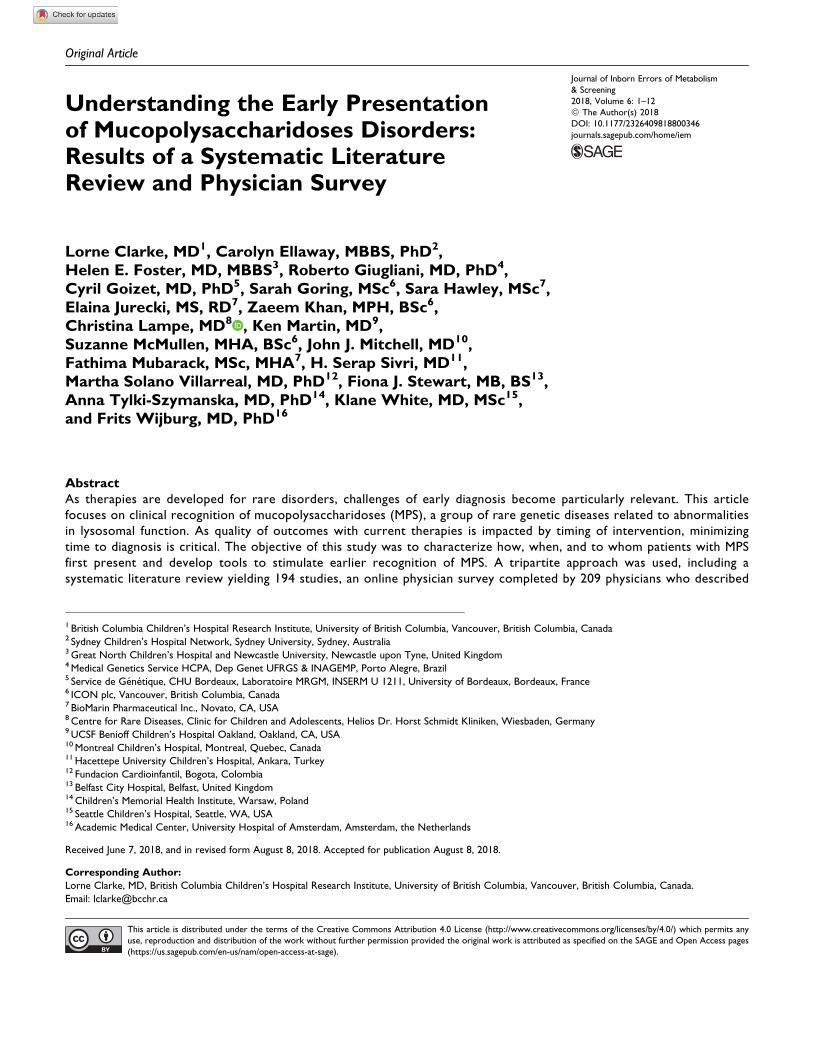
Figure 2. Literature review: duration of delays in diagnosis by mucopolysaccharidosis (MPS) type.*Some studies excluded due to nonreporting. Age of diagnosis and duration of delay in diagnosis may not be consistent as results have beensynthesized from different sources in the literature. Ranges are provided in brackets.
4 Journal of Inborn Errors of Metabolism & Screening

red flag signs and symptoms underwent iterative review
cycles with the panel as well as cross-referencing with the
physician survey and literature review results to ensure that
each sign or symptom was common enough among patients
with MPS to warrant inclusion and seen sufficiently infre-
quently by the physician type in question to be a realistic
trigger for screening. The exact balance of specificity versus
sensitivity determining inclusion/exclusion of an individual
sign or symptom was based on the collective clinical judg-
ment of the expert panel.
Results
Systematic Literature Review
A total of 1466 unique abstracts were identified. From these,
379 full-text publications were reviewed and 194 met the
inclusion criteria (Figure 1). The majority (76%) of these
studies reported on a single MPS type. A total of 330 MPS
type-specific records, representing 194 unique studies, were
identified.
Of the 194 studies included, 38% were reviews, 22% were
case reports, and the remainder included case series, observa-
tional studies, surveys, and guidelines. MPS type-specific sam-
ple sizes ranged from 1 patient to 1041 patients; 42% of records
contained fewer than 10 patients and only 3% of records con-
tained 75 or greater patients. Overall, 44% of studies were
European in origin, followed by Asian (15% of studies), and
North American (14% of studies). Each MPS type was
described in at least 1 study; descriptions of MPS I (39% of
studies) and MPS IV (32% of studies) were most frequent.
In studies where the specialty of the diagnosing physician
was described, it was most frequently geneticists, pediatri-
cians, or metabolic specialists. Delays in diagnosis are
reported in Figure 2 (additional data in Supplemental File
4). The typical delay in diagnosis, from time of symptom
onset, was reported to be 2.9 years (ranging from 0 to 38
years). In approximately 20% of records, the median delay
in diagnosis was reported to be at least 10 years. Some varia-
bility was observed across MPS types; the longest delays in
diagnosis tended to be reported for MPS IV.
Coarse facial features, short stature, corneal clouding, hepa-
tomegaly, and/or splenomegaly were the predominant signs
and symptoms at presentation. Other frequently published pre-
senting signs and symptoms included heart valve abnormality,
neurological abnormality, joint abnormality, and varying levels
of facial dysmorphism (additional data in Supplemental File 5).
Physician Survey
A total of 521 physicians were screened; 209 were eligible, and
they participated in the survey. Between 5 and 20 participants
were included per country, with general medicine being the
most frequently represented specialty (20% of participants) and
otorhinolaryngology the least frequent (5% of participants;
Table 1). The majority of participants practiced in an academic
setting (65%), and the study sample had a median of 16 years’
experience postresidency (range: 2-56 years). Participants had
experience with a median of 1 current MPS case (range: 0 to
200), and 3 previous MPS cases (range: 0 to 300). The majority
(78%) of physicians had experience with MPS I. Participants
reported having the least experience with MPS VII (14%reported any experience).
Physician participants described a total of 859 cases with
MPS, with just over half of the cases being MPS I (n ¼ 435)
and only 17 cases being MPS VII. For all MPS types, 67% of
cases described first presented under the age of 12 years, with
25% presenting under the age of 4 years.
Skeletal malformations and joint problems were the present-
ing signs and symptoms most frequently noted by the physi-
cians, reported in more than 20% of cases across all MPS types
(Figure 3). Gait disturbances, growth retardation, dysmorphic
facial features, psychomotor retardation, and liver enlargement
were also frequently reported, observed in more than 20% of
reported cases for most MPS types. Corneal clouding was the
most frequently observed ophthalmological sign for most MPS
types (26% of all cases with MPS). Heart abnormalities and
liver enlargement were observed at presentation in more than
20% of all cases with MPS reported, with heart abnormalities in
over 30% of cases with MPS I, IV, and VI. MPS III was the
type for which neurological signs and symptoms were most
frequently reported at presentation.
Among cases presenting under the age of 4, just over half of
patients presented with skeletal abnormalities or dysmorphic
facial features (52% and 51%, respectively). Other frequently
reported signs and symptoms in this age-group included devel-
opmental delay (45% of patients), growth retardation (38%),
psychomotor retardation (36%), hypotonia (31%), and cogni-
tive impairment (30%).
Forty-one percent of physicians reported that their patient
had signs and symptoms that they did not know were associated
Table 1. Physician Survey: Number of Surveyed Physicians bySpecialty.
Physician Respondents(n ¼ 209)
n (%)
SpecialtyGeneral practice/family medicine 42 (20.1)Orthopedics 12 (5.7)Rheumatology 18 (8.6)Neurology 13 (6.2)Internist 23 (11)Ophthalmology 23 (11)Medical geneticsa 0 (0)Cardiology 25 (12)Pediatrics 25 (12)Metabolic disease/metabolic genetics 18 (8.6)Otorhinolaryngologist 10 (4.8)
aPhysicians were recruited from specialist panels; however, a panel of pre-specified medical geneticists was not available
Clarke et al 5

with MPS, including various behavioral abnormalities (5%),
abdominal conditions such as hernias or hepatosplenomegaly
(4%), skeletal manifestations (3%), and visual conditions (2%).
Large variation was observed in the duration that patients
had MPS features prior to being referred or tested for MPS,
ranging from 1 month to 5 years. The signs and symptoms that
were present for the longest mean duration prior to referral
included carpal tunnel syndrome, skin abnormalities, recurrent
ear, nose, and/or sinus infection, gibbus, heart abnormalities,
and a family history of MPS, all present for a mean of over 13
months prior to referral (Table 2).
Referral patterns demonstrated that a large number of spe-
cialties were involved in the pathway to an MPS diagnosis
(Figure 4). General practice and pediatrics were the 2 special-
ties from which the surveyed physicians most frequently
received referrals. Metabolic specialists and pediatricians were
the specialties to which the surveyed physicians most fre-
quently referred patients. Among the surveyed physicians,
27% indicated that they would order an initial screening test
for MPS prior to referring a patient with suspected MPS. This
highlights the need for better education of the medical commu-
nity on MPS screening and testing.
MPS I MPS II MPS III MPS IV MPS VI MPS VII MPS IX Unknown Overall(n=435) (n=42) (n=148) (n=106) (n=21) (n=17) (n=21) (n=69) (n=859)
% % % % % % % % %Skeletal malforma�ons 49.4 50 30.4 68.9 61.9 29.4 52.4 34.8 47.4Joint problems (s�ffness or hypermobility) 33.8 28.6 37.2 34.9 42.9 35.3 52.4 30.4 34.7Gait disturbance 26.9 19 18.9 39.6 28.6 29.4 33.3 24.6 26.8Growth delays/short stature 30.6 26.2 27.7 42.5 57.1 11.8 23.8 27.5 31.2Knee deformi�es 18.4 26.2 12.8 28.3 28.6 23.5 23.8 13 19.1Hip deformi�es 17 4.8 16.9 27.4 19 5.9 14.3 11.6 17Gibbus deformity 10.1 14.3 2.7 15.1 14.3 5.9 14.3 7.2 9.5Other 1.1 0 0 0 0 5.9 4.8 1.4 0.9
Dysmorphic/coarse facial features 40.2 35.7 37.8 36.8 38.1 29.4 19 27.5 37.4 Psychomotor retarda�on 32 33.3 38.5 35.8 42.9 11.8 28.6 24.6 32.8 Hypotonia 22.3 21.4 30.4 17 19 35.3 23.8 14.5 22.6Other 0.5 0 0 0 0 0 0 0 0.2Heart abnormali�es 30.6 16.7 12.8 36.8 42.9 5.9 14.3 17.4 26Breathing abnormali�es 16.8 9.5 16.2 21.7 19 11.8 23.8 24.6 17.7Sleep apnea/sleep disordered breathing 13.1 9.5 18.9 13.2 28.6 11.8 23.8 17.4 14.9Liver enlargement 26.2 23.8 29.1 23.6 23.8 23.5 19 8.7 24.6Spleen enlargement 21.6 21.4 18.2 21.7 23.8 11.8 9.5 13 19.9Hearing deficits 11.3 16.7 16.9 10.4 14.3 5.9 4.8 15.9 12.6Mul�ple or recurrent hernia 5.3 14.3 4.1 10.4 14.3 17.6 4.8 4.3 6.5Other 0.2 0 0 0 0 0 0 0 0.1Clouding of the cornea 34 19 16.2 24.5 33.3 35.3 9.5 7.2 26.3Re�nal degenera�on 14.3 9.5 14.2 12.3 9.5 5.9 38.1 8.7 13.6Glaucoma/increased intraocular pressure 9.7 11.9 16.9 13.2 14.3 29.4 4.8 8.7 11.8Other 0.9 0 2 0 0 0 9.5 1.4 1.2Developmental delays 33.8 19 32.4 23.6 23.8 11.8 28.6 20.3 29.7Cogni�ve delays 22.1 9.5 34.5 13.2 28.6 11.8 28.6 18.8 22.4Hyperac�vity 12 11.9 41.9 17.9 19 11.8 23.8 14.5 18.5Au�sm spectrum disorder 8.7 0 26.4 9.4 14.3 11.8 4.8 8.7 11.5A�en�on deficits 13.3 19 31.8 10.4 19 17.6 23.8 7.2 16.4Carpal tunnel syndrome 6.2 4.8 5.4 5.7 9.5 0 0 5.8 5.7Other 0.5 0 1.4 0 0 0 0 0 0.5 Frequent ear infec�ons 20.2 21.4 10.8 19.8 9.5 11.8 28.6 15.9 18 Frequent sinus infec�ons 13.8 9.5 20.3 19.8 14.3 23.5 9.5 15.9 15.7 Frequent tonsil infec�ons 9.4 11.9 18.9 6.6 23.8 5.9 19 10.1 11.4 Frequent placement of ear tubes 6 7.1 11.5 9.4 9.5 5.9 19 5.8 7.8 Hearing impairment 9.4 9.5 15.5 12.3 23.8 0 14.3 4.3 10.7Other 0.2 0 0 0 0 0 0 0 0.1Skin abnormali�es 12.4 26.2 11.5 14.2 14.3 11.8 14.3 10.1 13Hair abnormali�es 12.6 11.9 16.9 7.5 19 17.6 9.5 1.4 12Hydrops fetalis 4.1 0 11.5 9.4 4.8 11.8 14.3 2.9 6.2Poor den��on/dental abnormali�es 13.1 14.3 15.5 16 19 0 9.5 7.2 13.3Frequent colds/upper respiratory tract infec�ons 9 2.4 10.8 9.4 9.5 23.5 33.3 10.1 10Macroglossia 12.2 7.1 15.5 13.2 19 0 9.5 11.6 12.5Family history of MPS 5.7 0 6.1 4.7 14.3 17.6 4.8 5.8 5.8Other 0.7 2.4 0.7 0 0 0 4.8 4.3 1
Skel
etal/
mus
cula
rDe
velo
p-m
enta
lOr
gan
syst
em
invo
lvem
ent
Opht
halm
o-lo
gical
Neur
olog
ical
Otor
hino
laryn
go-
logic
alOt
her
Figure 3. Physician survey: signs/symptoms present when mucopolysaccharidosis (MPS) first suspected or patient referred with suspectedMPS*.*Proportion among reported cases. Physicians were permitted to enter data for up to 10 patients; however, they were not asked to pull thesedata from patient charts—these are likely from memory. Cells are shaded if greater than 20%, with darkest shading for maximum values;“Other” fields within each subsection are populated by free-text fields. Ns in the column headers represent the number of patients enteredwhere at least one symptom was also provided.
6 Journal of Inborn Errors of Metabolism & Screening

Clinical Expert Panel
The expert panel identified red flag signs and symptoms for
consideration based on the systematic literature review, physi-
cian survey, and their own clinical experience. The red flag
signs and symptoms were selected to be specific to physicians
in pediatrics or general medicine as well as those in 5 subspeci-
alty types to whom patients with early signs of MPS are likely
to be referred: cardiology, pediatric neurology, otorhinolaryn-
gology, rheumatology, and orthopedics. These specialties were
selected based on the referral patterns reported in the physician
survey, the early signs and symptoms identified in the systema-
tic literature review and physician survey, and expert experi-
ence. One to 6 key red flag signs and symptoms were identified
for each specialty type and were further augmented with lists of
corroborating signs/symptoms to aid in the establishment of
Table 2. Physician Survey: Duration of Specific Signs/Symptoms at the Time of Decision to Test/Referral.a
Duration of Symptom Prior to Testing or Referral (months)
Patients with symptom (n) Mean (SD) Median (Range)
Skeletal/muscularSkeletal malformations 407 12.2 (11.3) 10 (1-60)Joint problems (stiffness or hypermobility) 298 12.8 (11.5) 9 (1-60)Gait disturbance 230 10.4 (9.6) 6 (1-60)Growth delays/short stature 268 11.6 (10.9) 7 (1-53)Knee deformities 164 12.2 (10.6) 9 (1-48)Hip deformities 146 12.9 (11.9) 10 (1-53)Gibbus deformity 82 14.5 (12.8) 10 (1-60)
DevelopmentalDysmorphic/coarse facial features 321 11.0 (10.7) 7 (1-60)Psychomotor retardation 282 10.2 (9.9) 6 (1-60)Hypotonia 194 7.3 (6.4) 6 (1-30)
Organ system involvementHeart abnormalities 223 14.8 (11.8) 12 (1-53)Breathing abnormalities 152 11.2 (9.8) 6.5 (1-53)Sleep apnea/sleep disordered breathing 128 10.6 (9.0) 7 (1-60)Liver enlargement 211 12.4 (12.1) 9 (1-60)Spleen enlargement 171 12.5 (12.3) 10 (1-60)Hearing deficits 108 11.3 (9.4) 7 (1-48)Multiple or recurrent hernia 56 12.6 (11.9) 10 (1-60)
OphthalmologicalClouding of the cornea 226 11.9 (11.2) 10 (1-60)Retinal degeneration 117 8.7 (5.8) 6 (1-24)Glaucoma/increased intraocular pressure 101 10.5 (10.2) 6.5 (1-48)
NeurologicalDevelopmental delays 255 10.8 (10.8) 6 (1-60)Cognitive delays 192 12.2 (11.4) 7 (1-60)Hyperactivity 159 9.7 (10.8) 6 (1-60)Autism spectrum disorder 99 10.6 (12.2) 6 (1-60)Attention deficits 141 9.9 (8.7) 6 (1-40)Carpal tunnel syndrome 49 15.1 (13.3) 13 (1-60)
OtorhinolaryngologicalFrequent ear infections 155 13.2 (10.9) 12 (1-60)Frequent sinus infections 135 13.2 (11.3) 11 (1-60)Frequent tonsil infections 98 9.6 (9.3) 6 (1-48)Frequent placement of ear tubes 67 9.2 (7.0) 7 (1-30)Hearing impairment 92 12.6 (10.5) 10 (1-48)
OtherSkin abnormalities 112 14.9 (12.5) 12 (1-60)Hair abnormalities 103 12.4 (11.0) 10 (1-60)Hydrops fetalis 53 6.2 (4.0) 6 (2-20)Poor dentition/ dental abnormalities 114 11.6 (10.1) 8 (1-48)Frequent colds/ upper respiratory tract infections 86 12.3 (9.7) 12 (1-40)Macroglossia 107 11.1 (9.7) 7 (1-48)Family history of MPS 50 16.1 (14.1) 12 (1-60)
Abbreviations: MPS: Mucopolysaccharidosis; SD: Standard deviation.aAt time the surveyed physician decided to test for MPS or refer on or before the case was referred to the surveyed physician.
Clarke et al 7

clinical suspicion of MPS (Table 3). These red flag signs and
symptoms were then converted into specialty-specific clinical
awareness diagnostic tools (Supplemental Files 6, 7, 8, 9, 10,
11, and 12). The tools were developed to be simple and visual.
They also highlight the incidence of MPS and urgency of early
diagnosis, provide information on additional signs and symp-
toms to help establish stronger clinical suspicion of MPS and
motivate testing, and list next steps.
While discussing next steps, the panel noted the differing
availability of screening and diagnostic test options globally
and the continuous improvement in testing technologies over
time. These factors, combined with those previously noted
highlight the need to better educate the medical community
on MPS screening and testing, led to the identification of the
need for a global MPS testing website with up-to-date, region-
specific information. As a result, www.test4mps.com website
was developed to provide information on how to test for MPS
and to house a searchable database of laboratories that conduct
MPS testing around the world.
Discussion
The largely unappreciated need for early diagnosis of MPS was
evident in the physician survey and systematic review. Patient
cases described in the physician survey were observed to have
signs and symptoms for over a year prior to referral, ranging up
to 5 years. Additionally, the observed referral patterns indicate
that patients are presenting to a variety of specialists and are
being referred on to a variety of other specialists. The systema-
tic literature review reflected similar trends, with nearly 40% of
records reporting a delay in diagnosis of 1 to 4 years and 20%reporting a delay of greater than 10 years. This clearly demon-
strates a need to improve the early recognition of signs and
symptoms to facilitate earlier MPS screening and referral to
metabolic specialists or clinical geneticists.
Results of the survey and literature review also highlighted
challenges of early MPS diagnosis: rarity and the varied and
sometimes subtle nature of signs and symptoms at presentation.
Forty one percent of physicians in the survey reported that their
patient(s) had signs or symptoms that they were not initially
Figure 4. Physician survey: alluvial plots showing which specialties the surveyed physicians received the mucopolysaccharidosis (MPS) referralsfrom (A) and which specialties the surveyed physicians referred suspected MPS patients to (B).Notes: The alluvial plot is weighted by the number of patients such that if a specialist said they had received 10 referrals from a generalpractitioner and 20 from a pediatrician, these weights are captured in the diagram.
8 Journal of Inborn Errors of Metabolism & Screening

Tab
le3.
List
ofSp
ecia
lty-
Spec
ific
Red
Flag
Sym
pto
ms
for
MPS
Dev
eloped
by
Inte
rnat
ional
Pan
elofM
PS
Exper
ts.
Spec
ialty
Red
Flag
Sym
pto
ms
Corr
obora
ting
Sym
pto
ms
Ort
hoped
ics
Bila
tera
lhip
dys
pla
sia/
ost
eonec
rosi
s(p
erth
es-lik
e)V
erte
bra
lbody
abnorm
ality
(hyp
opla
sia,
bea
king,
pla
tysp
ondyl
y,ky
phosi
s/gi
bbus)
Abnorm
alsk
elet
alfe
ature
s(s
uch
asgi
bbus,
pec
tus,
bro
adri
bs,
hyp
opla
stic
odonto
id,
enla
rged
sella
turc
ica,
genu
valg
us)
Aty
pic
alM
ongo
lian
spots
Car
pal
tunnel
syndro
me
(bila
tera
l)C
hro
nic
rhin
orr
hea
Cla
wed
han
ds
Coar
sefa
cial
feat
ure
sC
orn
ealcl
oudin
g
Den
talab
norm
alitie
sD
evel
opm
enta
ldel
ayD
iffic
ulty
open
ing
mouth
Dila
ted
Vir
chow
-Robin
spac
es
Enla
rged
tonsi
ls/a
den
oid
sG
lauco
ma
(bila
tera
l)H
eari
ng
loss
Hea
rtva
lve
dis
ease
Her
nia
s,in
cludin
gpre
vious
her
nia
repai
rH
irsu
tism
His
tory
ofher
nia
repai
rH
ydro
cephal
us
Join
tab
norm
alitie
s(r
estr
iction/s
tiffnes
sor
hyp
er-m
obili
ty/lax
ity)
Kyp
hosi
sLe
ftve
ntr
icula
rhyp
ertr
ophy
Live
ran
d/o
rsp
leen
enla
rgem
ent
Mac
rogl
oss
iaM
ulti-sy
stem
icin
volv
emen
tN
erve
com
pre
ssio
nsy
ndro
me
Psy
chom
oto
rdel
ay/r
egre
ssio
nR
ecurr
ent
ear
nose
and
thro
atin
fect
ions
Seiz
ure
sSh
ort
stat
ure
Slee
pap
nea
Spin
aldef
orm
ity
Unex
pla
ined
arth
ropat
hy,
with/w
ithout
pai
n
Car
dio
logy
Car
dia
cva
lve
thic
kenin
gO
tola
ryngo
logy
Rec
urr
ent
ear,
nose
,or
thro
atin
fect
ion
Upper
airw
ayobst
ruct
ion
Pro
gres
sive
hea
ring
loss
Ped
iatr
icN
euro
logy
Dev
elopm
enta
ldel
ay/r
egre
ssio
nLa
ngu
age
del
ayH
yper
activi
tyR
heu
mat
olo
gyU
nex
pla
ined
arth
ropat
hy
(with
or
without
pai
n)
No
evid
ence
of
infla
mm
atio
nPed
iatr
icia
ns
Short
stat
ure
/D
ecre
asin
ggr
ow
thve
loci
ty
Gib
bus
Dev
elopm
enta
ldel
ayR
ecurr
ent
EN
T-r
elat
edsy
mpto
ms
or
infe
ctio
ns
Enla
rged
liver
and/o
rsp
leen
Ingu
inal
or
um
bili
cal
her
nia
,es
pec
ially
recu
rren
t/his
tory
ofher
nia
repai
rG
ener
alPra
ctitio
ner
sU
nex
pla
ined
arth
ropat
hy
with
or
without
pai
n
Ear
lyonse
tsp
inal
dis
ease
Join
tre
stri
ctio
ns/
stiff
nes
sor
laxity/
hyp
erm
obili
tyC
ardia
cva
lvula
rdis
ease
Ear
lyco
rnea
lcl
oudin
g
Abbre
viat
ions:
EN
T,ea
r,nose
,th
roat
;M
PS,
muco
poly
sacc
har
idose
s.
9

aware were associated with MPS. Even with increased aware-
ness among those with little or no experience in managing an
MPS patient, a further challenge lies in providing a description
of a common presentation of MPS. While skeletal deformities
and joint problems are the hallmarks of most of the MPS dis-
orders, we compiled an extensive list of other clinical features
with early onset that should make one consider MPS. For
example, some of the early presenting signs and symptoms that
were frequently reported in the literature and physician survey
included growth retardation; recurrent ear, nose, and/or throat
infections; coarse facial features; developmental delay; heart
valve thickening; corneal clouding; progressive hearing loss;
and hernias.
Although others have attempted to generate tools and/or
algorithms based on presenting signs and symptoms to aid in
the diagnosis of MPS, these efforts have resulted in no notice-
able change in the average length of the diagnostic delay over
time.8 The difficulty in distributing this information and its
very limited half-life with the target audiences are substantial
contributing factors in the failure of these previous attempts.
Unfortunately, we will be facing the same challenges with
these tools. However, previous efforts have also had several
notable limitations. They often targeted only a subset of the
specialists likely to encounter undiagnosed individuals with
MPS,12–15 were limited to a particular MPS type14,15 or a
sub-group of the MPS population,12,13 resulted in complex,
multitiered algorithms with a high degree of specificity and
inadequate sensitivity,12,13 and relied solely on expert opin-
ion.12–15 Several steps were taken to overcome these previous
limitations in the current project.
The majority of specialists to whom individuals with MPS
present were included. All MPS disorders were targeted collec-
tively, thereby increasing the incidence to within a range that
may be more relevant for the target audience8 and enabling
physicians to proceed with the suspicion of MPS in general,
without needing specific knowledge of the types of MPS. The
proposed screening is based on simple red flags that are easier
to recall than a complex algorithm. And importantly, the qual-
ity and quantity of data gathered through systematic review and
physician survey provided a solid evidence-based foundation.
The literature review was conducted using systematic and
reproducible methods and captured a broad range of study
designs, including reviews, case reports, case series, and larger
observational studies. The physician survey was internationally-
based, multilingual, and included data collected from a wide
range of specialties, thus reflecting the variation in symptom
presentation and patterns of referrals across geographies and
specialties. This multimethod approach is important for rare
diseases where there is a paucity of large studies that capture
the data needed to inform the research question.
Despite the extensive set of data collected, there were some
notable limitations. For example, in the literature review, there
is an inherent risk of publication bias (ie, there may have been a
risk that particularly unusual presentations of MPS were
included, as these were considered worthy of publication).
However, given the large number of studies returned, and
inclusion of several large observational studies, it is expected
that these extremely unusual presentations would also be rela-
tively infrequent in the synthesized data set. This limitation of
the systematic literature review was also addressed by supple-
menting the review with a physician survey and by seeking
clinical expert feedback on the generalizability of the results.
A main limitation of the physician survey was that the data
were, by the design of the study, based on physician recall
rather than from a chart review. A sign or symptom not
reported at presentation may have been due to it not being
present, or alternatively, the physician may not have recalled
the sign or symptom, or it may not have been identified at that
time. Nonspecific signs and symptoms that could be identified
by specialists outside of those included in this analysis may
also have been missed. Furthermore, the MPS type was
reported by the responding physician who may not have been
the diagnosing physician, and thus, there may have been inac-
curacies. Additionally, not all MPS types were equally repre-
sented; however, this limitation was mitigated by analyzing the
data by MPS type and by including clinical experts to provide
insight into all MPS types. To increase the likelihood of finding
eligible physicians to participate in the survey, physicians
were selected only from countries where there were known
MPS clinics. Although this includes the majority of countries
with a well-known population of patients with MPS, it is a
limitation that not all countries with potential MPS patients
were represented in the analysis.
The compilation of evidence from the systematic literature
review and survey is extensive; however, through review and
discussion with the international panel of clinical experts, the
findings were distilled into targeted lists of specialty-specific
red flag signs and symptoms, together with an additional list of
corroborating signs and symptoms. These were synthesized
into simple, specialty-specific tools that can help raise aware-
ness while reducing the potentially overwhelming amount of
information for practitioners who may only ever see 1 or 2 MPS
patients in their entire career. The specialty-specific nature of
these tools further maximizes their relevance. Importantly,
these tools have also been coupled with real-world, up-to-
date laboratory information to facilitate rapid action following
initial suspicion of MPS.
The ability of these tools to have an impact for MPS patients
and their families is completely dependent on what happens
next. Due to the rarity of the disease, specialists may only see
1 patient with MPS during their career, perhaps years after this
paper is published. The key knowledge we have assembled here
will need to be available to physicians whenever they happen to
encounter that patient. Using repetitive dissemination of our
simple, evidence-based, specialty-specific tools and striving to
ensure their continual presence (ie, tools posted on hospital
walls, incorporation into guidelines, inclusion in an online appli-
cation), we hope to have the critical information available at the
right time for as many cases as possible. The effectiveness of our
approach will need to be evaluated in a future study.
Ultimately, though these clinical awareness diagnostic tools
will not completely close the diagnostic gap, we hope that they
10 Journal of Inborn Errors of Metabolism & Screening

will help to narrow it until a more comprehensive solution, such
as newborn screening, automated electronic medical record
flagging, or mandatory subpopulation screening, is feasible.
Authors’ Note
Sarah Goring, Suzanne McMullen, Sara Hawley, and Elaina Jurecki
contributed to study design, and physician survey conduct. Fathima
Mubarack and Elaina Jurecki designed the systematic literature review
and Fathima Mubarack performed data extraction and Sarah Goring
and Zaeem Khan contributed to the analysis of the systematic litera-
ture review. Lorne Clarke, Carolyn Ellaway, Helen E. Foster, Roberto
Giugliani, Cyril Goizet, Christina Lampe, Ken Martin, John J. Mitch-
ell, Martha Solano Villarreal, H. Serap Sivri, Fiona J. Stewart, Anna
Tylki-Szymanska, Klane White, and Frits Wijburg were members of
the clinical expert panel, and provided interpretation of the study
results and selection of the relevant symptoms based on the study
results. All authors read and approved the final manuscript.
Acknowledgments
We would like to acknowledge Dr Mira de Kruijf (Royal Manchester
Children’s Hospital), Dr Kuo Sheng Lee (Mackay Memorial Hospi-
tal), Dr Elizabeth Braunlin (University of Minnesota), Neil James,
Tiffany Wong, Grace Chen, and Emanuela Izzo (BioMarin Pharma-
ceutical Inc.) for their contributions to this study.
Declaration of Conflicting Interests
The author(s) declared the following potential conflicts of interest
with respect to the research, authorship, and/or publication of this
article: Lorne Clarke receives honoraria and travel support from
Sanofi, BioMarin, and Shire related to participation in disease registry
boards and speakers bureaus. Carolyn Ellaway has received travel
support and honoraria from BioMarin, Sanofi Genzyme, and Shire.
Helen E. Foster on behalf of Newcastle University, United Kingdom
has received speaker/chair honoraria and unrestricted educational bur-
saries from BioMarin and Sanofi Genzyme. Roberto Giugliani has
received investigator fees, and/or travel grants, and/or speaker honor-
aria from Actelion, Amicus, Armagen, BioMarin, GC Pharma, JCR
Pharmaceuticals, Lysogene, Sanofi Genzyme, Shire, and Ultragenyx.
Cyril Goizet has received consulting fees from Sanofi Genzyme and
BioMarin; honorarium for participation in advisory boards from Bio-
Marin and Sanofi Genzyme; financial support for research activities
from Sanofi Genzyme, Shire, and BioMarin; and funding for inscrip-
tions and travels for congresses from Sanofi Genzyme, Shire, and
BioMarin. Sara Hawley, Elaina Jurecki, and Fathima Mubarack are
employees and stockholders of BioMarin. Christina Lampe has
received honoraria/consultation fees from Shire, Alexion, Actelion,
BioMarin and Genzyme, and has participated in company sponsored
speaker’s bureau for Shire, Alexion, Actelion, BioMarin and Gen-
zyme. Ken Martin has accepted consulting fees or honoraria from the
following companies engaged in developing MPS-related therapies:
BioMarin, Sangamo, REGENXBIO, Ultragenyx, and Shire. John J.
Mitchell has received speaker fees, consulting fees and research sup-
port from BioMarin and Shire, and receives funding support from the
Harpur Foundation. H. Serap Sivri has received travel support and
honoraria from BioMarin. Martha Solano Villarreal has received
speaker fees and consulting fees from BioMarin. Fiona J. Stewart has
received speaker fees and consultancy fees from BioMarin. Anna
Tylki-Szymanska has received honoraria/consultation fees from Shire,
BioMarin, Chiesi and Genzyme, and has participated in company
sponsored speaker’s bureau for Shire, BioMarin and Genzyme. Klane
White has received honoraria/travel support from BioMarin,
Genzyme, and Medicrea, and receives royalties from UpToDate.com
and grant support from BioMarin. Frits Wijburg has received honor-
aria for presentations and board meetings, travel expenses to meetings
and honoraria for consultancy work from Sanofi Genzyme, BioMarin,
Actelion and Shire, and has received unrestricted educational grants
and research grants from Sanofi Genzyme, BioMarin and Actelion.
Sarah Goring, Zaeem Khan, and Suzanne McMullen declare that they
have no competing interests.
Funding
The author(s) disclosed receipt of the following financial support for
the research, authorship, and/or publication of this article: This study
was funded by BioMarin Pharmaceutical Inc.
ORCID iD
Christina Lampe http://orcid.org/0000-0003-4953-7119
Supplemental Material
Supplemental material for this article is available online
References
1. Muenzer J. Overview of the mucopolysaccharidoses. Rheumatol-
ogy. 2011;50:v4-v12.
2. Bittar T. Mucopolysaccharidosis. In: Grogan DP, ed. Medscape.
New York, NY: Medscape; 2013.
3. Neufeld EU, Muenzer J. The mucopolysaccharidoses. In: Scriver
CR, Beaudet AL, Sly WS, Valle D, Childs B, Kinzler KW, Vogel-
stein B, eds. The metabolic and molecular bases of inherited
disease. New York: McGraw-Hill; 2001: pp. 3421-3452.
4. Baehner F, Schmiedeskamp C, Krummenauer F, et al. Cumulative
incidence rates of the mucopolysaccharidoses in Germany.
J Inherit Metab Dis. 2005;28(6):1011-1017.
5. Meikle PJ, Hopwood JJ, Clague AE, Carey WF. Prevalence of
lysosomal storage disorders. JAMA. 1999;281(3):249-254.
6. Poorthuis BJ, Wevers RA, Kleijer WJ, et al. The frequency of
lysosomal storage diseases in the Netherlands. Human Genet.
1999;105(1-2):151-156.
7. Lin HY, Lin SP, Chuang CK, et al. Incidence of the mucopoly-
saccharidoses in Taiwan, 1984-2004. Am J Med Genet A. 2009;
149A(5):960-964.
8. Kuiper GA, Meijer OL, Langereis EJ, Wijburg FA. Failure to
shorten the diagnostic delay in two ultra-orphan diseases (muco-
polysaccharidosis types I and III): potential causes and implica-
tions. Orphanet J Rare Dis. 2018;13(1):2.
9. Wilson GN. Hunter syndrome (mucopolysaccharidosis II): diag-
nosis, genetic testing, treatment, and referral. Consultant for
Pediatricians. 2015;14(5):206-212.
10. Centre for Reviews and Dissemination. Systematic Reviews:
CRD’s Guidance for Undertaking Reviews in Health Care.
Layerthorpe, York: University of York. 2009. https://www.york.
ac.uk/media/crd/Systematic_Reviews.pdf
11. Moher D, Liberati A, Tetzlaff J, Altman DG. Preferred reporting
items for systematic reviews and meta-analyses: the PRISMA
statement. BMJ. 2009;339:b2535.
Clarke et al 11

12. Cimaz R, Coppa GV, Kone-Paut I, et al. Joint contractures in the
absence of inflammation may indicate mucopolysaccharidosis.
Pediatr Rheumatol. 2009;7:18.
13. Lehman TJA, Miller N, Norquist B, Underhill L, Keutzer J. Diag-
nosis of the mucopolysaccharidoses. Rheumatology. 2011;50(5):
v41-v48.
14. Wood T, Bainbridge K, Beck M, et al. Diagnosing mucopo-
lysaccharidosis IVA. J Inherit Metab Dis. 2013;36(2):
293-307.
15. Wood T, Bodamer OA, Burin MG, et al. Expert recommendations
for the laboratory diagnosis of MPS VI. Mol Genet Metab. 2012;
106(1):73-82.
12 Journal of Inborn Errors of Metabolism & Screening

Correction Notice
Journal of Inborn Errors of Metabolism
& Screening
2018, Volume 6: e20180103
© The Author(s) 2018
journals.sagepub.com/home/iem
Clarke L, et al. Understanding the Early Presentation of Mucopolysaccharidoses Disorders: Results of a Systematic Literature
Review and Physician Survey. Journal of Inborn Errors of Metabolism & Screening. 2018; 7: 1-12. DOI:
10.1177/2326409818800346
It was noted that the volume of the above-mentioned article was v6, which was wrongly published as v7.
DOI: 10.1177/2326409818800346er