Embed Size (px)
Citation preview

Unexpected Gynecologic Findings at Laparotomy
Susan A. Davidson, MDUniversity of Colorado, Denver
School of Medicine

Adnexal Mass: Gyn Etiologies
• Uterine– Leiomyomas– Pregnancy– Malignancy
• Tubal– Pregnancy– Hydrosalpinx– Malignancy
• Tubo-Ovarian– Abscess (TOA)– Torsion
• Ovarian– Functional cyst– Endometrioma– Neoplasm
• Benign• Malignant

Factors affecting intra-operative decision-making
• Autonomy– Consent
• Beneficence– Pathology
• Fertility• Hormonal

Uterine Leiomyomas
• Most common pelvic tumor– 1 in 4 white women– 1 in 2 black women
• Pathogenesis elusive– Each is monoclonal– Probable somatic mutation, influenced by
estrogen, progesterone, & local growth factors

Uterine Leiomyomas
• Rarely malignant– 0.3 to 0.7% of myomatous uteri
• Usually multiple• Symptoms (none in majority of women)
– Pressure– Pain– Abnormal bleeding

Uterine Leiomyomas: Surgical Management
• Intramural or sub-serosal with no “stalk”: leave in-situ
• Sub-serosal with “stalk”:– Leave in-situ:
• Premenopausal, unknown fertility issues, &• Pelvic pain was NOT indication for surgery
– Remove:• Very large• Pelvic pain WAS indication for surgery
Intrauterine Pregnancy
• Enlarged uterus• May be associated with ovarian luteal
cysts, especially early in pregnancy:– Do not remove: will resolve spontaneously– Intra-operative ultrasound to document
pregnancy

Uterine Malignancy
• Rarely diagnosed as an adnexal mass but may be associated with an adnexalmass– Uterine papillary-serous & clear cell
adenocarcinomas can spread intraperitoneally (e.g., omentum, tubes & ovaries) like ovarian cancer
– 20% of endometrioid ovarian cancers associated with uterine endometrial cancer

Ectopic (Tubal) Pregnancy
• Location:– 81% Ampullary (distal 2/3 of tube)– 12% Isthmus (proximal 1/3 of tube)– 5% Fimbrial– 2% Interstitial
• Triage points– Ruptured vs. Unruptured– Desires fertility vs. sterility

Ectopic (Tubal) Pregnancy
• Salpingectomy required– Uncontrolled bleeding– Recurrent ectopic pregnancy in same tube– Severely damaged tube– Ectopic 5 cm
• Salpingectomy recommended– Does not desire fertility

Ectopic (Tubal) Pregnancy
• Desires fertility– Linear salpingostomy– Risk of persistent ectopic pregnancy
• Post-op incidence: 3.9-8.3%• Follow serial bHCG’s weekly• Treat with methotrexate if rising bHCG

Ectopic (Tubal) Pregnancy
• Linear salpingostomy– Vasopressin (0.2 IU/ml NS) injection into tube
wall at area of maximal distention– Longitudinal incision– Evacuate products– Flush tube– Control bleeders with bipolar cautery, small
(6-0) sutures PRN.

Hydrosalpinx
• End-stage of pyosalpinx:– Distended with watery, sterile fluid
• Management:– Desires fertility
• Pelvic pain reason for surgery: salpingostomy• No pain: leave in-situ
– Does not desire fertility, postmenopausal:• Pelvic pain reason for surgery: Salpingectomy• No pain: leave in-situ

Tubal Malignancy
• Rare• Spread pattern similar to ovarian CA
– Intraperitoneal– Lymphatic
• Usually firm, distended tubal mass– Excise with adjacent ovary– Triage & surgical management same as for
ovarian cancer

Tubo-Ovarian Abscess
• Management dependent on:– Rupture– Reproductive wishes– Intra-operative findings

Tubo-Ovarian Abscess
• Immediate surgery– Uncertain diagnosis, suspicion of rupture:
• Septic shock• Generalized peritonitis• Falling WBC
– Extent of disease & desire for fertility define the extent of surgery
Tubo-Ovarian Abscess
• Discovered at exploration, stable patient, unruptured, desires fertility or fertility wishes unknown:– Leave in-situ:
• 70% respond to abx treatment alone• Greater surgical risk in acute inflammatory
phase• 14% had subsequent intra-uterine pregnancy in
one study• Retain ovarian function
Wagaman; J Reprod Med 1990;35:833. Cohen; J Am Assoc
Gynecol Laparosc 1999;6:139.
Torsed Adnexal Mass• More common in pregnant than non-
pregnant state (28% vs. 7%)• Management:
– Detorsion with ovarian cystectomy:• No increase in risk of venous embolism• Follow-up U/S: 93% with black-bluish ovaries
had normal follicle development– Frozen section, if suspicious, to rule out
malignancy

Functional Ovarian Cysts
• Follicular cysts• Corpus luteum cysts• Theca lutein cysts

Follicular Cysts
• Most frequent ovarian cystic structure• Usually multiple, thin-walled• Size variable:
– 3 to 15 cm, usually <6-8 cm • Dependent on gonadotrophins for growth • Most resolve spontaneously (rupture or
resorption)

Corpus Luteum Cysts
• Develop from mature graafian follicle• Size: 3 to 10 cm, occasionally larger• Development:
– Hemorrhage into cyst few days after ovulation
– Blood resorbs, leaving cyst• Signs & symptoms
– Unilateral pelvic pain, tender adnexa, occasionally massive bleeding
Spanos; AJOG 116:551, 1973
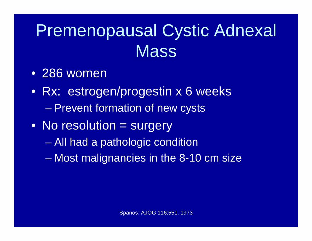
Premenopausal Cystic AdnexalMass
• 286 women• Rx: estrogen/progestin x 6 weeks
– Prevent formation of new cysts• No resolution = surgery
– All had a pathologic condition– Most malignancies in the 8-10 cm size

Theca Lutein Cysts
• Least common physiologic cyst• Bilateral• Moderate to massive ovarian
enlargement– Honeycombed, lobulated appearance– Gray to bluish-tinged cysts– Up to 20-30 cm in diameter

Theca Lutein Cysts
• Etiology: excess gonadotrophins– Exogenous: drugs to induce ovulation– Endogenous: molar pregnancy, pregnancies
with large placenta• Management:
– Leave in-situ & handle gently• Slowly resolve spontaneously• Bleeding from cysts hard to control

ACOG Practice Bulletin #11, 12/99
Endometriosis & Endometriomas
• Incidence– 7-10% of women in general population– Up to 50% of premenopausal women– 38% prevalence in infertile women– 71-87% prevalence in women with chronic
pelvic pain
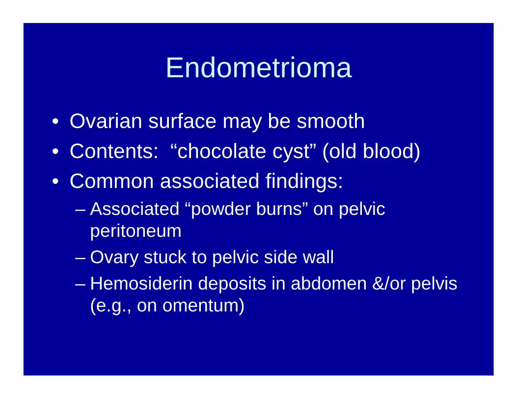
Endometrioma
• Ovarian surface may be smooth• Contents: “chocolate cyst” (old blood)• Common associated findings:
– Associated “powder burns” on pelvic peritoneum
– Ovary stuck to pelvic side wall– Hemosiderin deposits in abdomen &/or pelvis
(e.g., on omentum)

Endometrioma• Frequently associated with extra-ovarian
endometriosis– Biopsy to document disease
• Multiple treatment options:– Hormonal– Cystectomy with ovarian reconstruction– Cyst stripping or ablation– USO, BSO, +/- Hysterecomy
• Frequently rupture: copiously irrigate• As surprise finding: if looks like endometriosis,
no obvious malignancy, leave in situ for postopdiscussion of management

Ovarian Neoplasm: Risk of Malignancy
• Risk by menopausal status:– Premenopausal: 7-13% malignant– Postmenopausal: 30-45% malignant
• Excludes non-neoplastic masses

Koonings; Obstet Gynecol 74:921, 1989
Ovarian Neoplasm: Risk of Malignancy by Age
Age % Malignant Tumor histology <20 (n=61) 8 80% Germ cell, 20%
Stromal 20-29 (n=294) 4 33% ea: EOC, Germ cell,
Stromal 30-39 (n=171) 14 70% EOC, 20% Stromal 40-49 (n=128) 35 93% EOC 50-59 (n=104) 46 95% EOC 60-69 (n=79) 49 100% EOC >70 (n=24) 24 100% EOC

Koonings; Obstet Gynecol 74:921, 1989
Distribution of Benign Ovarian Neoplasms by Age
N=650 Serous Mucinous Teratoma Stromal <20 20% 11% 70% 0% 20-29 15% 11% 72% 1% 30-39 17% 12% 67% 4% 40-49 43% 8% 43% 3% 50-59 46% 14% 21% 17% 60-69 59% 11% 16% 11% >70 53% 24% 0% 12% Total 25% 12% 58% 4%

Suspicious Ultrasound
• Bilateral > Unilateral• Predominant solid component• Size >10 cm• Thick & numerous septa• Papillary or nodular structures• Ascites

Roman; Gynecol Oncol 69:1, 1998
Postmenopausal UnilocularCystic Mass
• Review of 17 series• Cyst size
– <5 cm: 440 (3 malignant)– 5-10 cm: 46 (1 malignant)– >1 cm: 43– Not stated: 40
• Risk of malignancy– 4/569 (0.7%)

Intraoperative Diagnosis of an Ovarian Mass
• Management complex• Need to consider
– Mass characteristics– Evidence of peritoneal implants: tumor or
endometriosis– Age– Menopausal status– Reproductive wishes

Intraoperative Diagnosis of an Ovarian Mass
• If diagnosed at laparoscopy for another indication– Biopsy any peritoneal implants or ovarian
surface abnormality– Defer management
• If diagnosed at laparotomy for another indication– Follow algorithms

Premenopausal Adnexal Mass:Intraoperative Diagnosis
• When in doubt, DON’T cut it out!• Intraoperative or postoperative gyn
oncology consult

Premenopausal Adnexal Mass:Intraoperative Diagnosis
No intervention
<4-6 cm
Cystectomy
>6cm, cystic
Frozen section only ifprepared to completely
stage
Remove adnexa
Solid MassCyst > 15-20 cm
Remove mass
Frozen sectionMalignant
Biopsycontralateral
mass
Frozen sectionMalignant
Remove mostsuspicious mass
Bilateral Solid Masses
Desires Fertility
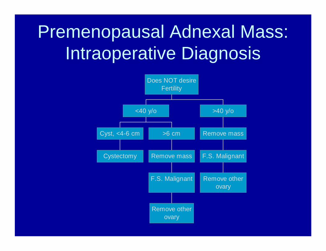
Premenopausal Adnexal Mass:Intraoperative Diagnosis
Cystectomy
Cyst, <4-6 cm
Remove otherovary
F.S. Malignant
Remove mass
>6 cm
<40 y/o
Remove otherovary
F.S. Malignant
Remove mass
>40 y/o
Does NOT desireFertility
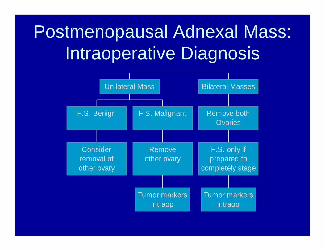
Postmenopausal Adnexal Mass:Intraoperative Diagnosis
Considerremoval ofother ovary
F.S. Benign
Tumor markersintraop
Removeother ovary
F.S. Malignant
Unilateral Mass
Tumor markersintraop
F.S. only ifprepared to
completely stage
Remove bothOvaries
Bilateral Masses

Intraoperative Diagnosis of Ovarian Malignancy
• Gyn oncology consult if available• May require second operation for staging• Obtain appropriate tumor markers
intraoperatively or immediately postoperative

Ovarian Cancer Surgical Staging
• Abdomino-pelvic washings• Peritoneal biopsies: pelvic side-walls,
cul-de-sac, bladder peritoneum, gutters, diaphragms
• Omentectomy• Bilateral pelvic & para-aortic
lymphadenectomy

Ovarian Cancer Surgical Staging
• Does not include hysterectomy or biopsy of a normal contralateral ovary– Usually TAH, BSO if no fertility issues
• Desires fertility– If contralateral ovary normal, leave in-situ– If both ovaries abnormal, leave uterus in-
situ• 30% upstaged (microscopic spread) if
completely staged

Use of Tumor Markers
• Germ cell tumors:– AFP, bHCG, LDH
• Epithelial tumors– CA-125, CEA
• Stromal tumors– Granulosa cell: Inhibin, CA-125– Sertoli Leydig: Testosterone, CA-125

Questions• A 26 y/o woman undergoes laparotomy for suspected
ruptured appendix and is found to have a 6 cm right ovarian mass that has torsed, with the fallopian tube, 2x on its pedicle. The most appropriate management is:– a) Detorsing the adnexa and leaving it in-situ– b) Detorsing the adnexa and performing an ovarian cystectomy– c) Detorsing the adnexa and removing the tube & ovary if they
remain blue-black– d) Removing the adnexa without detorsing

Questions• A 30 y/o nulligravid female undergoes laparotomy for
presumed appendicitis. Pre-operative WBC was 20,000, & she was non-toxic. Intraoperative, she is found to have bilateral 6cm tubo-ovarian abscesses that are fixed in the cul-de-sac & non-ruptured. The best management is:– a) Bilateral adnexectomy– b) Bilateral adnexectomy & hysterectomy– c) Drain abscesses & insert drains– d) Close patient & treat with antibiotics

Questions• A 34 y/o woman undergoes laparotomy for planned
colectomy. She is found to have an 8 cm irregular, solid left ovarian mass. The left adnexa is removed & sent for frozen section which reveals a low grade serous carcinoma. The other ovary is normal as is complete exploration. The most appropriate management is:– a) Biopsy right ovary for frozen section, draw tumor markers– b) Proceed with staging & tumor markers– c) Remove right tube & ovary, stage, & draw tumor markers– d) Remove uterus, right tube & ovary, stage, & draw tumor
markers