Embed Size (px)
Citation preview
81
Radiology UPdaTE Vol. 2 (3) iSSN 2424-5755
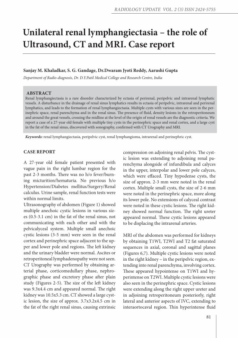
Unilateral renal lymphangiectasia – the role of Ultrasound, CT and MRI. Case report
Sanjay M. Khaladkar, S. G. Gandage, Dr.Dwaram Jyoti Reddy, Aarushi GuptaDepartment of Radio-diagnosis, Dr. D.Y.Patil Medical College and Research Centre, India
ABSTRACTRenal lymphangiectasia is a rare disorder characterized by ectasia of perirenal, peripelvic and intrarenal lymphatic vessels. A disturbance in the drainage of renal sinus lymphatics results in ectasia of peripelvic, intrarenal and perirenal lymphatics, and leads to the formation of renal lymphangiectasia. Multiple cysts with various sizes are seen in the per-inephric space, renal parenchyma and in the renal sinus. The presence of fluid, density lesions in the retroperitoneum and around the great vessels, crossing the midline at the level of the origin of renal vessels are the diagnostic criteria. We report a case of a 27-year old female with multiple tiny cysts in the perinephric space and renal cortex, and a large cyst in the fat of the renal sinus, discovered with sonography, confirmed with CT Urography and MRI.
Keywords: renal lymphangiectasia, peripelvic cyst, renal lymphangioma, intrarenal and perinephric cyst.
CASE REPORT
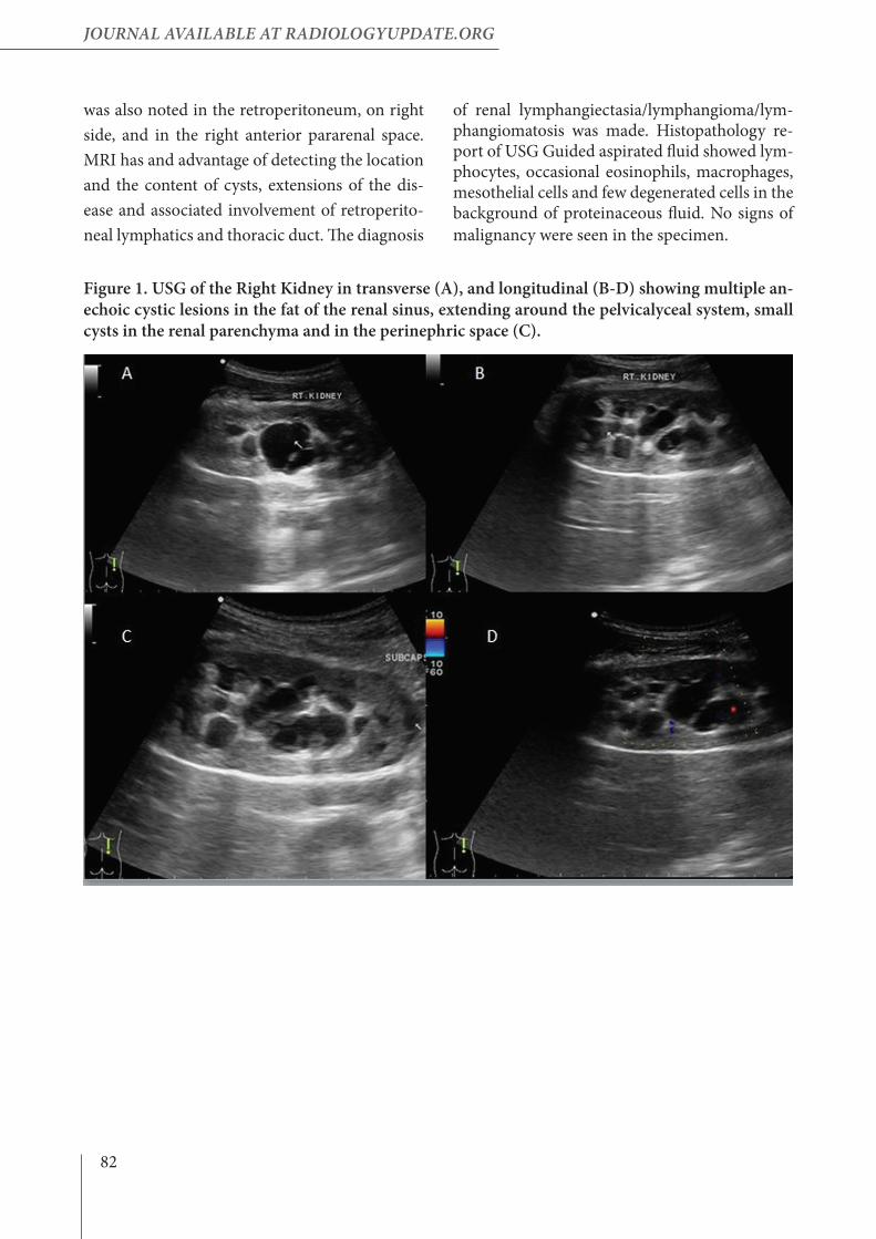
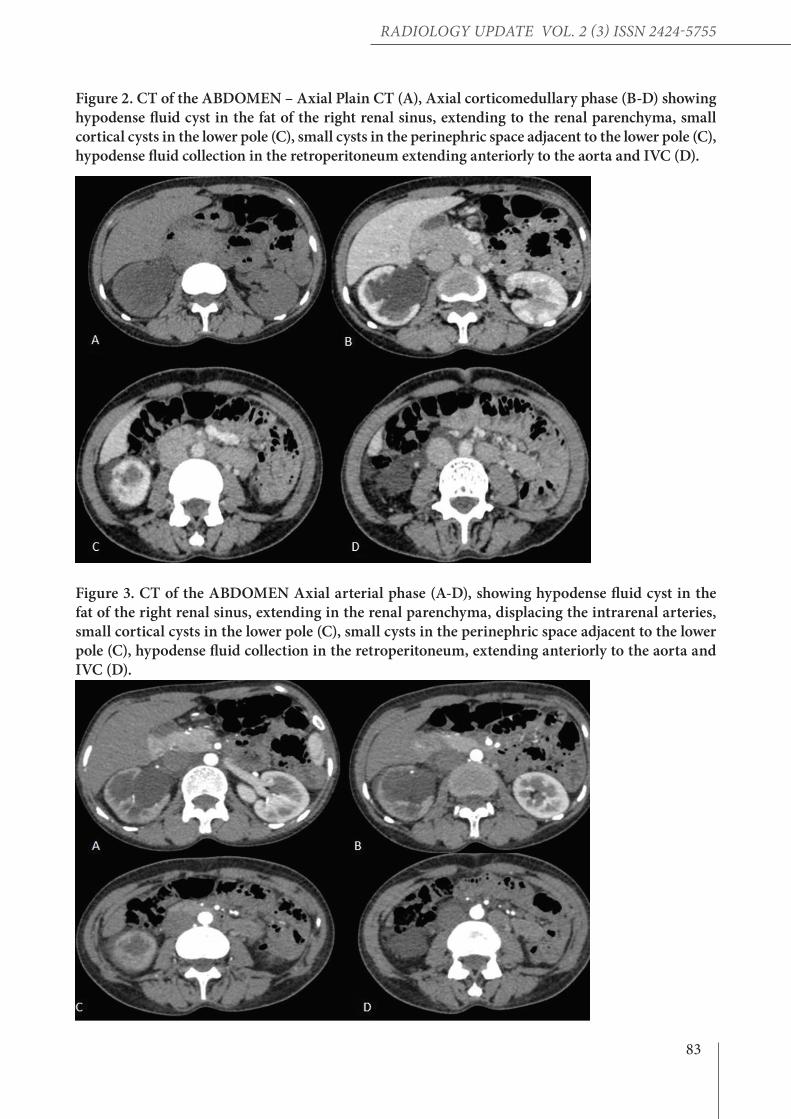
A 27-year old female patient presented with vague pain in the right lumbar region for the past 2-3 months. There was no h/o fever/burn-ing micturition/hematuria. No previous h/o Hypertension/Diabetes mellitus/Surgery/Renal calculus. Urine sample, renal function tests were within normal limits. Ultrasonography of abdomen (Figure 1) showed multiple anechoic cystic lesions in various siz-es (0.5-3.1 cm) in the fat of the renal sinus, not communicating with each other and with the pelvicalyceal system. Multiple small anechoic cystic lesions (3-5 mm) were seen in the renal cortex and perinephric space adjacent to the up-per and lower pole and regions. The left kidney and the urinary bladder were normal. Ascites or retroperitoneal lymphadenopathy were not seen.CT Urography was performed by obtaining ar-terial phase, corticomedullary phase, nephro-graphic phase and excretory phase after plain study (Figures 2-5). The size of the left kidney was 9.3x4.4 cm and appeared normal. The right kidney was 10.5x5.3 cm. CT showed a large cyst-ic lesion, the size of approx. 3.7x3.2x4.5 cm in the fat of the right renal sinus, causing extrinsic
compression on adjoining renal pelvis. The cyst-ic lesion was extending to adjoining renal pa-renchyma alongside of infundibula and calyces in the upper, interpolar and lower pole calyces, which were effaced. Tiny hypodense cysts, the size of approx. 2-3 mm were noted in the renal cortex. Multiple small cysts, the size of 2-6 mm were noted in the perinephric space, more along its lower pole. No extensions of calyceal contrast were noted in these cystic lesions. The right kid-ney showed normal function. The right ureter appeared normal. These cystic lesions appeared to be displacing the intrarenal arteries.
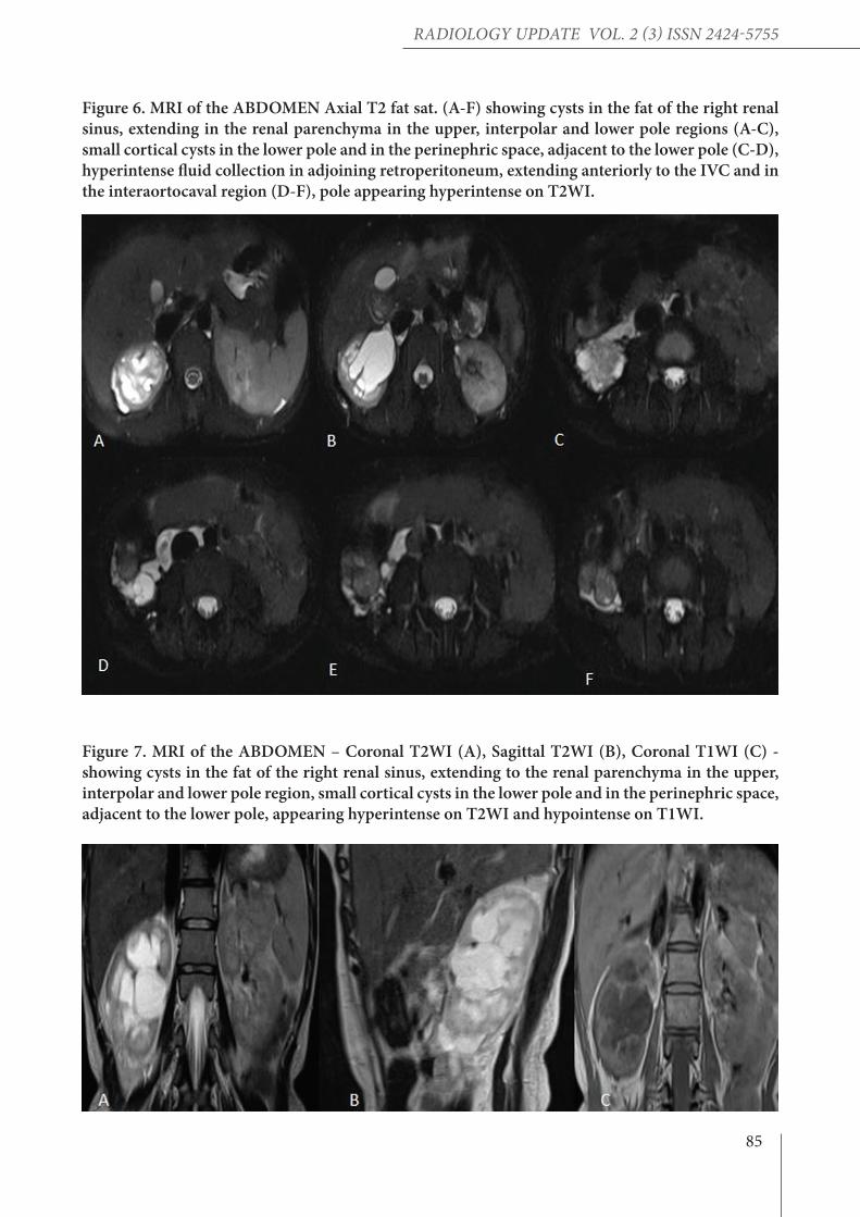
MRI of the abdomen was performed for kidneys by obtaining T1WI, T2WI and T2 fat saturated sequences in axial, coronal and sagittal planes (Figures 6,7). Multiple cystic lesions were noted in the right kidney – in the peripelvic region, ex-tending into renal parenchyma, involving cortex. These appeared hypointense on T1WI and hy-perintense on T2WI. Multiple cystic lesions were also seen in the perinephric space. Cystic lesions were extending along the right upper ureter and in adjoining retroperitoneum posteriorly, right lateral and anterior aspects of IVC, extending to interaortocaval region. Thin hyperintense fluid
82
JOURNAL AvAiLAbLe At RAdiOLOgyUpdAte.ORg
was also noted in the retroperitoneum, on right side, and in the right anterior pararenal space. MRI has and advantage of detecting the location and the content of cysts, extensions of the dis-ease and associated involvement of retroperito-neal lymphatics and thoracic duct. The diagnosis
Figure 1. USG of the Right Kidney in transverse (A), and longitudinal (B-D) showing multiple an-echoic cystic lesions in the fat of the renal sinus, extending around the pelvicalyceal system, small cysts in the renal parenchyma and in the perinephric space (C).
of renal lymphangiectasia/lymphangioma/lym-phangiomatosis was made. Histopathology re-port of USG Guided aspirated fluid showed lym-phocytes, occasional eosinophils, macrophages, mesothelial cells and few degenerated cells in the background of proteinaceous fluid. No signs of malignancy were seen in the specimen.
83
Radiology UPdaTE Vol. 2 (3) iSSN 2424-5755
Figure 2. CT of the ABDOMEN – Axial Plain CT (A), Axial corticomedullary phase (B-D) showing hypodense fluid cyst in the fat of the right renal sinus, extending to the renal parenchyma, small cortical cysts in the lower pole (C), small cysts in the perinephric space adjacent to the lower pole (C), hypodense fluid collection in the retroperitoneum extending anteriorly to the aorta and IVC (D).
Figure 3. CT of the ABDOMEN Axial arterial phase (A-D), showing hypodense fluid cyst in the fat of the right renal sinus, extending in the renal parenchyma, displacing the intrarenal arteries, small cortical cysts in the lower pole (C), small cysts in the perinephric space adjacent to the lower pole (C), hypodense fluid collection in the retroperitoneum, extending anteriorly to the aorta and IVC (D).

84
JOURNAL AvAiLAbLe At RAdiOLOgyUpdAte.ORg
Figure 4. CT of the ABDOMEN Axial excretory phase (A-D), showing hypodense fluid cyst in the fat of the right renal sinus, extending in the renal parenchyma alongside of the infundibula and calyces in the upper, interpolar and lower pole calyces, which were effaced, small cortical cysts in the lower pole and in the perinephric space, adjacent to the lower pole (D).
Figure 5. CT of the ABDOMEN Coronal (A), Sagittal (B-D), excretory phase showing hy-podense fluid cyst in the fat of the right renal sinus, extending in the renal parenchyma along-side of the infundibula and cal-yces in the upper, interpolar and lower pole calyces, which were effaced, small cortical cysts in the lower pole and in the per-inephric space, adjacent to the lower pole.
85
Radiology UPdaTE Vol. 2 (3) iSSN 2424-5755
Figure 6. MRI of the ABDOMEN Axial T2 fat sat. (A-F) showing cysts in the fat of the right renal sinus, extending in the renal parenchyma in the upper, interpolar and lower pole regions (A-C), small cortical cysts in the lower pole and in the perinephric space, adjacent to the lower pole (C-D), hyperintense fluid collection in adjoining retroperitoneum, extending anteriorly to the IVC and in the interaortocaval region (D-F), pole appearing hyperintense on T2WI.
Figure 7. MRI of the ABDOMEN – Coronal T2WI (A), Sagittal T2WI (B), Coronal T1WI (C) - showing cysts in the fat of the right renal sinus, extending to the renal parenchyma in the upper, interpolar and lower pole region, small cortical cysts in the lower pole and in the perinephric space, adjacent to the lower pole, appearing hyperintense on T2WI and hypointense on T1WI.
86
JOURNAL AvAiLAbLe At RAdiOLOgyUpdAte.ORg
INTRODUCTION
Renal lymphangiectasia is characterized by the ectasia of perirenal, peripelvic and intrarenal lymphatic vessels. Renal lymphangiectasia is a more appropriate term than other terms used, as renal lymphangioma. It is usually bilateral, but can be unilateral and may be asymptomatic or present with flank pain, hematuria, abdominal distension, and proteinuria. Neck (75%) and ax-illa (20%) are common sites for lymphangioma. 5% of cases occur in the retroperitoneum, me-diastinum, mesentery, omentum, pelvis and co-lon. Retroperitoneal lymphangiectasia accounts for 1% of all lymphangiectasias (1). Renal lym-phangiectasia is rare. Unilateral lymphangiecta-sia is extremely rare with only 3 out of 21 report-ed cases of renal lymphangiectasia in a literature review, done in the last decade (2).
DISCUSSION
The lymphatics of the renal capsule and renal pa-renchyma drain into the renal sinus lymphatics, which empty into the paracaval, paraortic and in-teraortocaval lymph nodes. Though pathophysi-ology of renal lymphangiectasia is unclear, both congenital and acquired obstructive inflamma-tory processes are suggested, and etiological fac-tors. A disturbance in the drainage of renal sinus lymphatics results in the ectasia of peripelvic, in-trarenal and perirenal lymphatics leading to the formation of renal lymphangiectasia.Hypertension is seen in about 50% of unilater-al cases and 15% of bilateral cases, and usually occurs due to compressions, caused by parapel-vic, intrarenal and perirenal cysts in the intrare-nal arterial circulation, which leads to the renin dependent hypertension. The natural history of renal lymphangiectasia is unclear. It can appear suddenly, grow rapidly, squeeze or regress spon-taneously (3). It can exacerbate in pregnancy. The dilated lym-phatic ducts can decompress the urinary tract leading to chyluria. Hemorrhages, ruptures, as-cites and hypertension are the most common complications secondary to perirenal fluid col-lection. Venous thrombosis is a less reported complication (1). It is usually seen in children
and less frequently occur in adults.Microscopically, they can be capillary or cavern-ous depending on size of the lymphatic spaces. Abnormal lymphatic channels can be unilocular or multilocular. Usually it occurs due to obstruc-tion of the lymphatic ducts through the renal pedicle (4).Renal lymphangiomatosis, renal lymphangio-ma, peripelvic lymphangiectasia, renal peripel-vic multicystic lymphangiectasia are the other terms used for renal lymphangiectasia. Renal lymphangiectasis is a preferred name and has replaced the others, as there is the ectasia of per-irenal, intrarenal and peripelvic lymphatics, re-nal polycystic disease, renal hygroma (5,6). Two different patterns of cystic lesions are observed in the renal sinus. The first pattern is called peripelvic - characterized by multiple small con-fluent cysts in the renal sinus, which are benign, usually bilateral and intraparenchymal. These usually occur due to lymphatic duct obstruc-tion with resultant renal sinus lymphangiectasis. These mimic hydronephrosis, but do not fill with excreted contrast and usually cause extrinsic compression and displacement of the collecting system. The second pattern is called the parapel-vic cyst and correspond to a large single cyst in the renal sinus, which originate from the medi-al renal parenchyma encroaching into the renal sinus. These appear like the renal cortical cyst. They may cause hydronephrosis due to compres-sion of the renal collecting system. In the ab-sence of radiological and pathological data, the term cystic lesions of the renal sinus can be used. Multiple small cystic lesions are seen in the per-inephric space surrounding the kidney, the view represents capsular lymphatic dilatative perire-nal lymphangiectasia. Sometimes perinephric fluid accumulation occurs in renal lymphangiec-tasia and is not surrounded by a wall. This occurs due to continuous secretion of fluid by the per-irenal lymphatics, associated with altered retro-peritoneal lymphatic pressure balance, that pre-vents reabsorption of the fluid. Multiple dilated tortuous cystic lesions are seen in the retroperi-toneum, around the great vessels, and represents the ectasia of the lymphatic channels (6).Renal lymphatic aspirate is not chylous or milky
87
Radiology UPdaTE Vol. 2 (3) iSSN 2424-5755
intrarenal and peripelvic regions. Extension of cysts can be seen in adjoining retroperitoneum. The signal intensity of cysts may vary, if there is high in protein content or hemorrhage within the cyst (3).The treatment is not required in asymptomatic cases, especially those, which are detected inci-dentally. In symptomatic cases, percutaneous cyst aspiration is the first line treatment. How-ever, high relapse rates are known in larger le-sions, as they are multiseptated. Aspiration and sclerosis of the cystic lesions of the renal sinus is also useful. Sclerotherapy is contraindicated for peripelvic cysts due to the risks of leading to ste-nosis. Due to the leakage of sclerosing agent (6). Symptomatic management includes antihyper-tensives for arterial Hypertension and diuretics for ascites. In severe cases, laparoscopic ablation and nephrectomy can be performed.Differential Diagnosis includes polycystic kid-ney disease, nephroblastomatosis, multilocular cystic nephroma, lymphoma and urinoma, poly-cystic renal disease (9).
CONCLUSION
Unilateral renal-lymphangiectasis is rare. It should be suspected if cystic lesions are seen in the perinephric space, within the renal paren-chyma and in the renal sinus. The presence of fluid density lesions in adjoining retroperitone-um, around the great vessels and crossing the midline at the level of origin of the renal vessels represents dilated renal lymphatics draining into the larger retroperitoneal lymphatics, and is a typical sign of renal lymphangiectasia.
like, the thoracic lymphatic duct as well as renal lymphatics are outside the pathway of mesenter-ic drainage. They contain lymphocytes and small amounts of fat and protein material.Imaging is based on Ultrasonography, the in-volved kidney may be normal or enlarged in size. Renal cortical echotexture may be normal or increased. Corticomedullary differentiation can be normal or lost. Multiseptated thin walled anechoic cystic lesions can be seen in the renal parenchyma, the fat of the renal sinus, in the peripelvic region and perinephric space. It can present as a solid mass, when small intrarenal lymphatics are obstructed. Retroperitoneal cyst-ic lesions may be seen adjacent to the aorta and Inferior vena cava along with ascites. In the CT scan, they are seen as multiple well defined hy-podense fluid lesions, the CT value (0-20HU), in the perinephric region. These can be unilocular or multilocular, high density fluid represents in-tracystic hemorrhage. Intervening renal paren-chyma shows normal enhancement with normal excretion of contrast, mass effect can be seen in adjoining pelvicalyceal system.The presence of fluid density lesions in adjoining retroperitoneum around the great vessels and crossing the midline at the level of origin of the renal vessels represents dilated renal lymphatics, draining into the larger retroperitoneal lymphat-ics, and is a typical sign of renal lymphangiec-tasia (7). Renal lymphangiectasia may show certain genetic mutations like Trisomy 7Q, mon-osomy X chromosome and mutations in VHL gene (Von-hippel Lindau gene) (8).On MRI, the cyst appears hypointense on T1WI and hyperintense on T2WI, and shows septa-tions. These cysts can be seen in the perinephric,
88
JOURNAL AvAiLAbLe At RAdiOLOgyUpdAte.ORg
REFERENCES
1) Elbanna KY, Almutairi BM, Zidan ATBilateral renal lym-phangiectasia: radiological findings by ultrasound, computed to-mography, and magnetic resonance imaging J Clin Imaging Sci. 2015 Jan 30;5:6. PMID: 25806141 PMCID: PMC4322375.2) Chua N, Wolfe K, Mehta S, Lodge RN, Liyanage SH.Tripara-metric ultrasound in differentiating multicystic renal masses: a rare presentation of unilateral focal renal lymphangioma. Radiol Case Rep. 2017 Aug 18;12(4):731-737.PMID: 29484059 PMCID: PMC58232983) Pianezza ML, Mokhtassi A, Wu L, D'A Honey RJ Case re-port: renal lymphangiectasia.Can J Urol. 2006 Aug;13(4):3204-7PMID: 16952331.4) Chaabouni A, Rebai N, Fourati M, Rekik S, Chabchoub K, Slimen MH, Bahloul A, Mhiri MNCystic lymphangioma of the kidney: Diagnosis and managementInt J Surg Case Rep. 2012;3(12):587-9. doi: 10.1016/j.ijscr.2012.01.013. Epub 2012 Mar 30PMID: 22963798 PMCID: PMC3484835.5) Ramseyer LT.Case 34: renal lymphangiectasia.Radiology. 2001 May;219(2):442-4PMID: 11323470.6) Restrepo JM, Amaya JEL, Sepúlveda NA,Vélez MU, Massaro M.RENAL LYMPHANGIECTASIA. MDCT AND MRI FIND-INGS.Rev Colomb Radiol. 2011; 22:(3):1-87) Vaidehi K. Pandya, Harsh C. Sutariya, Shruti P. Gandhi, Sajni I. Khemchandani, Himanshu V. Patel & Maulin K. Shah (2017) Role of CT scan in diagnosis of renal lymphangiectasia: our sin-gle-center experience, Renal Failure, 39:1, 533-5398) https://www.dovemed.com/diseases¬conditions/lymphangio-ma¬kidney/9) Al-Dofri SA.Renal lymphangiectasia presented by pleural ef-fusion and ascites.J Radiol Case Rep. 2009;3(10):5-10. PMID: 22470619 PMCID: PMC3303272


![Acute Unilateral Renal Infarction in the Setting of an …downloads.hindawi.com/journals/crihem/2017/3159363.pdf2007[12] 2011[13] 2012[14] 2014[15] 2016[16] 2016[17] Renal(unilateral](https://img.pdfslide.net/doc/110x75/5f05b7387e708231d41457d4/acute-unilateral-renal-infarction-in-the-setting-of-an-200712-201113-201214.jpg)