Embed Size (px)
Citation preview

SUPERCHARGED SCIENCE
Unit 19:
Biology Part 2 www.ScienceLearningSpace.com
Appropriate for Grades:
Grades K-8
Duration: 4-30 hours, depending on how many activities you do!
Your body does a tremendous number of things all the time. In these
sections, you’ll learn about your skeleton, bone joints, muscle tension, blood
cells, lungs, ears, eyes, and so much more!
We will go over integumentary, skeletal, and muscular systems by beginning
with a general overview of the body. We’ll also learn about what should we
eat, what happens to food once we swallow it, and how your digestive
system works, and why the standard American diet of fries, shakes, and
sodas wreaks havoc on our digestive system. Another system we’ll cover is
the respiratory system, which is responsible for providing your organs with
the oxygen it needs and removing the carbon dioxide it doesn’t. Speaking of
things your body doesn’t need, our next topic will be the excretory system,
the one responsible for getting rid of all waste from the body. We’ll talk
about how your body allows you to do all the things you do. In order to do
those things, your body must stay healthy, and keeping you healthy is the
job of the immune system.

Unit 19: Biology Part 2 Page 2 of 137
© 2011 Supercharged Science www.ScienceLearningSpace.com
Table of Contents
Materials for Experiments ...................................................................... 4
Key Vocabulary .................................................................................... 6
Unit Description .................................................................................. 19
Objectives ......................................................................................... 20
Lesson 1: Skin, Bones & Muscles ....................................................... 20
Lesson 2: Digestive System .............................................................. 21
Lesson 3: Cardiovascular System ....................................................... 23
Lesson 4: Respiratory & Excretory Systems ......................................... 24
Lesson 5: Controlling the Body .......................................................... 25
Lesson 6: Diseases and Defenses ....................................................... 26
Textbook Reading ............................................................................... 27
Lesson 1: Skin, Bones & Muscles ....................................................... 27
Lesson 2: Digestive Systems ............................................................. 35
Lesson 3: Cardiovascular System ....................................................... 45
Lesson 4: Respiratory and Excretory Systems...................................... 56
Lesson 5: Controlling the Body .......................................................... 66
Lesson 6: Diseases and Defenses ....................................................... 79
Activities and Experiments ................................................................... 88
Lesson 1: Skin, Bones & Muscles ....................................................... 88
Lesson 2: Digestive System .............................................................. 92
Lesson 3: Cardiovascular System ....................................................... 95
Unit 19: Biology Part 2 Page 3 of 137
© 2011 Supercharged Science www.ScienceLearningSpace.com
Lesson 4: Respiratory & Excretory Systems ......................................... 99
Lesson 5: Controlling the Body ........................................................ 105
Lesson 6: Diseases and Defenses ..................................................... 112
Exercises ......................................................................................... 120
Skin, Bones & Muscles Exercises ...................................................... 120
Digestive System Exercises ............................................................. 121
Cardiovascular System Exercises ..................................................... 122
Respiratory & Excretory Systems Exercises ....................................... 123
Controlling the Body Exercises ......................................................... 124
Diseases and Defenses Exercises ..................................................... 125
Answers to Exercises ........................................................................ 126

Unit 19: Biology Part 2 Page 4 of 137
© 2011 Supercharged Science www.ScienceLearningSpace.com
Materials for Experiments
Note: These materials are only for the experiments listed in this document
(Unit 19 Lesson Plan). If you’d like to do the additional experiments online
with the e-Science program, download the online shopping list for Unit 19 at
www.ScienceLearningSpace.com
Sand
1 ½ cups of instant potato flakes Food coloring
Plain paper Paper plates
Normal-sized books Chicken bones
2 jars Vinegar
Tape measure Measuring cup
Sugar Salt
Paper towels Paper and pen
Permanent marker
Notebook Protective eyewear
Cornstarch Iodine
Plate Two coffee stirrers
Cedarwood furniture oil Flashlight
Oblong balloon Two small balloons
One large balloon Syringe
Tennis Ball Clay
Straws
Plastic container Gelatin
Yardstick Unsweetened Iced Coffee
Lemonade or Lemon Juice
Q-tips Vanilla Extract
Rubbing Alcohol 30 pennies
12 nickels 6 dimes
4 quarters Spinning Office Chair
Two Washcloths Small pan
Candle Rubber bands
Rubber cement Milk jug
Milk carton
Aquarium tubing Masking tape
Cheesecloth Large bowl
Colander Regular yogurt (not ‘light’ or
lowfat) UV Light (black light)
Cups Water
Food Coloring (any color) Yellow Food Coloring
Vegetable Oil Corn Syrup
Apple
Plastic Wrap Sour milk, expired sour cream or
fresh yogurt Deck of Playing Cards

Unit 19: Biology Part 2 Page 5 of 137
© 2011 Supercharged Science www.ScienceLearningSpace.com
GloGerm or Germ Juice (refer to
website) 1 Gallon plastic bag
2 Similar sized glasses Measuring spoon
Teaspoon Plastic cups
1 Cardboard tube 1 Paper towel roll
Scissors or knife Stopwatch or clock
Paperclip 1 Teaspoon Bromthymol Blue
Sugar water or punch
Soap Salt water
10 Volunteers 10 Cups
½ Cup boiling water Fork
Methylene Blue Mineral Oil
Large clear bowl

Unit 19: Biology Part 2 Page 6 of 137
© 2011 Supercharged Science www.ScienceLearningSpace.com
Key Vocabulary
Absorption - Process in which substances are taken up by the blood; after
food is broken down into small nutrient molecules, the molecules are
absorbed by the blood.
Acne- Pimples caused by blocked oil glands.
Aerobic exercises- Types of exercises that cause the heart to beat faster and
allow the muscles to obtain energy to contract by using oxygen.
alveoli – grape-like sacs where gas exchange occurs in the lungs
Anabolic steroids- Hormones that cause the body to build up more protein in
its cells.
Anaerobic exercise- Types of exercises that involve short bursts of high-
intensity activity; forces the muscles to obtain energy to contract without
using oxygen.
Antibody – Chemical that identifies and destroys harmful substances
Artery – Blood vessel that carries blood away from the heart
asthma – chronic disease caused by an inflammation of the bronchioles
Atherosclerosis – Buildup of plaque in the arteries
Atrioventricular Valve – Valve separating each of the heart’s atria from the
ventricles
Atrium – One of the two chambers at the top of the heart that gets blood
from other parts of the body
Autonomic Nervous System – Part of the motor division of the PNS
controlling involuntary motions
Axon – Part of the neuron that sends impulses to other cells

Unit 19: Biology Part 2 Page 7 of 137
© 2011 Supercharged Science www.ScienceLearningSpace.com
Bacteria – Single-celled organisms without a nucleus
Ball and Socket joints- Joint structure in which the ball-shaped surface of
one bone fits into the cuplike depression in another bone; examples include
the shoulder and hip joints.
Body odor- Smell that is produced by the breakdown of sweat by bacteria
that live on the skin.
body system – group of organs and tissues working together towards a
common purpose
Bone marrow- Soft connective tissue found inside many bones; site of blood
cell formation.
Brain – Complex organ that is the control center of the body
Brain Stem – Part of the brain that controls basic body functions such as
breathing, heartbeat, and digestion
bronchi – tube leading from the trachea into the lungs
bronchiole – smaller tubes the bronchi branch into
bronchitis – disease caused by an inflammation of the bronchi
Capillary – Small blood vessel connecting arteries and veins where oxygen
transfer takes place
Capillary Bed – Network of capillaries providing oxygen and nutrients to
organs
Carbohydrates - Nutrients that include sugars, starches, and fiber; give your
body energy; organic compound.
Cardiac muscle- An involuntary and specialized kind of muscle found only in
the heart.

Unit 19: Biology Part 2 Page 8 of 137
© 2011 Supercharged Science www.ScienceLearningSpace.com
Cartilage- Smooth covering found at the end of bones; made of tough
collagen protein fibers; creates smooth surfaces for the easy movement of
bones against each other.
Cell Body – Part of the neuron that contains the nucleus and organelles
Central Nervous System (CNS) – Part of the nervous system consisting of
the brain and spinal cord
Cerebellum – Part of the brain that controls body position, coordination, and
balance
Cerebrum – Part of the brain that controls voluntary motion and speech
Chemical digestion - Digestion in which large food molecules are broken
down into small nutrient molecules.
Cilia – Small hairs that push mucus and pathogens out of your body
Circulation – The movement of blood around the body
Cochlea – Liquid-filled cavity in the ear
Collagen-
Compact bone- The dense, hard outer layer of a bone.
Connective tissue- Tissue that is made up of different types of cells that are
involved in structure and support of the body; includes blood, bone,
tendons, ligaments, and cartilage.
Constipation - Having three or less bowel movements each week.
Contraction - Shortening of muscle fibers.
Cornea – Clear protective layer on the outside of the eye
Coronary Circulation – The process of providing oxygen to the heart muscle
Coronary Heart Disease – Atherosclerosis blocking blood flow to the heart

Unit 19: Biology Part 2 Page 9 of 137
© 2011 Supercharged Science www.ScienceLearningSpace.com
Dairy - Milk products.
Dendrite – Part of the neuron that receives nerve impulses
Dermis- The layer of skin directly under the epidermis; made of a tough
connective tissue that contains the protein collagen.
dialysis – artificial kidney function
diaphragm – sheet of muscle that contracts or relaxes to let air into and out
of the lungs.
Diastolic Pressure – Measure of the lowest blood pressure
Diet - The sum of the food and drinks consumed by a person. Especially in
regard to his or her health.
Digestion - Process of breaking down food into nutrients.
Duodenum - The first part of the small intestine; where most chemical
digestion takes place.
Eardrum – Part of the ear that vibrates from sound waves
Elimination - The process in which solid food waste passes out of the body.
Enzymes - A substance—usually a protein—that speeds up chemical
reactions in the body.
Epidermis – Outer layer of skin
Epidermis- The outermost layer of the skin; forms the waterproof, protective
wrap over the body's surface; made up of many layers of epithelial cells.
epiglottis – flap of connective tissue that covers the trachea when eating to
prevent choking
Epithelial tissue- A tissue that is composed of layers of tightly packed cells
that line the surfaces of the body; examples of epithelial tissue include the

Unit 19: Biology Part 2 Page 10 of 137
© 2011 Supercharged Science www.ScienceLearningSpace.com
skin, the lining of the mouth and nose, and the lining of the digestive
system.
Esophagus - The narrow tube that carries food from the throat to the
stomach.
excretion – act of removing waste from the body
excretory system – group of organs that removes waste from the body
exhalation – movement of air out of the body
Extensor - The muscle that contracts to cause a joint to straighten.
external respiration – the process of air entering the body, going to the
lungs and exchanging oxygen for carbon dioxide
Fever – Raising of the body temperature above normal
Fixed joints- Joints which do not move. Skull joints, for example.
Flexor- The muscle that contracts to cause a joint to bend.
Food allergy - A condition in which the immune system reacts to harmless
substances in food as though they were harmful.
Fruit - A sweet, fleshy part of a plant which can both be eaten and has at
least one seed.
Fungi – Simple organisms that can have one or more cells
Genetic – Able to be passed on from parents to offspring
Gliding joints- Joint structure that allows one bone to slide over the other;
examples includes the joints in the wrists and ankles.
Grains - Any food made from wheat, rice, oats, cornmeal, barley or another
cereal grain is a grain product. Bread, pasta, oatmeal, breakfast cereals,
tortillas, and grits are examples of grain products.
Hearing – The ability to detect sound

Unit 19: Biology Part 2 Page 11 of 137
© 2011 Supercharged Science www.ScienceLearningSpace.com
Heart Attack – The complete blockage of a coronary artery
Hemoglobin – Oxygen-carrying protein
hereditary – able to be passed on from parents to children
Hinge joints- Joint structure in which the ends of bones are shaped in a way
that allows motion in two directions only (forward and backward); examples
include the knees and elbows.
Homeostasis- The ability of the body to maintain a stable internal
environment in the response to external changes.
Hormones - Regulatory molecules used in many bodily processes, including
digestion.
Hyperopia – Vision disorder in which light is focused behind the retina
Hypertension – Disease in which a person always has high blood pressure
Hypodermis- Fatty layer of tissue that lies under the dermis, but is not part
of the skin. Also called the subcutaneous tissue.
Ileum - The third part of the small intestine; covered with villi; the few
remaining nutrients are absorbed in the ileum.
Immune Response – Reaction of the body when a pathogen enters
Infectious – Able to be spread from one person to another
Inflammation – Reaction to infection involving increased blood flow
Ingredients - A specific item that a food contains.
inhalation –movement of air into the body
Insoluble fiber - Large, complex carbohydrate; does not dissolve in water;
moves through the large intestine and helps keep food waste moist so it can
pass easily out of the body.

Unit 19: Biology Part 2 Page 12 of 137
© 2011 Supercharged Science www.ScienceLearningSpace.com
Integumentary system- The outer covering of the body; made up of the
skin, hair, and nails.
internal respiration – the process of blood taking oxygen to the cells of the
body and exchanging it for carbon dioxide
Involuntary muscle- A muscle that a person cannot consciously control;
cardiac muscle and smooth muscle are involuntary.
Iris – Colored part of the eye around the pupil
Jejunum - The second part of the small intestine; where most nutrients are
absorbed into the blood; lined with tiny “fingers” called villi.
Joints- Point at which two or more bones meet.
Keratin- Tough, waterproof protein that is found in epidermal skin cells, nail,
and hair.
kidney – organ that filters urine
kidney stone – crystalized nitrogen-bearing compound that can lead to
intense pain
Ligaments- Fibrous tissue that connects bones to other bones; made of
tough collagen fibers.
Lipids - Nutrients such as fats that are rich in energy; organic compound.
Lymphocytes – White blood cells involved in the immune response
Lysozymes – Enzymes that kill pathogens
Mechanical digestion - Digestion with the teeth.
Melanin- The brownish pigment that gives skin and hair their color.
Minerals - Chemical elements that are needed for body processes.
Motor Division – Part of the PNS that sends messages from the brain back to
the internal organs

Unit 19: Biology Part 2 Page 13 of 137
© 2011 Supercharged Science www.ScienceLearningSpace.com
Motor Neuron – Neuron that carries messages from the brain and spinal cord
to the organs and muscles
Movable joints- Most mobile type of joint; the most common type of joint in
the body.
Mucus – Moist sticky substance that traps pathogens
Mucus Membrane – Area of the body not covered by skin
Muscle fibers - Long, thin cells that can contract; also called muscle cells.
Muscle tissue- Tissue that is composed of cells that have filaments that
move past each other and change the size of the cell. There are three types
of muscle tissue: smooth muscle, skeletal muscle, and cardiac muscle.
Myelin – Fatty layer that allows nerve impulses to move more quickly
Myopia – Vision disorder in which light is focused in front of the retina
MyPlate - Diagram that shows what portions of which food groups you
should include in your diet. Updated from MyPyramid.
MyPyramid - Diagram that shows how much you should eat each day of
foods from six different food groups.
Negative Feedback Loop- A mechanism of control in the body in which the
result of a bodily function acts as a signal to stop.
Nerve – Group of nerve cells
Nerve Impulse – Message sent by the nervous system
Nervous tissue- Composed of nerve cells and related cells.
Neuron – Nerve cell that sends messages throughout the body
Noninfectious – Not able to be spread from one person to another
Nutrients - Chemicals in food that your body needs.

Unit 19: Biology Part 2 Page 14 of 137
© 2011 Supercharged Science www.ScienceLearningSpace.com
Nutrition Facts - The label on packaged food that shows the nutrients in the
food.
Oil glands- Skin organ that secretes an oily substance, called sebum, into
the hair follicle.
Organ – A group of tissues working together
Organ – Group of specialized cells working together
Organ- A structure made of two or more tissues that work together.
Organ System – A group of organs working together
Organ System – Group of organs working together
Organ system- A group of organs that work together.
Organelle – Small structure inside a cell
Parasympathetic Division – Division of the autonomic nervous system that
controls involuntary motion under normal circumstances
Partly movable joints- Joints which can only move in one direction; for
example, elbows.
Pathogen – Something that causes disease
Pathogen – Substance capable of causing infection or disease
Periosteum- Tough, shiny, white membrane that covers all surfaces of
bones.
Peripheral Nervous System (PNS) – Part of the nervous system consisting of
all the nerve cells outside the CNS
Peristalsis - The wave-like movement of the intestinal muscles used to move
food from the esophagus to the anus.
Phagocyte – White blood cell that engulfs and destroys pathogens and debris

Unit 19: Biology Part 2 Page 15 of 137
© 2011 Supercharged Science www.ScienceLearningSpace.com
Phagocytosis – Process in which phagocytes destroy pathogens and debris
pharynx – tube through which food and air travels; commonly called the
throat
Pinna – The outer ear
Pivot joints- Joint structure in which the end on one bone rotates within a
ring-type structure which can be made partly of bone and partly of ligament;
example includes the joint between the radius and ulna.
Plaque – Material that can build up and block arteries
Plasma – The liquid part of blood
Platelet – Part of the blood that assists in clotting
Protein - Nutrients made up of smaller molecules called amino acids; give
your body energy; help control body processes; organic compound.
Protozoa – Single-celled organisms with nuclei
Pulmonary Artery – Artery that takes blood from the heart to the lungs
Pulmonary Circulation – Circulation of blood from the heart to the lungs, and
back to the heart
Pulmonary Vein – Vein that takes blood from the lungs back to the heart
Pupil – Small black opening in the eye that lets in light
Red Blood Cell – Disc-shaped cell that carries oxygen
Red marrow-
Reflex Arc – Nerve impulse that only makes it to the spinal cord, and never
gets to the brain
Retina – Area at the back of the eye on which light is focused

Unit 19: Biology Part 2 Page 16 of 137
© 2011 Supercharged Science www.ScienceLearningSpace.com
Sebum- An oily substance secreted by oil glands which breaks down
bacteria.
Secretions – Things that come out of the body
Seizure – Period of unconsciousness, possibly including violent muscle
movements
Semicircular Canals – Liquid filled part of the ear involved in balance
Semilunar Valve – Valve separating each of the heart’s ventricles from the
arteries leaving the heart
Sensory Division – Part of the PNS that sends messages from sense organs
to the brain
Sensory Neuron – Neuron that sends messages from the organs to the brain
and spinal cord
Skeletal muscle- The muscle that is usually attached to the skeleton.
Skeletal system- Body system that is made up of bones, cartilage, and
ligaments.
Skeletons- Sturdy scaffolding of bones and cartilage that is found inside
vertebrates.
Skin- The largest organ in the body. It covers the body; keeping water out,
and helping keep the temperature stable inside.
Skull – Bones that protect the brain
Small intestine - The narrow tube between the stomach and large intestine
where most chemical digestion and absorption of nutrients take place.
Smooth muscle- Involuntary muscle found within the walls of organs and
structures such as the esophagus, stomach, intestines, and blood vessels.
Soluble fiber - Large, complex carbohydrate; dissolves in water; helps keep
sugar and fat at normal levels in the blood.

Unit 19: Biology Part 2 Page 17 of 137
© 2011 Supercharged Science www.ScienceLearningSpace.com
Somatic Nervous System – Part of the motor division of the PNS controlling
voluntary motion
Sphygmomanometer – Tool used to measure blood pressure
Spinal Cord – Tube of neurons that carries messages to and from the brain
Spongy bone- Lighter and less dense than compact bone; found toward the
center of the bone.
Sprains- A ligament injury; usually caused by the sudden overstretching of a
joint which causes tearing.
Stretching exercises- Exercises which warm-up the muscles.
Stroke – Disease caused by atherosclerosis of the arteries providing blood to
the brain
Sweat glands- Gland that opens to the skin surface through skin pores;
found all over the body; secretes sweat.
Sympathetic Division – Division of the autonomic nervous system that
controls the “fight or flight” response
Synapse – Place where axons and dendrites meet
Systolic Blood Pressure – Measure of the highest blood pressure
Taste Buds – Clusters of sensory neurons found on the tongue
Tissue – A group of cells working together
Tissue- A group of cells that work together for a common purpose.
Touch – Sense of pain, pressure, and temperature
trachea – tube through which air travels on its way to the lungs
urea – nitrogen-containing compound in the urine
ureter – tube that moves urine from the kidneys to the urethra
Unit 19: Biology Part 2 Page 18 of 137
© 2011 Supercharged Science www.ScienceLearningSpace.com
urethra – tube through which urine leaves the body
urinary bladder – organ that stores urine before it is released
urinary system – group of organs that remove urine waste from the body
urine – combination of water and liquid wastes in the body
USDA - United States Department of Agriculture.
Vector – Organism that transfers disease
Vegetables - Any vegetable or 100% vegetable juice counts as a member of
the Vegetable Group. Vegetables may be raw or cooked; fresh, frozen,
canned, or dried/dehydrated; and may be whole, cut-up, or mashed.
Vein – Blood vessel that brings blood back to the heart
Ventricle – One of the two chambers at the bottom of the heart that pumps
blood to other parts of the body
Vertebrae – Bones that protect the spinal cord
Virus – Non-living pathogen that takes over cells by injecting genetic
material
Vitamins - Substances that the body needs in small amounts to function
properly.
Voluntary muscle- A muscle that a person can consciously control; skeletal
muscle is voluntary.
Water - One of the essential nutrients needed by the body.
White Blood Cell – Blood cell that protects the body from disease
Yellow marrow- The bone marrow that makes white blood cells.

Unit 19: Biology Part 2 Page 19 of 137
© 2011 Supercharged Science www.ScienceLearningSpace.com
Unit Description
Your body does a tremendous number of things all the time. In these
sections, you’ll learn about your skeleton, bone joints, muscle tension, blood
cells, lungs, ears, eyes, and so much more!
Some of the experiments you’ll be creating include: a working lung model so
you can see how pressure differences affect the lungs and diaphragm; a
robotic hand model with real tendons; working eye model which you can
adapt for near and far sighted conditions; how to do chemical
fingerprinting… and so much more!
We will go over integumentary, skeletal, and muscular systems by beginning
with a general overview of the body. We’ll also learn about what should we
eat, what happens to food once we swallow it, and how your digestive
system works, and why the standard American diet of fries, shakes, and
sodas wreaks havoc on our digestive system. Another system we’ll cover is
the respiratory system, which is responsible for providing your organs with
the oxygen it needs and removing the carbon dioxide it doesn’t. Speaking of
things your body doesn’t need, our next topic will be the excretory system,
the one responsible for getting rid of all waste from the body. We’ll talk
about how your body allows you to do all the things you do. In order to do
those things, your body must stay healthy, and keeping you healthy is the
job of the immune system.

Unit 19: Biology Part 2 Page 20 of 137
© 2011 Supercharged Science www.ScienceLearningSpace.com
Objectives
Lesson 1: Skin, Bones & Muscles
We will go over integumentary,
skeletal, and muscular systems by
beginning with a general overview
of the body.
The first section describes how the
organization of the body helps
maintain the normal state called
homeostasis. The body is made up
of millions of cells, which are
organized into tissues. Two or
more tissues helping perform the
same function is called an organ.
The second section deals with our
largest organ (our skin) as well as
our hair and nails. These three
together are called the
integumentary system. We’ll soon
discover the main functions of our
skin, hair, and nails, as well as
what they are made out of.
Bones are made up of several
parts; bone marrow (red and
yellow), spongy bone, compact
bone, and the periosteum.
Bones give the body its structure—
its shape. It holds up the tissue
against the pressure of gravity.
Bones also protect certain tissues.
The bones work in concert with the
muscles to give us the ability to
move. Certain parts of certain
types of bones make blood cells.
Bones store calcium and
phosphorus (mostly calcium).
It’s always good to have a strong
finish—so we end with muscles!
How do they do what they do?
What are they made of? What
would an absurdly muscular man
look like? All of these questions
and more are answered in this
section.
By the end you will have a whole
new vocabulary and a whole new—
and detailed—way to look at the
human body.

Unit 19: Biology Part 2 Page 21 of 137
© 2011 Supercharged Science www.ScienceLearningSpace.com
Objectives
Lesson 2: Digestive System
What should we eat? What
happens to food once we swallow
it? How long is our digestive
system? You’ll learn all about food,
nutrients, how your digestive
system works, and why the
standard American diet of fries,
shakes, and sodas wreaks havoc
on our digestive system.
Food and Nutrients
We need to eat and drink to have
energy, build and repair our
bodies, and maintain homeostasis.
The six things we need to consume
are protein (things like fish and
certain vegetables), carbohydrates
(things like bread), lipids (fat),
vitamins (found in high
concentrations in fruits and
vegetables), minerals (certain
molecules our systems need), and
water.
Creating a Healthy Diet
The new, simpler “My Plate”
division of dietary
recommendations from the USDA
has replaced the old “My Pyramid”.
A healthy diet is a balanced diet. A
diet that includes the right
amounts of protein, grains, fruits,
vegetables, and dairy.
We will also learn how to check the
nutrition facts found on the labels
of packaged food and drink. They
will tell you the information you
need to know to make smart food
decisions!
Exercise is just as important as a
good diet—we need both. A good
goal is 60 minutes of exercise 3
times a week.
The Digestive System
Digestion is broken down into four
steps: mechanical digestion (with
our teeth), chemical digestion
(with our enzymes), absorption
(when we take the nutrients into
our body), elimination (when we
get rid of the waste).
Enzymes are chemicals in our body
which make chemical reactions go
faster; they are catalysts.
The digestive organ—from mouth
to anus—is enormous. On average,
it’s over thirty feet long!

Unit 19: Biology Part 2 Page 22 of 137
© 2011 Supercharged Science www.ScienceLearningSpace.com
Keeping your digestive system
healthy involves several things:
Eating healthy food; making
sure to get all the right
nutrients and fiber.
Taking care of yourself if you
contract a food-borne illness.
Drinking lots of water.
It’s important to be aware of any
food allergies or intolerances you
may have. For example, some
people are allergic to peanuts and
can die from eating even one!
Other people cannot process dairy
products like milk and cheese due
to intolerance to lactose.

Unit 19: Biology Part 2 Page 23 of 137
© 2011 Supercharged Science www.ScienceLearningSpace.com
Objectives
Lesson 3: Cardiovascular System
Every living thing, from tiny
bacteria, to giant oak trees, to you
and me, is made of tiny things
called cells. When groups of cells
work together, they form
structures called tissues.
When groups of tissues work
together, they form structures
called organs. Your brain, lungs,
and heart, are all examples of
organs.
When groups of organs work
together, they make organ
systems, which are sometimes just
called systems. Your body has
many systems, including the
cardiovascular system.
In this lesson, you will learn more
about the cardiovascular system,
and the important things it does to
keep you alive. Here are the
highlights for this lesson:
Learn about blood, blood
vessels, and the heart
Learn how the flow of blood
throughout the body helps us
live
Learn the process of
circulation
Learn about common
diseases of the
cardiovascular systems and
how to minimize the risk of
getting these diseases

Unit 19: Biology Part 2 Page 24 of 137
© 2011 Supercharged Science www.ScienceLearningSpace.com
Objectives
Lesson 4: Respiratory & Excretory Systems
In this section, you will learn about
two different body systems:
respiratory and excretory systems.
The excretory system removes
waste from the body. The
respiratory system removes carbon
dioxide, which is one form of
waste, from the body. Along with
carbon dioxide, a major form of
waste is urine, which is removed
by the urinary system. This is
important because removing waste
is a crucial function of the body.
Diseases to any of these systems
can cause major problems for
individuals. At the end of this
section, you will know:
The parts of the respiratory
system and what they do
The path air takes as it goes
into and out of the body
The way pressure affects
breathing
The major parts of the
excretory system and the
other systems the organs of
the excretory system belong
to
Common diseases of the
respiratory and excretory
systems

Unit 19: Biology Part 2 Page 25 of 137
© 2011 Supercharged Science www.ScienceLearningSpace.com
Objectives
Lesson 5: Controlling the Body
How do you keep your balance
while riding a bike? Why do
certain things smell and taste so
good? How does our brain keep all
the parts of our body doing their
jobs, while still allowing us to learn
and remember new things? The
answer to these, and many more,
questions, can be answered by
learning about the nervous system.
This group of organs is so
important because it controls all
the other systems in our body.
There is just no way we’d be able
to survive without it. In this
section, we’ll talk about the most
important organs of the nervous
system, how they work, and what
happens if they don’t work. You
will learn:
The parts of the brain, and
what each part does
The structure and function of
nerve cells
How messages are passed
from the body to the brain
and back to the body
How the sense organs allow
us to see, hear, touch, smell,
taste and keep our balance
The causes and symptoms of
some common diseases of
the nervous system

Unit 19: Biology Part 2 Page 26 of 137
© 2011 Supercharged Science www.ScienceLearningSpace.com
Objectives
Lesson 6: Diseases and Defenses
Our body does a pretty remarkable
job of keeping us healthy. Every
day, we are bombarded by germs,
yet we generally stay pretty
healthy.
In this section, you will learn about
what the body does to prevent
germs from making their way into
the body, and what happens if the
germs do somehow get in. This is
important because knowing what
makes us sick, and what the body
does about it, can help you make
choices that will keep you and
those around you healthy. If you
do get sick, knowing these things
will help you get well as soon as
possible.
In this section, you will learn:
The difference between
infectious and noninfectious
diseases
How infectious diseases can
be prevented
How your skin protects your
body against infection
The importance of
inflammation, fever, mucus,
and fever in stopping
infections
What an immune responses
is, and how white blood cells
are involved in them
How people develop
immunity to disease

Unit 19: Biology Part 2 Page 27 of 137
© 2011 Supercharged Science www.ScienceLearningSpace.com
Textbook Reading
Lesson 1: Skin,
Bones & Muscles
Organization of the Body
Our cells are happiest when they
are in their normal or “home”
state. This is a state in which the
temperature, the concentrations of
molecules, and molecules being
produced are all at the levels at
which they normally function. This
“normal” or “home” state is called
homeostasis.
Our cells—and the tissues and
organs they constitute—work hard
to maintain homeostasis. We can
see this in action. When it is cold
out and we shiver, that’s our body
trying to get the temperature up to
normal levels. When it’s hot out
and we sweat, that’s our body
trying to get the temperature down
to normal levels. We see our body
trying to maintain homeostasis
when we feel hungry, or thirsty.
Homeostasis is an important
characteristic of living things.
If you were in the desert, your
body would be working hard to
maintain homeostasis—despite the
high temperature and the lack of
water.
Cells, Tissues, and Organs
There are many different types of
cells in the body, but all of them
work to maintain homeostasis. For
example, there are specific muscle
cells for muscles, specific heart
cells for the heart, specific
pancreas cells for the pancreas,
and skin cell cells making up the
skin. The different cell types differ
in how they function. They all work
together to make sure the body
functions normally.
A tissue is composed of specific
cells performing the same function.
An organ is made up of two or
more types of tissues working
together. Organs which work
together form organ systems.
Organ systems work together to
maintain homeostasis.
There are four main types of
tissue. Tissues are groups of cells
which together form specific
functions. These types are:
epithelial tissue, nervous
tissue, muscle tissue, and
connective tissue.
Epithelial tissue is found in tightly
packed surface layers; such as the
skin, as well as the lining of the

Unit 19: Biology Part 2 Page 28 of 137
© 2011 Supercharged Science www.ScienceLearningSpace.com
digestive system and the lining of
the mouth and nose.
Nervous tissue is responsible for
relaying information. All together
the nervous tissues form the
nervous system. The nervous
system includes the sensory nerves
in the body, nerves in the spinal
cord, and the brain.
There are three types of muscle
tissue: smooth muscle, skeletal
muscle, and cardiac muscle. All
three cell types have filaments
which change the size.
Bone, cartilage, and tendon tissues
are examples of connective tissue.
Connective tissue connects one
part of the body to another and is
involved in structural support.
Negative feedback loop. The low
blood temperature sends a
message to the brain which then
sends a message to the adrenal
gland, which increases the blood
temperature by raising the
metabolism. The raised blood
temperature then signals to the
brain that it’s time to stop sending
the message to the adrenal gland.
A key way organ systems maintain
homeostasis is via a negative
feedback loop.
A negative feedback loop simply
means that the result is a signal to
stop. For example, if you haven’t
eaten for a while your body will
sense that your blood sugar is low.
The low blood sugar acts as a
signal for the body to start
releasing sugar into your blood.
However, once the blood sugar
levels are back to normal—
homeostasis has been
reestablished—that normalcy acts
as a signal for the body to stop
releasing sugar into your blood.
Many problems can occur if these
negative feedback loops do not
function properly. For example,
diabetes is a disease which results
from the blood sugar negative
feedback loop functioning
abnormally.
The Integumentary System
The big deal is skin—the biggest
organ in the body. The skin, along
with the hair and nails make up
the integumentary system. They
help maintain homeostasis by:
Helping regulate temperature
Sending sensory information
about the environment
outside the body to the
brain.
Keeping water and germs out
of the body.

Unit 19: Biology Part 2 Page 29 of 137
© 2011 Supercharged Science www.ScienceLearningSpace.com
Acts as a barrier to sunlight.
The integumentary systems ability
to do these four things helps
maintain homeostasis and keep
your body healthy!
Figure 1 The Integumentary
System. The integumentary
system, and a cross-section of the
skin showing its three layers.
Skin
The skin has two layers. The
epidermis is the waterproof layer
of dead skin cells which sits on top
of the dermis. The hypodermis is
a fatty layer of tissue underneath
the dermis. As you can also see,
the hair and sweat pores originate
in the hypodermis.
The epidermis contains keratin
(which makes it waterproof) and
melanin (a brownish pigment
which gives the skin color, and
helps protect the lower layers from
the sun).
When you are scared, your dermis
might be causing goosebumps to
appear. That is because the dermis
contains tiny muscles which pull up
on the hair follicles and make them
stand up when you’re cold or
afraid. The dermis is made of the
protein collagen. Collagen is a
tough connective tissue.
Hair follicles also have oil glands
which secrete water-proofing oil
called sebum onto the hair.
Sebum can also prevent the
growth of bacteria underneath the
skin. If the oil gland becomes
blocked it can develop into pimples
called acne.
Figure 2 Acne. A woman with
acne on her face.
Lastly, the skin uses sweat
glands to control temperature.
The glands secrete sweat, which
evaporates off—taking heat with it.
Hair and Nails
What are hair and nails made of?
Do they have nerves?

Unit 19: Biology Part 2 Page 30 of 137
© 2011 Supercharged Science www.ScienceLearningSpace.com
Hair and nails are made of keratin.
Keratin is a tough, waterproof
protein. Hair color comes from
melanin—the same pigment that
acts as a sun-block in the skin.
Luckily, they do not have nerves—
otherwise it would be pretty painful
to get a hair cut or clip your nails!
Nails act as protective plates on
the fingers, while hair serves
several purposes. Hair can keep
use warm, although it’s mostly
used this way by other mammals.
The melanin protects us from the
sun. Hair can even act as filters—
filtering the air that comes up our
noses.
Keeping your skin, hair, and nails
clean is an important part of
keeping them healthy. Poor
hygiene can lead to offensive body
odor—a bad smell that comes off
your skin, hair, and nails. Good
skin hygiene can help fight both
body odor and acne.
Sunlight is a double-edged sword.
On the one hand, your skin makes
vitamin D when exposed to
sunlight. On the other, sunlight can
damage skin and even lead to skin
cancer. It’s important to get
sunlight, but to do so safely; using
a sun block that blocks UV rays.
The Skeletal System
What makes us so human-shaped?
Answer: Our bones! Without them
we would be a blob of tissue. Our
skeletal system—our bones,
ligaments, and cartilage—give us
our structure.
Maintaining a healthy skeletal
system is important. We must
make sure we get the right
nutrients—especially while we’re
still growing.
If our bones get broken, it’s
important to see a health-care
professional; if not correctly
treated, bones may not heal
properly.
Vertebrate (animals with
backbones) skeletons (the bones)
are connected to each other by
protein fibers called ligaments.
Where two bones meet, there’s a
layer of cartilage to create a
smooth movement-surface.
The main functions of bones are:
Support: Bones give the
body its structure—its shape.
It holds up the tissue against
the pressure of gravity.
Protection: The bones protect
certain tissues. For example,
the skull protects the brain,

Unit 19: Biology Part 2 Page 31 of 137
© 2011 Supercharged Science www.ScienceLearningSpace.com
and the ribs protect the heart
and lungs.
Movement: The bones work
in concert with the muscles
to give us the ability to
move.
Making Blood Cells: Certain
parts of certain types of
bones make blood cells.
Storage: Bones store calcium
and phosphorus (mostly
calcium).
What are bones?
Bones are made up of several
parts; bone marrow (red and
yellow), spongy bone, compact
bone, and the periosteum.
Bone marrow makes blood cells.
Red blood cells are made in the
red marrow, while white blood
cells are made in the yellow
marrow. When babies are born
they only have red marrow.
Spongy bone is a light, spongy
type of bone found inside bones.
Compact bone, on the other
hand, is hard and makes up the
outer layer of bones. The compact
bone layer is covered by a thin
white membrane called the
periosteum.
Figure 4 Cross-sections of
human bones.
Growth
Bones begin growing very early,
and stop growing between the ages
of 18-25. At about eight weeks of
development, we form a skeleton
of cartilage and other connective
tissues. As we grow up, the
cartilage becomes bone. Normally,
we have all of our bones by our
early twenties. We still keep some
of the cartilage in areas like our
nose and ears.
Movement
Joints are essential in how we
move. Bones work as levers and
the joints work as the fulcrums—
making our movement easier.
Some joints are fixed; for example,
many in the skull. Some allow only
little movement; for example, the

Unit 19: Biology Part 2 Page 32 of 137
© 2011 Supercharged Science www.ScienceLearningSpace.com
vertebrae which make up the
backbone. Lastly, there are
movable joints; for example, our
knees and elbows. These make up
the three classes of joints: fixed
joints, partly movable joints,
and movable joints.
Figure 5 Examples of fixed and
movable joints. Skull joints
(fixed) and a knee joint (movable).
There are four types of movable
joints:
1. Ball and Socket joints: In
these joints, one bone fits
into the other because one
has a ball-shaped ending,
and the other has a cup-
shaped ending which fits
around the ball. These are
the most common type of
joint. Hips, fingers, and toes
are all ball and socket joints.
Figure 6 Example of a ball
and socket joint. The
femoral head of the femur
(thigh bone) fits into the hip
socket.
2. Hinge joints: The joints that
only move two directions.
Elbows and knees fall into
this category.
Figure 7 Example of a
hinge joint. The elbow joint.
3. Pivot joints: This joints
allow the bones to rotate
within a ring—for example
the palm of your hand.

Unit 19: Biology Part 2 Page 33 of 137
© 2011 Supercharged Science www.ScienceLearningSpace.com
Figure 8 Pivot joint.
Human palm.
4. Gliding joints: bones are
only allowed to glide over
one another. For example the
joint that allows you to flex
your wrist.
Keeping the skeletal
system healthy
The keys to keeping the skeletal
system healthy are:
Eating well.
Getting exercise.
Taking care of injuries to the
skeletal system.
Eating a good balanced diet is very
important for overall health, but
making sure to get specific
nutrients help ensure a healthy
skeletal system throughout your
life! Those nutrients are: calcium
and vitamin D. 1300mg of Calcium
is recommended (one cup of milk
has about 300mg of Calcium) and
200IU of vitamin D (31/2 ounces of
cooked salmon is about 360IU of
vitamin D). Calcium can be found
in dairy products as well as broccoli
and cabbage. Your skin makes
vitamin D when exposed to
sunlight. Additionally, fish is rich in
vitamin D.
It’s important to get out an
exercise to maintain a healthy
skeletal system. When we exercise
we put stress on our bones and
stimulate them to stay strong.
Exercising also keeps the muscles
which work with the bones strong.
Just remember to stretch and wear
all the appropriate safety gear!
Lastly, if an injury occurs—a bone
breaks or a ligament tears, for
example—it’s important to see a
medical professional as soon as
possible. Otherwise, the skeletal
system may not heal properly.
The Muscular System
What do our muscles do—and how
do they do it? How many types of
muscles do we have? How do we
take care of them? These are the
questions we’ll answer in this
section.
Our muscular system helps us
move, and helps keep us alive. Our
muscles are involved in actions we
decide to do (kicking a ball, for
example) as well as actions we do
without thinking about (like
digesting food). There are three

Unit 19: Biology Part 2 Page 34 of 137
© 2011 Supercharged Science www.ScienceLearningSpace.com
types of muscles responsible for
these actions:
Skeletal muscle: Muscle
attached to our bone that
allows us to move. Generally,
skeletal muscle is voluntary
(we choose to use them),
however in some cases
(when we touch something
hot, for example) they move
involuntarily.
Smooth muscle: We do not
control this muscle. It is
found lining our organs. It
helps us digest, or our blood
vessels contract and dilate.
Cardiac muscle: As you
might be able to tell from the
name, cardiac muscle is
found only in the heart.
Cardiac muscle is
involuntary—thankfully!
Could you imagine how
stressful it would be to make
your heart beat all the time?
Bone movers
Muscle cells have the ability to
contract. Muscle fibers in the
muscle cells allow them to
contract. Through this contraction
we move. Muscles work in pairs;
the muscle that makes the joint
bend, and the muscle that makes
the joint straighten out. The
bender is called the flexor and the
straighten-er is called the
extensor.
Figure 10 Extensors and flexors
working together. In the first
image the elbow bends because
the biceps contract while the
triceps relax. In the second image
the knee straightens because the
biceps relax and the triceps
contract.
Nerves: how we control
our muscles
Nerves are both how we receive
sensory information about our
environment, and how we control
our muscles. Some signals we
control, and others we do not. For
example, when we go swimming
nerves tell us the temperature of
the water regardless if we want to
know it, we consciously move our
arms and legs to swim, and we
unconsciously (without thinking
about it) control our hearts.

Unit 19: Biology Part 2 Page 35 of 137
© 2011 Supercharged Science www.ScienceLearningSpace.com
Keeping muscles health
Keeping our muscles healthy
involves:
Eating a healthy diet.
Stretching exercises
Anaerobic exercises
Aerobic exercises
Taking immediate care of
muscle injuries
Eating healthily is especially
important when we are growing
up. Muscles are made from protein,
so it’s important to make sure that
we get enough protein while we
are still growing.
Exercising is important to stay
healthy overall (to avoid diseases
such as type 2 diabetes, for
example), and specifically
important for keeping our muscles
healthy. There are three main
types of exercise; stretching
exercises which make our
muscles more flexible, anaerobic
exercise which build our muscles
by making them work against
resistance, and aerobic exercises
which increase our endurance.
Touching our toes is an example of
a stretching exercise, push-ups are
an example of anaerobic exercise,
and jogging is an example of
aerobic exercise. Doing all three
regularly is important in
maintaining a healthy muscular
system. A good goal is to get sixty
minutes of these exercises five
days a week.
Taking care of muscle injuries, if
they occur, is equally important.
Sprains occur when the muscle
tears. Sprains can be painful and
result in swelling. Treatment
usually involves a combination of
stretching and exercises. The best
way to avoid muscle injuries is to
stretch well before exercising.
It goes without saying; anabolic
steroids should never be used to
increase muscle mass. The body
makes small amounts of anabolic
steroids to repair itself. However,
using anabolic steroids to increase
muscle mass damages the kidneys,
liver, reproductive system, and
more.
Lesson 2: Digestive
Systems
Food and Nutrients
Ironically,
people often
say “to our
health” when
they drink
alcohol; which is bad for our
health. In this chapter we will

Unit 19: Biology Part 2 Page 36 of 137
© 2011 Supercharged Science www.ScienceLearningSpace.com
cover food, drink and their effects
on our health.
You are what you eat. What you
eat gives you the energy to do
what you do, the building blocks to
build and repair yourself, and to
keep all your systems running well
(maintain homeostasis). Does that
mean that if you eat nothing but
beef you will start moo-ing?
No! But it does mean that if your
diet is healthy (contains the
energy, building blocks, and
nutrients you need in the right
amounts) then chances are you will
be healthy
A diet is simply the sum of the
food and drink consumed
considered in terms of its effect on
health. When we say “diet”, we
sometimes immediately think
about losing weight.
However, in this chapter on food
and its effects on our bodies we
will use the word diet to simply
mean the sum of food and drink
consumed.
In this chapter we will consider
several things:
Why we drink and eat.
What we drink and eat.
What happens to our food
and drink after we put it in
our mouths.
The Six Nutrients
Nutrients are the molecules our
body needs for:
a.) Energy
b.) To build and repair itself
c.) To maintain homeostasis
There are six types of nutrients
which the body needs:
Protein: Proteins are made up of
smaller molecules—called amino
acids—which are strung together
and then folded into a three-
dimensional shape. Proteins are
the main build blocks of our
tissues, they help fight bacteria
and other harmful invading
organisms and molecules, they are
also involved in many biological
processes in the body from cell
signaling to carrying oxygen in the
blood. They are very important.
High concentrations of protein are
found in meat as well as nuts and
some vegetables. It is important
not only to get the right quantities
(~34 grams/day) but also all

Unit 19: Biology Part 2 Page 37 of 137
© 2011 Supercharged Science www.ScienceLearningSpace.com
essential amino acids. The best
way to ensure a balanced protein
diet is to eat both plant and meat
high protein sources.
Carbohydrates: These are found
in things like bread, potatoes, and
sugar. They include sugars,
starches, and fiber. They provide
energy. There are two types of
fiber: water-soluble and water-
insoluble. Soluble fiber helps
maintain blood-sugar levels.
Insoluble fiber helps move food-
waste through the digestive
system.
Lipids: Fats. Lipids have many
functions in the body, from storing
energy to making up the cell-
membrane in cells. Lipids also help
the blood clot, protect nerves, and
control blood pressure. Fat is an
important part of the diet, but only
in small quantities. Consuming too
much fat can result in obesity as
well as diseases such as type 2
diabetes.
Vitamins: Vitamins help maintain
homeostasis by acting as key
elements of biochemical functions
in the body. A common example of
a vitamin is vitamin C—found in
oranges.
Minerals: Like vitamins, minerals
do not provide energy, but play
important roles in bodily functions.
A common example is fluoride,
which (among other functions)
helps maintain dental health.
Water: Yes, it is a nutrient! Up to
60% of the human body is water.
What percent of the brain is water,
you ask? 70%! We can only last for
a couple days without water.
Making sure to get enough water
each day is essential—especially
when it is warm out and/or you are
exercising!
Summary
Eating and drinking are essential
parts of our life. Part of
maintaining a healthy lifestyle is
making sure to eat and drink the
right quantities of the six essential
nutrients. Those nutrients are:
protein, carbohydrates, lipids,
vitamins, minerals, and water.

Unit 19: Biology Part 2 Page 38 of 137
© 2011 Supercharged Science www.ScienceLearningSpace.com
Creating a Healthy Diet
Figure 12 New and old USDA
dietary recommendation
schemes. The simpler MyPlate has
recently replaced the long-
standing, complicated food
pyramid scheme of representing a
balanced diet.
We already know what a diet is—
the sum of food and drink
consumed. Now, what does it
mean to have a healthy diet?
Does it mean to eat all vegetables?
Does it mean to eat only meat? No.
A healthy diet is a balanced diet.
This is what MyPlate
demonstrates. A balanced diet
means getting the right amounts of
nutrients.
MyPlate guidelines are
recommendations from the United
States Department of Agriculture
(USDA). Since 1958 the USDA has
recommended a balanced diet in
the form of the food pyramid.
However, the pyramid proved to be
too complicated for most
Americans to efficiently use to
create diets. In response, the
USDA simplified its
recommendations in 2011 into the
MyPlate format.
The specific advice of the USDA is:
1. Balance calorie intake.
Enjoy your food, but eat less.
Avoid oversized portions.
2. Eat certain foods. Make
half your plate fruit and
vegetables. Make at least
half your grains whole grains.
Switch to fat-free or low fat
(1%) milk.
3. Eat certain foods in
moderation. Compare
sodium in foods like sodium,
bread, and frozen meals—
and choose the foods with

Unit 19: Biology Part 2 Page 39 of 137
© 2011 Supercharged Science www.ScienceLearningSpace.com
lower numbers. Drink water
instead of sugary drinks.
Protein
According to the USDA: All foods
made from meat, poultry, seafood,
beans and peas, eggs, processed
soy products, nuts, and seeds are
considered part of the Protein
Foods Group.
The key to choosing the healthy
proteins is limiting the fat. On
meats the percentage is often
listed (for example, 96% lean
meat, 4% fat).
Grains
According to the USDA: Any food
made from wheat, rice, oats,
cornmeal, barley or another cereal
grain is a grain product. Bread,
pasta, oatmeal, breakfast cereals,
tortillas, and grits are examples of
grain products.
Grains are divided into 2
subgroups, whole grains and
refined grains. Whole grains
contain the entire grain
kernel ― the bran, germ, and
endosperm. For example: whole-
wheat flour, bulgur (cracked
wheat), oatmeal, whole cornmeal,
and brown rice.
Refined grains have been milled, a
process that removes the bran and
germ. This is done to give grains a
finer texture and improve their
shelf life, but it also removes
dietary fiber, iron, and many B
vitamins.
Some examples of refined grain
products are white flour, degermed
cornmeal, white bread ad white
rice.
Most refined grains are enriched.
This means certain B vitamins
(thiamin, riboflavin, niacin, folic
acid) and iron are added back after
processing. Fiber is not added back
to enriched grains. Check the
ingredient list on refined grain
products to make sure that the
word “enriched” is included in the
grain name. Some food products
are made from mixtures of whole
grains and refined grains.”
Vegetables
According to the USDA: Any
vegetable or 100% vegetable
juice counts as a member of the
Vegetable Group. Vegetables may
be raw or cooked; fresh, frozen,
canned, or dried/dehydrated; and
may be whole, cut-up, or mashed.
There are five subgroups of
vegetables. They are (according to
chooseMyPlate.gov):

Unit 19: Biology Part 2 Page 40 of 137
© 2011 Supercharged Science www.ScienceLearningSpace.com
1. Dark green vegetables
(broccoli, lettuce, kale,
spinach, etc)
2. Starchy vegetables (corn,
green peas, potatoes, etc.)
3. Red & Orange Vegetables
(winter squash, tomatoes,
peppers, yams, etc)
4. Beans and peas (chickpeas,
kidney, lentils, split peas,
black beans)
5. Other vegetables (artichokes,
asparagus, avocado, celery,
beets, cucumbers, onions,
zucchini, etc)
Fruits
According to the USDA: Any fruit or
100% fruit juice counts as part of
the Fruit Group. Fruits may be
fresh, canned, frozen, or dried, and
may be whole, cut-up, or pureed.
Dairy
According to the USDA: All fluid
milk products and many foods
made from milk are considered
part of this food group. Most dairy
choices should be fat-free or low-
fat. Foods made from milk that
retain their calcium content are
part of the group. Foods made
from milk that have little to no
calcium, such as cream cheese,
cream, and butter, are not.
Calcium-fortified soymilk (soy
beverage) is also part of the Dairy
Group.
Selection Tips: Choose fat-free or
low-fat milk, yogurt, and cheese. If
you choose milk or yogurt that is
not fat-free, or cheese that is not
low-fat, the fat in the product
counts against your maximum limit
for "empty calories" (calories from
solid fats and added sugars).
If sweetened milk products are
chosen (flavored milk, yogurt,
drinkable yogurt, desserts), the
added sugars also count against
your maximum limit for "empty
calories" (calories from solid fats
and added sugars).
For those who are lactose
intolerant, smaller portions (such
as 4 fluid ounces of milk) may be
well tolerated. Lactose-free and
lower-lactose products are
available. These include lactose-
reduced or lactose-free milk,
yogurt, and cheese, and calcium-
fortified soymilk (soy beverage).
Also, enzyme preparations can be
added to milk to lower the lactose
content. Calcium-fortified foods
and beverages such as cereals,
orange juice, rice milk, or almond
milk may provide calcium, but may
not provide the other nutrients
found in dairy products.

Unit 19: Biology Part 2 Page 41 of 137
© 2011 Supercharged Science www.ScienceLearningSpace.com
Check labels
A great way to manage what you
consume is to check the nutrition
facts on the labels. When you read
the labels, try to think of how the
nutrients fit into MyPlate.
Also important is the ingredient
list. The ingredients are listed from
most used to least used. If you see
corn syrup at the top of the
ingredients it means that the
greatest percentage is the
percentage of corn syrup compared
with the rest of the ingredients.
Exercise
Staying healthy means more than
just eating healthy—it also means
getting regular exercise every
week. Sixty minutes of exercise at
least three times a week supports
healthy eating habits. So, get out
there! Throw a ball! Go for a walk!
Get some of your friends together
and create your own game!
Exercise!
Leading a healthy lifestyle means
consuming the right quantities of
nutrients and getting weekly
exercise. The United States
Department of Agriculture suggests
the recommendations found at
chooseMyPlate.gov. Eating healthy
also means getting at least three
days of exercise in every week.
The Digestive System
Figure 15 The digestive system.
So far, we’ve talked about the food
and drinks we need. What happens
to those meals when they enter
our mouths?

Unit 19: Biology Part 2 Page 42 of 137
© 2011 Supercharged Science www.ScienceLearningSpace.com
1. Digestion. Digestion
involves the breakdown of
what we consume into
nutrients. The first step is
mechanical digestion—
chewing. After we
mechanically break down the
food with our teeth, we begin
chemical digestion.
Chemical digestion breaks
down what we eat and drink
chemically. Chemical
digestion is mostly
accomplished by proteins
called enzymes.
2. Absorption. After we’ve
broken down the nutrients
we need, we absorb them
into our body. This step is
called absorption.
3. Elimination. Lastly, we
excrete solid and liquid
waste.
Enzymes
Enzymes make reactions go
faster—they are catalysts. They
are found at every important step
of digestion.
Here are some of the key
enzymes:
Amylase is found in our
saliva (in our mouths). It
helps breaks down bread-like
things (starches) into smaller
sugar molecules.
Pepsin helps us digest
protein in our stomachs.
Pancreatic lipase breaks
down fats. It is secreted by
the pancreas.
Hormones
What is happening when we feel
hungry? Or when we feel thirsty?
What we are feeling is hormones
signaling our brains that we need
food or we need water.
Hormones—made by the endocrine
system—play a large role in our
digestion process. They help
maintain homeostasis by
stimulating appetite, thirst, as well
as many, many other bodily
functions.
Digestive Organs
The digestive system is essentially
one long tube. It begins with the
mouth and ends with the anus. On
average, it is thirty feet long! In
between the mouth and anus are
many organs which play various
roles; esophagus, stomach, small
intestine, large intestine, and anus,
to name a few.
Food is moved through the tube via
muscle contractions. The muscle
contractions start in the esophagus

Unit 19: Biology Part 2 Page 43 of 137
© 2011 Supercharged Science www.ScienceLearningSpace.com
and end in the anus; moving in a
wave called peristalsis. Peristalsis
is the name of the movement of
the muscle contractions moving
the food through the tube.
Mouth to Stomach
Digestion begins in the mouth. In
the mouth, the teeth digest food
mechanically, and the saliva
digests starches chemically. And so
begins the journey.
After the mouth, the food travels
to the stomach through a narrow
tube called the esophagus. The
esophagus moves the ball of
chewed and partially digested food
via peristalsis into the stomach.
Once in the stomach, the food is
further chemically digested. The
protein is digested with the
enzyme pepsin. Pepsin, along with
other chemicals such as
hydrochloric acid (HCl) chemically
digests the food. Water, salts, and
simple sugars are absorbed
through the walls of the stomach.
The rest of the nutrients are
absorbed after exiting the
stomach.
The small intestine
The small intestine is about 7m
long in adults and is composed of
three parts. Even though it’s only
seven feet long, if it were spread
out it would cover a basketball
court!
1. The duodenum. The
duodenum is the first part of
the small intestine. In the
duodenum the food from the
stomach is further digested
(chemically). Some of the
chemicals are secreted from
the duodenum itself, others
are secreted from the liver
and pancreas.
2. The jejunum. Most nutrients
are absorbed into the body at
this second part of the small
intestine. The nutrients are
absorbed through tiny blood
vessels.
3. The ileum. Here nutrients
are also absorbed into the
blood stream. What is not
absorbed in the ileum is
passed as waste through the
large intestine.
Figure 16 The intestines.

Unit 19: Biology Part 2 Page 44 of 137
© 2011 Supercharged Science www.ScienceLearningSpace.com
The Large Intestine
The large intestine takes the
liquid waste from the small
intestine, absorbs the excess
water, and excretes the solid was
through the anus.
The large intestine is home to
trillions of helpful bacteria.
Although we often think of bacteria
as harmful, we want the bacteria in
our small intestines. We have a
symbiotic relationship with the
bacteria in our small intestines—we
help each other to live. Among
other functions, the bacteria in our
large intestines produce vitamins
B12 and K, as well as break down
poisons.
The Liver
The liver is essential to digestion,
and life. The liver detoxifies the
blood, maintains the glucose
balance, synthesizes proteins, and
produces many chemicals needed
for digestion. The liver is essential
to life perhaps that is why it is
called the liver.
Maintaining a Healthy
Digestive System
Keeping your digestive system
healthy involves several things:
Eating healthy food; making
sure to get all the right
nutrients and fiber.
Taking care of yourself if you
contract a foodborne
illness.
Drinking lots of water.
Getting the right nutrients and
getting fiber in your diet is
extremely important. The nutrients
keep your system running well,
while fiber helps to move waste
through your digestive system. If
you do not get enough fiber you
may become constipated; unable
to pass waste.
Foodborne illnesses usually result
from food or drink contaminated by
harmful bacteria. This often results
in diarrhea. To avoid foodborne
illness you can:
Always wash your hands
after you go to the
bathroom.
Always wash your hands
before you eat.
Make sure that meats, fish,
poultry, and eggs are
thoroughly cooked before
you eat it.
Food allergies
Food allergies can be dangerous,
even deadly. Common food
allergies are: peanut, eggs, fish,

Unit 19: Biology Part 2 Page 45 of 137
© 2011 Supercharged Science www.ScienceLearningSpace.com
milk, and shellfish. If you suspect
that you have a food allergy, a
medical doctor could be able to
check.
Figure 17 Child suffering from
peanut allergy.
Some peoples’ bodies cannot break
down certain chemicals. These are
called intolerances. A common
intolerance is lactose intolerance—
an inability to break down lactose
found in dairy products. Seventy
five percent of the world is lactose
intolerant.
Summary
Digestion is the process of food
(and drink) being broken down and
absorbed. The mouth begins the
digestion by breaking down food
mechanically and beginning
chemical digestions. Protein is
digested in the stomach. The small
intestine finishes the chemical
digestion and absorption of food.
The large intestine absorbs excess
water from the waste and finally
passes it through the anus.
Maintaining a healthy digestive
system means maintaining a
healthy diet, and taking care of any
illnesses, allergies, or intolerances
which arise.
Lesson 3:
Cardiovascular
System
Every living thing, from tiny
bacteria, to giant oak trees, to you
and me, is made of tiny things
called cells.
When groups of cells work
together, they form structures
called tissues. When groups of
tissues work together, they form
structures called organs.
Your brain, lungs, and heart, are
all examples of organs. When
groups of organs work together,
they make organ systems, which
are sometimes just called systems.
Your body has many systems,
including the cardiovascular
system. In this section, you will
learn more about the
cardiovascular system and the
important things it does to keep
you alive.

Unit 19: Biology Part 2 Page 46 of 137
© 2011 Supercharged Science www.ScienceLearningSpace.com
Blood – It’s Not Just for
Vampires
The whole point of the
cardiovascular system is to get
blood to all the parts of the body.
If there’s a whole system just for
that, blood must be pretty
important. In fact, blood does
several important jobs in our body.
Figure 18 – Blood has many
important jobs in our body.
First, blood is the body’s delivery
system. You can think of it as both
the postal service (delivering
things you want) and the garbage
company (taking away the things
you don’t). It carries things you
need, including nutrients, oxygen,
and other important chemicals to
all the parts of your body. It also
takes away waste products,
including carbon dioxide, from all
the parts of your body. Besides
being a delivery system, blood
helps keep our bodies warm. By
moving warm blood around, our
body stays at the right
temperature.
What is Blood, Anyway?
Let’s take a closer look at what
blood is made of. Blood is made of
four things: plasma, platelets, red
blood cells, and white blood cells.
Let’s take a look at each one of
these parts individually.
Plasma
Plasma is the liquid part of blood.
Plasma is about 90% water, with
the other 10% made of dissolved
proteins, glucose, ions, hormones,
and gases. Blood is mostly
plasma, so since plasma is mostly
water, blood is mostly water. By
itself, plasma is a golden-brown
color.
Figure 19 – Plasma is a golden-
yellow color.
Red Blood Cells (or RBC’s for
short) are disc-shaped cells that
carry oxygen. Every cubic milliliter
of blood (that’s about 1/1,000 the

Unit 19: Biology Part 2 Page 47 of 137
© 2011 Supercharged Science www.ScienceLearningSpace.com
size of a sugar cube) has 4-6
million RBC’s. RBC’s are the most
common cells in blood. Every
second, 2-3 million new RBC’s are
produced. Unlike the other cells in
our body, RBC’s have no nucleus.
They do however, have a protein
called hemoglobin. In fact, RBC’s
are 95% hemoglobin.
Figure 20 – Red blood cells are
disc shaped cells with no
nucleus.
Hemoglobin is the protein that
carries oxygen. It also gives RBC’s
their red color.
White Blood Cells (WBC’s) are
larger than RBC’s, have a nucleus,
and do not have hemoglobin.
WBC’s only make up 1% of blood,
but still have a very important job
to do. These cells help keep us
healthy by seeking out and
destroying bacteria, viruses, and
pathogens, which are substances
that can cause infection and
disease.
Figure 21 – White blood cells
help keep us healthy by
destroying pathogens.
Some WBC’s create antibodies.
Antibodies destroy pathogens.
The last thing that blood is made of
is platelets. Platelets are the part
of blood that helps with clotting. If
you have ever fallen down and
scraped your knee, or received a
small cut, it was the platelets that
helped stopped the bleeding.
Platelets can do this important job
because they are sticky little pieces
of cells.
Figure 22 – Platelets are sticky
pieces of cells that help in
clotting.

Unit 19: Biology Part 2 Page 48 of 137
© 2011 Supercharged Science www.ScienceLearningSpace.com
If blood is traveling through a
blood vessel, and the vessel gets
cut, the platelets will stick to the
cut area. These small particles
then release chemicals called
clotting factors. The clotting
factors form a web of protein,
which catches red blood cells
leaving the body through the cut.
In this way, blood stops leaving the
body and bacteria are stopped
from entering.
Figure 23 – A web of protein is
created that catches RBC’s and
stops blood from leaving the
body.
The heart is the most important
organ in the cardiovascular
system. You may have seen
pictures of the heart like the one
below before, and you may have
heard your heart beat when you
were at the doctor’s office, but you
may never have thought about
what the heart was actually made
of.
The heart is made of four sections,
or chambers. The two chambers at
the top of the heart are called the
left atrium and right atrium.
The two chambers at the bottom of
the heart are called the left
ventricle and right ventricle.
Figure 24 – The heart Is the
most important organ in the
cardiovascular system
The job of the atria (that’s the
plural of atrium) is to get blood
from the other parts of the body.
The job of the ventricles is to pump
the blood from the heart to other
parts of the body. We’ll worry
about exactly where the atria are
getting the blood from and where
the ventricles are sending it to a
little later. For now, just make

Unit 19: Biology Part 2 Page 49 of 137
© 2011 Supercharged Science www.ScienceLearningSpace.com
sure you understand that atria get
blood in and ventricles pump blood
out.
Along with the heart, another
important part of the
cardiovascular system are blood
vessels. There are three main
types of blood vessels; arteries,
veins, and capillaries.
Arteries are blood vessels that
carry blood away from the heart.
Arteries have thick walls. They
need these thick walls because
there is a lot of pressure on the
blood in arteries.
Every time the heart contracts, it
creates a force on the walls of the
arteries. This force creates
pressure. You’ve probably heard of
blood pressure, and had your blood
pressure taken during a check-up.
Blood pressure is a measure of the
pressure on the walls of the
arteries caused by your beating
heart.
The next types of blood vessels are
veins. In many ways, veins are
the opposite of arteries. While
arteries move blood away from the
heart, veins bring blood back to
the heart. While arteries have
thick walls to handle blood under
high pressure, the walls of veins
are much thinner, because the
blood they carry is under a much
smaller amount of pressure. Veins
contain valves, which stop the
blood they are carrying from
moving backwards.
Figure 26 – As blood flows
through the veins back to the
heart, valves stop the blood
from flowing backwards.
The last types of blood vessels are
capillaries. Capillaries connect
veins and arteries, and they are
tiny. Their walls are only one cell
thick, and they are so narrow,
blood cells have to go through
them single file.

Unit 19: Biology Part 2 Page 50 of 137
© 2011 Supercharged Science www.ScienceLearningSpace.com
Figure 27 – Capillaries are the
smallest of the blood vessels,
and connect arteries and veins.
In spite of their small size,
however, capillaries are the place
where one of the most important
things in our body happens.
Networks of capillaries, called
capillary beds, are the places
where blood gives off oxygen to
the parts of the body, and collects
waste products like carbon dioxide.
One of the major purposes of the
cardiovascular system, oxygen
transfer, is happening in these tiny
vessels. The more active an organ
is, the more capillaries it will need
to get oxygen and other nutrients
from the blood.
Circulation
Now that we have examined blood,
blood vessels, and the heart
individually, lets take a look at how
everything works together to get
the organs of our body the oxygen
and nutrients they need, while
getting rid of the waste products
they don’t.
Circulation is the movement of
blood around the body and is really
divided into two “loops,”
pulmonary and systemic
circulation. The diagram below
provides an overview of circulation.
In the sections below, we will go
into much more detail.
Figure 28 – The flow of blood
into and out of the heart
Pulmonary Circulation
Let’s start our journey by
considering blood that has just
returned to the heart from some
organ in the body. Since the blood
gave oxygen to the organ, it now
has very little oxygen in it. Blood
that has very little oxygen is called
“oxygen-poor,” just like someone
who has very little money is poor.
As we said earlier, it is atria of the
heart that takes in blood. The
right atrium is the one that takes in
oxygen-poor blood. With a beat,
the oxygen-poor blood moves
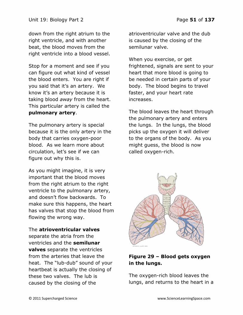
Unit 19: Biology Part 2 Page 51 of 137
© 2011 Supercharged Science www.ScienceLearningSpace.com
down from the right atrium to the
right ventricle, and with another
beat, the blood moves from the
right ventricle into a blood vessel.
Stop for a moment and see if you
can figure out what kind of vessel
the blood enters. You are right if
you said that it’s an artery. We
know it’s an artery because it is
taking blood away from the heart.
This particular artery is called the
pulmonary artery.
The pulmonary artery is special
because it is the only artery in the
body that carries oxygen-poor
blood. As we learn more about
circulation, let’s see if we can
figure out why this is.
As you might imagine, it is very
important that the blood moves
from the right atrium to the right
ventricle to the pulmonary artery,
and doesn’t flow backwards. To
make sure this happens, the heart
has valves that stop the blood from
flowing the wrong way.
The atrioventricular valves
separate the atria from the
ventricles and the semilunar
valves separate the ventricles
from the arteries that leave the
heat. The “lub-dub” sound of your
heartbeat is actually the closing of
these two valves. The lub is
caused by the closing of the
atrioventricular valve and the dub
is caused by the closing of the
semilunar valve.
When you exercise, or get
frightened, signals are sent to your
heart that more blood is going to
be needed in certain parts of your
body. The blood begins to travel
faster, and your heart rate
increases.
The blood leaves the heart through
the pulmonary artery and enters
the lungs. In the lungs, the blood
picks up the oxygen it will deliver
to the organs of the body. As you
might guess, the blood is now
called oxygen-rich.
Figure 29 – Blood gets oxygen
in the lungs.
The oxygen-rich blood leaves the
lungs, and returns to the heart in a

Unit 19: Biology Part 2 Page 52 of 137
© 2011 Supercharged Science www.ScienceLearningSpace.com
blood vessel. What type of vessel?
You’re right if you said it’s a vein,
because blood always goes back to
the heart in veins, and you’re also
right if you guessed that this vein
was called the pulmonary vein.
The pulmonary vein is the only
vein that carries oxygen-rich blood.
Once again, let’s save the question
about why this is the case for a
little later. This process of going
from the heart, to the lungs,
getting oxygen, and going back to
the heart is called pulmonary
circulation. Have you noticed
that we are using the word
“pulmonary” a lot? This is because
pulmonary refers to the lungs, and,
as you can see, the lungs are very
important in this part of circulation.
Systemic Circulation
The oxygen re-enters the heart
and goes into the left atrium. With
a beat, the blood goes through the
atrioventricular valve in to the left
ventricle. With another beat, it
goes through the semilunar valve
into an artery.
Since this is the first artery to carry
oxygen-rich blood from the heart
to the other parts of the body, it
gets a special name. It is called
the aorta. Even though it has this
special name, it is just like any
other artery. It carries blood away
from the heart and has thick walls
to handle the high pressure on the
blood inside of it.
Figure 30 – The aorta carries
blood from the heart to all
organs of the body.
The aorta branches out into smaller
and smaller arteries, until
eventually the blood reaches the
capillary bed of the organ oxygen
is being delivered to. The
exchange of oxygen from the blood
in the capillary bed to the organ
takes place, and the blood, which
is now oxygen poor, leaves the
capillary bed and enters a tiny
vein.
The blood travels back to the heart
through larger and larger veins.
Eventually, the blood gets back to
one of the two largest veins in the
body, which lead directly back into
the right atrium of the heart. Just
like the aorta has a special name
because it is the first artery out of

Unit 19: Biology Part 2 Page 53 of 137
© 2011 Supercharged Science www.ScienceLearningSpace.com
the heart, these veins, which are
the last ones blood goes through
before re-entering the heat, have
special names. They are called the
superior and inferior vena cava.
Blood returning from an organ in
the top half of the body enters
through the superior vena cava,
while blood coming from the
bottom half uses the inferior vena
cava.
Figure 31 – The superior vena
cava is one of two veins that
bring blood back into the heart.
Once the blood is back in the right
atrium, the process begins again.
Now that we have gone through
the entire process of both
pulmonary and systemic
circulation, we can answer the
questions about why the
pulmonary artery is the only artery
to carry oxygen-poor blood, and
the pulmonary vein is the only one
to carry oxygen rich blood. In
systemic circulation, all of the
arteries branch off of the aorta, so
they are all carrying oxygen-rich
blood to the various parts of the
body.
Similarly, all of the veins are
carrying oxygen-poor blood back to
the heart. It is only during
pulmonary circulation that oxygen-
poor blood leaves the heart to get
oxygen, or oxygen-rich blood
comes back to the heart to be
pumped out to the body. This is a
good reminder of an important
point. The heart is not where
blood gets oxygen. It is a pump
that sends blood where it needs to
go. It is just a pump, but a very
important one for our survival!
The figure below shows the major
parts of the heart. By now, you
should be able to describe what
goes on in each part.

Unit 19: Biology Part 2 Page 54 of 137
© 2011 Supercharged Science www.ScienceLearningSpace.com
Coronary Circulation
The heart is an organ, and like all
organs, it needs oxygen. The
process of providing the heart
muscle with oxygen is called
coronary circulation. As we said
before, during systemic circulation,
blood leaves the aorta and
branches out into many smaller
arteries. One set of arteries are
called the coronary arteries. These
arteries take blood to the heart
muscle and provide it with the
oxygen it needs. This seems
strange, because we have learned
that arteries always go away from
the heart. To understand this, it’s
helpful to think of the heart as two
different things – a pump that
sends blood everywhere it needs to
go and an organ that needs
oxygen. Arteries always go away
from the pump, but they
sometimes provide oxygen to the
organ.
Figure 33 – The coronary
arteries branch off of the aorta
and provide oxygen to the
heart.
Once the heart muscle receives the
oxygen it needs, the oxygen-poor
blood returns to the heart pump in
the cardiac vein.
Diseases of the Cardiovascular
System
The cardiovascular system is
essential for life. If one or more
parts of the system are not
working properly, the result can be
serious illness or death. Three
common diseases of the
cardiovascular system are
hypertension, coronary heart
disease and stroke.
Hypertension
As we learned earlier, blood in
arteries is under pressure, and this
pressure is referred to as blood
pressure. If you have ever had
your blood pressure taken, the
doctor or nurse probably used a
tool called a
sphygmomanometer, like one of
the ones shown below.

Unit 19: Biology Part 2 Page 55 of 137
© 2011 Supercharged Science www.ScienceLearningSpace.com
Figure 34 – Although neither of
these sphygmomanometers use
actual mercury, they both
measure blood pressure in the
unit millimeters of mercury
(mm Hg.)
Sphygmomanometers measure two
things – the highest pressure in the
artery, which is called systolic
pressure, and the lowest
pressure, which is called diastolic
pressure.
Old-fashioned
sphygmomanometers measured
pressure by making a column of
the liquid mercury rise a certain
number of millimeters. Most
modern sphygmomanometers don’t
use mercury, but blood pressure is
still measured in millimeters of
mercury. Millimeters are
abbreviated mm and mercury is
abbreviated Hg, so millimeters of
mercury can be abbreviated mm
Hg.
You are considered healthy if your
systolic blood pressure is less than
120 mm Hg and your diastolic
blood pressure is less than 80 mm
Hg. Blood pressure can change for
many reasons, but if a person
always has high blood pressure,
they have hypertension. If
someone has hypertension, they
have a greater risk of getting other
cardiovascular diseases.
Hypertension can be treated by
improving diet, increasing exercise,
or with medication.
Coronary Heart Disease
One of the most common forms of
cardiovascular disease occurs when
cell pieces, fatty substances,
calcium, and connective tissue,
which together are called plaque,
build up in the arteries and make it
harder for blood to flow. This is
called atherosclerosis.
Atherosclerosis is very serious if it
blocks blood from getting to the
heart muscle. When this happens,
it is known as coronary heart
disease.

Unit 19: Biology Part 2 Page 56 of 137
© 2011 Supercharged Science www.ScienceLearningSpace.com
Figure 35 – A buildup of plaque
from atherosclerosis makes it
harder for blood to get through
the artery.
If coronary heart disease
completely blocks the heart’s blood
supply, the result is a heart
attack. Coronary heart disease is
the most common cause of death
amongst adults in the United
States. Ways to prevent to
prevent coronary heart disease are
very similar to ways to prevent
hypertension. Healthy eating and
regularly exercising are very
important. The best way to
prevent heart disease is by not
smoking.
Stroke
A stroke is caused by
atherosclerosis of the arteries that
provide blood to the brain.
Sometimes the blockage of the
artery is caused by blood clot, a
free-floating object, or a bleeding
blood vessel. Reducing blood
pressure is the best way to avoid a
stroke, although not smoking is
also helpful.
Conclusion
The cardiovascular system is
crucial for survival. Blood pumped
around the body provides organs
with oxygen, and helps control
body temperature. Pulmonary
circulation takes blood to the lungs
to get oxygen, and systemic
circulation takes this oxygen-rich
blood to the various organs of the
body. We can keep our
cardiovascular systems healthy by
eating right, exercising, and not
smoking.
Lesson 4:
Respiratory and
Excretory Systems
Introduction to the
Respiratory System
Take a deep breath in, and slowly
let it out. As you do, think about
the breaths you take without
thinking about it. The truth is, you
probably only think about
breathing when you are coughing
and having a hard time breathing.
Even though breathing is not

Unit 19: Biology Part 2 Page 57 of 137
© 2011 Supercharged Science www.ScienceLearningSpace.com
something we think about
regularly, it is absolutely required
for the survival of the cells in your
body.
As you breathe, oxygen flows into
the body and carbon dioxide flows
out. This very important exchange
of gases is the main function of
your body’s respiratory system.
The Parts of the
Respiratory System
The respiratory system, like all
body systems, is a group of
organs and tissues all working
together for a common purpose, in
this case exchanging oxygen for
carbon dioxide. Some of the
respiratory system’s parts are
shown below.
Figure 36 – The parts of the
respiratory system work
together to provide oxygen and
get rid of carbon dioxide.
Diaphragm
One important muscle not shown in
the picture above is the
diaphragm. This sheet of muscle
goes along the bottom of the rib
cage. The diaphragm contracts to
make more space in the chest,
allowing air containing oxygen and
other gases to come in. It then
relaxes, causing there to be less
space in the chest and forcing air
containing carbon dioxide and
other gases out.
Figure 37 – The diaphragm
goes across the bottom of the
rib cage.
Nose and Nasal Cavity
The first stop for air entering your
body is your nose. In the nose,
the air is warmed and moistened.
The air also acts as a filter. Hair
and mucus in the nose catch
particles in the air that could be

Unit 19: Biology Part 2 Page 58 of 137
© 2011 Supercharged Science www.ScienceLearningSpace.com
harmful to you and keep them
from going any further into your
body and eventually reaching your
lungs.
Pharynx and Trachea
After leaving the nasal cavity, air
passes through the pharynx, a
long tube that carries both food
and air. In everyday language, the
pharynx would be referred to as
the throat. At the end of the
pharynx is a flap of tissue called
the epiglottis.
Have you ever been told not to talk
and eat at the same time? Good
manners are not the only reason
for this. The epiglottis is part of
why this is a smart idea too. When
you are eating, the epiglottis
covers the trachea, or windpipe,
which is a tube air is supposed to
travel down.
With the trachea covered, food can
go down the esophagus to the
stomach. If food gets into the
trachea, it can cause coughing and
choking. If you talk or laugh while
you eat, you are more likely to
have the epiglottis unable to cover
the trachea, and therefore more
likely to choke.
Figure 38 – When you eat, the
epiglottis covers the trachea
(the tube on the left) so the
food can go down the
esophagus (the tube on the
right.)
Air travels down the trachea until it
gets to the lungs. You have a left
and right lung, so the trachea
divides into two tubes, one going
into each lung. These tubes are
called the left and right bronchi.
Each bronchus (that’s the singular
form of bronchi) branches out into
smaller tubes called bronchioles.
Bronchioles lead to alveoli. Trace
the path or air from trachea, to
bronchus, to bronchiole, to alveoli
in the figure below.

Unit 19: Biology Part 2 Page 59 of 137
© 2011 Supercharged Science www.ScienceLearningSpace.com
Figure 39 – Air breathed in
eventually leads to the alveoli
in the lungs.
Alveoli are small sacs at the end of
bronchioles that look sort of like a
bunch of grapes. These sacs are
the places where gas exchange
takes place.
Gas exchange means that, in the
alveoli, oxygen moves into the
blood so it can be taken all over
the body, and carbon dioxide
crosses from the blood back to the
alveoli so it can be breathed out.
Figure 40 – In each alveolus
(that’s the singular form of
alveoli) oxygen flows into the
blood while carbon dioxide
flows out.
How We Breathe
We usually breathe without
thinking about it, which is a good
thing. Imagine if you had to stop
and think about it each time you
had to breathe. You wouldn’t be
able to do anything else! If you’ve
ever gone swimming, done yoga,
or sung, however, you know that it
is possible to control when you
breathe.
Earlier, we talked about how the
diaphragm is important for
breathing in and out, but how does
it work exactly? To understand
this, you have to understand
something about pressure.

Unit 19: Biology Part 2 Page 60 of 137
© 2011 Supercharged Science www.ScienceLearningSpace.com
Gases or liquids will always flow
from areas that have high pressure
to low pressure. If you’ve ever let
the air out of a balloon, you’ve
seen this in action. The pressure
inside the balloon is much higher
than the pressure outside the
balloon. So, if the air can, it will
flow quickly out of the balloon.
Figure 41 – The air pressure in
a balloon is greater than the air
pressure outside a balloon,
causing the air to flow out of it.
A similar thing goes on in the
lungs. When you want to breathe
out, the diaphragm relaxes and
moves up, reducing the size of the
chest. The air pressure is higher in
the chest than outside, so air goes
out. This is called exhalation.
Breathing in, or inhalation, is the
reverse of this. The diaphragm
contracts and moves down, making
more space in the chest. Now the
pressure becomes less inside the
chest than outside of it, so air goes
in.
Gas Exchange in the
Alveoli
The process of gas exchange –
oxygen going into the blood and
carbon dioxide leaving the blood to
be exhaled – also involves
differences in pressure, and is
actually very similar to inhalation
and exhalation. The amount, or
concentration, of oxygen in the air
found in alveoli is much greater
than it is in the blood that has just
come back from the cells of the
body. So, oxygen flows from the
alveoli to the blood, from high to
low concentration. Carbon dioxide
is simply the opposite. The
concentration is higher in the blood
than in the air, so the flow goes
from the blood to the alveoli.
Figure 42 – The difference in
concentrations of carbon
dioxide (CO2) and Oxygen (O2)

Unit 19: Biology Part 2 Page 61 of 137
© 2011 Supercharged Science www.ScienceLearningSpace.com
explain the flow of oxygen
carbon dioxide in the alveoli
Breathing Versus
Respiration
Sometimes, people will say
“breathing” and “respiration” as
though they were the same thing,
but they are actually very different.
What we have been talking about
so far is breathing.
This is the process where air enters
the body, goes into the lungs, and
exchanges its oxygen for carbon
dioxide. This is part of respiration,
and is known as external
respiration, but it only half of full
respiration.
Respiration also includes internal
respiration, where the blood, full
of oxygen, goes to the parts of the
body that need it. This process
can be learned about in a
discussion of the circulatory
system, another body system.
Respiratory System
Diseases
There are many diseases involving
the respiratory system. These
diseases, like all diseases, can be
acute, which means they last for a
short period of time, or chronic,
which means they last for a long
time. Many things cause
respiratory diseases. They can be
caused by bacteria, or things in the
environment like tobacco smoke.
Other respiratory diseases are
hereditary, which means that
parents can pass them on to their
children.
Bronchitis
Bronchitis is an extremely
common respiratory system
disease. In fact, you’ve probably
has bronchitis a few times in your
life. This disease occurs when the
bronchi become inflamed. Acute
bronchitis is usually caused by a
virus or bacterium, and can last
from several days to a week.
People with acute bronchitis
usually have a cough that produces
mucus. They may also be short of
breath or wheeze. Chronic
bronchitis lasts at least three
months over a two year period.
The most common cause of chronic
bronchitis is smoking, although it
can be caused by pollution or
smoke in the air. Bronchitis is
treated with antibiotics.

Unit 19: Biology Part 2 Page 62 of 137
© 2011 Supercharged Science www.ScienceLearningSpace.com
Figure 43 – In both acute and
chronic bronchitis, the bronchi
are inflamed.
Asthma
You probably know someone who
has asthma. Perhaps the person
with asthma has to be very careful
when doing very active things, or
avoid certain materials, like pet
hair, mold, or dust.
Asthma is a chronic disease caused
by an inflammation of the
bronchioles. People with asthma
can have an asthma attack
because of air that is warm, cool,
or moist, exercise, stress, diseases
like the common cold, or some of
the materials in the air mentioned
above, like pet hair, mold, or dust.
During an attack, not only do the
bronchioles become inflamed, but
the muscles in the bronchioles
contract, making them even
smaller. As if that wasn’t enough,
mucus lines the lungs, making it
even harder to breathe.
People with asthma exercise, play
sports, and do most other
activities. They just need to make
sure they control their asthma, and
look out for things that might
trigger an attack. Some people
with this disease take medicines
called bronchodilators to open up
the bronchioles.
Figure 44 – During an asthma
attack, the bronchioles get
inflamed, the muscles in the
bronchioles contract, and the
lungs line with mucus.
Pneumonia
Pneumonia occurs when the alveoli
become inflamed and full of fluid.
This makes it impossible for gas
exchange to happen the way it
should. As a result, people with
pneumonia have trouble breathing.
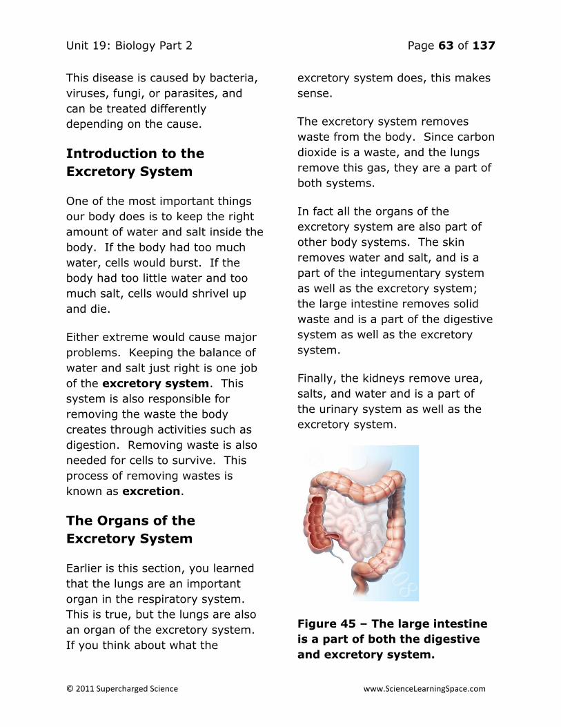
Unit 19: Biology Part 2 Page 63 of 137
© 2011 Supercharged Science www.ScienceLearningSpace.com
This disease is caused by bacteria,
viruses, fungi, or parasites, and
can be treated differently
depending on the cause.
Introduction to the
Excretory System
One of the most important things
our body does is to keep the right
amount of water and salt inside the
body. If the body had too much
water, cells would burst. If the
body had too little water and too
much salt, cells would shrivel up
and die.
Either extreme would cause major
problems. Keeping the balance of
water and salt just right is one job
of the excretory system. This
system is also responsible for
removing the waste the body
creates through activities such as
digestion. Removing waste is also
needed for cells to survive. This
process of removing wastes is
known as excretion.
The Organs of the
Excretory System
Earlier is this section, you learned
that the lungs are an important
organ in the respiratory system.
This is true, but the lungs are also
an organ of the excretory system.
If you think about what the
excretory system does, this makes
sense.
The excretory system removes
waste from the body. Since carbon
dioxide is a waste, and the lungs
remove this gas, they are a part of
both systems.
In fact all the organs of the
excretory system are also part of
other body systems. The skin
removes water and salt, and is a
part of the integumentary system
as well as the excretory system;
the large intestine removes solid
waste and is a part of the digestive
system as well as the excretory
system.
Finally, the kidneys remove urea,
salts, and water and is a part of
the urinary system as well as the
excretory system.
Figure 45 – The large intestine
is a part of both the digestive
and excretory system.

Unit 19: Biology Part 2 Page 64 of 137
© 2011 Supercharged Science www.ScienceLearningSpace.com
The Urinary System
Sometimes people think of the
urinary system and the excretory
system as the same thing. This is
not true. In fact, the urinary
system is just a part of the
excretory system. This system
consists of the organs that make,
store, and get rid of urine.
Urine and the Kidneys
Urine is a waste product made
mostly water, with dissolved salts
and molecules containing nitrogen
mixed in. One of the nitrogen-
containing molecules is urea,
which is formed when foods
containing protein, such as meat,
poultry, and certain vegetables are
eaten. Urine is usually a pale
yellow color, but can be colorless
to dark yellow, depending the
amount of water it contains. The
kidneys are about the size of your
fist and are found below your
ribcage, as shown in the figure
above. The human body has two
kidneys. The job of the kidneys is
to clean and filter the blood and
form urine. To do this, each
kidney contains millions of tubes
called nephrons. If the body
needs more water, water is
removed from nephrons and
returns to the blood.
Figure 46 – The kidney filters
blood in the body.
Other Organs in the
Urinary System
Urine leaves the kidneys and flows
into the ureters. These tubes
carry the urine from the kidney to
the urinary bladder. The urinary
bladder is hollow, muscular, and
elastic-walled. It looks sort of like
a balloon, and fills up with urine.
Once full, urine leaves the body
through the urethra.
Figure 66 – Urine is collected in the
bladder and leaves the body through the
urethra.

Unit 19: Biology Part 2 Page 65 of 137
© 2011 Supercharged Science www.ScienceLearningSpace.com
Diseases of the Excretory
System
Three common diseases of the
excretory system are kidney
stones, kidney failure, and urinary
tract infections. Kidney stones
are crystallized minerals created by
waste in the kidneys. Although the
stones form in the kidneys, they
can be found anywhere in the
urinary system. Depending on how
large the stones are, they can
cause no pain at all or cause a
great amount of pain. Sometimes
stones need to be removed in
surgery.
Figure 47 – Kidney stones form
in the kidneys, and can cause
great pain.
Kidney failure occurs when the
kidneys stop filtering blood. This
can happen because of an injury to
the kidneys, the loss of a lot of
blood, or by some drugs and
poisons. If the kidneys are not
seriously damaged, they may be
able to recover and continue to
function. However, in the event of
very serious kidney failure, a
person may need to have kidney
dialysis. Dialysis is a process in
which a machine, called a dialysis
machine, artificially filters the
blood, as the kidney normally
would.
A final disease of the excretory
system is urinary tract infections
(UTI.) UTI occur when bacteria get
into the bladder or kidney and
multiply. The most common type
of UTI is a bladder infection. It is
more common for women to get
UTI’s than men, and they are often
treated with antibiotics.
Figure 48 – A kidney dialysis
machine performs the job of
the kidney in the event of
kidney failure.

Unit 19: Biology Part 2 Page 66 of 137
© 2011 Supercharged Science www.ScienceLearningSpace.com
Conclusion
The respiratory and excretory
systems are both necessary for
life. The respiratory system takes
in oxygen-containing air and
releases carbon dioxide-containing
air. Additionally, it provides much-
needed oxygen to the organs of
the body. The excretory system
removes wastes from the body by
working together with other organ
systems. Diseases to either of
these systems can cause major
problems.
Lesson 5: Controlling
the Body
Your body is made of organs, like
your heart, lungs or stomach.
These organs work together to
allow you to breathe, eat, move,
and do just about everything else
you need to do. Organs working
together are called organ
systems. Although all organ
systems are important, and
necessary for us to survive, the
most important system might be
the nervous system, the system
that controls all the others. This
system not only controls all the
systems of your body but also
allows you to learn and use
language, senses conditions inside
and outside your body and
prepares you to fight or flee if you
are in a dangerous situation.
Nerves and Nerve
Impulses
Imagine you walk into your home
one night and it’s pitch black. You
quickly flick on a light switch and
the room is immediately lit up.
You may have never thought about
it, but it’s kind of amazing that this
happens. Even though it may be a
long distance from the light switch
to the actual light, the light comes
on.
Fortunately, messages in your
nervous system also travel at the
speed of electricity. The nervous
system is made of bundles of nerve
cells called nerves. Nerve cells
called neurons send messages,
called nerve impulses,
throughout the body. Since these
messages are electrical impulses,
they travel very quickly. Neurons
are also covered with a fatty
covering called myelin which
insulates the neuron, like the
plastic on the outside of a wire,
allowing the nerve impulse to
travel even faster.

Unit 19: Biology Part 2 Page 67 of 137
© 2011 Supercharged Science www.ScienceLearningSpace.com
Figure 49: Neurons send nerve
impulses throughout the body
at the speed of electricity.
Look at the shape of the neuron.
These cells are the perfect shape
for their job of sending messages
all over the body. The small
extensions on the end of the cell
are called dendrites and get nerve
impulses from other cells. The
longer extension at the other end
of the cell is called an axon and
sends nerve impulses to other
cells. The central part of the cell,
between the dendrites and axon, is
called the cell body. This part of
the cell contains the nucleus and
all the organelles, which are small
structures inside the cell. The
place where the axon of a nerve
cell meets the dendrites of another
nerve cell or another type of cell is
called a synapse.
Figure 50 – A synapse is found
where the dendrite and axon of
two neurons meet.
Neurons can be classified, or put
into groups, based on their job in
the body. Sensory neurons carry
messages from the organs to the
brain and spinal cord. Motor
neurons carry messages from the
brain and spinal cord to organs,
glands, and muscles.
The Central Nervous
System
The Central Nervous System (CNS)
is the part of the nervous system
including the brain and spinal cord.
Both of these organs are protected
by bones. The brain is protected
by the skull and the spine is
protected by bones called
vertebrae.

Unit 19: Biology Part 2 Page 68 of 137
© 2011 Supercharged Science www.ScienceLearningSpace.com
With 100 billion neurons each
connected to thousands of other
neurons, the brain is the control
center of the body and one of its
most complex organs. Like a
plane’s pilot or a ship’s captain, the
brain tells the other part of the
nervous system what to do.
Since the nervous system controls
the other systems, and the brain
controls the nervous system, the
brain is in charge of nearly
everything in our body, from
memory, to speech, to seeing and
hearing, to our overall ability to
learn.
Think about all the way your brain
is working as you read this
sentence. Your see the words,
read them, think about what they
mean, and try to remember them
as much as you can. It’s all
controlled by the brain!
The brain is made of three parts –
the cerebrum, the cerebellum, and
the brainstem. The cerebrum is
the largest part and controls the
things we are aware of, such as
speaking and solving problems. It
also controls voluntary motions,
meaning the motions we think
about doing, such as walking,
waving, or typing.
Figure 51 – The cerebrum is the
largest part of the brain.
The cerebrum is divided into two
halves called hemispheres. The
left hemisphere controls the right
part of the body, and vice versa.
Each hemisphere has four parts,
called lobes. These lobes are the
frontal, parietal, occipital, and
temporal lobe. Each lobe has
different things it is responsible for.
For example, the frontal lobe
controls speech, thinking, and
touch, while the temporal lobe
controls hearing and smell.
Under the cerebrum lies the
cerebellum. This part of the brain
controls balance, coordination, and
body position. All of the
cerebellum’s jobs are on display if
you are riding a bike.
The smallest part of the brain is
the brain stem. It is also located
under the cerebrum, but in front of

Unit 19: Biology Part 2 Page 69 of 137
© 2011 Supercharged Science www.ScienceLearningSpace.com
the cerebellum. The brain stem is
concerned with basic functions of
the body that we don’t even have
to think about doing. These
activities include such important
activities as digestion, heartbeats,
and even breathing.
Figure 52 – The cerebellum and
brainstem lie under the
cerebrum, which is divided into
four lobes.
The other organ of the CNS is the
spinal cord. The spinal cord is a
long, tube-shaped bundle of
neurons. Running from the
brainstem to the lower back, the
spinal cord is like the highway of
the nervous system. Messages get
sent from the parts of the body to
the brain, providing the brain with
critical information about the inside
and outside surroundings. The
brain sends back messages telling
the parts of the body what to do
based on the information it was
given. All this information is sent
in the form of nerve impulses up
and down the spinal cord.
Figure 53 – The spinal cord
runs from the brainstem to the
lower back.
Peripheral Nervous System
The nervous system is not just
made of your brain and spinal cord.
From your arms and hands, down
to your feet and legs, up to your
scalp and face, and even to the
organs inside you, all your body
parts have nerves traveling to and
from them, so that messages can
get to the CNS. This network of
nerve cells outside the CNS is
called the peripheral nervous
system (PNS.) The nerves of the
PNS are connected, either directly
or indirectly, to the spinal cord.
Within the PNS is two divisions, the
sensory division and the motor

Unit 19: Biology Part 2 Page 70 of 137
© 2011 Supercharged Science www.ScienceLearningSpace.com
division. The sensory division
carries messages from the sense
organs and internal organs to the
CNS. Human senses include sight,
smell, taste, touch, smell, and
balance.
Other animals have senses we
don’t have. Sharks can sense a
mild electric current and birds can
sense magnetism, allowing them to
find their way like a hiker with a
compass.
Every sense we have has a sense
organ associated with it. For
example, our eyes are associated
with sight. The optic nerve travels
from the eye to the brain, sending
messages.
Figure 54 – The optic nerve
sends messages from the eye
to the brain.
Imagine you get home and smell
something delicious. You think you
know the smell, but just to be
sure, you peek into the oven and
see chocolate chip cookies baking.
It is tempting to say that your nose
and eyes “told” you about the
cookies, but this is not actually
correct. Although your nose
detects chemicals coming from the
cookies, and your eyes detect light
that bounce off the cookies, all
these sense organs can do is send
a message about what they
sensed. The brain then has the job
of interpreting these messages,
trying to make sense of them.
Each sense organ is associated
with a certain region of the brain
that interprets its messages.
The motor division of the PNS
carries messages from the CNS to
the internal organs and muscles.
The motor division is further
divided into two parts called the
somatic nervous system and
autonomic nervous system. The
somatic nervous system is in
charge of voluntary movements, or
movements over which you have
control. If you play soccer, your
eyes send a message to your brain
each time you see the ball coming.
Your somatic nervous system is
then responsible for kicking the
ball. Your brain sends a message
to your leg instructing it to kick the
ball.

Unit 19: Biology Part 2 Page 71 of 137
© 2011 Supercharged Science www.ScienceLearningSpace.com
Figure 55 – Seeing and kicking
a soccer ball requires the
sensory division and the
somatic nervous system, within
motor division of the PNS.
The autonomic nervous system
is in charge of involuntary motions,
or motions over which you have
more control. These include such
essential actions as breathing,
digestion, and even the circulation
of blood. Under normal
circumstances, the
parasympathetic division of the
autonomic nervous system handles
all of the necessary involuntary
motions. However, in and
emergency, the sympathetic
division takes over, controlling
what is known as the “fight or
flight” response to dangerous or
frightening situations.
As you can see, the nervous
system does many things and has
many parts. Consider the chart
below, which lists the divisions of
the system. Each part of the
system plays a unique and
important role in allowing the other
body systems to function.
Eyes and Vision
Try to think of an activity you do,
while you are awake, that does
involve sight. If you are not
visually impaired, this is probably
hard to do. Human beings, more
than many other animals, rely of
their sense of sight to survive.
Sight is our single most important
sense. For this reason, human
sight is very highly developed.
As humans, we can see objects
that are both close up and far
away. We can also tell that they
are close ore far apart because we
can see in three dimensions. We
are able to see in three dimensions
because we have two eyes, both
facing the same direction, a few
inches apart. So, we see things
with both our eyes, but from
slightly different angles. The closer
an object is to you, the more
different the angle from each eye
is. Your brain can use this
information to see how far apart
objects are.
Along with being able to see in
three dimensions, humans have
the ability to see in color. We can
do this because the eye contains
special cells called rods and cones.

Unit 19: Biology Part 2 Page 72 of 137
© 2011 Supercharged Science www.ScienceLearningSpace.com
Rods allow us to see in dim light,
while cones allow us to see light of
different colors.
Figure 56: Rods allow us to see
in dim light while cones allow
us to see in color.
The Structure of the Eye
The eye is a complex structure that
detects and focuses light. Light
first enters the eye through the
cornea, a clear protective layer on
the outside of the eye. The pupil,
a black opening in the eye, lets
light in. In dark rooms, the pupil
will become larger, or dilate, in
order to let in more light. If the
room suddenly becomes bright, the
pupil will become smaller. The
pupil is surrounded by the brown,
blue, grey, or green iris.
After passing through the pupil,
light goes to the lens which, like a
hand lens, is a clear curved
structure that helps focus light on
the retina, in the back of the eye.
The retina is where the rods and
cones are found.
Vision Problems
Do you or someone you know
where glasses or contact lenses.
Glasses and contacts are used to
correct vision problems. The two
most common eye problems are
myopia and hyperopia, and each is
fixed a different way.
Myopia is sometimes called
nearsightedness. About one third
of people have this problem.
Myopia happens when the eye is
too long. With eye too long,
images seen are focused a little bit
in front of the retina, instead of on
the retina as they would be in a
normal eye.
Figure 57 – If a person has
myopia, light focuses in front of
the retina.
People with myopia can see nearby
objects clearly. Objects that are
far away, however, look blurry.
This can be corrected with a

Unit 19: Biology Part 2 Page 73 of 137
© 2011 Supercharged Science www.ScienceLearningSpace.com
concave lens on glasses or
contacts. “Concave” means that
the lens curves inward, like the
inside of a bowl. The concave lens
helps focus light on the retina
instead of in front of it, correcting
the problem.
Hyperopia, which is also called
farsightedness, affects about one
fourth of people, is pretty much the
opposite of myopia. In this
disorder, the eye is too short, so
images become focused behind the
retina. As a result, people with
hyperopia can see things clearly
when they are far away, but not
when they are close up.
Figure 58 – When the eye is too
short, hyperopia occurs, and
images are focused behind the
retina.
To correct hyperopia, a convex
lens, which is a lens that curves
out, like the outside of a bowl, is
used in glasses or contacts.
Some people suffer from both
myopia and hyperopia. How can
this be? It doesn’t seem possible
that the eye could be too long and
too short.
The answer lies in the lens of your
eye. You can think of the lens like
a trampoline. As you jump up and
down on the trampoline, it moves
closer or further from the ground.
The lens does a similar thing when
you look at things closer together
or further away. This makes the
overall shape of the eye change
slightly.
As your trampoline gets older, the
springs may begin to wear down,
and it may become harder for the
trampoline to move. The same
thing happens with the lens in your
eyes.
As a result, many older people
develop hyperopia. If these people
already had myopia, they will need
a lens that has both a concave and
convex part. Bifocals are glasses
that have these two parts.
These glasses were invented by
Benjamin Franklin over 200 years
ago, and are still used today.

Unit 19: Biology Part 2 Page 74 of 137
© 2011 Supercharged Science www.ScienceLearningSpace.com
Figure 59 – Although modern
bifocals look more stylish than
these Benjamin Franklin-style
glasses, they still use the same
basic idea.
Today, some people with vision
problems choose to have surgery
called LASIK. In this surgery, a
laser changes the shape of the
cornea correcting the vision
problem. As a result, the person
usually no longer needs to wear
glasses or contacts.
Hearing and Balance
It may seem like walking across a
balance beam and listening to your
favorite song are very different
activities, but they both depend on
your ears. Ears are the sense
organs that control hearing, which
is the ability to detect sound. Ears
also sense the position of the body
and help maintain balance when
you walk a balance beam or ride a
bike.
Imagine a pebble being dropped
into a lake. Waves of water go off
in all directions. A similar thing
happens when a car driving down
the street honks its own. Waves
go off from the car in all direction.
The difference is that these are not
waves of water, but instead are
sound waves, which travel through
the air. If you are nearby, some of
those sound waves make it to your
ear.
The pinna, or outer ear, which is
the part of your ear that you can
see, gathers up some of the sound
waves, sends them down the ear
canal, and eventually they strike
the eardrum. The eardrum is a
thin membrane that vibrates like a
drum when the waves hit it. The
vibrations pass three tiny bones,
called the hammer, anvil, and
stirrup, as well as a membrane
called the oval window, causing
them all to vibrate.

Unit 19: Biology Part 2 Page 75 of 137
© 2011 Supercharged Science www.ScienceLearningSpace.com
Figure 60 – Many parts of the
ear work together to make
hearing possible.
From the oval window, the
vibrations go to the cochlea,
liquid-filled space lined with hairs.
The vibrations make waves in the
cochlea’s liquid, just like waves in a
pond, causing the hairs to move.
The movement of the hairs sends a
nerve impulse through the auditory
nerve to the brain. The brain
interprets the message and “tells”
you what you have heard.
Along with hearing, the ears play a
major role in balance. Inside the
ears are semicircular canals
which are lined with hairs and full
of liquid. When the body moves in
one direction, the liquid in the
semicircular canals also moves.
This causes the hairs inside to
move, which sends a message to
your brain, which gives instructions
for the body’s muscles to contract
or relax. This keeps you balanced.
Figure 61 – The semicircular
canals help with balance

Unit 19: Biology Part 2 Page 76 of 137
© 2011 Supercharged Science www.ScienceLearningSpace.com
Touch
If you’ve ever accidently touched a
hot stove, prickly cactus plant, or a
block of ice, you know how strong
the sense of touch can be. Touch
is the sense of pain, pressure, or
temperature. All of our skin
contains sensory neurons. When
you touch something, messages go
from the sensory neurons to the
brain, which interprets what you’ve
touched.
Although all of the skin, as well as
many internal organs, have
sensory neurons allowing them to
experience touch, certain areas of
the body, including the palms of
the hands, soles of the feet, lips,
and tongue, have more sensory
neurons than other parts of the
body. This makes them more
sensitive to touch.
As we’ve learned, messages travel
from sense organs, like the skin, to
the brain very quickly. There are
times, however, when something
we touch is so dangerous that our
body does not even want to take
the time to send the message to
the brain. For example, if you
touch a hot stove, the message
that you are touching something
hot will only go to your spinal cord,
instead of making it all the way to
your brain. This is called a reflex
arc.
Figure 62 – A reflex arc causes
the spinal cord to send a
message pulling your hand
away from a hot stove.
You often hear people say they did
something, “without thinking.” The
truth, however, is that our brain is
usually very much involved in the
things we do. A reflex arc is one of
the rare times when the brain is
truly not involved. Other
responses may come later. You
may scream in pain, say “ouch” or
cry after touching the hot stove.
All these involve your brain and
therefore happen after the reflex
arc action of removing your hands.
It is only the initial movement, to
prevent damage to the tissue of
the skin, that is brain-free.
Taste and Smell
Neurons found in your nose detect
chemicals in the air. These
neurons send messages to your
brain, and the brain determines

Unit 19: Biology Part 2 Page 77 of 137
© 2011 Supercharged Science www.ScienceLearningSpace.com
what you are smelling. The nose is
able to detect thousands of
different odors.
The foods you eat also contain
chemicals, and these chemicals are
sensed by sensory neurons on your
tongue. These neurons are
organized into groups called taste
buds.
There are five types of taste buds
on the tongue, and each one
senses a different type of taste.
You have taste buds for sweet,
salty, sour, bitter, and umami (a
meaty taste) each in a different
spot on the tongue. Just like with
the other sense organs, when the
tongue’s sensory neurons get
information, they pass it on to the
brain. The brain is then able to
determine what you are tasting.
Notice that we spoke about five
different tastes, as opposed to
thousands of odors, and nearly
limitless textures, sights, and
sounds. Taste is our weakest
sense.
Figure 63 – Taste buds are
found in clusters on the tongue.
You may have noticed that in
general, kids are not as into foods
with strong flavors as adults. Much
of the reason for this has to do
with taste buds.
In babies, taste buds just are
found not just on the tongue but all
over the mouth. As you get older,
taste buds become a little weaker,
and are only found on the tongue.
As your taste buds change, so do
your likes and dislikes in food.
With less sensitive taste buds, you
may be interested in trying
something more adventurous that
you would have eaten as a young
child.
Keeping the Nervous
System Healthy
Think about all the things the
nervous system does. It not only
controls the senses, but also
controls feeling, thinking, moving,
and just about everything else we

Unit 19: Biology Part 2 Page 78 of 137
© 2011 Supercharged Science www.ScienceLearningSpace.com
do. It should not be a surprise,
then, that health problems
involving the nervous system can
have a big impact in the whole
body.
Encephalitis and Meningitis both
affect the CNS. Encephalitis can
cause swelling of the brain as the
body tries to fight off a virus. The
problem is that the brain has no
room to swell or expand, and
pushes against the skull. This can
cause brain injury or death. In
meningitis, the membranes that
cover the brain and spinal cord
become infected. If the infection is
caused by a virus, it usually will
clear up on its own. If it is caused
by bacteria, it is much more
serious and should be dealt with
immediately using an antibiotic.
One additional disease of the CNS
that affects young people is Reye’s
Syndrome. This rare but serious
disease occurs when young people
take aspirin to treat a viral
infection. Reye’s Syndrome causes
swelling of the brain, which can be
fatal. Many cold medicines contain
aspirin, so it is important to read
labels carefully if you have a viral
infection.
Figure 64 – Illustrations like
this one remind parents not to
treat viral infections with
aspirin, because of the
possibility of Reye’s Syndrome.
Other nervous system diseases
include cerebral palsy and epilepsy.
Cerebral palsy occurs when the
developing brain is injured around
the time of birth. In mild cases,
people with cerebral palsy may just
have weak muscles. In more
serious cases, they may have
trouble walking or talking.
Epilepsy is a disease in which
people may have a seizure.
Seizures are periods of time when
people lose consciousness, and
sometimes include violent muscle
movement. Epilepsy happens due
to unusual electrical activity in the
brain.

Unit 19: Biology Part 2 Page 79 of 137
© 2011 Supercharged Science www.ScienceLearningSpace.com
Figure 65: A person having a
seizure is not able to control
their body.
Although it is not possible to
prevent all diseases of the nervous
system, there are steps you can
take to minimize your risk.
Eeating a variety of healthy foods
and exercising regularly. Of
course, if your exercise includes
biking or some other activity that
could lead to a head injury, be sure
to wear a helmet so you don’t
damage your nervous system while
you are trying to keep it in good
condition.
Just the act of using your brain can
help make sure that it stays
healthy. Doing crossword puzzles,
reading, and playing musical
instruments have all been shown to
help keep your brain active and
healthy.
Lesson 6: Diseases
and Defenses
Infectious Diseases
Wash your hands! Cover your
mouth when you cough! Toss your
tissues after you’ve used them!
Do these sound familiar to you? If
you’ve heard these pieces of
advice, consider yourself lucky.
The person who was telling you to
do these things was trying to keep
you, and those around you,
healthy.
By doing simple things like
covering your cough and washing
your hands, you can help keep
yourself from getting or spreading
diseases that are infectious, or
able to be spread from one person
to another. Taking these
preventative measures is
important, but if you do get sick,
your body has ways of fighting off
diseases as well.
What Causes Infectious
Diseases?
You may have heard that diseases
are caused by germs. That’s true,
but “germ” is not a scientific term.
The word pathogen is the
scientific way of saying “germ,”
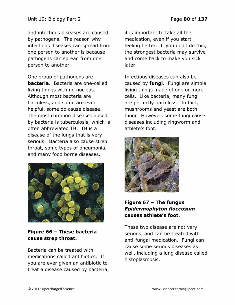
Unit 19: Biology Part 2 Page 80 of 137
© 2011 Supercharged Science www.ScienceLearningSpace.com
and infectious diseases are caused
by pathogens. The reason why
infectious diseases can spread from
one person to another is because
pathogens can spread from one
person to another.
One group of pathogens are
bacteria. Bacteria are one-celled
living things with no nucleus.
Although most bacteria are
harmless, and some are even
helpful, some do cause disease.
The most common disease caused
by bacteria is tuberculosis, which is
often abbreviated TB. TB is a
disease of the lungs that is very
serious. Bacteria also cause strep
throat, some types of pneumonia,
and many food borne diseases.
Figure 66 – These bacteria
cause strep throat.
Bacteria can be treated with
medications called antibiotics. If
you are ever given an antibiotic to
treat a disease caused by bacteria,
it is important to take all the
medication, even if you start
feeling better. If you don’t do this,
the strongest bacteria may survive
and come back to make you sick
later.
Infectious diseases can also be
caused by fungi. Fungi are simple
living things made of one or more
cells. Like bacteria, many fungi
are perfectly harmless. In fact,
mushrooms and yeast are both
fungi. However, some fungi cause
diseases including ringworm and
athlete’s foot.
Figure 67 – The fungus
Epidermophyton floccosum
causes athlete’s foot.
These two disease are not very
serious, and can be treated with
anti-fungal medication. Fungi can
cause some serious diseases as
well, including a lung disease called
histoplasmosis.

Unit 19: Biology Part 2 Page 81 of 137
© 2011 Supercharged Science www.ScienceLearningSpace.com
A third group of pathogens are
protozoa. Protozoa have a single
cell, like bacteria, but they have a
nucleus.
One serious disease caused by
protozoa is malaria. More than a
million people die from this
infectious disease each year.
Mosquitoes transfer the protozoan
that causes malaria from one
person to another when they bite
them.
Some protozoa also cause
diarrhea. Although diarrhea may
just seem like an inconvenience to
us, in some poor countries people
die of the dehydration caused by
diarrhea.
Figure 68 – The protozoan
Giardia lamblia causes
intestinal diseases, which may
lead to diarrhea.
A final group of pathogens are
viruses. Viruses are different than
the other pathogens we have
described because they are not
alive. Viruses inject DNA, the
genetic material into cells. This
allows the viruses to take over the
cells and multiply. Viruses are
responsible for many common
infectious diseases, including colds
and the flu.
Figure 69 – Viruses like H1N1,
the virus responsible for
“swine” flu, inject DNA into
cells to take them over.
Unfortunately, viruses do not
respond to antibiotics. Medicines
called antiviral drugs can treat
diseases caused by viruses, but
often times the best thing for a
person with a cold or the flu to do
is drink plenty of fluids and get lots
of rest.
How Are Pathogens
Spread?
Different pathogens are spread in
different ways. Giardia lamblia,

Unit 19: Biology Part 2 Page 82 of 137
© 2011 Supercharged Science www.ScienceLearningSpace.com
the protozoan shown in the picture
above which causes diarrhea, is
spread in water.
Some pathogens, including the
fungus that causes athlete’s foot,
or the bacterium that causes the
skin disease impetigo, are spread
when they get onto objects or
surfaces.
If someone with athlete’s foot
walks on the floor of a public
shower, and then you walk on that
same floor, the disease can be
spread to you. Similarly sharing a
towel or piece of clothing with
someone who has impetigo can
spread this disease to you.
A common way for pathogens to be
spread is in the air. This is the
method for spreading many
pathogens that causes diseases of
the throat and lungs. When a
person sneezes or coughs, tiny
droplets are released into the air.
Each of these drops contains
pathogens, such as the pathogens
for the cold and flu. If you breathe
in these pathogens, you can get
the disease.
Another method for spreading
pathogens is through the use of a
vector. Vectors are organisms
that carry the disease from one
person (or other animal) to
another. Vectors are usually
insects, and include ticks and
mosquitoes. When the insect bites
a person with the disease, it picks
up the pathogen. Usually, the
pathogen does not make the vector
sick. However, when the vector
bites another person, they transfer
the disease to them.
Figure 70 – A mosquito can act
as a vector when it bites a
person with a disease.
As you read above, malaria is one
disease transferred by a vector.
Ticks act as a vector for Lyme
disease. Mosquitoes can also be
vectors for West Nile Virus, a
disease that has spread rapidly in
the United States over the last
dozen years.
Avoid the Spread of
Infectious Diseases
The single most effective way of
not getting an infectious disease is
to wash your hands often. This

Unit 19: Biology Part 2 Page 83 of 137
© 2011 Supercharged Science www.ScienceLearningSpace.com
includes washing your hands after
using the restroom, after handling
raw meat, after being around
someone who is sick, and before
preparing or eating food. Soap
helps make your hands an
undesirable place for pathogens to
live, which is, of course, exactly
the way you want it.
You can also use a hand sanitizer
with alcohol. Alcohol kills many
bacteria.
To prevent getting a disease
caused by vectors, the best
strategy is, of course, to avoid
vectors. If you’re going
somewhere where you know ticks
or mosquitoes are likely to be
found, it’s a good idea to wear long
sleeves and long pants. There are
also some insect repellants that
can be used to keep vectors away.
Figure 71 – These “gaiters”
cover the lower leg and provide
some protection against
vectors.
As hard as you might try to
prevent it, at some point your will
likely get an infectious disease.
When this happens, try to avoid
being around others so you don’t
infect them. Also, be sure to cover
your cough and continue to wash
your hands frequently to keep from
spreading your pathogens to
others.
Noninfectious Diseases
Not all diseases can be spread from
one person to another. Diseases
that are not able to be passed from
person to person are called
noninfectious diseases. Some
noninfectious diseases are caused
by pathogens, but the pathogens
are not able to spread from person
to person.
Often times, noninfectious diseases
are caused by a combination of
genetics and lifestyle choices. This
means that a characteristic can be
inherited, or passed on from
parents to children.
Your eye and hair color are both
genetic traits. It is also possible to
inherit the tendency to have high
blood pressure, a noninfectious
disease.
Inheriting this characteristic,
however, does not mean you will
definitely have high blood

Unit 19: Biology Part 2 Page 84 of 137
© 2011 Supercharged Science www.ScienceLearningSpace.com
pressure. If you eat healthy foods
and exercise regularly, you will be
less likely to have high blood
pressure, regardless of your
genetics. It is not possible to
change your genetics, but you can
control your lifestyle choices.
Your Body’s First Line of
Defense
Up to this point, we’ve been talking
about a lot of things you can do to
prevent disease. Eating healthy,
exercising, avoiding cigarette
smoking, washing your hands, and
avoiding vectors are all great
choices you can make. To go
along with these lifestyle choices,
your body has some built in
defenses against disease that
happen without you even having to
think about it.
Have you ever seen pictures of old
castles? You probably noticed the
moat around the castle, designed
to keep invaders away. Believe it
or not, your body has its own
version of a moat, keeping
invading pathogens away. Your
“moat” is your skin! Your skin is
your largest organ, and is a
physical barrier between your
insides and the outside world. The
outer layer of skin, called the
epidermis, is tough and
waterproof. This makes it hard for
pathogens to get in.
Figure 72 – The epidermis is
thick and keeps out many
pathogens.
Some parts of your body, including
your mouth and nose, are not
covered by skin. These areas,
called mucus membranes, are
not tough like skin, making them
possible places for pathogens to
enter.
Fortunately, mucus membranes
have some defenses against
pathogens. Mucus is one defense.
It is a moist, sticky substance, and
many pathogens get stuck in it
before they can do any damage.
Many mucus membranes also have
cilia. Cilia are small hairs that
sweep your mucus and any
pathogens they may have trapped
toward openings in your body, like
your nose and mouth. When you

Unit 19: Biology Part 2 Page 85 of 137
© 2011 Supercharged Science www.ScienceLearningSpace.com
clear your throat or blow your
nose, the mucus and pathogens
leave your body.
Figure 73 – Under high
magnification, you can see that
the cilia of the nose look sort of
like a brush, sweeping away
mucus and pathogens.
Things that come out of your body
are called body secretions, and
many of these secretions can also
help protect you from disease.
Mucus, sweat, tears, and saliva all
have chemicals called lysozymes.
Lysozymes are enzymes that kill
pathogens. The stomach secretes
a strong acid called hydrochloric
acid, which kills most pathogens in
food and water. Finally, very few
pathogens can grow in urine
because it is acidic.
The Second Line of
Defense
As good as the skin is at protecting
against infection, sometimes
pathogens do manage to get past
it. One common way for
pathogens to enter is through cuts
and scrapes. When the skin has
been cut, pathogens have an easy
point of entry.
If pathogens get through the skin,
and cause an infection, they will
face the body’s second line of
defense. For example, there might
be an inflammation in the area of
the infection. An inflammation
happens when chemicals are
released causing the blood vessels
in the area to get larger, or dilate.
The area around the infection
becomes red, warm, and painful,
while blood flow to the area
increases.
Figure 74 – Although they can
be painful, inflammations help
the skin to heal.

Unit 19: Biology Part 2 Page 86 of 137
© 2011 Supercharged Science www.ScienceLearningSpace.com
The increased blood flow allows
white blood cells (WBC) to come to
the infected area. There are
several types of WBC’s and all are
very important in fighting off
infections and disease. The WBC’s
involved in an inflammation are
called phagocytes. Phagocytes
literally means “eating cells” and
eating is exactly what these cells
do. The phagocyte engulfs the
pathogens and destroy them in a
process called phagocytosis.
Phagocytosis can also be used for
“eating” debris in the cell.
Figure 75 – Phagocytosis can
be used to engulf and destroy
pathogens or debris.
WBC’s also produce chemicals that
cause a fever. As you probably
know and have experienced, a
fever occurs when the body
temperature gets above its normal
temperature of 98.6 degrees F (37
degrees C). Although fevers are
no fun for you, they are even
worse for many pathogens. Most
bacteria and viruses that infect
people reproduce most rapidly at
normal body temperature. With
the higher temperature caused by
the fever, the pathogens cannot
multiply as quickly.
The Immune System
If a pathogen makes it through
both the skin and the second line
defenses, the body begins an
immune response. An immune
response is a reaction to pathogens
involving WBC’s called
lymphocytes.
Lymphocytes are so important to
the immune system that this
system is sometimes called the
lymphatic system. In addition to
lymphocytes, the lymph organs –
red bone marrow, the thymus
gland, the spleen, and tonsils – are
very important in this system. The
pictures below illustrate and
describe these organs.

Unit 19: Biology Part 2 Page 87 of 137
© 2011 Supercharged Science www.ScienceLearningSpace.com
Figure 76 – Red bone marrow is
found in many bones. It
produces lymphocytes, as well
as other blood cells.
Figure 77 – The thymus gland
is found behind the breastbone
and stores lymphocytes until
they mature.

Unit 19: Biology Part 2 Page 88 of 137
© 2011 Supercharged Science www.ScienceLearningSpace.com
Activities and Experiments
Lesson 1: Skin, Bones & Muscles
Experiment: Does drinking soda weaken your bones?
According to the data in the table below, does drinking soda weaken your
bones?
Bones fractured Bones not fractured
Soda drinkers 38 69
Non soda drinkers 5 52
Answer: What percent of the soda-drinkers had fractured bones? What
percent of non-soda-drinkers had fractured bones? 38+69= 107. That
means out of 107, 38 had fractured bones which mean 36% (38 divided by
107). 5+52 = 57. That means that out of 57 only 5 or 9% had fractures.
That makes a strong case for the effect of soda, but it’s not a cause. In order
to know if drinking soda causes weak bones, further research would have to
be done.
Experiment: Make your own brain
Materials:
2 cups of sand
1 ½ cups of instant potato flakes
2 ½ cups of warm water
A couple drops of red food coloring
A 1 gallon plastic bag.
Make the brain:
Mix together in the plastic bag. When fully mixed together you’ll have
something that’s the approximate weight, shape, and size of a human brain.
Watch out for zombies!

Unit 19: Biology Part 2 Page 89 of 137
© 2011 Supercharged Science www.ScienceLearningSpace.com
Experiment: Test bone design
In this experiment you’ll test how strong hollow bones are compared with
solid bones.
Materials:
Plain paper
Paper plates
Normal-sized books
Build your bones
1. Roll three pieces of paper into cylinders; so that they’re hollow in the
middle.
2. Place them on their ends.
3. Place the paper plates on top of them.
4. Stack books on top of the plate. See how many books it takes until the
plate collapses.
5. Roll three more pieces of paper tightly into rolls—so that they are not
hollow.
6. Place them on their ends.
7. Place a paper plate on top of them.
8. Stack books on top of the plate. See how many books it takes until the
plate collapses.
What happened? The hollow paper “bones” supported more weight. This is
one of the reasons our bones are hollow! Additionally, since our bones are
hollow, they take less energy to move.

Unit 19: Biology Part 2 Page 90 of 137
© 2011 Supercharged Science www.ScienceLearningSpace.com
Experiment: Bones in vinegar; the power of calcium
Materials:
Chicken bones
2 Jars
Water
Vinegar
What to do:
1. Fill one jar with water.
2. Fill the other jar with vinegar.
3. Place one chicken bone in each jar.
4. Wait a day.
5. Take the bones out and compare. What do you notice?
What happened? The bone in vinegar became more flexible and breakable.
This is because the acid in the vinegar dissolved some of the calcium on the
bone. The calcium in our bones contribute it it’s hardness.
Experiment: Compare size and strength
Materials:
Tape measure
Paper
Milk jug
Measure:
1. Find some friends.
2. Measure your biceps. Write down the measurements in the table
below.
3. With the same arm use your biceps to lift the jug of milk as many
times as you can (without hurting yourself!).
4. Write down how many times you can lift the jug in the table.
Unit 19: Biology Part 2 Page 91 of 137
© 2011 Supercharged Science www.ScienceLearningSpace.com
Name Bicep Size Number of Times Jug
Lifted
What next? Graph it. On the Y axis of the graph mark the bicep sizes, on the
X mark the number of times the jug was lifted. What does the graph tell you
about the relationship between size and strength?

Unit 19: Biology Part 2 Page 92 of 137
© 2011 Supercharged Science www.ScienceLearningSpace.com
Activities and Experiments
Lesson 2: Digestive System
Experiment: Magic Spit
Materials:
Protective eyewear
Cornstarch
Plate
Iodine (can be purchased at drug stores)
Water
Two similar sized glasses. Label them “A” and “B”.
Measuring spoon
Teaspoon
Two coffee stirrers
Notebook
What to do:
1. Put a teaspoon of cornstarch on a plate.
2. Put a couple drops of iodine on the cornstarch. Note the color.
3. Generate at least 2 millimeters of spit in glass A. (Tip: think of lemons
helps generate spit).
4. Pour 2 millimeters of water in glass B.
5. Put 1/10 teaspoon in both glasses.
6. Mix glass A with the first coffee stirrer.
7. Mix glass B with the second coffee stirrer.
8. Add two drops of iodine to each.
9. Mix and place in a warm place (i.e. direct sunlight) for twenty minutes.
10. Observe changes.
What happened? When the iodine touched the cornstarch on the plate it
turned blue-black. This is because it is a starch indicator (it always turns this
color in the presence of starch).

Unit 19: Biology Part 2 Page 93 of 137
© 2011 Supercharged Science www.ScienceLearningSpace.com
Glass A is much lighter than glass B. Why? This is because the enzyme
Amylase in saliva digests starch. The iodine reacted with the starch in glass
B, but there was no starch in glass A with which the iodine could react.
Experiment: All in Good Taste?
Find out your sensitivity to sweet and salty tastes. This is best done with a
partner.
Materials:
Measuring cup
Plastic cups
Water
Sugar
Salt
Paper towels
Paper and pen
Permanent marker
Notebook
What to do:
1. Create sugar and salt solutions.
a. Mix 1: Mix 1 2/3 cups of water with ¼ sugar. Label the solution
“12.5% Sugar”.
b. Mix 2: Make mix 1 take ½ cup of it and then add 1 ½ cup
water. Label this solution “3.1% sugar”.
c. Mix 3: Take ½ cup of mix 2 and add it to 1 ½ cups of water.
Label this solution “0.78% Sugar”.
d. Mix 4: Mix ½ cup of Mix 3 with 1 ½ cups of water. Label this
solution “0.19% Sugar”.
e. Make the same solutions with salt.
2. Have your partner pour a little of one of the solutions onto a paper
towel and put it on your tongue. Tell her or him with you can taste the
solution, and whether it is salty or sweet. Between each taste rinse out
your mouth.
3. Do the same for your partner.
4. If you can, test three more people.
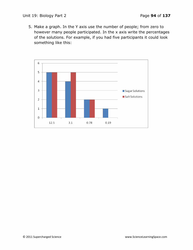
Unit 19: Biology Part 2 Page 94 of 137
© 2011 Supercharged Science www.ScienceLearningSpace.com
5. Make a graph. In the Y axis use the number of people; from zero to
however many people participated. In the x axis write the percentages
of the solutions. For example, if you had five participants it could look
something like this:

Unit 19: Biology Part 2 Page 95 of 137
© 2011 Supercharged Science www.ScienceLearningSpace.com
Activities and Experiments
Lesson 3: Cardiovascular System
Experiment: A Very Simple Stethoscope
Materials:
1 cardboard tube
1 paper towel roll
The first stethoscope was simply a wooden tube. In this activity, you will
make your own simple stethoscope.
1. Place a cardboard tube from a paper towel roll over the heart. Try to
hear the “lub-dub” sound.
2. Experiment with various exercises to change the heart rate you hear in
your stethoscope.
Experiment: The Best Workout
Materials:
Pen
Paper
Stopwatch or clock
1. Make a hypothesis about what exercise will get your heart beating the
fastest: running in place, jumping jacks, hopping on one foot, or
stepping onto and off of a chair.
2. Measure your pulse for 30 seconds and multiply by 2, to get resting
pulse in beats per minute.
3. Run in place for two minutes
4. Measure your pulse immediately after running, again for 30 seconds,
multiplying by two.
5. Rest for several minutes
6. Repeat steps 2-5 for jumping jacks, hopping, and chair steps
7. Make a bar graph showing the increase in pulse for each exercise

Unit 19: Biology Part 2 Page 96 of 137
© 2011 Supercharged Science www.ScienceLearningSpace.com
8. Evaluate the accuracy of your hypothesis. Are you surprised?
Experiment: The Heart – A Special Muscle
1. Make a fist with your hand. This is about the size of your heart.
2. Begin to slightly open and close your hand, as though your “heart” is
beating.
3. Measure how long it takes before your hand hurts
4. Your heart beats 24 hours a day. But it only hurts when a person is
having a heart attack. How can this be?
Answer: Your muscles need oxygen. When a muscle like your hand does not
get enough oxygen, it still makes the motion, but it uses a different method
of getting energy that produces lactic acid, which causes the pain. The heart
is constantly supplied with oxygen from coronary circulation. So, lactic acid
is never needed.
Experiment: A Tennis Ball Heart
Materials:
1 tennis ball
Water
Scissors or knife
1. Carefully cut a hole in the tennis ball
2. Fill the ball with water
3. Squeeze the ball, then release it. What do you observe?
4. How is the ball similar to our heart, as it pumps blood? What does
your hand’s squeeze represent?
Experiment: Observing Your Capillaries
Materials:
Cedarwood furniture oil
Flashlight
1. Soak your finger in cedarwood furniture oil.
2. Shine a strong flashlight on your finger

Unit 19: Biology Part 2 Page 97 of 137
© 2011 Supercharged Science www.ScienceLearningSpace.com
3. Your finger becomes so clean, you can see the small capillaries where
oxygen transfer is happening.
Experiment: Modeling Blood Pressure
Materials:
Oblong balloon
Syringe
1. Fill an oblong balloon with water
2. Cover the tip of a syringe with the open end of the balloon
3. Using the syringe, remove and replace water to simulate the
expansion and contraction of blood vessels
4. Which situation represents systolic blood pressure? How about
diastolic?
Experiment: Clogged Arteries
Materials:
Plastic container
2/3 cup water
2 straws
Bean sized lump of clay
Paperclip
1. Fill a plastic container 2/3 full of water.
2. Insert a clean straw about halfway down the container's length
measured from the bottom of the vessel to its top.
3. Blow into the straw with a steady air stream that produces about a
continual stream of bubbles. Describe the blowing effort needed to
produce this steady, but non-violent stream of bubbles.
4. Completely pack the end of a second straw with a bean-sized lump of
clay.
5. Unbend a paperclip and use it to poke a small hole through the clay
plug.
6. Insert this straw to the same depth as in step 2. Again, blow a steady
air stream into the straw. How does the effort compare to when the

Unit 19: Biology Part 2 Page 98 of 137
© 2011 Supercharged Science www.ScienceLearningSpace.com
passageway was larger? Does the appearance of the bubble stream
change? How?
7. What does the clay represent?

Unit 19: Biology Part 2 Page 99 of 137
© 2011 Supercharged Science www.ScienceLearningSpace.com
Activities and Experiments
Lesson 4: Respiratory & Excretory Systems
Experiment: Oxygen All Around
We breathe in the air that is around us. In order for respiration to work,
there must be oxygen in this air. But can we prove that there actually is
oxygen in the air around us? This experiment will do exactly that.
Materials:
Small amount of water
Pan
Candle
1. Place a small amount of water in the bottom of a pan. The bottom
of the pan should be covered.
2. Light a candle and carefully place it in the water on the bottom of
the pan.
3. Cover the lit candle with the glass and observe what happens.
What’s happening: Fire needs fuel, heat, and oxygen. The candle was
getting oxygen from the air around it – the same air we breathe. When the
candle had no more oxygen, the flame went out.
Experiment: How Long Can You Hold Your Breath?
Although you normally breathe without thinking about it, you can, of course,
hold your breath. Your brain will not allow you to hold your breath longer
than is safe.
Materials:
Plastic bag
Pen
Paper

Unit 19: Biology Part 2 Page 100 of 137
© 2011 Supercharged Science www.ScienceLearningSpace.com
1. Hold your breath, with your nose plugged, and count how long you can
do it
2. Record your time.
3. Breathe into a plastic bag for one minute.
4. Hold your breath again and count how long you can do it now.
What’s happening: You should have found that you could hold your breath
less time after breathing into the bag. As you breathed into the bag, there
was more carbon dioxide in the bag, meaning there was less oxygen going
into your body with each breath. With less oxygen, you can hold your
breath for less time.
Experiment: A Diaphragm’s Duty
When you breathe in, the diaphragm muscle contacts, making more space in
the chest, which allows air to come in. When you breathe out, the opposite
happens. In this activity, you will create a model diaphragm to demonstrate
this important job.
Materials:
2 pieces of straw (2 inches)
Scissors
Small balloon
Large balloon
Rubber bands
Rubber cement or glue
1. Take a piece of straw about 5 cm (2 inches) long and cut a small triangle
in the center. The cut-out should only be on one side of the triangle.
2. Place a small balloon over each end of the straw and secure it with a
small rubber band.
3. Blow into the triangle cut-out to make sure that air will go into each
balloon.
4. Bend the straw in the middle of the hole.
5. Take a second piece of straw and cut a V-shape on the end. Fit the

Unit 19: Biology Part 2 Page 101 of 137
© 2011 Supercharged Science www.ScienceLearningSpace.com
V-shape of the second straw into the triangle hole of the bent straw.
6. Cement the two straws together, and allow plenty of time to dry.
7. Cut a hole in the bottom of a clear plastic cup, the size of the bottom of a
straw.
8. Push the open end of the straw into the bottom of the cup, going through
the hole. Cement the straw into the hole.
9. Cut the neck off of a large balloon.
10. Carefully stretch the cut balloon over the top opening of the cup. Do not
crack the cup. Secure the edges with a large rubber band.
11. Squeeze the bottom balloon gently and observe what happens to the
small balloons.
What’s happening: The large balloon represents the diaphragm. When you
squeeze it, air is forced out, slightly inflating the small balloons. This is a
model of an exhale. How could you model an inhale?
Experiment: What’s in an Exhale?
When you breathe out, you breathe out many things. Carbon dioxide, the
waste product of respiration, is the most important. “See” the carbon
dioxide in this activity.
Materials:
1 teaspoon Bromthymol Blue
1 cup
Straw
1. Place about a teaspoon of Bromthymol Blue (BTB) in a cup
2. Place the straw in the solution and exhale (blow) into the straw.
3. Observe what happens to the blue liquid.

Unit 19: Biology Part 2 Page 102 of 137
© 2011 Supercharged Science www.ScienceLearningSpace.com
What’s Happening: With enough blowing, the solution becomes green (or
even yellow if you really blow.) BTB is an indicator that becomes yellow
when the pH is lowered. The presence of carbon dioxide in your breathe
lowers the pH slightly, changing the color.
Experiment: How Much Air is in There?
Lung capacity is the amount of air in the lungs. When people have
respiratory diseases, their lung capacity is often decreased. What’s your
lung capacity? Let’s find out!
Materials:
Milk jug
Masking tape
Water
1 cup measuring cup
Aquarium tubing
1. Place a strip of masking tape down the side of a milk jug from the
top to the bottom.
2. Fill the jug all the way up with water using a 1 cup measuring cup. Make
a mark on the tape after each cup you pour in.
3. Dump out about half the water, and record exactly how many cups of
water are left in the jug.
4. Place one end of a piece of aquarium tubing inside the mouth of the jug.
5. Take a normal breath and exhale through the tubing. Mark how high the
water goes.
6. Subtract the water amount after your breathe with the water height
before, to see how much air was in your breath.
7. Repeat steps 5 and 6, but breathe out as much air as you possibly can.

Unit 19: Biology Part 2 Page 103 of 137
© 2011 Supercharged Science www.ScienceLearningSpace.com
What’s happening: The air you breathed out displaced, or moved the water,
allowing you to see how much air you exhaled.
Experiment: The Pressure’s On
Materials:
Milk carton
Tape
Water
You are able to breathe because of differences in the pressure in your body
as compared to the pressure outside your body. But what happens when
the pressure changes? Conduct this experiment to see.
1. Punch three pencil-sized holes in a ½ liter carton of milk.
2. Tape over the holes with one long piece of tape, so they are
completely covered
3. Add water to the milk carton, so it is completely full
4. Quickly rip the tape off the carton and make observations.
What’s happening: You should have noticed that the lowest hole had the
most water flowing out of it. This is where pressure was the greatest.
Similarly, air pressure at low elevations (such as sea level) is greater than at
high elevations (such as when you are flying in an airplane.) Some people
have a harder time breathing at high elevations, and if you get high enough
artificial pressure must be added.
Experiment: The Job of a Kidney
Materials:
Sand
Water
Yellow food coloring
Cheesecloth
Large bowl
Unit 19: Biology Part 2 Page 104 of 137
© 2011 Supercharged Science www.ScienceLearningSpace.com
The job of the kidney is to filter liquid waste. In this activity, you will model
the action of the kidney by filtering sandy water.
1. Mix sand, water, and yellow food coloring.
2. Place a piece of cheesecloth over a large bowl.
3. Slowly pour the sandy water over the cheesecloth and into the bowl.
4. Make observations about the liquid that goes into the bowl.
What’s happening: The cheesecloth, just like the kidney, serves as a filter.
Sand is kept out while the water gets through. This is important in
maintaining the body’s salt balance.
Experiment: A Model Colon
The colon, also called the large intestine, is an important part of both the
digestive and excretory systems, which removes solid waste. This model
colon will demonstrate the creation of solid waste.
Materials:
Cheesecloth
Colander
Regular yogurt
Bowl
1. Place a piece of cheesecloth into a colander.
2. Add regular yogurt (not light) into the colander.
3. Place the colander with the yogurt in it into a bowl and place it into the
refrigerator for several days
4. After several days, make observations about both what’s in the bowl
and what’s left in the colander.
What’s happening: The colon filters out liquids from the digested material
(that’s modeled by what’s in the bowl) and leaves us with solid waste
(modeled by what’s in the colander.)

Unit 19: Biology Part 2 Page 105 of 137
© 2011 Supercharged Science www.ScienceLearningSpace.com
Activities and Experiments
Lesson 5: Controlling the Body
Experiment: Measuring Reflexes
Usually our brain sends messages to the muscles of our body, causing them
to expand or contract. This makes our body move the way we need it too.
Sometimes, such as when something unexpected happens, our body doesn’t
take the time to send the message all the way to the brain. Instead, the
message goes as far as your spinal cord, and the spinal cord gives the
muscles the messages they need. This is called a reflex. How good are your
reflexes?
Materials:
Yardstick
Partner
1. Have a partner hold a yardstick in front of you.
2. Put your fingers around the bottom of the yardstick, but don’t touch it.
3. Without warning you, have your partner let go of the yardstick.
4. Try to stop the falling yardstick by squeezing it with your fingers as
soon as you notice it falling.
5. When you stop it, record how high up on the yardstick you are (in
inches or centimeters.) The smaller the number, the quicker your
reflex.
What’s Happening: Stopping the ruler is a reflex. Your brain was not
involved. Other reflexes include removing your hand from a hot or painful
object.
Experiment: Message Received
Your brain sends messages to your muscles, as well as to all the other
organs in your body. Sometimes, you have no control over these messages,
such as when the brain controls breathing, digestion, or blood circulation.
Other times, such as when you are exercising or just walking around, you
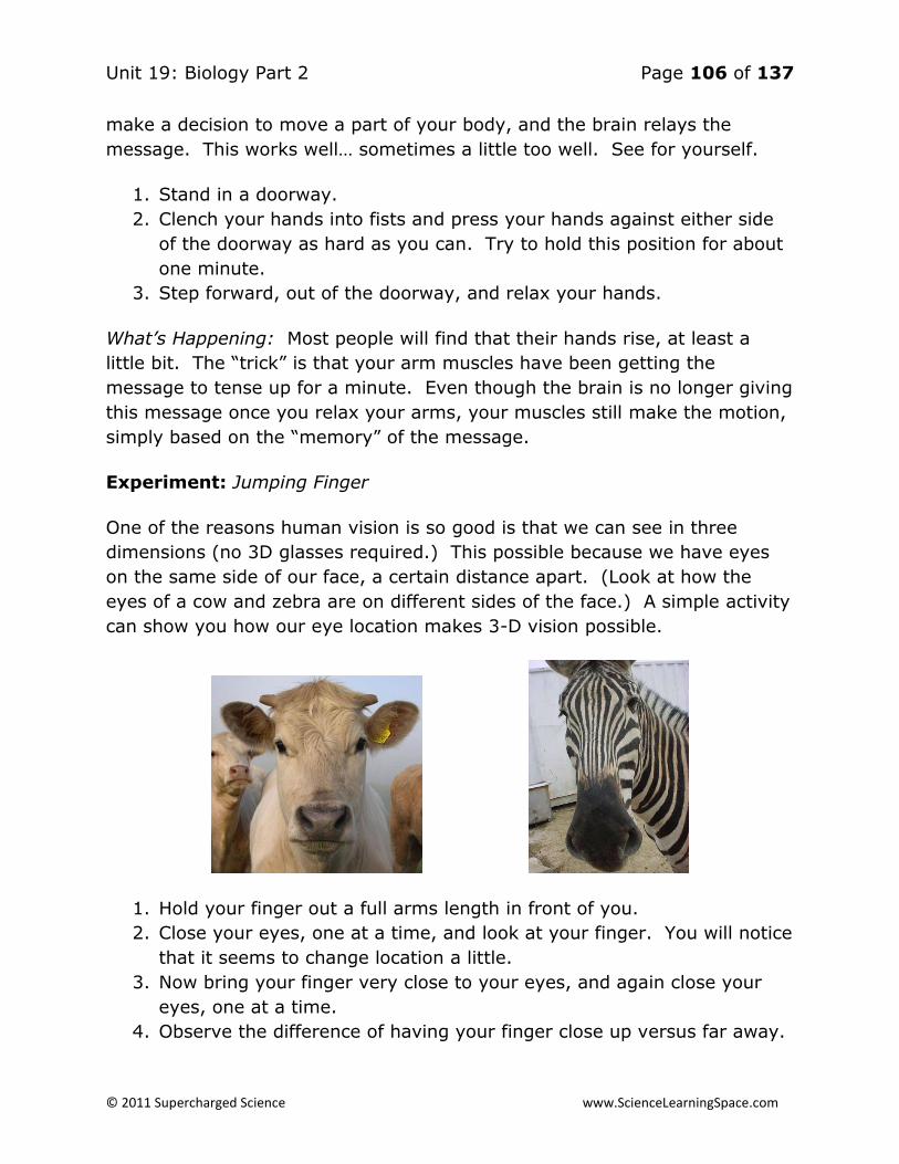
Unit 19: Biology Part 2 Page 106 of 137
© 2011 Supercharged Science www.ScienceLearningSpace.com
make a decision to move a part of your body, and the brain relays the
message. This works well… sometimes a little too well. See for yourself.
1. Stand in a doorway.
2. Clench your hands into fists and press your hands against either side
of the doorway as hard as you can. Try to hold this position for about
one minute.
3. Step forward, out of the doorway, and relax your hands.
What’s Happening: Most people will find that their hands rise, at least a
little bit. The “trick” is that your arm muscles have been getting the
message to tense up for a minute. Even though the brain is no longer giving
this message once you relax your arms, your muscles still make the motion,
simply based on the “memory” of the message.
Experiment: Jumping Finger
One of the reasons human vision is so good is that we can see in three
dimensions (no 3D glasses required.) This possible because we have eyes
on the same side of our face, a certain distance apart. (Look at how the
eyes of a cow and zebra are on different sides of the face.) A simple activity
can show you how our eye location makes 3-D vision possible.
1. Hold your finger out a full arms length in front of you.
2. Close your eyes, one at a time, and look at your finger. You will notice
that it seems to change location a little.
3. Now bring your finger very close to your eyes, and again close your
eyes, one at a time.
4. Observe the difference of having your finger close up versus far away.

Unit 19: Biology Part 2 Page 107 of 137
© 2011 Supercharged Science www.ScienceLearningSpace.com
What’s Happening: When your finger is far away, your eyes perceive it in
more or less the same place. This happens because the angle between your
left eye and your finger is not that different that the angle between your
finger and right eye. As your finger gets closer, the difference becomes
more significant. Your brain constantly uses this information to allow you to
detect depth, the key to seeing in three dimensions.
Experiment: Can You Believe Your Eyes?
Our brain is a remarkable organ that receives information from sense
organs, processes the information, and then sends out a message in
response. But what happens when the brain gets conflicting information?
That’s what this experiment is all about.
1. Take a look at
the chart. Say
the COLOR of
the word.
Don’t read the
word.
What’s Happening: It’s harder than it looks, isn’t it? The problem is that
our brain is getting a mixed message. The color of the word is one
message, but the word itself if another. Your brain can overcome this and
say the right color, but it requires more work simply looking at the sky and
saying it’s blue.
Experiment: Selective Listening
Have you ever been accused of having “selecting listening?” This term
refers to the idea that when your mom or dad tells you to clean your room,
you somehow can’t hear them, but when they ask if you want ice cream,
your ears work great. Is this a real, scientific thing? It turns out that there
actually is something going on your brain that makes your hearing selective.
Check it out!
1. Go to a noisy place where you’ll be surrounded by people. (A fast food
restaurant around lunchtime will work well.)

Unit 19: Biology Part 2 Page 108 of 137
© 2011 Supercharged Science www.ScienceLearningSpace.com
2. Hold a pen slightly above the table and drop it. Observe any
reactions from other people.
3. Wait a minute or two so you aren’t too obvious, then drop another
object, like a set of keys. Again, look for reactions.
4. Drop a few more things, and, for the last thing, drop several coins.
What’s Happening: You probably got much more of a reaction from the
dropped coins than from the other objects. The reason. People care about
money… not so much about pens and keys. Our brain filters out the sounds
we don’t find important. This is a good thing, because we are bombarded
with sounds every day. The problem is that since your brain decides what’s
important, you don’t get to completely control. So, if you decide you are
going to watch TV while doing schoolwork, this probably won’t work, unless
you choose a TV show you don’t find interesting (and if that was the case,
why would you watch it in the first place.) Some people do find it helpful to
listen to quiet music while working, and are able to tune this out enough to
focus on the work.
Experiment: Go For a Spin
Your ears do more than just help you hear. They are also very important for
your balance. Inside your ear are liquid-filled tubes called semicircular
canals. As your body moves, the liquid in the semicircular canals moves too.
But what about when you stop? Let’s find out.
Materials:
Office chair
Adult
1. Sit on an office chair that spins.
2. Have an adult spin you quickly on the chair. Hold on to the handles
and be careful!
3. Have the adult suddenly stop the chair, and observe how you feel.
What’s Happening: Did you feel as though you were still moving? Part of
you was. Although your body stopped, the fluid in your semicircular canals
kept on going for a little while. As long as the fluid kept move, your brain
was giving the message that you were moving, and it felt that way.

Unit 19: Biology Part 2 Page 109 of 137
© 2011 Supercharged Science www.ScienceLearningSpace.com
Experiment: Can You Feel It?
Every bit of your skin has sensory neurons. These neurons sense pain,
pressure, and temperature, and send the message to your brain. This is the
sense of touch. Not all parts of your body have the same amount of
neurons, and the more neurons a certain body part has, the more sensitive
it will be to touch. In this experiment, you’ll use indirect evidence to figure
out if you have more neurons in your palm or lower back.
Materials:
2 washcloths
Water
Partner
1. Prepare two washcloths – one with cool water and one with slightly
warm water. The two washcloths should not be dramatically different
in temperature, but there should be some difference.
2. Have a partner place a washcloth on your palm 20 times. He or she
should not tell you which washcloth it is, and should not do it in any
kind of pattern. Your job is to tell if it is the cool or warm washcloth
that is being touched to your palm.
3. Have your partner record how many times you answered correctly.
4. Repeat steps two and three placing the washcloth on your lower back.
What’s Happening: Your palm has more sensory neurons than your lower
back, so you should have had an easier time identifying cool versus warm on
this part of your body. This means your palm is also more sensitive to
pressure and pain. Now that you have compared the palm and lower back,
why not compare some other parts of your body?
Experiment: The Nose Knows (Or Does It?)
Our sense of smell is controlled by sensory neurons in the nose. When a
smell is detected, a message is sent to the brain. What the brain does with
the information in some cases may surprise you.
Materials:

Unit 19: Biology Part 2 Page 110 of 137
© 2011 Supercharged Science www.ScienceLearningSpace.com
Vanilla extract
Rubbing alcohol
1. Smell some vanilla extract. Continue to smell it until you no longer
sense the smell.
2. Place some rubbing alcohol under your nose. Observe the smell.
What’s Happening: The human body has a remarkable ability to get used to
odors we smell all the time. If the brain keeps getting the same message
from the nose, it will eventually learn to ignore it. This is important if you
are an animal that could be someone’s prey. If you’re a rabbit, for example,
you’ll get used to the normal smells around you and be able to sense if a
predator is coming. For humans, who don’t use our smell for survival, this
ability to block out smells isn’t all that helpful, and can actually be a
problem. Someone with bad body odor will get used to it, and won’t realize
they need a shower!
Experiment: Taste Bud Map
On your tongue are thousands of sensory neurons. These neurons are
organized into groups called taste buds. When you eat something, the taste
buds send a message to your brain. Your brain interprets the message, and
“tells” you what you are eating. Certain areas of the tongue have higher
concentrations of certain types of taste buds. In this experiment, you will
have a chance to see where they are.
Materials:
3 cups
Sugar water or punch
Salt water
Unsweetened iced coffee
Q tips
1. Prepare a “sweet,” “salty,” “sour,” and “bitter” cup. In the “sweet”
cup, place sugar water or punch. In the “salty” cup place salt water.

Unit 19: Biology Part 2 Page 111 of 137
© 2011 Supercharged Science www.ScienceLearningSpace.com
In the “sour” cup, place lemon juice or lemonade. In the “bitter” cup,
place unsweetened iced coffee.
2. Dip a q-tip into the sweet cup. Place a drop of the sweet liquid on the
tip of your tongue. Dip it in again, and place it on the side of your
tongue towards the front. Place another drop on the side towards the
back, and a final drop in the back. Record where you tasted the
sweetness most strongly.
3. With a different q-tip, repeat step two for the other cups.
What’s Happening: Look at the diagram below, which shows where the
concentration of taste buds for each of these tastes are. How closely do
your results match the diagram? If your results seem a little off, don’t
worry. You actually have taste buds for all kinds of tastes everywhere. The
map just shows where they are the strongest.

Unit 19: Biology Part 2 Page 112 of 137
© 2011 Supercharged Science www.ScienceLearningSpace.com
Activities and Experiments
Lesson 6: Diseases and Defenses
Experiment: How Clean is Clean?
The single best way to prevent the spread of infectious disease is to wash
your hands. But how much do you need to wash them to actually get rid of
all the disease causing pathogens you are carrying? Using a model to
represent pathogen-filled hands, you can find out.
Materials:
GloGerm, Germ Juice or Glitter Bug
1. Copy a chart like the one below:
0 5 10 15 20
++++
2. Cover your hands with a pathogen simulator, such as GloGerm, Germ
Juice, or Glitter Bug. Cover both your palms and the backs of your
hands, and try to get it on the skin under your fingernails.
3. Look at your hands under a UV light. You will see that the “germs” are
clearly visible. This is time 0, meaning that you have not washed your
hands at all. You will call this level of dirtiness “++++,” or the
dirtiest. This level of dirtiness has already been filled in on the data
table.
4. Wash your hands for five seconds. Again look at your hands under the
UV light. Rate their dirtiness as either ++++, +++, ++, +, or –,
where ++++ represents as dirty as they were at the start, and –
represents totally clean.
5. Wash your hands for an additional 5 seconds, and record their
cleanliness after 10, 15, and 20 seconds total.

Unit 19: Biology Part 2 Page 113 of 137
© 2011 Supercharged Science www.ScienceLearningSpace.com
What’s Happening: The Centers for Disease Control (CDC) recommends
washing your hands with warm, soapy water for 10-15 seconds. This
assumes that you scrub vigorously, and that your hands are not abnormally
dirty.
Experiment: Don’t Forget the Soap
Materials:
Vegetable oil
Water
Soap
1. Put a spoonful of vegetable oil on your hands
2. Try to wash it off in only cold water, without soap.
3. Repeat with only warm water and with water and soap.
What’s Happening: Plain water, especially plain cold water, is not as
effective at getting the oil off your hands as warm water and soap.
This model used vegetable oil, but we have natural oils that build up in
our skin. To clean them, and to get rid of pathogens, soap is needed!
Experiment: Don’t Sneeze On Me!
Materials:
10 volunteers
10 cups
Water
Food coloring
When we sneeze, we release a huge number of pathogens into the air. If we
don’t cover our mouth, these pathogens are more likely to infect another
person, making them sick. The person we infected can now spread the
disease to others. In this way, pathogens can spread very quickly. You’ll
need about 10 volunteers for this activity. (You can be one of the 10.) Prior
to the activity, prepare 10 cups, by filling them up with water and adding

Unit 19: Biology Part 2 Page 114 of 137
© 2011 Supercharged Science www.ScienceLearningSpace.com
food coloring to one of them. Number the cups 1-10. You can make the
colored cup whatever number you want.
1. Explain to your volunteers that we are going to imagine they work
together in an office, and have frequent appointments with each other.
Have everyone fill out a chart like the one below. To fill the chart out,
the volunteers need to make an “appointment” with each other at a
certain time. For example, if Jessica and Raul are two of your
volunteers, they might agree to have an “appointment” at 9:00.
Jessica would write “Raul” under 9:00, and Raul would write “Jessica”
under 9:00. You should not meet with the same person twice.
Time Appointment With
9:00
10:00
11:00
2. Assign everyone a number. The oldest volunteer is number 1, and the
youngest is number 10, with everyone else somewhere in between
based on their ages.
3. Give the volunteers their water cup, based on their number and tell
them to go to their 9:00 appointment.
4. During the appointment, we will imagine that each individual sneezed
once. We’ll model this by exchanging a little bit of water. In the
example above, for instance, Raul would spill some water into Jessica’s
cup, and Jessica would spill some into Raul’s.
5. Repeat the procedure for the 10:00 and 11:00 appointments.
6. Once the appointments are over, announce that anyone who has
colored water (even just a little coloring) is now sick. Ask who is sick
and record the number.
What’s Happening: At the start of the activity, only one person was sick (had
colored water.) Now, it is likely that most people are. Diseases spread
quickly, especially when people do not make good decisions about their
health and the health of others. The sick person should really have stayed
home from work. If he or she had, no one else would have become sick. If
they did come in, they should have at least made an effort to cover their
mouths when sneezing.
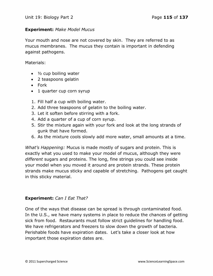
Unit 19: Biology Part 2 Page 115 of 137
© 2011 Supercharged Science www.ScienceLearningSpace.com
Experiment: Make Model Mucus
Your mouth and nose are not covered by skin. They are referred to as
mucus membranes. The mucus they contain is important in defending
against pathogens.
Materials:
½ cup boiling water
2 teaspoons gelatin
Fork
1 quarter cup corn syrup
1. Fill half a cup with boiling water.
2. Add three teaspoons of gelatin to the boiling water.
3. Let it soften before stirring with a fork.
4. Add a quarter of a cup of corn syrup.
5. Stir the mixture again with your fork and look at the long strands of
gunk that have formed.
6. As the mixture cools slowly add more water, small amounts at a time.
What’s Happening: Mucus is made mostly of sugars and protein. This is
exactly what you used to make your model of mucus, although they were
different sugars and proteins. The long, fine strings you could see inside
your model when you moved it around are protein strands. These protein
strands make mucus sticky and capable of stretching. Pathogens get caught
in this sticky material.
Experiment: Can I Eat That?
One of the ways that disease can be spread is through contaminated food.
In the U.S., we have many systems in place to reduce the chances of getting
sick from food. Restaurants must follow strict guidelines for handling food.
We have refrigerators and freezers to slow down the growth of bacteria.
Perishable foods have expiration dates. Let’s take a closer look at how
important those expiration dates are.

Unit 19: Biology Part 2 Page 116 of 137
© 2011 Supercharged Science www.ScienceLearningSpace.com
Materials:
4 cups
Water
Sour milk, expired sour cream or fresh yogurt
Methylene blue
Mineral oil
Large clear bowl
1. Label four cups as “control,” “milk,” “yogurt,” and “sour cream”
2. Fill the control cup with water.
3. Fill each of the other cups half full with sour milk, expired sour cream, or
fresh yogurt.
4. Add 20 drops of methylene blue to each cup.
5. Add 2 drops of mineral oil to each cup.
6. Place the cups in a large clear bowl filled with warm water.
7. Record the time it takes for the color to fade.
What’s Happening: Methylene blue is a dye used to detect bacteria. The
bacteria use up oxygen, and the dye turns colorless in the absence of
oxygen. So, the faster the color fades when the dye is added to a substance,
the less oxygen there is, and the more bacteria the substance has. (There
are some bacteria that don’t need oxygen, so this test wouldn’t work for
them.) You may have noticed that there are bacteria present in the yogurt
although it’s fresh. These bacteria are helpful to you. They improve your
ability to digest food.

Unit 19: Biology Part 2 Page 117 of 137
© 2011 Supercharged Science www.ScienceLearningSpace.com
Experiment: The Skin You’re In
Your skin is your first line of defense against pathogens. Everyday, many
germs come your way, but most of them are blocked by your skin. This
simple model can show how.
Materials:
Apple
Plastic wrap
Food coloring
1. Cut an apple in half.
2. Cover one half in plastic wrap; leave the other uncovered.
3. Add several drops of food coloring to each apple, and make
observations.
What’s Happening: The plastic wrap represents the skin, and the food
coloring represents a pathogen. Without skin, pathogens could easily enter
the body. The thick skin keeps most pathogens out.
Experiment: Take All Your Medicine!
If you have ever been prescribed an antibiotic, your doctor probably
reminded you to take all the medication prescribed, even if you started to
feel better. Why is this so important?
Materials:
30 pennies
12 nickels
6 dimes
4 quarters
Deck of cards
1. For this experiment, assume the following:
a. Coins represent bacteria. The higher the monetary value of the
coin, the more resistant it is. (The harder it is to kill.)
b. If you have more than 35 bacteria, you will feel sick.

Unit 19: Biology Part 2 Page 118 of 137
© 2011 Supercharged Science www.ScienceLearningSpace.com
c. A deck of cards represents an antibiotic. Each card is one dose
of medicine.
2. Place 52 coins on a table in the following amounts: 30 pennies, 12
nickels, 6 dimes, and 4 quarters.
3. Shuffle a deck of cards.
4. Pull the top card. This one card is one dose of antibiotic, and will kill
one bacterium. Follow these rules to determine which bacterium dies.
a. Any card numbered 2-7 kills a penny.
b. Eights, 9, 10’s, and Jacks kill nickels
c. Queens and Kings kill dimes
d. Aces kill quarters.
e. If there are no coins in the correct denomination to be killed (for
example, you get a jack but all the nickels are already dead,) the
card can kill a penny instead.
5. “Dead” bacteria can be taken off the table.
6. The person in this simulation decides to ignore their doctors advice to
take all 52 doses, and stop when they feel better. So, when you have
35 coins left on the table, stop.
7. The bacteria reproduce, and their offspring are just like them. So, for
every penny still on the table, add another penny. Add another nickel
for every nickel, etc. Count how many of each type of coin you have
(there will be 70 total), and add it to the data table. Then, figure our
the percent of each coin type by dividing the number present by 70.
Add this to your data table.
Sample Data Table:
Coin Type Number to
Start
Percent at
Start
Number
after
Reproduction
Percent After
Reproduction
Penny 30
Nickel 12
Dime 6
Quarter 4

Unit 19: Biology Part 2 Page 119 of 137
© 2011 Supercharged Science www.ScienceLearningSpace.com
What’s happening: Typically, the percent of the highly resistant bacteria
(dimes and quarters) will increase while the less resistant ones decrease in
percentage. This is exactly what happens in real life if you don’t take all the
antibiotic. The bacteria that are hardest to kill stay and reproduce. This
makes it harder to cure the disease caused by the bacteria. If you had gone
through all the cards to begin with, you would have killed all the bacteria for
sure. Now one deck won’t do it. To avoid needing more medication, or
stronger medication, later on, the best thing to do is take all the antibiotic
the first time around.

Unit 19: Biology Part 2 Page 120 of 137
© 2011 Supercharged Science www.ScienceLearningSpace.com
Exercises
Skin, Bones & Muscles Exercises
1. What is homeostasis?
2. What is an example of the body maintaining homeostasis?
3. Cells make up tissues, at least how many tissues make up an organ?
4. What are the four main types of tissues?
5. Describe how a negative feedback works and give an example.
6. What is the biggest organ in the body?
7. The skin, hair, and nails make up which system?
8. What are three ways the integumentary system helps maintain
homeostasis?
9. What are the two layers of skin? What is the fatty layer underneath the
skin called?
10. What role does melanin play in the skin?
11. What causes acne?
12. What three components make up the skeletal system?
13. What are the main functions of bones?
14. What is the difference between red bone marrow and yellow bone
marrow?
15. What are the three classes of joints?
16. What are the four types of movable joints?
17. What are two key nutrients bones need?
18. What’s the difference between skeletal muscle, smooth muscle, and
cardiac muscle?
19. Muscles work in pairs, what do we call the one that bends the joint?
What do we call the one that straightens the joint?
20. What is the difference between stretching exercises, aerobic
exercises, and anaerobic exercises?
Unit 19: Biology Part 2 Page 121 of 137
© 2011 Supercharged Science www.ScienceLearningSpace.com
Exercises
Digestive System Exercises
1. What is a diet?
2. Why does our body need nutrients?
3. What are the six essential nutrients? Describe them in your own words.
4. What model of a proper diet do we currently use; MyPlate or
MyPyramid?
5. What are the three general pieces of advice the USDA gives in regards
to maintaining a healthy diet?
6. Does the protein food group, according to the USDA, include only
meat?
7. Is 30% lean meat the type of meat you should buy often? Why or why
not?
8. What is the difference between “enriched” grains and “non- enriched”
grains?
9. Do “enriched” refined grains contain fiber?
10. Who should avoid dairy products?
11. If you eat a healthy, balanced diet, is it necessary to get weekly
exercise? If so, how much?
12. What is the difference between mechanical digestion and chemical
digestion?
13. What are the two steps after digestion?
14. Name two key enzymes used for digestion.
15. What is peristalsis?
16. Is protein chemically digested in the mouth? If not there, where?
17. What are the three parts of the small intestine? What are their
functions?
18. What is the main function of the large intestine?

Unit 19: Biology Part 2 Page 122 of 137
© 2011 Supercharged Science www.ScienceLearningSpace.com
Exercises
Cardiovascular System Exercises
1. What is the overall purpose of the cardiovascular system?
2. How could body temperature be affected if someone has poor
circulation?
3. What are the four parts of blood?
4. Sickle-cell anemia is a disease in which RBC’s cannot carry hemoglobin
properly. What could happen to a person who has sickle-cell anemia?
5. What protein give’s RBC’s their red color?
6. What would happen to a person with too few WBC’s?
7. Hemophilia is a disease in which platelets do not work properly. What
could happen to someone who has hemophilia?
8. Why do arteries have thick walls?
9. Why do veins have valves?
10. Name the four chambers of the heart.
11. Where does the pulmonary artery start? Where does it lead?
12. What is the difference between oxygen-rich and oxygen poor blood?
13. What happens to blood in the lungs?
14. What is the name of the only vein that carries oxygen poor blood?
15. Which chamber of the heart does blood enter after leaving the
pulmonary vein?
16. In which blood vessel does oxygen transfer happen?
17. Where do the superior and inferior vena cava lead?
18. Which organ gets blood from the coronary artery?
19. What is the difference between systolic and diastolic blood pressure?
20. How can hypertension be treated?
21. When can atherosclerosis cause a heart attack?
22. What happens if blood vessels leading to the brain are blocked?
23. What are three benefits of not smoking related to the cardiovascular
system
Unit 19: Biology Part 2 Page 123 of 137
© 2011 Supercharged Science www.ScienceLearningSpace.com
Exercises
Respiratory & Excretory Systems Exercises
1. What is the job of the respiratory system?
2. What happens to air in the nasal cavity?
3. What does the diaphragm do when you inhale?
4. Why is it important for the epiglottis to cover the trachea when you are
eating?
5. What happens in the alveoli?
6. When pressure is different, in what direction do fluids flow?
7. How does your body use differences in pressure when you exhale?
8. What is the difference between breathing and respiration?
9. What is internal respiration?
10. What disease results from an inflammation on the bronchi?
11. What types of things can cause an asthma attack?
12. What types of things cause pneumonia?
13. Why are the lungs part of both the respiratory and excretory system?
14. Besides the excretory system, what system are the kidneys a part of?
15. What do the kidneys do?
16. What is urine?
17. After traveling down the ureters, where does urine go?
18. How do kidney stones develop?
19. What is the purpose of a kidney dialysis machine?
20. What are the most common types of UTI?

Unit 19: Biology Part 2 Page 124 of 137
© 2011 Supercharged Science www.ScienceLearningSpace.com
Exercises
Controlling the Body Exercises
1. How does the nervous system relate to the other organ systems?
2. What is a synapse?
3. What organs are in the central nervous system?
4. What are the three parts of the brain?
5. If the left side of the cerebrum is injured, will the right or left side of the
body most likely be affected? Why?
6. What do the vertebrae do?
7. What is the function of myelin?
8. What is the difference between the somatic and autonomic nervous
sytem?
9. When is the sympathetic nervous system used?
10. How is a reflex different from a typical message to the brain?
11. What is the purpose of cones in the eyes?
12. What is the path of light coming into the cornea?
13. What happens to images if a person has myopia?
14. What two things are ears responsible for?
15. If the hairs in the ears semicircular canals move, what message is sent
to the brain?
16. What are some parts of the body that have many touch receptors?
17. Where are taste buds found?
18. What causes Reye’s Syndrome?
19. What occurs during a seizure?
20. What are some things you can do to “exercise” your brain?

Unit 19: Biology Part 2 Page 125 of 137
© 2011 Supercharged Science www.ScienceLearningSpace.com
Exercises
Diseases and Defenses Exercises
1. What is the difference between an infectious and noninfectious disease?
2. What are four types of pathogens?
3. How are viruses different than other pathogens?
4. Can a noninfectious disease be caused by a pathogen?
5. How do vectors spread disease?
6. What is the single best way to avoid getting an infectious disease?
7. How can you avoid coming into contact with vectors when you are out in
nature?
8. What is the body’s first line of defense?
9. How do body excretions keep you from getting sick?
10. How does mucus stop pathogens?
11. What role does cilia play in the nose?
12. What comes out of your body when you cough or clear your throat?
13. When does your body send out signals for an inflammation?
14. How are inflammations helpful?
15. How are fevers helpful?
16. What types of white blood cells are involved in inflammations?
17. When would your body produce an immune response?
18. Where are lymphocytes produced?
19. What is the function of the thymus gland?
20. What do killer t-cells do?

Answers to Exercises
Answers to Skin, Bones & Muscles Exercises
1. What is homeostasis? The ability of the body to maintain a stable
internal environment in the response to external changes.
2. What is an example of the body maintaining homeostasis? Many
examples to choose from. One might be when we sweat to keep our
body temperature down, or when we shiver to keep the temperature
up. Another example is how we regulate the amount of sugar in our
blood. All of the examples should show how the body uses a negative
feedback loop to maintain homeostasis. We sweat just until we are
cooled down, we shiver until we are warmed up, and we put sugar in
our system just until we have enough.
3. Cells make up tissues, at least how many tissues make up an organ?
Two or more tissues working together to serve the same function
constitute an organ.
4. What are the four main types of tissues? Epithelial tissue, muscular
tissue, nervous tissue, and connective tissue.
5. Describe how a negative feedback works and give an example. A
mechanism of control in the body in which the result of a bodily
function acts as a signal to stop. Plus one example (like the control of
blood sugar, or blood temperature).
6. What is the biggest organ in the body? The skin.
7. The skin, hair, and nails make up which system? The integumentary
system.
8. What are three ways the integumentary system helps maintain
homeostasis? 1.) Helping regulate temperature, 2.) Sending sensory
information about the environment outside the body to the brain 3.)
Keeping water and germs out of the body 4.) Acts as a barrier to
sunlight.
9. What are the two layers of skin? What is the fatty layer underneath the
skin called? The two layers are the epidermis and the dermis. The fatty
layer beneath is the hypodermis or subcutaneous tissue.
10. What role does melanin play in the skin? It pigments the skin, and
helps protect the lower layers from harmful UV rays.
11. What causes acne? Clogging of the oil glands.

Unit 19: Biology Part 2 Page 127 of 137
© 2011 Supercharged Science www.ScienceLearningSpace.com
12. What three components make up the skeletal system? Bones,
ligaments, and cartilage.
13. What are the main functions of bones? Support: Bones give the body
its structure—its shape. It holds up the tissue against the pressure of
gravity. protection: The bones protect certain tissues. For example,
the skull protects the brain, and the ribs protect the heart and lungs.
Movement: The bones work in concert with the muscles to give us the
ability to move. Making Blood Cells: Certain parts of certain types of
bones make blood cells. Storage: Bones store calcium and phosphorus
(mostly calcium).
14. What is the difference between red bone marrow and yellow bone
marrow? Red bone marrow makes red blood cells while yellow bone
marrow makes white blood cells.
15. What are the three classes of joints? Fixed, partly-movable, and
movable.
16. What are the four types of movable joints? Ball and socket joints,
hinge joints, pivot joints, and gliding joints.
17. What are two key nutrients bones need? Calcium and vitamin D.
18. What’s the difference between skeletal muscle, smooth muscle, and
cardiac muscle? Skeletal muscle is attached to our bones and allow us
to move. We do not control smooth muscle. Cardiac muscle is only
found in the heart.
19. Muscles work in pairs, what do we call the one that bends the joint?
What do we call the one that straightens the joint? The muscle that
bends the joint is called the flexor, and the one that straightens the
joint is called the extensor.
20. What is the difference between stretching exercises, aerobic
exercises, and anaerobic exercises? Stretching exercises warm-up our
muscles and make them more flexible, anaerobic exercise build our
muscles by making them work against resistance, and aerobic
exercises increase our endurance.

Unit 19: Biology Part 2 Page 128 of 137
© 2011 Supercharged Science www.ScienceLearningSpace.com
Answers to Exercises
Answers to Digestive System Exercises
1. What is a diet? The sum of the food and drink consumed considered in
terms of its effect on health
2. Why does our body need nutrients? Because it needs energy, it needs
to grow/repair itself, and it needs to maintain the systems that
maintain homeostasis.
3. What are the six essential nutrients? Describe them in your own words.
Protein, carbohydrates, lipids, vitamins, minerals, water.
4. What model of a proper diet do we currently use; MyPlate or
MyPyramid? MyPlate.
5. What are the three general pieces of advice the USDA gives in regards
to maintaining a healthy diet? Balance calories intake, eat certain
[nutrient rich] foods, eat fatty and sugary foods in moderation.
6. Does the protein food group, according to the USDA, include only
meat? No it also includes vegetables, fish, eggs, and poultry.
7. Is 30% lean meat the type of meat you should buy often? Why or why
not? No, because it has far too much fat. You should look for 94% lean
meat.
8. What is the difference between “enriched” grains and “non- enriched”
grains? Enriched grains contain vitamins and minerals we need that
are lost in the refining process.
9. Do “enriched” refined grains contain fiber? No, fiber is not added back
to enriched refined grains.
10. Who should avoid dairy products? People who cannot digest lactose—
people who are lactose intolerant.
11. If you eat a healthy, balanced diet, is it necessary to get weekly
exercise? If so, how much? Yes, exercise is always necessary. 60
minutes of exercise three days a week is a good level of activity.
12. What is the difference between mechanical digestion and chemical
digestion? Mechanical digestion is accomplished with the teeth, which
chemical digestion breaks food down into the nutrients via chemicals
and enzymes (catalysts).
13. What are the two steps after digestion? Absorption and elimination.

Unit 19: Biology Part 2 Page 129 of 137
© 2011 Supercharged Science www.ScienceLearningSpace.com
14. Name two key enzymes used for digestion. Amylase, pepsin, or
pancreatic lipase.
15. What is peristalsis? Peristalsis is the wave-like movement of the
digestive system used to move food from the mouth to anus.
16. Is protein chemically digested in the mouth? If not there, where? No,
protein is digested by pepsin in the stomach.
17. What are the three parts of the small intestine? What are their
functions? The duodenum, the jejunum, and the ileum. The duodenum
is the first part of the small intestine. In the duodenum the food from
the stomach is further digested (chemically). Some of the chemicals
are secreted from the duodenum itself, others are secreted from the
liver and pancreas.The jejunum; Most nutrients are absorbed into the
body at this second part of the small intestine. The nutrients are
absorbed through tiny blood vessels. The ileum Here nutrients are also
absorbed into the blood stream. What is not absorbed in the ileum is
passed as waste through the large intestine.
18. What is the main function of the large intestine? Eliminate solid waste
from the body.

Unit 19: Biology Part 2 Page 130 of 137
© 2011 Supercharged Science www.ScienceLearningSpace.com
Answers to Exercises
Answers to Cardiovascular System Exercises
1. What is the overall purpose of the cardiovascular system? to move
blood to the various parts of the body
2. How could body temperature be affected if someone has poor
circulation? the body would not be able to heat up or cool down to the
ideal temperature
3. What are the four parts of blood? plasma, platelets, RBC’s, WBC’s
4. Sickle-cell anemia is a disease in which RBC’s cannot carry hemoglobin
properly. What could happen to a person who has sickle-cell anemia?
Oxygen would not get to the organs of the body.
5. What protein give’s RBC’s their red color? hemoglobin
6. What would happen to a person with too few WBC’s? they would have
a weak immune system and could more easily get diseases
7. Hemophilia is a disease in which platelets do not work properly. What
could happen to someone who has hemophilia? their blood would not
clot when they got minor cuts and scrapes
8. Why do arteries have thick walls? because the blood in them is under
high pressure
9. Why do veins have valves? to keep the blood from flowing backwards
10. Name the four chambers of the heart. right atrium, left atrium, right
ventricle, left ventricle
11. Where does the pulmonary artery start? Where does it lead? it starts
at the heart and leads to the lungs
12. What is the difference between oxygen-rich and oxygen poor blood?
oxygen-rich blood has oxygen and oxygen-poor blood has very little
13. What happens to blood in the lungs? it gets oxygen
14. What is the name of the only vein that carries oxygen poor blood?
pulmonary vein
15. Which chamber of the heart does blood enter after leaving the
pulmonary vein? left atrium
16. In which blood vessel does oxygen transfer happen? capillaries
17. Where do the superior and inferior vena cava lead? the right atrium of
the heart
18. Which organ gets blood from the coronary artery? heart

Unit 19: Biology Part 2 Page 131 of 137
© 2011 Supercharged Science www.ScienceLearningSpace.com
19. What is the difference between systolic and diastolic blood pressure?
systolic is the highest blood pressure; diastolic is the lowest
20. How can hypertension be treated? improved diet, increased exercise,
or medication
21. When can atherosclerosis cause a heart attack? when a coronary
artery becomes completely blocked
22. What happens if blood vessels leading to the brain are blocked? a
stroke
23. What are three benefits of not smoking related to the cardiovascular
system decreased risk of hypertension, coronary heart disease, and
stroke

Unit 19: Biology Part 2 Page 132 of 137
© 2011 Supercharged Science www.ScienceLearningSpace.com
Answers to Exercises
Answers to Respiratory & Excretory Systems
Exercises
1. What is the job of the respiratory system? to provide the body with
oxygen and remove carbon dioxide
2. What happens to air in the nasal cavity? air is warmed and moistened
3. What does the diaphragm do when you inhale? contracts to make more
room in the chest
4. Why is it important for the epiglottis to cover the trachea when you are
eating? this prevents food from going down the trachea
5. What happens in the alveoli? oxygen travels from the alveoli to the blood
and carbon dioxide travels from the blood to the alveoli
6. When pressure is different, in what direction do fluids flow? from high to
low pressure
7. How does your body use differences in pressure when you exhale? the
pressure inside the body increases, becoming greater than the pressure
outside, causing air to flow out
8. What is the difference between breathing and respiration? breathing is
the act of air coming in and out of the body; respiration includes all gas
transfer
9. What is internal respiration? the transfer of oxygen from the blood to the
various parts of the body
10. What disease results from an inflammation on the bronchi? bronchitis
11. What types of things can cause an asthma attack? change of
temperature, physical activity, poor air quality
12. What types of things cause pneumonia? bacteria, viruses, fungi, and
parasites
13. Why are the lungs part of both the respiratory and excretory system?
they are involved gas exchange as well as removing a waste product
(carbon dioxide)
14. Besides the excretory system, what system are the kidneys a part of?
urinary
15. What do the kidneys do? filter urine

Unit 19: Biology Part 2 Page 133 of 137
© 2011 Supercharged Science www.ScienceLearningSpace.com
16. What is urine? a mixture of water and nitrogen-containing compounds,
including urea
17. After traveling down the ureters, where does urine go? to the bladder
18. How do kidney stones develop? nitrogen-containing compounds
crystallize in the urinary system
19. What is the purpose of a kidney dialysis machine? to act as the
kidneys by filtering blood, if the kidneys have shut down
20. What are the most common types of UTI? bladder infections

Unit 19: Biology Part 2 Page 134 of 137
© 2011 Supercharged Science www.ScienceLearningSpace.com
Answers to Exercises
Answers to Controlling the Body Exercises
1. How does the nervous system relate to the other organ systems? The
nervous system controls the other systems
2. What is a synapse? The place where the axon of one neuron meets the
dendrite of another
3. What organs are in the central nervous system? The brain and spinal cord
4. What are the three parts of the brain? Cerebrum, cerebellum, and brain
stem
5. If the left side of the cerebrum is injured, will the right or left side of the
body most likely be affected? Why? The right, because each hemisphere
of the cerebrum controls the opposite side of the body
6. What do the vertebrae do? Protect the spinal cord
7. What is the function of myelin? Allow messages to move quickly along
nerve cells
8. What is the difference between the somatic and autonomic nervous
sytem? The somatic system controls voluntary system, while the
autonomic system controls involuntary motion
9. When is the sympathetic nervous system used? In emergencies
10. How is a reflex different from a typical message to the brain? Reflexes
bypass the brain, and are controlled by the spinal cord
11. What is the purpose of cones in the eyes? To see in color
12. What is the path of light coming into the cornea? After passing the
cornea, the light goes through the pupil, then the lens, and is focused on
the retina
13. What happens to images if a person has myopia? The image focuses in
front of the retina instead of on it
14. What two things are ears responsible for? Hearing and body position
(balance)
15. If the hairs in the ears semicircular canals move, what message is sent
to the brain? That the body is moving
16. What are some parts of the body that have many touch receptors?
Palms, soles of feet, lips, tongue
17. Where are taste buds found? On the tongue

Unit 19: Biology Part 2 Page 135 of 137
© 2011 Supercharged Science www.ScienceLearningSpace.com
18. What causes Reye’s Syndrome? Giving aspirin to young children with
viral infections
19. What occurs during a seizure? A person becomes unconscious and may
have violent jerking motions
20. What are some things you can do to “exercise” your brain? Read,
learn, and do activities like crossword puzzles

Unit 19: Biology Part 2 Page 136 of 137
© 2011 Supercharged Science www.ScienceLearningSpace.com
Answers to Exercises
Answers to Diseases and Defenses Exercises
1. What is the difference between an infectious and noninfectious
disease? Infectious disease can be spread, but noninfectious disease cannot
2. What are four types of pathogens? Bacteria, viruses, protozoa, and
fungi
3. How are viruses different than other pathogens? They are not alive
4. Can a noninfectious disease be caused by a pathogen? Yes
5. How do vectors spread disease? They bite someone with the pathogen,
keep the pathogen with them, then spread that pathogen to the next person
they bite
6. What is the single best way to avoid getting an infectious disease?
Washing your hands
7. How can you avoid coming into contact with vectors when you are out
in nature? Wear long sleeves and long pants
8. What is the body’s first line of defense? The thick outer layer of the
skin called the epidermis
9. How do body excretions keep you from getting sick? Many body
excretions contain pathogen-killing chemicals
10. How does mucus stop pathogens? Pathogens get stuck in the sticky
mucus
11. What role does cilia play in the nose? Cilia sweeps up pathogens and
moves them out of the body
12. What comes out of your body when you cough or clear your throat?
Mucus and pathogens

Unit 19: Biology Part 2 Page 137 of 137
© 2011 Supercharged Science www.ScienceLearningSpace.com
13. When does your body send out signals for an inflammation? When
pathogens have entered the body through the skin
14. How are inflammations helpful? They bring white blood cells to the site
of the infection
15. How are fevers helpful? They raise your body temperature, making it
harder for many pathogens to reproduce quickly
16. What types of white blood cells are involved in inflammations?
Phagocytes
17. When would your body produce an immune response? When both the
first and second lines of defense have failed
18. Where are lymphocytes produced? In red bone marrow
19. What is the function of the thymus gland? To store lymphocytes until
they are mature
20. What do killer t-cells do? Attack and destroy specific pathogens