Embed Size (px)
Citation preview

Unit 5: Understanding Unit 5: Understanding Athletic-Related Injuries to Athletic-Related Injuries to
the Lower Extremitythe Lower ExtremityThe Knee: Anatomy and The Knee: Anatomy and
InjuriesInjuries
Sports MedicineSports MedicineMr. SmithMr. Smith


• Hinge JointHinge Joint• Knee movementKnee movement
– FlexionFlexion– ExtensionExtension– Slight RotationSlight Rotation– GlidingGliding
• Knee stability Knee stability depends on depends on ligaments, joint ligaments, joint capsule, and capsule, and muscles.muscles.
Mechanics of the Knee

Joint CapsuleJoint Capsule

• Medial and lateral Medial and lateral condylescondyles
• Medial condyle is Medial condyle is longer than the longer than the lateral condyle.lateral condyle.
• Trochlea – groove Trochlea – groove that receives the that receives the patella.patella.
• Articular cartilageArticular cartilage
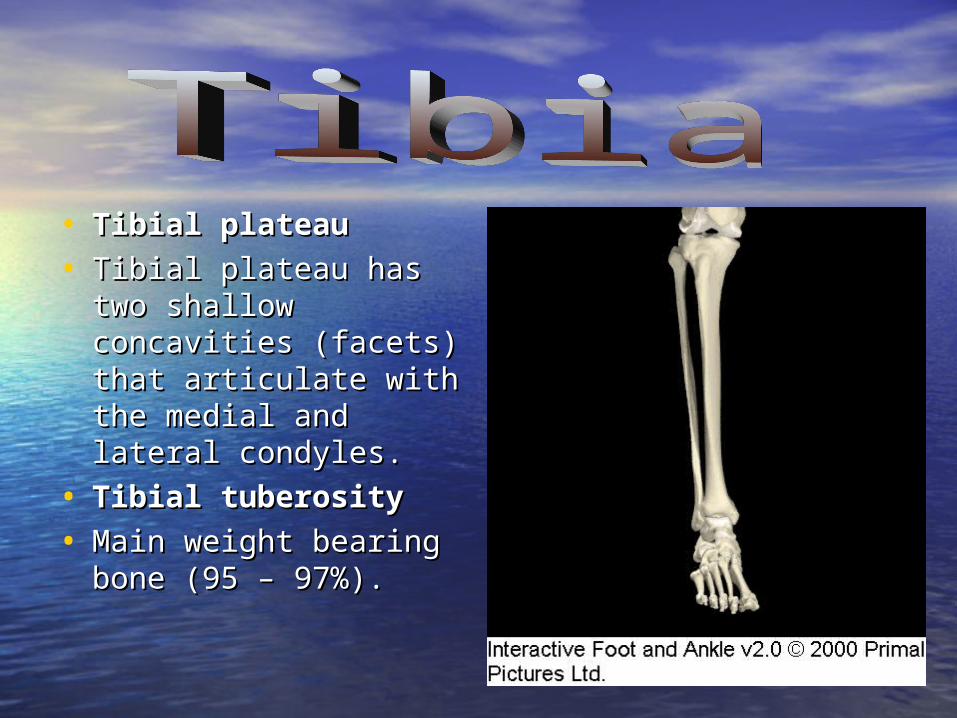
• Tibial plateauTibial plateau
• Tibial plateau has two Tibial plateau has two shallow concavities shallow concavities (facets) that articulate (facets) that articulate with the medial and with the medial and lateral condyles.lateral condyles.
• Tibial tuberosityTibial tuberosity
• Main weight bearing Main weight bearing bone (95 – 97%).bone (95 – 97%).


• Largest sesamoid Largest sesamoid bone in the body.bone in the body.
• Articulates in the Articulates in the groove between groove between the femoral the femoral condyles.condyles.
• Provides a better Provides a better line of pull for the line of pull for the quadricep muscles. quadricep muscles.


• Lateral collateral Lateral collateral ligament and ligament and muscle muscle attachment.attachment.
• Non – Weight Non – Weight bearing bone.bearing bone.
• Located on lateral Located on lateral aspect.aspect.
• Minimal knee Minimal knee function.function.

• Femur and tibiaFemur and tibia
• Femur and patellaFemur and patella
• Tibia and fibulaTibia and fibula


• Attachment: femur – Attachment: femur – intersurface of intersurface of lateral condyle. lateral condyle.
• Attachment: Tibia – Attachment: Tibia – Anterior tibial Anterior tibial plateau.plateau.
• Prevents excessive Prevents excessive anterior movement anterior movement and internal rotation and internal rotation of the tibia.of the tibia.
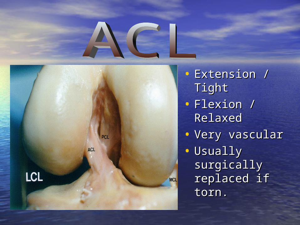
• Extension / Extension / TightTight
• Flexion / Flexion / RelaxedRelaxed
• Very vascularVery vascular
• Usually Usually surgically surgically replaced if replaced if torn.torn.

• Attachment: Femur -Attachment: Femur -Anterior portion of the Anterior portion of the lateral surface of the lateral surface of the medial condyle.medial condyle.
• Tibia – Posterior medial Tibia – Posterior medial tibial plateau.tibial plateau.
• Prevents excessive Prevents excessive posterior movement of posterior movement of the tibia on the femur.the tibia on the femur.
• Prevents Prevents hyperextension of the hyperextension of the kneeknee

• Attachment: Femur – Attachment: Femur – superior epicondyle.superior epicondyle.
• Tibia – Medial aspect / Tibia – Medial aspect / medial meniscus.medial meniscus.
• Resists valgus forces.Resists valgus forces.
• Resists external rotation of Resists external rotation of the tibia.the tibia.

• Attachment: Femur – Attachment: Femur – lateral epicondyle.lateral epicondyle.
• Fibula: Fibular headFibula: Fibular head• ExtracapsularExtracapsular• Resists varus forces.Resists varus forces.
• Aided by popliteal Aided by popliteal muscle / IT-bandmuscle / IT-band

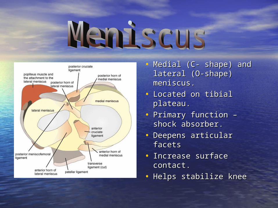
• Medial (C- shape) and Medial (C- shape) and lateral (O-shape) lateral (O-shape) meniscus.meniscus.
• Located on tibial Located on tibial plateau.plateau.
• Primary function – shock Primary function – shock absorber.absorber.
• Deepens articular facetsDeepens articular facets
• Increase surface Increase surface contact.contact.
• Helps stabilize kneeHelps stabilize knee


• 33 vascularvascular zoneszones– Red – RedRed – Red Zone- Zone-
outer 1/3 which has outer 1/3 which has good blood supply.good blood supply.
– RedRed – White – White Zone- Zone- middle 1/3 which middle 1/3 which has minimal blood has minimal blood supply.supply.
– White – WhiteWhite – White Zone- Zone- inner 1/3 which is inner 1/3 which is avascular (no blood avascular (no blood supply)supply)

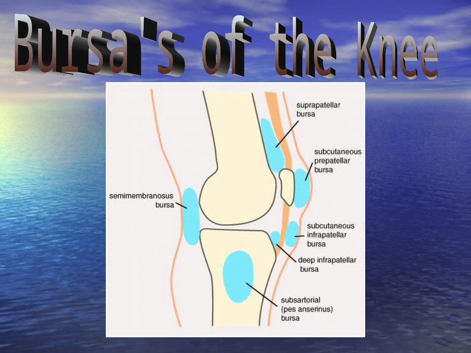
• Bursa’s are fluid filled Bursa’s are fluid filled sac’s .sac’s .
• Reduce friction Reduce friction between anatomical between anatomical structures.structures.
• Two dozen bursa’s Two dozen bursa’s within the knee.within the knee.
• Prepatellar most often Prepatellar most often injured. injured.


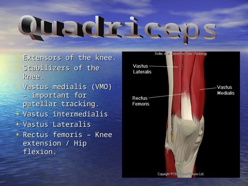
• Extensors of the knee.Extensors of the knee.
• Stabilizers of the knee.Stabilizers of the knee.
• Vastus medialis (VMO) Vastus medialis (VMO) – important for patellar – important for patellar tracking.tracking.
• Vastus intermedialisVastus intermedialis
• Vastus LateralisVastus Lateralis
• Rectus femoris – Knee Rectus femoris – Knee extension / Hip flexion.extension / Hip flexion.

• Patellar Tendon – Patellar Tendon – Common tendon Common tendon for the quadriceps for the quadriceps muscle group.muscle group.
• Attachment: Tibial Attachment: Tibial tuberosity.tuberosity.
• Houses the Houses the patella.patella.


• Flexors of the Flexors of the knee.knee.
• Extensors of the Extensors of the hip.hip.
• Prevents anterior Prevents anterior tibial movement / tibial movement / Aids ACL.Aids ACL.

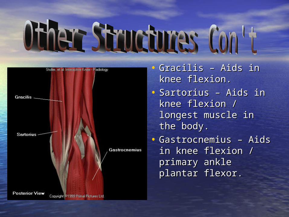
• Gracilis – Aids in Gracilis – Aids in knee flexion.knee flexion.
• Sartorius – Aids in Sartorius – Aids in knee flexion / knee flexion / longest muscle in longest muscle in the body.the body.
• Gastrocnemius – Gastrocnemius – Aids in knee flexion / Aids in knee flexion / primary ankle primary ankle plantar flexor.plantar flexor.

• Lateral side of Lateral side of knee.knee.
• Assist LCL in the Assist LCL in the lateral stability of lateral stability of the knee.the knee.


• Mechanism – Mechanism – Compression and Compression and rotation femur / rotation femur / tibia.tibia.
• Can be associated Can be associated with ligament with ligament injury (MCL).injury (MCL).
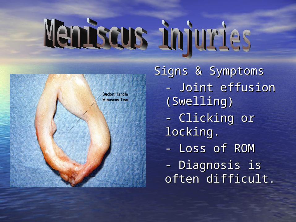
Signs & SymptomsSigns & Symptoms
- Joint effusion - Joint effusion (Swelling)(Swelling)
- Clicking or - Clicking or locking.locking.
- Loss of ROM- Loss of ROM
- Diagnosis is often - Diagnosis is often difficult.difficult.

Meniscus MRIMeniscus MRI

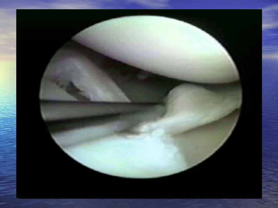
Meniscus TearMeniscus Tear
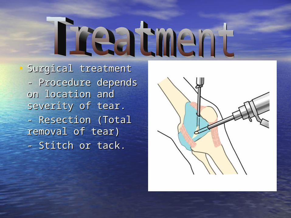
• Surgical treatmentSurgical treatment
- Procedure - Procedure depends on depends on location and location and severity of tear.severity of tear.
- Resection (Total - Resection (Total removal of tear)removal of tear)
- Stitch or tack.- Stitch or tack.


- Indicated only for - Indicated only for minor tears.minor tears.
- RICE (Control - RICE (Control Swelling)Swelling)
- Maintain ROM / - Maintain ROM / StrengthStrength
- Non surgical - Non surgical treatment can treatment can result in further result in further damagedamage

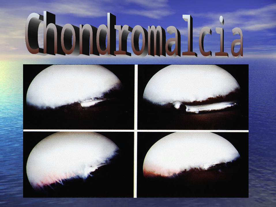
ArthriisArthriis

• Common in sports.Common in sports.
• Mechanism – External Mechanism – External rotation of tibia, knee rotation of tibia, knee in valgus position, foot in valgus position, foot fixed.fixed.
• Signs & SymptomsSigns & Symptoms– Feeling popFeeling pop– Knee feels unstableKnee feels unstable– Joint effusionJoint effusion– Positive testingPositive testing

• RICE (Control RICE (Control Swelling)Swelling)
• Immobilizer / Immobilizer / CrutchesCrutches
• Refer to physicianRefer to physician
• Rehabilitation Rehabilitation (Strengthening & (Strengthening & ROM)ROM)

MRI of ACLMRI of ACL
NORMAL ACLNORMAL ACL ACL TEARACL TEAR

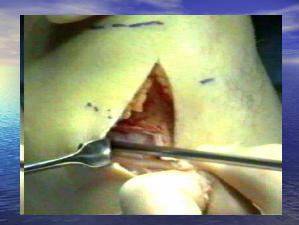
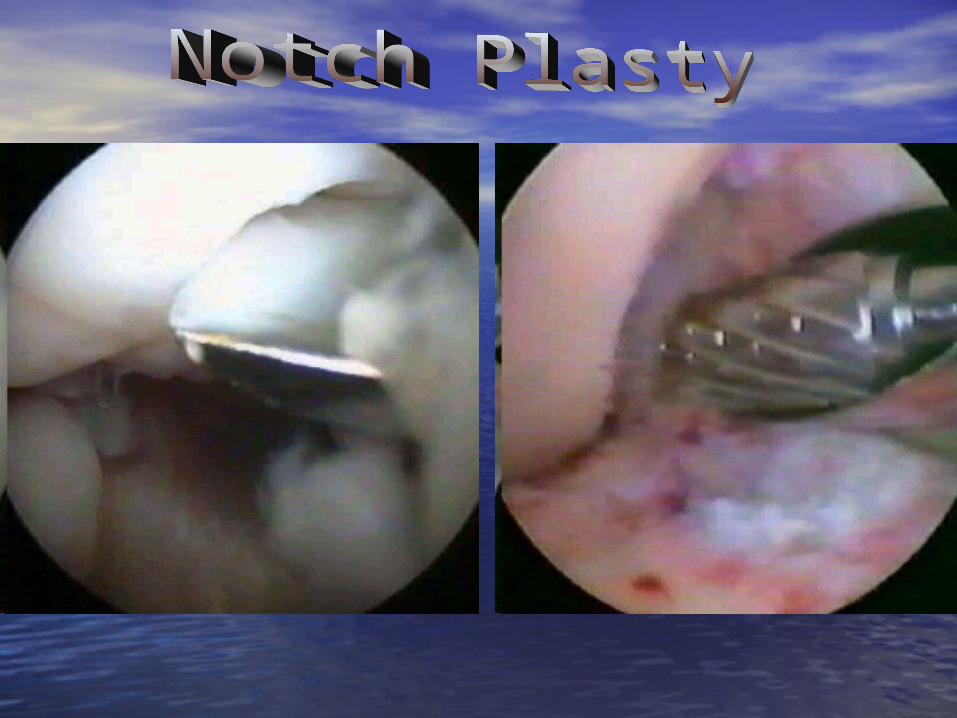
• Arthroscopic Arthroscopic surgerysurgery
• Various grafts can Various grafts can be used for be used for repair.repair.– Patellar tendon Patellar tendon
(Autograft)(Autograft)– Hamstring Hamstring
(semitendinosus / (semitendinosus / Gracillis)Gracillis)
– Cadaver Cadaver (Allograft)(Allograft)


Patellar Tendon (autograft)


• Injured less Injured less frequentfrequent
• Mechanism:Mechanism:
- Fall on anterior - Fall on anterior aspect of bent aspect of bent knee with foot knee with foot plantar flexed.plantar flexed.
- Hyperextension- Hyperextension


• Signs / SymptomsSigns / Symptoms
- Experience “Pop”- Experience “Pop”
- Effusion (swelling)- Effusion (swelling)
- Tenderness - Tenderness posterior aspect of posterior aspect of kneeknee
- Knee feels unstable- Knee feels unstable
- Positive special test- Positive special test

MCL / LCL InjuriesMCL / LCL Injuries
• MCL injuries are usually caused by a MCL injuries are usually caused by a lateral to medial blow to the knee. lateral to medial blow to the knee. Also known as a valgus force. Also known as a valgus force.
• LCL injuries are usually caused by LCL injuries are usually caused by medial to lateral blow to the knee. medial to lateral blow to the knee. Also known as a varus force.Also known as a varus force.

• Mechanism of InjuryMechanism of Injury
- direct blow from - direct blow from the lateral side the lateral side (Valgus Stress)(Valgus Stress)
- severe rotation of - severe rotation of the tibiathe tibia
- can be a - can be a combination of bothcombination of both

MRI OF MCL TEARMRI OF MCL TEAR

• 11stst degree degree- ligamentous fibers are - ligamentous fibers are stretchedstretched- joint is stable during - joint is stable during valgus stress testvalgus stress test- little or no joint effusion- little or no joint effusion- may be some joint - may be some joint stiffness and medial joint stiffness and medial joint line tendernessline tenderness- almost full range of - almost full range of motionmotion

• 22ndnd degree sprain degree sprain
- partial tear of the - partial tear of the ligamentligament
- slight to moderate - slight to moderate laxity during valgus laxity during valgus stress teststress test
- there is little joint - there is little joint effusioneffusion
- moderate to severe - moderate to severe joint stiffness with loss joint stiffness with loss of ROMof ROM

• 33rdrd degree sprain degree sprain
- Complete tear - Complete tear
- severe laxity - severe laxity revealed with revealed with valgus stress testvalgus stress test
- moderate joint - moderate joint effusioneffusion
- loss of ROM- loss of ROM

• 11stst degree – RICE, degree – RICE, Rehab to increase Rehab to increase strength, ROM.strength, ROM.
• 22ndnd degree – RICE, degree – RICE, Immobilize, Crutch, Immobilize, Crutch, 24 hours. Re-24 hours. Re-evaluate. Refer to evaluate. Refer to physicianphysician
• 33rdrd degree – RICE, degree – RICE, Immobilize, Crutch. Immobilize, Crutch. Refer to physicianRefer to physician

• Not very common Not very common in athletics.in athletics.
• Occurs by a medial Occurs by a medial blow to knee which blow to knee which produces a varus produces a varus stressstress

Patellar DislocationPatellar Dislocation
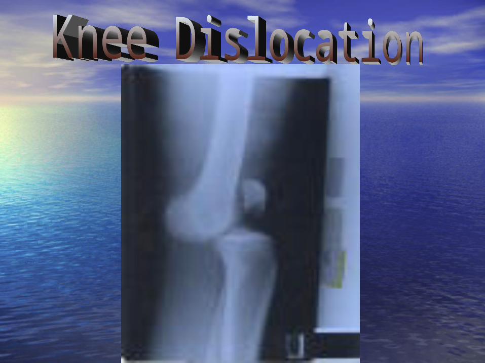

Knee DislocationKnee Dislocation

Assessing Assessing the Knee the Knee JointJoint

HISTORYHISTORY
– What did you feel, hear, …. Was there a What did you feel, hear, …. Was there a pop or snap?pop or snap?
– Did you get hit by another player? Was Did you get hit by another player? Was your foot planted? Did this happen your foot planted? Did this happen without being hit?without being hit?
– Exactly where does you knee hurt, and Exactly where does you knee hurt, and be specific?be specific?
– Have you hurt this knee before, when, Have you hurt this knee before, when, what was the injury?what was the injury?

HISTORYHISTORY
• When did you first notice the condition?When did you first notice the condition?
• Is there swelling or recurrent swelling?Is there swelling or recurrent swelling?
• What activity hurts the most? What activity hurts the most?
• Does it ever catch or lock?Does it ever catch or lock?
• Do you fell as if the knee is going to Do you fell as if the knee is going to give way, or has it already done so?give way, or has it already done so?
• Does it hurt to go up and down stairs?Does it hurt to go up and down stairs?

ObservationObservation
• Does the athlete have a limp, or is it Does the athlete have a limp, or is it easy to walk?easy to walk?
• Cant the athlete be full weight Cant the athlete be full weight bearing?bearing?
• Is the athlete able to perform a half-Is the athlete able to perform a half-squat to extension?squat to extension?
• Cant the athlete do up and down Cant the athlete do up and down stairs?stairs?

Testing for Knee Joint Testing for Knee Joint InstabilityInstability• Testing helps one get a better idea of the Testing helps one get a better idea of the
stability of the joint and an informed stability of the joint and an informed decision can be made about playing decision can be made about playing status. status.
• Many tests may point to ligamentous Many tests may point to ligamentous damage, while others will help detect damage, while others will help detect meniscus damage.meniscus damage.
• Knowing these test and how to perform Knowing these test and how to perform them takes practice and time to them takes practice and time to understand the degrees of damage done understand the degrees of damage done to the knee.to the knee.
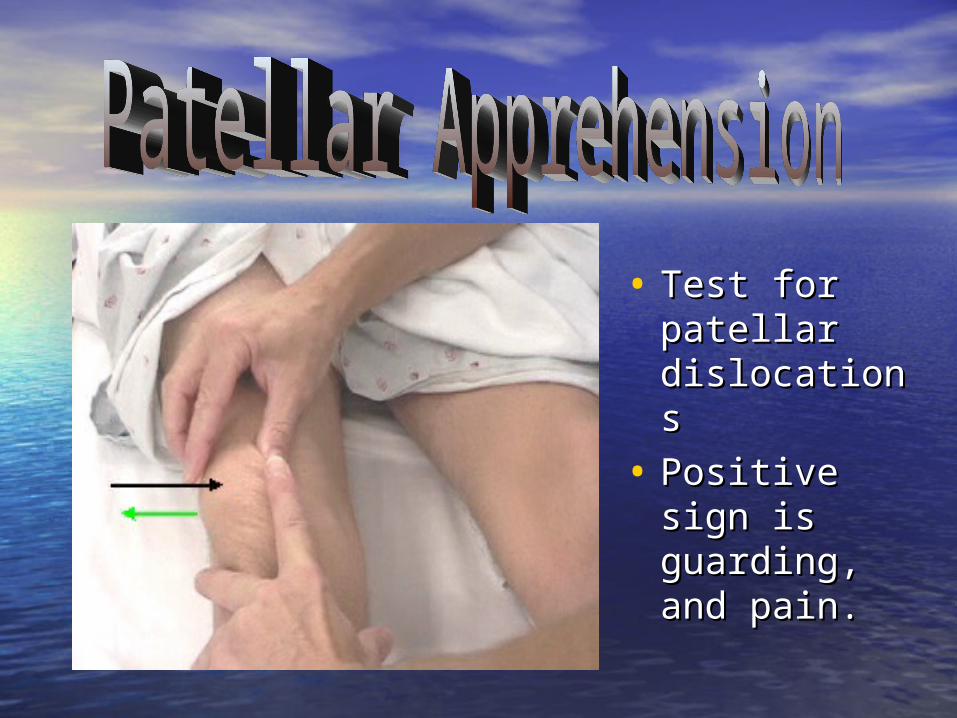
• Test for Test for patellar patellar dislocationsdislocations
• Positive sign Positive sign is guarding, is guarding, and pain.and pain.

• Tests for torn meniscusTests for torn meniscus
• Positive sign is popping, Positive sign is popping, clicking or pain. clicking or pain.
• Apley’s Apley’s Compression/DistractionCompression/Distraction
- Tests for torn meniscus- Tests for torn meniscus
- Compression signs - Compression signs same as McMurraysame as McMurray
- Distraction sign is - Distraction sign is reduction of pain.reduction of pain.

Valgus and Varus Stress Valgus and Varus Stress TestsTests
• Purpose: intended to reveal laxity of the Purpose: intended to reveal laxity of the medial and lateral collaterals. medial and lateral collaterals.
• The athlete lie supine with the leg The athlete lie supine with the leg extended.extended.
• Apply stress to the knee either medially Apply stress to the knee either medially or laterallyor laterally

Valgus Stress TestsValgus Stress Tests
• The valgus examination in full The valgus examination in full extension tests the MCL, extension tests the MCL, posteromedial capsule, and the posteromedial capsule, and the cruciates. The exam at 30 degrees cruciates. The exam at 30 degrees flexion isolates the MCL.flexion isolates the MCL.

• Tests for MCL Tests for MCL stabilitystability
• Positive sign Positive sign indicates an injury indicates an injury to the MCLto the MCL
• Test at 0 and 15 Test at 0 and 15 degreesdegrees


Varus Stress TestsVarus Stress Tests
• The examiner reverses hand The examiner reverses hand positions and tests the lateral side positions and tests the lateral side with a varus force on the fully with a varus force on the fully extended knee and then with 30 extended knee and then with 30 degrees of flexion. With the knee degrees of flexion. With the knee extended, the LCL and posterolateral extended, the LCL and posterolateral capsule are examined. At 30 degrees capsule are examined. At 30 degrees of flexion, the LCL is isolated.of flexion, the LCL is isolated.

• Tests for LCL Tests for LCL stabilitystability
• Positive sign Positive sign indicates an injury indicates an injury to the LCLto the LCL
• Test at 0 and 15 Test at 0 and 15 degreesdegrees


Anterior Cruciate Ligament Anterior Cruciate Ligament TestsTests
• Anterior Drawer TestAnterior Drawer Test
• Lachman’s TestLachman’s Test
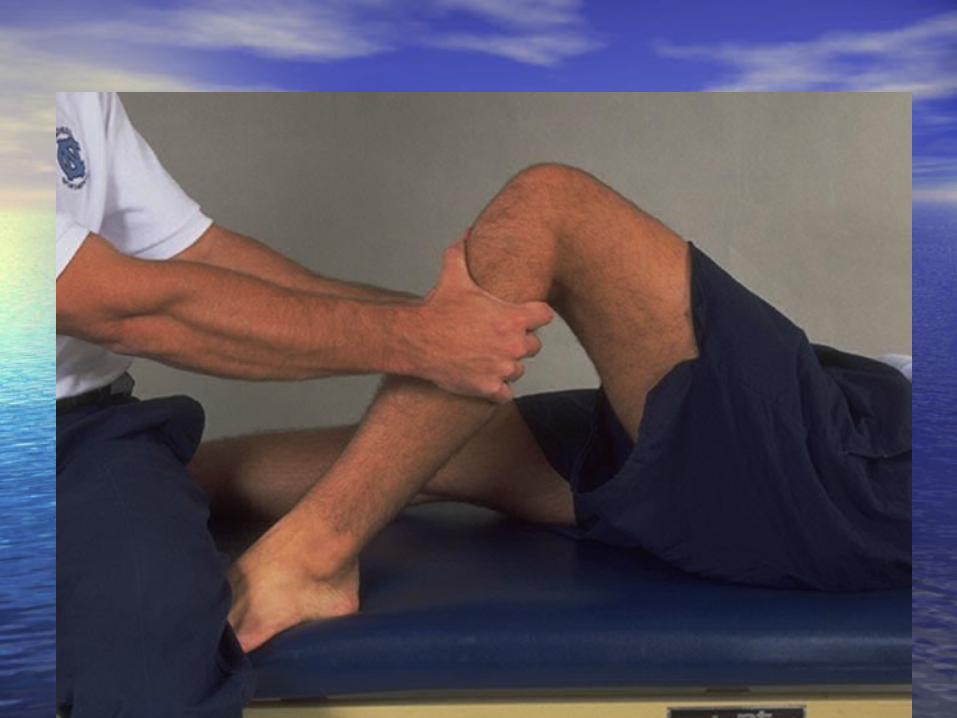
Anterior Drawer TestAnterior Drawer Test
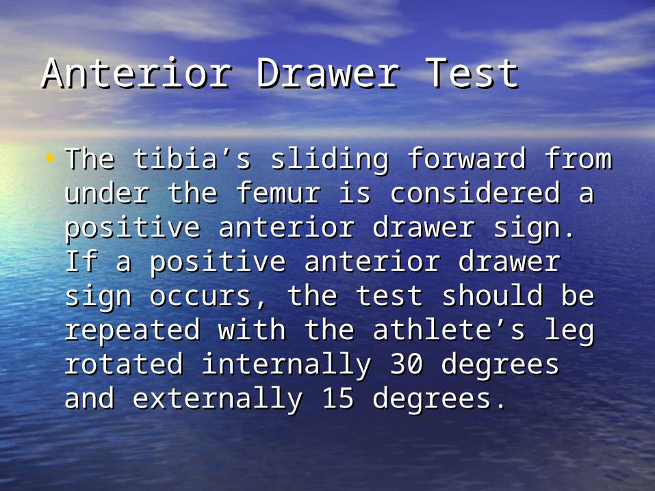
• The tibia’s sliding forward from under The tibia’s sliding forward from under the femur is considered a positive the femur is considered a positive anterior drawer sign. If a positive anterior drawer sign. If a positive anterior drawer sign occurs, the test anterior drawer sign occurs, the test should be repeated with the athlete’s should be repeated with the athlete’s leg rotated internally 30 degrees and leg rotated internally 30 degrees and externally 15 degrees. externally 15 degrees.

• Anterior Drawer Anterior Drawer TestTest
- Tests for ACL - Tests for ACL instability / laxityinstability / laxity
• Positive sign is Positive sign is increase in anterior increase in anterior translation. (No translation. (No end point)end point)


Anterior Cruciate Ligament Anterior Cruciate Ligament TestsTests
• Lachman’s TestLachman’s Test: is considered to be a : is considered to be a better test than the drawer test at 90 better test than the drawer test at 90 degrees of flexion. degrees of flexion.

Lachman’s TestsLachman’s Tests
• The Lachman’s test is administered by The Lachman’s test is administered by positioning the knee in approximately 30 positioning the knee in approximately 30 degrees of flexion. One hand of the degrees of flexion. One hand of the examiner stabilizes the leg by grasping examiner stabilizes the leg by grasping the distal end of the thigh, and the other the distal end of the thigh, and the other hand grasps the proximal aspect of the hand grasps the proximal aspect of the tibia and attempts to move it anteriorly. tibia and attempts to move it anteriorly. A positive Lachman’s test indicated A positive Lachman’s test indicated damage to the ACLdamage to the ACL

• Lachman TestLachman Test
- Test for ACL - Test for ACL instbility/laxityinstbility/laxity
• Positive sign is Positive sign is increased increased anterior tibial anterior tibial translation. translation. (No end point)(No end point)


Posterior Cruciate Ligament Posterior Cruciate Ligament TestsTests
• Posterior Drawer TestPosterior Drawer Test: is : is performed with the knee flexed at 90 performed with the knee flexed at 90 degrees and the foot in neutral degrees and the foot in neutral position. Force is exerted in a position. Force is exerted in a posterior direction at the proximal posterior direction at the proximal tibial plateau. A positive posterior tibial plateau. A positive posterior drawer test indicates damage to the drawer test indicates damage to the posterior cruciate ligament.posterior cruciate ligament.

• Posterior Posterior DrawerDrawer
- Test for PCL - Test for PCL instability/laxityinstability/laxity
• Positive sign is Positive sign is posterior posterior movement of movement of the tibia.the tibia.


Posterior Cruciate Ligament Posterior Cruciate Ligament TestsTests
• Posterior Sag TestPosterior Sag Test: With the : With the athlete supine, both knees are flexed athlete supine, both knees are flexed to 9- degrees. Observing laterally on to 9- degrees. Observing laterally on the injured side, the tibia will appear the injured side, the tibia will appear to sag posteriorly when compared to to sag posteriorly when compared to the opposite extremity if the the opposite extremity if the posterior cruciate ligament is posterior cruciate ligament is damaged.damaged.


Prevention of Knee InjuriesPrevention of Knee Injuries
• To avoid injuries to the knee, the To avoid injuries to the knee, the athlete must be as highly conditioned athlete must be as highly conditioned as possible, which means total body as possible, which means total body conditioning that includes strength, conditioning that includes strength, flexibility, cardiovascular and muscle flexibility, cardiovascular and muscle endurance, agility, speed and balance.endurance, agility, speed and balance.
• THE MUSCLES around the knee MUST THE MUSCLES around the knee MUST be strong and flexible. be strong and flexible.

Prevention of Knee InjuriesPrevention of Knee Injuries
• Athletes participating in a particular sport Athletes participating in a particular sport should acquire a strength ratio between the should acquire a strength ratio between the quadriceps and hamstring muscle groups. quadriceps and hamstring muscle groups. Fro example: the hamstring muscles of Fro example: the hamstring muscles of football players should have 60 to 70 football players should have 60 to 70 percent of the strength of the quadriceps percent of the strength of the quadriceps muscles. The gastrocnemius muscle should muscles. The gastrocnemius muscle should also be strengthened to help stabilize the also be strengthened to help stabilize the knee. Although maximizing muscle strength knee. Although maximizing muscle strength may prevent some injuries, it fails to may prevent some injuries, it fails to prevent rotary-type injuries.prevent rotary-type injuries.
Prevention of Knee InjuriesPrevention of Knee Injuries
• Shoe Type: Shoe Type: – Cleat LengthCleat Length– Astro Turf shoes: more grip=more Astro Turf shoes: more grip=more
injuriesinjuries– Sneakers are good for artificial surfacesSneakers are good for artificial surfaces

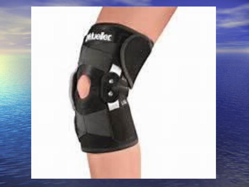
Functional and Prophylactic Functional and Prophylactic Knee BracesKnee Braces
• Functional Knee Braces are used to Functional Knee Braces are used to protect grade 1 and 2 sprains of the ACL protect grade 1 and 2 sprains of the ACL and MCL, and reconstructed ACL knees. and MCL, and reconstructed ACL knees. Most of them are bilateral knee braces, Most of them are bilateral knee braces, meaning there is a hinge on both sides meaning there is a hinge on both sides of the brace. These braces have an of the brace. These braces have an important part within the athletic important part within the athletic community. They will also give the community. They will also give the athlete confidence while playing.athlete confidence while playing.


Functional and Prophylactic Functional and Prophylactic Knee BracesKnee Braces• Prophylactic Knee Braces are Prophylactic Knee Braces are
designed to prevent or reduce the designed to prevent or reduce the severity of knee injuries. They are severity of knee injuries. They are worn on the lateral surface of the worn on the lateral surface of the knee to protect the medial collateral knee to protect the medial collateral ligament.ligament.

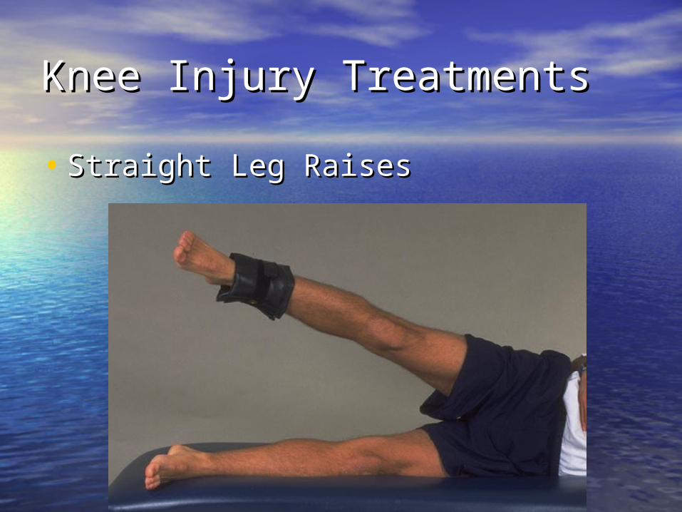
Knee Injury TreatmentsKnee Injury Treatments
• Straight Leg RaisesStraight Leg Raises

Knee Injury TreatmentsKnee Injury Treatments
• Side Leg RaisesSide Leg Raises

Knee Injury TreatmentsKnee Injury Treatments
• Side Leg RaisesSide Leg Raises

Knee Injury TreatmentsKnee Injury Treatments
• Terminal Knee ExtensionsTerminal Knee Extensions

Knee Injury TreatmentsKnee Injury Treatments• Step UpsStep Ups

BOSUBOSU