Embed Size (px)
Citation preview
Chapter 5UNIT III
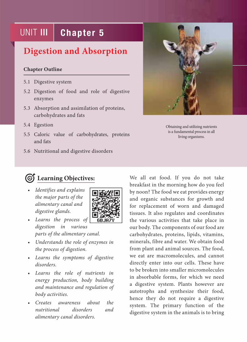
Obtaining and utilizing nutrients
is a fundamental process in all
living organisms.
Digestion and Absorption
Chapter Outline
5.1 Digestive system
5.2 Digestion of food and role of digestive
enzymes
5.3 Absorption and assimilation of proteins,
carbohydrates and fats
5.4 Egestion
5.5 Caloric value of carbohydrates, proteins
and fats
5.6 Nutritional and digestive disorders
• Identifies and explains
the major parts of the
alimentary canal and
digestive glands.
• Learns the process of
digestion in various
parts of the alimentary canal.
• Understands the role of enzymes in
the process of digestion.
• Learns the symptoms of digestive
disorders.
• Learns the role of nutrients in
energy production, body building
and maintenance and regulation of
body activities.
• Creates awareness about the
nutritional disorders and
alimentary canal disorders.
We all eat food. If you do not take
breakfast in the morning how do you feel
by noon? The food we eat provides energy
and organic substances for growth and
for replacement of worn and damaged
tissues. It also regulates and coordinates
the various activities that take place in
our body. The components of our food are
carbohydrates, proteins, lipids, vitamins,
minerals, fibre and water. We obtain food
from plant and animal sources. The food,
we eat are macromolecules, and cannot
directly enter into our cells. These have
to be broken into smaller micromolecules
in absorbable forms, for which we need
a digestive system. Plants however are
autotrophs and synthesize their food,
hence they do not require a digestive
system. The primary function of the
digestive system in the animals is to bring
Learning Objectives:
99
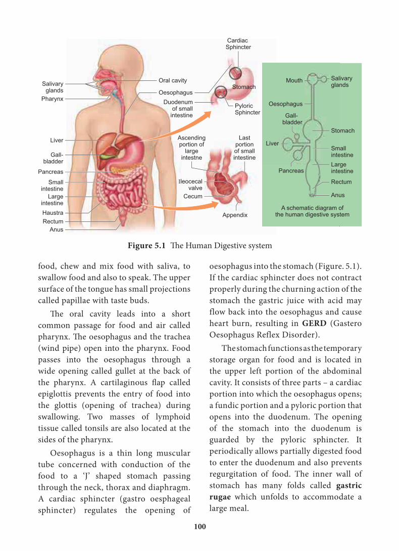
Mechanical digestion is initiated in the
buccal cavity by chewing with the help of
teeth and tongue. Chemical digestion is
through salivary enzymes secreted by the
salivary glands.
Each tooth is embedded in a socket
in the jaw bone; this type of attachment
is called thecodont. Human beings and
many mammals form two sets of teeth
during their life time, a set of 20 temporary
milk teeth (deciduous teeth) which gets
replaced by a set of 32 permanent teeth
(adult teeth). This type of dentition is
called diphyodont. The permanent teeth
are of four different types (heterodont),
namely, Incisors (I) chisel like cutting
teeth, Canines (C) dagger shaped tearing
teeth, Pre molars (PM) for grinding, and
Molars (M) for grinding and crushing.
Arrangement of teeth in each half of the
upper and lower jaw, in the order of I, C,
PM and M can be represented by a dental
formula, in human the dental formula is
2123/2123.
Mineral salts like calcium and
magnesium are deposited on the teeth and
form a hard layer of ‘tartar’ or calculus called plaque. If the plaque formed on
teeth is not removed regularly, it would
spread down the tooth into the narrow
gap between the gums and enamel and
causes inflammation, called gingivitis,
which leads to redness and bleeding of the
gums and to bad smell. The hard chewing
surface of the teeth is made of enamel and
helps in mastication of food.
Tongue is a freely movable muscular
organ attached at the posterior end by
the frenulum to the floor of the buccal
cavity and is free in the front. It acts as a
universal tooth brush and helps in intake
the nutrients, water and electrolytes from
the external environment into every
cell in the body through the circulatory
system.
Alimentary canal faces
a confl ict between
the need of nutrient
absorption and to keep
our intestinal tract free from pathogenic
bacteria and virus. About 7 litres of
digestive juice are poured into the
alimentary canal and are reabsorbed
each day. If this does not happen the
body gets rapidly dehydrated and may
lead to reduction in the blood pressure.
5.1. Digestive systemThe process of digestion involves intake
of the food (Ingestion), breakdown of the
food into micromolecules (Digestion),
absorption of these molecules into the
blood stream (Absorption), the absorbed
substances becoming components of
cells (Assimilation) and elimination of
the undigested substances (Egestion).
Digestive system includes the alimentary
canal and associated digestive glands.
5.1.1. Structure of the alimentary canalThe alimentary canal is a continuous,
muscular digestive tract that begins with
an anterior opening, the mouth and
opens out posteriorly through the anus.
The alimentary canal consists of mouth,
buccal cavity, pharynx, oesophagus,
stomach, intestine, rectum and anus
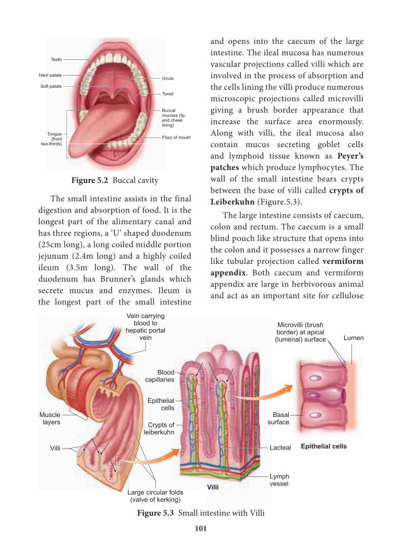
(Figure. 5.1). The mouth is concerned
with the reception of food and leads to the
buccal cavity or oral cavity (Figure. 5.2).
100
food, chew and mix food with saliva, to
swallow food and also to speak. The upper
surface of the tongue has small projections
called papillae with taste buds.
The oral cavity leads into a short
common passage for food and air called
pharynx. The oesophagus and the trachea
(wind pipe) open into the pharynx. Food
passes into the oesophagus through a
wide opening called gullet at the back of
the pharynx. A cartilaginous flap called
epiglottis prevents the entry of food into
the glottis (opening of trachea) during
swallowing. Two masses of lymphoid
tissue called tonsils are also located at the
sides of the pharynx.
Oesophagus is a thin long muscular
tube concerned with conduction of the
food to a ‘J’ shaped stomach passing
through the neck, thorax and diaphragm.
A cardiac sphincter (gastro oesphageal
sphincter) regulates the opening of
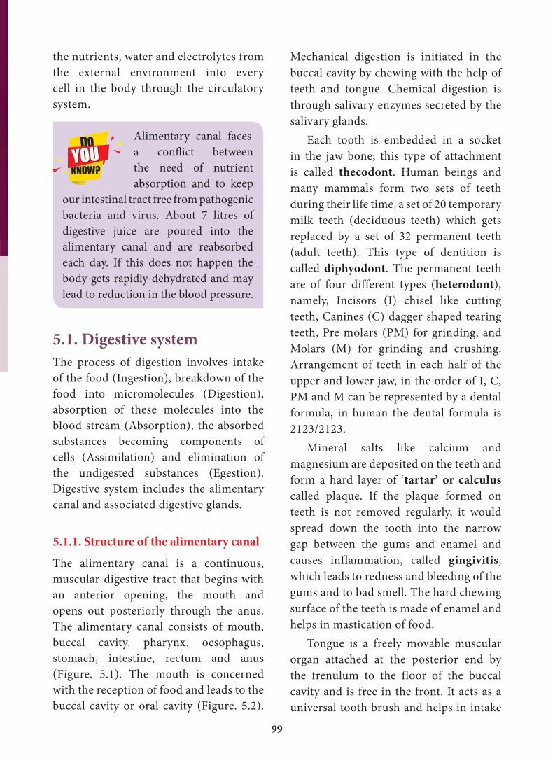
oesophagus into the stomach (Figure. 5.1).
If the cardiac sphincter does not contract
properly during the churning action of the
stomach the gastric juice with acid may
flow back into the oesophagus and cause
heart burn, resulting in GERD (Gastero
Oesophagus Reflex Disorder).
The stomach functions as the temporary
storage organ for food and is located in
the upper left portion of the abdominal
cavity. It consists of three parts – a cardiac
portion into which the oesophagus opens;
a fundic portion and a pyloric portion that
opens into the duodenum. The opening
of the stomach into the duodenum is
guarded by the pyloric sphincter. It
periodically allows partially digested food
to enter the duodenum and also prevents
regurgitation of food. The inner wall of
stomach has many folds called gastric rugae which unfolds to accommodate a
large meal.
�
Figure 5.1 The Human Digestive system
101
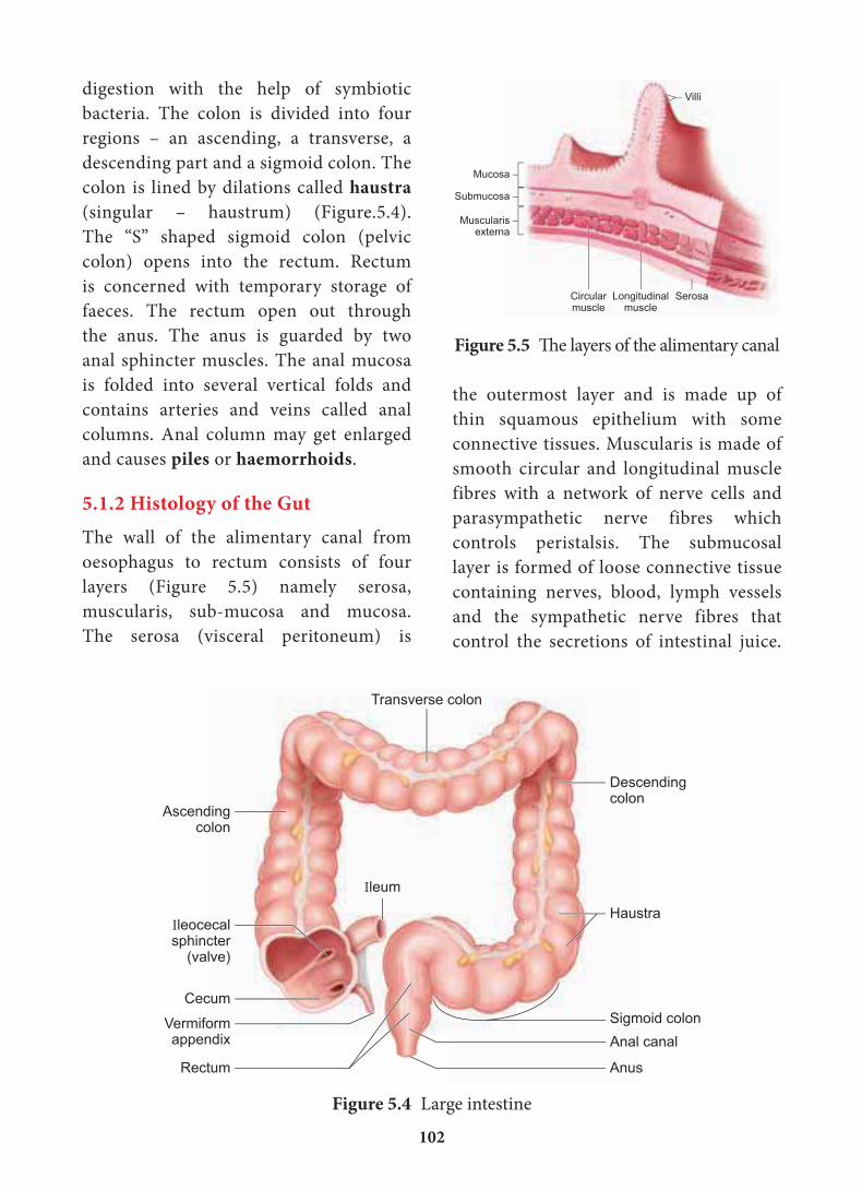
and opens into the caecum of the large
intestine. The ileal mucosa has numerous
vascular projections called villi which are
involved in the process of absorption and
the cells lining the villi produce numerous
microscopic projections called microvilli
giving a brush border appearance that
increase the surface area enormously.
Along with villi, the ileal mucosa also
contain mucus secreting goblet cells
and lymphoid tissue known as Peyer’s patches which produce lymphocytes. The
wall of the small intestine bears crypts
between the base of villi called crypts of Leiberkuhn ( Figure.5.3).
The large intestine consists of caecum,
colon and rectum. The caecum is a small
blind pouch like structure that opens into
the colon and it possesses a narrow finger
like tubular projection called vermiform appendix. Both caecum and vermiform
appendix are large in herbivorous animal
and act as an important site for cellulose
The small intestine assists in the final
digestion and absorption of food. It is the
longest part of the alimentary canal and
has three regions, a ‘U’ shaped duodenum
(25cm long), a long coiled middle portion
jejunum (2.4m long) and a highly coiled
ileum (3.5m long). The wall of the
duodenum has Brunner’s glands which
secrete mucus and enzymes. Ileum is
the longest part of the small intestine
Figure 5.2 Buccal cavity
Figure 5.3 Small intestine with Villi
102
the outermost layer and is made up of
thin squamous epithelium with some
connective tissues. Muscularis is made of
smooth circular and longitudinal muscle
fibres with a network of nerve cells and
parasympathetic nerve fibres which
controls peristalsis. The submucosal
layer is formed of loose connective tissue
containing nerves, blood, lymph vessels
and the sympathetic nerve fibres that
control the secretions of intestinal juice.
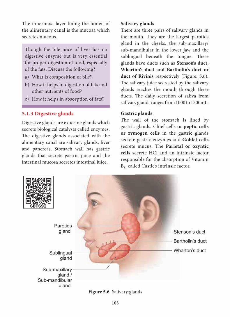
digestion with the help of symbiotic
bacteria. The colon is divided into four
regions – an ascending, a transverse, a
descending part and a sigmoid colon. The
colon is lined by dilations called haustra (singular – haustrum) (Figure.5.4).
The “S” shaped sigmoid colon (pelvic
colon) opens into the rectum. Rectum
is concerned with temporary storage of
faeces. The rectum open out through
the anus. The anus is guarded by two
anal sphincter muscles. The anal mucosa
is folded into several vertical folds and
contains arteries and veins called anal
columns. Anal column may get enlarged
and causes piles or haemorrhoids.
5.1.2 Histology of the GutThe wall of the alimentary canal from
oesophagus to rectum consists of four
layers (Figure 5.5) namely serosa,
muscularis, sub-mucosa and mucosa.
The serosa (visceral peritoneum) is
Ascendingcolon
Ileocecalsphincter
(valve)
Cecum
Vermiformappendix
Anus
Ileum
Anal canal
Rectum
Sigmoid colon
Haustra
Descendingcolon
Transverse colon
Figure 5.4 Large intestine
Figure 5.5 The layers of the alimentary canal
103
Salivary glandsThere are three pairs of salivary glands in
the mouth. They are the largest parotids
gland in the cheeks, the sub-maxillary/
sub-mandibular in the lower jaw and the
sublingual beneath the tongue. These
glands have ducts such as Stenson’s duct, Wharton’s duct and Bartholin’s duct or duct of Rivinis respectively (Figure. 5.6). The salivary juice secreated by the salivary
glands reaches the mouth through these
ducts. The daily secretion of saliva from
salivary glands ranges from 1000 to 1500mL.
Gastric glandsThe wall of the stomach is lined by
gastric glands. Chief cells or peptic cells or zymogen cells in the gastric glands
secrete gastric enzymes and Goblet cells
secrete mucus. The Parietal or oxyntic cells secrete HCl and an intrinsic factor
responsible for the absorption of Vitamin
B12 called Castle’s intrinsic factor.
The innermost layer lining the lumen of
the alimentary canal is the mucosa which
secretes mucous.
Figure 5.6 Salivary glands
Though the bile juice of liver has no
digestive enzyme but is very essential
for proper digestion of food, especially
of the fats. Discuss the following?
a) What is composition of bile?
b) How it helps in digestion of fats and
other nutrients of food?
c) How it helps in absorption of fats?
5.1.3 Digestive glandsDigestive glands are exocrine glands which
secrete biological catalysts called enzymes.
The digestive glands associated with the
alimentary canal are salivary glands, liver
and pancreas. Stomach wall has gastric
glands that secrete gastric juice and the
intestinal mucosa secretes intestinal juice.
104
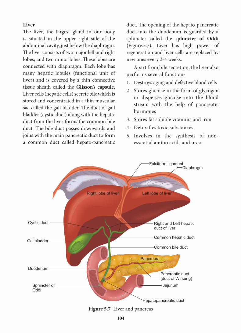
duct. Th e opening of the hepato-pancreatic
duct into the duodenum is guarded by a
sphincter called the sphincter of Oddi (Figure.5.7). Liver has high power of
regeneration and liver cells are replaced by
new ones every 3-4 weeks.
Apart from bile secretion, the liver also
performs several functions
1. Destroys aging and defective blood cells
2. Stores glucose in the form of glycogen
or disperses glucose into the blood
stream with the help of pancreatic
hormones
3. Stores fat soluble vitamins and iron
4. Detoxifies toxic substances.
5. Involves in the synthesis of non-
essential amino acids and urea.
LiverTh e liver, the largest gland in our body
is situated in the upper right side of the
abdominal cavity, just below the diaphragm.
Th e liver consists of two major left and right
lobes; and two minor lobes. Th ese lobes are
connected with diaphragm. Each lobe has
many hepatic lobules (functional unit of
liver) and is covered by a thin connective
tissue sheath called the Glisson’s capsule.
Liver cells (hepatic cells) secrete bile which is
stored and concentrated in a thin muscular
sac called the gall bladder. Th e duct of gall
bladder (cystic duct) along with the hepatic
duct from the liver forms the common bile
duct. Th e bile duct passes downwards and
joins with the main pancreatic duct to form
a common duct called hepato-pancreatic
Jejunum
Falciform ligament
Right lobe of liver Left lobe of liver
Diaphragm
Right and Left hepaticduct of liver
Common hepatic duct
Common bile duct
Pancreas
Pancreatic duct(duct of Wirsung)
Gallbladder
Cystic duct
Duodenum
Hepatopancreatic duct
Sphincter ofOddi
Figure 5.7 Liver and pancreas
105
antibacterial agent lysozyme and a lubricating
agent mucus (a glycoprotein). The mucus
in saliva prepares the food for swallowing
by moistening, softening, lubricating and
adhering the masticated food into a bolus.
About 30 percent of polysaccharide, starch
is hydrolyzed by the salivary amylase
enzyme into disaccharides (maltose). The
bolus is then passed into the pharynx and
then into the oesophagus by swallowing or
deglutition. The bolus further passes down
through the oesophagus to the stomach by
successive waves of muscular contraction
called peristalsis. The gastro oesphageal
sphincter controls the passage of food into
the stomach.
Digestion in the stomachFood remains in the stomach for 4 to 5
hours, the rhythmic peristaltic movement
churns and mixes the food with gastric
juice and make it into a creamy liquid
called chyme. The gastric secretion is
partly controlled by autonomic reflexes.
The secretion of gastric juice begins when
the food is in the mouth. The gastric
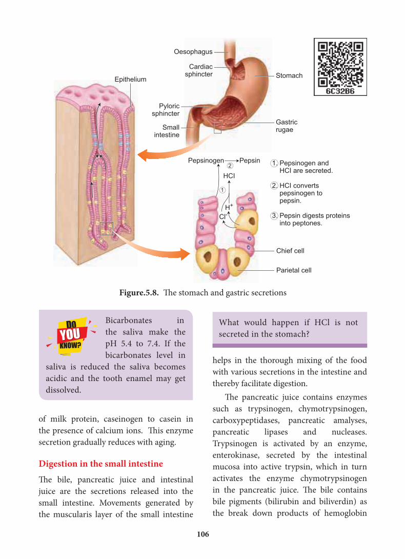
juice contains HCl and proenzymes. The
proenzyme pepsinogen, on exposure to
HCl gets converted into the active enzyme
pepsin which converts proteins into
proteoses and peptones (peptides). The HCl
provides an acidic medium (pH1.8) which is
optimum for pepsin, kills bacteria and other
harmful organisms and avoids putrifaction.
The mucus and bicarbonates present in
the gastric juice play an important role in
lubrication and protection of the mucosal
epithelium from the eroding nature of the
highly acidic HCl (Figure. 5.8). Another
proteolytic enzyme found in gastric juice
of infants is rennin helps in the digestion
PancreasThe second largest gland in the digestive
system is the Pancreas, which is a yellow
coloured, compound elongated organ
consisting of exocrine and endocrine cells.
It is situated between the limbs of the ‘U’
shaped duodenum. The exocrine portion
secretes pancreatic juice containing
enzymes such as pancreatic amylase,
trypsin and pancreatic lipase and the
endocrine part called Islets of Langerhans
secretes hormones such as insulin and
glucagon. The pancreatic duct directly
opens into the duodenum.
5.2 Digestion of food and role of digestive enzymesThe process of digestion converts the solid
food into absorbable and assimilable forms.
This is accomplished by mechanical and
chemical processes.
Digestion in the buccal cavityThe smell, sight and taste as well as the
mechanical stimulation of food in the
mouth, triggers a reflex action which results
in the secretion of saliva. The mechanical
digestion starts in the mouth by grinding
and chewing of food. It is called mastication.
The saliva contain water, electrolytes (Na+,
K+, Cl–, HCO3), salivary amylase (ptyalin),
Activity
List the chemical preservatives, artificial
enhancers found in the food items
available in the market. How can you
avoid such harmful substances in your
food?
106
of milk protein, caseinogen to casein in
the presence of calcium ions. This enzyme
secretion gradually reduces with aging.
Digestion in the small intestineThe bile, pancreatic juice and intestinal
juice are the secretions released into the
small intestine. Movements generated by
the muscularis layer of the small intestine
Oesophagus
Cardiacsphincter
Pyloricsphincter
Smallintestine
Gastricrugae
StomachEpithelium
Pepsinogen Pepsin
HCI
Cl-H+
1. Pepsinogen and HCI are secreted.
2. HCI converts pepsinogen to pepsin.
3. Pepsin digests proteins into peptones.
Chief cell
Parietal cell
1
2
Figure.5.8. The stomach and gastric secretions
Bicarbonates in
the saliva make the
pH 5.4 to 7.4. If the
bicarbonates level in
saliva is reduced the saliva becomes
acidic and the tooth enamel may get
dissolved.
What would happen if HCl is not
secreted in the stomach?
helps in the thorough mixing of the food
with various secretions in the intestine and
thereby facilitate digestion.
The pancreatic juice contains enzymes
such as trypsinogen, chymotrypsinogen,
carboxypeptidases, pancreatic amalyses,
pancreatic lipases and nucleases.
Trypsinogen is activated by an enzyme,
enterokinase, secreted by the intestinal
mucosa into active trypsin, which in turn
activates the enzyme chymotrypsinogen
in the pancreatic juice. The bile contains
bile pigments (bilirubin and biliverdin) as
the break down products of hemoglobin
107
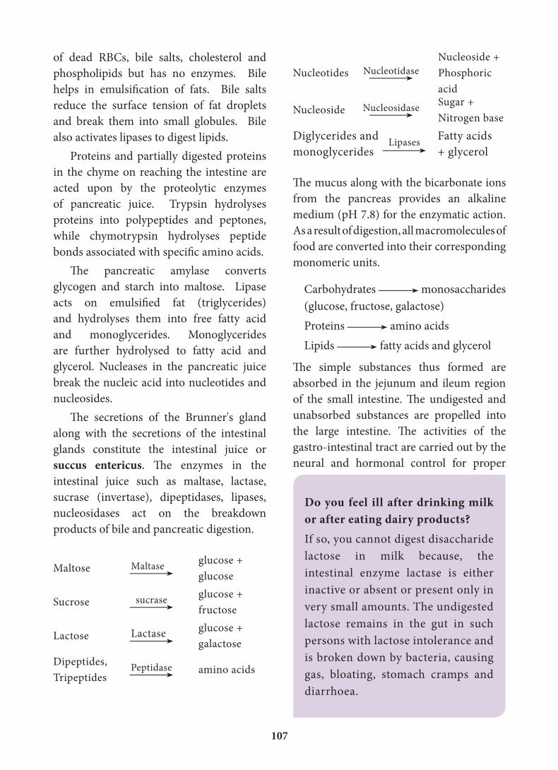
Nucleotides Nucleotidase
Nucleoside +
Phosphoric
acid
Nucleoside Nucleosidase Sugar +
Nitrogen base
Diglycerides and
monoglyceridesLipases Fatty acids
+ glycerol
The mucus along with the bicarbonate ions
from the pancreas provides an alkaline
medium (pH 7.8) for the enzymatic action.
As a result of digestion, all macromolecules of
food are converted into their corresponding
monomeric units.
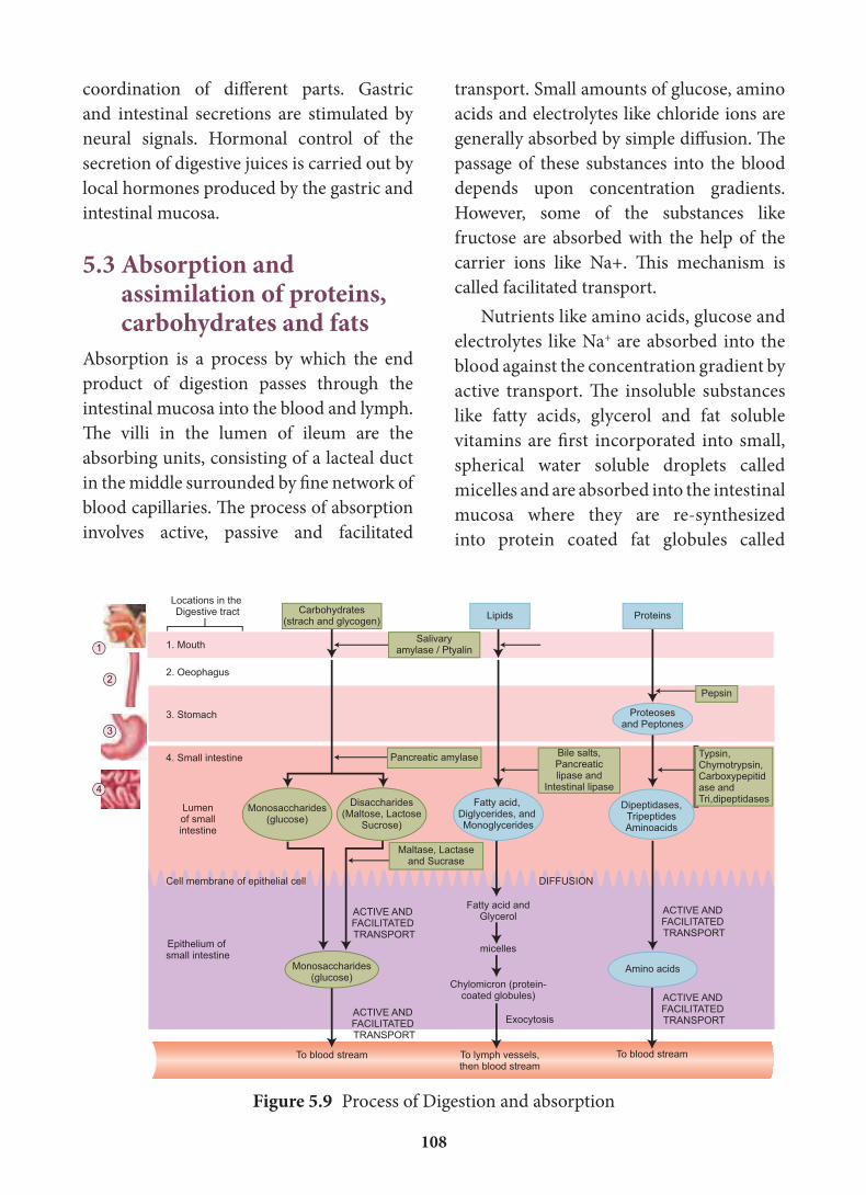
Carbohydrates monosaccharides
(glucose, fructose, galactose)
Proteins amino acids
Lipids fatty acids and glycerol
The simple substances thus formed are
absorbed in the jejunum and ileum region
of the small intestine. The undigested and
unabsorbed substances are propelled into
the large intestine. The activities of the
gastro-intestinal tract are carried out by the
neural and hormonal control for proper
of dead RBCs, bile salts, cholesterol and
phospholipids but has no enzymes. Bile
helps in emulsification of fats. Bile salts
reduce the surface tension of fat droplets
and break them into small globules. Bile
also activates lipases to digest lipids.
Proteins and partially digested proteins
in the chyme on reaching the intestine are
acted upon by the proteolytic enzymes
of pancreatic juice. Trypsin hydrolyses
proteins into polypeptides and peptones,
while chymotrypsin hydrolyses peptide
bonds associated with specific amino acids.
The pancreatic amylase converts
glycogen and starch into maltose. Lipase
acts on emulsified fat (triglycerides)
and hydrolyses them into free fatty acid
and monoglycerides. Monoglycerides
are further hydrolysed to fatty acid and
glycerol. Nucleases in the pancreatic juice
break the nucleic acid into nucleotides and
nucleosides.
The secretions of the Brunner's gland
along with the secretions of the intestinal
glands constitute the intestinal juice or
succus entericus. The enzymes in the
intestinal juice such as maltase, lactase,
sucrase (invertase), dipeptidases, lipases,
nucleosidases act on the breakdown
products of bile and pancreatic digestion.
Maltose Maltase glucose +
glucose
Sucrose sucrase glucose +
fructose
Lactose Lactase glucose +
galactose
Dipeptides,
Tripeptides Peptidase
amino acids
Do you feel ill after drinking milk or after eating dairy products?If so, you cannot digest disaccharide
lactose in milk because, the
intestinal enzyme lactase is either
inactive or absent or present only in
very small amounts. The undigested
lactose remains in the gut in such
persons with lactose intolerance and
is broken down by bacteria, causing
gas, bloating, stomach cramps and
diarrhoea.
108
coordination of different parts. Gastric
and intestinal secretions are stimulated by
neural signals. Hormonal control of the
secretion of digestive juices is carried out by
local hormones produced by the gastric and
intestinal mucosa.
5.3 Absorption and assimilation of proteins, carbohydrates and fats
Absorption is a process by which the end
product of digestion passes through the
intestinal mucosa into the blood and lymph.
The villi in the lumen of ileum are the
absorbing units, consisting of a lacteal duct
in the middle surrounded by fine network of
blood capillaries. The process of absorption
involves active, passive and facilitated
Figure 5.9 Process of Digestion and absorption
transport. Small amounts of glucose, amino
acids and electrolytes like chloride ions are
generally absorbed by simple diffusion. The
passage of these substances into the blood
depends upon concentration gradients.
However, some of the substances like
fructose are absorbed with the help of the
carrier ions like Na+. This mechanism is
called facilitated transport.
Nutrients like amino acids, glucose and
electrolytes like Na+ are absorbed into the
blood against the concentration gradient by
active transport. The insoluble substances
like fatty acids, glycerol and fat soluble
vitamins are first incorporated into small,
spherical water soluble droplets called
micelles and are absorbed into the intestinal
mucosa where they are re-synthesized
into protein coated fat globules called
109
Do you know?
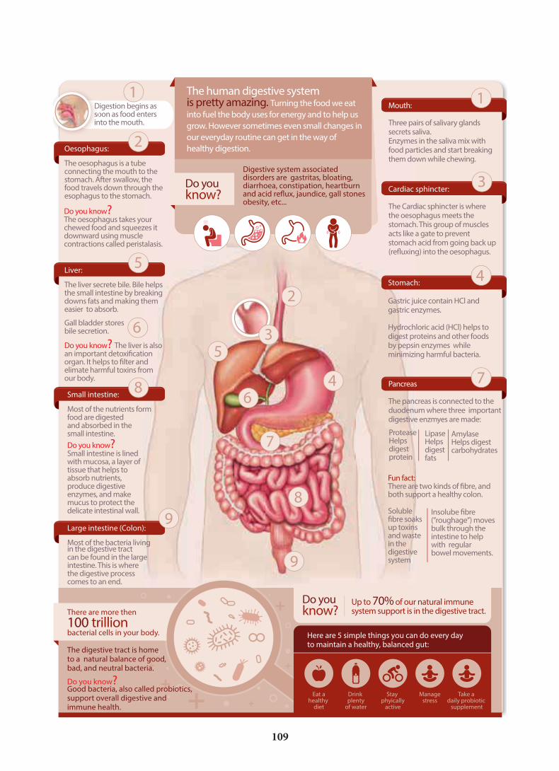
Here are 5 simple things you can do every dayto maintain a healthy, balanced gut:
Drink plenty
of water
Stay phyically
active
Managestress
Take a daily probiotic
supplement
Do you know? The oesophagus takes your chewed food and squeezes it downward using muscle contractions called peristalasis.
Do you know? The liver is also an important detoxification organ. It helps to filter and elimate harmful toxins fromour body.
Do you know? Small intestine is linedwith mucosa, a layer oftissue that helps toabsorb nutrients,produce digestiveenzymes, and makemucus to protect thedelicate intestinal wall.
Fun fact:There are two kinds of fibre, and both support a healthy colon.
Soluble fibre soaks up toxins and waste in the digestive system
Insolube fibre(”roughage”) movesbulk through theintestine to helpwith regularbowel movements.
Gall bladder stores bile secretion.
ProteaseHelps digestprotein
LipaseHelps digestfats
AmylaseHelps digestcarbohydrates
Digestive system associated disorders are gastritas, bloating, diarrhoea, constipation, heartburn and acid reflux, jaundice, gall stones obesity, etc...
Oesophagus:
The oesophagus is a tube connecting the mouth to the stomach. After swallow, the food travels down through the esophagus to the stomach.
Liver:
The liver secrete bile. Bile helps the small intestine by breaking downs fats and making them easier to absorb.
Small intestine:
Most of the nutrients form food are digestedand absorbed in the small intestine.
Large intestine (Colon):
Most of the bacteria livingin the digestive tractcan be found in the largeintestine. This is wherethe digestive processcomes to an end.
Mouth:
Three pairs of salivary glands secrets saliva.Enzymes in the saliva mix with food particles and start breaking them down while chewing.
Cardiac sphincter:
The Cardiac sphincter is where the oesophagus meets the stomach. This group of muscles acts like a gate to prevent stomach acid from going back up (refluxing) into the oesophagus.
Stomach:
Gastric juice contain HCl and gastric enzymes.
Hydrochloric acid (HCl) helps to digest proteins and other foods by pepsin enzymes while minimizing harmful bacteria.
Pancreas
The pancreas is connected to theduodenum where three importantdigestive enzmyes are made:
Up to 70% of our natural immunesystem support is in the digestive tract.
Eat ahealthy
diet
Do you know?
The human digestive system is pretty amazing. Turning the food we eat
into fuel the body uses for energy and to help us
grow. However sometimes even small changes in
our everyday routine can get in the way of
healthy digestion.
There are more then
100 trillion bacterial cells in your body.
The digestive tract is home to a natural balance of good,bad, and neutral bacteria.
Do you know? Good bacteria, also called probiotics, support overall digestive and immune health.
1
4
7
7
8
9
9
Digestion begins assoon as food entersinto the mouth.
DDsssoin
1
2
5
8
2
3
3
4
5
6
6
110
substance for their activities and
incorporate into their protoplasm, this
process is called assimilation.
5.4 EgestionThe digestive waste and unabsorbed
substances in the ileum enter into the
large intestine and it mostly contains
fibre called roughage. The roughage is
utilized by symbiotic bacteria in the large
intestine for the production of substances
like vitamin K and other metabolites.
All these substances are absorbed in the
colon along with water. The waste is then
solidified into faecal matter in the rectum.
The faecal matter initiates a neural reflex
causing an urge or desire for its removal.
The egestion of faeces through the anal
opening is called defaecation. It is a
voluntary process and is carried out by a
peristaltic movement.
5.5 Caloric value of carbohydrates, proteins and fatsWe obtain 50% energy from carbohydrates
35% from fats and 15% from proteins. We
require about 400 to 500 gm of carbohydrates,
60 to 70 gm of fats and 65 to 75 gm of proteins
per day. Balanced diet of each individual will
vary according to their age, gender, level of
physical activity and others conditions such
as pregnancy and lactation.
Carbohydrates are sugar and starch.
These are the major source of cellular fuel
which provides energy. The caloric value of
carbohydrate is 4.1 calories per gram and its
physiological fuel value is 4 Kcal per gram.
Lipids are fats and derivatives of fats,
are also the best reserved food stored in
our body which is used for production of
chylomicrons which are then transported
into the lacteals within the intestinal villi
and eventually empty into lymphatic duct.
The lymphatic ducts ultimately release
the absorbed substances into the blood
stream. While the fatty acids are absorbed
by the lymph duct, other materials are
absorbed either actively or passively by the
capillaries of the villi (Figure. 5.9). Water
soluble vitamins are absorbed by simple
diffusion or active transport. Transport of
water depends upon the osmotic gradient.
Absorption of substances in the
alimentary canal takes place in mouth,
stomach, small intestine and large intestine.
However maximum absorption takes
place in the small intestine. Absorption
of simple sugars, alcohol and medicines
takes place in the stomach. Certain drugs
are absorbed by blood capillaries in the
lower side of the tongue and mucosa of
mouth. Large intestine is also involved
in absorption of more amounts of water,
vitamins, some minerals and certain
drugs.
1. What features of the small intestine
enables it to absorb digested food
efficiently?
2. What happens to the protein
molecules in food, from the time it
is swallowed, to the time its products
are built up in the cytoplasm of a
muscle cell.
Absorbed substances are transported
through blood and lymph to the liver
through the hepatic portal system. From
the liver, nutrients are transported to all
other regions of the body for utilization.
All the body tissues utilize the absorbed
111
inner lining of colon called colitis. The
most common symptoms of colitis are
rectal bleeding, abdominal cramps, and
diarrhoea.
Protein energy malnutrition: (PEM)Growing children require more amount of
protein for their growth and development.
Protein deficient diet during early stage
of children may lead to protein energy
malnutrition such as Marasmus and Kwashiorkor. Symptoms are dry skin,
pot-belly, oedema in the legs and face,
stunted growth, changes in hair colour,
weakness and irritability. Marasmus is an
acute form of protein malnutrition. This
condition is due to a diet with inadequate
carbohydrate and protein. Such children
are suffer from diarrhoea, body becomes
lean and weak (emaciated) with reduced
fat and muscle tissue with thin and folded
skin.
Indigestion: It is a digestive disorder
in which the food is not properly digested
leading to a feeling of fullness of stomach.
It may be due to inadequate enzyme
secretion, anxiety, food poisoning, over
eating, and spicy food.
Constipation: In this condition, the
faeces are retained within the rectum
because of irregular bowel movement due
to poor intake of fibre in the diet and lack
of physical activities.
Vomiting: It is reverse peristalsis.
Harmful substances and contaminated
food from stomach are ejected through
the mouth. This action is controlled by
the vomit centre located in the medulla
oblongata. A feeling of nausea precedes
vomiting.
energy. Fat has a caloric value of 9.45 Kcal
and a physiological fuel value of 9 Kcal per
gram.
Proteins are source of amino acids
required for growth and repair of body
cells. They are stored in the body only
to a certain extent; large quantities are
excreted as nitrogenous waste. The
caloric value and physiological fuel value
of one gram of protein are 5.65 Kcal and 4
Kcal respectively. According to ICMR
(Indian Council of Medical Research
and WHO (World Health Organization),
the daily requirement of protein for an
average Indian is 1gm per 1 kg body
weight.
5.6. Nutritional and digestive disordersIntestinal tract is more prone to bacterial,
viral and parasitic worm infections. This
infection may cause inflammation of the
Many research
findings have proven
that usage of chemical
preservatives and
artificial enhancers lead to highly
harmful effects. It includes heart
ailments, hypertension, infertility,
gastrointestinal disorders, early puberty
in girls, weakening of bones, damage
in organs like kidney and liver, chronic
obstructive pulmonary diseases,
headache, allergies, asthma, skin rashes
and even cancer. Remember that
nothing will beat and overtake the taste
and safety of homemade foods. "East or
west home preparation is the best."
112
Jaundice: It is the condition in which
liver is affected and the defective liver
fails to break down haemoglobin and to
remove bile pigments from the blood.
Deposition of these pigments changes the
colour of eye and skin yellow. Sometimes,
jaundice is caused due to hepatitis viral
infections.
Liver cirrhosis: Chronic disease of liver
results in degeneration and destruction of
liver cells resulting in abnormal blood vessel
and bile duct leading to the formation of
fibrosis. It is also called deserted liver or
scarred liver. It is caused due to infection,
consumption of poison, malnutrition and
alcoholism.
Gall Stones: Any alteration in the
composition of the bile can cause the
formation of stones in the gall bladder. The
stones are mostly formed of crystallized
cholesterol in the bile. The gall stone causes
obstruction in the cystic duct, hepatic duct
and also hepato-pancreatic duct causing
pain, jaundice and pancreatitis.
Appendicitis: It is the inflammation of
the vermiform appendix, leading to severe
abdominal pain. The treatment involves
the removal of appendix by surgery. If
treatment is delayed the appendix may
rupture and results in infection of the
abdomen, called peritonitis.
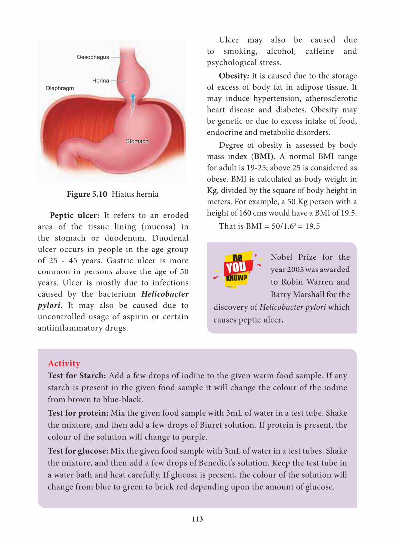
Hiatus hernia (Diaphragmatic hernia): It is a structural abnormality
in which superior
part of the stomach
protrudes slightly
above the diaphragm.
The exact cause of
hiatus hernias is not
known. In some people, injury or other
damage may weaken muscle tissue, by
applying too much pressure (repeatedly)
on the muscles around the stomach while
coughing, vomiting, and straining during
bowel movement and lifting heavy object.
Heart burn is also common in those with
a hiatus hernia. In this condition, stomach
contents travel back into the oesophagus or
even into oral cavity and causes pain in the
centre of the chest due to the eroding
nature of acidity (Figure.5.10).
Diarrhoea: It is the most common
gastrointestinal disorder worldwide. It
is sometimes caused by bacteria or viral
infections through food or water. When the
colon is infected, the lining of the intestine
is damaged by the pathogens, thereby
the colon is unable to absorb fluid. The
abnormal frequency of bowel movement
and increased liquidity of the faecal
discharge is known as diarrhoea. Unless
the condition is treated, dehydration
can occur. Treatment is known as oral hydration therapy. This involves drinking
plenty of fluids – sipping small amounts
of water at a time to rehydrate the body.
Food adulterants cause harmful effects in the form of headaches, palpitations, allergies, cancers and in addition reduces the quality of food. Common adulterants are addition of citric acid to lemon juice, papaya seeds to pepper, melamine to milk, vanillin for natural vanillin, red dyes to chillis, lead chromate and lead tetraoxide to turmeric powder, etc.,
113
Peptic ulcer: It refers to an eroded
area of the tissue lining (mucosa) in
the stomach or duodenum. Duodenal
ulcer occurs in people in the age group
of 25 - 45 years. Gastric ulcer is more
common in persons above the age of 50
years. Ulcer is mostly due to infections
caused by the bacterium Helicobacter pylori. It may also be caused due to
uncontrolled usage of aspirin or certain
antiinflammatory drugs.
Figure 5.10 Hiatus hernia
ActivityTest for Starch: Add a few drops of iodine to the given warm food sample. If any
starch is present in the given food sample it will change the colour of the iodine
from brown to blue-black.
Test for protein: Mix the given food sample with 3mL of water in a test tube. Shake
the mixture, and then add a few drops of Biuret solution. If protein is present, the
colour of the solution will change to purple.
Test for glucose: Mix the given food sample with 3mL of water in a test tubes. Shake
the mixture, and then add a few drops of Benedict’s solution. Keep the test tube in
a water bath and heat carefully. If glucose is present, the colour of the solution will
change from blue to green to brick red depending upon the amount of glucose.
Ulcer may also be caused due
to smoking, alcohol, caffeine and
psychological stress.
Obesity: It is caused due to the storage
of excess of body fat in adipose tissue. It
may induce hypertension, atherosclerotic
heart disease and diabetes. Obesity may
be genetic or due to excess intake of food,
endocrine and metabolic disorders.
Degree of obesity is assessed by body
mass index (BMI). A normal BMI range
for adult is 19-25; above 25 is considered as
obese. BMI is calculated as body weight in
Kg, divided by the square of body height in
meters. For example, a 50 Kg person with a
height of 160 cms would have a BMI of 19.5.
That is BMI = 50/1.62 = 19.5
Nobel Prize for the
year 2005 was awarded
to Robin Warren and
Barry Marshall for the
discovery of Helicobacter pylori which
causes peptic ulcer.
114
ICT Corner
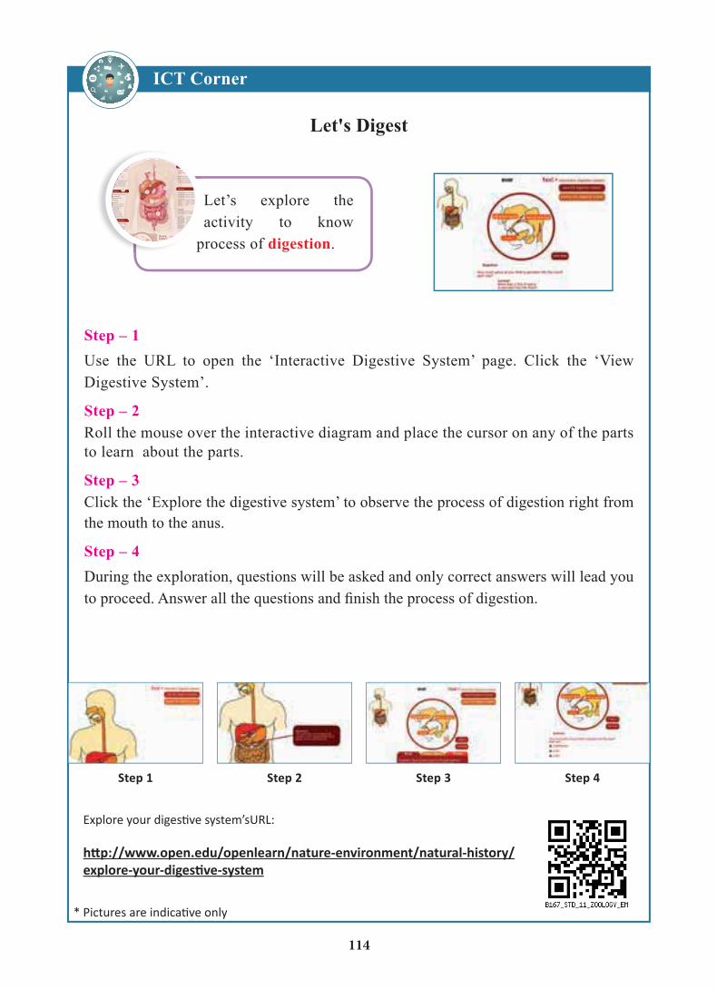
Let's Digest
Step – 1Use the URL to open the ‘Interactive Digestive System’ page. Click the ‘View Digestive System’.
Step – 2Roll the mouse over the interactive diagram and place the cursor on any of the parts to learn about the parts.
Step – 3Click the ‘Explore the digestive system’ to observe the process of digestion right from the mouth to the anus.
Step – 4During the exploration, questions will be asked and only correct answers will lead you
Do you know?
ctions called peristalasis.
u know? The liver is also portant detoxification
It helps to filter and e harmful toxins fromdy.
ou know? intestine is lined
mucosa, a layer ofe that helps tob nutrients,
uce digestivemes, and makes to protect the
ate intestinal wall.
Fun fact:There are two kinds of fibboth support a healthy co
Soluble fibre soaks up toxins and waste in the digestive system
Insolube fib(”roughagebulk througintestine towith regulabowel mov
adder stores cretion.
ProteaseHelps digestprotein
LipaseHelps digestfats
AmyHelpscarbo
er secrete bile. Bile helps mall intestine by breaking
s fats and making them to absorb.
intestine:
of the nutrients form are digestedbsorbed in the intestine.
e intestine (Colon):
of the bacteria livingdigestive tract
e found in the largeine. This is wheregestive processs to an end.
stomach acid from going(refluxing) into the oesop
Stomach:
Gastric juice contain HCl gastric enzymes.
Hydrochloric acid (HCl) hdigest proteins and otheby pepsin enzymes whileminimizing harmful bact
Pancreas
The pancreas is connecteduodenum where three digestive enzmyes are m
Up to 70% of our natural immunsystem support is in the digestive e are more then
0 trillion
7
8
9
9
5
8
2
3
4
5
6
6
Let’s explore the activity to know
process of digestion.
115
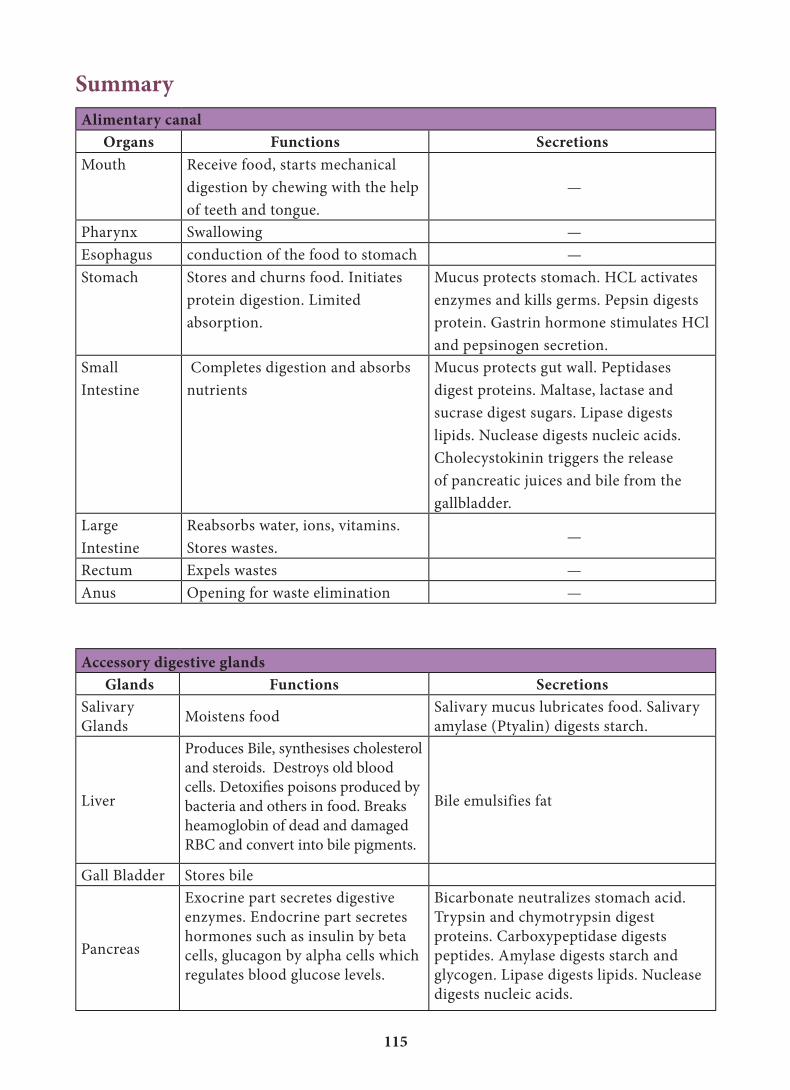
Accessory digestive glands Glands Functions Secretions
Salivary
GlandsMoistens food
Salivary mucus lubricates food. Salivary
amylase (Ptyalin) digests starch.
Liver
Produces Bile, synthesises cholesterol
and steroids. Destroys old blood
cells. Detoxifies poisons produced by
bacteria and others in food. Breaks
heamoglobin of dead and damaged
RBC and convert into bile pigments.
Bile emulsifies fat
Gall Bladder Stores bile
Pancreas
Exocrine part secretes digestive
enzymes. Endocrine part secretes
hormones such as insulin by beta
cells, glucagon by alpha cells which
regulates blood glucose levels.
Bicarbonate neutralizes stomach acid.
Trypsin and chymotrypsin digest
proteins. Carboxypeptidase digests
peptides. Amylase digests starch and
glycogen. Lipase digests lipids. Nuclease
digests nucleic acids.
SummaryAlimentary canal
Organs Functions SecretionsMouth Receive food, starts mechanical
digestion by chewing with the help
of teeth and tongue.
—
Pharynx Swallowing —
Esophagus conduction of the food to stomach —
Stomach Stores and churns food. Initiates
protein digestion. Limited
absorption.
Mucus protects stomach. HCL activates
enzymes and kills germs. Pepsin digests
protein. Gastrin hormone stimulates HCl
and pepsinogen secretion.
Small
Intestine
Completes digestion and absorbs
nutrients
Mucus protects gut wall. Peptidases
digest proteins. Maltase, lactase and
sucrase digest sugars. Lipase digests
lipids. Nuclease digests nucleic acids.
Cholecystokinin triggers the release
of pancreatic juices and bile from the
gallbladder.
Large
Intestine
Reabsorbs water, ions, vitamins.
Stores wastes. —
Rectum Expels wastes —
Anus Opening for waste elimination —
116
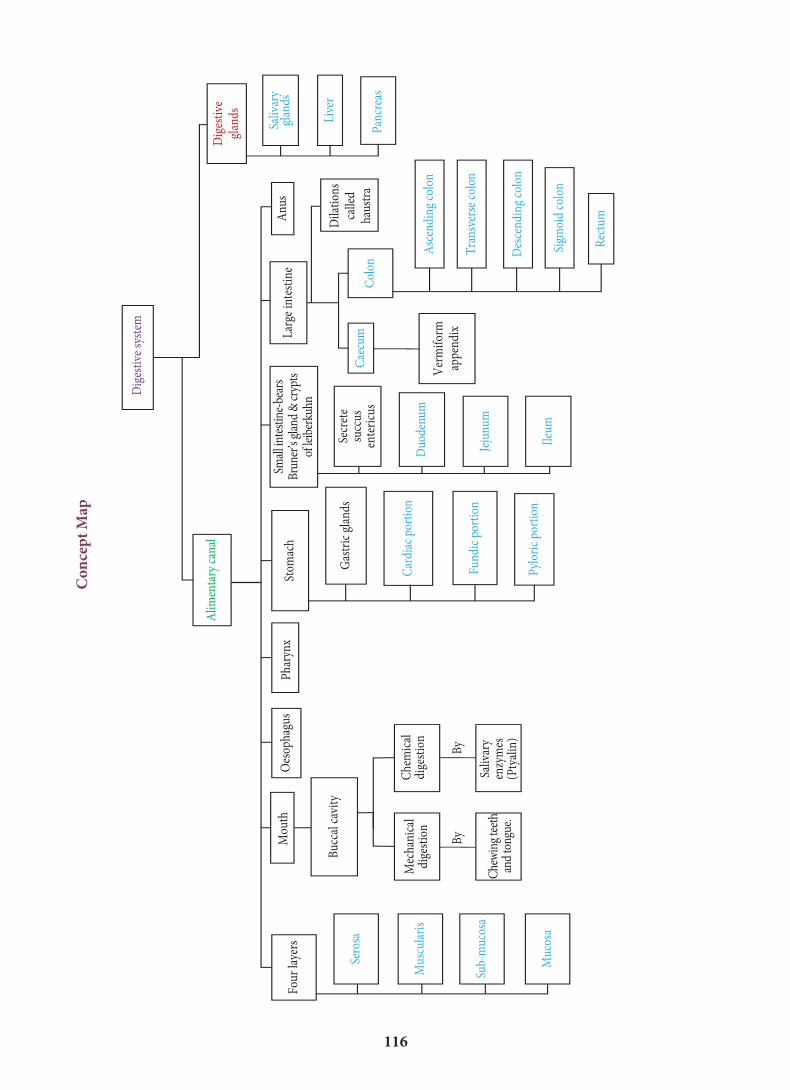
Con
cept
Map
Car
diac
por
tion
Fun
dic
port
ion
Pyl
oric
por
tion
Ileu
m
Jeju
num
Duo
den
um
Col
on
Asc
endi
ng
colo
n
Pan
crea
s
Live
r
Saliv
ary
glan
ds
Dig
esti
vegl
ands
Ver
mif
orm
appe
ndi
x
Dila
tion
sca
lled
haus
tra
An
usLa
rge
inte
stin
eSm
all i
ntes
tine-
bear
sB
rune
r’s g
land
& c
rypt
sof
leib
erku
hn
Secr
ete
succ
usen
teri
cus
Gas
tric
gla
nds
Stom
ach
Alim
enta
ry c
anal
Dig
esti
ve s
yste
m
Pha
ryn
xO
esop
hagu
sM
outh
Four
laye
rs
Buc
cal c
avit
y
Saliv
ary
enzy
mes
(Pty
alin
)
Sub-
muc
osa
Mus
cula
ris
Sero
sa
Muc
osa
Che
win
g te
eth
and
tong
ue.
Tra
nsv
erse
col
on
Des
cen
din
g co
lon
Sigm
oid
colo
n
Rec
tum
Cae
cum
Mec
han
ical
dige
stio
n
By
By
Che
mic
aldi
gest
ion
117
GlossaryAmpulla of vater – Common duct called
hepato-pancreatic duct
Bartholin’s duct or duct of rivinis – Duct
of sublingual gland
Crypts of leiberkuhn – crypts between the
base of villi in the wall of the small intestine
Falciform ligament – It seperate lobes of
liver connect the liver with diaphragm
Gastric rugae – Folds in wall of stomach
Glisson’s capsule – Thin connective tissue
sheath which covers the hepatic lobules
Goblet cells – Mucus secreting glands
Haustra – Pouch like dilation in the colon
Sphincter of boydon – Sphincter which
guard opening of the bile duct before it joins
with the pancreatic duct
Sphincter of oddi – Sphincter which guard
the opening of the ampulla of vater into the
duodenum
Stenson’s duct – Duct of parotids gland
Succus entericus – Intestinal juice
Taeniae coli – Longitudinal muscular
chords in the colon
Valves of kerkring or plicae circulares – Circular folds in the lumen of ileum
Wharton’s duct – Duct of sub-maxillary/
sub-mandibular gland
3. Which of the following hormones
stimulate the production of pancreatic
juice and bicarbonate?
a. Angiotensin and epinephrine
b. Gastrin and insulin
c. Cholecysokinin and secretin
d. Insulin and glucagon
4. The sphincter of Oddi guards
a. Hepatopancreatic duct
b. Common bile duct
c. Pancreatic duct
d. Cystic duct
5. In small intestine, active absorption
occurs in case of
a. Glucose
b. Amino acids
c. Na+
d. All the above
Evaluation1. Choose the incorrect sentence from the
following:
a. Bile juice emulsifies the fat.
b. Chyme is a digestive acidic food
in stomach.
c. Pancreatic juice converts lipid
into fatty acid and glycerol.
d. Enterokinase stimulates the
secretion of pancreatic juice.
2. What is chyme….?
a. The process of conversion of fat into
small droplets.
b. The process of conversion of micelles
substances of glycerol into fatty droplet.
c. The process of preparation of
incompletely digested acidic food
through gastric juice.
d. The process of preparation of
completely digested liquid food in
midgut.
118
6. Which one is incorrectly matched?
a. Pepsin – stomach
b. Renin – liver
c. Trypsin – intestine
d. Ptyalin – mouth
7. Absorption of glycerol, fatty acids and
monoglycerides takes place by
a. Lymph vessels within villi
b. Walls of stomach
c. Colon
d. Capillaries within villi
8. First step in digestion of fat is
a. Emulsification
b. Enzyme action
c. Absorption by lacteals
d. Storage in adipose tissue
9. Enterokinase takes part in the
conversion of
a. Pepsinogen into pepsin
b. Trypsinogen into trypsin
c. Protein into polypetide
d. Caseinogen into casein
10. Which of the following combinations
are not matched?
Column I Column II
a.Bilirubin and
biliverdin
(i) intestinal juice
b.Hydrolysis of
starch
(ii) Amylases
c. Digestion of fat (iii) Lipases
d. Salivary gland (iv) Parotid
11. Match column I with column II and
choose the correct option
Column – I Column – II(P) Small intestine (i) Largest factory
(Q) Pancreas (ii) Absorpstion of
glucose
(R) Liver (iii) Carrying
electrolytic
solution (S) Colon (iv) Digestion and
absorption
a. ( P-iv ) ( Q -iii ) ( R- i ) ( S – ii )
b. ( P-iii ) ( Q -ii ) ( R- i ) ( S – iv )
c. ( P-iv ) ( Q -iii ) ( R- i ) ( S – ii )
d. ( P-ii ) ( Q -iv ) ( R- iii ) ( S – i )
12. Match column I with column II and
choose the correct option
Column – I Column – II
(P) Small intestine (i) 23 cm
(Q) Large intestine (ii) 4 meter
(R) Oesophagus (iii) 12.5 cm
(S) Pharynx (iv) 1.5 meter
a. ( P-iv ) ( Q -ii ) ( R- i ) ( S – iii )
b. ( P-ii ) ( Q -iv ) ( R- i ) ( S – iii )
c. ( P-i ) ( Q -iii ) ( R- ii ) ( S – iv )
d. ( P-iii ) ( Q -i ) ( R- ii ) ( S – iv )
13. Match column I with column II and
choose the correct option
Column – I Column – II
(P) Lipase (i) Starch
(Q) Pepsin (ii) Cassein
(R) Renin (iii) Protein
(S) Ptyalin (iv) Lipid
a. ( P-iv ) ( Q -ii ) ( R- i ) ( S – iii )
b. ( P-iii ) ( Q -iv ) ( R- ii ) ( S – i )
c. ( P-iv ) ( Q -iii ) ( R- ii ) ( S – i )
d. ( P-iii ) ( Q -ii ) ( R- iv ) ( S – i )
14. Which of the following is not the
function of liver?
a. Production of insulin
b. Detoxification
c. Storage of glycogen
d. Production of bile
119
15. Assertion : (A) Large intestine also shows
the presence of villi like small intestine.
Reason: (B) Absorption of water takes
place in large intestine.
a. Both A and B are true and B is the
correct explanation of A
b. Both A and B are true but B is not
the correct explanation of A
c. A is true but B is false
d. A is false but B is true
16. Which of the following is not true
regarding intestinal villi?
a. Th ey possess microvilli.
b. Th ey increase the surface area.
c. Th ey are supplied with capillaries
and the lacteal vessels.
d. Th ey only participate in digestion
of fats.
17. Why are villi present in the intestine
and not in the stomach?
18. Bile juice contains no digestive enzymes,
yet it is important for digestion. Why?
19. List the chemical changes that starch
molecule undergoes from the time it
reaches the small intestine.
20. How do proteins diff er from fats in their
energy value and their role in the body?
21. Digestive secretions are secreted only
when needed. Discuss.
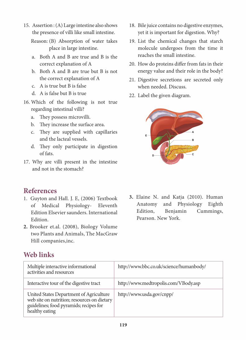
22. Label the given diagram.
A
B
CD
E
Web linksMultiple interactive informational activities and resources
http://www.bbc.co.uk/science/humanbody/
Interactive tour of the digestive tract http://www.medtropolis.com/VBody.asp
United States Department of Agriculture web site on nutrition; resources on dietary guidelines; food pyramids; recipes for healthy eating
http://www.usda.gov/cnpp/
References1. Guyton and Hall. J. E, (2006) Textbook
of Medical Physiology- Eleventh
Edition Elsevier saunders. International
Edition.
2. Brooker et.al. (2008), Biology Volume
two Plants and Animals, The MacGraw
Hill companies,inc.
3. Elaine N. and Katja (2010). Human
Anatomy and Physiology Eighth
Edition, Benjamin Cummings,
Pearson. New York.