Embed Size (px)
Citation preview
Unit III: HomeostasisBlood
Chapter 17
pp. 575-585
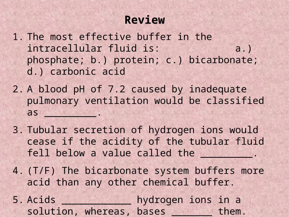
Review
1. The most effective buffer in the intracellular fluid is: a.) phosphate; b.) protein; c.) bicarbonate; d.) carbonic acid
2. A blood pH of 7.2 caused by inadequate pulmonary ventilation would be classified as _________.
3. Tubular secretion of hydrogen ions would cease if the acidity of the tubular fluid fell below a value called the _________.
4. (T/F) The bicarbonate system buffers more acid than any other chemical buffer.
5. Acids ____________ hydrogen ions in a solution, whereas, bases _______ them.
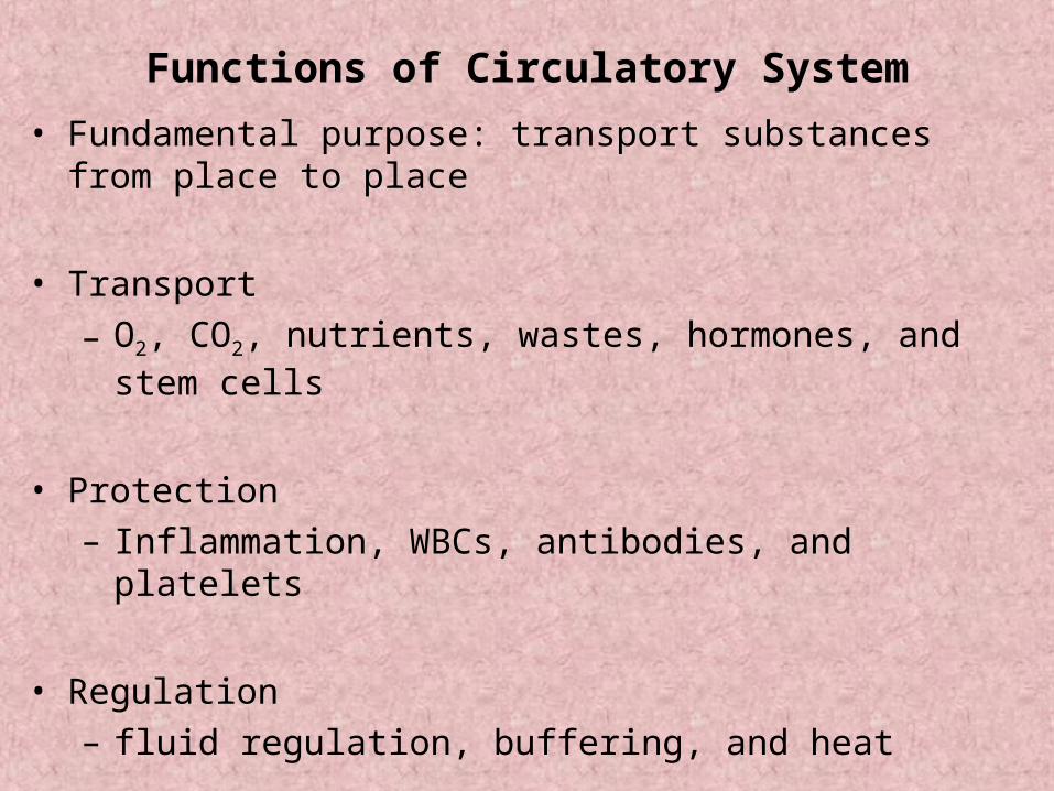
Functions of Circulatory System
• Fundamental purpose: transport substances from place to place
• Transport
– O2, CO2, nutrients, wastes, hormones, and stem cells
• Protection
– Inflammation, WBCs, antibodies, and platelets
• Regulation
– fluid regulation, buffering, and heat
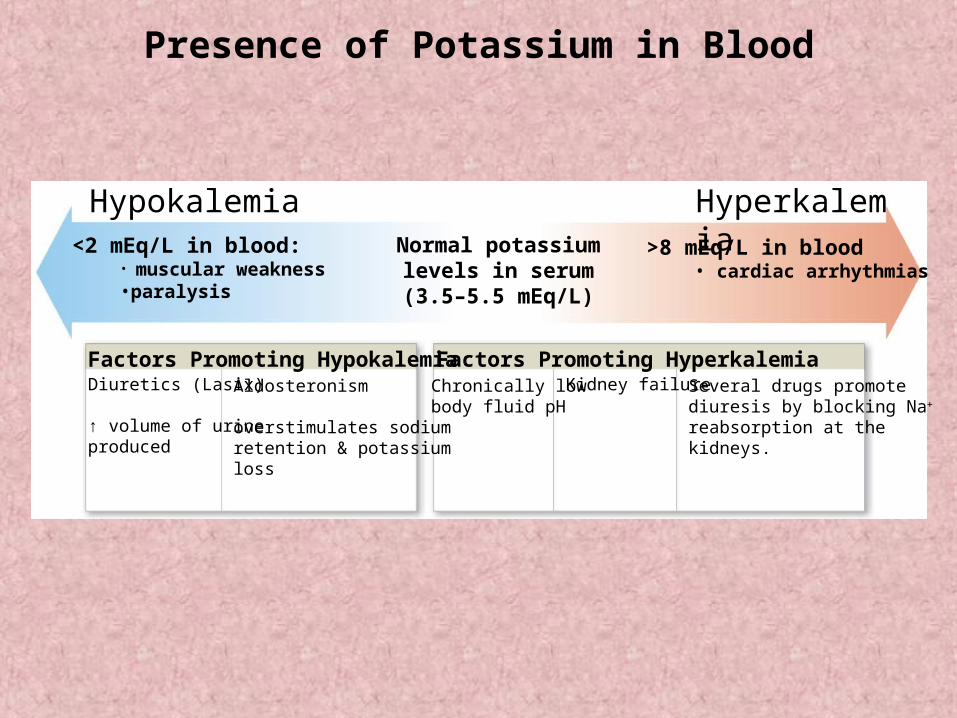
Presence of Potassium in Blood
>8 mEq/L in blood • cardiac arrhythmias
<2 mEq/L in blood:• muscular weakness•paralysis
Normal potassiumlevels in serum(3.5–5.5 mEq/L)
Factors Promoting Hypokalemia Factors Promoting HyperkalemiaDiuretics (Lasix)
↑ volume of urineproduced
Aldosteronism
overstimulates sodiumretention & potassiumloss
Chronically lowbody fluid pH
Kidney failure Several drugs promotediuresis by blocking Na
reabsorption at thekidneys.
Hypokalemia Hyperkalemia
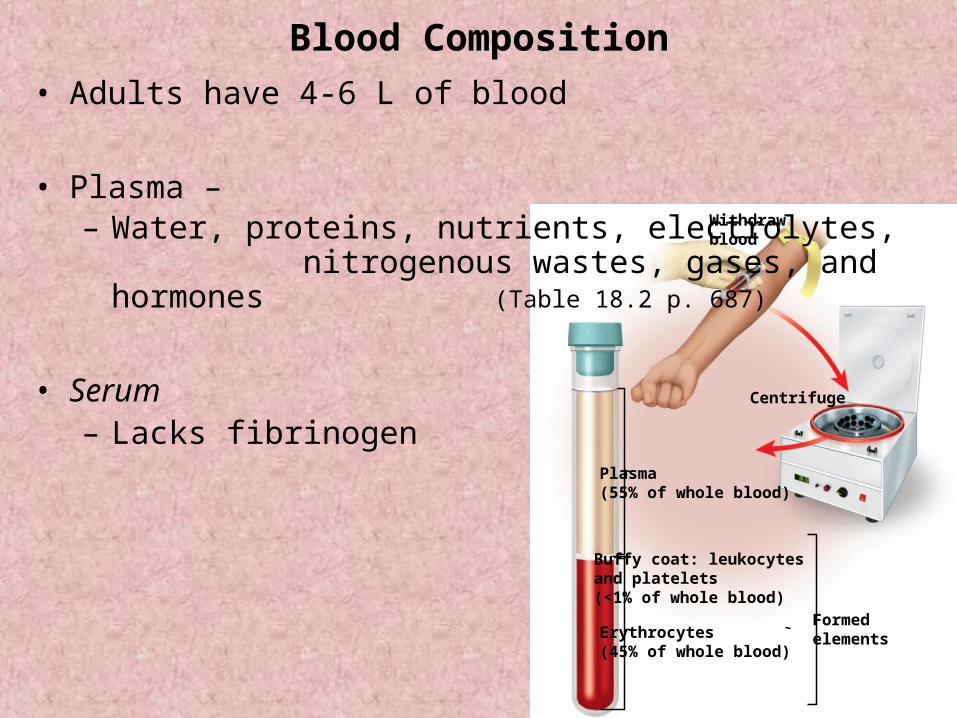
Centrifuge
Withdrawblood
Plasma(55% of whole blood)
Buffy coat: leukocytesand platelets(<1% of whole blood)
Erythrocytes(45% of whole blood)
Formedelements
Blood Composition
• Adults have 4-6 L of blood
• Plasma – – Water, proteins, nutrients, electrolytes,
nitrogenous wastes, gases, and hormones (Table 18.2 p. 687)
• Serum– Lacks fibrinogen
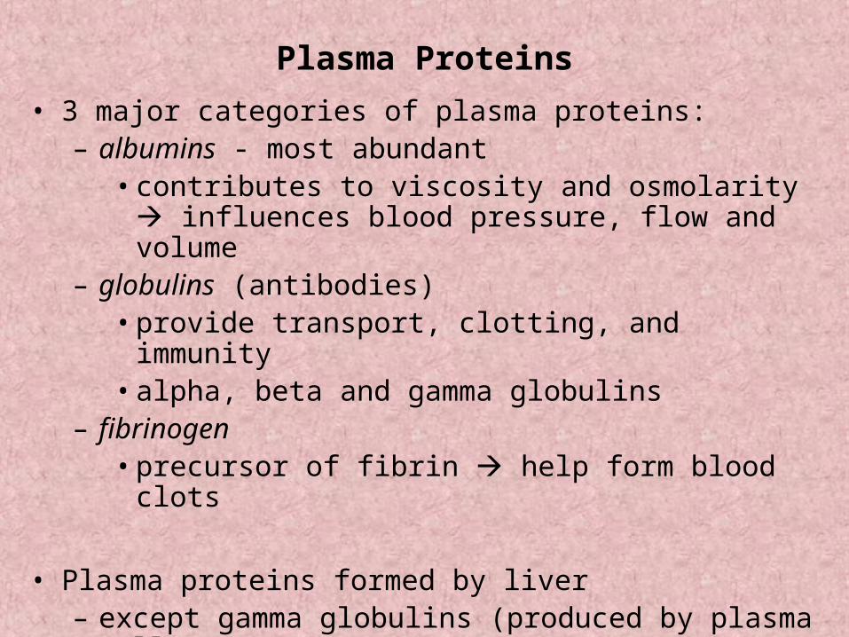
Plasma Proteins
• 3 major categories of plasma proteins:– albumins - most abundant
• contributes to viscosity and osmolarity influences blood pressure, flow and volume
– globulins (antibodies) • provide transport, clotting, and immunity• alpha, beta and gamma globulins
– fibrinogen • precursor of fibrin help form blood clots
• Plasma proteins formed by liver – except gamma globulins (produced by plasma cells)
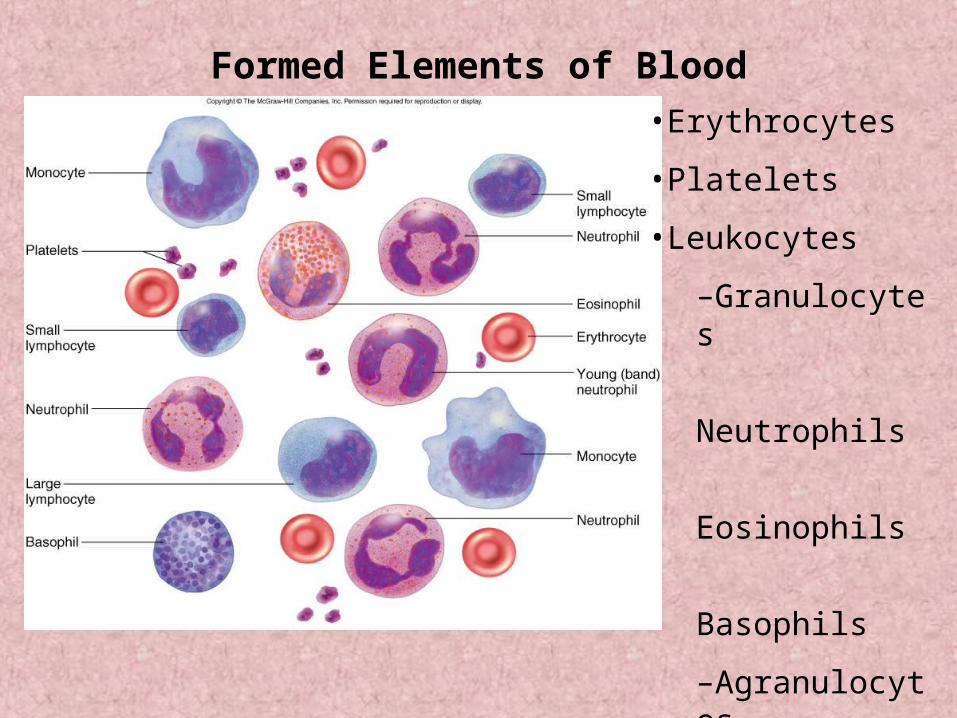
Formed Elements of Blood
•Erythrocytes
•Platelets
•Leukocytes
–Granulocytes
Neutrophils
Eosinophils
Basophils
–Agranulocytes
Lymphocytes
Monocytes
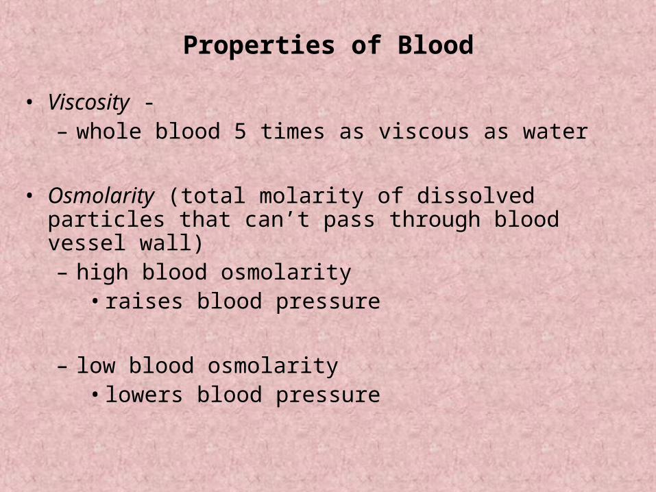
Properties of Blood
• Viscosity -– whole blood 5 times as viscous as water
• Osmolarity (total molarity of dissolved particles that can’t pass through blood vessel wall)– high blood osmolarity
• raises blood pressure
– low blood osmolarity • lowers blood pressure
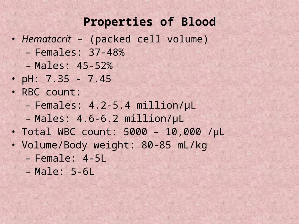
Properties of Blood
• Hematocrit – (packed cell volume)– Females: 37-48%– Males: 45-52%
• pH: 7.35 - 7.45• RBC count:
– Females: 4.2-5.4 million/µL– Males: 4.6-6.2 million/µL
• Total WBC count: 5000 – 10,000 /µL• Volume/Body weight: 80-85 mL/kg
– Female: 4-5L– Male: 5-6L
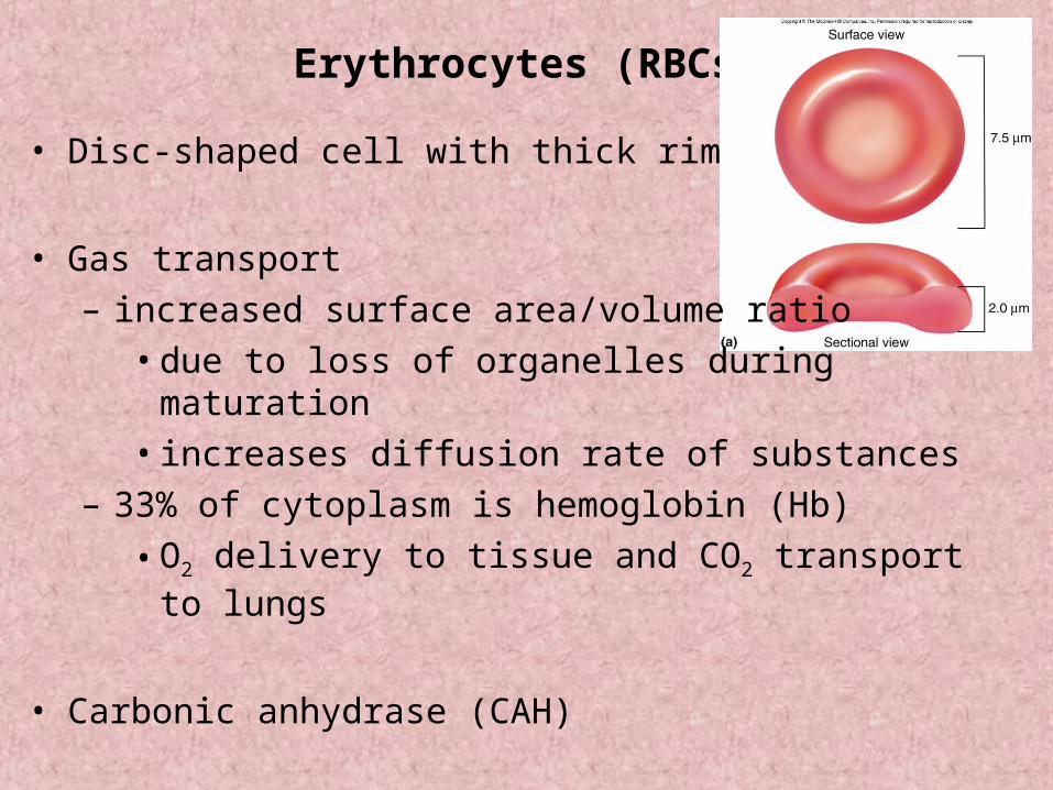
Erythrocytes (RBCs)
• Disc-shaped cell with thick rim
• Gas transport
– increased surface area/volume ratio
• due to loss of organelles during maturation
• increases diffusion rate of substances
– 33% of cytoplasm is hemoglobin (Hb)
• O2 delivery to tissue and CO2 transport to lungs
• Carbonic anhydrase (CAH)
Erythrocytes and Hemoglobin
• Common measurements:
– Hematocrit (packed cell volume)
– Red blood cell count
– hemoglobin concentration of whole blood
• men 13-18g/dL; women 12-16g/dL
• Values are lower in women
– androgens stimulate RBC production
– women have periodic menstrual losses
– Hematocrit is inversely proportional to % body fat
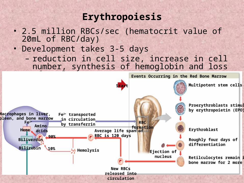
Erythropoiesis
• 2.5 million RBCs/sec (hematocrit value of 20mL of RBC/day)• Development takes 3-5 days
– reduction in cell size, increase in cell number, synthesis of hemoglobin and loss of nucleus
Macrophages in liver,spleen, and bone marrow
Fe2+
90%
10%
Fe2+ transported in circulationby transferrin
Average life span ofRBC is 120 days
Hemolysis
Heme
Biliverdin
Bilirubin
Amino acids
New RBCsreleased into
circulation
RBCformation
Ejection ofnucleus
Events Occurring in the Red Bone Marrow
Multipotent stem cells
Proerythroblasts stimulatedby erythropoietin (EPO)
Erythroblast
Roughly four days of differentiation
Retilculocytes remain in the bone marrow for 2 more days
Start
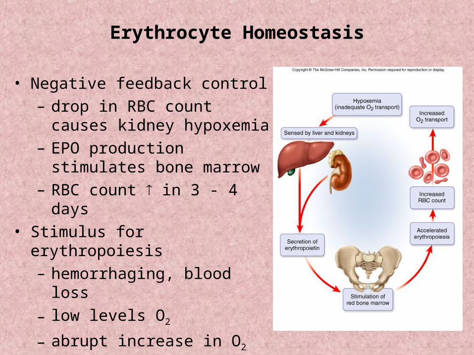
Erythrocyte Homeostasis
• Negative feedback control
– drop in RBC count causes kidney hypoxemia
– EPO production stimulates bone marrow
– RBC count in 3 - 4 days
• Stimulus for erythropoiesis
– hemorrhaging, blood loss
– low levels O2
– abrupt increase in O2 consumption
– loss of lung tissue in emphysema
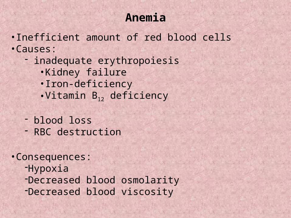
Anemia
•Inefficient amount of red blood cells•Causes:
inadequate erythropoiesis•Kidney failure•Iron-deficiency•Vitamin B12 deficiency
blood loss RBC destruction
•Consequences:HypoxiaDecreased blood osmolarityDecreased blood viscosity
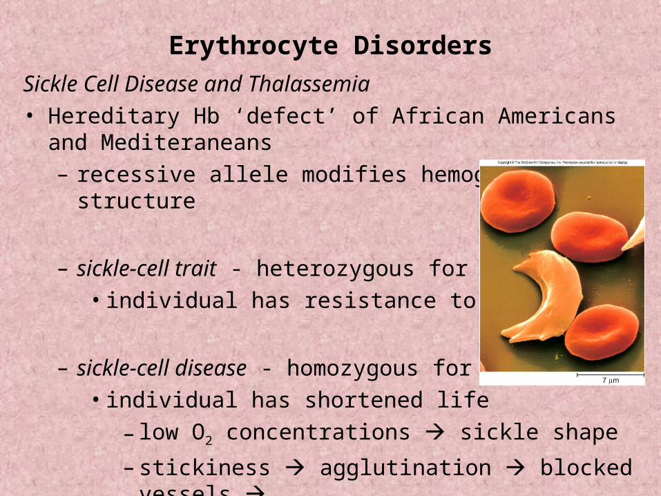
Erythrocyte Disorders
Sickle Cell Disease and Thalassemia
• Hereditary Hb ‘defect’ of African Americans and Mediteraneans
– recessive allele modifies hemoglobin structure
– sickle-cell trait - heterozygous for HbS
• individual has resistance to malaria
– sickle-cell disease - homozygous for HbS
• individual has shortened life
– low O2 concentrations sickle shape
– stickiness agglutination blocked vessels – intense pain; kidney and heart failure; paralysis; stroke
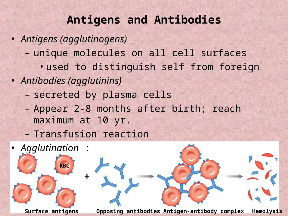
RBC
Surface antigens Antigen-antibody complexOpposing antibodies Hemolysis
Antigens and Antibodies
• Antigens (agglutinogens)
– unique molecules on all cell surfaces
• used to distinguish self from foreign
• Antibodies (agglutinins)
– secreted by plasma cells
– Appear 2-8 months after birth; reach maximum at 10 yr.
– Transfusion reaction
• Agglutination :
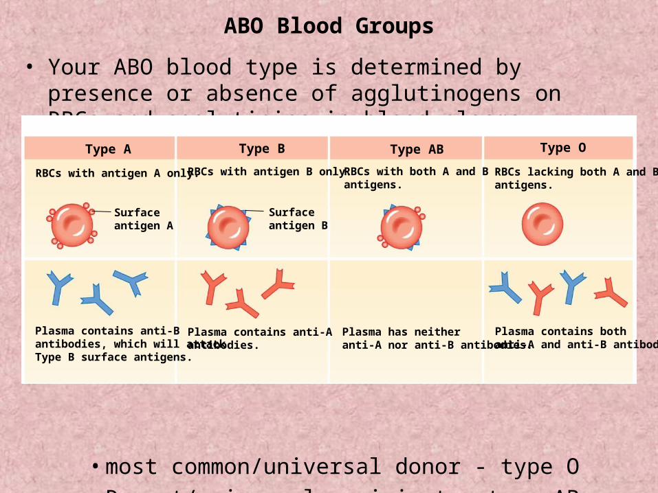
ABO Blood Groups
• Your ABO blood type is determined by presence or absence of agglutinogens on RBCs and agglutinins in blood plasma.
• most common/universal donor - type O
• Rarest/universal recipient - type AB
Type A Type AB Type O
RBCs lacking both A and Bantigens.
RBCs with antigen A only. RBCs with antigen B only. RBCs with both A and B antigens.
Type B
Surfaceantigen A
Surfaceantigen B
Plasma contains anti-Bantibodies, which will attackType B surface antigens.
Plasma contains anti-Aantibodies.
Plasma has neitheranti-A nor anti-B antibodies.
Plasma contains bothanti-A and anti-B antibodies.
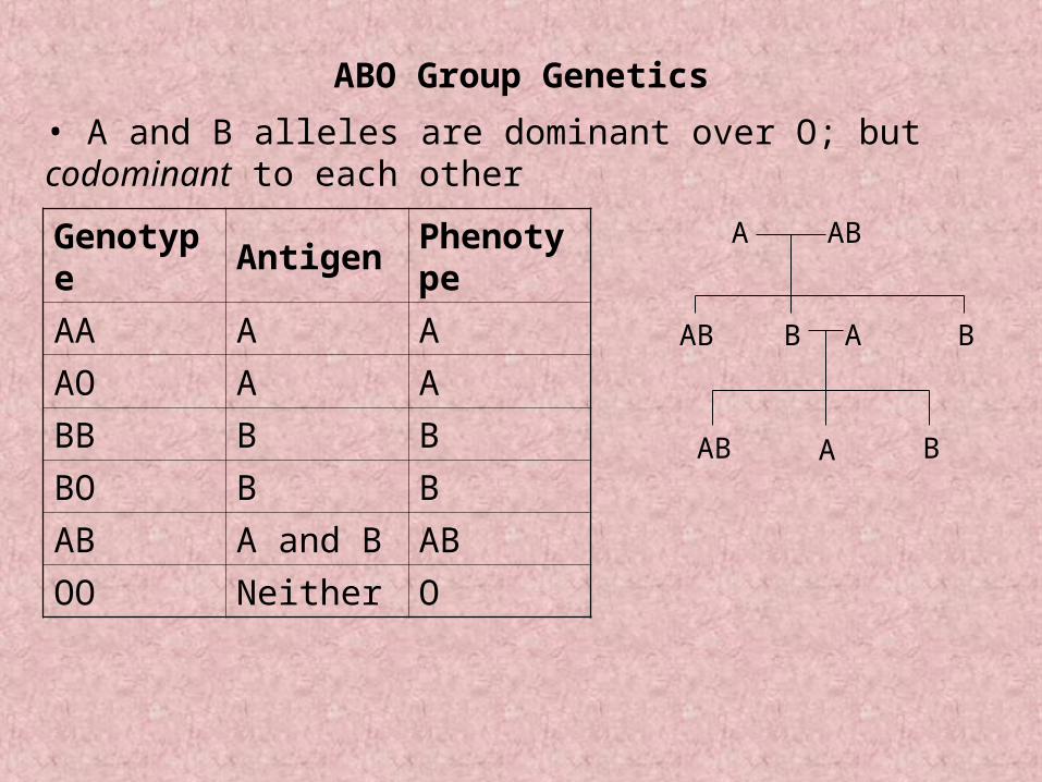
ABO Group Genetics
• A and B alleles are dominant over O; but codominant to each other
Genotype Antigen Phenotype
AA A A
AO A A
BB B B
BO B B
AB A and B AB
OO Neither O
A AB
BAB BA
AB A B
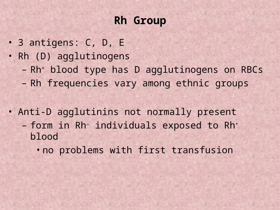
Rh Group
• 3 antigens: C, D, E
• Rh (D) agglutinogens
– Rh+ blood type has D agglutinogens on RBCs
– Rh frequencies vary among ethnic groups
• Anti-D agglutinins not normally present
– form in Rh- individuals exposed to Rh+ blood
• no problems with first transfusion
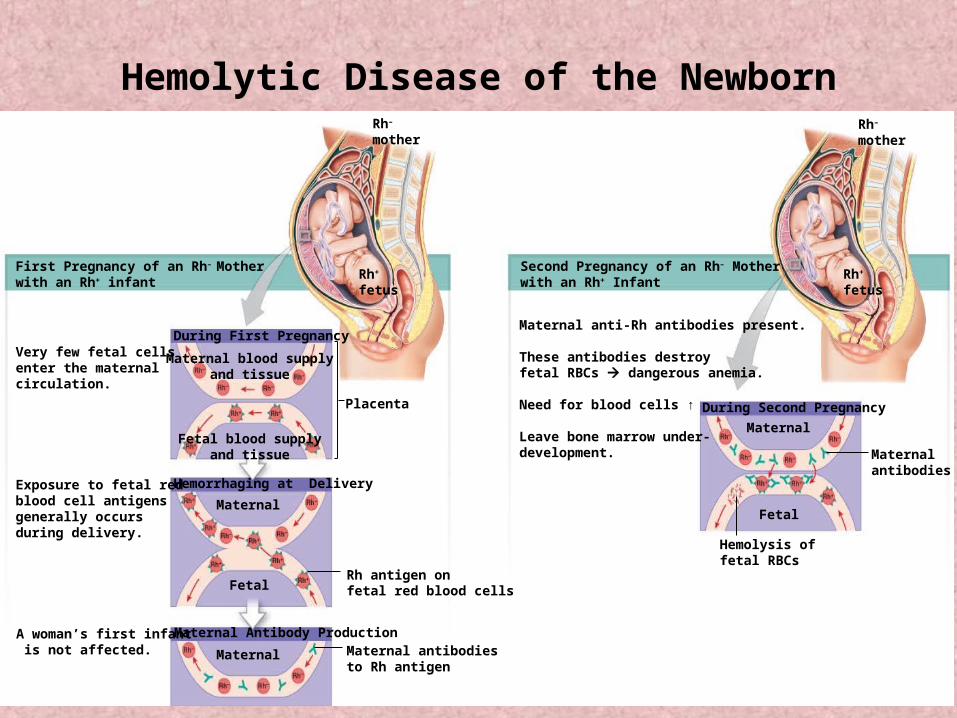
First Pregnancy of an Rh– Motherwith an Rh+ infant
Very few fetal cells enter the maternal circulation.
Exposure to fetal red blood cell antigens generally occurs during delivery.
A woman’s first infant is not affected.
During First Pregnancy
Hemorrhaging at Delivery
Maternal Antibody Production
During Second Pregnancy
Rh antigen onfetal red blood cells
Maternal antibodiesto Rh antigen
Maternal
Fetal
Maternal
Fetal blood supplyand tissue
Maternal blood supplyand tissue
Placenta
Rh+
fetus
Rh–
motherRh–
mother
Rh+
fetus
Maternal
Fetal
Maternalantibodies
Hemolysis offetal RBCs
Second Pregnancy of an Rh– Motherwith an Rh+ Infant
Maternal anti-Rh antibodies present.
These antibodies destroyfetal RBCs dangerous anemia.
Need for blood cells ↑
Leave bone marrow under-development.
Hemolytic Disease of the Newborn
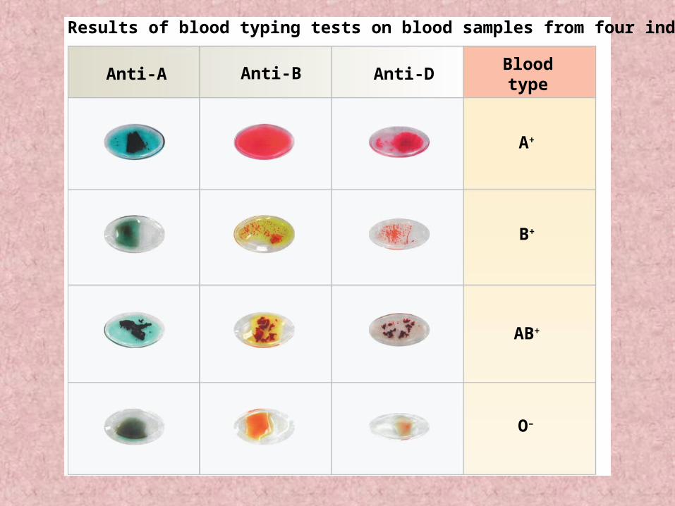
Anti-A Anti-B Anti-DBloodtype
A+
B+
AB+
O–
Results of blood typing tests on blood samples from four individuals